Article

The Stress of Fungicides Changes the Expression of Clock Protein CmFRQ and the Morphology of Fruiting Bodies of Cordyceps militaris

College of Life Science, Jiangxi Normal University, No. 99, Ziyang Avenue, Nanchang 330022, China
*
Author to whom correspondence should be addressed.
These authors contributed equally to this work.
J. Fungi 2024, 10(2), 150; https://doi.org/10.3390/jof10020150
Submission received: 9 January 2024 / Revised: 1 February 2024 / Accepted: 10 February 2024 / Published: 13 February 2024

Abstract

:
The physiological, biochemical, and morphological changes brought about by fungi in response to fungicides can undoubtedly bring diversity to fungi. Cordyceps militaris strains TN (mating type genes MAT1-1-1, MAT1-1-2, and MAT1-2-1) and CmFRQ-454 (mating type genes MAT1-1-1 and MAT1-1-2) were treated with non-lethal doses of fungicides amphotericin B, L-cysteine, terbinafine, and 5-fluorocytosine. The results showed that the treatment with amphotericin B, terbinafine, and 5-fluorocytosine promoted an increase in the relative content of clock protein CmFRQ (C. militaris FREQUENCY) in the mycelium of strain TN, while the high concentration of L-cysteine inhibited the expression of CmFRQ in strain TN. These four fungicides could reduce the relative contents of CmFRQ in the mycelium of strain CmFRQ454. The relative contents of CmFRQ in the mycelium of strain TN were increased after removing the four fungicides, but the relative contents of CmFRQ in the mycelium of strain CmFRQ454 were decreased after removing the four fungicides. This indicates that the effect of fungicides on CmFRQ on mycelium was still sustained after removing the stress of fungicides, and the operation of the circadian clock was changed. The fruiting bodies of C. militaris strain TN and CmFRQ-454 were still degenerated to varying degrees after removing amphotericin B, L-cysteine, and terbinafine. However, the fruiting bodies of strain TN after removing 5-fluorocytosine did not show significant degeneration; the fruiting bodies of strain CmFRQ-454 after removing 5-fluorocytosine obtained rejuvenation. These results indicate that the stress of fungicides could lead to the degeneration of fruiting bodies as well as the rejuvenation of fruiting bodies, resulting in the morphological diversity of C. militaris. The increase or decrease of the CmFRQ-454, the main component of the circadian clock, caused by the stress of fungicants, might lead to the differential degeneration of different mating-type strains of C. militaris.

1. Introduction

The effects of fungicides on fungi are varied, especially on the diversity of fungi. The use of fungicides creates a strong selection pressure on the fungus, which reduces the effectiveness of the fungicide. Scanlan et al. showed that the DNA insertions into the promoter of the erg11/CYP51 DMI target gene resulted in changes in the canola pathogen Leptosphaeria maculans resistance to demethylation inhibitor (DMI) fungicides [1]. Some fungicides induce a genetic change in Monilinia fructicola in the form of transposon transposition [2]. The genomic instability (single nucleotide polymorphisms, insertions/deletions, copy number variants, and transposable element insertions) in fungal plant pathogens caused by exposure to sublethal fungicide doses accelerates the emergence of fungicide resistance or other adaptive traits [3]. Many studies have been reported on the genetic diversity of fungi driven by fungicides, although there are differences in the characteristics that appear [4,5,6,7]. In addition, antifungal resistance caused by the use of fungicides has also received more attention [8,9,10].
Circadian clock mechanisms exist in many eukaryotic cells to regulate morphology, physiology, biochemistry, growth, and development [11,12,13]. The changes in the external environment can lead to a reset of the circadian clock to maintain synchronization with environmental conditions [14,15,16]. A typical circadian clock consists of three parts: environmental signal input, circadian oscillator, and time information output, which regulate a variety of different molecular and physiological processes and show circadian rhythm [17]. In Neurospora crassa, the key components of the core circadian oscillator include clock protein frequency (FRQ), blue light receptor white collar-1 (WC-1) and white collar-2 (WC-2), etc., and participate in the transcription-/translation-based negative feedback loop [18,19]. WC-1 and WC-2 can form a dimer (WCC) that acts as a photoreceptor, as well as a transcription factor or a positive element promoting frq transcription in the dark [20,21,22,23,24]. FRQ and FRH (FRQ-interacting RNA helicase) form an FRQ-FRH complex (FFC) functioning as the negative limb in the negative feedback loop [25,26,27]. After phosphorylation of FRQ by several kinases (CKI and CKII), the FRQ ubiquitination leads to its degradation, releasing WCC dimers, reactivating FRQ expression, and restarting the circadian cycle [14,28,29,30,31,32,33,34,35,36,37].
As a powerful external factor that interferes with fungi, fungicides undoubtedly bring about genetic diversity in fungi, and thus affect the physiological and biochemical characteristics of fungi, thereby affecting their morphology. In this study, we selected four fungal inhibitors: amphotericin B, L-cysteine, terbinafine, and 5-fluorocytosine. Amphotericin B is a polyene antifungal drug whose main mechanism of action is to damage the permeability of the cell membrane by combining with steroids on the cell membrane of sensitive fungi, causing the leakage of important substances in the cell, thus destroying the normal metabolism of the cell and inhibiting the growth of the fungus. Amphotericin B has the advantages of high antifungal activity, a broad antifungal spectrum, and low drug resistance [38]. Amino acids are important components of proteins, but a certain concentration of amino acids has an inhibitory effect on fungi [39,40,41]. Previous studies have shown that a certain concentration of L-cysteine can inhibit the growth of the mycelium of Villosiclava virens and inhibit the growth of the mycelium, spore germination, and germ tube elongation of Alternaria alternata [42,43]. Obviously, amino acid antifungal agents have good safety in food applications. Terbinafine is an allylamine broad-spectrum antifungal agent that inhibits squalene cyclooxygenase during the synthesis of ergosterol in fungal cells and accumulates squalene in cells, thus exerting a fungicidal effect. Previous studies have shown that terbinafine has antifungal effects on various fungi [44]. Under the action of fungal cytosine deaminase, 5-fluorocytosine enters fungal cells and is converted into fluorouracil, replacing uracil in RNA, thereby interfering with the synthesis of normal fungal proteins and achieving antifungal effects [45,46]. It can act on yeast (Saccharomyces cerevisiae), Aspergillus, dematiaceous fungi, Candida albicans, and Cryptococcus neoformans [47,48,49].
Cordyceps militaris is an economically important edible and medicinal fungus in China. With the increase in the number of subcultures, the fungal fruiting body is prone to degeneration, which affects its economic value [50]. Many causes of C. militaris degeneration have been analyzed, including environmental causes [50]. Circadian clocks can be directly influenced by the environment, and the FRQ of fungal circadian clocks is a centric component [17,51]. Therefore, C. militaris frequency (CmFRQ) was selected as the research object in this study.

2. Materials and Methods

2.1. C. Militaris Strains

C. militaris strain TN had mating-type genes MAT1-1-1, MAT1-1-2, and MAT1-2-1. C. militaris strain CmFRQ454 had mating-type genes MAT1-1-1 and MAT1-1-2, but no MAT1-2-1. Strain CmFRQ-454 had been degenerated in the process of subculture.

2.2. Strain Activation

The C. militaris strains were activated before use. The strains were inoculated on potato dextrose agar (PDA; 200 g potatoes, 1 g KH2PO4, 1 g MgSO4·7H20, 10 mg vitamin B1, 20 g glucose, 20 g agar powder, the fixed volume of 1000 mL after preparation) medium and cultured under constant temperature and darkness at 25 °C for 6 d. The obtained strains can be used in subsequent experiments.

2.3. Culture of Mycelium with the Treatment of Fungicides

C. militaris mycelium was cultured in PDA medium at 25 °C under dark conditions. When inoculating, 4-point holes were drilled on the plate of the PDA medium with a hole puncher (diameter 6 mm), and then the strain obtained in the same way was filled into the holes of the PDA medium. If the treatment of fungicides was required in the mycelium growth process, fungicides should be added to the cooled but unsolidified PDA medium according to the required concentration when preparing the PDA medium. The final concentrations of the fungicides added were 2 μg/mL for amphotericin B, 125 μg/mL for L-cysteine, 0.5 μg/mL for terbinafine, and 4.65 × 10⁻⁶ nmol/L for 5-fluorocytosine. At the same time, a blank control was set. The subculture mycelium of C. militaris obtained by the treatment of fungicides was S0. The S0 colony morphology was observed on the 5th and 10th days of culture. All S0 samples, together with Petri dishes, were wrapped in aluminum foil and placed in a –55 °C refrigerator to facilitate simultaneous protein extraction.

2.4. Transfer Culture of C. Militaris Mycelium after the Treatment of Fungicides

After the subculture S0 samples were treated with fungicides, the S0 mycelium of C. militaris was transferred onto the PDA without fungicides and then cultured at 25 °C under dark conditions to obtain the subculture S1. All S1 samples, together with Petri dishes, were wrapped in aluminum foil and placed in a –55 °C refrigerator to facilitate simultaneous protein extraction.

2.5. Culture of Fruiting Bodies for S1

C. militaris subculture S1 was inoculated into 100 mL potato dextrose broth medium (PDB; 200 g potatoes, 1 g KH2PO4, 1 g MgSO4·7H20, 10 mg vitamin B1, and 20 g glucose, with a fixed volume of 1000 mL after preparation), shaking the fungus to form spherical mycelium with a diameter of about 4.0 mm at 25 °C at 200 rpm, in order to obtain a liquid strain. Then, 5 mL of the liquid strain were inoculated into the rice culture medium (30 g high-quality rice and 35 mL PDB medium were added to a 350 mL culture flask), cultured for 5 d at 25 °C under dark conditions, and then exposed to light at 20 °C for 2 days. The mycelium surface was scratched with inoculation rings to induce fruiting body buds. When the orange primordium was formed, the cap was replaced with a breathable cap to facilitate the development of fruiting bodies. During the continuous culture of fruiting bodies, two 4-W fluorescent lamps for continuous light were placed in the incubator with an illumination intensity of 450–600 Lux. The morphology of the fruiting body was observed and recorded.

2.6. Sample Treatment and Western Blot Analysis

Mycelium samples stored at –55 °C were ground into a powder in a mortar, frozen at –55 °C, and then added to a 2 mL centrifuge tube. In the centrifuge tube, 300 μL phosphate-buffered saline (PBS) (0.1 mol/L, pH 7.2, containing protease inhibitors for fungi and yeast) were pre-added, immediately shaken, and mixed, and the biomass concentration of the samples was adjusted to 1.0 g/mL. Samples were extracted overnight at 4 °C. After overnight extraction, centrifugation was performed at 12,000 rpm, and the supernatant was collected. If there was still suspended matter, it would be centrifuged again. After adding 5× denatured protein loading buffer and bathing at 100 °C for 5 min, the protein samples were loaded successively with a loading volume of 40 μL for each sample (32 μL sample protein + 8 μL loading buffer). The protein samples were subjected to SDS-PAGE (concentration gel 5%, separation gel 12%) at 120 V for 2.5 h, then electrotransferred to a PVDF membrane. The PVDF membrane was incubated in a closed solution (5% skim milk powder) for 2 h and then washed with PBST (pH 7.4 PBS added 0.1% Tween-20) 3 times, 10 min each time. The membrane was incubated in anti-CmFRQ2 at 4 °C overnight, washed with PBST 3 times, then incubated with sheep anti-rabbit IgG-HRP for 1 h, washed with PBST 3 times, and then a DAB staining kit was used for color development. The grayscale value of the protein bands was determined by the software ImageJ (version ImageJ 1.x, https://imagej.net/software/imagej/, accessed on 1 April 2023).

3. Results

3.1. Morphology of C. militaris Strain TN Colony Treated with Fungicides

C. militaris strain TN was inoculated onto PDA medium containing a certain concentration of fungicides, and colony morphology was observed on the 5th and 10th days (Figure 1). The results showed that the colonies treated with amphotericin B were the smallest on the 5th day of growth (Figure 1B). L-cysteine-treated colonies (Figure 1C), terbinafine-treated colonies (Figure 1D), and 5-fluorocytosine-treated colonies (Figure 1E) grew similarly to control colonies (Figure 1A). On the 10th day of growth, amphotericin B-treated colonies were also the smallest (Figure 1G), followed by terbinafine-treated colonies (Figure 1I). L-cysteine-treated colonies (Figure 1H) and 5-fluorocytosine-treated colonies (Figure 1J) were similar to the control colonies (Figure 1F), and both were in contact with the Petri plate wall. The mycelium in the colonies treated by the four fungicides was relatively tight (Figure 1B–E,G–J). These results indicate that amphotericin B inhibited the growth of the C. militaris strain TN mycelium more obviously, followed by terbinafine.

3.2. Morphology of C. militaris Strain CmFRQ454 Colony Treated with Fungicides

C. militaris strain CmFRQ454 was inoculated onto PDA medium containing a certain concentration of fungicides, and colony morphology was observed on the 5th and 10th days (Figure 2). The results showed that the colonies treated with terbinafine were the smallest on the 5th day of growth (Figure 2D). Amphotericin B-treated colonies (Figure 2B), L-cysteine-treated colonies (Figure 2C), and 5-fluorocytosine-treated colonies (Figure 2E) grew faster than control colonies (Figure 2A). On the 10th day of growth, terbinafine-treated colonies were also the smallest (Figure 2I), amphotericin B-treated colonies (Figure 2G), L-cysteine-treated colonies (Figure 2H), and 5-fluorocytosine-treated colonies (Figure 2J) were similar to the control colonies (Figure 2F). The mycelium in the colony treated with 5-fluorocytosine was more loose, which is beneficial for ventilation in the mycelium (Figure 2E,J). However, the mycelia in the colonies treated with other fungicides were relatively tight (Figure 2B–D,G–I). These results indicate that terbinafine inhibited the growth of C. militaris strain CmFRQ454 mycelium more obviously. In the early stage of colony culture, amphotericin B, L-cysteine, and 5-fluorocytosine could promote the C. militaris strain CmFRQ454 colony growth to some extent.

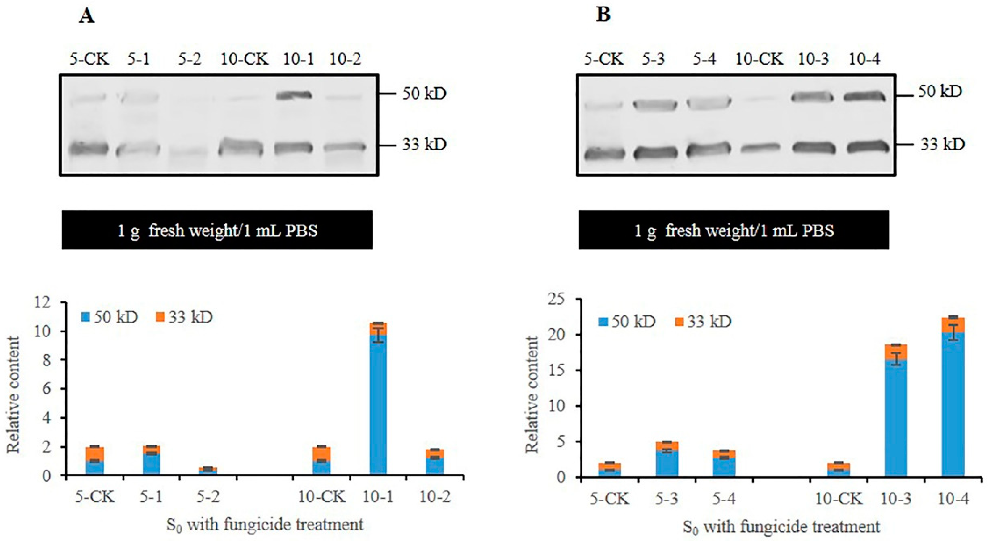
3.3. Analysis of CmFRQ in the Subculture S0 Mycelium of C. militaris Strain TN Treated with Fungicides

Previous studies had shown that the CmFRQ antibody Anti-FRQ2 prepared by us could detect the CmFRQ degradation forms of 50 kD and 33 kD proteins (to be published separately). When extracting mycelium protein, the content of mycelium was always fixed, and the total protein in 1 g of mycelium sample was extracted with 1 mL of PBS. Therefore, the relative content of CmFRQ in the mycelium was compared under the same biomass, as was the case in subsequent studies.
Western blot analysis was performed on the proteins extracted from the C. militaris strain TN mycelium treated with fungicides for 5 and 10 days. The detection of the antibody Anti-FRQ2 showed that the relative content of CmFRQ in S0-amphotericin B mycelium (mycelium of C. militaris strain TN treated with amphotericin B) (Figure 3A: 5-1) showed no significant change compared to the control group (Figure 3A: 5-CK) on the 5th day, and there was a significant increase compared to the control group (Figure 3A: 10-CK) on the 10th day (Figure 3A: 10-1). The relative content of CmFRQ in the C. militaris strain TN treated with L-cysteine was lower compared to that of the control group on the 5th day (Figure 3A: 5-CK and 5-2), and there was no significant change compared to the control group on the 10th day (Figure 3A: 10-CK and 10-2). On the 5th day of treatment with terbinafine, the relative content of CmFRQ in the C. militaris strain TN showed a certain increase compared to the control group (Figure 3B: 5-CK and 5-3), and on the 10th day, there was a sharp increase compared to the control group (Figure 3B: 10-CK and 10-3). On the 5th day, the relative content of CmFRQ in the C. militaris strain TN treated with 5-fluorocytosine showed a certain increase compared to the control group (Figure 3B: 5-CK and 5-4), and on the 10th day, there was a sharp increase compared to the control group (Figure 3B: 10-CK and 10-4). These results indicate that the treatment of amphotericin B, terbinafine, and 5-fluorocytosin could all increase the relative content of CmFRQ in the mycelium of C. militaris strain TN to a certain extent, especially in the late stage of culture close to the aging stage of the mycelium. L-cysteine had little effect on CmFRQ expression.

3.4. Analysis of CmFRQ in Subculture S0 Mycelium of C. militaris Strain CmFRQ454 Treated with Fungicides

The mycelium of C. militaris strain CmFRQ454 treated with amphotericin B showed no significant changes in the relative content of CmFRQ on the 5th day compared to the control group (Figure 4A: 5-CK and 5-1) but significantly decreased on the 10th day compared to the control group (Figure 4A: 10-CK and 10-1). The relative content of CmFRQ in the mycelium of C. militaris strain CmFRQ454 treated with L-cysteine decreased compared to the control group on the 5th and 10th days (Figure 4A: 5-CK, 5-2, 10-CK, and 10-2). The amount of CmFRQ in the mycelium of C. militaris strain CmFRQ454 treated with terbinafine decreased significantly compared to the control group on the 5th and 10th days (Figure 4B: 5-CK, 5-3, 10-CK, and 10-3). The mycelium of C. militaris strain CmFRQ454 was treated with 5-fluorocytosine, and the amount of CmFRQ significantly decreased on the 5th and 10th days (Figure 4B: 5-CK, 5-4, 10-CK, and 10-4). These results indicate that none of these four fungicides significantly increased the amount of CmFRQ in the C. militaris strain CmFRQ454 and even had a certain inhibitory effect.

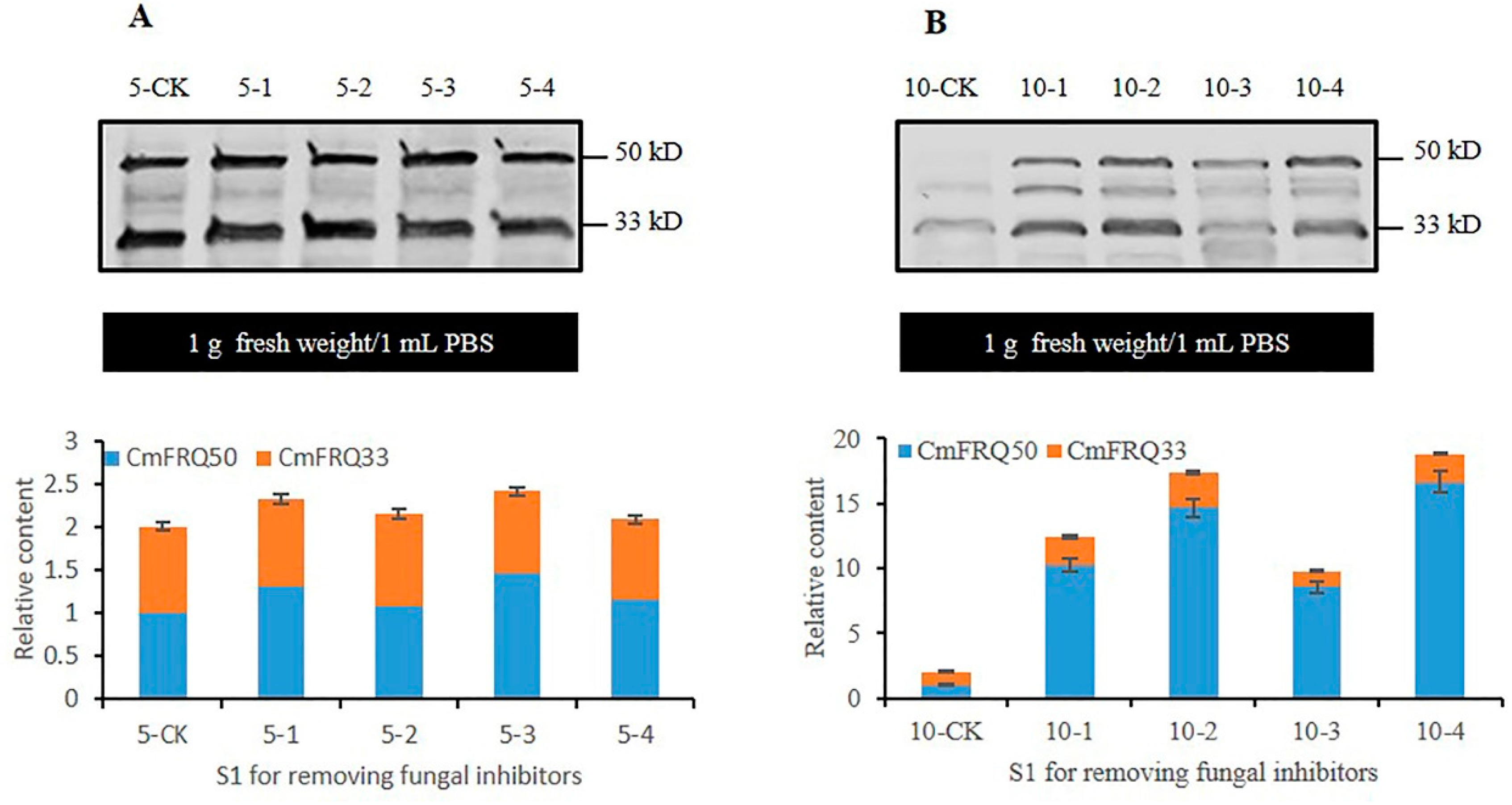
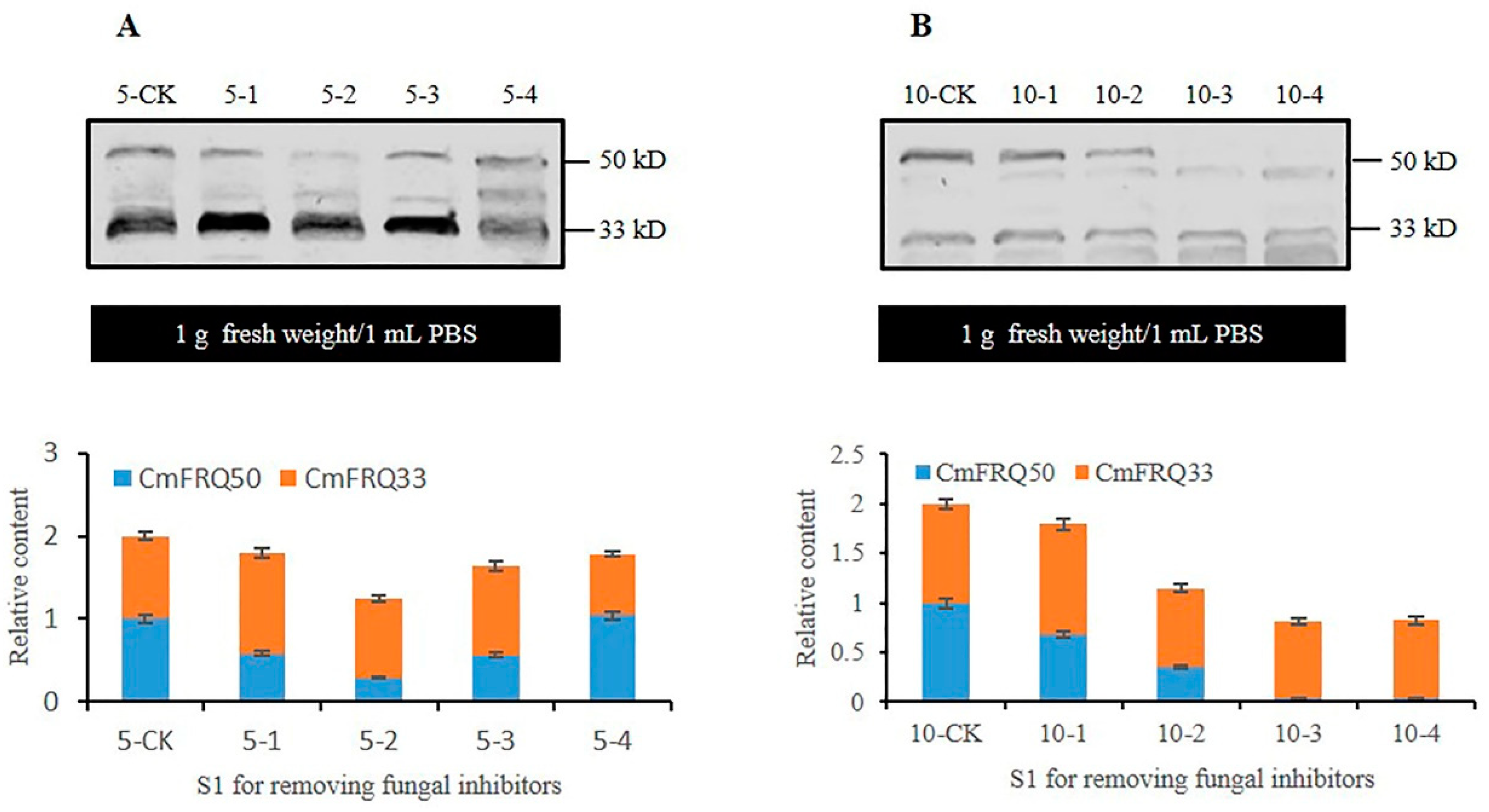
3.5. Analysis of CmFRQ in Subculture S1 Mycelium of C. militaris Strain TN after Removal of Fungicide

The subculture S0 mycelium of the C. militaris strain TN treated with fungicides was transferred to the PDA medium without fungicides to obtain the subculture S1 mycelium without fungicide treatment. When the subculture S1 mycelium was cultured for 5 days, the relative content of CmFRQ in the S1-amphotericin B mycelium (Figure 5A: 5-1), S1-L-cysteine mycelium (Figure 5A: 5-2), S1-terbinafine mycelium (Figure 5A: 5-3), and S1-5-fluorocytosine mycelium (Figure 5A: 5-4) compared to that in the control group (Figure 5A: 5-CK) showed no significant changes. However, when the subculture S1 mycelia was cultured for 10 days, the relative content of CmFRQ in the S1-amphotericin B mycelium (Figure 5A: 10-1), S1-L-cysteine (Figure 5A: 10-2), S1-terbinafine (Figure 5A: 10-3), and S1-5-fluorocytosine (Figure 5A: 10-4) compared to that in the control group (Figure 5A: 10-CK) increased significantly. This indicates that in subculture S1 mycelium treated with the removal of these four fungicides, the early circadian clock in the C. militaris strain TN mycelium did not change much, but the late circadian clock changed greatly.

3.6. Analysis of CmFRQ in Subculture S1 Mycelium of C. militaris Strain CmFRQ454 after Removal of Fungicide

Subculture S0 mycelium of C. militaris strain CmFRQ454 treated with fungicides was transferred to the culture medium without fungicide to obtain subculture S1 mycelium without fungicide treatment. When subculture S1 mycelium was cultured for 5 days, the relative content of CmFRQ in S1-amphotericin B (Figure 6A: 5-1), S1-L-cysteine (Figure 6A: 5-2), S1-Terbinefin (Figure 6A: 5-3) and S1-5-fluorocytosine (Figure 6A: 5-4) mycelium compared with the control (Figure 6A: 5-CK) was decreased, and that in the S1-L-cysteine was more decreased (Figure 6A: 5-2). When subculture S1 mycelia was cultured for 10 days, the relative content of CmFRQ in S1-amphotericin B mycelium (Figure 6A: 10-1), S1-L-cysteine (Figure 6A: 10-2), S1-terbinafine (Figure 6A: 10-3) and S1-5-fluorocytosine (Figure 6A: 10-4) mycelium compared with the control group (Figure 6A: 10-CK) was decreased significantly. These results indicated that the expression of CmFRQ in subculture S1 mycelium after removal of these four fungicides is down-regulated, and the operation of the circadian clock may be weakened.

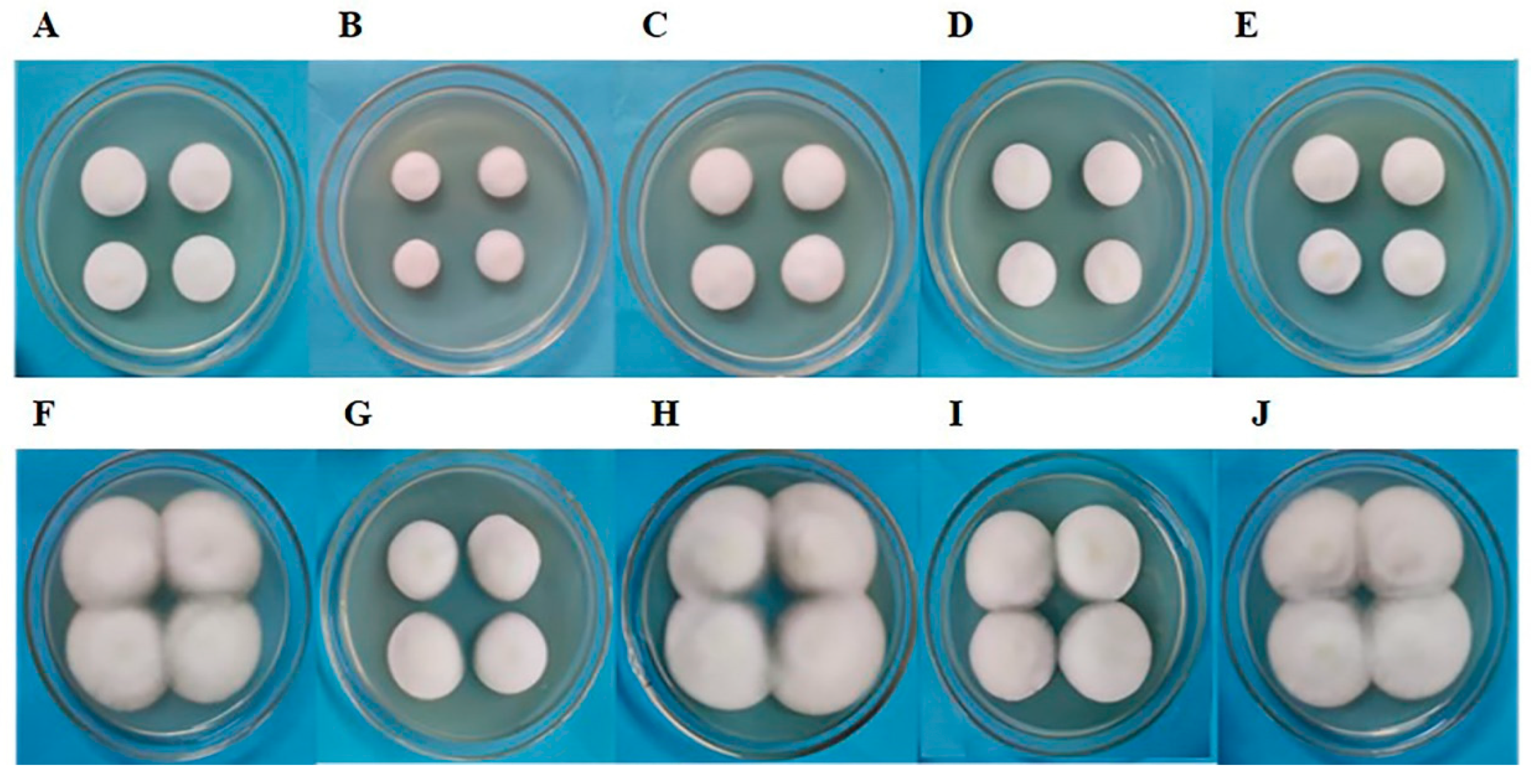
3.7. Morphological Analysis of Subculture S1 Fruiting Bodies of C. militaris Strain TN after Removal of Fungicides

The subculture S1 mycelium of C. militaris strain TN was inoculated in a culture bottle for fruiting body culture, and the fruiting body morphology was observed after 40 days of culture (Figure 7). Compared with the control group (Figure 7A), the differences appeared in the fruiting bodies of subculture S1 despite the absence of fungicide effect. The results of S1-amphotericin B (Figure 7B), S1-L-cysteine (Figure 7C) and S1- terbinafine (Figure 7D) cultures showed certain degenerated, shorter, and more deformed fruiting body than the control group (Figure 7A). The appearance of albino mycelium on the stroma obtained from S1-L-cysteine (Figure 7C) and S1-terbinafine (Figure 7D) cultures affected the normal growth of the fruiting body. The fruiting bodies obtained by S1-5-fluorocytosine culture were thinner than those in the control group, but no malformed fruiting body and albino mycelium was present on the stroma (Figure 7E). These results indicated that the effect of fungicide on C. militaris strain TN could continue to the next generation subculture without fungicide treatment, resulting in worse fruiting body growth. However, the fruiting body obtained after 5-fluorocytosine treatment showed relatively good performance, with almost no degeneration.

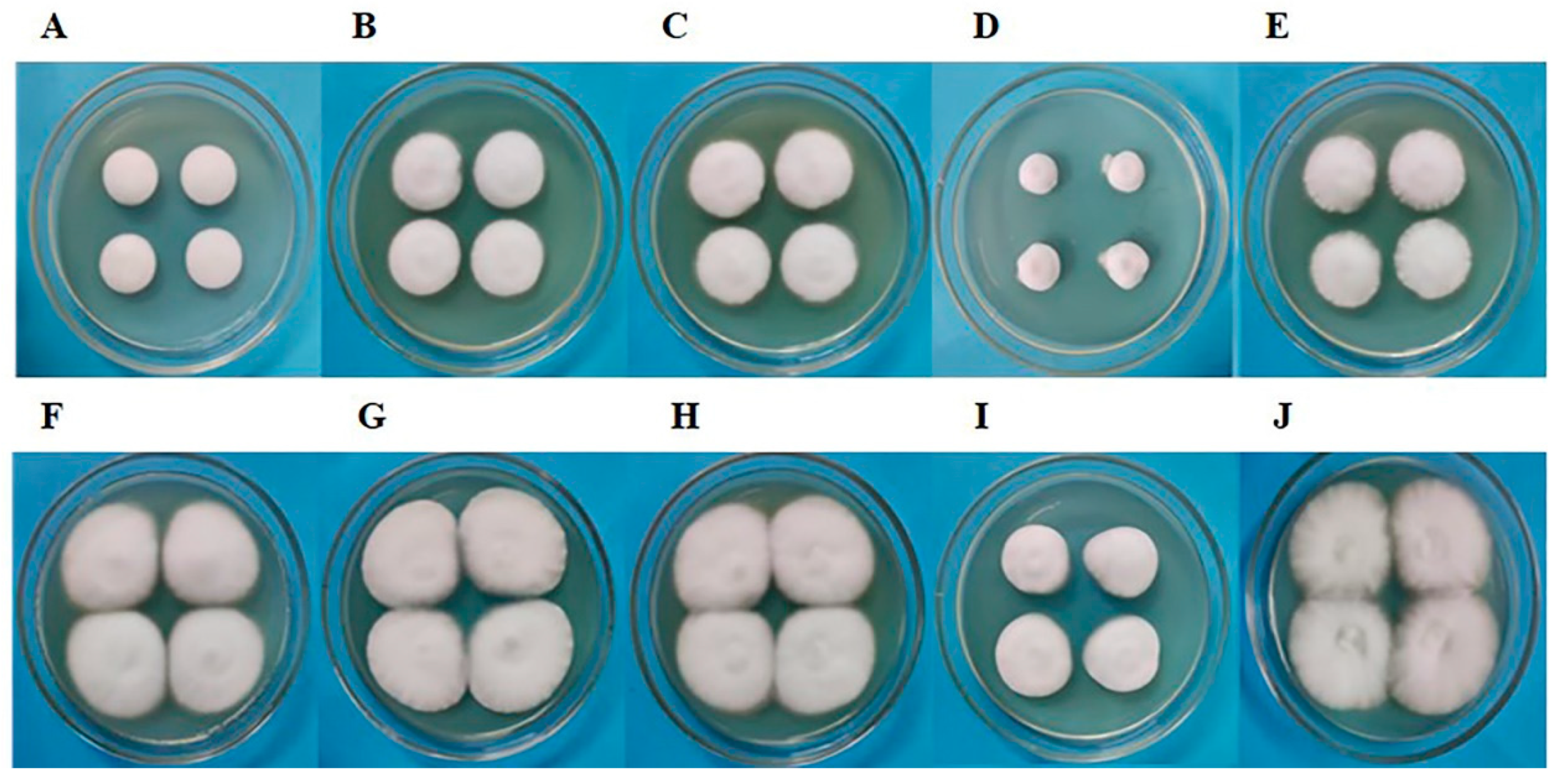
3.8. Morphological Analysis of Subculture S1 Fruiting Bodies of C. militaris Strain CmFRQ454 after Removal of Fungicide

The C. militaris strain CmFRQ454 was once a rejuvenated strain, and after multiple generations of asexual transfer culture, the fruiting body showed degeneration (Figure 8A). The subculture S1 mycelium of C. militaris strain CmFRQ454 was inoculated in a culture bottle for fruiting body culture, and the morphology of fruiting body was observed after 40 days of culture (Figure 8). Compared with the control group (Figure 8A), the fruiting bodies obtained from S1 amphotericin B (Figure 8B), S1-L-cysteine (Figure 8C), and S1-terbinafine (Figure 8D) cultures were still degenerated, similar to the control group (Figure 8A), showing deformed enlargement of fruiting bodies, bifurcation, shorter than normal fruiting bodies, and albino mycelium on the stroma. Among them, the degeneration of fruiting bodies obtained from S1- terbinafine culture was more obvious (Figure 8D), and the number of fruiting bodies decreased and were shorter than that of the control group (Figure 8A). However, the fruiting body obtained by S1-5-fluorocytosine culture showed rejuvenation, normal growth, few deformities, and longer than control fruiting body (Figure 8E). These results indicated that the treatment of C. militaris strain CmFRQ454 with fungicides had different effects on fruiting bodies, which could lead to further degeneration of fruiting bodies, and also lead to the rejuvenation of the fruiting body.

4. Discussion

At present, research on the relationship between fungicides and fungal diversity mainly focuses on the relationship between the resistance of fungicides and the genetic diversity of fungi [1,4,5,6,52]. The main idea of our research was to investigate the changes in the circadian clock and fruiting body of C. militaris under the influence of fungicides. After treating the mycelium of C. militaris with four fungicides at a certain concentration, the mycelium was not completely killed, but a certain non-lethal selection pressure was given. Under the action of four fungicides, the clock protein CmFRQ, mycelial growth, and fruiting body development of C. militaris were all affected; in particular, the impact on fruiting bodies was bidirectional, which can lead to the degeneration of fruiting bodies and also promote their rejuvenation.
In this study, two strains of C. militaris with different mating types, TN and CmFRQ454, were used. Obviously, the genetic characteristics of these two strains were different. Therefore, the expression of CmFRQ in subculture S0 mycelium treated with four fungicides was different between the two strains. Three fungicides, namely amphotericin B, terbinafine, and 5-flucytocytosin, could promote an increase in the relative content of CmFRQ in the mycelium of C. militaris strain TN, and this promotion effect could increase with time. However, high concentrations of L-cysteine inhibited the expression of CmFRQ in C. militaris strain TN mycelium. These four fungicides could reduce the relative content of CmFRQ in strain CmFRQ454 mycelium, and 5-fluorocytosin promoted the highest degree of CmFRQ reduction. These results indicate that the circadian clock of C. militaris with different mating types was disturbed differently during the stress of fungicides on the mycelium of C. militaris.
After the stress of the fungicide was removed, the effect of the fungicide on the clock protein CmFRQ in mycelium was still retained in the subculture S1 obtained from the S0 transferring culture. The results showed that the relative expression of CmFRQ was increased in the mycelium of subculture S1 of strain TN treated with amphotericin B, L-cysteine, terbinafine, and 5-fluorocytosine. However, in the mycelium of subculture S1 of C. militaris strain CmFRQ454, the situation was exactly the opposite, with a relative decrease in CmFRQ expression. After removing the stress of fungicides, the effect of the stress will continue, and the response to different mating types of C. militaris is exactly the opposite.
After treating the C. militaris strains TN and CmFRQ454 with these four fungicides, the mycelium in the colonies was relatively tight, but only the mycelium in the colonies of strain CmFRQ454 treated with 5-fluorocytosine was relatively loose, which was conducive to the formation of fruiting bodies. Most of the fruiting bodies obtained from subculture S1 after removing the fungicides showed the morphological degeneration. However, 5-fluorocytosine led to the revival of the degenerated C. militaris strain CmFRQ454. It is known that 5-fluorocytosine interferes with RNA transcription, thereby affecting protein synthesis [45,46]. However, from the performance of these two strains, 5-fluorocytosine promotes the synthesis of CmFRQ in C. militaris strain TN while inhibiting it in C. militaris strain CmFRQ454. Therefore, the expression of circadian clock proteins could correspond to the degeneration or rejuvenation of the fruiting body. There are few studies on the bidirectional effects of fungicides on fungal growth and reproduction. Some studies have shown that DMI fungicides could promote the spore production of Colletotrichum spp. at low concentrations [53]. Obviously, the bidirectional action of fungicides brings more diversity to fungal physiology and morphology.
C. militaris is a fungus prone to degeneration, especially during subculture [50]. Some researchers have regarded the degeneration of fungi as an aging problem and determined that the mechanism of degeneration is the same as that of aging, and high levels of reactive oxygen species (ROS) lead to molecular damage and degeneration [54,55,56,57]. Studies have shown that ROS can regulate the important components of the circadian oscillator in N. crassa, and it has also been determined that ROS homeostasis is controlled by the circadian clock, and ROS levels exhibit a circadian rhythm [58,59,60,61,62]. The mycelium of the degenerated C. militaris strain was also found to contain more ROS and higher levels of oxidative stress, with a significant negative correlation between its oxidative stress and strain degeneration [63]. Therefore, we speculate that the effect of fungicides on C. militaris could affect the balance of ROS in C. militaris cells, thereby affecting the degeneration of C. militaris, which is closely related to its circadian clock.

5. Conclusions

Four fungicides, amphotericin B, L-cysteine, terbinafine, and 5-fluorocytosine, were used to treat two C. militaris strains, strain TN and strain CmFRQ454. Treatment with the four fungicides resulted in changes in the expression of the clock protein CmFRQ and the fruiting body morphology of the two strains.
Amphotericin B, terbinafine, and 5-fluorocytosine could increase the relative content of CmFRQ in the subculture S0 mycelium of C. militaris strain TN, while high concentrations of L-cysteine reduced the relative content of CmFRQ in the subculture S0 mycelium of C. militaris strain TN. These four fungicides could reduce the relative content of CmFRQ in the subculture S0 mycelium of C. militaris strain CmFRQ454. After removing the stress of fungicides, the effect of the stress of fungicides would continue. The relative content of CmFRQ was increased in the subculture S1 mycelium of strain TN treated with amphotericin B, L-cysteine, terbinafine, and 5-fluorocytosine, but the opposite result appeared in the subculture S1 mycelium of C. militaris strain CmFRQ454.
The fruiting bodies of subculture S1 of strain TN or strain CmFRQ454 still showed degeneration after removing the treatment of four fungicides, but rejuvenation was provided to the fruiting bodies of subculture S1 of strain CmFRQ454 after removing the treatment of 5-fluorocytosine.

Author Contributions

Conceptualization, M.-J.F. and Z.-Y.H.; methodology, M.-J.F. and Z.-Y.H.; software, J.-M.P., D.-D.Z. and Z.-Y.H.; validation, Z.-Y.H.; resources, M.-J.F., and J.-M.P.; data curation, J.-M.P., D.-D.Z. and Z.-Y.H.; writing—original draft preparation, M.-J.F.; writing—review and editing, M.-J.F., J.-M.P., D.-D.Z. and Z.-Y.H.; visualization, J.-M.P. and Z.-Y.H.; project administration, M.-J.F.; and funding acquisition, M.-J.F. All authors have read and agreed to the published version of the manuscript.

Funding

This work was supported by the National Natural Science Foundation of China (31760601).

Institutional Review Board Statement

Not applicable.

Informed Consent Statement

Not applicable.

Data Availability Statement

Data are contained within the article.

Conflicts of Interest

The authors declare no conflicts of interest.

References

  1. Scanlan, J.L.; Mitchell, A.C.; Marcroft, S.J.; Forsyth, L.M.; Idnurm, A.; Van de Wouw, A.P. Deep amplicon sequencing reveals extensive allelic diversity in the erg11/CYP51 promoter and allows multi-population DMI fungicide resistance monitoring in the canola pathogen Leptosphaeria maculans. Fungal. Genet. Biol. 2023, 168, 103814. [Google Scholar] [CrossRef] [PubMed]
  2. Chen, F.; Everhart, S.E.; Bryson, P.K.; Luo, C.; Song, X.; Liu, X.; Schnabel, G. Fungicide-induced transposon movement in Monilinia fructicola. Fungal. Genet. Biol. 2015, 85, 38–44. [Google Scholar] [CrossRef]
  3. Gambhir, N.; Kamvar, Z.N.; Higgins, R.; Amaradasa, B.S.; Everhart, S.E. Spontaneous and Fungicide-Induced Genomic Variation in Sclerotinia sclerotiorum. Phytopathology 2021, 111, 160–169. [Google Scholar] [CrossRef] [PubMed]
  4. Baker, S.T.; Froelich, M.H.; Boatwrigh, H.; Wang, H.; Schnabel, G.; Kerrigan, J. Genetic diversity and fungicide sensitivity of Cytospora plurivora on peach. Plant Dis. 2023, 107, 2112–2118. [Google Scholar] [CrossRef]
  5. Brito, F.S.D.; Santos, J.R.P.; Azevedo, V.C.R.; Peixouto, Y.S.; de Oliveira, S.A.; Ferreira, C.F.; Haddad, F.; Amorim, E.P.; Fraaije, B.; Miller, R.N.G. Genetic diversity and azole fungicide sensitivity in Pseudocercospora musae field populations in Brazil. Front. Microbiol. 2020, 11, 99. [Google Scholar] [CrossRef] [PubMed]
  6. Shrestha, S.K.; Cochran, A.; Mengistu, A.; Lamour, K.; Castro-Rocha, A.; Young-Kelly, H. Genetic diversity, QoI fungicide resistance, and mating type distribution of Cercospora sojina-Implications for the disease dynamics of frogeye leaf spot on soybean. PLoS ONE 2017, 12, e0177220. [Google Scholar] [CrossRef] [PubMed]
  7. Walker, A.S.; Ravigne, V.; Rieux, A.; Ali, S.; Carpentier, F.; Fournier, E. Fungal adaptation to contemporary fungicide applications: The case of Botrytis cinerea populations from Champagne vineyards (France). Mol. Ecol. 2017, 26, 1919–1935. [Google Scholar] [CrossRef]
  8. Barber, A.E.; Riedel, J.; Sae-Ong, T.; Kang, K.; Brabetz, W.; Panagiotou, G.; Deising, H.B.; Kurzai, O. Effects of agricultural fungicide use on Aspergillus fumigatus abundance, antifungal susceptibility, and population structure. mBio 2020, 11, e02213-20. [Google Scholar] [CrossRef]
  9. Mahmoud, D.E.; Faraag, A.H.I.; Abu El-Wafa, W.M. In vitro study on the potential fungicidal effects of atorvastatin in combination with some azole drugs against multidrug resistant Candida albicans. World J. Microbiol. Biotechnol. 2021, 37, 191. [Google Scholar] [CrossRef]
  10. Woods, M.; McAlister, J.A.; Geddes-McAlister, J. A One Health approach to overcoming fungal disease and antifungal resistance. WIREs Mech. Dis. 2023, 15, e1610. [Google Scholar] [CrossRef]
  11. Dunlap, J.C. Molecular bases for circadian clocks. Cell 1999, 96, 271–290. [Google Scholar] [CrossRef]
  12. Reppert, S.M.; Weaver, D.R. Molecular analysis of mammalian circadian rhythms. Annu. Rev. Physiol. 2001, 63, 647–676. [Google Scholar] [CrossRef] [PubMed]
  13. Bell-Pedersen, D. Understanding circadian rhythmicity in Neurospora crassa: From behavior to genes and back again. Fungal. Genet. Biol. 2000, 29, 1–18. [Google Scholar] [CrossRef] [PubMed]
  14. Pregueiro, A.M.; Liu, Q.; Baker, C.L.; Dunlap, J.C.; Loros, J.J. The Neurospora checkpoint kinase 2: A regulatory link between the circadian and cell cycles. Science 2006, 313, 644–649. [Google Scholar] [CrossRef]
  15. Liu, Y.; Merrow, M.; Loros, J.J.; Dunlap, J.C. How temperature changes reset a circadian oscillator. Science 1998, 281, 825–829. [Google Scholar] [CrossRef]
  16. Devlin, P.F. Signs of the time: Environmental input to the circadian clock. J. Exp. Bot. 2002, 53, 1535–1550. [Google Scholar] [CrossRef] [PubMed]
  17. Baker, C.L.; Loros, J.J.; Dunlap, J.C. The circadian clock of Neurospora crassa. FEMS Microbiol. Rev. 2012, 36, 95–110. [Google Scholar] [CrossRef]
  18. Chen, C.H.; Dunlap, J.C.; Loros, J.J. Neurospora illuminates fungal photoreception. Fungal. Genet. Biol. 2010, 47, 922–929. [Google Scholar] [CrossRef]
  19. Heintzen, C.; Liu, Y. The Neurospora crassa circadian clock. Adv. Genet. 2007, 58, 25–66. [Google Scholar]
  20. Crosthwaite, S.K.; Dunlap, J.C.; Loros, J.J. Neurospora wc-1 and wc-2: Transcription, photoresponses, and the origins of circadian rhythmicity. Science 1997, 76, 763–769. [Google Scholar] [CrossRef]
  21. Cheng, P.; Yang, Y.; Gardner, K.H.; Liu, Y. PAS domain-mediated WC-1/WC-2 interaction is essential for maintaining the steady-state level of WC-1 and the function of both proteins in circadian clock and light responses of Neurospora. Mol. Cell Biol. 2002, 22, 517–524. [Google Scholar] [CrossRef]
  22. Cheng, P.; Yang, Y.; Wang, L.; He, Q.; Liu, Y. WHITE COLLAR-1, a multifunctional Neurospora protein involved in the circadian feedback loops, light sensing, and transcription repression of wc-2. J. Biol. Chem. 2003, 278, 3801–3808. [Google Scholar] [CrossRef]
  23. Froehlich, A.C.; Loros, J.J.; Dunlap, J.C. Rhythmic binding of a WHITE COLLAR-containing complex to the frequency promoter is inhibited by FREQUENCY. Proc. Natl. Acad. Sci. USA 2003, 100, 5914–5919. [Google Scholar] [CrossRef]
  24. Belden, W.J.; Loros, J.J.; Dunlap, J.C. Execution of the circadian negative feedback loop in Neurospora requires the ATPdependent chromatin-remodeling enzyme CLOCKSWITCH. Mol. Cell 2007, 25, 587–600. [Google Scholar] [CrossRef]
  25. Aronson, B.D.; Johnson, K.A.; Loros, J.J.; Dunlap, J.C. Negative feedback defining a circadian clock: Autoregulation of the clock gene frequency. Science 1994, 263, 1578–1584. [Google Scholar] [CrossRef]
  26. Cheng, P.; He, Q.L.; Wang, L.; Liu, Y. Regulation of the Neurospora circadian clock by an RNA helicase. Genes Dev. 2005, 19, 234–241. [Google Scholar] [CrossRef]
  27. Dunlap, J.C.; Loros, J.J. The Neurospora circadian system. J. Biol. Rhythm. 2004, 19, 414–424. [Google Scholar] [CrossRef] [PubMed]
  28. Liu, Y.; Loros, J.; Dunlap, J.C. Phosphorylation of the Neurospora clock protein FREQUENCY determines its degradation rate and strongly influences the period length of the circadian clock. Proc. Natl. Acad. Sci. USA 2000, 97, 234–239. [Google Scholar] [CrossRef] [PubMed]
  29. Görl, M.; Merrow, M.; Huttner, B.; Johnson, J.; Roenneberg, T.; Brunner, M. A PEST-like element in FREQUENCY determines the length of the circadian period in Neurospora crassa. EMBO J. 2001, 20, 7074–7084. [Google Scholar] [CrossRef] [PubMed]
  30. He, Q.; Cheng, P.; Yang, Y.; He, Q.; Yu, H.; Liu, Y. FWD1-mediated degradation of FREQUENCY in Neurospora establishes a conserved mechanism for circadian clock regulation. EMBO J. 2003, 22, 4421–4430. [Google Scholar] [CrossRef] [PubMed]
  31. He, Q.; Cha, J.; Lee, H.C.; Yang, Y.; Liu, Y. CKI and CKII mediate the FREQUENCY-dependent phosphorylation of the WHITE COLLAR complex to close the Neurospora circadian negative feedback loop. Genes Dev. 2006, 20, 2552–2565. [Google Scholar] [CrossRef]
  32. He, Q.; Cheng, P.; He, Q.; Liu, Y. The COP9 signalosome regulates the Neurospora circadian clock by controlling the stability of the SCFFWD-1 complex. Genes Dev. 2005, 19, 1518–1531. [Google Scholar] [CrossRef]
  33. He, Q.; Liu, Y. Degradation of the Neurospora circadian clock protein FREQUENCY through the ubiquitin-proteasome pathway. Biochem. Soc. Trans. 2005, 33, 953–956. [Google Scholar] [CrossRef]
  34. Yang, Y.; Cheng, P.; Zhi, G.; Liu, Y. Identification of a calcium/calmodulin-dependent protein kinase that phosphorylates the Neurospora circadian clock protein FREQUENCY. J. Biol. Chem. 2001, 276, 41064–41072. [Google Scholar] [CrossRef]
  35. Yang, Y.; Cheng, P.; He, Q.; Wang, L.; Liu, Y. Phosphorylation of FREQUENCY protein by casein kinase II is necessary for the function of the Neurospora circadian clock. Mol. Cell. Biol. 2003, 23, 6221–6228. [Google Scholar] [CrossRef]
  36. Schafmeier, T.; Haase, A.; Káldi, K.; Scholz, J.; Fuchs, M.; Brunner, M. Transcriptional feedback of Neurospora circadian clock gene by phosphorylation-dependent inactivation of its transcription factor. Cell 2005, 122, 235–246. [Google Scholar] [CrossRef] [PubMed]
  37. Wang, B.; Kettenbach, A.N.; Zhou, X.; Loros, J.J.; Dunlap, J.C. The Phospho-Code Determining Circadian Feedback Loop Closure and Output in Neurospora. Mol. Cell 2019, 74, 771–784.e3. [Google Scholar] [CrossRef] [PubMed]
  38. Ostrosky-Zeichner, L.; Marr, K.A.; Rex, J.H.; Cohen, S.H. Amphotericin B: Time for a new “gold standard”. Clin. Infect. Dis. 2003, 37, 415–425. [Google Scholar] [PubMed]
  39. Zhao, P. Effect of amino acids on growth and development of Auricularia auricula. J. Gansu Agric. Univ. 1997, 32, 159–192. (In Chinese) [Google Scholar]
  40. Pan, K.; Wu, F. Effects of amino acids on growth and development of Fusarium oxysporum f. sp. Cucumerinum. North. Hortic. 2008, 2, 228–231. (In Chinese) [Google Scholar]
  41. Liu, A.; Chen, J.; Zhang, J.; Fang, Q.; Wang, M.; Zha, Q. Effect of organic acids and amino acids on the mycelial growth of Agaricus bisporus 2796. Chin. Agric. Sci. Bull. 2010, 26, 46–51. (In Chinese) [Google Scholar]
  42. Kang, H.; Qiao, Y.; Liu, C.; Zhang, Y.; Sun, D. Inhibitory effect of L-cysteine against Alternaria alternata rot of grapes. Food Sci. 2020, 41, 228–235. (In Chinese) [Google Scholar]
  43. Zhang, L.; Zhao, X.; Guo, Y.; Hu, D.; Liang, W. Inhibitory effect of L-cysteine on the mycelial growth of Villosiclava virens and its transcriptome analysis. Acta Phytopathol. Sin. 2022, 52, 352–363. (In Chinese) [Google Scholar]
  44. Hou, S.; Deng, L.; Cai, J.; Sun, R. Agricultural antifungal activity of terbinafine and terbinafine hydrochloride. J. Trop. Biol. 2021, 12, 448–455. (In Chinese) [Google Scholar]
  45. Waldorf, A.R.; Polak, A. Mechanisms of action of 5-fluorocytosine. Antimicrob. Agents Chemother. 1983, 23, 79–85. [Google Scholar] [CrossRef] [PubMed]
  46. Polak, A. Mode of action of 5-fluorocytosine and 5-fluorouracil in dematiaceous fungi. Sabouraudia 1983, 21, 15–25. [Google Scholar] [CrossRef] [PubMed]
  47. Polak, A.; Wain, W.H. The influence of 5-fluorocytosine on nucleic acid synthesis in Candida albicans, Cryptococcus neoformans, and Aspergillus fumigatus. Chemotherapy 1977, 23, 243–259. [Google Scholar] [CrossRef] [PubMed]
  48. Wain, W.H.; Polak, A. The effect of 5-fluorocytosine on the synthesis of 80S ribosomes by pathogenic fungi. Sabouraudia 1981, 19, 187–197. [Google Scholar] [CrossRef] [PubMed]
  49. Whelan, W.L. The genetic basis of resistance to 5-fluorocytosine in Candida species and Cryptococcus neoformans. Crit. Rev. Microbiol. 1987, 15, 45–56. [Google Scholar] [CrossRef]
  50. Lou, H.; Lin, J.; Guo, L.; Wang, X.; Tian, S.; Liu, C.; Zhao, Y.; Zhao, R. Advances in research on Cordyceps militaris degeneration. Appl. Microbiol. Biotechnol. 2019, 103, 7835–7841. [Google Scholar] [CrossRef]
  51. Cha, J.; Zhou, M.; Liu, Y. Mechanism of the Neurospora Circadian Clock, a FREQUENCY-centric View. Biochemistry 2015, 54, 150–156. [Google Scholar] [CrossRef] [PubMed]
  52. Scruggs, A.C.; Basaiah, T.; Adams, M.L.; Quesada-Ocampo, L.M. Genetic diversity, fungicide sensitivity, and host resistance to Ceratocystis fimbriata infecting sweetpotato in north carolina. Plant Dis. 2017, 101, 994–1001. [Google Scholar] [CrossRef] [PubMed]
  53. Han, Y.C.; Zeng, X.G.; Guo, C.; Zhang, Q.H.; Chen, F.Y.; Ren, L.; Chen, W.D.; Qin, L. Reproduction response of Colletotrichum fungi under the fungicide stress reveals new aspects of chemical control of fungal diseases. Microb. Biotechnol. 2022, 15, 431–441. [Google Scholar] [CrossRef] [PubMed]
  54. Scheckhuber, C.Q.; Mitterbauer, R.; Osiewacz, H.D. Molecular basis of and interference into degenerative processes in fungi: Potential relevance for improving biotechnological performance of microorganisms. Appl. Microbiol. Biotechnol. 2009, 85, 27–35. [Google Scholar] [CrossRef] [PubMed]
  55. Silar, P.; Lalucque, H.; Vierny, C. Cell degeneration in the model system Podospora anserina. Biogerontology 2001, 2, 1–17. [Google Scholar] [CrossRef] [PubMed]
  56. Soerensen, M.; Gredilla, R.; Müller-Ohldach, M.; Werner, A.; Bohr, V.A.; Osiewacz, H.D.; Stevnsner, T. A potential impact of DNA repair on ageing and lifespan in the ageing model organism Podospora anserina: Decrease in mitochondrial DNA repair activity during ageing. Mech. Ageing Dev. 2009, 130, 487–496. [Google Scholar] [CrossRef] [PubMed]
  57. Dufour, E.; Boulay, J.; Rincheval, V.; Sainsard-Chanet, A. A causal link between respiration and senescence in Podospora anserina. Proc. Natl. Acad. Sci. USA 2000, 97, 4138–4143. [Google Scholar] [CrossRef]
  58. Gyöngyösi, N.; Nagy, D.; Makara, K.; Ella, K.; Káldi, K. Reactive oxygen species can modulate circadian phase and period in Neurospora crassa. Free Radic. Biol. Med. 2013, 58, 134–143. [Google Scholar] [CrossRef]
  59. Gyöngyösi, N.; Káldi, K. Interconnections of reactive oxygen species homeostasis and circadian rhythm in Neurospora crassa. Antioxid. Redox Signal. 2014, 20, 3007–3023. [Google Scholar] [CrossRef]
  60. Xia, Y.; Cheng, Z.; Wang, S.; Guan, D.; Liu, F. Modeling the crosstalk between the circadian clock and ROS in Neurospora crassa. J. Theor. Biol. 2018, 458, 125–132. [Google Scholar] [CrossRef]
  61. Wang, C.S.; Butt, T.M.; St Leger, R.J. Colony sectorization of Metarhizium anisopliae is a sign of ageing. Microbiology 2005, 151 Pt 10, 3223–3236. [Google Scholar] [CrossRef] [PubMed]
  62. Li, L.; Pischetsrieder, M.; St Leger, R.J.; Wang, C.S. Associated links among mtDNA glycation, oxidative stress and colony sectorization in Metarhizium anisopliae. Fungal Genet. Biol. 2008, 45, 1300–1306. [Google Scholar] [CrossRef] [PubMed]
  63. Lin, Q.Q.; Qiu, X.H.; Zheng, Z.L.; Xie, C.H.; Xu, Z.F.; Han, R.H. Characteristics of the degenerate strains of Cordyceps militaris. Mycosystema 2010, 29, 670–677. [Google Scholar]
Figure 1. The colonies of C. militaris strain TN treated with amphotericin B (B), L-cysteine (C), terbinafine (D), and 5-fluorocytosine (E) were cultured for 5 days, and the colonies of strain TN treated with amphotericin B (G), L-cysteine (H), terbinafine (I), and 5-fluorocytosine (J) were cultured for 10 days. At the same time, the control colonies of C. militaris TN were also cultured for 5 days (A) and 10 days (F), respectively.
Jof 10 00150 g001
Figure 2. The colonies of C. militaris strain CmFRQ454 treated with amphotericin B (B), L-cysteine (C), terbinafine (D), and 5-fluorocytosine (E) were cultured for 5 days, and the colonies of strain CmFRQ454 treated with amphotericin B (G), L-cysteine (H), terbinafine (I), and 5-fluorocytosine (J) were cultured for 10 days. At the same time, the control colonies of C. militaris CmFRQ454 were also cultured for 5 days (A) and 10 days (F), respectively.
Jof 10 00150 g002
Figure 3. The relative content of CmFRQ in C. militaris strain TN treated with different fungicides was analyzed by Western blotting. (A) The Western blotting analysis of CmFRQ in C. militaris strain TN treated with amphotericin B (A: 5-1 and 10-1) and L-cysteine (A: 5-2 and 10-2) was implemented (p < 0.05); (B) The Western blotting analysis of CmFRQ in C. militaris strain TN treated with terbinafine (B: 5-3 and 10-3) and 5-fluorocytosine (B: 5-4 and 10-4) was implemented (p < 0.05). Note: Total protein was extracted from 0.1 g mycelium samples with 0.1 mL PBS, i.e., 1.0 g fresh weight/1 mL PBS.
Jof 10 00150 g003
Figure 4. The relative content of CmFRQ in C. militaris strain CmFRQ454 treated with different fungicides was analyzed by Western blotting. (A) The Western blotting analysis of CmFRQ in C. militaris strain CmFRQ454 mycelium treated with amphotericin B (A: 5-1 and A: 10-1) and L-cysteine (A: 5-2 and A: 10-2) was implemented (p < 0.05); (B) The Western blotting analysis of CmFRQ in C. militaris strain CmFRQ454 mycelium treated with terbinafine (B: 5-3 and B: 10-3) and 5-fluorocytosine (B: 5-4 and B: 10-4) was implemented (p < 0.05). Note: Total protein was extracted from 0.1 g mycelium samples with 0.1 mL PBS, i.e., 1.0 g fresh weight/1 mL PBS.
Jof 10 00150 g004
Figure 5. The relative content of CmFRQ in S1 mycelium of C. militaris strain TN was analyzed by Western blotting. (A) The Western blotting analysis of CmFRQ in the S1-amphotericin B (from S0 with amphotericin B treatment) (A: 5-1 and 10-1), S1-L-cysteine (from S0 with L-cysteine treatment) (A: 5-2 and 10-2) was implemented (p < 0.05); (B) The western blotting analysis of CmFRQ in the S1- terbinafine (from S0 with terbinafine treatment) (A: 5-3 and 10-3) and S1-5-fluorocytosine (from S0 with 5-fluorocytosine treatment) (A: 5-4 and 10-4) mycelium are was implemented (p < 0.05). Note: Total protein was extracted from 0.1 g mycelium samples with 0.1 mL PBS, i.e., 1.0 g fresh weight/1 mL PBS.
Jof 10 00150 g005
Figure 6. The relative content of CmFRQ in S1 mycelium of C. militaris strain CmFRQ454 was analyzed by Western blotting. (A) The Western blotting analysis of CmFRQ in the S1-amphotericin B (from S0 with amphotericin B treatment) (A: 5-1 and 10-1) and S1-L-cysteine (from S0 with L-cysteine treatment) (A: 5-2 and 10-2) was implemented (p < 0.05); (B) The Western blotting analysis of CmFRQ in the S1- terbinafine (from S0 with terbinafine treatment) (A: 5-3 and 10-3) and S1-5-fluorocytosine (from S0 with 5-fluorocytosine treatment) (A: 5-4 and 10-4) mycelium was implemented (p < 0.05). Note: Total protein was extracted from 0.1 g mycelium samples with 0.1 mL PBS, i.e., 1.0 g fresh weight/1 mL PBS.
Jof 10 00150 g006
Figure 7. The fruiting body of subculture S1 of C. militaris strain TN after removal of fungicide. The fruiting bodies obtained by S1-amphotericin B, S1-L-cysteine, S1-terbinafine and S1-5-fluorocytosine culture, respectively, are shown in (BE). The control fruiting body was shown in (A).
Jof 10 00150 g007
Figure 8. The fruiting body of subculture S1 of C. militaris strain CmFRQ454 after removal of fungicide. The fruiting bodies obtained by S1-amphotericin B, S1-L-cysteine, S1-terbinafine and S1-5-fluorocytosine culture, respectively, are shown in (BE). The control fruiting body is shown in (A).
Jof 10 00150 g008
Disclaimer/Publisher’s Note: The statements, opinions and data contained in all publications are solely those of the individual author(s) and contributor(s) and not of MDPI and/or the editor(s). MDPI and/or the editor(s) disclaim responsibility for any injury to people or property resulting from any ideas, methods, instructions or products referred to in the content.

