Article

Five New Species of Gymnopilus from Xizang Autonomous Region of China and Surrounding Areas

1
College of Science, Gansu Agricultural University, Lanzhou 730070, China
2
State Key Laboratory of Mycology, Institute of Microbiology, Chinese Academy of Sciences, Beijing 100101, China
3
College of Life Science, University of Chinese Academy of Sciences, Beijing 100049, China
4
Tibet Plateau Institute of Biology, Lhasa 850000, China
5
College of Food Science and Engineering, Gansu Agricultural University, Lanzhou 730070, China
*
Authors to whom correspondence should be addressed.
J. Fungi 2024, 10(3), 220; https://doi.org/10.3390/jof10030220
Submission received: 2 February 2024 / Revised: 10 March 2024 / Accepted: 14 March 2024 / Published: 18 March 2024
(This article belongs to the Special Issue Edible and Medicinal Macrofungi, 3rd Edition)

Abstract

:
The species of Gymnopilus (Hymenogastraceae, Agricales) are commonly recognized as wood-decaying fungi. Certain members of this genus have been identified as psilocybin-producing mushrooms. Gymnopilus exhibits a diverse range and has a global distribution. In this study, a total of seventy-eight specimens were gathered from ten provinces in China. A comprehensive molecular phylogenetic analysis was conducted, employing gene sequences including ITS, nrLSU, nrSSU, rpb1, rpb2, and tef1-α. Additionally, morphological examinations were also carried out. The phylogenetic topology of Gymnopilus from this study generally agreed with previous studies and facilitated the identification of all those specimens. As a result, eleven species, including five newly discovered ones named Gy. gyirongensis, Gy. variisporus, Gy. tomentosiceps, Gy. tenuibasidialis, and Gy. aurantipileatus, were recognized. Significantly, four of the five newly identified species are native to the Xizang Autonomous Region, emphasizing their specialization in this distinctive habitat. This research contributes to our comprehension of Gymnopilus diversity and lays the groundwork for the conservation and sustainable utilization of Gymnopilus resources.

1. Introduction

The genus Gymnopilus P. Karst. (Hymenogastraceae, Agaricales), which was originally described in Sweden in 1879, has more than 200 species in the world [1,2,3]. The species of Gymnopilus are commonly known as wood-decaying fungi in forests and play the role of decomposers in the cycle of matter [1,4]. Gymnopilus species can be characterized by the morphological characteristics of their hemispherical or convex cap; yellowish, brownish, greenish, or purplish pileus; adnate or adnexed lamellae; fibrous and sometimes membranous or filamentous curtain; the presence of an arachnoid to membranous veil; roughened or verrucose appearance; and basidiospores that are yellow to yellowish brown and then rusty brown. Some species usually have a bitter taste and clamp connections present on almost all kinds of hyphae [5,6,7,8,9]. The majority of Gymnopilus species are saprobes, and some species generate psilocybin, a psychedelic compound that can lead to nerve poisoning [7,10].
Romagnesi (1942) first divided the genus into two groups: Annulatae Romagn. (1942: 89) and Cortinatae Romagn. (1942: 89). Group Annulatae is characterized by having a persistent, membranous annulus, while in the other group, the annulus is absent. Hesler (1969) and Singer (1986) accepted those two groups of Gymnopilus and put this genus under Cortinariaceae, while Kühner (1980) and Guzmán-Dávalos et al. (2003) classified it under Strophariaceae. Kirk et al. (2008) considered it to belong to Hymenogastraceae [1,2]. Later, Guzmán-Dávalos (1995) established a section, Macrospori Guzm.-Dáv (1995: 119) to Gymnopilus, which contains species characterized by large spores and a lack of an annulus [7,11]. Recently, morphological examination combined with molecular phylogenetic analysis has been widely used to study this genus and describe new species from different parts of the world [2,12,13,14,15,16,17,18,19,20].
The taxonomic study of Gymnopilus started relatively late in China, and Shu-Qun Deng (1963) was the first person to record Gymnopilus species from China in his monumental work Fungi of China. Recently, Li (2012) reported a total of thirty Gymnopilus species, including six new species from the tropical region [7,21]. Liu (2019) reported a species from northeast China, and she also constructed a phylogenetic tree of Gymnopilus based on ITS sequences. This phylogenetic tree, which was identical to those of previous studies, showed six stable clades [5,7,12]. In terms of geographic distribution, most Gymnopilus species were found in parts of southern China, such as the Guangdong, Yunnan, Sichuan, and Hainan provinces, and some were from northeastern China (Jilin province). Generally, the species of Gymnopilus in China is poorly reported, and up to now, only thirty-one species have been recorded [7,21].
In this study, we collected seventy-eight Gymnopilus specimens from ten provinces of China and two from Thailand. Molecular phylogenetic analysis combined with morphological examination revealed they belong to eleven species, of which five are proposed as new species.

2. Materials and Methods

2.1. Specimen Collection and Morphological Study

We collected specimens in the field from the Xizang Autonomous Region and surrounding provinces of China and Thailand. The macroscopic features of the collected specimens were photographed and recorded, along with their odor and the fresh specimens’ changes in color upon injury. The specimens were dried completely overnight at 55 °C using a food desiccator, sealed in plastic bags, and deposited in the Herbarium Mycologicum Academiae Sinicae, Beijing, China (HMAS).
The anatomical and cytological features, including lamellae, pileipellis, basidiospores, basidia, and cystidia, of the dried specimens were observed following the protocols described in [22]. For microscopic characterization, the sections were mounted in 5% KOH solution. Thirty measurements of mature spores were taken for each specimen, along with twenty measurements of basidia and cystidia, which included x, the mean of length by width ± SD; Q, the quotient of basidiospore length to width; and Qm, the mean of Q-values ± SD [23]. The hymenial cystidia and pileocystidia were stained with 1% aqueous Congo red solution [24]. The color designation referred to methuen handbook of colour.

2.2. DNA Extraction, PCR Amplification, and Sequencing

Genomic DNA was extracted from 5 to 10 mg of dried specimen using a Broad-spectrum Plant Rapid Genomic DNA Kit (Biomed, Beijing, China) and preserved at −20 °C.
We used primers ITS4 and ITS5 [25] for the internal transcribed spacer (ITS) region of the nuclear ribosomal DNA repeat, primers LROR and LR5 [26] for the Large Subunit (LSU) region, primers NS1 and NS4 [27] for the nuclear nrSSU-rDNA region, primers EF1-983F and EF1-1567R [28] for translation elongation factor alpha (tef1-α), and RNA polymerase II gene (rpb2) with 6F/7CR [29] and primers Af/Cr for RNA polymerase II (largest subunit) (rpb1) [30]. Genes were amplified by polymerase chain reaction (PCR) using the procedures mentioned in [26,28,29,31]. The PCR products were sent to a commercial biotech company (Baimaide Biotechnique Company, Beijing, China) for sequencing.

2.3. Molecular Phylogenetic Study

The sequences produced from this study and some generated by a previous work and deposited in the NCBI GenBank database were used in our phylogenetic analyses [2,7,9,19,20,32,33] (Table 1). Sequences of multigene data were aligned separately using Muscle version 3.6 [34], then manually adjusted to remove ambiguous regions in BioEdit version 7.0.4 [35]. The six partitions were assembled in PhyloSuite v1.2.2 [36] in the order of six loci (ITS, nrLSU, nrSSU, rpb1, rpb2, tef1-α). Maximum likelihood (ML) analysis was performed using RAxmlGUI 1.3 under a GTRGAMMA model with one thousand rapid bootstrap (BS) replicates [37]. Bayesian Inference (BI) analysis was performed using MrBayes v3.2.6 [38]. Six Markov chains were run for 2,000,000 generations, and trees were sampled every 100th generation. Burn-ins were determined in Tracer version 1.6 with an ESS value higher than 200, and the remaining trees were used to calculate Bayesian posterior probabilities (PP). The trees were displayed in Fig Tree version 1.4.0 [39].
A total of fifty-nine ITS sequences and four nrLSU sequences of Gymnopilus were downloaded from GenBank for phylogenetic analysis. Galerina marginata (Batsch) Kühner was selected as the outgroup. The newly generated sequence samples from this study have been deposited in GenBank.
The resulting file after tree construction was used to view the phylogenetic tree using iTOL [40]. Bootstrap Support (BS) ≥ 70% was considered significantly supported. Bayesian posterior probability (PP) ≥ 0.90 was regarded as significant.

3. Results

3.1. Molecular Phylogenetic Analysis Results
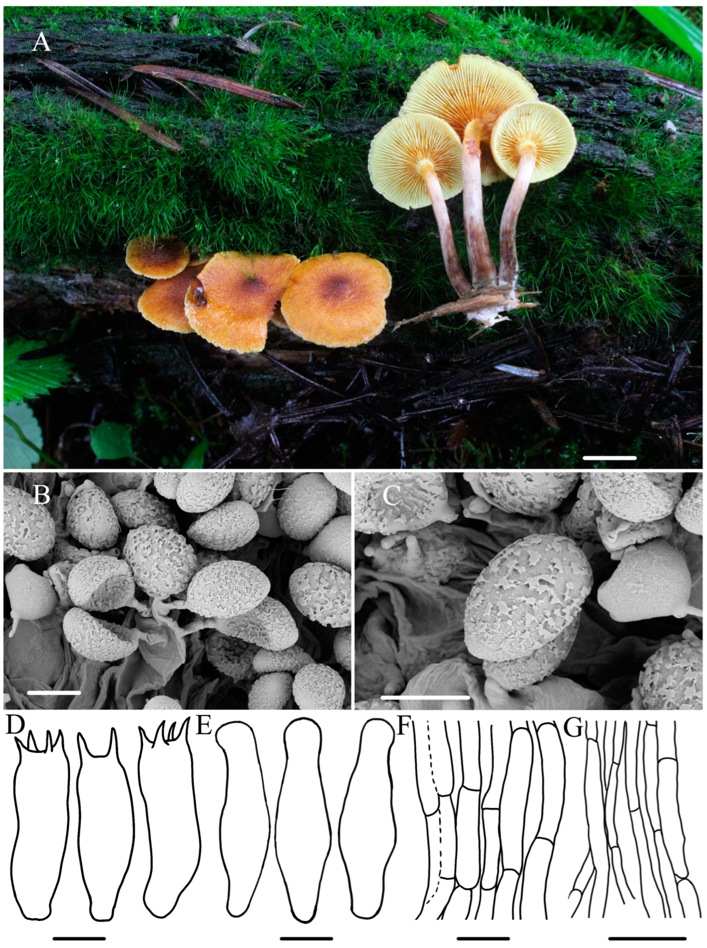
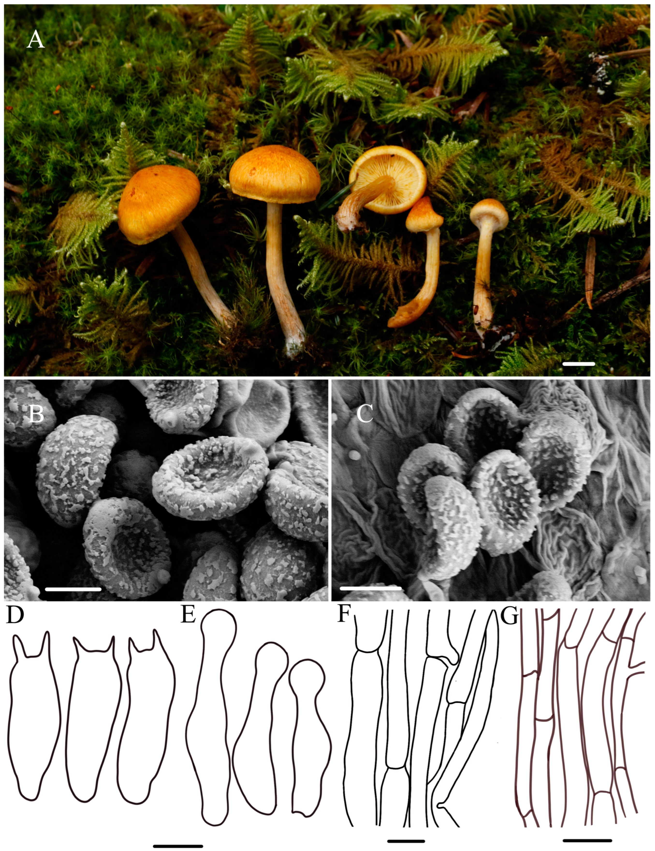
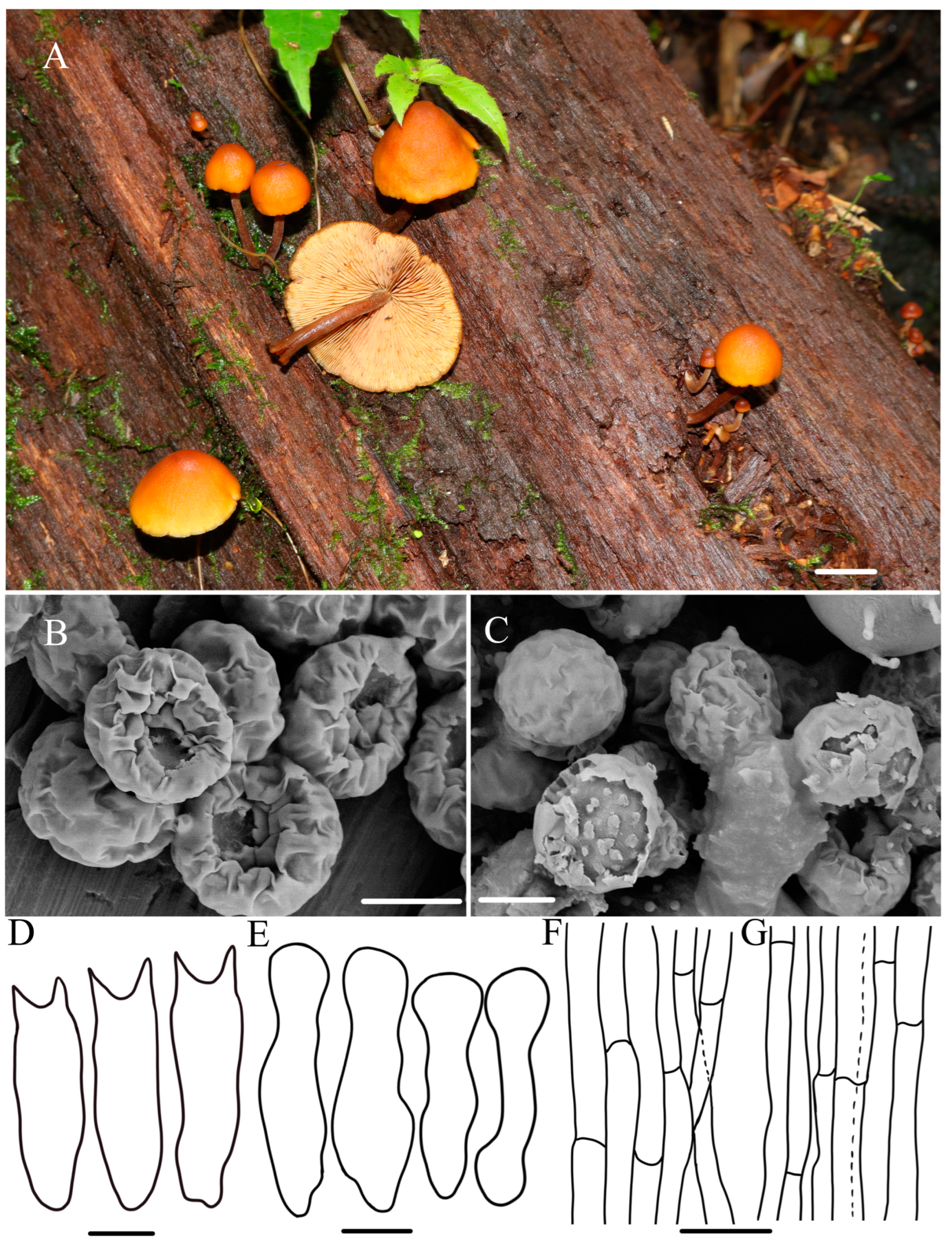
The seventy-eight specimens of Gymnopilus collected in this study, which represented eleven species, were included in the phylogenetic analyses with the outgroup species Galerina marginata. During our study, seventy-six ITS sequences, seventy-three nrLSU sequences, seventy-five nrSSU sequences, fifteen rpb1 sequences, sixty rpb2 sequences, and sixteen tef1-α sequences were newly generated in this study (Table 1). There was a total of five thousand and thirty-one bp (base pairs) in the final alignment after assembling those six gene sequences, of which eight hundred and ninety-one characters are from nrLSU, five hundred and forty-four characters are from tef1-α, seven hundred and six characters are from rpb2, one thousand three hundred and twenty-seven characters are from rpb1, eight hundred and ninety characters are from nrSSU, and six hundred and seventy-three characters are from ITS. The phylogenetic tree of the ML and MrBayes topology were generally the same using those six gene sequences. The Maximum Likelihood tree is shown in Figure 1, with bootstrap and posterior probability values indicated on the branches.

3.2. Taxonomy

Gymnopilus gyirongensis R.L. Zhao and W.Q. Yang, sp. nov., Figure 2.
Fungal Names: FN 571665.
Etymology: —‘gyirong’ refers to the location where the type specimen was collected.
Typification: China, Xizang Autonomous Region, Shigatse, Gyirong County, Gyirong Town, N 28°23′45″, E 85°23′34″, 3441 m asl, 2 August 2022, collected by Rui-Lin Zhao and Xin-Yu Zhu, ZRL20220779 (holotype HMAS 287478).
Diagnosis: Pileus medium-sized with orange–yellow to brownish orange, covered with yellowish rust squamules and umbonate center; basidiospores medium-sized (6.2–6.8 × 3.4–4.3) μm with ornamentation moderately developed; scattered or gathered on mossy wood with rotting pine needles.
Macroscopic description: Pileus 12–30 mm diameter, plano-convex to plane, with a low broad umbo, orange-–yellow (4–6AB4–6) to brownish orange (5–6BC4–5), slightly wavy to serrate margins, moist surface covered with yellow and rust squamules, denser and darker in the middle of the cap. Context thin, pale white (1A1) to pale yellow (2B3), color unchanging upon cutting. Lamellae adnate to slightly adnexed, pale yellow (4A3) to yellowish brown (4B8), with a few ferruginous spots (7B6), crowded, unequal, L = 0.7–1.3 cm, I = 4.8–6.1 mm. Stipe 2.8–3.6 × 0.2–0.5 cm, cylindrical hollow, sometimes thickening at the base, fibrillose, pale white (1A1) from to the top and brown (7E7) from center to the base, color changing to yellowish brown (4B8), base with whitish or cream mycelium. Odor indistinct.
Microscopic description: Basidiospores (5.9–)6.2–6.8(–7.0) × (2.9–)3.4–4.3(–4.5) μm, Q = 1.6–1.8, avQ = 1.7, broadly ellipsoid, distinct suprahilar depression in side view, ornamentation moderately developed, germ pore absent. Basidia (18.5–)19.5–21.4(–21.8) × 5.1–6.0 μm, narrowly clavate, mostly four-sterigmate, rarely two-sterigmate, sterigmata 2.2–3.5 μm long, hyaline in KOH, oil-like droplets present. Cheilocystidia 21.2–26.8 × 4.8–6.8 μm, lecythiform to narrowly utriform, cylindrical, narrowly clavate, capitate to sub-capitate, hyaline, oil droplets present, thin-walled, well-defined basal clamp. Pleurocystidia not observed. Pileipellis is a cutis of filamentous hyphae, 3.6–11 μm diam, with very light rusty brown plasmatic pigment, thin-walled. Stipitipellis filamentous hyphae 4.4–7.5 μm diam, some inflated hyphae observed with constrictions at septa, thin-walled, clamp connections present. A yellowish pigment dissolves when lamellae are mounted in KOH.
Habitat: —Scattered or clustered on mossy, damp wood with rotting pine needles.
Other material examined: China, Xizang Autonomous Region, Shigatse, Gyirong County, Gyiron Town, N 28°23′45″, E 85°23′34″, 3441 m asl, 2 August 2022, collected by Rui-Lin Zhao and Xin-Yu Zhu, ZRL20220790 (HMAS 287479); China, Xizang Autonomous Region, Nyingchi City, Forest ecological monitoring station, N 29°39′2″, E 94°42′58″, 3880 m asl, 27 July 2021, collected by Bin Cao and Xin-Yu Zhu, ZRL20211058 (HMAS 287474); China, Sichuan Province, Tibetan Autonomous Prefecture of Garzê, Yajiang County, Gexigou National Nature Reserve, 15 August 2020, collected by Rui-Lin Zhao and Xi-Xi Han, ZRL20200269 (HMAS 287446).
Notes: Gymnopilus gyirongensis sp. nov. can be easily distinguished by its orange-yellow to orange–brown pileus covered with yellowish rust squamules and umbonate center. Microscopically, this new species has medium-sized spores with moderately developed ornamentation. In the field, another proposed new species, Gy. tomentosiceps, may be confused with Gy. gyirongensis due to the fact that they both have yellowish caps; however, they have different habitats: Gy. tomentosiceps usually grows on mossy, soil under coniferous forest trees, but Gy. gyirongensis prefers fruiting on damp wood with rotting pine needles. In this study, phylogenetic analysis demonstrated that four specimens of this new species formed a cohesive cluster with full support. Additionally, they were positioned closely to Gy. penetrans, Gy. hybridus, and Gy. tomentosiceps (Figure 1). Gymnopilus penetrans is similar to Gy. gyirongensis because they share the characteristic of a smooth pileus covered with yellow and rusty brown squamules on the surface and can be found under conifers and deciduous trees [2,32]. However, Gy. penetrans develop bigger basidiopores (7.2–9.9 × 4–5.5 μm) than those of Gy. gyirongensis [6]. Moreover, these two species have 29 base pair differences in their ITS sequences. The four ITS sequences of this new species display four or five base pair differences. Another similar species, Gy. hybridus, can easily be distinguished from this species due to it having bigger basidiospores (6–8 × 4–4.5 μm) and basidia (20–28 × 5–8 μm) [5,7,41].
Figure 2. The morphology of Gymnopilus gyirongensis sp. nov. of holotype ZRL20220779 (HMAS 287478). (A) Basidiomata; (B,C) basidiospores; (D) basidia; (E) cheilocystidia; (F) pileipellis; (G) stipitipellis. Scale bar = 1 cm for (A); 5 μm for (BD); 4 μm for (E); 5 μm for (F,G).
Jof 10 00220 g002
Gymnopilus variisporus R.L. Zhao and W.Q. Yang, sp. nov., Figure 3.
Fungal Names: FN 571764.
Etymology: —‘variisporus’ refers to variable ornamentation on the spore surface during maturity.
Typification: China, Xizang Autonomous Region, Shigatse, Gyirong County, Gyirong Town, N 28°23′45″, E 85°23′34″, 3441 m asl, 2 August 2022, collected by Mao-Qiang He and Bin Cao, ZRL20220827 (holotype HMAS 287480).
Diagnosis: Pileus medium-sized, covered with yellow to rusty squamules; stipe yellowish brown to rusty brown; basidiospores 6.7–7.3 × 4.5–5.1 μm, golden brown to yellow–brown; scattered or clustered in meadows on moist ground of decaying pine needles.
Macroscopic description: Pileus 4–39 mm across, convex to plano-convex when young, becoming hemispherical to plano-convex or slightly concaved at maturity, yellow–brown (4B8), smooth in dried specimens, glabrous, with inflexed to involute margin, moist surface covered with yellow and rusty squamules. Context thin but thicker at the center, pale white (1A1) to yellowish brown (4B8), unchanging upon cutting. Lamellae adnate to slightly adnexed, crowded to close, pale yellow (4A3) to yellowish brown (4B8), and finally ferruginous with spots (7B6). Stipe 2.9–6.3 × 0.2–0.7 cm, cylindrical, hollow, fibrous, yellowish brown to rusty brown (2B3–5C5), base with white mycelium, lacking veil. Odor indistinct.
Microscopic description: Basidiospores (6.5–)6.7–7.3(–7.6) × (3.8)4.5–5.1(–5.4) μm, Q = 1.43–1.49, avQ = 1.46, ellipsoid, distinct suprahilar depression in side view, ornamentation moderately developed, not more than 0.2 μm high, golden brown to yellow–brown. Basidia 14.1–16.9 × 4.3–5.0 μm, clavate, colorless, with oil droplets or pigment, sterigmata 1.6–2.3 μm long with yellowish brown. Cheilocystidia 14.1–18.0 × 5.2–5.6 μm, utriform to narrowly lageniform, some cylindrical to narrowly clavate, hyaline, with obtuse or sub-capitate apex, base pedunculate or pedicellate, non-gut, well-defined basal clamp. Pleurocystidia absent. Pileipellis cutis hyphae 4–7 (8.5) µm in diameter, septate, crowded, branched, clamp connection present, non-guttulated, thin-walled. Stipitipellis cutis hyphae 6.5–9 µm in diameter, branched, filamentous, clamp connection present. A yellowish pigment dissolves when lamellae are mounted in KOH.
Habitat: —Scattered or clustered in meadows on moist ground of decaying pine needles, characterized by the absence of bark and cork, summer.
Other material examined: China, Xizang Autonomous Region, Shigatse, Gyirong County, Gyirong Town, N 28°23′45″, E 85°23′34″, 3441 m asl, 2 August 2022, collected by Mao-Qiang He and Bin Cao, ZRL20220830 (HMAS 287481).
Notes:Gymnopilus variisporus sp. nov. is distinctive in having a hemispherical to plano-convex pileus covered with yellowish squamules and possessing ellipsoid spores with moderate ornamentation. In the field, Gy. variisporus has relatively similar features to Gy. gyirongensis sp. nov. because they both possess yellowish pileus covered with rust squamules, but Gy. variisporus has bigger basidiospores (6.7–7.3 × 4.5–5.1 μm). Phylogenetic analyses in this study showed that Gy. variisporus formed a distinct lineage with fully supported values and was a sister to Gy. swaticus, Gy. stabilis, and Gy. sapineus (Figure 1). The three known species have bigger basidiospores and different habitats compared to this new species. Their respective characterization are Gy. stabilis having yellowish orange to light orange pilei and possessing bigger basidispores (7.5–8.5 × 4.5–5 µm), growing in sandy soil [32]; Gy. sapineus has yellowish brown to rusty brown pileus covered with fibrillose-tomentum to tomentum–scaly, and it possess bigger basidiospores (7.3–8.2 × 4.5–5.2 µm), fruiting on dead wood, but Gy. variisporus usually scatter or cluster in meadows, specifically on moist ground [6,32]; Gy. swaticus has velutinous to slightly tomentose and pileus and bigger basidiospores (8.6–10.0 × 4.5–5.6 μm), growing on Piceae smithiana [2,19].
Figure 3. The morphology of Gy. variisporus sp. nov. of holotype ZRL20220827 (HMAS 287480). (A). Basidiomata; (B,C) basidiospores; (D) basidia; (E) cheilocystidia; (F) pileipellis; (G) stipitipellis. Scale bar = 1 cm for (A); 5 μm for (BD); 4 μm for (E); 5 μm for (F,G).
Jof 10 00220 g003
Gymnopilus tomentosiceps R.L. Zhao and W.Q. Yang, sp. nov., Figure 4.
Fungal Names: FN 571765.
Etymology: —‘tomentosiceps’ means the pileus of this species is covered with tomentose scales.
Typification: China, Xizang Autonomous Region, Zayü County, N 28°36′46″, E 98°5′22″, 4110 m asl, 21 July 2021, collected by Rui-Lin Zhao, Ming-Yu Zhu, and Bin Cao, ZRL20210594 (holotype HMAS 287463).
Diagnosis: Pileus medium-sized, covered with tomentose scales; basidiospores 5.5–6.8 × 3.8–4.5 μm, coarsely roughened with large and irregular wart; cheilocystidia present, 21.2–26.8 × 4.8–6.8 μm, lecythiform to narrowly utriform, capitate to sub-capitate.
Macroscopic description: Pileus 8–26 mm diameter, hemispherical, pale yellow (4A3) to darker ochre–yellow (6C8), with undulating margin, covered with tomentose scales. Context thin, pale white (1A1) to yellowish brown (4B8), unchanging upon cutting. Lamellae adnate to slightly adnexed, pale yellow (4A3) to yellowish brown (4B8), with ferruginous spots and slightly wrinkled in dried specimens, glabrous, crowded, unequal, L = 0.7–1.3 cm, I = 4.8–6.1 mm. Stipe 2.4–3.9 × 0.2–0.4 cm, cylindrical, hollow, sometimes tapering at base, pearl white to brown (3B1-7E7), base with or without whitish or cream mycelium. Pleats yellow to rusty brown. Odor indistinct.
Microscopic description: Basidiospores (4.6–)5.5–6.8(–7.1) × (3.7–)3.8–4.5(–4.8) μm, Q = 1.45–1.51 μm, aveQ = 1.48 μm, ellipsoid, distinct suprahilar depression in side view, slightly thickened wall, coarsely roughened with large and irregular wart, germ pore absent, yellowish brown. Basidia 14.5–19.1 × 4.5–5.0 μm, narrowly clavate, hyaline, oil-like droplets present, two to four sterigmata, sterigmata 2.8–3.6 μm long. Cheilocystidia 21.2–26.8 × 4.8–6.8 μm, lecythiform to narrowly utriform, capitate to sub-capitate, hyaline, oil droplets present, thin-walled, well-defined basal clamp. Pileipellis cutis hyphae 2.7–7.1 µm in diameter, septate, crowded, branched, clamp connection present, non-guttulated, thin-walled. Stipitipellis hyphae 1.2–4.8 µm in diameter, branched, filamentous, clamp connection present. A yellowish pigment dissolves when lamellae are mounted in KOH.
Habitat: —Grows on mossy, moist soil under coniferous forest trees.
Other material examined: China, Xizang Autonomous Region, Bomi County, N 29°47′30″, E 95°41′50″, 3670 m asl, 24 July 2021, collected by Xin-Yu Zhu, Ming-Zhe Zhang, ZRL20210795 (HMAS 287466).
Notes: Gymnopilus tomentosiceps sp. nov. is easily distinguished by its pileus, which is completely covered with a pale yellow tinge. In the field, Gy. tomentosiceps is morphologically similar to Gy. orientispectabilis because it possesses minutely fibrillose–scaly, but Gy. orientispectabilis has bigger basidiospores (7.2–9.0 × 4.8–6 μm) and basidia (22.2–36.0 × 6.6–9.6 μm) [20]. In the phylogenetic tree (Figure 1), Gy. penetrans and Gy. arenophilus are sisters to Gy. tomentosiceps, consistent with previous studies [2,7,41]. Morphologically, they can be easily distinguished. Gymnopilus penetrans has finely innately rusty ochre to rusty brown fibrillose-striped pileus and moderately developed ornamentation in terms of the ellipsoidal spores, growing on the dead wood of conifers and deciduous trees, and all of those are different from Gy. tomentosiceps. [2,32]. Gy. arenophilus differs by its bigger basidiospores (8.5–10.5 × 5.5–6.5 μm) and longer basidia (26–35 × 7–9 μm). Additionally, it exhibits distinct growing conditions, being found on sandy soil and occasionally attached to wood chips or charcoal [14].
Figure 4. The morphology of Gy. tomentosiceps sp. nov. of holotype ZRL20210594 (HMAS 287463). (A). Basidiomata; (B,C) basidiospores; (D) basidia; (E) cheilocystidia; (F) pileipellis; (G) stipitipellis. Scale bar = 1 cm for (A); 5 μm for (B,C); 6 μm for (D,E); 1 μm for (F); 1 μm for (G).
Jof 10 00220 g004
Gymnopilus tenuibasidialis R.L. Zhao and W.Q. Yang, sp. nov., Figure 5.
Fungal Names: FN 571766.
Etymology: —‘tenuibasidialis’ refers to the shape of the basidia of this species (narrow and long).
Typification: China, Xizang Autonomous Region, Nyingchi City, Bomi County, Spruce forest, N 29°52′50″, E 95°34′43″, 2700 m asl, 26 July 2021, collected by Xin-Yu Zhu and Ming-Zhe Zhang, ZRL20210911 (holotype HMAS 287467).
Diagnosis: Pileus medium-sized, abrupt papilla, yellowish to mustard with brown patches of varying sizes; basidiospores 4.8–5.1 × 3.7–4.0 μm, broadly ellipsoid; cheilocystidia present, 30.1–35.5 × 3.7–4.8 μm, slender.
Macroscopic description: Pileus 18–34 mm diameter, with abrupt papilla, yellowish to mustard (4B6–5C8), surface fibrillose, brown patches of varying sizes. Context thin, pale white (1A1), color unchanging upon cutting. Lamellae 4–6 mm broad, yellowish brown (4B8), adnate to sinuate, straight, sparser, unequal in length, edge entire or slightly serrulate, lamellulae mostly in two tiers. Stipe 1.3–5.7 × 0.2–0.6 cm, yellowish brown (4B8), cylindrical, hollow, fibrillose, dull surface, equal in thickness at the top and bottom. Odor indistinct.
Microscopic description: Basidiospores (4.1–)4.8–5.1(–5.6) × (3.5–)3.7–4.0(–4.2) μm, Q = 1.28–1.30 μm, broadly ellipsoid, with broadly rounded apices, moderately roughened with irregular warts and short ridges, non-dextrinoid to obscurely dextrinoid. Basidia 21.4–24.0 × 3.6–4.8 μm, four-spored, sterigmata 1.8–5.1 μm long, clavate to cylindrical, oil-like droplets present, usually constricted near or above the middle. Cheilocystidia 30.1–35.5 × 3.7–4.8 μm, lecythiform to narrowly utriform, capitate to sub-capitate but occasionally without a swollen apex, hyaline, well-defined basal clamp. Pleurocystidia not observed. Pileipellis cutis hyphae 3.8–13.2 µm in diameter, septate, crowded, branched, clamp connection present, non-guttulated, thin-walled. Stipitipellis hyphae 1.4–7.4 µm in diameter, branched, filamentous, clamp connection present. A yellowish pigment dissolves when lamellae are mounted in KOH.
Habitat: —Single or scattered under pines, usually in a wet environment.
Other material examined: No other specimens.
Notes: Gymnopilus tenuibasidialis sp. nov. is easily distinguished by possessing abrupt papill pileus with brown patches and presenting slender cheilocystidia (30.1–35.5 × 3.7–4.8 μm). In the phylogenetic tree (Figure 1), Gy. tenuibasidialis formed a distinct lineage separated from other Gymnopilus species with fully supported lineages. Gymnopilus purpureosquamulosus, Gy. dunensis, Gy. suberis, and Gy. dilepis were close to this new species in phylogeny; however, they can be easily distinguished by macro- and micro-morphology. Gymnopilus dilepis can differ by its convex to plano-convex pileus with ruby to violet brown squamules, bigger basidiospores (6–7.5 × 4.8–6 µm), shorter and thicker cheilocystidia (20–30.2 × 8–12 µm) [12,42]. Gymnopilus purpureosquamulosus differs in having brown–purple to purple pileus with a central depression, a surface covered with reddish violet squamules and lamellae that turn grey with KOH and FeSO4, and bigger basidiospores (7–8 × 4–5.5 µm) [18]. Gymnopilus dunensis can differ by its pileus, covered with greyish squamules when young; bigger basidiospores (8.5–10.7 × 4.9–6.6 μm) and basidia (24.9–29.4 × 8.5–9.7 μm); and the fact that it is gregarious in small groups on sandy soil [19]. Gymnopilus suberis can differ by its pileus (initially cream to white before finally tunin sulfur yellow), a fibrous stipe with a cortinoid veil, and bigger basidiospores (7.2–8.8 × 4.4–4.8 µm) and basidia (24–27 × 6.5 µm) [17]. Phylogenetically, an unidentified specimen represented by ZRL20151336 (HMAS 287411) is a sister to this new species; however, they exhibit large differences in spore ornamentation (Figure 1).
Figure 5. The morphology of Gy. tenuibasidialis sp. nov. of holotype ZRL20210911 (HMAS 287467). (A). Basidiomata; (B,C) basidiospores; (D) basidia; (E) cheilocystidia; (F) pileipellis; (G) stipitipellis. Scale bar = 1 cm for (A); 5 μm for (B,C); 6 μm for (D); 4 μm for (E); 5 μm for (F,G).
Jof 10 00220 g005
Gymnopilus aurantipileatus R.L. Zhao and W.Q. Yang, sp. nov., Figure 6.
Fungal Names: FN 571767.
Etymology: —‘aurantipileatus’ means the pileus color of the type of specimen is orange.
Typification: China, Yunnan Province, Jingdong Yi Autonomous County, Ailao Mountains, N 23°36′–24°56′, E 100°44′–101°30′, 3166 m asl, 4 July 2021, collected by Rui-Lin Zhao, Mao-Qiang He, Min-Zhe Zhang, Xin-Yu Zhu, and Mei-Qi Wang, ZRL20210361 (HMAS 287460).
Diagnosis: Pileus medium-sized, with lemon yellow to orange yellow, not scaly; stipe 0.7–1.9 × 0.1–0.3 cm, yellowish brown; basidiospores 5.7–6.5 × 4.5–5.3 μm, ornamentation developed; scattered or clumped on damp wood.
Macroscopic description: Pileus 2–30 mm diameter, hemispherical, lemon yellow (4A8) to orange–yellow (5A8), surface smooth and glabrous, not scaly. Context thin, yellowish brown (4B8), unchanging upon cutting. Lamella adnate, pale yellow (4A3), close to crowded, with ferruginous patches, edge even. Stipe 0.7–1.9 × 0.1–0.3 cm, moderately yellowish brown (4B8), hollow, terete, equal or widened to the base, context moist, with spots of the same color as the lamella and gradually becomes denser near the cap. Odor indistinct.
Microscopic description: Basidiospores (5.3–)5.7–6.5(–6.8) × (4.3–)4.5–5.3(–6.0) μm, Q = 1.2–1.3 μm, broadly ellipsoid, verrucose with small to medium warts, coarsely roughened with large and irregular warts, ornamentation developed, golden brown to yellow-brown. Basidia 19.4–23.1 × 5.4–7.0 μm, four- or two-spored, clavate, colorless, with oil droplets or pigment, sterigmata 2.3–4.2 μm long, yellow–brown with occasional individual sterigmata yellowish brown. Cheilocystidia 9–16.5 × 3–4.8 μm, clavate, lageniform to narrowly lageniform with capitate apex, yellowish brown. Pleurocystidia not seen. Pileipellis cutis hyphae 2.4–6.7 µm in diameter, septate, crowded, branched, clamp connection present, non-guttulated, thin-walled. Stipitipellis hyphae 3.2–6.3 µm in diameter, branched, filamentous, clamp connection present. A yellowish pigment dissolves when lamellae are mounted in KOH.
Habitat: —Scattered or in clusters overgrown with wood, usually in a wet environment.
Other material examined: China, Yunnan Province, Jingdong Yi Autonomous County, Ailao Mountains, N 23°36′–24°56′, E 100°44′–101°30′, 3166 m asl, 4 July 2021, collected by Rui-Lin Zhao, Mao-Qiang He, Ming-Zhe Zhang, Xin-Yu Zhu, and Mei-Qi Wang, ZRL20210364 (HMAS 287461).
Notes: Gymnopilus aurantipileatus sp. nov. is easily distinguished by having smooth to glabrous pileus and small cheilocystidia (9–16.5 × 3–4.8 μm). In the field, Gy. aurantipileatus sp. nov. is similar to Gy. purpureosquamulosus, because they both have pileus broadly convex to plano-convex, light orange to orange–yellow. Gymnopilus picreus and this new species have the type of same habitat, dead coniferous trees. However, Gy. picreus is distinguished by its squamulose pileus, rusty stained lamellae, dark brown stipe with white pulverulence, and bigger basidiospores (7.0–9.5 × 4.5–6.0 μm) [7]. Compared to Gy. crociphyllus, which has oxide yellow to light brown pileus and sometimes mustard yellowish smudges, bigger basidia (27.8–30.9 × 6.3–7.5 μm) and bigger cheilocystidia (23.3–26.9 × 6.7–8.7 μm) [43], Gy. aurantipileatus sp. nov. is also different. Gy. austropicreus differs by featuring a pale lemon-yellow pileus and lamellae that darken to black with the application of KOH. Furthermore, it possesses bigger basidiospores (6.6–9 × 4.5–6 µm) and cheilocystidia (15–27 × 4–6 µm) [44].
Figure 6. The morphology of Gy. aurantipileatus sp. nov. of holotype ZRL20210361 (HMAS 287460). (A). Basidiomata; (B,C) basidiospores; (D) basidia; (E) cheilocystidia; (F) pileipellis; (G) stipitipellis. Scale bar = 1 cm for (A); 4 μm for (B,C); 5 μm for (D); 3 μm for (E); 3 μm for (F,G).
Jof 10 00220 g006

4. Discussion

Based on a combination of morphological and phylogenetic analyses, seventy-eight specimens were identified as eleven Gymnopilus species. Six of them are known species, and they are Gy. penetrans (Fr) Murrill, Gy. hybridus (Gillet) Maire, Gy. suberis (Maire) Singer, Gy. dilepis (Berk. & Broome) Singer, Gy. minisporus (T. Bau & M. T. Liu), and Gy. picreus (Pers.) P. Karst. These known species have been reported in tropical and subtropical areas based on previous studies [2,4,5,6,7,8,42,45], and in this study, we found them in ten provinces. Five new species—Gy. gyirongensis sp. nov.; Gy. tomentosiceps sp. nov.; Gy. variisporus sp. nov.; Gy. aurantipileatus sp. nov.; Gy. tenuibasidialis sp. nov.—mainly from the Xizang Autonomous Region and surrounding provinces of China have been described.
In this study, the macroscopic and microscopic morphology of those five new species are described in detail. Some macro- and micro-differences can be used to separate them from each other. Gymnopilus tomentosiceps is characterized by having a small and tomentose cap, while Gy. tenuibasidialis has an abrupt papilla cap. The mature spores of these five new species are all distinct, with suprahilar depressions in side view, but the shapes and ornamentation on the surface (Figure 2, Figure 3, Figure 4, Figure 5 and Figure 6) are different among these species. The basidiospores of Gy. variisporus and Gy. tomentosiceps are ellipsoid, while those of the other species are broadly ellipsoid; Gy. aurantipileatus has developed ornamentation on basidiospores, while the ornamentation of Gy. variisporus and Gy. aurantipileatus is very variable in the process of maturity (Figure 3 and Figure 6). In general, even though these five new species have similar macro characteristics, their characteristics in terms of spore shape and ornamentation are distinct and different from each other, as supported by our molecular phylogenetic analysis.
In the phylogenetic tree (Figure 1), twenty-nine included species of Gymnopilus segregate into three distinct clades (/penetrans, /crociphyllus, and /picreus), and these three clades are part of the six clades delineated in a previous study and are phylogenetically positioned in the same location [7,12]. In the /penetrans clade, Gy. hybridus is regarded as a synonym of Gy. penetrans by Holec (2005) [6]. However, in Europe, Gy. penetrans is classified as a singular species, with Gy. hybridus considered as a synonym [44,45], or as distinct and formed as two separate species (Gy. penetrans and Gy. hybridus) [46], and these species differ in color, the development of the veil, the presence of rusty spots on lamellae, and the shape of cystidia [6]. In our phylogenetic tree (Figure 1), Gy. tomentosiceps, Gy. hybridus, and Gy. penetrans show a close relationship but in different phylogenetic positions. Furthermore, a total of 21 nucleotide differences were detected in the ITS region between Gy. tomentosiceps and Gy. penetrans, and 24 nucleotide differences were detected between Gy. tomentosiceps and Gy. hybridus. Thus, those three species are identified as separate species. Differences in macro characteristics exist: Gy. prnetrans typically exhibits smooth or slightly wrinkled caps and stipe with white fibrillose partial veil remnants; Gy. hybrids feature hygrophanous streaks, as well as smooth and scale-free caps; and Gy. tomentosiceps is characterized by a cap covered with tomentose scales. Gymnopilus crociphyllus (Cooke and Massee) Pegler has distinctive characteristics, such as a fasciculate fruiting body, large pileus size, and a rumpled pileus margin [43]. In the /picreus clade, which includes Gy. austropicreus B.J. Rees and Gy. aurantipileatus sp. nov, this clade can be morphologcially characterized as having the same ornamentation on spores encased in a membrane which will gradually fall off with maturity. In another study [46], Gy. picreus (FT.) P. Karsten and Gy. austropicreus always emerged basal to the remainder of the species in the phylogenetic tree, which could refer to how they may be ancestral to other species in the genus [47]. Gymnopilus picreus groups grow as a saprophytes on the dead wood of conifers (mostly Picea abies; less frequently Pinus sylvestris) and, rarely, deciduous trees (Betula, Fagus sylvatica) [6], but other Gymnopilus species could grow on dead wood or on mulch-rich soil [2]. Thus, we suspect that the rationale for the types of wood rot they seek for nutrition is possibly ancestral in this genus.
In China, a total of thirty-one Gymnopilus species, including seven species originally described to be from China, have been published so far [7,9,21]. Gymnopilus is a species-rich genus, and the previous studies from China included several provinces, such as the Guangdong, Yunnan, Sichuan, Hainan, and Jilin provinces. In this paper, we have reported species from more parts of China, namely the Xizang Autonomous Region, resulting in a total of thirty-six species of Gymnopilus that have been recorded in China. Furthermore, four of the five new species are originally from the Xizang Autonomous Region, which indicates the special nature of the species, as they are from such a unique habitat. This study complements our understanding of Gymnopilus diversity and lays a foundation for the conservation and utilization of related Gymnopilus resources.

Author Contributions

Conceptualization, W.-Q.Y.; methodology, W.-Q.Y.; validation, W.-Q.Y. and J.-X.L.; formal analysis, W.-Q.Y. and J.-X.L.; resources, M.-Q.H., X.-Y.Z., S.-H.W. and D.P.; data curation, W.-Q.Y. and J.-X.L.; writing—original draft preparation, W.-Q.Y.; writing—review and editing, R.-L.Z. and J.-M.Y.; supervision, R.-L.Z.; project administration, R.-L.Z.; funding acquisition, R.-L.Z. All authors have read and agreed to the published version of the manuscript.

Funding

This research was funded by the Survey of Wildlife Resources in Key Areas of Tibet (ZL202203601); the Projects of Science and Technology Programs of Tibet (Project ID: XZ202202YD0031C), the Biological Resources Programme, Chinese Academy of Sciences (KFJ-BRP-009); the National Natural Science Foundation of China (Project ID: 31961143010, 31970010, 31470152); the Biodiversity Survey and Assessment Project of the Ministry of Ecology and Environment, China (2019HJ2096001006).

Institutional Review Board Statement

Not applicable.

Informed Consent Statement

Not applicable.

Data Availability Statement

All sequence data are available in NCBI GenBank following the accession numbers in the manuscript.

Acknowledgments

This project was supported by the Survey of Wildlife Resources in Key Areas of Tibet (ZL202203601); the Projects of Science and Technology Programs of Tibet (Project ID: XZ202202YD0031C), the Biological Resources Programme, Chinese Academy of Sciences (KFJ-BRP-009); the National Natural Science Foundation of China (Project ID: 31961143010, 31970010, 31470152); the Biodiversity Survey and Assessment Project of the Ministry of Ecology and Environment, China (2019HJ2096001006). We thank Bin Cao, Ming-Yu Zhu, Min-Zhe Zhang, Xi-Xi Han and Mei-Qi Wang for assistance in specimen collecting and Luis A. Parra for the changes to the naming of these new species.

Conflicts of Interest

The authors report there are no competing interests to declare.

References

  1. Kirk, P.; Cannon, P.; Minter, D.; Stalpers, J. Dictionary of the Fungi; CABI: Wallingford, UK, 2008; p. 335. [Google Scholar]
  2. Khan, J.; Kiran, M.; Jabeen, S.; Sher, H.; Khalid, A.N. Gymnopilus penetrans and G. swaticus sp. nov. (Agaricomycota: Hymenogastraceae); A new record and a new species from northwest Pakistan. Phytotaxa 2017, 312, 60–70. [Google Scholar] [CrossRef]
  3. He, M.-Q.; Zhao, R.-L.; Hyde, K.D.; Begerow, D.; Kemler, M.; Yurkov, A.; McKenzie, E.H.C.; Raspé, O.; Kakishima, M.; Sánchez-Ramírez, S.; et al. Notes, outline and divergence times of Basidiomycota. Fungal Divers. 2019, 99, 105–367. [Google Scholar] [CrossRef]
  4. Hesler, H. North American Species of Gymnopilus. Mycol Mem 3; Hafner Publishing Company: New York, NY, USA; London, UK, 1969; Volume 3, pp. 1–117. [Google Scholar]
  5. Rees, B.J.; Zuccarello, G.C.; Orlovich, D.A. Relationships between Australian and Northern Hemisphere Gymnopilus species II. A preliminary phylogeny of species of Gymnopilus and related genera based on Internal Transcribed Spacer (ITS) region of ribosomal DNA. Mycotaxon 2002, 84, 93–110. [Google Scholar]
  6. Holec, J. Taxonomy and nomenclature of Gymnopilus bellulus and G. microsporus (Agaricales, Cortinariaceae). Mycotaxon 2005, 92, 361–370. [Google Scholar]
  7. Liu, M.; Bau, T. Gymnopilus minisporus sp. nov., a new species and a new record of the European species G. hybridus from northeast China. Phytotaxa 2019, 397, 159–168. [Google Scholar] [CrossRef]
  8. Guzmán-Dávalos, L. Type studies of Gymnopilus (Agaricales) I. Mycotaxon 2003, 86, 395–423. [Google Scholar]
  9. Khurshid, R.; Naseer, A.; Khalid, A.N. Gymnopilus rubellus sp. nov. from the coniferous forests of Azad Jammu and Kashmir, Pakistan. Plant Syst. Evol. 2023, 309, 3. [Google Scholar] [CrossRef]
  10. Gartz, J. Occurrence of psilocybin, psilocin and baeocystin in Gymnopilus purpuratus. Persoonia 1989, 14, 19–22. [Google Scholar]
  11. Guzman-Davalos, L. Further investigations on Gymnopilus (Agaricales, Cortinariaceae). A new section and a new species from Mexico. Mycotaxon 1995, 54, 117–124. [Google Scholar]
  12. Guzmán-Dávalos, L.; Mueller, G.M.; Cifuentes, J.; Miller, A.N.; Santerre, A. Traditional infrageneric classification of Gymnopilus is not supported by ribosomal DNA sequence data. Mycologia 2003, 95, 1204–1214. [Google Scholar] [CrossRef] [PubMed]
  13. Guzmán-Dávalos, L.; Contu, M.; Ortega, A.; Vizzini, A.; Herrera, M.; Ovrebo, C.; Rodríguez, A.; Villalobos-Arámbula, A.; Palomera, V.; Vargas, G.; et al. New morphological and molecular data on Gymnopilus purpureosquamulosus and its phylogenetic relationships among similar species. Sydowia 2008, 60, 41–56. [Google Scholar]
  14. Guzmán-Dávalos, L.; Ortega, A.; Contu, M.; Vizzini, A.; Rodríguez, A.; Villalobos-Arámbula, A.R.; Santerre, A. Gymnopilus maritimus (Basidiomycota, Agaricales), a new species from coastal psammophilous plant communities of northern Sardinia, Italy, and notes on G. arenophilus. Mycol. Prog. 2009, 8, 195–205. [Google Scholar] [CrossRef]
  15. Rees, B.; Marchant, A.; Zuccarello, G. A tale of two species—Possible origins of red to purple-coloured Gymnopilus species in Europe. Australas. Mycol. 2004, 22, 57–72. [Google Scholar]
  16. Ortega, A.; Esteve-Raventós, F. A new species of Gymnopilus (Cortinariaceae) from sandy soils in Pinus forests. Persoonia 2005, 18, 505–510. [Google Scholar]
  17. Holec, J.; Kříž, M.; Kolařík, M.; Žák, M. Mediterranean fungus Gymnopilus suberis discovered in Central Europe–A consequence of global warming. Sydowia 2016, 68, 69–85. [Google Scholar]
  18. Acharya, K.; Paloi, S.; Dutta, A.K.; Sikder, R.; Saha, T. Gymnopilus purpureosquamulosus Høil. (Agaricales, Basidiomycota): A new distributional record from India. Check List 2017, 13, 2064. [Google Scholar] [CrossRef]
  19. Bashir, H.; Jabeen, S.; Bashir, H.; Khalid, A.N. Gymnopilus dunensis, a new species from Punjab province, Pakistan. Phytotaxa 2020, 428, 51–59. [Google Scholar] [CrossRef]
  20. Thorn, R.G.; Malloch, D.W.; Saar, I.; Lamoureux, Y.; Nagasawa, E.; Redhead, S.A.; Margaritescu, S.; Moncalvo, J.-M. New species in the Gymnopilus junonius group (Basidiomycota: Agaricales). Botany 2020, 98, 293–315. [Google Scholar] [CrossRef]
  21. Li, Y.J.; Li, T.H.; Tu, L.G. Distribution of known species of Gymnopilus from China and a new record to the Chinese mainland. J. Fungal Res. 2012, 10, 8–12. [Google Scholar]
  22. Li, J.-X.; He, M.-Q.; Zhao, R.-L. Three new species of Micropsalliota (Agaricaceae, Agaricales) from China. Phytotaxa 2021, 491, 167–176. [Google Scholar] [CrossRef]
  23. He, M.; Hyde, K.D.; Cheewangkoon, R.; Zhao, R. Two new species of Micropsalliota (Agaricaceae/Agaricales) from Thailand. Phytotaxa 2020, 453, 137–144. [Google Scholar] [CrossRef]
  24. Heilmann-Clausen, J.; Verbeken, A.; Vesterholt, J.; Leonard, P. The genus Lactarius: Book review. Field Mycol. 2000, 1, 6. [Google Scholar] [CrossRef]
  25. White, T.J.; Bruns, T.; Lee, S.; Taylor, J. Amplification and direct sequencing of fungal ribosomal RNA genes for phylogenetics. In PCR Protocols: A Guide to Methods and Applications; Academic Press: New York, NY, USA, 1990; Volume 18, pp. 315–322. [Google Scholar]
  26. Moncalvo, J.-M.; Lutzoni, F.M.; Rehner, S.A.; Johnson, J.; Vilgalys, R. Phylogenetic Relationships of Agaric Fungi Based on Nuclear Large Subunit Ribosomal DNA Sequences. Syst. Biol. 2000, 49, 278–305. [Google Scholar] [CrossRef] [PubMed]
  27. O’Donnell, K.; Cigelnik, E.; Weber, N.S.; Trappe, J.M. Phylogenetic relationships among ascomycetous truffles and the true and false morels inferred from 18S and 28S ribosomal DNA sequence analysis. Mycologia 1997, 89, 48–65. [Google Scholar] [CrossRef]
  28. Morehouse, E.A.; James, T.Y.; Ganley, A.R.D.; Vilgalys, R.; Berger, L.; Murphy, P.J.; Longcore, J.E. Multilocus sequence typing suggests the chytrid pathogen of amphibians is a recently emerged clone. Mol. Ecol. 2003, 12, 395–403. [Google Scholar] [CrossRef] [PubMed]
  29. Matheny, P.B.; Wang, Z.; Binder, M.; Curtis, J.M.; Lim, Y.W.; Nilsson, R.H.; Hughes, K.W.; Hofstetter, V.; Ammirati, J.F.; Schoch, C.L.; et al. Contributions of rpb2 and tef1 to the phylogeny of mushrooms and allies (Basidiomycota, Fungi). Mol. Phylogenet. Evol. 2007, 43, 430–451. [Google Scholar] [CrossRef]
  30. Matheny, P.B.; Liu, Y.J.; Ammirati, J.F.; Hall, B.D. Using RPB1 sequences to improve phylogenetic inference among mushrooms (Inocybe, Agaricales). Am. J. Bot. 2002, 89, 688–698. [Google Scholar] [CrossRef]
  31. Zhao, R.; Karunarathna, S.; Raspé, O.; Parra, L.A.; Guinberteau, J.; Moinard, M.; De Kesel, A.; Barroso, G.; Courtecuisse, R.; Hyde, K.D.; et al. Major clades in tropical Agaricus. Fungal Divers. 2011, 51, 279–296. [Google Scholar] [CrossRef]
  32. Holec, J.; Borovička, J.; Peintner, U.; Kolařík, M. Towards consolidation of Gymnopilus taxonomy: The case of G. stabilis, G. sapineus, and G. penetrans. Mycol. Prog. 2022, 21, 327–343. [Google Scholar] [CrossRef]
  33. Kasuya, T.; Kobayashi, T.; Kurokawa, E.; Pham, H.N.; Hosaka, K.; Terashima, Y. Three species of Gymnopilus newly recorded in Japan. Jpn. J. Mycol. 2016, 57, 31–45. [Google Scholar]
  34. Edgar, R.C. MUSCLE: Multiple sequence alignment with high accuracy and high throughput. Nucleic Acids Res. 2004, 32, 1792–1797. [Google Scholar] [CrossRef]
  35. Hall, T.A. BioEdit: A user-friendly biological sequence alignment editor and analysis program for Windows 95/98/NT. Nucl. Acids Symp. Ser. 1999, 41, 95–98. [Google Scholar]
  36. Zhang, D.; Gao, F.; Jakovlić, I.; Zhou, H.; Zhang, J.; Li, W.X.; Wang, G.T. PhyloSuite: An integrated and scalable desktop platform for streamlined molecular sequence data management and evolutionary phylogenetics studies. Mol. Ecol. Resour. 2020, 20, 348–355. [Google Scholar] [CrossRef]
  37. Silvestro, D.; Michalak, I. raxmlGUI: A graphical front-end for RAxML. Org. Divers. Evol. 2012, 12, 335–337. [Google Scholar] [CrossRef]
  38. Ronquist, F.; Huelsenbeck, J.P. MrBayes 3: Bayesian phylogenetic inference under mixed models. Bioinformatics 2003, 19, 1572–1574. [Google Scholar] [CrossRef]
  39. Rambaut, A. Free Figure Drawing Tool Version 1.2.2. Institute of Evolutionary Biology, University of Edinburgh [J]. URL. 2006. Available online: http://tree.bio.ed.ac.uk (accessed on 30 December 2023).
  40. Letunic, I.; Bork, P. Interactive Tree of Life (iTOL) v5: An online tool for phylogenetic tree display and annotation. Nucleic Acids Res. 2021, 49, W293–W296. [Google Scholar] [CrossRef] [PubMed]
  41. Rees, B.; Strid, Å. Relationships between Australian and Northern hemisphere Gymnopilus species 1: New species and common misconceptions regarding earlier names. Australas. Mycol. 2001, 20, 30–49. [Google Scholar]
  42. Suwannarach, N.; Kumla, J.; Sri-Ngernyuang, K.; Lumyong, S. Gymnopilus dilepis, a new record in Thailand. Mycotaxon 2017, 132, 337–341. [Google Scholar] [CrossRef]
  43. Cho, H.J.; Lee, H.; Park, J.Y.; Park, M.S.; Kim, N.K.; Eimes, J.A.; Kim, C.; Han, S.-K.; Lim, Y.W. Seven New Recorded Species in Five Genera of the Strophariaceae in Korea. Mycobiology 2016, 44, 137–145. [Google Scholar] [CrossRef]
  44. Rees, B.J. A Taxonomic Study of the Genera Gymnopilus, Pyrrhoglossum and Phaeocollybia in Southern Australia. Ph.D. Thesis, School of Biological Science, University of New South Wales, Sydney, Australia, 1996. Volume 4. pp. 238–240. [Google Scholar]
  45. Guzman-Davalos, L. New records of the genus Gymnopilus (Agaricales, Cortinariaceae) from Mexico. Mycotaxon 1996, 67, 61–78. [Google Scholar]
  46. Rees, B.; Marchant, A.; Zuccarello, G.; Heslewood, M.; Bartlett, J. A southern hemisphere contribution to the phylogenetic study of the agarics. Australas. Mycol. 2003, 21, 102–110. [Google Scholar]
  47. Høiland, K. The genus Gymnopilus in Norway. Mycotax 1990, 39, 257–279. [Google Scholar]
Figure 1. Maximum Likelihood (ML) phylogram of Gymnopilus inferred from partial ITS, nrLSU, nrSSU, rpb1, rpb2, and tef1-α sequences. The tree is rooted by Ga. marginata. Maximum Likelihood support values (>70) and posterior probabilities (>0.90) are shown on the branch (ML/PP). The sequences produced from this study are in bold. “T” refers to the sequences from the types of specimens in this study.
Jof 10 00220 g001
Table 1. Information of the sequences generated from this study. Missing sequences are indicated by “-”.
SpeciesSpecimen-VoucherCountryITSLSUSSUrpb1rpb2tef1-αSource
Gymnopilus penetransHMAS 287405China: Yunnan-OR915134OR915221-PP058997-This study
Gy. penetransHMAS 287412China: ZhejiangOR913484OR915135OR915178PP210943PP058994PP165679This study
Gy. penetransHMAS 287413China: ZhejiangOR913499OR915128OR915204---This study
Gy. penetransHMAS 287415China: Xizang Autonomous RegionOR913483OR915122OR915208PP210941PP058992PP165677This study
Gy. penetransHMAS 287417China: Xizang Autonomous RegionOR913494OR915118OR915190PP210948PP058998PP165675This study
Gy. penetransHMAS 287418China: Xizang Autonomous RegionOR913504OR915130OR915174--PP165678This study
Gy. penetransHMAS 287419China: Xizang Autonomous RegionOR913497OR915117OR915179PP210937-PP165676This study
Gy. penetransHMAS 287423China: GansuOR913486OR915114OR915222---This study
Gy. penetransHMAS 287424China: YunnanOR982115OR915132OR915180PP210947PP058990PP165668This study
Gy. penetransHMAS 287426China: YunnanOR982116OR915133OR915188PP210946PP058996PP165669This study
Gy. penetransHMAS 287433China: YunnanOR982119OR915125OR915173-PP059007-This study
Gy. penetransHMAS 287434China: YunnanOR982120OR915126OR915193-PP059009-This study
Gy. penetransHMAS 287438China: SichuanOR913507OR915140OR915201-PP058989-This study
Gy. penetransHMAS 287439China: SichuanOR913506OR915129OR915210PP210940PP058988-This study
Gy. penetransHMAS 287441China: SichuanOR913496OR915112OR915176-PP059002-This study
Gy. penetransHMAS 287442China: SichuanOR913489OR915119OR915177-PP059000PP165674This study
Gy. penetransHMAS 287450China: SichuanOR913501OR915124OR915192-PP059003-This study
Gy. penetransHMAS 287451China: SichuanOR913493OR915127OR915195-PP059004PP165680This study
Gy. penetransHMAS 287452China: YunnanOR913524-OR915220---This study
Gy. penetransHMAS 287445China: SichuanOR913485OR915121OR915191-PP059005-This study
Gy. penetransHMAS 287454China: SichuanOR913508OR915131OR915182PP210942PP058999-This study
Gy. penetransHMAS 287455China: SichuanOR913495OR915139OR915189-PP059008PP165681This study
Gy. penetransHMAS 287456China: SichuanOR913492OR915115OR915197-PP059001-This study
Gy. penetransHMAS 287457China: SichuanOR913500OR915137OR915214PP210939PP058993-This study
Gy. penetransHMAS 287465China: Xizang Autonomous RegionOR913505OR915120OR915183PP210944PP059006-This study
Gy. penetransHMAS 287469China: Xizang Autonomous RegionOR913488OR915141OR915206PP210950PP058987PP165673This study
Gy. penetransHMAS 287482China: HubeiOR913502-----This study
Gy. penetransHMAS 287483China: HubeiOR982114OR976241---PP165671This study
Gy. penetransHMAS 287452China: YunnanOR913524-OR915220---This study
Gy. penetransHMAS 287471China: Xizang Autonomous RegionOR913503OR915138OR915185PP210949PP058986PP165682This study
Gy. penetransHMAS 287472China: Xizang Autonomous RegionOR913487OR915113OR915203PP210951PP058995-This study
Gy. penetransHMAS 287473China: Xizang Autonomous RegionOR913491OR915123OR915199PP210945PP058985PP165670This study
Gy. penetransHMAS 287475China: Xizang Autonomous RegionOR913498OR915116OR915186PP210938PP058991PP165683This study
Gy. penetransHMAS 287477China: ChongqingOR913490OR915136OR915196-PP059010PP165672This study
Gy. penetransTNS-F-61961JapanKT368684-----Kasuya et al. (2016)
Gy. penetransTNS-F-61963JapanKT368685-----Khan et al. (2017)
Gy. penetransPRM 901944Czech R.MW750184-----Holec et al. (2021)
Gy. penetransPRM 900954Czech R.MW750186-----Holec et al. (2021)
Gy. penetransPRM 901951Czech R.MW750185-----Holec et al. (2021)
Gy. penetransPRM 946166PolandMW750183-----Holec et al. (2021)
Gy. hybridusMT163ChinaMK036417-----Liu and Bau (2019)
Gy. hybridusIB-78-226SwedenAF501548-----Liu and Bau (2019)
Gy. hybridusBH11USAMF773630-----Liu and Bau (2019)
Gy. hybridusHMAS 287432China: HeilongjiangOR913479-----This study
Gy. hybridusHMAS 287414China: HeilongjiangOR913478OR915144OR915200-PP059011-This study
Gy. suberisPRM-923698Czech RepublicHG969653-----Liu and Bau (2019)
Gy. suberisPRM-923697Czech RepublicHG969652-----Holec et al. (2016)
Gy. suberisPRM-923203SpainHG969654-----Holec et al. (2016)
Gy. suberisTNS-F-61959JapanKT368689-----Khan et al. (2017)
Gy. suberisHMAS 287416China: Xizang Autonomous RegionOR913522OR915107OR915209-PP059023-This study
Gy. suberisHMAS 287435China: SichuanOR913520OR915106OR915194-PP059021-This study
Gy. suberisHMAS 287440China: SichuanOR913523OR915103OR915211---This study
Gy. suberisHMAS 287431China: Inner Mongolia Autonomous RegionOR913521OR915108OR915175-PP059020-This study
Gy. suberisHMAS 287443China: SichuanOR913518OR915105OR915181-PP059024-This study
Gy. suberisHMAS 287476China: YunnanOR913519OR915104OR915186-PP059022-This study
Gy. suberisHMAS 287421Thailand: NanOR913511OR915098OR915225-PP059016-This study
Gy. suberisHMAS 287422Thailand: NanOR913512OR915099OR915205-PP059014-This study
Gy. suberisHMAS 287425China: YunnanOR913509OR915100OR915223-PP059018-This study
Gy. suberisHMAS 287427China: YunnanOR913515OR976239OR976274---This study
Gy. suberisHMAS 287428China: GuangxiOR913514OR915101OR915226-PP059017-This study
Gy. suberisHMAS 287429China: GuangxiOR913513OR976240OR915224---This study
Gy. suberisHMAS 287430China: GuangxiOR913510OR976238OR915227-PP059015-This study
Gy. dilepisINM-2-71867JapanKT368680-----Khan et al. (2017)
Gy. dilepisTNS-F-61955JapanKT368681-----Khan et al. (2017)
Gy. dilepisTNS-F-70390JapanKU727215-----Kasuya et al. (2016)
Gy. picreusHMAS 287406China: Xizang Autonomous RegionOR913466OR915146OR915171-PP058976-This study
Gy. picreusHMAS 287407China: ZhejiangOR913470OR915145OR915164---This study
Gy. picreusHMAS 287408China: ZhejiangOR913471OR915147OR915168-PP058970-This study
Gy. picreusHMAS 287409China: ZhejiangOR913461OR915149OR915172-PP058971-This study
Gy. picreusHMAS 287410China: ZhejiangOR913469OR915148OR915165-PP058969-This study
Gy. picreusHMAS 287436China: SichuanOR913462OR915157OR915169-PP058972-This study
Gy. picreusHMAS 287437China: SichuanOR913463-OR915161-PP058968-This study
Gy. picreusHMAS 287448China: SichuanOR913467OR915154OR915162-PP058974-This study
Gy. picreusHMAS 287449China: SichuanOR913465OR915153OR915167-PP058973-This study
Gy. picreusHMAS 287464China: Xizang Autonomous Region-OR915151OR915170-PP058977-This study
Gy. picreusHMAS 287462China: Xizang Autonomous RegionOR913468OR915152OR915163-PP058978-This study
Gy. picreusHMAS 287444China: SichuanOR913464OR915155OR915166-PP058975-This study
Gy. picreusTNS-F-61965JapanKT368687-----Kasuya et al. (2016)
Gy. picreusTNS-F-61964JapanKT368686-----Kasuya et al. (2016)
Gy. picreusIBUG-HFinlandAY281003-----Holec et al. (2021)
Gy. picreusAS 97-103AustraliaAF501557-----Rees et al.. (2002)
Gy. minisporusHMAS 287468China: Xizang Autonomous RegionOR913517-OR915228---This study
Gy. minisporusHMAS 287447China: SichuanOR913516OR915096OR915212-PP058982-This study
Gy. minisporusMT005ChinaMK036415-----Liu and Bau (2019)
Gy. minisporusMT012ChinaMK036416-----Liu and Bau (2019)
Gy. sp. HMAS 287470China: Xizang Autonomous RegionOR913480OR915097OR915219-PP058981-This study
Gy. sp. HMAS 287453China: SichuanOR913460OR915150OR915213-PP058967-This study
Gy. sp. HMAS 287458China: YunnanOR982118OR915109OR915215---This study
Gy. sp. HMAS 287459China: YunnanOR982117OR915102OR915216-PP059019-This study
Gy. sp. HMAS 287411China: ZhejiangOR913457OR915094OR915207-PP058979-This study
Gy. aurantipileatusHMAS 287460China: YunnanOR913458OR915110OR915159-PP058965-This study
Gy. aurantipileatusHMAS 287461China: YunnanOR913459OR915111OR915160-PP058966-This study
Gy. tomentosicepsHMAS 287463China: Xizang Autonomous RegionOR913481OR915142OR915217-PP059012-This study
Gy. tomentosicepsHMAS 287466China: Xizang Autonomous RegionOR913482OR915143OR915184-PP059013-This study
Gy. tenuibasidialisHMAS 287467China: Xizang Autonomous RegionOR913456OR915095OR915218-PP058980-This study
Gy. gyirongensisHMAS 287478China: Xizang Autonomous RegionOR913476OR976246OR976275---This study
Gy. gyirongensisHMAS 287479China: Xizang Autonomous RegionOR913477OR976245OR976276---This study
Gy. gyirongensisHMAS 287474China: Xizang Autonomous RegionOR913475OR915156OR915198-PP058984-This study
Gy. gyirongensisHMAS 287446China: SichuanOR913474OR976244OR915202-PP058983-This study
Gy. variisporusHMAS 287480China: Xizang Autonomous RegionOR913472OR976242OR976272---This study
Gy. variisporusHMAS 287481China: Xizang Autonomous RegionOR913473OR976243OR976273---This study
Gy. sapineusPRM 924999Czech R.MW750187-----Holec et al. (2021)
Gy. sapineusPRM 915496Czech R.MW750188-----Holec et al. (2021)
Gy. stabilisPRM 954258Czech R.MW750182-----Holec et al. (2021)
Gy. stabilisM 0159312GermanyMW750189-----Holec et al. (2021)
Gy. subspectabilisTRTC 152281CanadaMN206898-----Holec et al. (2021)
Gy. subspectabilisCMMF001425CanadaMN206902-----Thorn et al. (2020)
Gy. subspectabilisMICH 10995USAMN206901-----Thorn et al. (2020)
Gy. swaticusSWAT 000133PakistanMF149864MF149865----R. Khurshid et al. (2023)
Gy. swaticusGJ1640PakistanMF149866MF149867----R. Khurshid et al. (2023)
Gy. swaticusGJ1612PakistanMF149863-----Holec et al. (2021)
Gy. turficolaIB 1998098aNorwayAF325669-----Holec et al. (2021)
Gy. voitkiiNBM-F00947CanadaMN206872-----Thorn et al. (2020)
Gy. voitkiiFNL 2009 MS7-056CanadaMN206879-----Thorn et al. (2020)
Gy. voitkiiNBM-F00943CanadaMN206867-----Thorn et al. (2020)
Gy. arenophilusGDA-47384SpainEU518421-----Khan et al. (2017)
Gy. maritimusM.Contu s.n. IBUGItalyEU518419-----Guzmán-Dávalos et al. (2009)
Gy. orientispectabilisTMI37361JapanMN206910-----Thorn et al. (2020)
Gy. speciosissimusCMMF002481CanadaMN206895-----Thorn et al. (2020)
Gy. luteusCMMF006463CanadaMN206889-----Thorn et al. (2020)
Gy. dunensisHum-46PakistanMK088249-----R. Khurshid et al. (2023)
Gy. dunensisL90PakistanMK088248-----R. Khurshid et al. (2023)
Gy. dunensisL04PakistanMK088247-----R. Khurshid et al. (2023)
Gy. rubellusLAH36995PakistanOL964420OL964421----R. Khurshid et al. (2023)
Gy. rubellusLAH36996PakistanOL964403OL964404----R. Khurshid et al. (2023)
Gy. crociphyllusTNS-F-61956JapanKT368675-----Kasuya et al. (2016)
Gy. crociphyllusTNS-F-61966JapanKT368679-----Kasuya et al. (2016)
Gy. crociphyllusINM-2-87471JapanKU727211-----Kasuya et al. (2016)
Gy. crociphyllusINM-2-87449JapanKU727210-----Kasuya et al. (2016)
Gy. spectabilisTNS-F-61962JapanKT368688-----Kasuya et al. (2016)
Gy. purpureosquamulosusIBUG-89-16SwitzerlandAY280998-----R. Khurshid et al. (2023)
Gy. purpureosquamulosusK(M) 75214NigeriaAY280979-----Liu and Bau (2019)
Gy. bellulusTENN 069859USAKY744149-----Liu and Bau (2019)
Gy. bellulusSMNS-STU-F-0900398GermanyMF039254-----Eberhardt et al. (2018)
Gy. austropicreusOTA 60208New ZealandOQ064819-----Beaumont
et al. (2002)
Gy. austropicreusOTA 70412New ZealandOQ064892-----Beaumont
et al. (2002)
Galerina marginataLE-BIN-2272RussiaKY327302-----Liu and Bau (2019)
Ga. marginataIBUG-5246MexicoAY281020-----R. Khurshid et al. (2023)
The bolded parts are the sequences generated in this study.
Disclaimer/Publisher’s Note: The statements, opinions and data contained in all publications are solely those of the individual author(s) and contributor(s) and not of MDPI and/or the editor(s). MDPI and/or the editor(s) disclaim responsibility for any injury to people or property resulting from any ideas, methods, instructions or products referred to in the content.

