
Pestalotiopsis jiangsuensis sp. nov. Causing Needle Blight on Pinus massoniana in China


Abstract
1. Introduction
2. Materials and Methods
2.1. Field Survey and Fungal Isolation
2.2. Morphological Identification
2.3. Genomic DNA Extraction, PCR, and Sequencing
2.4. Phylogenetic Analyses
2.5. Genealogical Concordance Phylogenetic Species Recognition Analyses
2.6. Pathogenicity Test
3. Results
3.1. Disease Symptoms and Fungal Isolation
3.2. Phylogenetic Analyses
3.3. Taxonomy
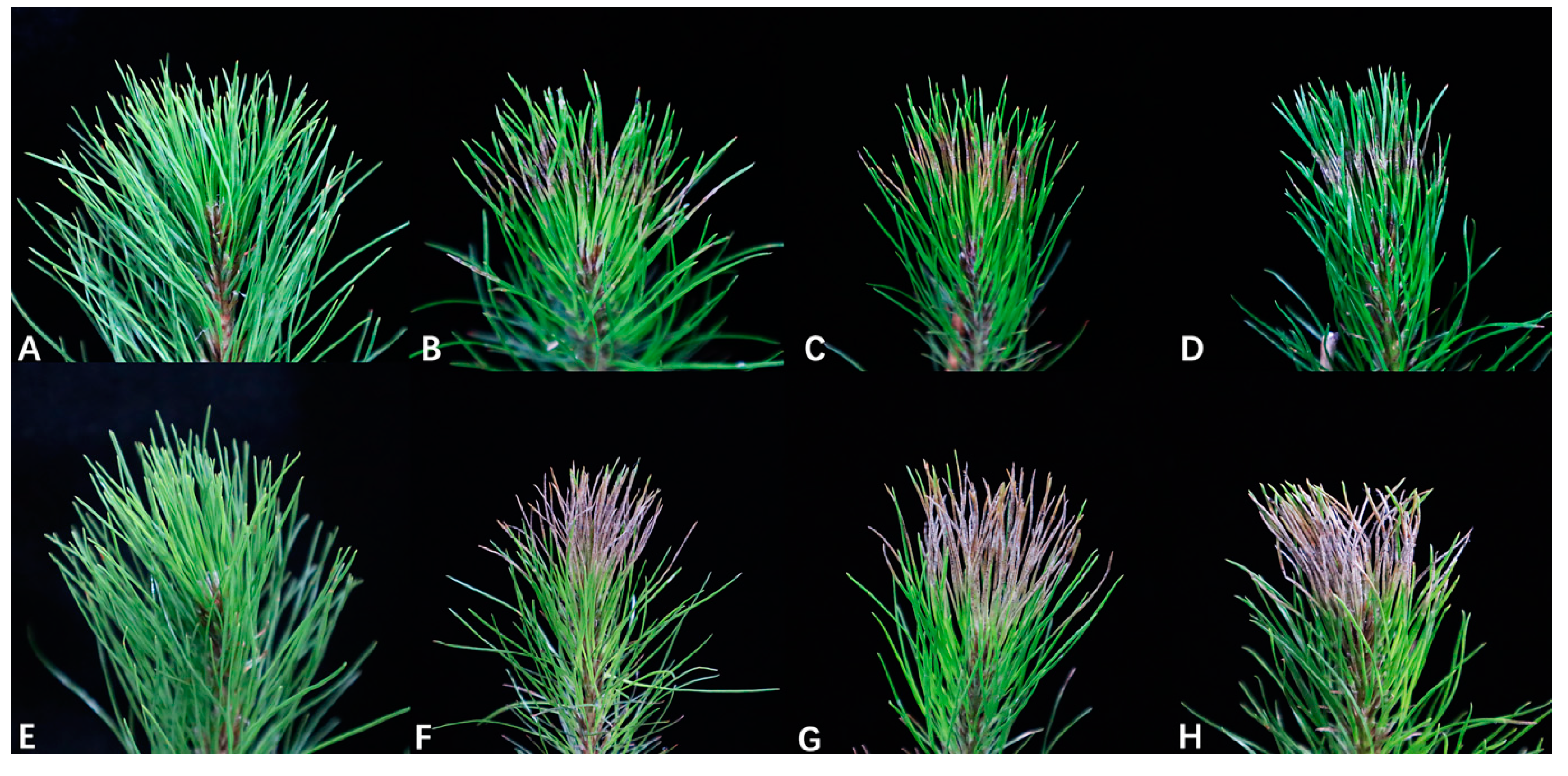
3.4. Pathogenicity Test
4. Discussion
5. Conclusions
Author Contributions
Funding
Institutional Review Board Statement
Informed Consent Statement
Data Availability Statement
Acknowledgments
Conflicts of Interest
References
- Lu, J.Y.; Chen, H.; Yang, Z.Q.; Sun, S.; Luo, Q.F.; Xie, J.K.; Tan, J.H. Physiological and molecular mechanisms of the response of roots of Pinus massoniana Lamb. to low-temperature stress. Front. Plant Sci. 2022, 13, 954324. [Google Scholar] [CrossRef]
- Liu, Q.H.; Zhou, Z.C.; Wei, Y.C.; Shen, D.Y.; Feng, Z.P.; Hong, S.P. Genome-wide identification of differentially expressed genes associated with the high yielding of oleoresin in secondary xylem of masson pine (Pinus massoniana Lamb.) by transcriptomic analysis. PLoS ONE 2015, 10, e0132624. [Google Scholar] [CrossRef]
- Kang, B.; Liu, S.R.; Zhang, G.G.; Chang, J.G.; Wen, Y.G.; Ma, J.M.; Hao, W.F. Carbon accumulation and distribution in Pinus massoniana and Cunninghamia lanceolata mixed forest ecosystem in Daqingshan, Guangxi, China. Acta Ecol. Sin. 2006, 26, 1320–1327. [Google Scholar] [CrossRef]
- Yang, Z.; Xia, H.; Tan, J.; Feng, Y.; Huang, Y. Selection of superior families of Pinus massoniana in southern China for large-diameter construction timber. J. For. Res. 2020, 31, 475–484. [Google Scholar] [CrossRef]
- He, L.; Zhao, C.; Yan, M.; Zhang, L.Y.; Xia, Y.Z. Inhibition of P-glycoprotein function by procyanidine on blood-brain barrier. Phytother. Res. 2009, 23, 933–937. [Google Scholar] [CrossRef]
- Xie, Y.; Liu, B.; Gao, K.; Zhao, Y.; Li, W.; Deng, L.; Zhou, Z.; Liu, Q. Comprehensive analysis and functional verification of the Pinus massoniana NBS-LRR gene family involved in the resistance to Bursaphelenchus xylophilus. Int. J. Mol. Sci. 2023, 24, 1812. [Google Scholar] [CrossRef]
- Luo, X.; Yu, C. First report of damping-off disease caused by Fusarium oxysporum in Pinus massoniana in China. J. Plant Dis. Prot. 2020, 127, 401–409. [Google Scholar] [CrossRef]
- Yu, C.; Luo, X. Trichoderma koningiopsis controls Fusarium oxysporum causing damping-off in Pinus massoniana seedlings by regulating active oxygen metabolism, osmotic potential, and the rhizosphere microbiome. Biol. Control 2020, 150, 104352. [Google Scholar] [CrossRef]
- Li, G.Q.; Wu, W.X.; Lu, L.Q.; Chen, B.Y.; Chen, S.F. Characterization of Pseudofusicoccum species from diseased plantation-grown Acacia mangium, Eucalyptus spp., and Pinus massoniana in Southern China. Pathogens 2023, 12, 574. [Google Scholar] [CrossRef] [PubMed]
- Liang, Q.X.; Pan, F.Y.; Li, D.X. Regularity of outbreak and control techniques of Pinus massoniana cercospora needle blight. J. Zhejiang For. Sci. Technol. 2002, 4, 64–65+84. [Google Scholar]
- Wu, S.X.; Wu, J.; Wang, Y.; Qu, Y.F.; He, Y.; Wang, J.Y.; Cheng, J.H.; Zhang, L.Q.; Cheng, C.H. Discovery of entomopathogenic fungi across geographical regions in southern China on pine sawyer beetle Monochamus alternatus and implication for multi-pathogen vectoring potential of this beetle. Front. Plant Sci. 2022, 13, 1061520. [Google Scholar] [CrossRef]
- Sun, H.T.; Cao, R.B. Identification of Pestalotiopsis parasitized on fruit crops. Acta Agric. Zhejiangensis 1990, 16, 179–185. (In Chinese) [Google Scholar]
- Xu, L.; Kusakari, S.; Hosomi, A. Postharvest disease of grape caused by Pestalotiopsis species. Jpn. J. Phytopathol. 1999, 65, 305–311. [Google Scholar] [CrossRef]
- Maharachchikumbura, S.S.N.; Guo, L.D.; Cai, L.; Chukeatirote, E.; Wu, W.P.; Sun, X.; Hyde, K.D. A multi-locus backbone tree for Pestalotiopsis, with a polyphasic characterization of 14 new species. Fungal Divers. 2012, 56, 95–129. [Google Scholar] [CrossRef]
- Maharachchikumbura, S.S.N.; Hyde, K.D.; Groenewald, J.Z.; Xu, J.; Crous, P.W. Pestalotiopsis revisited. Stud. Mycol. 2014, 79, 121–186. [Google Scholar] [CrossRef]
- Jayawardena, R.S.; Zhang, W.; Liu, M.; Maharachchikumbura, S.S.N.; Zhou, Y.; Huang, J.B.; Nilthong, S.; Wang, Z.Y.; Li, X.H.; Yan, J.Y.; et al. Identification and characterization of Pestalotiopsis-like fungi related to grapevine diseases in China. Fungal Biol. 2015, 119, 348–361. [Google Scholar] [CrossRef] [PubMed]
- Gu, R.; Bao, D.F.; Shen, H.W.; Su, X.J.; Li, Y.X.; Luo, Z.L. Endophytic Pestalotiopsis species associated with Rhododendron in Cangshan Mountain, Yunnan Province, China. Front. Microbiol. 2022, 13, 1016782. [Google Scholar] [CrossRef]
- Bate-Smith, E.C.; Metcalfe, C.R. Leucanthocyanins. 3. The nature and systematic distribution of tannin in dicotyledonous plants. Bot. J. Linn. Soc. 1957, 55, 669–705. [Google Scholar] [CrossRef]
- Maharachchikumbura, S.S.N.; Guo, L.D.; Chukeatirote, E.; Ekachai, C.; Bahkali, A.H.; Hyde, K.D. Pestalotiopsis-morphology, phylogeny, biochemistry and diversity. Fungal Divers. 2011, 50, 167–187. [Google Scholar] [CrossRef]
- Guba, E.F. Monograph of Pestalotia and Monochaetia; Harvard University Press: Cambridge, MA, USA, 1961. [Google Scholar]
- Sutton, B.C. The Coelomycetes. Fungi Imperfecti with Pycnidia, Acervuli and Stromata; Commonwealth Mycological Institute: Surrey, UK, 1980. [Google Scholar] [CrossRef]
- Jeewon, R.; Liew, E.C.Y.; Simpson, J.A.; Hodgkiss, I.J.; Hyde, K.D. Phylogenetic significance of morphological characters in the taxonomy of Pestalotiopsis species. Mol. Phylogenet. Evol. 2003, 27, 372–383. [Google Scholar] [CrossRef]
- Hu, H.L.; Jeewon, R.; Zhou, D.Q.; Zhou, T.X.; Hyde, K.D. Phylogenetic diversity of endophytic Pestalotiopsis species in Pinus armandii and Ribes spp.: Evidence from rDNA and β-tubulin gene phylogenies. Fungal Divers. 2007, 24, 1–22. [Google Scholar]
- Liu, A.R.; Chen, S.C.; Wu, S.Y.; Xu, T.; Guo, L.D.; Jeewon, R.; Wei, J.G. Cultural studies coupled with DNA based sequence analyses and its implication on pigmentation as a phylogenetic marker in Pestalotiopsis taxonomy. Mol. Phylogenet. Evol. 2010, 57, 528–535. [Google Scholar] [CrossRef]
- Jeewon, R.; Liew, E.C.Y.; Hyde, K.D. Phylogenetic relationships of Pestalotiopsis and allied genera inferred from ribosomal DNA sequences and morphological characters. Mol. Phylogenet. Evol. 2002, 25, 378–392. [Google Scholar] [CrossRef]
- Tejesvi, M.V.; Tamhankar, S.A.; Kini, K.R.; Rao, V.S.; Prakash, H.S. Phylogenetic analysis of endophytic Pestalotiopsis species from ethnopharmaceutically important medicinal trees. Fungal Divers. 2009, 38, 167–183. [Google Scholar]
- Huang, Q.; He, J.S. Identification and biological characteristics of pathogen of Pinus tabulaeformis blight. J. Sichuan For. Sci. Technol. 2000, 21, 28–30. [Google Scholar] [CrossRef]
- Huang, G.F. The best method of comprehensive control of loblolly pine blight and defoliation by exploring suspected pine wood nematode disease. Flowers 2018, 12, 364–368. [Google Scholar]
- Xu, Y.; Ren, H.T.; Wang, P.; Zhao, H.X.; Song, Y.Q.; Yu, Q.F.; Liu, X.F. Pathogenic fungi of Pinus sylvestris var. mongolica red blight. J. West China For. Sci. 2017, 46, 91–95. [Google Scholar] [CrossRef]
- Chen, S.C. Research progress and prospect of forest diseases in China. For. Pest Dis. 1997, 4, 38–40. [Google Scholar]
- Bednářová, M.; Dvořák, M.; Janoušek, J.; Jankovský, L. Other foliar diseases of coniferous trees. In Infectious Forest Diseases; Gonthier, P., Nicolotti, G., Eds.; CABI: Wallingford, UK, 2013; pp. 458–487. [Google Scholar]
- Orlikowski, L.B.; Ptaszek, M.; Warabieda, W. Occurrence and harmfulness of Pestalotiopsis funerea to ornamental coniferous plants. Prog. Plant Prot. 2014, 54, 25–30. [Google Scholar]
- Hu, R.R.; Liang, J.; Xie, X.; Zhang, Y.J.; Zhang, X.Y. Incidence of pine needle blight and its relationship with site factors of Japanese red pine forests in the Kunyushan Mountains, East China. Glob. Ecol. Conserv. 2020, 22, e00922. [Google Scholar] [CrossRef]
- Monteiro, P.; Gonçalves, M.F.M.; Pinto, G.; Silva, B.; Martín-García, J.; Diez, J.J.; Alves, A. Three novel species of fungi associated with pine species showing needle blight-like disease symptoms. Eur. J. Plant Pathol. 2022, 162, 183–202. [Google Scholar] [CrossRef]
- White, T.J.; Bruns, T.; Lee, S.; Taylor, J. Amplification and direct sequencing of fungal ribosomal RNA genes for phylogenetics. In PCR Protocols: A Guide to Methods and Applications; Innis, M.A., Gelfand, D.H., Sninsky, J.J., White, T.J., Eds.; Academic Press Inc.: New York, NY, USA, 1990; pp. 315–322. [Google Scholar] [CrossRef]
- Carbone, I.; Kohn, L.M. A method for designing primer sets for speciation studies in filamentous ascomycetes. Mycologia 1999, 91, 553–556. [Google Scholar] [CrossRef]
- Glass, N.L.; Donaldson, G.C. Development of primer sets designed for use with the PCR to amplify conserved genes from filamentous ascomycetes. Appl. Environ. Microbiol. 1995, 61, 1323–1330. [Google Scholar] [CrossRef]
- O’Donnell, K.; Cigelnik, E. Two divergent intragenomic rDNA ITS2 types within a monophyletic lineage of the fungus Fusarium are nonorthologous. Mol. Phylogenet. Evol. 1997, 7, 103–116. [Google Scholar] [CrossRef]
- Katoh, K.; Standley, D.M. MAFFT multiple sequence alignment software version 7: Improvements in performance and usability. Mol. Biol. Evol. 2013, 30, 772–780. [Google Scholar] [CrossRef]
- Hall, T.A. BioEdit: A user-friendly biological sequence alignment editor and analysis program for Windows 95/98/NT. Nucleic Acids Symp. Ser. 1999, 41, 95–98. [Google Scholar] [CrossRef]
- Nguyen, L.T.; Schmidt, H.A.; von Haeseler, A.; Minh, B.Q. IQ-TREE: A fast and effective stochastic algorithm for estimating maximum-likelihood phylogenies. Mol. Biol. Evol. 2015, 32, 268–274. [Google Scholar] [CrossRef] [PubMed]
- Quaedvlieg, W.; Binder, M.; Groenewald, J.Z.; Summerell, B.A.; Carnegie, A.J.; Burgess, T.I.; Crous, P.W. Introducing the consolidated species concept to resolve species in the Teratosphaeriaceae. Persoonia 2014, 33, 1–40. [Google Scholar] [CrossRef]
- Mundkur, B.B.; Kheswalla, K.F. Indian and Burman species of the d=genera Pestalotia and Monochaetia. Mycologia 1942, 34, 308–317. [Google Scholar] [CrossRef]
- Yuan, C. Study on Molecular Detection Technologies for Two Forest Fungus Diseases Pathogen in Sichuan. Master’s Thesis, Sichuan Agricultural University, Ya’an, China, 2012. [Google Scholar]
- Steyaert, R.L. Contribution à l’étude monographique de Pestalotia de Not. et Monochaetia Sacc. (Truncatella gen. nov. et Pestalotiopsis gen. nov.). Bull. Du Jard. Bot. De L’état A Brux. 1949, 19, 285–347. [Google Scholar] [CrossRef]
- Nag Raj, T.R. Coelomycetous Anamorphs with Appendage-Bearing Conidia; Mycologue Publications: Waterloo, ON, Canada, 1993. [Google Scholar]
- Ge, Q.X.; Chen, Y.X.; Xu, T. Flora Fungorum Sinicorum, V. 38 Pestalotiopsis; Science Press: Beijing, China, 2009. [Google Scholar]
- Griffiths, D.A.; Swart, H.J. Conidial structure in two species of Pestalotiopsis. Trans. Br. Mycol. Soc. 1974, 62, 295. [Google Scholar] [CrossRef]
- Liu, F.; Hou, L.; Raza, M.; Cai, L. Pestalotiopsis and allied genera from Camellia, with description of 11 new species from China. Sci. Rep. 2017, 7, 866. [Google Scholar] [CrossRef]
- Jeewon, R.; Liew, E.C.Y.; Hyde, K. Phylogenetic evaluation of species nomenclature of Pestalotiopsis in relation to host association. Fungal Divers. 2004, 17, 39–55. [Google Scholar]
- Liu, A.R.; Xu, T.; Guo, L.D. Molecular and morphological description of Pestalotiopsis hainanensis sp. nov., a new endophyte from a tropical region of China. Fungal Divers. 2007, 24, 23–36. [Google Scholar]
- Li, L.L.; Yang, Q.; Li, H. Morphology, phylogeny, and pathogenicity of Pestalotioid species on Camellia oleifera in China. J. Fungi 2021, 7, 1080. [Google Scholar] [CrossRef]
- Zhang, Z.X.; Liu, X.Y.; Zhang, X.G.; Meng, Z. Morphological and phylogenetic analyses reveal two new species and a new record of Phyllosticta (Botryosphaeriales, Phyllostictaceae) from Hainan, China. MycoKeys 2022, 91, 1–23. [Google Scholar] [CrossRef]
- Silva, A.C.; Diogo, E.; Henriques, J.; Ramos, A.P.; Sandoval-Denis, M.; Crous, P.W.; Bragança, H. Pestalotiopsis pini sp. nov., an emerging pathogen on stone pine (Pinus pinea L.). Forests 2020, 11, 805. [Google Scholar] [CrossRef]
- Bezos, D.; Martínez-Álvarez, P.; Sanz-ros, A.V.; Martín-García, J.; Fernandez, M.M.; Diez, J.J. Fungal communities associated with bark beetles in Pinus radiata plantations in Northern Spain affected by pine pitch canker, with special focus on Fusarium species. Forests 2018, 9, 698. [Google Scholar] [CrossRef]
- Chen, J.; Hao, X.; Liu, X.; Ma, L. First report of Pestalotiopsis neglecta causing black spot needle blight of Pinus sylvestris var. mongolica in China. Plant Dis. 2020, 104, 1545. [Google Scholar] [CrossRef]
- Martínez-Álvarez, P.; Martín-García, J.; Rodríguez-Ceinós, S.; Diez, J.J. Monitoring endophyte populations in pine plantations and native oak forests in Northern Spain. For. Syst. 2012, 21, 373–382. [Google Scholar] [CrossRef]
- Qiu, D.X.; Tan, S.B.; Wu, J.C. Preliminary study on of Pinus massoniana. Sci. Silvae Sin. 1980, 3, 203–207. [Google Scholar]
- Jiang, N.; Voglmayr, H.; Xue, H.; Piao, C.G.; Li, Y. Morphology and phylogeny of Pestalotiopsis (Sporocadaceae, Amphisphaeriales) from Fagaceae leaves in China. Microbiol. Spectr. 2022, 10, e03272-22. [Google Scholar] [CrossRef]


| Locus | PCR Primers (Forward/Reverse) | PCR: Thermal Cycles: (Annealing Temperature in Bold) |
|---|---|---|
| ITS | ITS5/ITS4 | 94 °C: 3 min, (94 °C: 45 s, 55 °C: 45 s, 72 °C: 1 min) ×35 cycles, 72 °C: 10 min |
| TEF1 | EF1-728F/EF1-986R | 94 °C: 3 min, (94 °C: 45 s, 55 °C: 45 s, 72 °C: 1 min) ×35 cycles, 72 °C: 10 min |
| TUB2 | T1/Bt-2b | 94 °C: 3 min, (94 °C: 45 s, 56 °C: 60 s, 72 °C: 1 min) ×35 cycles, 72 °C: 10 min |
| Species a | Strain Number b | Host | Origin | GenBank Accession Number c | ||
|---|---|---|---|---|---|---|
| ITS | TUB2 | TEF1 | ||||
| Pestalotiopsis abietis | CFCC 53011 T | Abies fargesii | China | MK397013 | MK622280 | MK622277 |
| P. adusta | ICMP 6088 T | Prunus cerasus | Fiji | JX399006 | JX399037 | JX399070 |
| P. aggestorum | LC6301 T | Camellia sinensis | China | KX895015 | KX895348 | KX895234 |
| P. anacardiacearum | IFRDCC 2397 T | Mangifera indica | China | KC247154 | KC247155 | KC247156 |
| P. anhuiensis | CFCC 54791 T | Cyclobalanopsis glauca | China | ON007028 | ON005056 | ON005045 |
| P. appendiculata | CGMCC 3.23550 T | Rhododendron decorum | China | OP082431 | OP185516 | OP185509 |
| P. arengae | CBS 331.92 T | Arenga undulatifolia | Singapore | KM199340 | KM199426 | KM199515 |
| P. arceuthobii | CBS 434.65 T | Arceuthobium campylopodum | USA | KM199341 | KM199427 | KM199516 |
| P. australasiae | CBS 114126 T | Knightia sp. | New Zealand | KM199297 | KM199409 | KM199499 |
| P. australis | CBS 114193 T | Grevillea sp. | Australia | KM199332 | KM199383 | KM199475 |
| P. biciliata | CBS 124463 T | Platanus × hispanica | Slovakia | KM199308 | KM199399 | KM199505 |
| P. brachiata | CGMCC 3.18151 T | Rhizophora apiculata | Thailand | MK764274 | MK764340 | MK764318 |
| P. brassicae | CBS 170.26 T | Brassica napus | New Zealand | KM199379 | - | KM199558 |
| P. camelliae | MFLUCC 12-0277 T | Camellia japonica | China | JX399010 | JX399041 | JX399074 |
| P. camelliae-oleiferae | CSUFTCC 08 T | Camellia oleifera | China | OK493593 | OK562368 | OK507963 |
| P. cangshanensisi | CGMCC 3.23544 T | Rhododendron delavayi | China | OP082426 | OP185517 | OP185510 |
| P. castanopsidis | CFCC 54430 T | Castanopsis lamontii | China | OK339732 | OK358508 | OK358493 |
| P. chamaeropis | CBS 186.71 T | Chamaerops humilis | Italy | KM199326 | KM199391 | KM199473 |
| P. changjiangensis | CFCC 54314 T | Castanopsis tonkinensis | China | OK339739 | OK358515 | OK358500 |
| P. changjiangensis | CFCC 54433 | Castanopsis tonkinensis | China | OK339740 | OK358516 | OK358501 |
| P. chiaroscuro | BRIP 72970 T | Sporobolus natalensis | Australia | OK422510 | - | - |
| P. chinensis | MFLUCC 12-0273 T | Taxus sp. | China | JX398995 | - | - |
| P. clavata | MFLUCC 12-0268 T | Buxus sp. | China | JX398990 | JX399025 | JX399056 |
| P. colombiensis | CBS 118553 T | Eucalyptus urograndis | Colombia | KM199307 | KM199421 | KM199488 |
| P. cyclobalanopsidis | CFCC 54328 T | Cyclobalanopsis glauca | China | OK339735 | OK358511 | OK358496 |
| P. daliensis | CGMCC 3.23548 T | Rhododendron decorum | China | OP082429 | OP185511 | OP185518 |
| P. dianellae | CBS 143421 T | Dianella sp. | Australia | MG386051 | MG386164 | - |
| P. digitalis | MFLU 14-0208 T | Digitalis purpurea | New Zealand | KP781879 | KP781883 | - |
| P. diploclisiae | CBS 115587 T | Diploclisia glaucescens | China | KM199320 | KM199419 | KM199486 |
| P. disseminata | CBS 143904 | Persea americana | New Zealand | MH554152 | MH554825 | MH554587 |
| P. distincta | LC3232 T | Camellia sinensis | China | KX894961 | KX895293 | KX895178 |
| P. diversiseta | MFLUCC12-0287 T | Rhododendron sp. | China | JX399009 | JX399040 | JX399073 |
| P. dracaenae | HGUP 4037 T | Dracaena fragrans | China | MT596515 | MT598645 | MT598644 |
| P. dracaenicola | MFLUCC 18-0913 T | Dracaena sp. | Thailand | MN962731 | MN962733 | MN962732 |
| P. dracontomelon | MFLUCC 10-0149 T | Dracontomelon dao | Thailand | KP781877 | - | KP781880 |
| P. eleutherococci | HMJAU 60190 | Eleutherococcus brachypus | China | OL996127 | OL898722 | - |
| P. endophytica | MFLUCC 18-0932 T | Magnolia garrettii | Thailand | MW263946 | - | MW417119 |
| P. ericacearum | IFRDCC 2439 T | Rhododendron delavayi | China | KC537807 | KC537821 | KC537814 |
| P. etonensis | BRIP 66615 T | Sporobolus jacquemontii | Australia | MK966339 | MK977634 | MK977635 |
| P. ficicola | SAUCC230046 T | Ficus microcarpa | China | OQ691974 | OQ718749 | OQ718691 |
| P. foliicola | CFCC 54440 T | Castanopsis faberi | China | ON007029 | ON005057 | ON005046 |
| P. formosana | NTUCC 17-009 T | Neolitsea villosa | China | MH809381 | MH809385 | MH809389 |
| P. furcata | MFLUCC 12-0054 T | Camellia sinensis | Thailand | JQ683724 | JQ683708 | JQ683740 |
| P. fusoidea | CGMCC 3.23545 T | Rhododendron delavayi | China | OP082427 | OP185519 | OP185512 |
| P. gaultheriae | IFRD 411-014 T | Gaultheria forrestii | China | KC537805 | KC537819 | KC537812 |
| P. gibbosa | NOF 3175 T | Gaultheria shallon | Canada | LC311589 | LC311590 | LC311591 |
| P. grandis-urophylla | E72-04 | Eucalyptus grandis | Brazil | KU926710 | KU926718 | KU926714 |
| P. grevilleae | CBS 114127 T | Grevillea sp. | Australia | KM199300 | KM199407 | KM199504 |
| P. guangxiensis | CFCC 54308 T | Quercus griffithii | China | OK339737 | OK358513 | OK358498 |
| P. guizhouensis | CFCC 57364 T | Cyclobalanopsis glauca | China | ON007035 | ON005063 | ON005052 |
| P. hawaiiensis | CBS 114491 T | Leucospermum sp. | USA | KM199339 | KM199428 | KM199514 |
| P. hispanica | CBS 115391 | Eucalyptus globulus | Portugal | MW794107 | MW802840 | MW805399 |
| P. hollandica | CBS 265.33 T | Sciadopitys verticillata | Netherlands | KM199328 | KM199388 | KM199481 |
| P. humus | CBS 336.97 T | Soil | Papua New Guinea | KM199317 | KM199420 | KM199484 |
| P. hydei | MFLUCC 20-0135 T | Litsea petiolata | Thailand | MW266063 | MW251112 | MW251113 |
| P. iberica | CAA 1004 T | Pinus radiata | Spain | MW732248 | MW759035 | MW759038 |
| P. inflexa | MFLUCC 12-0270 T | Unidentified tree | China | JX399008 | JX399039 | JX399072 |
| P. intermedia | MFLUCC 12-0259 T | Unidentified tree | China | JX398993 | JX399028 | JX399059 |
| P. italiana | MFLUCC 12-0657 T | Cupressus glabra | Italy | KP781878 | KP781882 | KP781881 |
| P. jiangsuensis | CFCC 59538 | Pinus massoniana | China | OR533577 | OR539191 | OR539186 |
| CFCC 59539 | OR533578 | OR539192 | OR539187 | |||
| CFCC 59540 | OR533579 | OR539193 | OR539188 | |||
| CFCC 59541 | OR533580 | OR539194 | OR539189 | |||
| CFCC 59542 | OR533581 | OR539195 | OR539190 | |||
| P. jiangxiensis | LC4399 T | Camellia sp. | China | KX895009 | KX895341 | KX895227 |
| P. jinchanghensis | LC6636 T | Camellia sinensis | China | KX895028 | KX895361 | KX895247 |
| P. kaki | KNU-PT-1804 T | Diospyros kaki | Korea | LC552953 | LC552954 | LC553555 |
| P. kandelicola | NCYUCC 19-0355 T | Kandelia candel | China | MT560723 | MT563100 | MT563102 |
| P. kenyana | CBS 442.67 T | Coffea sp. | Kenya | KM199302 | KM199395 | KM199502 |
| P. knightiae | CBS 114138 T | Knightia sp. | New Zealand | KM199310 | KM199408 | KM199497 |
| P. krabiensis | MFLUCC 16-0260 T | Pandanus sp. | Thailand | MH388360 | MH412722 | MH388395 |
| P. lespedezae | SY16E | Pinus armandii | China | EF055205 | - | EF055242 |
| P. leucadendri | CBS 121417 T | Leucadendron sp. | South Africa | MH553987 | MH554654 | MH554412 |
| P. licualacola | HGUP4057 T | Licuala grandis | China | KC492509 | KC481683 | KC481684 |
| P. linearis | MFLUCC 12-0271 T | Trachelospermum sp. | China | JX398992 | JX399027 | JX399058 |
| P. linguae | ZHKUCC 22-0159 | Pyrrosia lingua | China | OP094104 | OP186108 | OP186110 |
| P. lithocarpi | CFCC 55100 T | Lithocarpus chiungchungensis | China | OK339742 | OK358518 | OK358503 |
| P. lushanensis | LC4344 T | Camelia sp. | China | KX895005 | KX895337 | KX895223 |
| P. macadamiae | BRIP 63738b T | Macadamia integrifolia | Australia | KX186588 | KX186680 | KX186621 |
| P. malayana | CBS 102220 T | Macaranga triloba | Malaysia | KM199306 | KM199411 | KM199482 |
| P. menhaiensis | CGMCC 3.18250 T | Camellia sinensis | China | KU252272 | KU252488 | KU252401 |
| P. microspora | SS1-033I | Cornus canadensis | Canada | MT644300 | - | - |
| P. monochaeta | CBS 144.97 T | Quercus robur | Netherlands | KM199327 | KM199386 | KM199479 |
| P. montellica | MFLUCC12-0279 T | Fagraea bodeni | China | JX399012 | JX399043 | JX399076 |
| P. nanjingensis | CSUFTCC 16 T | Camellia oleifera | China | OK493602 | OK562377 | OK507972 |
| P. nanningensis | CSUFTCC 10 T | Camellia oleifera | China | OK493596 | OK562371 | OK507966 |
| P. neglecta | TAP1100 T | Quercus myrsinaefolia | Japan | AB482220 | LC311599 | LC311600 |
| P. neolitseae | NTUCC 17-011 T | Neolitsea villosa | China | MH809383 | MH809387 | MH809391 |
| P. novae-hollandiae | CBS 130973 T | Banksia grandis | Australia | KM199337 | KM199425 | KM199511 |
| P. olivacea | SY17A | Pinus armandii | China | EF055215 | EF055251 | - |
| P. oryzae | CBS 353.69 T | Oryza sativa | Denmark | KM199299 | KM199398 | KM199496 |
| P. pallidotheae | MAFF 240993 T | Pieris japonica | Japan | AB482220 | LC311584 | LC311585 |
| P. pandanicola | MFLUCC 16-0255 T | Pandanus sp. | Thailand | MH388361 | MH412723 | MH388396 |
| P. papuana | CBS 331.96 T | Coastal soil Papua | New Guinea | KM199321 | KM199413 | KM199491 |
| P. parva | CBS 278.35 | Leucothoe fontanesiana | Thailand | KM199313 | KM199405 | KM199509 |
| P. phoebes | SAUCC230093 T | Phoebe zhenna | China | OQ692028 | OQ718803 | OQ718745 |
| P. photinicola | YB28-2 | Mango | China | MK228997 | MK360938 | MK512491 |
| P. pini | MEAN 1092 T | Pinus pinea | Portugal | MT374680 | MT374705 | MT374693 |
| P. pinicola | KUMCC 19-0183 T | Pinus armandii | China | MN412636 | MN417507 | MN417509 |
| P. portugallica | CBS 393.48 T | - | Portugal | KM199335 | KM199422 | KM199510 |
| P. rhizophorae | MFLUCC 17-0416 T | Rhizophora apiculata | Thailand | MK764283 | MK764349 | MK764327 |
| P. rhododendri | IFRDCC 2399 T | Rhododendron sinogrande | China | KC537804 | KC537818 | KC537811 |
| P. rhodomyrtus | CFCC 55052 | Cyclobalanopsis augustinii | China | OM746311 | OM839984 | OM840083 |
| P. rosarioides | CGMCC 3.23549 T | Rhododendron decorum | China | OP082430 | OP185513 | OP185520 |
| P. rosea | MFLUCC 12-0258 T | Pinus sp. | China | JX399005 | JX399036 | JX399069 |
| P. scoparia | CBS 176.25 T | Chamaecyparis sp. | China | KM199330 | KM199393 | KM199478 |
| P. sequoiae | MFLUCC 13-0399 T | Sequoia sempervirens | Italy | KX572339 | - | - |
| P. shaanxiensis | CFCC 54958 T | Quercus variabilis | China | ON007026 | ON005054 | ON005043 |
| P. shorea | MFLUCC 12-0314 T | Shorea obtusa | Thailand | KJ503811 | KJ503814 | KJ503817 |
| P. sichuangensis | CGMCC 3.18244 T | Camellia sinensis | China | KX146689 | KX146807 | KX146748 |
| P. silvicola | CFCC 55296 T | Cyclobalanopsis kerrii | China | ON007032 | ON005060 | ON005049 |
| P. spatholobi | SAUCC231201 T | Spatholobus suberectus | China | OQ692023 | OQ718798 | OQ718740 |
| P. spathulata | CBS 356.86 T | Gevuina avellana | Chile | KM199338 | KM199423 | KM199513 |
| P. spathuliappendiculata | CBS 144035 T | Phoenix canariensis | Australia | MH554172 | MH554845 | MH554607 |
| P. suae | CGMCC3.23546 T | Rhododendron delavayi | China | OP082428 | OP185521 | OP185514 |
| P. telopeae | CBS 114161 T | Telopea sp. | Australia | KM199296 | KM199403 | KM199500 |
| P. terricola | CBS 141.69 T | Soil | Pacific Islands | MH554004 | MH554680 | MH554438 |
| P. thailandica | MFLUCC 17-1616 T | Rhizophora apiculata | Thailand | MK764285 | MK764351 | MK764329 |
| P. trachycarpicola | IFRDCC 2240 T | Trachycarpus fortunei | China | JQ845947 | JQ845945 | JQ845946 |
| P. tumida | CFCC 55158 T | Rosa chinensis | China | OK560610 | OL814524 | OM158174 |
| P. unicolor | MFLUCC 12-0276 T | Rhododendron sp. | China | JX398999 | JX399030 | - |
| P. verruculosa | MFLUCC 12-0274 T | Rhododendron sp. | China | JX398996 | - | JX399061 |
| P. vismiae | HHL-DG | Rhizophora stylosa | China | HM535704 | HM573246 | - |
| P. yanglingensis | LC4553 T | Camellia sinensis | China | KX895012 | KX895345 | KX895231 |
| P. yunnanensis | HMAS 96359 T | Podocarpus macrophyllus | China | AY373375 | - | - |
| Neopestalotiopsis protearum | CBS 114178 T | Leucospermum cuneiforme | Zimbabwe | JN712498 | KM199463 | LT853201 |
Disclaimer/Publisher’s Note: The statements, opinions and data contained in all publications are solely those of the individual author(s) and contributor(s) and not of MDPI and/or the editor(s). MDPI and/or the editor(s) disclaim responsibility for any injury to people or property resulting from any ideas, methods, instructions or products referred to in the content. |
© 2024 by the authors. Licensee MDPI, Basel, Switzerland. This article is an open access article distributed under the terms and conditions of the Creative Commons Attribution (CC BY) license (https://creativecommons.org/licenses/by/4.0/).
Share and Cite
Li, H.; Peng, B.-Y.; Xie, J.-Y.; Bai, Y.-Q.; Li, D.-W.; Zhu, L.-H. Pestalotiopsis jiangsuensis sp. nov. Causing Needle Blight on Pinus massoniana in China. J. Fungi 2024, 10, 230. https://doi.org/10.3390/jof10030230
Li H, Peng B-Y, Xie J-Y, Bai Y-Q, Li D-W, Zhu L-H. Pestalotiopsis jiangsuensis sp. nov. Causing Needle Blight on Pinus massoniana in China. Journal of Fungi. 2024; 10(3):230. https://doi.org/10.3390/jof10030230
Chicago/Turabian StyleLi, Hui, Bing-Yao Peng, Jun-Ya Xie, Yu-Qing Bai, De-Wei Li, and Li-Hua Zhu. 2024. "Pestalotiopsis jiangsuensis sp. nov. Causing Needle Blight on Pinus massoniana in China" Journal of Fungi 10, no. 3: 230. https://doi.org/10.3390/jof10030230
APA StyleLi, H., Peng, B.-Y., Xie, J.-Y., Bai, Y.-Q., Li, D.-W., & Zhu, L.-H. (2024). Pestalotiopsis jiangsuensis sp. nov. Causing Needle Blight on Pinus massoniana in China. Journal of Fungi, 10(3), 230. https://doi.org/10.3390/jof10030230