Article

Control of Fusarium Head Blight of Wheat with Bacillus velezensis E2 and Potential Mechanisms of Action

1
School of Agricultural Engineering, Jiangsu University, Zhenjiang 212013, China
2
Jiangsu Suhe Socialized Agriculture Service Co., Ltd., Nanjing 210012, China
*
Author to whom correspondence should be addressed.
J. Fungi 2024, 10(6), 390; https://doi.org/10.3390/jof10060390
Submission received: 24 April 2024 / Revised: 25 May 2024 / Accepted: 27 May 2024 / Published: 30 May 2024
(This article belongs to the Special Issue Fungal Metabolism in Filamentous Fungi: 2nd Edition)

Abstract

:
Wheat plants are impacted by Fusarium head blight (FHB) infection, which poses a huge threat to wheat growth, development, storage and food safety. In this study, a fungal strain was isolated from diseased wheat plants and identified as Fusarium asiaticum F1, known to be a member of the Fusarium graminearum species complex, agents causally responsible for FHB. In order to control this disease, new alternatives need to be developed for the use of antagonistic bacteria. Bacillus velezensis E2 (B. velezensis E2), isolated from a previous investigation in our laboratory, showed a notable inhibitory effect on F. asiaticum F1 growth and deoxynivalenol (DON) synthesis in grains. The spore germination of F. asiaticum F1 was significantly reduced and the spores showed vesicular structures when treated with B. velezensis E2. Observations using scanning electron microscopy (SEM) showed that the hyphae of F. asiaticum F1 were shrunken and broken when treated with B. velezensis E2. The RNA-seq results of F1 hyphae treated with B. velezensis E2 showed that differentially expressed genes (DEGs), which were involved in multiple metabolic pathways such as toxin synthesis, autophagy process and glycan synthesis, especially the genes associated with DON synthesis, were significantly downregulated. In summary, those results showed that B. velezensis E2 could inhibit F. asiaticum F1 growth and reduce the gene expression of DON synthesis caused by F1. This study provides new insights and antagonistic mechanisms for the biological control of FHB during wheat growth, development and storage.

1. Introduction

Fusarium head blight (FHB) has caused great agricultural and economic losses worldwide. Wheat yields were reduced by 20 to 60 percent when severe FHB epidemics occurred in some regions [1]. In the USA, China, the UK, Africa, Brazil and elsewhere, severe FHB epidemics occur at a minimum of every fourth or fifth year. In the USA, yield losses as a result of FHB were estimated to be worth USD 3 billion between the early 1990s and 2008 [2]. FHB is mainly caused by the Fusarium graminearum complex (FSGC), which includes F. asiaticum, F. graminearum, F. meridionale, F. ussurianum, F. boothii, F. nepalense, F. mesoamericaum, F. vorosii, F. louisianense, F. aethiopicum, F. brasilicum, F. gerlachii, F. acaciae-mearnsii, F. cortaderiae and F. austroamericanum [3]. Fusarium graminearum is the main pathogen in FSGC. It may exist in all stages of grain growth and can spoil grains, rendering them inedible, by interfering with grain field growth and harvest storage.
Fusarium mycotoxins are a group of toxic secondary metabolisms secreted by Fusarium spp., such as DON, nivalenol and modified mycotoxins (3AcDON,15AcDON) [4,5,6], among which DON has the largest contamination range and amount in terms of grain. DON is synthesized during the infection of wheat by the Fusarium graminearum complex and is difficult to degrade under high temperatures and pressures. DON can pose a great threat to human and animal health, alter normal immune function and be serious enough to cause death [7]. In many countries, the detection rate of DON in wheat generally exceeds 50%. Therefore, DON on wheat has raised more attention regarding food safety incidents, leading many countries to introduce stringent foodstuff regulatory restrictions [6,7,8,9].
The conventional prevention and control methods for FHB include the chemical control and cross-breeding [10,11]. At present, chemical fungicides are most widely used to control FHB, but they are not eco-friendly and may cause resistance of the pathogen [10]. In contrast, biological control is eco-friendly and a cheap way to control these diseases [12]. Antagonistic Bacillus is one of the main research hotspots in the biological control of head blight [13]. At present, the Bacillus species widely used in biological control mainly include Bacillus subtilis [14], Bacillus amyloliquefaciens [15,16], Bacillus velezensis [17] and a combination of compatible biocontrol agents [18]. Antagonistic Bacillus is widely used in the biological control of other crops, such as maize [19], citrus [20], loquat [21], pepper and tomato [22]. For example, Bacillus velezensis S1 and S6 isolated from wheat ears showed bio-antimicrobial activity against the fungal pathogen of Septoria tritici blotch (Zymoseptoria tritici) [23]. The lipopeptide mycosubtilin from the beneficial bacterium Bacillus subtilis could also protect wheat against Z. tritici through a dual mode of action (direct and indirect), such as the priming of plant defense-related mechanisms [24]. Studies have found that the inhibition mechanisms of Bacillus amyloliquefaciens on Fusarium oxysporum include the inhibition of cell wall and membrane synthesis, the induction of increased membrane permeability and the destruction of ribosomes and mitochondria [25].
The purpose of this research was to study the antagonistic effect and mechanism of B. velezensis E2 on the pathogenic fungus of FHB and to find a biological approach to controlling FSGC. Therefore, this study was carried out with the following objectives: (1) To isolate and identify the main pathogen of FHB from the wheat region of the middle-lower Yangtze River; (2) To study the inhibitory effect of B. velezensis E2 on the pathogen by using scanning electron microscopy (SEM) (JEOL, Beijing, China); (3) To study the differentially expressed genes (DEGs) associated with Fusarium asiaticum F1 and their associated metabolic pathways under the influence of B. velezensis E2 using RNA sequencing and transcriptional analysis.

2. Materials and Methods

2.1. Wheat

The wheat variety Zhenmai 15 (Triticum aestivum L.) was sampled at Jiangsu Runguo Agricultural Development Co., Ltd., Zhenjiang City, Jiangsu Province, China. The wheat variety is susceptible to FHB, and the disease is easily contracted from the seedling stage to the panicle stage. Wheat plants and rhizosphere soil were multipoint-sampled during the flowering period in fields with severe FHB disease. The samples were used for the culture and isolation of the pathogen.

2.2. Isolation, Identification and Pathogenicity Verification of the Fungal Pathogen

The diseased wheat kernels that were characterized by a shriveled, slightly pinkish color were first picked out. The wheat kernels were soaked in 1% sodium hypochlorite solution for 3 min and then rinsed three times with sterile water. The diseased wheat kernels were briefly placed in a tube, and 10 mL of sterilized distilled water was added, followed by shaking in a shaker for 1 h. Gradient dilution was carried out in an ultra-clean workbench. Yang’s method [26] was applied, with slight modifications. Single colonies were isolated and purified by plate streaking on the potato dextrose agar (PDA) plate. DNA was extracted after multiple purification cultures. F. asiaticum F1 genomic DNA was extracted using the Shanghai Shenggong Fungal Genomic DNA Rapid Extraction kit (SK8229, Sangon Biotech, Shanghai, China). Fungi were identified by PCR amplification and sequencing with primers specific for the internal transcribed spacer (ITS) and transcription elongation factor (TEF-α) genes. The primer sequences are shown in Table 1. Afterwards, sequence alignments were performed according to the BLAST program of the National Center for Biotechnology Information (http://www.ncbi.nlm.nih.gov, accessed on 24 May 2023).
F1 was cultured in carboxylmethyl cellulose medium, and the cultured conidia suspension was sufficiently diluted in sterile distilled water and counted using a hemocytometer. The final concentration was adjusted to 1 × 10⁵ spores/mL using a hemocytometer. A pathogenicity experiment caused by the isolated fungus was carried out according to the method of Wang [27]. By using the spikelet single-flower drip method, 10 μL 1 × 10⁴ spores/mL F1 spores was injected into each ear of wheat, with an equal amount of sterile water as a control. The samples were wrapped in plastic wrap and placed in an incubator, and the ears were removed after 7 days. Each treatment was repeated three times.

2.3. Analysis of the Toxigenic Chemotype of F1

With reference to Wang’s method [28], the specific primers were used to detect the toxigenic chemotype of Fusarium. The selected primers are shown in Table 2. The PCR reaction procedure of the primers is as follows: 95 °C, 4 min; 94 °C, 1 min; 58 °C, 50 s; 72 °C, 50 s; 30 cycles; 72 °C, 10 min. PCR products were added to 1% concentration gel sampling holes. After electrophoresis, the gel plate was placed in the ultraviolet transmissometer for detection.

2.4. Culturing the Potential Antagonist

The antagonistic bacteria obtained by screening in our laboratory was stored in the China Center for Type Culture Collection (CCTCC). The preservation number was CCTCC NO: M 2022579. The recommended classification name was B. velezensis E2. The antagonistic bacteria culture and concentration adjustment procedures were performed using Li’s method [29]. B. velezensis E2 was inoculated into LB medium at a 1% inoculation rate, cultured at 28 °C and 180 rpm for 18 h and then transferred to the new LB medium at the same inoculation rate. It was activated three times, and the final concentration was adjusted to 1 × 10⁹ cfu/mL.

2.5. Effect of B. velezensis E2 on F. asiaticum F1 Growth

The plate confrontation method was carried out on the potato dextrose agar (PDA) plate [30]. A hole punch with a diameter of 6 mm was used to drill four holes at equal spacing about 20 mm from the center of the PDA plate, and the agar in the holes was removed. Then, 30 μL 109 cfu/mL B. velezensis E2 suspension was added to the left and right holes, and sterile water was added to the other two holes as the control. An F. asiaticum F1 plug with a diameter of 6 mm was placed in the center of the PDA plate. Then, the diameter of the pathogenic fungal hyphae was observed and determined after 7 d. There were three replicates, and the experiment was repeated twice.
The conidia of F. asiaticum F1 were obtained using the carboxylmethyl cellulose medium, and the concentration of spores was increased after brief centrifugation and resuspension. Then, 500 μL 1 × 10⁹ cfu/mL of the B. velezensis E2 suspension and 500 μL 1 × 10⁶ spores/mL of the F1 conidia suspension were added to 25 mL of potato dextrose broth (PDB), and an equal amount of sterile water was added as the control. After shaking at 180 rpm for 6.5 h, the spore germination rate and germ tube length of about 200 F. asiaticum F1 spores were statistically measured. If the length of the germ tube is at least half the length of the conidium, a conidium is regarded as having germinated. The spore germination rate was calculated according to the following formula.
Spore germination rate (%) = (Number of germinated spores/200) × 100%
There were three replicates for each treatment, and the experiment was repeated twice.

2.6. SEM Analysis

Meng’s method was used, with slight modifications [31]. The collected F. asiaticum F1 hyphae were fixed in glutaraldehyde solution at 4 °C overnight. The specimens were washed three times with 0.1% moL/L phosphate-buffered saline for 15 min each time. Then, 30, 50, 70, 90 and 95% ethanol were used to dehydrate the specimens in gradients for 15 min each. The specimens were dehydrated with 100% ethanol three times for 20 min each time. Next, the ethanol was replaced with pure tertiary alcohol three times, and the standing time for each session was 15 min. Finally, the mixed mycelial pellet and tert-butanol suspension were sucked and dropped on the sample table covered with a cover glass. They were vacuum-dried in a freeze dryer that was pre-cooled for 1 h. The specimens were taken out after the air pressure dropped below 10 Pa. The dehydrated specimens were coated with gold-palladium and observed under the thermal field emission SEM (JSM-7001F).

2.7. Effect of Bacillus velezensis E2 on Toxin Accumulation in Wheat

A total of 50 g of wheat seeds was placed in a 150 mL conical flask. After high-temperature autoclave sterilization, 1 mL 1 × 10⁵ spores/mL F1 spore suspension was added to the conical flask. After 24 h, 3 mL 1 × 10⁹ cfu/mL B. velezensis E2 suspension was added to the conical flask, and an equal amount of sterile water was added as a control. There were three replicates for each treatment, and the experiment was repeated twice. The samples were kept at room temperature and in the dark for 30 days. Referring to the national food safety standard GB5009.111-2016 [32], the enzyme-linked immunosorbent assay (ELISA) screening method was used to detect the DON content in wheat grains. The method referred to the instructions of the vomiting toxin enzyme-linked immunosorbent assay kit (Qingdao Purebon Biotechnology Co., Ltd., Qingdao, China).

2.8. Sample Preparation and RNA Extraction

First, 100 µL 1 × 10⁵ spores/mL F. asiaticum F1 spore suspension was added to 50 mL PDB medium and cultured at 28 °C and 180 rpm for 18 h. The inoculum was inoculated into the new PDB medium according to the same inoculum amount and incubated for 18 h under the same conditions. B. velezensis E2 was activated in the LB medium and incubated at 28 °C and 180 rpm in a shaker protected from light for 18 h. After that, it was inoculated into the new LB medium according to the 1% inoculum and incubated for 18 h under the same conditions. The above two kinds of bacteria were centrifuged, and then the bacterial body was collected and dissolved in 100 mL PDB medium, while the control group was the same amount of F. asiaticum F1 body dissolved in 100 mL of the PDB medium.
All the above samples were repeated three times (CK1, CK2, CK3, T1, T2 and T3, respectively). After incubation in a shaker at 28 °C and 180 rpm for 10 h in the dark, the organisms were collected by centrifugation at 5000 rpm for 5 min and washed three times with PBS buffer, and then the samples were frozen in liquid nitrogen and stored at −80 °C at an ultra-low temperature in the refrigerator. The samples were pre-cooled with liquid nitrogen, and the total RNA was extracted according to the instructions of the RNA kit. The concentration, purity and integrity of RNA were detected by a microspectrophotometer.

2.9. Transcriptome Sequencing and DEGs Obtention for F1

The concentration, purity and integrity of the RNA in the sample were verified with Agilent 2100, and the qualified samples were sent to Gene Denovo Biotechnology Co. (Guangzhou, China) for the construction and sequencing of the cDNA libraries. After sequencing, a Clean Data sequence was assembled to obtain the unigene library. Then, the quality of the library was evaluated, and the gene expression quantity was carried out. DESeq2 software (v1.44.0) was used to screen differentially expressed genes. The genetic parameter of a false discovery rate (FDR) ≤ 0.05 and an absolute fold change (FC) ≥ 4 were considered to be differentially expressed genes.

2.10. GO and KEGG Classification

The expressed genes (FDR ≤ 0.05 and absolute FC ≥ 4) were functionally annotated according to three databases, the Kyoto Encyclopedia of Genes and Genomes (KEGG) and Gene Ontology (GO). Moreover, both upward and downward gene expression profiles were subjected to GO and KEGG enrichment analysis. In addition, GO and KEGG enrichment analyses were performed on both the upward and downward gene expression profiles.

2.11. Gene Expression Validation by RT-qPCR

2.11.1. Primer Design

Partial differential genes were selected from the transcriptome analysis results for reference using the method of Hsu et al., and Primer design software Primer 5 (v5.5.0) was used to design primers based on gene sequence information [33]. The cpc-1 gene and rhoA gene were used as an internal control. The specific information is shown in Table 3. The RNA obtained was used for reverse transcription.

2.11.2. RT-qPCR Reaction System and Procedure

Reverse transcription-quantitative polymerase chain reaction (RT-qPCR) was carried out to analyze the gene expression of F. asiaticum F1. In this section, a 20 μL reaction system was used, and the parameters in the real-time fluorescence quantitative PCR analyzer were set according to the method of Xu, as follows [34]: pre-denaturation for 90 s at 95 °C, denaturation for 5 s at 95 °C, annealing for 15 s at 60 °C and, finally, elongation for 20 s at 72 °C for 40 cycles; The dissolution curve was: 95 °C, 15 s; 60 °C, 1 min; 95 °C, 15 s. The procedures were performed according to the Vazyme Reverse Transcription Kit (R223) instructions. The RT-qPCR experiment was repeated three times. The relative expression level of the genes was calculated using the 2−ΔΔCT method, and the standard deviation was calculated between three biological replicates.

2.12. Statistical Analyses

The experimental data were processed and analyzed using Excel 2021 and SPSS Statistics 18. A significant difference in the means was defined by Tukey’s test as p < 0.05.

3. Results

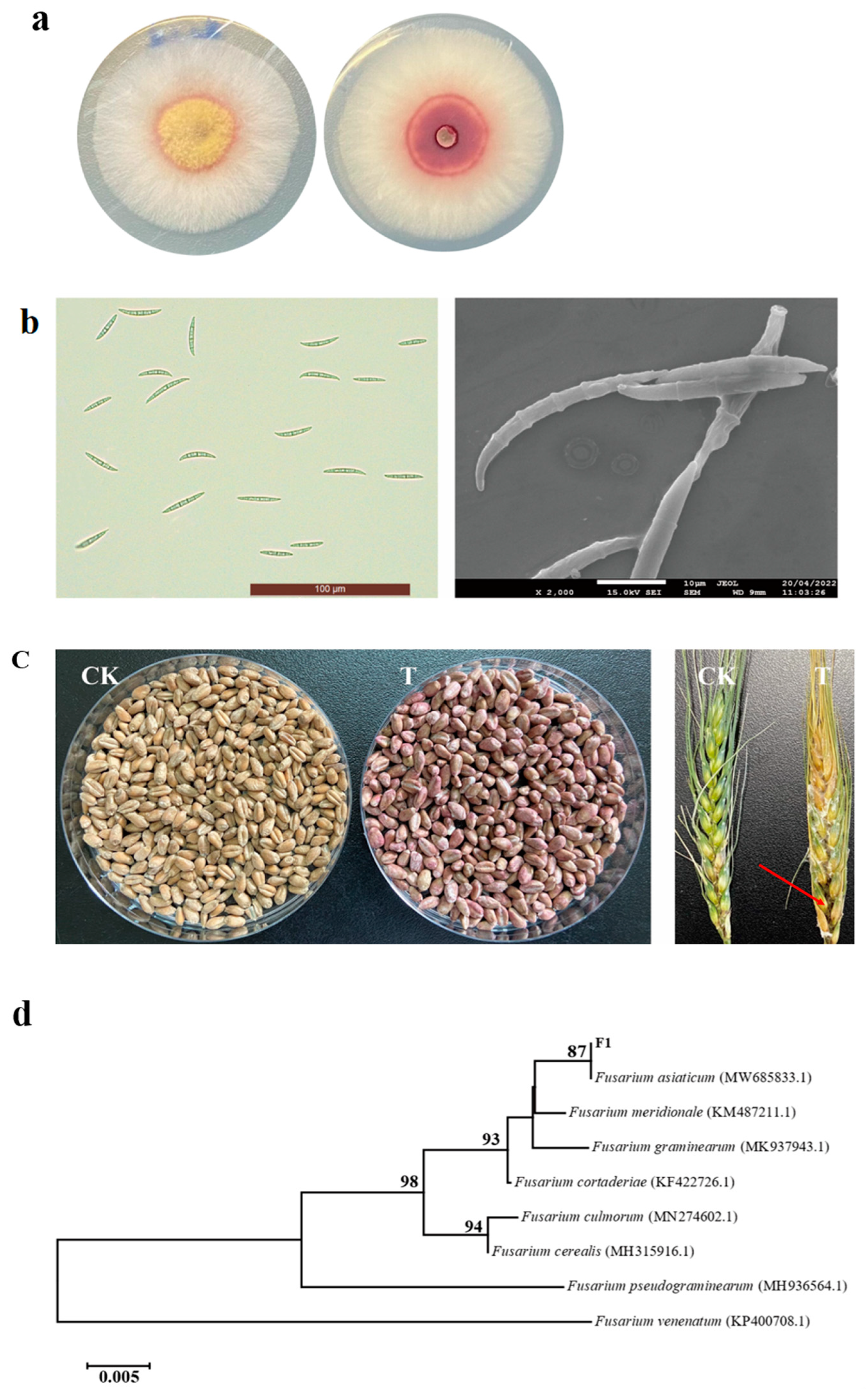
3.1. Isolation, Identification and Pathogenicity Verification of FHB Pathogens

In this section, the initial form of the pathogen is shown in Figure 1a. Because FSGC could not be accurately identified based on the ITS sequence, the phylogenetic tree was constructed based on the TEF-1α sequence. The acquired TEF-α rDNA gene sequence was analyzed using the Blast program on NCBI (http://www.ncbi.nlm.nih.gov, accessed on 24 May 2023). There was more than 99% homology between the F1 strain and F. asiaticum strains in the NCBI database. The phylogenetic tree was created, as depicted in Figure 1b. As shown in Figure 1c, the wheat seeds turned red after a month of F1 infection, and the wheat was fully rotten after 2 weeks of F1 infection. Based on the morphological characteristics and molecular biological identification, the pathogen F1 was identified as F. asiaticum.

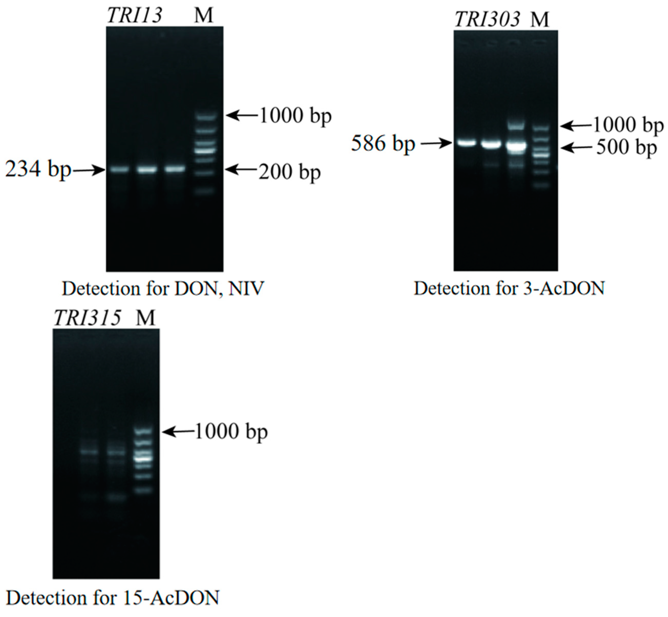
3.2. Analysis of the Toxigenic Chemotype of F1

The DNA of F1 was amplified by using primers for detecting DON and NIV. As shown in Figure 2, the amplified fragment of F1 was about 200 bp, indicating that F1 produced the DON toxin. Subsequently, the primers for detecting 3-AcDON and 15-AcDON were used to amplify F1, and the amplified fragment of F1 was about 586 bp, indicating that F1 produces 3-AcDON. There were no bands at 864 bp, indicating that F1 did not produce 15-AcDON. This indicated that the toxigenic chemotypes of F1 were DON and 3-AcDON.

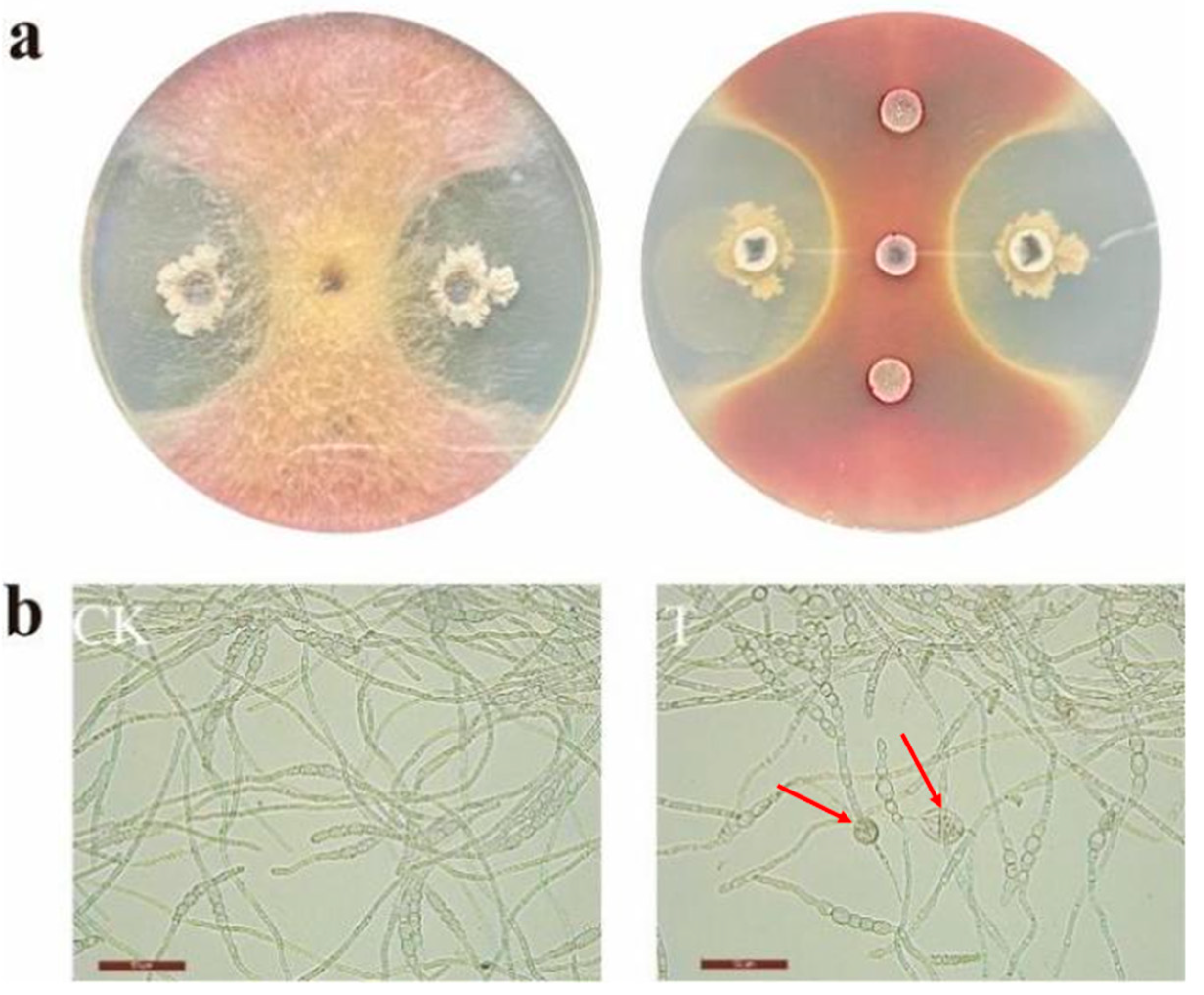
3.3. Antagonistic Activity of Bacillus velezensis
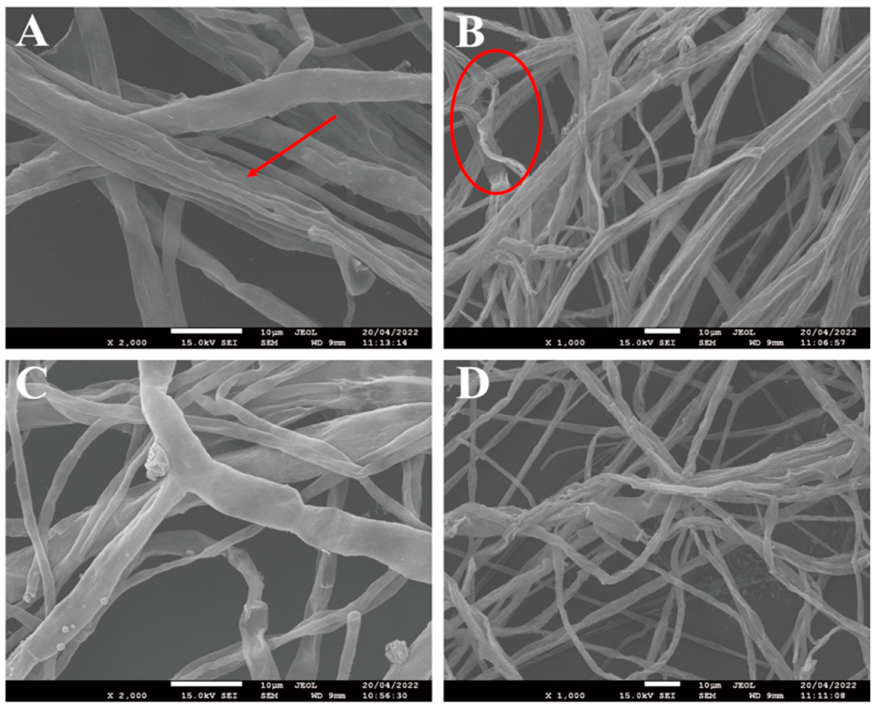
As shown in Figure 3a, the radial growth of F. asiaticum F1 on a PDA plate was inhibited by B. velezensis E2. As shown in Table 4, the spore germination rate of F1 treated with B. velezensis E2 was only 25.17%, while that of the control group was 93.33%, indicating that B. velezensis E2 could effectively inhibit the spore germination of F. asiaticum F1. Figure 3b showed that a vesicle-like structure was formed at the end of the spore germ tube with the treatment of B. velezensis E2. These results demonstrated the inhibitory effect of B. velezensis E2 on F. asiaticum F1.

3.4. SEM

The results of the electron microscopy further showed the inhibitory effect of B. velezensis E2 on F. asiaticum F1. As shown in Figure 4, under the influence of B. velezensis E2, the hyphae of F. asiaticum F1 exhibit shrinkage, twisting and breaking. The same changes also occurred in the mycelia morphology when F. graminearum was treated with myriocin [35].

3.5. Effect of B. velezensis E2 on DON Accumulation in Storage Wheat

As shown in Table 5, compared with wheat treated with only F1 spore suspension (CK), the amount of DON in wheat treated with B. velezensis E2 (T) was significantly reduced after 30 days of storage. This indicates that B. velezensis E2 can inhibit the growth of F1, thereby reducing the generation of vomiting toxins on wheat.

3.6. Sequencing Data and Its Quality Control

As shown in Table 6, 40,583,875 high-quality reads were obtained in the treatment group (F. asiaticum co-cultivation with B. velezensis E2) and control group (F. asiaticum co-cultivation with sterile water) using Illumina HiSeq2500 (Illumina, San Diego, CA, USA). After filtering low-quality reads, the GC percentage was approximately 52.70%. Moreover, among the sequenced samples, the Q20 varied from 98.64% to 98.76%. In total, 87.48% of readings could be mapped uniquely with the reference genome.

3.7. Differential Expression Gene Analysis

The differentially expressed genes between the control group and the treatment group are shown in Figure 5. A total of 3482 differentially expressed genes were screened using |log2 (Fold Change)| ≥ 4, FDR ≤ 0.05 as the criteria, among which 2071 genes were down-regulated and 1411 genes were up-regulated.

3.8. GO Enrichment Analysis of Differentially Expressed Genes

To examine the assignment and distribution of GO terms among the DEGs, GO enrichment analysis was carried out, as shown in Figure 6. There were 3414 genes which were annotated to the GO database among the DEGs. The GO functionality included biological processes, molecular function and cell components. These three categories contain 18, 10 and 11 GO terms, respectively.
There were 2817 DEGs in the biological process category. The category with the highest number of DEGs is the cellular metabolic process (769 DEGs). The other GO terms included the cellular process, single-organism process, multicellular organismal process, detoxification, growth, multi-organism process, reproduction, cellular component organization or biogenesis, reproductive process, localization, positive regulation of the biological process, signaling, developmental process, negative regulation of the biological process, response to the stimulus, biological regulation and regulation of the biological process.
In the molecular function category, 10 GO terms were enriched and included with 1181 DEGs. Most genes were enriched into catalytic activity, which included 581 DEGs. The other GO terms included the structural molecule activity, antioxidant activity, transporter activity, nucleic acid binding transcription factor activity, signal transducer activity, molecular function regulator, transcription factor activity, protein binding, molecular transducer activity and binding.
In the cell components category, 11 GO terms were enriched and included with 2144 DEGs. Most genes were enriched into the cell and cell part, and they have an equal number of DEGs. The other GO terms included the nucleoid, membrane part, membrane, membrane-enclosed lumen, supramolecular fiber, macromolecular complex, extracellular region, organelle and organelle part.

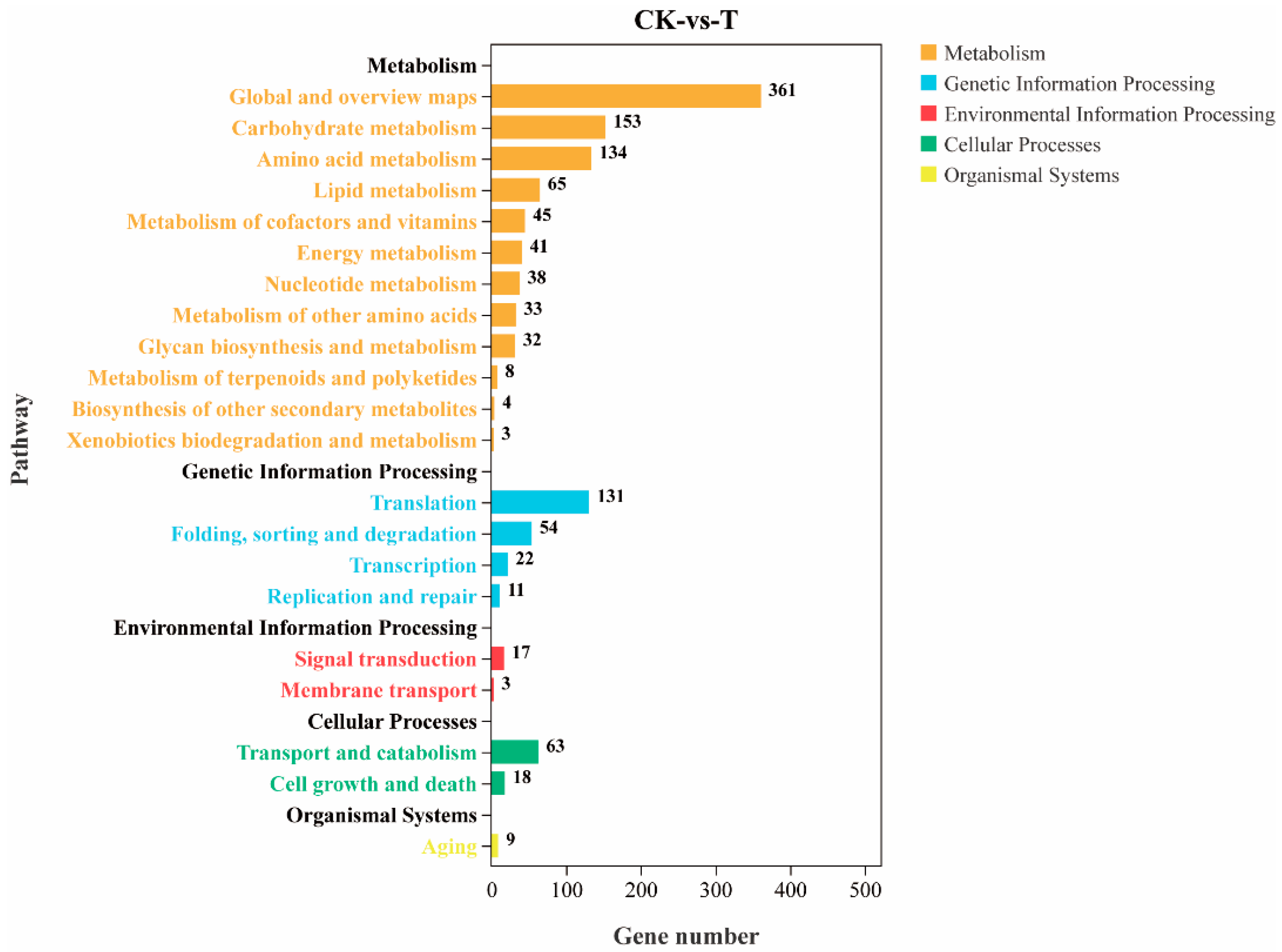
3.9. KEGG Enrichment Analysis of Differentially Expressed Genes

The KEGG enrichment and classification of differential genes showed that a total of 679 differential genes were involved in 117 metabolic pathways. Figure 7 shows that differential genes are mainly involved in metabolism, genetic information processing, environmental information processing, cellular processes and organismal systems. These primary metabolic pathways are divided into 21 secondary metabolic pathways, among which genomics processes include replication and repair, translation, transcription, etc. Cellular processes include transport and catabolism. Environmental information processes include membrane transport and signal transduction. The basal metabolic process includes lipid metabolism, the biosynthesis of other secondary metabolites and the metabolism of other amino acids. Biological systems involve environmental adaptation. The top 10 KEGG metabolic pathways with the most significant enrichment include the ribosome, biosynthesis of amino acids, metabolic pathways, biosynthesis of antibiotics, ribosome biogenesis in eukaryotes, gluconeogenesis and pyruvate metabolism, biosynthesis of secondary metabolites, N-Glycan biosynthesis and fructose and mannose metabolism.

3.9.1. Effects on DON Synthesis Pathways

F. asiaticum F1 treated with B. velezensis E2 produced many different genes in the DON synthesis pathway, as shown in Table 7. The Tri101, Tri10 and Tri1 expression levels were down-regulated in the F. asiaticum F1 treated with B. velezensis E2. An analysis of the transcriptome data indicated that the gene cut6, which is involved in acetyl-CoA synthesis, was significantly down-regulated under the action of antagonizing B. velezensis E2.

3.9.2. Effects on Secondary Metabolic Pathways

The genes related to mannose synthesis, such as ALG9, alg-11, ALG12, dpm1 and gls2, were all down-regulated, as shown in Table 5. The gene TCB1, which is involved in the synthesis of transmembrane proteins, was also down-regulated, indicating that the B. velezensis E2 affected the cell membrane formation and material transport of the pathogenic fungi. Transcriptome data also showed that under the influence of B. velezensis E2, the expression of ATG8 was up-regulated, and the expression of other genes related to autophagy protein synthesis such as ATG2, ATG9, ATG12 and ATG13 were also up-regulated. In addition, most of the DEGs related to ribosome biogenesis in eukaryotes pathways were up-regulated under B. velezensis E2 treatment. By analyzing the transcriptome data, it was found that the DEGs of F. asiaticum F1 related to ribosome synthesis were up-regulated, among which mpp10, UTP14, UTP15 and UTP4 were related to the synthesis of nucleolar RNA-related proteins, and NOG1 was related to the synthesis of nucleolar GTP-binding proteins, as shown in Table 7.

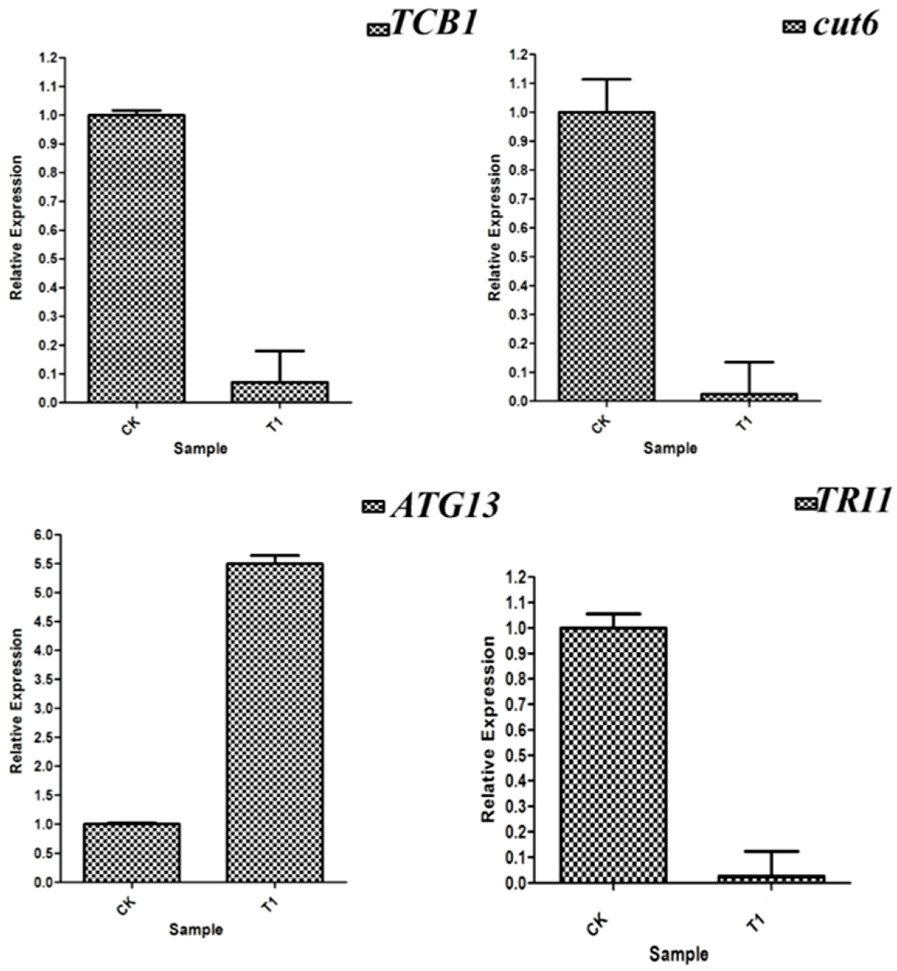
3.10. RT-qPCR Verification of Differentially Expressed Genes

RT-qPCR was used to verify the expression levels of the DEGs, as shown in Figure 8. The expression levels of KRE33, ATG8, TRI101 and ATG13 were up-regulated, while the expressions of alg-11, ade5, TCB1 and cut6 were down-regulated, which was basically consistent with the results of the transcriptional analysis.

4. Discussion

As mentioned above, FHB causes serious losses in the wheat yield and quality worldwide every year. In China, the middle-lower reaches of the Yangtze River are the main regions of FHB. The Zhenjiang area belongs to the lower reaches of the Yangtze River. In this study, the sampling and screening of wheat in the Zhenjiang area were carried out, and the pathogen of wheat FHB was identified as Fusarium asiaticum by morphological characteristics and DNA sequencing. After analyzing the toxigenic chemotype of F1, it was found that its toxigenic chemotypes are DON and 3-AcDON. DON mainly contaminates wheat, seriously affecting the yield and quality of grain, feed and food and even causing serious food safety incidents. Studies have further shown that DON was the most abundant mycotoxin found in infected wheat heads, and the concentrations were consistently higher than those of its acetylated derivatives, 15-AcDON and 3-AcDON [27]. Therefore, it is important to find a suitable method for inhibiting the infection of Fusarium asiaticum and the accumulation of DON.
Many studies have found that Bacillus has an obvious inhibitory effect on Fusarium, so it is an important resource for the development of microbial fungicides [36,37]. However, there are few studies on the effect of Bacillus on toxin accumulation and the mechanism of inhibiting toxin production by Fusarium. In this study, B. velezensis E2, previously screened by our laboratory, was used to conduct related research. The plate confrontation experiment revealed that B. velezensis E2 had a significant inhibitory effect on F. asiaticum F1, and the inhibition effect of B. velezensis E2 on F. asiaticum F1 was further revealed through the spore germination experiment and scanning electron microscope observation. It was also found that B. velezensis E2 can significantly reduce the content of DON in wheat. Other studies have shown that Bacillus amyloliquefaciens degraded DON mainly by secreting extracellular enzymes [38]. In order to better understand the antagonistic mechanism of B. velezensis E2 against F. asiaticum F1, the hyphae of F. asiaticum F1 under B. velezensis E2 were studied by transcriptomics techniques.
The RNA-seq result showed that from the total gene reads, 3482 genes were differentially expressed (either up-regulated or down-regulated). Then, the DEGs were analyzed to determine their GO enrichment. The GO enrichment was highly concentrated in the metabolic process (769 DEGs), cellular processes (620 DEGs), single-organism process (614 DEGs) and catalytic activity (581 DEGs). Then, KEGG pathway enrichment analysis was conducted to determine the DEGs that have functional networks and biological pathways in the system. In the KEGG enrichment classification of DEGs, the DEGs in the treated F1 hyphae related to toxin synthesis, glycan synthesis, ribosome synthesis and the autophagy process were analyzed.
The FHB not only leads to a huge decrease in the wheat yield but also increases the risk of excessive levels of DON in the storage period of wheat [39]. Mycotoxins are secondary metabolites of fungi that cause serious damage to agricultural products and foods in the food supply chain. These harmful pollutants have been directly linked with poor socio-economic patterns and human health issues [40]. In the DON synthesis pathways, B. velezensis E2 treatment produced many different genes in the DON synthesis pathway, as shown in Table 5. The biosynthesis of DON is a series of complex processes, which are closely associated with the expression of a series of TRI genes [41]. The 12-gene core TRI cluster, the two-gene TRI1-TRI16 locus and the single-gene TRI101 locus all generate trichothecene biosynthesis enzymes [41,42]. Tri1 plays a crucial role in DON synthesis. It encodes the cytochrome P450 monooxygenase. Tri6 and Tri10 regulate the transcription of TRI genes [42]. An acetyltransferase that catalyzes the esterification of acetyl to the C-3 hydroxyl of trichothecenes is encoded by the gene TRI101 [42]. Previous research by Xu showed that succinate dehydrogenase inhibitors (SDHIs) could decrease the DON biosynthesis of F. asiaticum [10]. This fungicide can inhibit the synthesis of DON, and its mechanism of action is mainly to inhibit the expression of key genes in glycolysis, thereby reducing the content of pyruvate and acetyl-CoA, the raw materials for DON synthesis. It can be seen in Table 7 that Tri101, Tri10 and Tri1, related to DON synthesis, and cut6, related to acetyl-CoA synthesis, were all down-regulated in F. asiaticum F1 treated with B. velezensis E2. This suggested that B. velezensis E2 might inhibit DON synthesis by down-regulating the expression of genes related to enzymes and raw materials for DON toxin synthesis.
N-glycans or asparagine-linked glycans are the main components of eukaryotic glycoproteins, which are located on cell membranes. By analyzing the transcriptome data, it was found that the genes related to mannose synthesis, such as ALG9, alg-11, ALG12, dpm1 and gls2, were all down-regulated, as shown in Table 5. The gene TCB1, which is involved in the synthesis of transmembrane proteins, was also down-regulated, indicating that the B. velezensis E2 affected the cell membrane formation and material transport of the pathogenic bacteria. This also explains the vesicle-like structure of the mycelium of F. asiaticum F1, observed under the microscope earlier, in the presence of B. velezensis E2. Peptidoglycan is an important part of the cell wall. It was found that under the treatment of B. velezensis E2, genes related to glycan synthesis in F. asiaticum F1 were down-regulated, suggesting that B. velezensis E2 could affect the formation of the cell wall. Many biocontrol microorganisms can inhibit the growth and reproduction of pathogenic fungi by producing glucanase and chitinase to destroy the integrity of the cell wall of pathogenic fungi [43]. Some studies have also shown that lipopeptide compounds produced by B. velezensis E2 can destroy the cell membrane and cell wall of pathogenic bacteria, which is consistent with the expression of several key genes above. Therefore, it was speculated that B. velezensis E2 destroyed the integrity of the cell wall and interfered with the composition of the membrane lipids of F. asiaticum F1 by affecting the expression of related genes involved in N-glycan synthesis. It was also an important reason why B. velezensis E2 inhibits the growth of F. asiaticum F1 mycelium.
Ribosomes are large macromolecular assemblies, with approximately two-thirds of their mass consisting of RNA, with the rest being proteins [44], and their main function is to synthesize proteins. By analyzing transcriptome data, it was found that genes related to ribosome synthesis were up-regulated in F. asiaticum F1. Among those up-regulated genes, mpp10, UTP14, UTP15 and UTP4 were related to nucleolar RNA-related protein synthesis, and NOG1 was related to nucleolar GTP-binding protein synthesis. This indicated that F. asiaticum F1 maintained protein synthesis by synthesizing ribosomes to resist the harmful effects of B. velezensis E2. Ma et al. also found that, in response to the stress of Bacillus amyloliquefaciens, the genes involved in the pathway of ribosome synthesis were significantly up-regulated in Fusarium oxysporum to alleviate the degradation of the protein by bacteriostatics [25]. The down-regulated gene ade5 encodes a phosphoribosamine-glycine ligase. Kim et al. constructed a deletion mutant of ade5 and found that its hyphal growth rate was slowed down and almost no conidia were produced [45]. This also explained why B. velezensis E2 could significantly inhibit the spore germination rate of F. asiaticum F1 from the molecular level.
Autophagy is a degradation pathway that removes cytoplasmic material from eukaryotic cells and is characterized by the formation of two-membrane structures called autophagosomes. The autophagy balance of a pathogen is very important for their growth, pathogenicity and environmental competitiveness [46]. Biocontrol Streptomyces sp. S89 inhibits the growth of scab hyphae by secreting the active substance rapamycin, reducing the pathogenicity of the pathogen and the synthesis of mycotoxins [47]. The effect of rapamycin on the Target of Rapamycin signaling pathway of Gibberella spp. could promote the proteasome-dependent degradation of histone acetyltransferase Gcn5, thereby reducing the level of acetylation of Atg8 and promoting autophagy [47]. The expression of genes related to eukaryotic autophagy in the autophagy pathway of F. asiaticum F1 was up-regulated under the treatment of B. velezensis E2. It can be seen in Table 7 that ATG2, ATG9, ATG12 and ATG13, involved in the autophagy protein synthesis, were all up-regulated. It is speculated that F. asiaticum F1 can defend against the damage of B. velezensis E2 by autophagy.

5. Conclusions

In this study, the pathogen of FHB was identified as F. asiaticum F1, which can produce a toxic secondary metabolite named DON. It was found that B. velezensis E2 can not only inhibit the pathogen growth but also inhibit the accumulation of DON. Compared to the control group, a total of 3482 differentially expressed genes were screened by the transcriptome sequencing of F. asiaticum F1 from the B. velezensis E2 treatment group. Then, GO functional annotation and KEGG enrichment analysis were performed for these differentially expressed genes. The results showed that these differentially expressed genes were involved in DON synthesis, glycan synthesis, ribosome synthesis and the autophagy process. In addition, the RT-qPCR validation results of the differentially expressed genes were consistent with the basic trend of the sequencing results. These results provide a theoretical basis for further revealing the molecular mechanism of disease resistance induced by B. velezensis E2.

Author Contributions

Methodology, J.M.; software, T.Z.; validation, J.M. and C.G.; formal analysis, Z.S. and M.L.; investigation, Y.Z. and C.G.; resources, X.X.; data curation, X.L.; writing—original draft preparation, J.M.; writing—review and editing, M.L.; visualization, X.L.; supervision, Y.Z.; project administration, W.S.; funding acquisition, W.S. All authors have read and agreed to the published version of the manuscript.

Funding

This research was funded by the National Science and Technology Major Project (2022ZD0115801), State Key Laboratory of Crop Biology in Shandong Agriculture University (2020KF07) and Funds for Independent Innovation of Agricultural Science & Technology in Jiangsu Province (CX(22)3114).

Institutional Review Board Statement

Not applicable.

Informed Consent Statement

Not applicable.

Data Availability Statement

The raw sequence data reported in this paper have been deposited in the Genome Sequence Archive (Genomics, Proteomics & Bioinformatics 2021) in the National Genomics Data Center (Nucleic Acids Res 2022), China National Center for Bioinformation/Beijing Institute of Genomics, Chinese Academy of Sciences (GSA: CRA013405), and they are publicly accessible at https://ngdc.cncb.ac.cn/gsa (accessed on 12 November 2023).

Acknowledgments

All authors are thankful to their representative universities/institutes for the support and services used in this study.

Conflicts of Interest

Author Zhenzhong Sun was employed by the company Jiangsu Suhe Socialized Agriculture Service Co., Ltd. The remaining authors declare that the research was conducted in the absence of any commercial or financial relationships that could be construed as a potential conflict of interest.

References

  1. Niu, H.; Jiang, Y.; Niu, J. Research Advances in the Genetics and Breeding of Wheat (Triticum aestivum) Resistance to Fusarium Head Blight. J. Agric. Biotechnol. 2020, 28, 530–542. [Google Scholar]
  2. Figueroa, M.; Hammond-Kosack, K.E.; Solomon, P.S. A review of wheat diseases-a field perspective. Mol. Plant Pathol. 2018, 19, 1523–1536. [Google Scholar] [CrossRef] [PubMed]
  3. Yli-Mattila, T.; Gagkaeva, T.; Ward, T.J.; Aoki, T.; Kistler, H.C.; O‘Donnell, K. A novel Asian clade within the Fusarium graminearum species complex includes a newly discovered cereal head blight pathogen from the Russian Far East. Mycologia 2009, 101, 841–852. [Google Scholar] [CrossRef] [PubMed]
  4. Dong, F.; Xing, Y.J.; Lee, Y.W.; Mokoena, M.P.; Olaniran, A.O.; Xu, J.H.; Shi, J.R. Occurrence of Fusarium mycotoxins and toxigenic Fusarium species in freshly harvested rice in Jiangsu, China. World Mycotoxin J. 2020, 13, 201–211. [Google Scholar] [CrossRef]
  5. Lee, T.; Lee, S.H.; Shin, J.Y.; Kim, H.K.; Yun, S.H.; Kim, H.Y.; Lee, S.; Ryu, J.G. Comparison of Trichothecene Biosynthetic Gene Expression between Fusarium graminearum and Fusarium asiaticum. Plant Pathol. J. 2014, 30, 33–42. [Google Scholar] [CrossRef]
  6. Wegulo, S.N. Factors influencing deoxynivalenol accumulation in small grain cereals. Toxins 2012, 4, 1157–1180. [Google Scholar] [CrossRef]
  7. Liu, N.; Yang, Y.; Chen, J.; Jia, H.; Zhang, Y.; Jiang, D.; Wu, G.; Wu, Z. 3-Acetyldeoxynivalenol induces lysosomal membrane permeabilization-mediated apoptosis and inhibits autophagic flux in macrophages. Env. Pollut. 2020, 265, 114697. [Google Scholar] [CrossRef]
  8. Choi, J.-H.; Nah, J.-Y.; Lee, M.-J.; Jang, J.-y.; Lee, T.; Kim, J. Fusarium diversity and mycotoxin occurrence in proso millet in Korea. LWT 2021, 14, 110964. [Google Scholar] [CrossRef]
  9. Rotter, B.A.; Prelusky, D.B.; Pestka, J.J. Toxicology of deoxynivalenol (vomitoxin). J. Toxicol. Env. Health 1996, 48, 1–34. [Google Scholar] [CrossRef]
  10. Xu, C.; Li, M.; Zhou, Z.; Li, J.; Chen, D.; Duan, Y.; Zhou, M. Impact of Five Succinate Dehydrogenase Inhibitors on DON Biosynthesis of Fusarium asiaticum, Causing Fusarium Head Blight in Wheat. Toxins 2019, 11, 272. [Google Scholar] [CrossRef]
  11. Wang, H.W.; Sun, S.L.; Ge, W.Y.; Zhao, L.F.; Hou, B.Q.; Wang, K.; Lyu, Z.F.; Chen, L.Y.; Xu, S.S.; Guo, J.; et al. Horizontal gene transfer of Fhb7 from fungus underlies Fusarium head blight resistance in wheat. Science 2020, 368, 844. [Google Scholar] [CrossRef] [PubMed]
  12. Zhang, P.G.; Zhu, Y.X.; Ma, D.F.; Xu, W.J.; Zhou, J.J.; Yan, H.W.; Yang, L.; Yin, J.L. Screening, Identification, and Optimization of Fermentation Conditions of an Antagonistic Endophyte to Wheat Head Blight. Agronomy 2019, 9, 476. [Google Scholar] [CrossRef]
  13. Stumbriene, K.; Gudiukaite, R.; Semaskiene, R.; Svegzda, P.; Jonaviciene, A.; Suproniene, S. Screening of new bacterial isolates with antifungal activity and application of selected Bacillus sp. cultures for biocontrol of Fusarium graminearum under field conditions. Crop Prot. 2018, 113, 22–28. [Google Scholar] [CrossRef]
  14. Siahmoshteh, F.; Hamidi-Esfahani, Z.; Spadaro, D.; Shams-Ghahfarokhi, M.; Razzaghi-Abyaneh, M. Unraveling the mode of antifungal action of Bacillus subtilis and Bacillus amyloliquefaciens as potential biocontrol agents against aflatoxigenic Aspergillus parasiticus. Food Control 2018, 89, 300–307. [Google Scholar] [CrossRef]
  15. Liu, Y.; Lu, J.; Sun, J.; Lu, F.; Bie, X.; Lu, Z. Membrane disruption and DNA binding of Fusarium graminearum cell induced by C16-Fengycin A produced by Bacillus amyloliquefaciens. Food Control 2019, 102, 206–213. [Google Scholar] [CrossRef]
  16. Marin-Bruzos, M.; Grayston, S.J.; Forge, T.; Nelson, L.M. Isolation and characterization of streptomycetes and pseudomonad strains with antagonistic activity against the plant parasitic nematode Pratylenchus penetrans and fungi associated with replant disease. Biol. Control 2021, 158, 104599. [Google Scholar] [CrossRef]
  17. Cai, X.C.; Liu, C.H.; Wang, B.T.; Xue, Y.R. Genomic and metabolic traits endow Bacillus velezensis CC09 with a potential biocontrol agent in control of wheat powdery mildew disease. Microbiol. Res. 2017, 196, 89–94. [Google Scholar] [CrossRef] [PubMed]
  18. Zalila-Kolsi, I.; Ben Mahmoud, A.; Ali, H.; Sellami, S.; Nasfi, Z.; Tounsi, S.; Jamoussi, K. Antagonist effects of Bacillus spp. strains against Fusarium graminearum for protection of durum wheat (Triticum turgidum L. subsp. durum). Microbiol. Res. 2016, 192, 148–158. [Google Scholar] [CrossRef] [PubMed]
  19. Liu, Y.; Teng, K.; Wang, T.; Dong, E.; Zhang, M.; Tao, Y.; Zhong, J. Antimicrobial Bacillus velezensis HC6: Production of three kinds of lipopeptides and biocontrol potential in maize. J. Appl. Microbiol. 2020, 128, 242–254. [Google Scholar] [CrossRef]
  20. Chen, K.; Tian, Z.; He, H.; Long, C.-A.; Jiang, F. Bacillus species as potential biocontrol agents against citrus diseases. Biol. Control 2020, 151, 104419. [Google Scholar] [CrossRef]
  21. Ye, W.-Q.; Sun, Y.-F.; Tang, Y.-J.; Zhou, W.-W. Biocontrol potential of a broad-spectrum antifungal strain Bacillus amyloliquefaciens B4 for postharvest loquat fruit storage. Postharvest Biol. Technol. 2021, 174, 111439. [Google Scholar] [CrossRef]
  22. Srikhong, P.; Lertmongkonthum, K.; Sowanpreecha, R.; Rerngsamran, P. Bacillus sp. strain M10 as a potential biocontrol agent protecting chili pepper and tomato fruits from anthracnose disease caused by Colletotrichum capsici. BioControl 2018, 63, 833–842. [Google Scholar] [CrossRef]
  23. Platel, R.; Sawicki, M.; Esmaeel, Q.; Randoux, B.; Trapet, P.; El Guilli, M.; Chtaina, N.; Arnauld, S.; Bricout, A.; Rochex, A.; et al. Isolation and Identification of Lipopeptide-Producing Bacillus velezensis Strains from Wheat Phyllosphere with Antifungal Activity against the Wheat Pathogen Zymoseptoria tritici. Agronomy 2022, 12, 95. [Google Scholar] [CrossRef]
  24. Platel, R.; Lucau-Danila, A.; Baltenweck, R.; Maia-Grondard, A.; Trapet, P.; Magnin-Robert, M.; Randoux, B.; Duret, M.; Halama, P.; Hilbert, J.L.; et al. Deciphering immune responses primed by a bacterial lipopeptide in wheat towards Zymoseptoria tritici. Front. Plant Sci. 2023, 13, 1074447. [Google Scholar] [CrossRef] [PubMed]
  25. Ma, J.; Hu, D.; Tian, S.; Peng, J.; Zhang, C.; Jia, N.; Wang, Z. Mechanism Analysis of Fusarium oxysporum HG-11 Responses to Biocontrol Agent Bacillus amyloliquefaciens B501. Acat Agric. Boreali-Occident. Sin. 2021, 30, 1695–1707. [Google Scholar]
  26. Yang, H.; Wang, L.; Li, S.; Gao, X.; Wu, N.; Zhao, Y.; Sun, W. Control of postharvest grey spot rot of loquat fruit with Metschnikowia pulcherrima E1 and potential mechanisms of action. Biol. Control 2021, 152, 104406. [Google Scholar] [CrossRef]
  27. Wang, Q.; Song, R.; Fan, S.; Coleman, J.J.; Xu, X.; Hu, X. Diversity of Fusarium community assembly shapes mycotoxin accumulation of diseased wheat heads. Mol. Ecol. 2022, 32, 2504–2518. [Google Scholar] [CrossRef] [PubMed]
  28. Wang, J.H.; Zhang, J.B.; Chen, F.F.; Li, H.P.; Ndoye, M.; Liao, Y.C. A multiplex PCR assay for genetic chemotyping of toxigenic Fusarium graminearum and wheat grains for 3-acetyldeoxynivalenol, 15-acetyldeoxynivalenol and nivalenol mycotoxin. J. Food Agric. Environ. 2012, 10, 505–511. [Google Scholar]
  29. Yan, F.; Zhang, D.; Wang, X.; Liu, C.; Zhang, F. Reduction of postharvest diseases of loquat fruit by serine protease and possible mechanisms involved. Sci. Hortic. 2022, 304, 111246. [Google Scholar] [CrossRef]
  30. Ren, Y.; Yao, M.; Chang, P.; Sun, Y.; Li, R.; Meng, D.; Xia, X.; Wang, Y. Isolation and characterization of a Pseudomonas poae JSU-Y1 with patulin degradation ability and biocontrol potential against Penicillium expansum. Toxicon 2021, 195, 1–6. [Google Scholar] [CrossRef]
  31. Meng, D.; Garba, B.; Ren, Y.; Yao, M.; Xia, X.; Li, M.; Wang, Y. Antifungal activity of chitosan against Aspergillus ochraceus and its possible mechanisms of action. Int. J. Biol. Macromol. 2020, 158, 1063–1070. [Google Scholar] [CrossRef]
  32. GB5009.111-2016; Determination of Deoxynivalenol in Food—High Performance Liquid Chromatographic Method with Immunoaffinity Column Clean-Up. National Health and Family Planning Commission of the People’s Republic of China, State Food and Drug Administration: Beijing, China, 2016.
  33. Hsu, S.C.; Tsen, H.Y. PCR primers designed from malic acid dehydrogenase gene and their use for detection of Escherichia coli in water and milk samples. Int. J. Food Microbiol. 2001, 64, 1–11. [Google Scholar] [CrossRef] [PubMed]
  34. Xu, M.; Yang, Q.; Serwah Boateng, N.A.; Ahima, J.; Dou, Y.; Zhang, H. Ultrastructure observation and transcriptome analysis of Penicillium expansum invasion in postharvest pears. Postharvest Biol. Technol. 2020, 165, 111198. [Google Scholar] [CrossRef]
  35. Shao, J.; Pei, Z.; Jing, H.; Wang, L.; Jiang, C.; Du, X.; Jiang, C.; Lou, Z.; Wang, H. Antifungal activity of myriocin against Fusarium graminearum and its inhibitory effect on deoxynivalenol production in wheat grains. Physiol. Mol. Plant Pathol. 2021, 114, 101635. [Google Scholar] [CrossRef]
  36. Wang, W.; Zhao, J.; Huang, W.; Guo, X.; Li, W.; Zhang, Z. Research Progress in Metabolites Produced by Bacillus Against Three Common Plant Pathogenic Fungi. Biotechnol. Bull. 2023, 39, 59–68. [Google Scholar]
  37. Adedire, D.E.; Owoeye, T.E.; Farinu, O.M.; Ogundipe, W.F.; Adedire, O.M. Management of Fusarium Wilt Disease of Maize (Zea mays L.) with Selected Antimycotic Plant Extracts and Inhibitory Bacillus Strains. Curr. Microbiol. 2023, 80, 204. [Google Scholar] [CrossRef]
  38. Wang, J.; Geng, K.; Liu, Y.; Shi, Z.; Gong, Y.; Tian, Y.; Kang, X.; Wang, Y.; Sun, X. Screening and Identification of Strains Degrading Deoxynivalenol. Chin. J. Anim. Nutr. 2023, 35, 5430–5440. [Google Scholar]
  39. Su, P.; Ge, W.; Wang, H.; Kong, L. Advances in understanding the mechanisms of wheat-Fusarium graminearum interactions. Sci. Sin. Vitae 2021, 51, 1493–1507. [Google Scholar] [CrossRef]
  40. Ganesan, A.R.; Mohan, K.; Rajan, D.K.; Pillay, A.A.; Palanisami, T.; Sathishkumar, P.; Conterno, L. Distribution, toxicity, interactive effects, and detection of ochratoxin and deoxynivalenol in food: A review. Food Chem. 2022, 378, 131978. [Google Scholar] [CrossRef]
  41. Alexander, N.J.; Proctor, R.H.; McCormick, S.P. Genes, gene clusters, and biosynthesis of trichothecenes and fumonisins in Fusarium. Toxin Rev. 2009, 28, 198–215. [Google Scholar] [CrossRef]
  42. Kimura, M.; Tokai, T.; Takahashi-Ando, N.; Ohsato, S.; Fujimura, M. Molecular and genetic studies of Fusarium trichothecene biosynthesis: Pathways, genes, and evolution. Biosci. Biotechnol. Biochem. 2007, 71, 2105–2123. [Google Scholar] [CrossRef] [PubMed]
  43. Abbasi, S.; Safaie, N.; Sadeghi, A.; Shamsbakhsh, M. Streptomyces Strains Induce Resistance to Fusarium oxysporum f. sp. lycopersici Race 3 in Tomato Through Different Molecular Mechanisms. Front. Microbiol. 2019, 10, 1505. [Google Scholar] [CrossRef] [PubMed]
  44. Ramakrishnan, V. What we have learned from ribosome structures. Biochem. Soc. Trans. 2008, 36, 567–574. [Google Scholar] [CrossRef] [PubMed]
  45. Kim, J.E.; Myong, K.; Shim, W.B.; Yun, S.H.; Lee, Y.W. Functional characterization of acetylglutamate synthase and phosphoribosylamine-glycine ligase genes in Gibberella zeae. Curr. Genet. 2007, 51, 99–108. [Google Scholar] [CrossRef] [PubMed]
  46. Li, F.Q.; Vierstra, R.D. Arabidopsis ATG11, a scaffold that links the ATG1-ATG13 kinase complex to general autophagy and selective mitophagy. Autophagy 2014, 10, 1466–1467. [Google Scholar] [CrossRef]
  47. Wang, J.; Xu, C.; Sun, Q.; Xu, J.; Chai, Y.; Berg, G.; Cernava, T.; Ma, Z.; Chen, Y. Post-translational regulation of autophagy is involved in intra-microbiome suppression of fungal pathogens. Microbiome 2021, 9, 131. [Google Scholar] [CrossRef]
Figure 1. (a) The colony morphology of F. asiaticum F1 cultured on the PDA plate for 5 d; (b) Morphology of F. asiaticum F1 conidia under an optical microscope (left) and scanning electron microscope (right); (c) Verification of the pathogenicity of F. asiaticum F1 in storage (left) and growing wheat (right). Changes in wheat inoculated with the F1 spore suspension for one month in the storage period and 7 d in the growth period. The wheat seeds turned red (left); The spikes of wheat turned yellow and moldy. The area indicated by the arrow showed symptom of blackening (right). (d) Phylogenetic tree of the strain F1 based on the sequences of the TEF-α rDNA gene. Using MEGA 7.0 software, the phylogenetic tree was created using the neighbor-joining method. The branch points display the bootstrap values.
Jof 10 00390 g001
Figure 2. Determination of the F1 toxigenic chemotype by agarose gel electrophoresis.
Jof 10 00390 g002
Figure 3. Antagonistic B. velezensis E2 on F. asiaticum F1. (a) Effect on the growth of F. asiaticum F1 after 7 d on the PDA plate. B. velezensis E2 suspension (30 μL) was added to the left and right holes, and an equal amount of sterile water was added to the upper and lower holes as a control. (b) Effect on the germination of F. asiaticum F1 spores. The area indicated by the arrow showed vesicle-like structures.
Jof 10 00390 g003
Figure 4. Scanning electron microscopy of F. asiaticum growth without the presence of B. velezensis E2 (control: (C,D)) and faced with B. velezensis E2 (A,B). The areas indicated by arrow and circle showed shrinkage and twisting of the F1 hyphae with the treatment of B. velezensis E2.
Jof 10 00390 g004
Figure 5. Volcano plot of differentially expressed genes. The horizontal coordinate of the volcanic map shows the difference multiple between the two groups, and the vertical coordinate shows the −log10 value of the FDR value of the difference between the two groups. Each dot in the volcanic map represents a gene, with the red representing up-regulated genes, the yellow representing down-regulated genes and the blue representing non-differentially expressed genes.
Jof 10 00390 g005
Figure 6. Classification of differentially expressed genes based on GO annotation. The horizontal coordinate of the bar chart shows the second-order GO term, and the vertical coordinate shows the number of differentially expressed genes in this term, with yellow indicating up-regulation and red indicating down-regulation.
Jof 10 00390 g006
Figure 7. KEGG pathway classification of differentially expressed genes. The vertical coordinate represents primary and secondary metabolic pathways, and the horizontal coordinate represents the number of differentially expressed genes in this metabolic pathway.
Jof 10 00390 g007
Figure 8. Results of the verification of some differentially expressed genes by RT-qPCR. The internal reference gene was cpc-1 gene. The RT-qPCR experiment was repeated three times, and the error bar indicated the degree of dispersion.
Jof 10 00390 g008aJof 10 00390 g008b
Table 1. Primer sequences.
Gene NameAccession Number PrimerPrimer Sequence (5′→3′)Product Length
ITSNR_121320.1ITS1TCCGTAGGTGAACCTGCGG559
ITS4TCCTCCGCTTATTGATATGC
TEF-αNW_022157945.1TEF-FATGGGTAAGGAGGAGAAGAC700
TEF-RGGAAGTACCAGTGATCATGTT
Note: Accession number was from the NCBI database.
Table 2. Primer sequences of the toxigenic chemotype.
Gene NameAccession NumberPrimerPrimer Sequence (5′→3′)Toxigenic ChemotypeProduct Length
Tri13NC_058400.1Tri13-FTACGTGAAACATTGTTGGCdeoxynivalenol (DON), nivalenol (NIV)234, 415
Tri13-RGGTGTCCCAGGATCTGCG
Tri3NC_058400.1Tri303-FGATGGCCGCAAGTGGA3-AcDON (3-acetyldeoxynivalenol)586
Tri303-RGCCGGACTGCCCTATTG
Tri315-FCTCGCTGAAGTTGGACGTAA15-AcDON (15-acetyldeoxynivalenol)864
Tri315-RGTCTATGCTCTCAACGGACAAC
Note: The accession number was from the NCBI database.
Table 3. Primer design of differentially expressed genes.
Gene IDGene NameAccession NumberRevers Primer (5′ to 3′)
FGSG_00732KRE33NC_026474.1F: CGCAAAGCGGTGGACT
R: GAATCTTGTCGGTCTCCTTGT
FGSG_06580cut6NC_026477.1F: AGCGAGCCATTCACTTCACT
R: TCCAGGGGGTCCGATAAAGA
FGSG_07896TRI101NC_026477.1F: GTTCTGCCGTGCTGTTGATG
R: GTCTCACAGTCTCGGGCTTAC
FGSG_08429ade5NC_026475.1F: GGGCAGACAAAGCAGGCA
R: TAGGCTCGCTCAATGGCAC
FGSG_08491ATG13NC_026475.1F: AACGACCCAGCCGAATCTAC
R: ACTGCTCCATCACATCCCAC
FGSG_10740ATG8NC_026476.1F: GAGGTTCTACCCCCGACAG
R: CGCCAAAAGTGTTCTCGCC
FGSG_10858ALG-11NC_026476.1F: CCGACCCGAGAAGAACCATC
R: CTCAGCCAGTCCAGAACCTC
FGSG_06885TCB1NC_026477.1F: TCAAGGGCGAGGATGGAC
R: GGCAGGTCGGAACAGAAGTC
FGSG_09286cpc-1NC_026477.1F: GCCTTTTCCTCACCTGCTGT
R: CCGACTTGCGACGGTTCA
FGSG_04400rhoANC_026475.1F: GGCGATGGTGCTTGTGGTAA
R: GAGGGAGTCGGGAGAGTCAA
Note: Accession number was from the NCBI database.
Table 4. Effect of B. velezensis E2 on the germination of F. asiaticum F1 spores.
TreatmentCKB. velezensis E2
Spore germination rate93.33 ± 0.7625.17 ± 3.32 **
The results indicated the mean ± standard deviation and were tested by an independent sample T test, ** p < 0.01.
Table 5. Effect of B. velezensis E2 on the deoxynivalenol production of F. asiaticum F1.
ExperimentControl GroupTreatment Group
DON content (μg/kg)46.02 ± 1.1936.72 ± 0.83 **
The results indicated the mean ± standard deviation and were tested by an independent sample T test, ** p < 0.01.
Table 6. Sequencing data tables of F. asiaticum F1.
SamplesRaw Data (bp)Clean Data (bp)Clean ReadsGC (%)Q20 (%)Unique-Mapped (%)
CK16,112,122,0006,075,824,99940,647,97452.9198.7035,427,322 (87.32%)
CK26,529,115,4006,479,907,69643,433,24852.9698.6938,186,463 (88.06%)
CK36,884,337,0006,836,297,20745,781,10653.1098.6540,361,865 (88.30%)
T15,622,680,7005,585,596,45337,401,95252.3698.7232,604,351 (87.36%)
T25,586,865,5005,542,412,17137,159,50652.2698.7632,255,302 (86.95%)
T35,876,491,8005,830,758,06739,079,46652.5898.6433,887,979 (86.88%)
Table 7. Effects of B. velezensis E2 on F. asiaticum F1 genes related to DON synthesis.
Gene IDGene Namelog2FCDefinition
FGSG_07896TRI101−5.02richothecene 3-O-acetyltransferase
FGSG_03538TRI10−10.75TRI10 [Fusarium graminearum]
FGSG_00071TRI1−6.28cytochrome P450 monooxygenase
FGSG_06580cut6−4.3acetyl-CoA carboxylase
FGSG_05243ALG9−3.13alpha-1,2-mannosyltransferase
FGSG_10858alg-11−2.24alpha-1,2-mannosyltransferase
FGSG_01233ALG12−3.25alpha-1,6-mannosyltransferase
FGSG_04044dpm1−2.37dolichol-phosphate mannosyltransferase
FGSG_05973gls2−3.28alpha-glucosidase
FGSG_06885TCB1−2.62transmembrane protein
FGSG_08429ade5−2.64phosphoribosylglycinamide formyltransferase
FGSG_10740ATG84.44GABA(A) receptor-associated protein
FGSG_08491ATG133.34autophagy-related protein 13
FGSG_10033mpp103.56U3 small nucleolar RNA-associated protein
FGSG_08759UTP144.90U3 small nucleolar RNA-associated protein 14
FGSG_10060UTP153.70U3 small nucleolar RNA-associated protein 15
FGSG_00270UTP44.69U3 small nucleolar RNA-associated protein 4
FGSG_06165NOG13.57nucleolar GTP-binding protein
Disclaimer/Publisher’s Note: The statements, opinions and data contained in all publications are solely those of the individual author(s) and contributor(s) and not of MDPI and/or the editor(s). MDPI and/or the editor(s) disclaim responsibility for any injury to people or property resulting from any ideas, methods, instructions or products referred to in the content.

