Review

Insights into Fungal Mitochondrial Genomes and Inheritance Based on Current Findings from Yeast-like Fungi

Zhejiang Provincial Key Laboratory of Biometrology and Inspection & Quarantine, College of Life Sciences, China Jiliang University, Hangzhou 310018, China
*
Author to whom correspondence should be addressed.
These authors contributed equally to this work.
J. Fungi 2024, 10(7), 441; https://doi.org/10.3390/jof10070441
Submission received: 17 April 2024 / Revised: 19 June 2024 / Accepted: 20 June 2024 / Published: 21 June 2024
(This article belongs to the Section Fungal Genomics, Genetics and Molecular Biology)

Abstract

:
The primary functions of mitochondria are to produce energy and participate in the apoptosis of cells, with them being highly conserved among eukaryotes. However, the composition of mitochondrial genomes, mitochondrial DNA (mtDNA) replication, and mitochondrial inheritance varies significantly among animals, plants, and fungi. Especially in fungi, there exists a rich diversity of mitochondrial genomes, as well as various replication and inheritance mechanisms. Therefore, a comprehensive understanding of fungal mitochondria is crucial for unraveling the evolutionary history of mitochondria in eukaryotes. In this review, we have organized existing reports to systematically describe and summarize the composition of yeast-like fungal mitochondrial genomes from three perspectives: mitochondrial genome structure, encoded genes, and mobile elements. We have also provided a systematic overview of the mechanisms in mtDNA replication and mitochondrial inheritance during bisexual mating. Additionally, we have discussed and proposed open questions that require further investigation for clarification.

1. Introduction

Mitochondria are crucial organelles in eukaryotic organisms, playing roles in the synthesis of cellular energy, the maintenance of membrane structure dynamics, and the regulation of apoptosis, among other functions [1]. Mitochondria are semi-autonomous organelles that contain their own set of genetic information, known as the mitochondrial genome. The mitochondrial genome varies significantly among different eukaryotic organisms. The endosymbiotic theory suggests that mitochondria originate from a common ancestor with the incorporation of an endosymbiotic alpha-proteobacterium into a host cell [2]. However, as mitochondria have evolved, mitochondrial genomes have diverged significantly among the major groups of eukaryotes. In animals, the mitochondrial genome is a closed circular DNA molecule, encoding a small number of genes related to energy production. It lacks intergenic regions and introns, and its gene arrangement is conserved throughout evolution [3,4]. The mitochondrial genome in plants mostly exists in the form of closed circular DNA molecules, although a small fraction is present as linear DNA molecules. In addition to encoding a large number of genes related to energy production, the plant mitochondrial genome also codes for some plant-specific genes. It possesses intergenic regions and a variable number of introns, making it more complex compared to the mitochondrial genome in animals (Table 1) [4]. Fungi represent a unique kingdom of organisms, closely related to animals and more distantly related to plants [5]. However, the configuration and composition of the mitochondrial genome in fungi are more similar to that in plant mitochondria than in animals. Linear and circular molecules of mitochondrial genomes also exist in fungi [6] There are significant variations in the size of mitochondrial genomes among different fungal species. To date, the smallest fungal mitochondrial genome discovered is 11,198 bp (Hanseniaspora guilliermondii), while the largest is 343,690 bp (Malassezia furfur) (Table 2). These differences in the number and arrangement of core genes, the quantity and types of introns, and the sequences of intergenic regions contribute to the diversity in the size and structure of fungal mitochondrial genomes. Similar to the structural diversity of mitochondrial genomes, the replication strategies of fungal mtDNA are also complex and are different from those of mammals [7,8]. Fungi also exhibit mitochondrial inheritance during bisexual mating. In the progeny cells of sexual reproduction, mitochondria primarily originate from one parent to avoid cellular heteroplasmy. While the mechanism of mitochondrial inheritance varies among different fungal species, current studies indicate that most mitochondrial inheritance in fungi is tightly regulated and often associated with their mating type loci [9]. In this review, we would like to mainly provide a summary of the constitution of yeast-like fungal mitochondrial genomes, the replication strategies of fungal mtDNA, and the mechanisms of fungal mitochondrial inheritance during bisexual mating, which will provide a reference for the systematic study of fungal mitochondrial genomes.

2. Fungal Mitochondrial Genome Structure

Fungal mitochondrial genomes are variable-sized double-stranded DNA molecules, commonly existing in circular or linear form [10]. Saccharomyces cerevisiae was the first fungus reported to have a circular mitochondrial genome [11]. Subsequently, a linear mitochondrial genome was revealed in Hansenula mrakii [12]. Additionally, the occurrence of coexisting circular and linear forms of mitochondrial genomes in the same fungal species has been demonstrated through restriction enzyme mapping analysis [13].
A circular mitochondrial genome is formed by the fusion of both ends of a linear mitochondrial genome that lacks telomeric loops (t-loops) [13]. These t-loops are recombinant basic units of repetitive sequences that can prevent the formation of circular mitochondrial genomes [14]. In fungi, a notable feature distinguishing the linear mitochondrial genome from the circular mitochondrial genome is the presence of a terminal inverted repeat sequence (TIR) or one to six open reading frames (uORFs) that may potentially generate DNA or RNA polymerases [15]. Studies have revealed that the linear mitochondrial genome of Batrachochytrium dendrobatidis consists of three linear segments, each possessing TIRs and containing a uORF that encodes a homolog of the DNA polymerase type B (dpoB) [16]. In Synchytrium, the mitochondrial genome of S. endobioticum is linear, whereas that of S. microbalum is circular [17]. Nucleic acid sequence analysis has shown that a dpoB homolog exists in the uORF of the S. endobioticum mitochondrial genome, and both ends of its mitochondrial genome contain TIRs [17]. In addition, the mitochondrial genome of S. microbalum is one-third that of S. endobioticum [17]. However, among the 37 linear mitochondrial genomes, only seven have been found to have the presence of dpoB homologs, indicating that dpoB homologs are not essential in all linear mitochondrial genomes [18]. Interestingly, studies on Candida parapsilosis have revealed the existence of linear or circular mitochondrial genomes in strains from different groups [13]. Moreover, the mitochondrial telomeric repeat unit is found in most of the linear mitochondrial genomes in C. parapsilosis strains [13].
Therefore, t-loops, TIRs, and dpoB homologs may all contribute to the mitochondrial genome adopting either a linear or circular structure, whereas the mechanism still needs further study. In addition to research on the mechanisms and genetic evolution of linear and circular mitochondrial genome formation, studies on C. orthopsilosis and C. metapsilosis revealed that strains containing linear mtDNA grow faster in YPD medium and form colonies of a certain size earlier compared to strains with circular mtDNA, which shows that strains with linear mitochondrial genomes exhibit a greater adaptive capacity to the growth environment [19]. Thus, the biological functions of linear and circular mitochondrial genomes in fungi are also worthy of in-depth investigation.

3. The Coding Genes of the Fungal Mitochondrial Genome

The fungal mitochondrial genome contains 14 conserved core genes, including NADH dehydrogenase subunit (Complex I) genes (nad1-nad6 and nad4L), Complex III and IV subunit genes involved in the respiratory chain (cob, cox1, cox2, and cox3), and ATP synthase genes (atp6, atp8, and atp9). It also includes small and large subunit rRNA genes (rns and rnl) and an unknown number of tRNA genes [20,21,22].
However, not all fungal mitochondrial genomes contain these 14 core genes. In terms of the mitochondrial respiratory chain, Complex I is absent in the mitochondrial genomes of most types of yeast, suggesting a possible correlation with the facultative anaerobic respiratory mode in yeast [23]. Nevertheless, multiple alternative NAD(P)H dehydrogenases, which can transfer electrons bypassing complex I, have been identified in the nuclear genomes of S. cerevisiae, C. glabrata, Kluyveromyces lactis, Eremothecium gossypii, and S. pombe that lack Complex I [24,25]. Moreover, the additional alternative oxidases (AOX), which can transfer electrons from ubiquinone to O2 bypassing Complex III and IV, have been identified in the nuclear genomes of various phytopathogens, including Botrytis cinerea, Magnaporthe oryzae, Ustilago maydis, Sporisorium reilianum, and the like. And the AOX is mainly responsible for the environmental stresses in fungi [24,26,27,28]. In terms of the ATP synthase gene, studies have found that in the mitochondrial genome of Erysiphe necator, there is a significant loss of amino acid sequences in the conserved functional domains of the N-terminal region in atp9 [29]. This loss results in the functional defect of atp9. However, a homolog of atp9 has been found in the nuclear genome of E. necator. It is speculated that this gene might have undergone migration between the mitochondrial and nuclear genomes. The loss of function in mitochondrial atp9 could potentially be compensated for by the homolog present in the nuclear genome [29]. Moreover, artificially nuclear expressing atp9 in a mitochondrial atp9 gene deletion mutant of yeast can lead to its phosphorylation function being achieved, but it may also trigger a stress response in the cell [30]. This suggests that the function of atp9 is strictly regulated in fungi. Surprisingly, atp9 has neither been identified in the mitochondrial genome nor the nuclear genome of Ophiostoma himal-ulmi [31]. In addition to the absence of atp9, atp8 is also missing in Stemphylium lycopersici [22]. The mechanism of how to compensate for the functional loss of these mitochondrial genes in fungi still requires further study. The small and large subunits of rRNA encoded by the fungal mitochondrial genome mainly participate in the synthesis of mitochondrial proteins. However, in the mitochondrial genome of Morchella conica, coding sequences for rRNA subunits (rnl and rns) have not been identified [32]. While the mitochondrial genomes of most fungi encode more than 20 tRNA genes, only 7–9 tRNA coding sequences were found in the mitochondrial genomes of Harpochytrium hedenii and Hyaloraphidium curvatum. It is speculated that the tRNAs required for protein synthesis are translated by nuclear sequences and then imported into the mitochondria [33,34].
In fungi, the mitochondrial genome may contain additional atypical genes whose products contribute to ribosome assembly (e.g., rps3), the promotion of transcription (N-acetyltransferase), RNA maturation, and intron splicing. The ribosomal protein rps3 is the only one that is encoded by the fungal mitochondrial genome. It plays a regulatory role in protein synthesis, exerting various functions during the translation process. In fungi, the rps3 homolog VAR1 was initially discovered in yeast, and later, it was subsequently found in ascomycetes and basidiomycetes [35,36]. The rps3 homologs can be located either within introns or in intergenic regions. In most fungi of Pezizomycotina, rps3 is located within the introns of rnl. However, in fungi of Saccharomycotina, rps3 is found in intergenic regions, with some located between trnS and rnl and others located downstream of trnM [37]. In Allomyces and Blastocladiella, rps3 is located downstream of trnF [38]. However, in Basidiomycota, rps3 is not located in a conserved syntenic unit but instead scattered throughout the different mitochondrial genomes [37]. In the fungi of Aspergillus, rps3 can be located either within the introns of rnl or nad5 [39]. In Bipolaris sorokiniana, two independently existing rps3 homologs have been identified to be located between cox3 and nad1, with lengths of 399 bp and 555 bp, respectively, indicating the possibility of the simultaneous existence of two rps3 homologs in the same strain [40]. Furthermore, rps3 has also been found to exist in the nuclear genome. The protein generated by nuclear rps3 possesses an N-terminal KH domain involved in rRNA binding and a C-terminal ribosomal protein S3 domain. In contrast, the rps3 protein produced by mitochondrial genome expression lacks the KH domain but retains the conserved S3 domain [37]. The absence, structural variations in position, and sequence diversity of rps3 in mitochondrial genomes may be related to the evolutionary changes in mitochondrial genomes and functions among different fungi.
N-acetyltransferase can catalyze the transfer of an acetyl group between acetyl coenzyme A and an amine. In the mitochondrial genome of fungi, it can be encoded by open reading frames (ORFs) within introns and also exist independently in intergenic regions. The N-acetyltransferase domain-containing protein with a length of 217 aa was initially discovered in the mitochondrial genome intron in S. lycopersici [22]. Furthermore, in Pestalotiopsis fici, the encoding of aminotransferase has been found as a standalone aminotransferase gene located in an intergenic region [41,42]. In Annulohypoxylon stygium, two putative copies of N-acetyltransferase genes are located downstream of cox2 and within the ORF of the third intron of cob [43]. Phylogenetic analysis of N-acetyltransferase genes has shown that it may originate from the nuclear genome and undergo horizontal transfer from the nucleus to the mitochondrial genome [39]. The mechanisms underlying this phenomenon remain to be further investigated.
The mitochondrial RNase P (rnpB) gene, which is responsible for tRNA cleavage, was identified in the fungal mitochondrial genome. The presence of rnpB has been identified in some species of basidiomycetes, including Microbotryum lychnidis-dioicae, Schizophyllum commune, and U. maydis [42]. Additionally, the copy number of rnpB varies among different fungal mitochondrial genomes, with M. conica identified to have two independently existing rnpB [32,44]. However, the occurrence of rnpB in fungal mitochondrial genomes is generally low. Phylogenetic and RNA secondary structures analyses have shown that the mitochondrial rnpB homologs have been lost several times independently in fungi [45].

4. Mobile Elements in the Fungal Mitochondrial Genome

Mobile elements are a type of DNA sequence capable of altering their position within a genome or inserting into another genome [46]. One of the widely present mobile elements in the fungal mitochondrial genome is the self-splicing intron. Introns are distributed across various fungal mitochondrial genes, with cox1 being the primary intron reservoir, followed by rrnl, cox2, cob, cox3, and nad2 [47]. Generally, based on the RNA secondary structure, mitochondrial introns in fungi are typically classified into Group I and II.
Group I introns primarily consist of 10 conserved helices with a structurally conserved catalytic core, crucial for self-splicing [48]. There are two main mechanisms currently recognized for the migration of Group I introns. One is driven by the homing endonucleases (HEs). Based on the conserved amino acid sequences at the active sites involved in enzyme activity, HEs are classified into four families: GIY-YIG, LAGLIDADG, His-Cys, and HNH [49]. Currently, in the fungal mitochondrial genome, genes encoding GIY-YIG (GIY) and LAGLIDADG (LD) endonucleases have been identified [50]. During intron homing, the HE initiates a double-strand break repair, causing cleavage at a specific HE recognition site within the invading gene (approximately 14–45 nucleotides). After cleavage, the gene containing the intron serves as a template to repair the break, and the intron, along with the HE gene, integrates as a single unit into the new host genome [51]. The frequency of horizontal gene transfer induced by HEs varies significantly among fungal populations and is increasingly recognized as a crucial evolutionary process in fungal evolution [50]. The other one involves reverse splicing via an RNA intermediate. Typically, long-distance intron migration is achieved through the reverse splicing pathway. During the reverse splicing process, the internal guide sequence (IGS) within Group I introns recognizes a complementary target region in the 5′ exon sequence of the host gene through 4–6 base pairings. Subsequently, it inserts into the transcribed RNA. Later, the IGS-containing RNA undergoes reverse transcription, and the resulting cDNA undergoes genomic recombination, integrating the Group I intron into the genome [52]. Group I introns can invade both homologous and heterologous sites during this process [53].
Group II introns are always composed of catalytically active intron RNAs and intron-encoded proteins (IEPs), thereby forming an enzyme with self-splicing functionality. Group II introns contain six secondary structure domains (D1-6). D1 is the largest domain serving as a scaffold for the assembly of other domains, capable of recognizing exon sequences. D2 and D3 domains enhance catalytic efficiency, while the D4 domain encodes a multifunctional IEP in certain intron ORFs. D5 and D6 serve as the catalytic center and branch point in the splicing pathway of introns, respectively [54,55,56]. The intron RNA catalyzes its splicing through a transesterification reaction, followed by binding with the IEP protein to form a catalytically active ribonucleoprotein complex. After recognizing the target sequence at the insertion site, the complex cleaves the target DNA through reverse splicing, using the intron RNA as a template to synthesize cDNA. Finally, the insertion of the Group II intron onto the mitochondrial genome is completed through the host’s DNA repair mechanisms [57].
Interestingly, some mitochondrial plasmids, which contain ORFs of reverse transcriptase or DNA/RNA polymerase and exist in the mitochondria of fungi, have the ability to integrate either wholly or partially into the mitochondrial genome of their host, thus becoming a kind of mobile element. Evidence of the transfer of gene fragments and tRNA fragments between mitochondrial plasmids and mitochondrial genomes has been found in some species of Neurospora, Podospora, Agaricus, Termitomyces, and Moniliophthora [58,59,60,61,62]. Surprisingly, the occurrence of DNA fragment transfer between mitochondrial plasmids and mitochondrial genomes has been found to be associated with fungal growth and aging in Neurospora and Podospora [63,64].

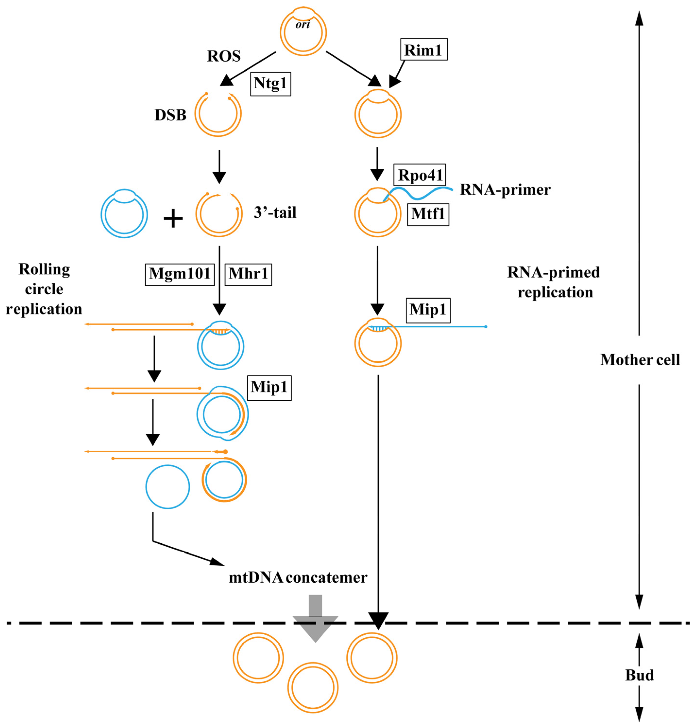
5. The Replication Strategies of Fungal mtDNA

The replication mechanism of fungal mtDNA is mainly studied in yeast, whereas it is less understood than in humans [58,59,60,61,62]. Except for the similar mechanism of RNA-primed replication in humans, yeast has developed alternative ways by coupling recombination and rolling circle replication [63,64].
In mammalian cells, RNA-primed replication initiates from the rep/ori region [65]. However, sequence analysis shows that S. cerevisiae and close relatives have rep/ori-like elements whereas, no rep/ori-like elements have been identified in the mitochondrial genome in C. glabrata and H. wingei [66,67]. Thus, this indicates that the rep/ori-like elements are not universally present in the mitochondrial genome of yeast. In the process of RNA-primed replication in S. cerevisiae, the separated single-stranded DNA is stabilized by a single-strand binding protein, Rim1. RNA polymerase Rpo41 recognizes transcriptional promoters located at oris with the help of transcription factor Mtf1 and generates RNA transcripts. These transcripts are subsequently processed to generate primers, thereby leading DNA polymerase γ catalytic subunit Mip1 to proceed with mitochondrial DNA (mtDNA) replication (Figure 1) [65]. Interestingly, the mtDNA can be maintained in the mtDNA defective mutant rho- of S. cerevisiae, in which Rpo41 is absent [68]. Therefore, neither rep/ori-like elements nor Rpo41 are essential for mtDNA replication, which indicates the existence of another mechanism.
The discovery of overlapping replication forks and recombination sites in both C. albicans and C. parapsilosis has provided increasing evidence that a recombination-mediated rolling circle replication model is a common mode of mtDNA replication in yeast [69,70]. The replication origin on the mitochondrial genome is attacked by reactive oxygen species (ROS), leading to the formation of bubble-like structures. The base damage is ultimately transformed into a double-strand break (DSB) by the base excision repair enzyme Ntg1. The DSB is subsequently processed to create a 3′-tail, which is recognized and bound by the mitochondrial homologous recombinase Mhr1 [71,72]. In addition to Mhr1 mediated recombination, a single-stranded annealing protein Mgm101, which belongs to the Rad52 protein family, has been identified to functionally operate like bacteriophage proteins and catalyze recombination by the single-strand annealing mode [73]. After recombination, the nucleoprotein filament invades a template circular mtDNA molecule, forming a heteroduplex joint and initiating rolling circle replication with Mip1. This process can generate mtDNA concatemers consisting of multiple genome units that are selectively transmitted to growing buds, where concatemers are circularized into monomers (Figure 1). Moreover, single-stranded circles displaced by rolling circle replication can serve as a substrate for single-strand annealing to initiate replication [7,74,75]. Although the recombination-mediated rolling circle replication model has been widely studied and discussed in yeast and is considered the primary mechanism for mtDNA replication, evidence suggests that other mtDNA replication mechanisms also exist in yeast. The diversity of mtDNA replication mechanisms in yeast may be related to its evolution [7]. Furthermore, research on mtDNA replication mechanisms in fungi has mainly focused on yeast, and whether there are more mtDNA replication mechanisms in other fungi remains to be studied.

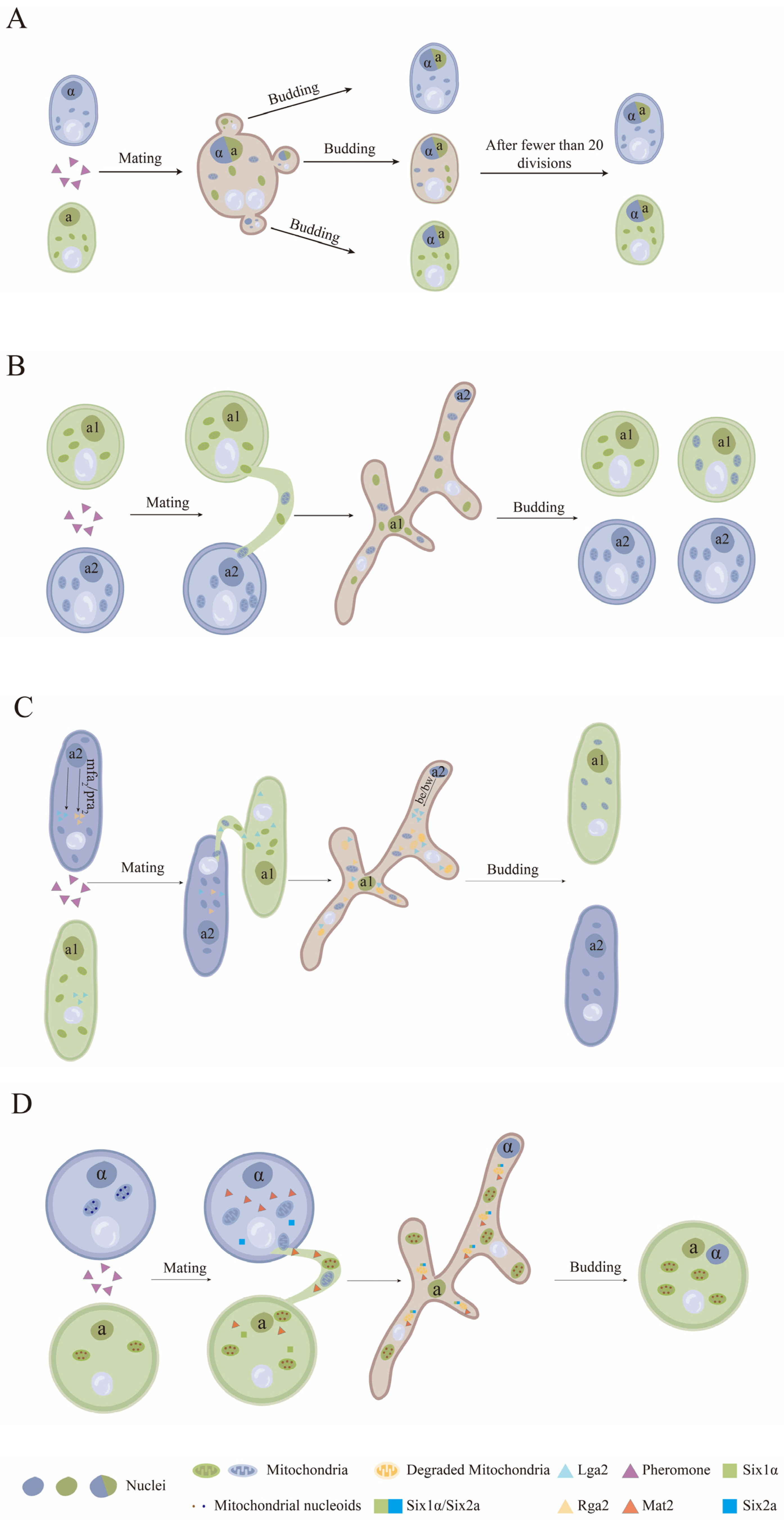
6. The Inheritance of the Fungal Mitochondrial Genome during Bisexual Mating

The inheritance of the mitochondrial genome in fungi during bisexual mating is independent of the nuclear genome and typically exhibits two modes, biparental (BPI) and unpaternal (UPI) inheritance. BPI refers to the inheritance phenomenon where progeny inherit traits from both parents. In S. cerevisiae and Schizosaccharomyces pombe, the fusion of mating partners can lead to the BPI of the mitochondrial genome and simultaneous recombination of mtDNA [76,77]. UPI is typically associated with inheritance from only one parent, which is a common phenomenon of mitochondrial inheritance observed in many ascomycetes and basidiomycetes. Except for the traditional modes of BPI and UPI in mitochondrial inheritance, some fungal species also exhibit mixed BPI and UPI, with the recombination of mtDNA [78,79,80]. In addition, the mode of mitochondrial inheritance may be different within the same species and can even be influenced by environmental factors [79,81].
For the BPI mode of the mitochondrial genome in S. cerevisiae and S. pombe, after the fusion of haploid cells following bisexual mating, the nuclei from the two parent cells fuse while the mitochondria remain individually and relatively stable in their positions. Mitochondria near the budding site are then allocated to the progeny cells. The mitochondria in these progeny cells may contain only the mitochondrial genome from one of the parents, or they may contain mitochondrial genomes from both parents, resulting in potentially different mitochondrial genotypes [82,83]. Thus, the position of the budding sites determines whether either or both parents contribute to the mitochondrial genome of progeny cells. Moreover, the progeny cells, which contain mitochondrial genomes of both parents, generate homoplasmic progeny cells after approximately twenty rounds of mitotic cell division [9].
For the UPI mode of the mitochondrial genome, fungi have evolved several different mitochondrial elimination mechanisms that can actively avoid mitochondrial heteroplasmy during bisexual mating [78]. M. violaceum, as an obligate pathogen of the Caryophyllaceae, exhibits a heterothallic bipolar mating system, with two forms of mating type loci (a1 and a2). Both forms of a locus encode specific pheromone receptors that are homologous to the Ste3 pheromone receptors of S. cerevisiae [84]. Upon the fusion of haploid cells of a1 and a2 mating types, a conjugation tube forms, through which nuclei and mitochondria are transferred. In the progeny cells with a1 mating type locus, the frequency of mitochondria from either parent is equal, whereas in the progeny cells with the a2 mating type, mitochondria are almost entirely (94%) derived from the a2 parental strain (Figure 2) [85]. However, the mechanism of the UPI in mitochondrial in M. violaceum is still unknown.
Bisexual mating of U. maydis is regulated by a tetrapolar mating system consisting of the a and b mating type loci. Genes in a locus encode a precursor for a small lipopeptide pheromone (mfa) and a G-protein-coupled pheromone receptor (pra), while genes in the b locus encode two subunits of a heterodimeric regulatory protein (bE/bW), which is responsible for hyphae growth and virulence [86,87]. There are two different forms (non-homologous) of the a mating type locus (a1 and a2) and multiple forms (multiple alleles) of the b mating type locus [86]. The a2 mating type locus contains two ORFs that code lga2 and rga2. The absence of lga2 promotes the recombination of mtDNA and BPI, whereas the absence of rga2 favors the inheritance of the mitochondrial genome in strains with the a1 loci. Studies have shown that the Rga2 protein protects the mitochondria of the a2 loci strain from degradation by the Lga2 protein [88]. Thus, the a2 locus has the predominance of mitochondrial genome inheritance in strains. In addition, the common mitochondria degradation pathway, mitophagy, is found to be efficiently triggered by Lga2 in U. maydis. However, the lga2-triggered mitophagy is mechanistically distinct from starvation-triggered mitophagy, though the mitophagy triggered by these two different patterns is in all cases dependent on autophagy associated genes atg8 and atg11. Moreover, mitochondrial fission factor Dnm1 (dynamin-like protein) has been verified to participate in the process of mitochondrial fission during mitophagy triggered by Lga2 (Figure 2) [89].
Cryptococcus neoformans, as a basidiomycete human fungal pathogen, possesses a bipolar mating system with two mating types, MATα and MATa. The mating type locus of C. neoformans contains genes encoding homeodomain transcription factors (Sxi1α and Sxi2a) as well as genes for pheromones and pheromone receptors [89]. Cells containing opposite mating types (α and a) undergo bisexual mating under conditions of nutrient starvation. The cell of MATα generates a conjugation tube when receiving pheromone signals secreted by the cells of MATa nearby [90]. A zygote is formed after cell fusion through the conjugation tube. Meanwhile, the UPI of the mitochondrial genome is strictly controlled, and the mitochondrial genome of hypha germinating from the zygote was determined to come from the MATa parent only (Figure 2) [91,92]. A transcription factor Mat2, which is not encoded by the mating type locus, is activated in MATa cells and is involved in mitochondrial preservation [91,92]. Thus, overexpression of Mat2 in the MATα strain leads to BPI of the mitochondrial genome while mating with the MATa strain in C. neoformans [93]. Another set of transcription factors, Sxi1α and Sxi2a, have been identified to form a complex that plays a leading role in the elimination of MATα mitochondria by activating unknown downstream factors. Deletion of either Sxi1α in the MATα strain or Sxi2a in the MATa strain results in BPI of mitochondrial inheritance in C. neoformans [93]. Recently, a negative regulator (Crg1) of the pheromone pathway was determined to influence UPI of mitochondrial inheritance in C. neoformans under the regulation of Mat2 [93]. Deletion of Crg1 in both parents results in BPI of mitochondrial inheritance in progeny cells with the increase of MATα mitochondrial genome [94]. A recent study on the observation of mitochondrial elimination process in the zygote of C. neoformans showed that the mitochondrial nucleoids and mtDNA of the MATα parent are preferentially reduced and then, the mitochondrial structures are degraded, with the remaining mitochondrial nucleoids of the MATa parent proliferating during zygote development [95]. Further studies on the mechanism of UPI in C. neoformans mitochondrial inheritance have shown that none of the common mitochondrial degradation pathways, including mitophagy, ubiquitination, or methylation, are the key participants involved in MATα mitochondrial elimination [94]. However, ATG8-mediated autophagy is responsible for the clearance of mitochondrial structures after the reduction in the nucleoids of the MATα parent [95]. Therefore, some unknown mechanisms for mitochondrial degradation are speculated based on the existence of bisexual mating in C. neoformans.
Figure 2. Mitochondrial inheritance model of fungi. (A) For the BPI mode in S. cerevisiae, the mitochondria are allocated to the progeny cells depending on their positions. (B) For the UPI mode, in M. violaceum, the mitochondria in the progeny cells of the a1 mating type are from either the a1 or a2 parent, whereas the mitochondria in the progeny cells of the a2 mating type are almost entirely from the a2 parental strain. (C) In U. maydis, Lga2 in the a2 loci drives the degradation of mitochondria that are from a1 mating type cells, and Rga2 in the a2 loci protects the mitochondria that are from a2 mating type cells. (D) In C. neoformans, Six2a/Six1α and Mat2 drive the degradation of mitochondrial nucleoids and structures that are from α mating type cells to maintain the UPI of mitochondria. This figure is reproduced from the references of [9,78,88,91,95].
Jof 10 00441 g002

7. Conclusions

The structure, size, encoded genes, and mobile elements of mitochondrial genomes vary greatly among different fungal species. This diversity may be related to the mitochondrial functions of fungi and their modes of growth, reproduction, and parasitism, which is an intriguing area for further research. Additionally, understanding the relationship between mitochondrial genome differences and genetic evolution in fungal species is crucial for further elucidating fungal phylogenetics. Many unknown mechanisms remain in the replication of mtDNA and mitochondrial inheritance during bisexual mating in fungal mitochondria, which are important for revealing the origin and evolution of mitochondria. This review summarizes existing research on fungal mitochondrial genomes, hoping to provide some assistance to related studies.

Author Contributions

Writing—original draft, J.T. and L.Z. Data curation, J.S., Q.Y., Y.L., and D.L. Writing—review and editing, H.C., Y.Z., and Z.Y. All authors have read and agreed to the published version of the manuscript.

Funding

This research was funded by the National Natural Science Foundation of China (32100154), the Joint Funds of the National Natural Science Foundation of China (U20A2043), and the Zhejiang Provincial National Natural Science Foundation of China (LY24C140004).

Data Availability Statement

Data are contained within the article.

Acknowledgments

The authors thank Huilan Fu of the College of JunCao Science and Ecology (College of Carbon Neutrality), Fujian Agriculture and Forestry University, Fuzhou City, Fujian Province, China, for her suggestions on this work.

Conflicts of Interest

The authors declare no conflicts of interest.

References

  1. Pfanner, N.; Warscheid, B.; Wiedemann, N. Mitochondrial proteins: From biogenesis to functional networks. Nat. Rev. Mol. Cell Biol. 2019, 20, 267–284. [Google Scholar] [CrossRef]
  2. Roger, A.J.; Muñoz-Gómez, S.A.; Kamikawa, R. The Origin and Diversification of Mitochondria. Curr. Biol. 2017, 27, R1177–R1192. [Google Scholar] [CrossRef]
  3. Greimann, E.S.; Ward, S.F.; Woodell, J.D.; Hennessey, S.; Kline, M.R.; Moreno, J.A.; Peters, M.; Cruise, J.L.; Montooth, K.L.; Neiman, M.; et al. Phenotypic Variation in Mitochondria-Related Performance Traits Across New Zealand Snail Populations. Integr. Comp. Biol. 2020, 60, 275–287. [Google Scholar] [CrossRef]
  4. Bonen, L. Mitochondrial genomes: A paradigm of organizational diversity. In Advances in Genome Biology; Verma, R.S., Ed.; Genes and Genomes; JAI: Greenwich, CT, USA, 1998; Volume 5, pp. 415–461. [Google Scholar]
  5. Kitazaki, K.; Kubo, T. Cost of Having the Largest Mitochondrial Genome: Evolutionary Mechanism of Plant Mitochondrial Genome. J. Bot. 2010, 2010, 620137. [Google Scholar] [CrossRef]
  6. Nosek, J.; Tomáska, L. Mitochondrial genome diversity: Evolution of the molecular architecture and replication strategy. Curr. Genet. 2003, 44, 73–84. [Google Scholar] [CrossRef]
  7. Chen, X.J.; Clark-Walker, G.D. Unveiling the mystery of mitochondrial DNA replication in yeasts. Mitochondrion 2018, 38, 17–22. [Google Scholar] [CrossRef]
  8. Fontana, G.A.; Gahlon, H.L. Mechanisms of replication and repair in mitochondrial DNA deletion formation. Nucleic Acids Res. 2020, 48, 11244–11258. [Google Scholar] [CrossRef]
  9. Mendoza, H.; Perlin, M.H.; Schirawski, J. Mitochondrial Inheritance in Phytopathogenic Fungi-Everything Is Known, or Is It? Int. J. Mol. Sci. 2020, 21, 3883. [Google Scholar] [CrossRef]
  10. Smith, D.R.; Keeling, P.J. Mitochondrial and plastid genome architecture: Reoccurring themes, but significant differences at the extremes. Proc. Natl. Acad. Sci. USA 2015, 112, 10177–10184. [Google Scholar] [CrossRef]
  11. Hollenberg, C.P.; Borst, P.; van Bruggen, E.F. Mitochondrial DNA. V. A 25 micron closed circular duplex DNA molecule in wild-type yeast mitochondria. Stucture and genetic complexity. Biochim. Biophys. Acta 1970, 209, 1–15. [Google Scholar] [CrossRef]
  12. Wesolowski, M.; Fukuhara, H. Linear mitochondrial deoxyribonucleic acid from the yeast Hansenula mrakii. Mol. Cell. Biol. 1981, 1, 387–393. [Google Scholar] [CrossRef]
  13. Rycovska, A.; Valach, M.; Tomaska, L.; Bolotin-Fukuhara, M.; Nosek, J. Linear versus circular mitochondrial genomes: Intraspecies variability of mitochondrial genome architecture in Candida parapsilosis. Microbiol. Read. Engl. 2004, 150, 1571–1580. [Google Scholar] [CrossRef]
  14. Steinert, G.; Firket, H.; Steinert, M. Synthesis of desoxyribonucleic acid in the parabasal body of Trypanosoma mega. Exp. Cell Res. 1958, 15, 632–635. [Google Scholar] [CrossRef]
  15. Handa, H. Linear plasmids in plant mitochondria: Peaceful coexistences or malicious invasions? Mitochondrion 2008, 8, 15–25. [Google Scholar] [CrossRef]
  16. O’Hanlon, S.J.; Rieux, A.; Farrer, R.A.; Rosa, G.M.; Waldman, B.; Bataille, A.; Kosch, T.A.; Murray, K.A.; Brankovics, B.; Fumagalli, M.; et al. Recent Asian origin of chytrid fungi causing global amphibian declines. Science 2018, 360, 621–627. [Google Scholar] [CrossRef]
  17. van de Vossenberg, B.T.L.H.; Brankovics, B.; Nguyen, H.D.T.; van Gent-Pelzer, M.P.E.; Smith, D.; Dadej, K.; Przetakiewicz, J.; Kreuze, J.F.; Boerma, M.; van Leeuwen, G.C.M.; et al. The linear mitochondrial genome of the quarantine chytrid Synchytrium endobioticum; insights into the evolution and recent history of an obligate biotrophic plant pathogen. BMC Evol. Biol. 2018, 18, 136. [Google Scholar] [CrossRef]
  18. Fonseca, P.L.C.; De-Paula, R.B.; Araújo, D.S.; Tomé, L.M.R.; Mendes-Pereira, T.; Rodrigues, W.F.C.; Del-Bem, L.-E.; Aguiar, E.R.G.R.; Góes-Neto, A. Global Characterization of Fungal Mitogenomes: New Insights on Genomic Diversity and Dynamism of Coding Genes and Accessory Elements. Front. Microbiol. 2021, 12, 787283. [Google Scholar] [CrossRef]
  19. Kosa, P.; Valach, M.; Tomaska, L.; Wolfe, K.H.; Nosek, J. Complete DNA sequences of the mitochondrial genomes of the pathogenic yeasts Candida orthopsilosis and Candida metapsilosis: Insight into the evolution of linear DNA genomes from mitochondrial telomere mutants. Nucleic Acids Res. 2006, 34, 2472–2481. [Google Scholar] [CrossRef]
  20. Wallen, R.M.; Perlin, M.H. An Overview of the Function and Maintenance of Sexual Reproduction in Dikaryotic Fungi. Front. Microbiol. 2018, 9, 503. [Google Scholar] [CrossRef]
  21. Kurtzman, C.P.; Robnett, C.J. Relationships among genera of the Saccharomycotina (Ascomycota) from multigene phylogenetic analysis of type species. FEMS Yeast Res. 2013, 13, 23–33. [Google Scholar] [CrossRef]
  22. Franco, M.E.E.; López, S.M.Y.; Medina, R.; Lucentini, C.G.; Troncozo, M.I.; Pastorino, G.N.; Saparrat, M.C.N.; Balatti, P.A. The mitochondrial genome of the plant-pathogenic fungus Stemphylium lycopersici uncovers a dynamic structure due to repetitive and mobile elements. PLoS ONE 2017, 12, e0185545. [Google Scholar] [CrossRef]
  23. Schikora-Tamarit, M.À.; Marcet-Houben, M.; Nosek, J.; Gabaldón, T. Shared evolutionary footprints suggest mitochondrial oxidative damage underlies multiple complex I losses in fungi. Open Biol. 2021, 11, 200362. [Google Scholar] [CrossRef]
  24. Lavín, J.L.; Oguiza, J.A.; Ramírez, L.; Pisabarro, A.G. Comparative genomics of the oxidative phosphorylation system in fungi. Fungal Genet. Biol. 2008, 45, 1248–1256. [Google Scholar] [CrossRef]
  25. Kerscher, S.J. Diversity and origin of alternative NADH:ubiquinone oxidoreductases. Biochim. Biophys. Acta 2000, 1459, 274–283. [Google Scholar] [CrossRef]
  26. Romero-Aguilar, L.; Vázquez-Meza, H.; Guerra-Sánchez, G.; Luqueño-Bocardo, O.I.; Pardo, J.P. The Mitochondrial Alternative Oxidase in Ustilago maydis Is Not Involved in Response to Oxidative Stress Induced by Paraquat. J. Fungi 2022, 8, 1221. [Google Scholar] [CrossRef]
  27. Cárdenas-Monroy, C.A.; Pohlmann, T.; Piñón-Zárate, G.; Matus-Ortega, G.; Guerra, G.; Feldbrügge, M.; Pardo, J.P. The mitochondrial alternative oxidase Aox1 is needed to cope with respiratory stress but dispensable for pathogenic development in Ustilago maydis. PLoS ONE 2017, 12, e0173389. [Google Scholar] [CrossRef]
  28. Mendoza, H.; Culver, C.D.; Lamb, E.A.; Schroeder, L.A.; Khanal, S.; Müller, C.; Schirawski, J.; Perlin, M.H. Identification and Functional Characterization of a Putative Alternative Oxidase (Aox) in Sporisorium reilianum f. sp. zeae. J. Fungi 2022, 8, 148. [Google Scholar] [CrossRef]
  29. Zaccaron, A.Z.; De Souza, J.T.; Stergiopoulos, I. The mitochondrial genome of the grape powdery mildew pathogen Erysiphe necator is intron rich and exhibits a distinct gene organization. Sci. Rep. 2021, 11, 13924. [Google Scholar] [CrossRef]
  30. Bietenhader, M.; Martos, A.; Tetaud, E.; Aiyar, R.S.; Sellem, C.H.; Kucharczyk, R.; Clauder-Münster, S.; Giraud, M.-F.; Godard, F.; Salin, B.; et al. Experimental relocation of the mitochondrial ATP9 gene to the nucleus reveals forces underlying mitochondrial genome evolution. PLoS Genet. 2012, 8, e1002876. [Google Scholar] [CrossRef]
  31. Wai, A.; Hausner, G. The mitochondrial genome of Ophiostoma himal-ulmi and comparison with other fungi causing Dutch elm disease. Can. J. Microbiol. 2021, 67, 584–598. [Google Scholar] [CrossRef]
  32. Li, W.; Zhang, F.; Gao, L.-Z. SMRT-based mitochondrial genome of the edible mushroom Morchella conica. Mitochondrial DNA Part B Resour. 2020, 5, 3201–3202. [Google Scholar] [CrossRef]
  33. Paquin, B.; Laforest, M.J.; Forget, L.; Roewer, I.; Wang, Z.; Longcore, J.; Lang, B.F. The fungal mitochondrial genome project: Evolution of fungal mitochondrial genomes and their gene expression. Curr. Genet. 1997, 31, 380–395. [Google Scholar] [CrossRef]
  34. Turmel, M.; Lemieux, C.; Burger, G.; Lang, B.F.; Otis, C.; Plante, I.; Gray, M.W. The complete mitochondrial DNA sequences of Nephroselmis olivacea and Pedinomonas minor. Two radically different evolutionary patterns within green algae. Plant Cell 1999, 11, 1717–1730. [Google Scholar] [CrossRef]
  35. Groot, G.S.; Mason, T.L.; Van Harten-Loosbroek, N. Var1 is associated with the small ribosomal subunit of mitochondrial ribosomes in yeast. Mol. Gen. Genet. MGG 1979, 174, 339–342. [Google Scholar] [CrossRef]
  36. Burke, J.M.; RajBhandary, U.L. Intron within the large rRNA gene of N. crassa mitochondria: A long open reading frame and a consensus sequence possibly important in splicing. Cell 1982, 31, 509–520. [Google Scholar] [CrossRef]
  37. Korovesi, A.G.; Ntertilis, M.; Kouvelis, V.N. Mt-rps3 is an ancient gene which provides insight into the evolution of fungal mitochondrial genomes. Mol. Phylogenet. Evol. 2018, 127, 74–86. [Google Scholar] [CrossRef]
  38. Paquin, B.; Lang, B.F. The mitochondrial DNA of Allomyces macrogynus: The complete genomic sequence from an ancestral fungus. J. Mol. Biol. 1996, 255, 688–701. [Google Scholar] [CrossRef]
  39. Wai, A.; Shen, C.; Carta, A.; Dansen, A.; Crous, P.W.; Hausner, G. Intron-encoded ribosomal proteins and N-acetyltransferases within the mitochondrial genomes of fungi: Here today, gone tomorrow? Mitochondrial DNA Part DNA Mapp. Seq. Anal. 2019, 30, 573–584. [Google Scholar] [CrossRef]
  40. Smits, P.; Smeitink, J.A.M.; van den Heuvel, L.P.; Huynen, M.A.; Ettema, T.J.G. Reconstructing the evolution of the mitochondrial ribosomal proteome. Nucleic Acids Res. 2007, 35, 4686–4703. [Google Scholar] [CrossRef] [PubMed]
  41. Zhang, S.; Wang, X.-N.; Zhang, X.-L.; Liu, X.-Z.; Zhang, Y.-J. Complete mitochondrial genome of the endophytic fungus Pestalotiopsis fici: Features and evolution. Appl. Microbiol. Biotechnol. 2017, 101, 1593–1604. [Google Scholar] [CrossRef] [PubMed]
  42. Aguileta, G.; de Vienne, D.M.; Ross, O.N.; Hood, M.E.; Giraud, T.; Petit, E.; Gabaldón, T. High variability of mitochondrial gene order among fungi. Genome Biol. Evol. 2014, 6, 451–465. [Google Scholar] [CrossRef] [PubMed]
  43. Deng, Y.; Hsiang, T.; Li, S.; Lin, L.; Wang, Q.; Chen, Q.; Xie, B.; Ming, R. Comparison of the Mitochondrial Genome Sequences of Six Annulohypoxylon stygium Isolates Suggests Short Fragment Insertions as a Potential Factor Leading to Larger Genomic Size. Front. Microbiol. 2018, 9, 2079. [Google Scholar] [CrossRef] [PubMed]
  44. Yang, Y.; Liu, X.-Y.; Huang, B. The complete mitochondrial genome of Linnemannia amoeboidea (W. Gams) Vandepol & Bonito (Mortierellales: Mortierellaceae). Mitochondrial DNA Part B Resour. 2022, 7, 374–376. [Google Scholar] [CrossRef]
  45. Seif, E.R.; Forget, L.; Martin, N.C.; Lang, B.F. Mitochondrial RNase P RNAs in ascomycete fungi: Lineage-specific variations in RNA secondary structure. RNA 2003, 9, 1073–1083. [Google Scholar] [CrossRef]
  46. Deng, Y.; Zhang, X.; Xie, B.; Lin, L.; Hsiang, T.; Lin, X.; Lin, Y.; Zhang, X.; Ma, Y.; Miao, W.; et al. Intra-specific comparison of mitochondrial genomes reveals host gene fragment exchange via intron mobility in Tremella fuciformis. BMC Genom. 2020, 21, 426. [Google Scholar] [CrossRef] [PubMed]
  47. Fonseca, P.L.C.; Badotti, F.; De-Paula, R.B.; Araújo, D.S.; Bortolini, D.E.; Del-Bem, L.-E.; Azevedo, V.A.; Brenig, B.; Aguiar, E.R.G.R.; Góes-Neto, A. Exploring the Relationship Among Divergence Time and Coding and Non-coding Elements in the Shaping of Fungal Mitochondrial Genomes. Front. Microbiol. 2020, 11, 765. [Google Scholar] [CrossRef] [PubMed]
  48. Nielsen, H.; Johansen, S.D. Group I introns: Moving in new directions. RNA Biol. 2009, 6, 375–383. [Google Scholar] [CrossRef]
  49. Stoddard, B.L. Homing endonucleases from mobile group I introns: Discovery to genome engineering. Mob. DNA 2014, 5, 7. [Google Scholar] [CrossRef]
  50. Wu, B.; Hao, W. Horizontal transfer and gene conversion as an important driving force in shaping the landscape of mitochondrial introns. G3 Genes Genomes Genet. 2014, 4, 605–612. [Google Scholar] [CrossRef]
  51. Sanchez-Puerta, M.V.; Abbona, C.C.; Zhuo, S.; Tepe, E.J.; Bohs, L.; Olmstead, R.G.; Palmer, J.D. Multiple recent horizontal transfers of the cox1 intron in Solanaceae and extended co-conversion of flanking exons. BMC Evol. Biol. 2011, 11, 277. [Google Scholar] [CrossRef]
  52. Lang, B.F.; Laforest, M.-J.; Burger, G. Mitochondrial introns: A critical view. Trends Genet. 2007, 23, 119–125. [Google Scholar] [CrossRef] [PubMed]
  53. Bhattacharya, D.; Reeb, V.; Simon, D.M.; Lutzoni, F. Phylogenetic analyses suggest reverse splicing spread of group I introns in fungal ribosomal DNA. BMC Evol. Biol. 2005, 5, 68. [Google Scholar] [CrossRef] [PubMed]
  54. Fedorova, O.; Zingler, N. Group II introns: Structure, folding and splicing mechanism. Biol. Chem. 2007, 388, 665–678. [Google Scholar] [CrossRef] [PubMed]
  55. Lambowitz, A.M.; Zimmerly, S. Mobile group II introns. Annu. Rev. Genet. 2004, 38, 1–35. [Google Scholar] [CrossRef] [PubMed]
  56. Lehmann, K.; Schmidt, U. Group II introns: Structure and catalytic versatility of large natural ribozymes. Crit. Rev. Biochem. Mol. Biol. 2003, 38, 249–303. [Google Scholar] [CrossRef] [PubMed]
  57. Lambowitz, A.M.; Zimmerly, S. Group II introns: Mobile ribozymes that invade DNA. Cold Spring Harb. Perspect. Biol. 2011, 3, a003616. [Google Scholar] [CrossRef] [PubMed]
  58. Griffiths, A.J.F.; Kraus, S.R.; Barton, R.; Court, D.A.; Myers, C.J.; Bertrand, H. Heterokaryotic transmission of senescence plasmid DNA in Neurospora. Curr. Genet. 1990, 17, 139–145. [Google Scholar] [CrossRef]
  59. van Diepeningen, A.D.; Debets, A.J.M.; Slakhorst, S.M.; Hoekstra, R.F. Mitochondrial pAL2-1 plasmid homologs are senescence factors in Podospora anserina independent of intrinsic senescence. Biotechnol. J. 2008, 3, 791–802. [Google Scholar] [CrossRef]
  60. Andrade, B.S.; Góes-Neto, A. Phylogenetic analysis of DNA and RNA polymerases from a Moniliophthora perniciosa mitochondrial plasmid reveals probable lateral gene transfer. Genet. Mol. Res. 2015, 14, 14105–14114. [Google Scholar] [CrossRef]
  61. Férandon, C.; Xu, J.; Barroso, G. The 135 kbp mitochondrial genome of Agaricus bisporus is the largest known eukaryotic reservoir of group I introns and plasmid-related sequences. Fungal Genet. Biol. 2013, 55, 85–91. [Google Scholar] [CrossRef]
  62. Nieuwenhuis, M.; Groeneveld, J.; Aanen, D.K. Horizontal transfer of tRNA genes to mitochondrial plasmids facilitates gene loss from fungal mitochondrial DNA. Curr. Genet. 2023, 69, 55–65. [Google Scholar] [CrossRef] [PubMed]
  63. Maas, M.F.P.M.; de Boer, H.J.; Debets, A.J.M.; Hoekstra, R.F. The mitochondrial plasmid pAL2-1 reduces calorie restriction mediated life span extension in the filamentous fungus Podospora anserina. Fungal Genet. Biol. 2004, 41, 865–871. [Google Scholar] [CrossRef]
  64. Maas, M.F.P.M.; van Mourik, A.; Hoekstra, R.F.; Debets, A.J.M. Polymorphism for pKALILO based senescence in Hawaiian populations of Neurospora intermedia and Neurospora tetrasperma. Fungal Genet. Biol. 2005, 42, 224–232. [Google Scholar] [CrossRef]
  65. Lecrenier, N.; Foury, F. New features of mitochondrial DNA replication system in yeast and man. Gene 2000, 246, 37–48. [Google Scholar] [CrossRef]
  66. Clark-Walker, G.D.; McArthur, C.R.; Sriprakash, K.S. Location of transcriptional control signals and transfer RNA sequences in Torulopsis glabrata mitochondrial DNA. EMBO J. 1985, 4, 465–473. [Google Scholar] [CrossRef]
  67. Sekito, T.; Okamoto, K.; Kitano, H.; Yoshida, K. The complete mitochondrial DNA sequence of Hansenula wingei reveals new characteristics of yeast mitochondria. Curr. Genet. 1995, 28, 39–53. [Google Scholar] [CrossRef]
  68. Fangman, W.L.; Henly, J.W.; Brewer, B.J. RPO41-independent maintenance of [rho-] mitochondrial DNA in Saccharomyces cerevisiae. Mol. Cell. Biol. 1990, 10, 10–15. [Google Scholar] [CrossRef]
  69. Gerhold, J.M.; Aun, A.; Sedman, T.; Jõers, P.; Sedman, J. Strand invasion structures in the inverted repeat of Candida albicans mitochondrial DNA reveal a role for homologous recombination in replication. Mol. Cell 2010, 39, 851–861. [Google Scholar] [CrossRef]
  70. Gerhold, J.M.; Sedman, T.; Visacka, K.; Slezakova, J.; Tomaska, L.; Nosek, J.; Sedman, J. Replication intermediates of the linear mitochondrial DNA of Candida parapsilosis suggest a common recombination based mechanism for yeast mitochondria. J. Biol. Chem. 2014, 289, 22659–22670. [Google Scholar] [CrossRef]
  71. Ling, F.; Shibata, T. Recombination-dependent mtDNA partitioning: In vivo role of Mhr1p to promote pairing of homologous DNA. EMBO J. 2002, 21, 4730–4740. [Google Scholar] [CrossRef]
  72. Ling, F.; Hori, A.; Shibata, T. DNA recombination-initiation plays a role in the extremely biased inheritance of yeast [rho-] mitochondrial DNA that contains the replication origin ori5. Mol. Cell. Biol. 2007, 27, 1133–1145. [Google Scholar] [CrossRef] [PubMed]
  73. Nardozzi, J.D.; Wang, X.; Mbantenkhu, M.; Wilkens, S.; Chen, X.J. A properly configured ring structure is critical for the function of the mitochondrial DNA recombination protein, Mgm101. J. Biol. Chem. 2012, 287, 37259–37268. [Google Scholar] [CrossRef] [PubMed]
  74. Shibata, T.; Ling, F. DNA recombination protein-dependent mechanism of homoplasmy and its proposed functions. Mitochondrion 2007, 7, 17–23. [Google Scholar] [CrossRef] [PubMed]
  75. Kaniak-Golik, A.; Skoneczna, A. Mitochondria-nucleus network for genome stability. Free Radic. Biol. Med. 2015, 82, 73–104. [Google Scholar] [CrossRef] [PubMed]
  76. Hayles, J.; Nurse, P. Genetics of the fission yeast Schizosaccharomyces pombe. Annu. Rev. Genet. 1992, 26, 373–402. [Google Scholar] [CrossRef] [PubMed]
  77. Birky, C.W. The inheritance of genes in mitochondria and chloroplasts: Laws, mechanisms, and models. Annu. Rev. Genet. 2001, 35, 125–148. [Google Scholar] [CrossRef] [PubMed]
  78. Wilson, A.J.; Xu, J. Mitochondrial inheritance: Diverse patterns and mechanisms with an emphasis on fungi. Mycology 2012, 3, 158–166. [Google Scholar] [CrossRef]
  79. Xu, J.; Wang, P. Mitochondrial inheritance in basidiomycete fungi. Fungal Biol. Rev. 2015, 29, 209–219. [Google Scholar] [CrossRef]
  80. Yan, Z.; Sun, S.; Shahid, M.; Xu, J. Environment factors can influence mitochondrial inheritance in the fungus Cryptococcus neoformans. Fungal Genet. Biol. 2007, 44, 315–322. [Google Scholar] [CrossRef] [PubMed]
  81. Wang, Z.; Wilson, A.; Xu, J. Mitochondrial DNA inheritance in the human fungal pathogen Cryptococcus gattii. Fungal Genet. Biol. 2015, 75, 1–10. [Google Scholar] [CrossRef]
  82. Chen, X.J.; Butow, R.A. The organization and inheritance of the mitochondrial genome. Nat. Rev. Genet. 2005, 6, 815–825. [Google Scholar] [CrossRef]
  83. Birky, C.W.; Demko, C.A.; Perlman, P.S.; Strausberg, R. Uniparental inheritance of mitochondrial genes in yeast: Dependence on input bias of mitochondrial DNA and preliminary investigations of the mechanism. Genetics 1978, 89, 615–651. [Google Scholar] [CrossRef]
  84. Yockteng, R.; Marthey, S.; Chiapello, H.; Gendrault, A.; Hood, M.E.; Rodolphe, F.; Devier, B.; Wincker, P.; Dossat, C.; Giraud, T. Expressed sequences tags of the anther smut fungus, Microbotryum violaceum, identify mating and pathogenicity genes. BMC Genom. 2007, 8, 272. [Google Scholar] [CrossRef]
  85. Wilch, G.; Ward, S.; Castle, A. Transmission of mitochondrial DNA in Ustilago violacea. Curr. Genet. 1992, 22, 135–140. [Google Scholar] [CrossRef]
  86. Kronstad, J.W.; Leong, S.A. The b mating-type locus of Ustilago maydis contains variable and constant regions. Genes Dev. 1990, 4, 1384–1395. [Google Scholar] [CrossRef]
  87. Gillissen, B.; Bergemann, J.; Sandmann, C.; Schroeer, B.; Bölker, M.; Kahmann, R. A two-component regulatory system for self/non-self recognition in Ustilago maydis. Cell 1992, 68, 647–657. [Google Scholar] [CrossRef]
  88. Fedler, M.; Luh, K.-S.; Stelter, K.; Nieto-Jacobo, F.; Basse, C.W. The a2 mating-type locus genes lga2 and rga2 direct uniparental mitochondrial DNA (mtDNA) inheritance and constrain mtDNA recombination during sexual development of Ustilago maydis. Genetics 2009, 181, 847–860. [Google Scholar] [CrossRef]
  89. Nieto-Jacobo, F.; Pasch, D.; Basse, C.W. The mitochondrial Dnm1-like fission component is required for lga2-induced mitophagy but dispensable for starvation-induced mitophagy in Ustilago maydis. Eukaryot. Cell 2012, 11, 1154–1166. [Google Scholar] [CrossRef]
  90. Yadav, V.; Sun, S.; Heitman, J. Uniparental nuclear inheritance following bisexual mating in fungi. eLife 2021, 10, e66234. [Google Scholar] [CrossRef]
  91. Matha, A.R.; Lin, X. Current Perspectives on Uniparental Mitochondrial Inheritance in Cryptococcus neoformans. Pathogens 2020, 9, 743. [Google Scholar] [CrossRef]
  92. Yan, Z.; Xu, J. Mitochondria are inherited from the MATa parent in crosses of the basidiomycete fungus Cryptococcus neoformans. Genetics 2003, 163, 1315–1325. [Google Scholar] [CrossRef] [PubMed]
  93. Gyawali, R.; Lin, X. Prezygotic and postzygotic control of uniparental mitochondrial DNA inheritance in Cryptococcus neoformans. mBio 2013, 4, e00112–e00113. [Google Scholar] [CrossRef] [PubMed]
  94. Sun, S.; Fu, C.; Ianiri, G.; Heitman, J. The Pheromone and Pheromone Receptor Mating-Type Locus Is Involved in Controlling Uniparental Mitochondrial Inheritance in Cryptococcus. Genetics 2020, 214, 703–717. [Google Scholar] [CrossRef] [PubMed]
  95. Nishimura, Y.; Shikanai, T.; Kawamoto, S.; Toh-E, A. Step-wise elimination of α-mitochondrial nucleoids and mitochondrial structure as a basis for the strict uniparental inheritance in Cryptococcus neoformans. Sci. Rep. 2020, 10, 2468. [Google Scholar] [CrossRef] [PubMed]
Figure 1. Model of mtDNA replication in yeast. In rolling circle replication (left), ROS attack the ori region and this leads to a DSB together with Ntg1. The 3′-tail forms subsequently and combines with another ori region with the action of Mhr1 and Mgm101, and rolling circle replication proceeds with Mip1. The mtDNA concatemer then moves to the bud. In RNA-primed replication (right), single-stranded DNA is stabilized by Rim1.
Jof 10 00441 g001
Table 1. Basic comparison of the mitochondrial genomes of animals, plants, and fungi.
EukaryotesMitochondrial Genome Size StructureCharacterization of Genes in Mitochondrial Genomes
Animals10–50 KbCircularHighly conserved set of genes with a few intergenic regions and introns
Plants60 Kb–12 MbCircular (most) or linear (a small amount)A large number of genes with large intergenic repetitions and a variable number of introns
Fungi10–350 KbCircular or linearHighly diverse sets of genes with various sequences of intergenic regions and types of introns
Table 2. The number and size range of published fungal mitochondrial genomes of four phyla in the kingdom of fungi.
PhylumNumber of Published Fungal Mitochondrial GenomesMitochondrial Genome Size (bp)
Ascomycota69311,198–272,497
Basidiomycota40313,032–343,690
Chytridiomycota18019,473–225,604
Zygomycota3026,612–83,361
Disclaimer/Publisher’s Note: The statements, opinions and data contained in all publications are solely those of the individual author(s) and contributor(s) and not of MDPI and/or the editor(s). MDPI and/or the editor(s) disclaim responsibility for any injury to people or property resulting from any ideas, methods, instructions or products referred to in the content.

