Review

Trichoderma and Mycosynthesis of Metal Nanoparticles: Role of Their Secondary Metabolites

by
Guillermo M. Herrera Pérez
1,
Laura E. Castellano
2 and
Claudia A. Ramírez Valdespino
3,*
1
Consejo Nacional de Humanidades, Ciencias y Tecnologías (CONAHCYT), Centro de Investigación en Materiales Avanzados, S. C. (CIMAV), Miguel de Cervantes #120, Complejo Industrial Chihuahua, Chihuahua 31136, Chih., Mexico
2
División de Ciencias e Ingenierías Campus León, Universidad de Guanajuato, Loma del Bosque #103, Lomas del Campestre, León de los Aldama 37150, Gto., Mexico
3
Centro de Investigación en Materiales Avanzados, S. C. (CIMAV), Av. Miguel de Cervantes #120, Complejo Industrial Chihuahua, Chihuahua 31136, Chih., Mexico
*
Author to whom correspondence should be addressed.
J. Fungi 2024, 10(7), 443; https://doi.org/10.3390/jof10070443
Submission received: 3 May 2024 / Revised: 24 May 2024 / Accepted: 28 May 2024 / Published: 22 June 2024

Abstract

:
Nanocompounds are widely used in many fields such as environmental, medicine, or agriculture. Nowadays, these nanocompounds are mainly synthesized by chemical methods, causing environmental pollution and potential health problems. Thus, microorganisms have been investigated as potential nanoparticle green biosynthesizers. The main research is focused on the synthesis of nanoparticles (NPs) using algae, yeast, bacteria, and fungi. Among them, fungi have been the most used, due to their simple and effective mycosynthesis. Fungi as well as other organisms involved in green synthesis of NPs use their secondary metabolites (SMs) to mediate and catalyze the reactions to produce metal nanoparticles (MNPs) as well as being able to act as capping agents producing different physicochemical characteristics and biological activities in the MNPs. Among the various fungi used for mycosynthesis are Trichoderma species, which mediate the production of Ag, Cu, CuO, Zn, ZnO, and other MNPs. Here, we review the main SMs from Trichoderma that have been reported or suggested to contribute to synthesize or act as capping agents and their applications, as well as present the main challenges faced by this type of synthesis.

1. Introduction

Nanomaterials, specifically MNPs, have peculiar physical and chemical properties not found in bulk metal. These properties are given by their larger surface area compared with their bulk counterparts [1]. The biogenic synthesis of NPs is an attractive nanotechnological alternative to other chemical and physical methods, offering simplicity, relatively low cost, lower generation of toxic waste, lower energy consumption, and higher yields [2]. Plant extracts, algae, yeast, bacteria, and fungi can be used as biofactories of NPs, acting as reducing agents and stabilizers [3]. The ability of such organisms to alter the chemical nature of metals is due to their development of mechanisms of defense against toxic agents, with the products being NPs with lower toxicity [3]. In general, green synthesis mediated by microorganisms is more complex than green synthesis using plant extracts because there are some limitations that make the process longer, including the isolation and cultivation of microorganisms under sterile conditions, as well as their maintenance. However, synthesis mediated by fungi is advantageous compared to plants, because fungi produce a wide quantity of SMs and proteins, which results in a high production of MNPs with longer stability [4].
The use of fungi to synthesize various materials, called mycosynthesis, has gained attention for its potential in various applications, specifically in sustainable agriculture and as antimicrobials, acting against plant and human pathogens [5,6,7,8]. This method has been particularly effective in the production of MNPs, with Aspergillus, Fusarium, and Trichoderma being the main genera for this purpose [9]. Fungi have been found to be effective in the green synthesis of NPs due to their ability to produce large quantities of proteins and SMs, which act as reducing and stabilizing agents [10,11].
Among the fungi listed before, Trichoderma is highlighted as an opportunistic avirulent plant symbiont due to its ability to establish mutualistic endophytic relationships with a wide number of plant species including cucumber, tomato, and maize plants; it can control phytopathogens and promote plant defense systems, root growth, and crop productivity [12,13,14,15,16]. By 2020, around 375 species from Trichoderma had been reported and validated. They belong to the Fungi Kingdom, Ascomycota Division, Pezizomycoina Subdivision, Sordariomycetes Class, Hypocreales Order, and Hypocraceae Family [17]. These fungi are considered among the most beneficial organisms for humanity, given their versatility to be used in agriculture, biotechnology, bioremediation and, recently, in nanotechnology [18,19]. Trichoderma species are of economic importance, due to them being characterized as having rapid growth, as well as their ability to produce of enzymes, antibiotics and other SMs and molecules that help them in their biocontrol activity, and in establishing interactions with plants [20,21].
Secondary metabolites are small molecules that are not necessary for normal growth or development [22]. Some of the SMs are produced by various species when they have ceased to grow and only under certain growth conditions, for example, when the organism responds to certain stresses, giving them the advantage of being able to survive in different environments [23]. Among the main SMs synthesized by fungi are polyketides and fatty acid-derived compounds, aflatoxins, methylsalicylic acid and related compounds, alkaloids, siderophores, quinones and related compounds, terpenes, and peptaibols [23].
It is well known that Trichoderma produces a wide variety of SMs with different applications, such as peptaibols, diketopiperazine-type compounds, polyketides, pyrones, and terpenes [24]. These SMs are synthesized through a series of reactions mediated by transferases, p450 monooxygenases, hydrolases, and isomerases, among others, whose genes are generally found in clusters [25]. The plasticity from Trichoderma to catabolize a wide variety of substrates as well as produce several SMs makes it a safe and ecofriendly biocontrol agent that is extremely well adapted to different ecological niches, mainly because SMs can act in chemical defense, communication or survive in stress conditions [26]. SMs from Trichoderma can also play a key role in reducing toxic metal ions to their metallic forms, producing NPs with different morphological features such as sizes, shapes, and characteristics that can have potential applications in biocatalysis, agriculture, and as antimicrobial agents [5,27]. In this review, we focus on the mycosynthesis mediated by SMs in Trichoderma. We also summarize the main SMs reported as key molecules involved in the synthesis and capping of MNPs and their applications and review the main challenges involved in mycosynthesis mediated by Trichoderma.

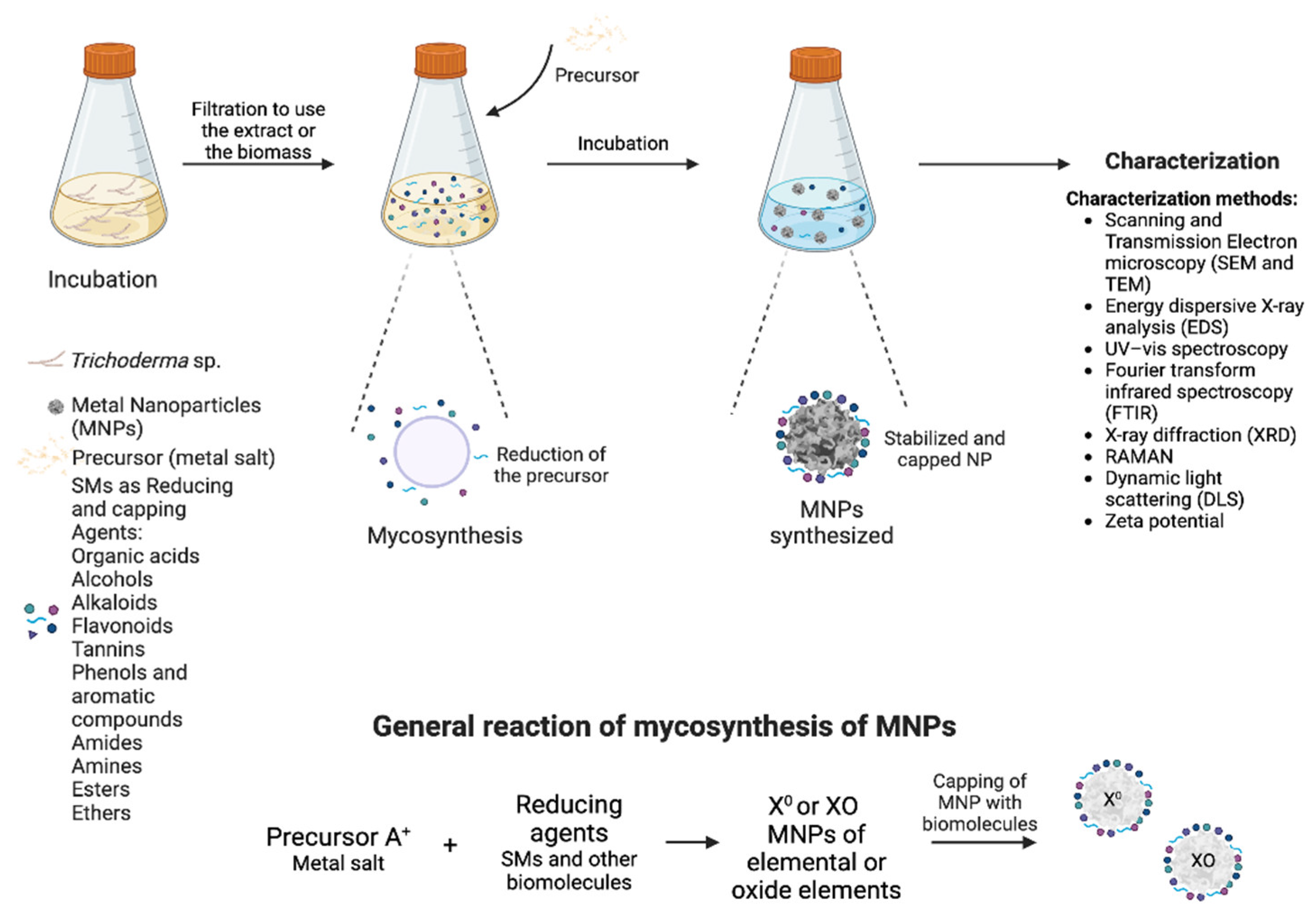
2. Mechanism of Green Synthesis of MNPs by Fungi

Compared with physical and chemical methods of synthesis of MNPs, mycosynthesis seems to be simple, but many factors need to be considered to control the size, shape, and stability of the MNPs. Among these factors are the culture conditions to obtain the biomass or the supernatant, the medium composition, temperature, pH, the amount of biomass or supernatant, the precursor and the amount used, and the strain/isolate used, among others [10,28].
Two mechanisms have been reported: intracellular and extracellular synthesis. However, most of the reports are hypotheses formulated on the assumption that certain data are obtained, with few or no convincing experimental support. Intracellular synthesis is less common and requires a series of subsequent processes for the purification of the MNPs, which makes it inefficient and cost ineffective. As to how the synthesis is carried out, Mukherjee and collaborators have hypothesized in the case of Ag NPs synthesized by Verticillium that Ag+ is trapped on the surface of the fungal cells through the electrostatic interaction with negatively charged carboxylate groups in enzymes present there, reducing the Ag+ and accumulating on the cytoplasmic membrane as well as within the cytoplasm [29].
Extracellular synthesis is the most widely used, as it is simple and cost-effective. As a general methodology for mycosynthesis, the fungus is grown in a nutrient-rich medium for 3–5 days, which can either be used as a source of SMs and other biomolecules, or the biomass is recovered and grown in distilled water for 2–4 days. Then, the supernatant containing the SMs and other biomolecules are recovered and placed in contact with the different precursors, at different concentrations and incubation times. Thus, the different SMs and other biomolecules carry out the reduction of the precursors and, consequently, the precipitation of the MNPs, generally observed as precipitates of different colors, depending on the synthesized NP. Figure 1 shows the general mechanism of how Trichoderma SMs are involved in the extracellular synthesis of NPs and the methods used by their characterization.
The extracellular mycosynthesis of MNPs is mediated by reactions with several types of biomolecules, including SMs. The main biomolecules reported are reductases (NADH and nitrate-dependent), quinones, amino acids, organic cofactors, and compounds with glucose, among others [30]. These molecules act by reducing several precursors such as sulfates, acetates, oxides, and chlorides, to obtain the different MNPs.
Some reports indicate that the molecules involved in mycosynthesis are enzymes or enzyme extracts, such as xylanases and amylases from T. longibrachiatum L2 and T. harzianum MTCC 801, respectively, which showed a potential and new use of enzyme extracts mediating mycosynthesis of Au and Ag NPs [31,32,33]. These results correlate with those previously reported by Gemishev and collaborators where they observed that when growing Trichoderma in a medium supplemented with corn steep liquor, the production of Ag NPs was higher compared to the one obtained when Trichoderma grew in media with other carbon sources (yeast extract, peptone, and casamino acids) [34]. To assimilate this carbon source, the fungus needs to produce lytic enzymes for its degradation, which are in turn used for the mycosynthesis of MNPs. It is important to highlight that Trichoderma is among the fungi with the highest production of lytic enzymes; therefore, those enzymes can be used to synthesize various MNPs with potential use. On the other hand, in T. viride and T. reesei, it was found that NADH-dependent reductase enzymes are involved in the reduction of Ag+ to Ag0, in the same way as in Fusarium species [34,35,36]. Although these reports indicate that enzymes are responsible for mediating mycosynthesis, this does not exclude the possibility that those other biomolecules, including SMs, could help during the synthesis and capping of MNPs. To corroborate this, one option is to perform the synthesis with the pure enzymes and compare it with a crude enzyme extract and contrast the characteristics of the MNPs. Another alternative is to carry out the deletion of the genes coding for these enzymes and determine if, even in their absence, the synthesis of the MNPs is carried out.
One of the most comprehensive studies on SMs acting by capping MNPs was performed on Se NPs synthesized by T. harzianum. By using mass spectrometry analyses via TripleTOF LC-MS, a total of 35 SMs were reported in this study, and 27 of them are antifungal agents. Among these are heptonic acid, ferulate, fumaric acid, threonic acid, mannitol, and glucose [37]. The authors point out that these organic compounds have a dual effect: acting as stabilizers and increasing fungicidal activity. As well, these MNPs had no significant side effects in human cells. These results support the importance of carrying out a careful study of the SMs present in MNPs, to ensure that these may be used in a safe manner, testing on cell lines or other organisms, such as mice, invertebrates, or beneficial plant microorganisms (including Trichoderma itself). In the following, we will focus on the SMs described during Trichoderma-mediated MNPs synthesis, as well as those that have been suggested for these species.

3. Main Secondary Metabolites from Trichoderma Involved in the Synthesis of Nanoparticles

Fungal SMs are mainly associated with activities such as competition, mycoparasitism, antibiosis against other microorganisms and predators, induction of microbial growth, communication with plant and other organisms, plant growth regulation, induction of plant resistance, protection against extreme environmental conditions, and as signaling or effector molecules. Thus, the synthesis of MNPs by fungi is a collateral effect resulting from a defense mechanism of the fungus against toxic agents found in the environment.
In mycosynthesis mediated by Trichoderma, less is known about what SMs are involved. Most studies report FTIR analysis, which indicates the functional groups present, and mention only that the molecules mediating the synthesis of NPs have organic functional groups in their structure, some of them being SMs [38] (Table 1).
As mentioned above, depending on the growth conditions of the fungi, they will produce certain SMs. In this regard, it has been reported that pH plays an important role in the production of SMs and, therefore, in the synthesis of MNPs. In Ag NP mycosynthesis, a directly proportional relationship is reported between the size of the NPs and the pH: if the pH is lower, the NPs are smaller. The authors suggest that it was because some fungi at low pH overproduce SMs and other biomolecules that will oxidize the nitrate [39]. On the other hand, it has been reported that Au NPs are unable to be synthesized at low pH, while at neutral and alkaline pH, the formation of MNPs is successfully carried out, probably by protonation of the carboxyl groups present in the SMs [27,40]. In contrast, for the synthesis of Fe NPs mediated by Trichoderma species, the optimum pH was found to be 4.5 [41]. It is possible that pH could affect not only the particle synthesis but also the capping and potentially the biological properties, and each presents its unique synthesis parameters.
Interestingly, the use of dead biomass or crude extracts influences the characteristics of the MNPs and their potential applications. do Nascimento and collaborators report the correlation between the biosorption process and the synthesis of Au NPs using biomass of T. harzianum, finding that the reduction, nucleation, agglomeration, and capping processes were mediated by molecules with methyl, amide, and amine groups [42]. These results suggest that Trichoderma can be used in bioremediation as well as as a biosynthesizer of MNPs. An interesting study that synthesized non-metal NPs used six Trichoderma isolates—T. asperellum, T. harzianum, T. atroviride, T. virens, T. longibrachiatum and T. brevicompactum—and evaluated the cultivation method in the form of culture filtrate, cell lysate, and crude cell wall; they found that culture filtrate was more effective and that T. asperellum produced the NPs with better results in the promotion of plant growth and in the control of diseases caused by phytopathogenic fungi [43]. These results suggest the versatility of Trichoderma in carrying out the mycosynthesis of MNPs, which influences the molecules anchored to them, giving them unique characteristics. Furthermore, these studies indicate that, depending on the MNPs to be synthesized and the Trichoderma strain to be used, conditions such as pH and temperature, among others, will have to be standardized to obtain MNPs with the desired characteristics.
One of the possible SMs that could be mediating the synthesis of MNPs are the naphthoquinones and anthraquinones, since it has been reported that these compounds have good reducing properties and these can act as mediators of the synthesis [44].
In the synthesis of Ag NPs, it has been reported that the main SMs involved are alkane, alkene, amine, amide, and organic acid compounds [30,45,46,47,48,49,50]. Interestingly, the synthesis of Ag NPs is one of the most studied, and this has led to the identification of some of the compounds involved in their synthesis. In T. harzianum, it was reported that kojic, acetic, and citric acid are the main components mediating the synthesis of MNPs [47]. Another study, also using T. harzianum, indicates that the compounds 1-benzoyl-3-[(S)-((2DS, 4R, 8R)-8-ethylquinuclidin-2-yl] (6-methoxyquinolin-4-yl)methyl)thiourea, puerarin, genistein, isotalathiazidine and ginsenoside are responsible for Ag NP production [48]. The first study reports organic acids and the second includes nitrogenous compounds, isoflavonoids and alkaloids, among others. This indicates that, despite using the same Trichoderma species, it can produce different SMs and, therefore, mediates the synthesis of the same type of MNPs.
For the synthesis of Au- and Cu-based NPs, SMs of nitrogenous nature stand out [42,47,48,51]. Finally, carboxylic acids, amide, ester, ether, and phenolic compounds mediate the synthesis of ZnO NPs [52]. This information suggests that, depending on the type of MNPs, it will be the nature of the SMs that Trichoderma will be using. However, further characterization is needed to determine which SMs are specifically responsible for mediating the synthesis, or at least to suggest candidates containing the identified functional groups.
Table 1. SMs involved or suggested in the biosynthesis of Trichoderma MNPs.
MNPsSMs Involved or Suggested to Be Involved in the BiosynthesisType of NP/Size of the NP (nm)Methods of CharacterizationTrichoderma SpeciesReferences
Ag55 exometabolites: alkane, dicarboxylic acid, aromatic ketone, amino acid, heteroacyclic compound, ketose sugar, sugars alcohol, aliphatic amine, polyol compound, steroidal pheromone, carbocyclic sugars groupsSpherical/
59.66
SEM, EDAX, Zeta potential, PSA, UV–vis and FTIRTrichoderma fusant Fu21[30]
AgCompounds with –OH group of phenols, alkyne groups, N-H of amine groups, –CH3 of aromatic and aliphatic compounds and –C–O stretch of alcohols, carboxylic acids, and estersSpherical or polyhedral/
5–50
Electron microscopy, EDS, UV–vis, FTIR and XRDT. longibranchiatum[45]
Amide I and amide II-like compounds and primary aminesSpherical/10UV–vis, TEM,
FTIR, DLS and Zeta potential
T. longibranchiatum[46]
Kojic, acetic and citric acidSpherical/20UV–vis and TEMT. harzianum[47]
1-benzoyl-3-[(S)-((2DS, 4R, 8R)-8-ethylquinuclidin-2-yl] (6-methoxyquinolin-4-yl)methyl)thiourea, puerarin, genistein, isotalatizidine and ginsenosideNot determined/21.49UV–vis, FTIR, EDS, DLS,
XRD and SEM
T. harzianum[48]
Alkaloids, flavonoids, tannins, and phenolsRoughly spherical/
12.7
XRD, TEM, SEM, EDX and FTIRT. harzianum[49]
Biomolecules with hydroxyl, alkane, amide, and carboxylate groupsSpherical, triangular and cuboid/5 to 11DLS, XRD, FTIR, FESEM and HRTEMT. longibrachiatum[50]
Compounds with primary and secondary aminesTriangular and spherical/
50–75
UV–vis, XRD, FTIR, SEM and EDAXT. atroviride[51]
Biochemicals with carbonyl, CH3 and alcohol groupsSpherical/
15–20
UV–vis, FTIR and SEMT. viride and T. longibraciatum[53]
Biomolecules with amine, amide, carbonyl, phenols, methylene, and alcohol groupsRoughly
spherical/5–35
UV–vis, XRD, TEM and FTIRTrichoderma strains[54]
Alkaline, amine and aromatic peptidesAnisotropic structural/
15–25
FTIR, TEM, EDXT. atroviride[55]
Secondary metabolites with aromatic, amide I and carbonyl groupsSpherical/
50–100
UV–vis, FTIR, SEM, EDX, XRD and Zeta potentialT. citrinoviride[56]
AuMolecules with methyl, amide, and amine groups Spherical, hexagonal, and octagonal/
20–50
XRD, FTIR, SEM-EDS, DLS and Zeta potential T. harzianum[42]
Compounds with primary and secondary amines Triangular nanoplates and spherical/
50–75
UV–vis, XRD, FTIR, SEM and EDAX T. atroviride[51]
Cu/CuOSecondary metabolites with amide and aromatic groupsSpherical/110FTIR, XRD, SEM, TEM and XPST. asperellum[57]
Primary amines, secondary amines, aliphatic amines, and amide groupsSpherical/
8–100
UV-vis, FTIR and SEMT. virens[58]
ZnOSecondary metabolites with carboxylic acid, amide, esters, ethers, and phenolic groupsSpherical or polyhedral/15–30.32UV-vis, FTIR, EDX, XRD, SEM and TEMT. longibranchiatum[52]
Abbreviations: Energy dispersive X-ray analysis (EDS); UV–vis spectroscopy (UV-vis); Fourier transform infrared spectroscopy (FTIR); X-ray diffraction (XRD); Transmission electron microscopy (TEM); Dynamic light scattering (DLS); Scanning electron microscopy (SEM); Energy dispersive X-ray (EDAX); Particle size analyzer (PSA); Field emission scanning electron microscope (FESEM); High-resolution transmission electron microscope (HRTEM); X-ray photoelectron spectroscopy (XPS); Raman spectroscopy (Raman) and Photoluminescence (PL).

4. Secondary Metabolites from Trichoderma Acting as Capping Agents in MNPs

Capping agent is an amphiphilic molecule comprising a polar head group and a non-polar hydrocarbon tail, conferring the functionality and enhancing the compatibility with another phase [59]. Among the molecules reported as stabilizers are amide groups with carbonyl stretch providing stability by enclosing their surface. Thus, secondary metabolites including quinone, alcohol, and organic acids compounds, among others, can serve as stabilizing agents [38,60].
The authors reported FTIR analysis where they identified biomolecules that are bound on the MNPs with functional groups such as alcohol, phenols, carbonyl, aromatic and aliphatic amines and amide are presented in the MNPs synthesized [61,62]. However, only a few reports suggest the main SMs involved in mycosynthesis mediated by Trichoderma; Table 2 summarizes those that have been described.
Some reports indicate that MNPs were relatively stable up to 6 months after synthesis [46]. Since some molecules involved in capping are SMs, it is important to note that, to maintain their nature, the MNPs must be maintained in a cool and dark environment, otherwise the molecules may be degraded, and the MNPs would lose their stability.
Capping is also responsible for the MNPs having a better antimicrobial capacity; this may be attributable to the biomolecules released by Trichoderma that exhibit diverse activities, including antimicrobial activity. Several reports indicate that mycosynthesized MNPs show better antimicrobial activity compared to MNPs synthesized by physical or chemical methods or even some commercial pesticides. In addition, several MNPs exhibit antimicrobial activity similar to those reported for certain commonly used antibiotics [63]. Moreover, an interesting study revealed that TiO2 NPs synthesized by T. citrinoviride were effective in inhibiting Pseudomonas aeruginosa, a drug-resistant bacteria that causes nosocomial infections, and the authors suggest that these MNPs can become an alternative to antibiotics or potentiate the ailing antibiotics [64]. Recently, the mycosynthesis of Ag and Au NPs mediated by T. saturnisporum was reported, finding that Ag NPs were more effective against pathogenic bacteria [65], suggesting that, despite being mycosynthesized by the same Trichoderma, MNPs from different materials will still have distinctive characteristics and probably different molecules acting as capping.
In addition, Ag NPs synthesized by T. harzianum were shown to be effective for controlling larvae and pupae of the dengue vector Aedes aegypti L., suggesting that presumably part of the observable effect is caused by the presence of SMs in the MNPs [63]. Chinnaperumal and collaborators found that TiO2 NPs synthesized by T. viride had higher larvicidal and pupicidal activity against Helicoverpa armigera compared to chemically synthesized TiO2 NPs [66]. These results suggest that MNPs synthesized by Trichoderma contain SMs capable of controlling organisms of medical and agricultural interest, even better than MNPs obtained by conventional methods.
Mycogenic MNPs produced by some Trichoderma species have shown the ability to inhibit the growth of plant pathogenic fungi or have been used in the nanopriming of seeds. Guilger and collaborators reported that Ag NPs synthesized using T. harzianum had a major activity against the fungus S. sclerotiorum compared to commercial or non-biogenic and uncapped Ag NPs. The authors suggest that this increase is because of the biomolecules and organic compounds present in the capping of the nanoparticles [67]. By using ZnO NPs synthesized by T. harzianum, Zaki and coworkers reported that these MNPs showed antifungal activity similar to that reported for two fungicides when evaluated against Fusarium sp., Rhizoctonia solani, and Macrophomina phaseolina [68].
Ag NPs were more effective in suppressing disease and improving life on cotton plants, compared with commercial chemical fungicides [69], probably because of the synergistic effect of the MNPs and the SMs contained in them. By using T. citrinoviride, TiO2 and Ag NPs were synthesized, and it was observed that TiO2 at 25 and 50 μg/mL had a positive effect on seed germination and seedling vigor. However, lower concentrations of Ag NPs reduced seed germination and seedling vigor and induced an increase in the activities of catalase, superoxide dismutase, and peroxidase enzymes [56]. The authors suggest that these results, among several factors, are related to the surface charge of MNPs, affecting the absorption and response of seedlings. Recently, MnO NPs were synthesized for the first time by using T. virens and they were effective against several phytopathogens, including Alternaria alternata and Helminthosporium sp. [70].
In addition, the MNPs synthesized by Trichoderma can be used as carriers of molecules or in photothermolysis, having potential medical applications [57,71]. Moreover, it has been seen that MNPs synthesized by Trichoderma can promote its own growth; this has been reported with FeO3 and TiO2 NPs and enhances their biocontrol activity [72,73].
One interesting study characterized the removal of contaminants by using MNPs synthesized by Trichoderma. The report indicates that SiO2 NPs synthesized from rice husks and T. harzianum were able to adsorb lead from water used for Nile tilapia culture, and the fish showed increased growth and improvement in physiological parameters; the authors confirmed the presence of functional groups of biomolecules encapsulating SiO2 NPs [74]. In these phenomena of adsorption, it is likely that the biomolecules capping the MNPs serve for better adsorption of contaminants; however, no supporting evidence was provided in this study, so further research is needed in this area.
Table 2. Secondary metabolites involved or suggested in the capping of MNPs synthesized by Trichoderma.
MNPsSMs Involved or Suggested to Act as CappingType of NP/Size of the NP (nm)Trichoderma SpeciesReferences
AgExometabolites: alkanes, aromatic alcohol, ketones, phenolic compounds, saturated fatty acids, furans, heterocyclen, steroid, sugar acids, acyclic alkanes, fatty alcohol, aromatic hydrocarbons, esters, and sulfur-containing compoundsSpherical/59.66Trichoderma fusant Fu21[30]
Aromatic secondary metabolitesNot determined/21.49T. harzianum[48]
Biomolecules with hydroxyl, alkane, amide, and carboxylate groupsSpherical, triangular and cuboid/5–11T. longibrachiatum[50]
Compounds with primary and secondary aminesTriangular nanoplates and spherical/50–75T. atroviride[51]
Biochemicals with carbonyl, CH3 and alcohol groupsSpherical/15–20T. viride and T. longibraciatum[53]
Biomolecules with amine, amide, carbonyl, phenols, methylene, and alcohol groupsRoughly spherical/5–35Trichoderma strains[54]
Secondary metabolites with aromatic, amide I and carbonyl groupsSpherical/50–100T. citrinoviride[56]
Compounds with alkane, phosphine, amide and aromatic ketones, aliphatic bending, silica, cycloalkane, aromatic mono-substitution and alkynes functional groupsSpherical/43.68T. harzianum[62]
Carbohydrate, and heterocyclic compound molecules, especially, gliotoxin moleculeSpherical and oval/5–50T. virens[75]
AuBiomolecules containing amide I and amide II groupsSpherical and pseudo-spherical/9.8Trichoderma sp.[40]
Molecules with methyl, amide, and amine groupsSpherical, hexagonal, and octagonal/20–50T. harzianum[42]
Compounds with primary and secondary aminesTriangular nanoplates and spherical/50–75T. atroviride[51]
Cu/CuOSecondary metabolites with amide and aromatic groupsSpherical/110T. asperellum[57]
Primary amines, secondary amines, aliphatic amines, and amide groupsSpherical/8–100T. virens[58]
MnOPhenols, alkaloids, carbohydrates, and amino acidsRod/35T. virens[70]
Fe/FeO3Compounds with alkene, carboxyl, and phenol groupsNot determinedTrichoderma strains[41]
Molecules with amide, alcohol, esters, ethers, and aromatic groupsSpherical/185T. harzianum[72]
Compounds with amide I and amide II groupsSpherical/25T. asperellum[76]
SiO2Molecules with different functional groups of biomolecules Oval, rod and cubical/89T. harzianum[74]
TiO2Molecules with carbonyl groups Triangular, pentagonal, spherical and rod/10–400T. citrinoviride[64]
Molecules with different functional groups: alkane, methylene, alkene, amine, and carboxylic acidRoughly spherical/74.4T. viride[66]
ZnOMycochemicals with phenolic, amino acids, aldehydes, and ketone functional groupsHexagonal, spherical and rod/8–25Trichoderma sp.[68]

5. Research Gaps and Future Directions in the Mycosynthesis of Nanoparticles Mediated by Trichoderma and Their SMs

Fungi produce several SMs that can be classified as mycotoxins; it is important to note that mycosynthesis can also lead to the production of these harmful compounds. Tomah and collaborators suggest that in T. virens-mediated mycosynthesis of Ag NPs, the SMs involved in capping are organic acids, amide, amine, phenolic and heterocyclic compound molecules, among others, and some of them display medicinal and biocontrol properties but, unfortunately, have toxic properties in humans, such as gliotoxin molecule [75,77]. This research shows one of the most important gaps in Trichoderma-mediated mycosynthesis because it is important to focus on the characterization of the SMs involved in their synthesis and capping, since some may contain compounds that could be toxic to humans or animals. It is important to highlight that most studies focus on synthesizing the MNPs, characterizing them by microscopy, XRD, and FTIR techniques and their possible antimicrobial activity or cytotoxicity; however, few are focused on studying the SMs responsible for mediating the synthesis and generating the capping. Thereon, it is important that Trichoderma-mediated mycosynthesis begins to be related to metabolomics and proteomics, using techniques such as LC-MS (Liquid Chromatography coupled to Mass Spectroscopy) or NMR (Nuclear Magnetic Resonance Spectroscopy) to know which molecules are involved in the synthesis and capping of MNPs.
Another of the main challenges in mycosynthesis is the fact that the first stages must be carried out under strict sterile conditions to maintain the purity of the strains used; otherwise, probable contaminations can alter the growth of Trichoderma, besides affecting the SMs that would be produced, causing a lack of reproducibility during the synthesis process. This consideration can become less cost effective for this type of synthesis; besides, this challenge is not presented in the chemical or physical synthesis as well as phytosynthesis of MNPs and could affect the cost of the synthesis. An alternative to overcome this disadvantage could be if, as mentioned above, a detailed characterization of the SMs or other molecules involved in Trichoderma-mediated MNPs synthesis could be performed to purify these compounds or to generate overexpressing strains of these SMs.
One of the sizeable challenges is the synthesis time, because the growth of Trichoderma and the obtaining of the extracts can take more than a week, even up to a month. In chemical synthesis, the MNPs are obtained in several hours. In this regard, as mentioned above, one option is to purify the SMs to make them available for the required synthesis, as long as the SMs remain stable for long periods of time. This disadvantage goes hand in hand with large-scale production, where chemical and phytosynthesis methods show better results. Therefore, if MNPs need to be produced with higher efficiency, chemical methods are the best option. However, if what is required are MNPs where their production is environmentally friendly, green synthesis, among them, Trichoderma-mediated MNPs synthesis, is one of the best options. One of the research areas to be developed in the following years is the efficiency of green synthesis of MNPs.
On the other hand, most of the fungi reported as nanosynthesizing agents are pathogenic to plants or humans, which makes the disposal of the generated biomass a challenge. In contrast, Trichoderma is not a plant pathogen and there are few reports where it has caused infection in immunosuppressed patients. This is an advantage, as only SMs or other biomolecules secreted by Trichoderma are used; the biomass produced would be considered a waste, which can be used for the formulation of a biocontroller or biostimulant that can be accompanied by the synthesized NPs and develop other types of agrochemicals, such as bio-nanoformulations with multiple benefits in agriculture.
To our knowledge, once the MNPs are synthesized by Trichoderma and summing up what was found in the cited literature, practically no residues are generated, since, in some syntheses, the MNPs are allowed to be dispersed in the same solution in which the synthesis was carried out and, on the other hand, in other methods, the remains of the extracts are calcined to obtain the desired MNPs. This is a great advantage over chemical methods, where a large amount of highly polluting secondary compounds is generated and for which a special disposal must be carried out. Moreover, the use of toxic and hazardous substances, and the addition of external reducing, stabilizing and capping substances is avoided in mycosynthesis.
Another area of potential interest is to determine the mechanisms of synthesis. An alternative is to generate knockout strains of genes that encode for enzymes involved in the synthesis of a potential SM involved in the synthesis of NPs; in this way, it can be known which SMs are essential for the synthesis to be carried out, as well as which others are important in the capping process. Another alternative is to use computational chemistry tools that allow the theoretical elucidation of which SMs are more feasible to be used in the synthesis and capping process.
As mentioned throughout this document, the MNPs synthesized by Trichoderma can be used in the medical area, as an antimicrobial agent; in agriculture, as an antifungal and pupicidal agent, as well as a stimulant of seed germination and plant growth; likewise, they can be used in remediation processes, by removing toxic agents from the environment, showing even better results than chemically synthesized MNPs. Although, in general, Trichoderma mycosynthesized MNPs offer more advantages than disadvantages, there are still areas of study to complement what is currently known and to fully elucidate the mechanisms of synthesis and capping, as well as beneficial or toxic activities for different organisms.

6. Conclusions

Trichoderma is an alternative for the synthesis of stable MNPs, with unique characteristics, and although Trichoderma-mediated mycosynthesis has been investigated for about 20 years, little is known about the SMs involved in this process. Therefore, it is a field of research with high potential that could lead to the search or generation of new strains with higher production of these SMs. Recently, more than 70 strains from Trichoderma were reported and they produce around 445 new metabolites, including terpenes, steroids, polyketides, peptides, and alkaloids, which can be used as reducing or capping agents [78].
However, it is important to conduct detailed studies to understand the biochemical mechanism that allows Trichoderma-mediated MNP synthesis to be carried out, as well as the composition of the Trichoderma SMs involved in the synthesis and capping of MNPs to ensure that their use will not generate undesirable side effects that could impact both animal health and ecosystems. This opens a new line of research, which will establish the guidelines to produce MNPs in a more sustainable and safe manner.

Author Contributions

G.M.H.P. and C.A.R.V. authors contributed equally to this work. C.A.R.V. conceptualized the idea. G.M.H.P., L.E.C. and C.A.R.V. wrote and revised the article. All authors have read and agreed to the published version of the manuscript.

Funding

The authors thank CIMAV Internal Project Grant No. PI-2024-11 (PI-26011) under the academic responsibility of CARV.

Acknowledgments

The authors acknowledge CONAHCYT-Mexico, CIMAV and UG.

Conflicts of Interest

The authors declare no conflicts of interest.

References

  1. Home|National Nanotechnology Initiative. Available online: https://www.nano.gov/ (accessed on 8 March 2024).
  2. Hussain, I.; Singh, N.B.; Singh, A.; Singh, H.; Singh, S.C. Green Synthesis of Nanoparticles and Its Potential Application. Biotechnol. Lett. 2016, 38, 545–560. [Google Scholar] [CrossRef]
  3. Li, X.; Xu, H.; Chen, Z.-S.; Chen, G. Biosynthesis of Nanoparticles by Microorganisms and Their Applications. J. Nanomater. 2011, 2011, e270974. [Google Scholar] [CrossRef]
  4. Mukherjee, B.; Santra, K.; Pattnaik, G.; Ghosh, S. Preparation, Characterization and in-Vitro Evaluation of Sustained Release Protein-Loaded Nanoparticles Based on Biodegradable Polymers. Int. J. Nanomedicine 2008, 3, 487–496. [Google Scholar] [CrossRef]
  5. Chhipa, H. Chapter 5—Mycosynthesis of Nanoparticles for Smart Agricultural Practice: A Green and Eco-Friendly Approach. In Green Synthesis, Characterization and Applications of Nanoparticles; Shukla, A.K., Iravani, S., Eds.; Micro and Nano Technologies; Elsevier: Amsterdam, The Netherlands, 2019; pp. 87–109. [Google Scholar] [CrossRef]
  6. Owaid, M.N.; Ibraheem, I.J. Mycosynthesis of Nanoparticles Using Edible and Medicinal Mushrooms. Eur. J. Nanomed. 2017, 9, 5–23. [Google Scholar] [CrossRef]
  7. Madakka, M.; Jayaraju, N.; Rajesh, N. Mycosynthesis of Silver Nanoparticles and Their Characterization. MethodsX 2018, 5, 20–29. [Google Scholar] [CrossRef]
  8. Azmath, P.; Baker, S.; Rakshith, D.; Satish, S. Mycosynthesis of Silver Nanoparticles Bearing Antibacterial Activity. Saudi Pharm. J. 2016, 24, 140–146. [Google Scholar] [CrossRef] [PubMed]
  9. Elsakhawy, T.; Omara, A.E.-D.; Abowaly, M.; El-Ramady, H.; Badgar, K.; Llanaj, X.; Törős, G.; Hajdú, P.; Prokisch, J. Green Synthesis of Nanoparticles by Mushrooms: A Crucial Dimension for Sustainable Soil Management. Sustainability 2022, 14, 4328. [Google Scholar] [CrossRef]
  10. Guilger-Casagrande, M.; Germano-Costa, T.; Pasquoto-Stigliani, T.; Fraceto, L.F.; de Lima, R. Biosynthesis of Silver Nanoparticles Employing Trichoderma harzianum with Enzymatic Stimulation for the Control of Sclerotinia sclerotiorum. Sci. Rep. 2019, 9, 14351. [Google Scholar] [CrossRef]
  11. Roy, L.; Bera, D.; Adak, S. Mycosynthesized Nanoparticles: Role in Food Processing Industries; Prasad, R., Kumar, V., Kumar, M., Wang, S., Eds.; Springer: Singapore, 2018. [Google Scholar] [CrossRef]
  12. Ferreira, F.V.; Musumeci, M.A. Trichoderma as Biological Control Agent: Scope and Prospects to Improve Efficacy. World J. Microbiol. Biotechnol. 2021, 37, 90. [Google Scholar] [CrossRef]
  13. Harman, G.E.; Howell, C.R.; Viterbo, A.; Chet, I.; Lorito, M. Trichoderma Species—Opportunistic, Avirulent Plant Symbionts. Nat. Rev. Microbiol. 2004, 2, 43–56. [Google Scholar] [CrossRef]
  14. Brotman, Y.; Briff, E.; Viterbo, A.; Chet, I. Role of Swollenin, an Expansin-Like Protein from Trichoderma, in Plant Root Colonization. Plant Physiol. 2008, 147, 779–789. [Google Scholar] [CrossRef]
  15. Cai, F.; Chen, W.; Wei, Z.; Pang, G.; Li, R.; Ran, W.; Shen, Q. Colonization of Trichoderma harzianum Strain SQR-T037 on Tomato Roots and Its Relationship to Plant Growth, Nutrient Availability and Soil Microflora. Plant Soil 2015, 388, 337–350. [Google Scholar] [CrossRef]
  16. Schweiger, R.; Padilla-Arizmendi, F.; Nogueira-López, G.; Rostás, M.; Lawry, R.; Brown, C.; Hampton, J.; Steyaert, J.M.; Müller, C.; Mendoza-Mendoza, A. Insights into Metabolic Changes Caused by the Trichoderma virens-Maize Root Interaction. Mol. Plant-Microbe Interact. 2021, 34, 524–537. [Google Scholar] [CrossRef] [PubMed]
  17. Cai, F.; Druzhinina, I.S. In Honor of John Bissett: Authoritative Guidelines on Molecular Identification of Trichoderma. Fungal Divers. 2021, 107, 1–69. [Google Scholar] [CrossRef]
  18. Schalamun, M.; Schmoll, M. Trichoderma—Genomes and Genomics as Treasure Troves for Research towards Biology, Biotechnology and Agriculture. Front. Fungal Biol. 2022, 3, 1002161. [Google Scholar] [CrossRef]
  19. Tomah, A.A.; Zhang, Z.; Alamer, I.S.A.; Khattak, A.A.; Ahmed, T.; Hu, M.; Wang, D.; Xu, L.; Li, B.; Wang, Y. The Potential of Trichoderma-Mediated Nanotechnology Application in Sustainable Development Scopes. Nanomaterials 2023, 13, 2475. [Google Scholar] [CrossRef] [PubMed]
  20. Olmedo-Monfil, V.; Mendoza-Mendoza, A.; Gómez, I.; Cortés, C.; Herrera-Estrella, A. Multiple Environmental Signals Determine the Transcriptional Activation of the Mycoparasitism Related Gene prb1 in Trichoderma atroviride. Mol. Genet. Genomics 2002, 267, 703–712. [Google Scholar] [CrossRef]
  21. Ramírez-Valdespino, C.A.; Casas-Flores, S.; Olmedo-Monfil, V. Trichoderma as a Model to Study Effector-Like Molecules. Front. Microbiol. 2019, 10, 1030. [Google Scholar] [CrossRef]
  22. Fox, E.M.; Howlett, B.J. Secondary Metabolism: Regulation and Role in Fungal Biology. Curr. Opin. Microbiol. 2008, 11, 481–487. [Google Scholar] [CrossRef]
  23. Hoffmeister, D.; Keller, N.P. Natural Products of Filamentous Fungi: Enzymes, Genes, and Their Regulation. Nat. Prod. Rep. 2007, 24, 393–416. [Google Scholar] [CrossRef]
  24. Hermosa, R.; Cardoza, R.E.; Rubio, M.B.; Gutiérrez, S.; Monte, E. Secondary Metabolism and Antimicrobial Metabolites of Trichoderma. In Biotechnology and Biology of Trichoderma; Gupta, V.K., Schmoll, M., Herrera-Estrella, A., Upadhyay, R.S., Druzhinina, I., Tuohy, M.G., Eds.; Elsevier: Amsterdam, The Netherlands, 2014; pp. 125–137. [Google Scholar] [CrossRef]
  25. Mukherjee, P.K.; Horwitz, B.A.; Kenerley, C.M. Secondary Metabolism in Trichoderma—A Genomic Perspective. Microbiol. Read. Engl. 2012, 158, 35–45. [Google Scholar] [CrossRef] [PubMed]
  26. Zeilinger, S.; Gruber, S.; Bansal, R.; Mukherjee, P.K. Secondary Metabolism in Trichoderma—Chemistry Meets Genomics. Fungal Biol. Rev. 2016, 30, 74–90. [Google Scholar] [CrossRef]
  27. Mishra, A.; Kumari, M.; Pandey, S.; Chaudhry, V.; Gupta, K.C.; Nautiyal, C.S. Biocatalytic and Antimicrobial Activities of Gold Nanoparticles Synthesized by Trichoderma Sp. Bioresour. Technol. 2014, 166, 235–242. [Google Scholar] [CrossRef] [PubMed]
  28. Gemishev, O.; Panayotova, M.; Gicheva, G.; Mintcheva, N. Green Synthesis of Stable Spherical Monodisperse Silver Nanoparticles Using a Cell-Free Extract of Trichoderma reesei. Materials 2022, 15, 481. [Google Scholar] [CrossRef] [PubMed]
  29. Mukherjee, P.; Ahmad, A.; Mandal, D.; Senapati, S.; Sainkar, S.R.; Khan, M.I.; Parishcha, R.; Ajaykumar, P.V.; Alam, M.; Kumar, R.; et al. Fungus-Mediated Synthesis of Silver Nanoparticles and Their Immobilization in the Mycelial Matrix: A Novel Biological Approach to Nanoparticle Synthesis. Nano Lett. 2001, 1, 515–519. [Google Scholar] [CrossRef]
  30. Hirpara, D.; Gajera, H.P. Green Synthesis and Antifungal Mechanism of Silver Nanoparticles Derived from Chitin-Induced Exometabolites of Trichoderma Interfusant. Appl. Organomet. Chem. 2019, 34, e5407. [Google Scholar] [CrossRef]
  31. Guleria, V.; Saxena, J. Bioinspired Trichogenic Silver Nanoparticles and Their Antifungal Activity Against Plant Pathogenic Fungi Sclerotinia sclerotiorum MTCC 8785. Nat. Environ. Pollut. Technol. 2023, 22, 253–259. [Google Scholar] [CrossRef]
  32. Elegbede, J.A.; Lateef, A.; Azeez, M.A.; Asafa, T.B.; Yekeen, T.A.; Oladipo, I.C.; Adebayo, E.A.; Beukes, L.S.; Gueguim-Kana, E.B. Fungal Xylanases-Mediated Synthesis of Silver Nanoparticles for Catalytic and Biomedical Applications. IET Nanobiotechnol. 2018, 12, 857–863. [Google Scholar] [CrossRef]
  33. Elegbede, J.A.; Lateef, A.; Azeez, M.A.; Asafa, T.B.; Yekeen, T.A.; Oladipo, I.C.; Aina, D.A.; Beukes, L.S.; Gueguim-Kana, E.B. Biofabrication of Gold Nanoparticles Using Xylanases Through Valorization of Corncob by Aspergillus niger and Trichoderma longibrachiatum: Antimicrobial, Antioxidant, Anticoagulant and Thrombolytic Activities. Waste Biomass Valorization 2020, 11, 781–791. [Google Scholar] [CrossRef]
  34. Gemishev, O.T.; Panayotova, M.I.; Mintcheva, N.N.; Djerahov, L.P.; Tyuliev, G.T.; Gicheva, G.D. A Green Approach for Silver Nanoparticles Preparation by Cell-Free Extract from Trichoderma reesei Fungi and Their Characterization. Mater. Res. Express 2019, 6, 095040. [Google Scholar] [CrossRef]
  35. Ingle, A.; Gade, A.; Pierrat, S.; Sonnichsen, C.; Rai, M. Mycosynthesis of Silver Nanoparticles Using the Fungus Fusarium acuminatum and Its Activity Against Some Human Pathogenic Bacteria. Curr. Nanosci. 2008, 4, 141–144. [Google Scholar] [CrossRef]
  36. Fayaz, M.; Tiwary, C.S.; Kalaichelvan, P.T.; Venkatesan, R. Blue Orange Light Emission from Biogenic Synthesized Silver Nanoparticles Using Trichoderma viride. Colloids Surf. B Biointerfaces 2010, 75, 175–178. [Google Scholar] [CrossRef] [PubMed]
  37. Hu, D.; Yu, S.; Yu, D.; Liu, N.; Tang, Y.; Fan, Y.; Wang, C.; Wu, A. Biogenic Trichoderma harzianum-Derived Selenium Nanoparticles with Control Functionalities Originating from Diverse Recognition Metabolites against Phytopathogens and Mycotoxins. Food Control 2019, 106, 106748. [Google Scholar] [CrossRef]
  38. Abdel-Hadi, A.; Iqbal, D.; Alharbi, R.; Jahan, S.; Darwish, O.; Alshehri, B.; Banawas, S.; Palanisamy, M.; Ismail, A.; Aldosari, S.; et al. Myco-Synthesis of Silver Nanoparticles and Their Bioactive Role against Pathogenic Microbes. Biology 2023, 12, 661. [Google Scholar] [CrossRef] [PubMed]
  39. Ramos, M.M.; Dos S Morais, E.; da S Sena, I.; Lima, A.L.; de Oliveira, F.R.; de Freitas, C.M.; Fernandes, C.P.; de Carvalho, J.C.T.; Ferreira, I.M. Silver Nanoparticle from Whole Cells of the Fungi Trichoderma Spp. Isolated from Brazilian Amazon. Biotechnol. Lett. 2020, 42, 833–843. [Google Scholar] [CrossRef] [PubMed]
  40. Qu, Y.; Shen, W.; Pei, X.; Ma, F.; You, S.; Li, S.; Wang, J.; Zhou, J. Biosynthesis of Gold Nanoparticles by Trichoderma Sp. WL-Go for Azo Dyes Decolorization. J. Environ. Sci. 2017, 56, 79–86. [Google Scholar] [CrossRef] [PubMed]
  41. Kareem, S.; Adeleye, T.; Ojo, R. Effects of pH, Temperature and Agitation on the Biosynthesis of Iron Nanoparticles Produced by Trichoderma Species. IOP Conf. Ser. Mater. Sci. Eng. 2020, 805, 012036. [Google Scholar] [CrossRef]
  42. do Nascimento, J.M.; Cruz, N.D.; de Oliveira, G.R.; Sá, W.S.; de Oliveira, J.D.; Ribeiro, P.R.S.; Leite, S.G.F. Evaluation of the Kinetics of Gold Biosorption Processes and Consequent Biogenic Synthesis of AuNPs Mediated by the Fungus Trichoderma harzianum. Environ. Technol. Innov. 2021, 21, 101238. [Google Scholar] [CrossRef]
  43. Nandini, B.; Hariprasad, P.; Prakash, H.S.; Shetty, H.S.; Geetha, N. Trichogenic-Selenium Nanoparticles Enhance Disease Suppressive Ability of Trichoderma against Downy Mildew Disease Caused by Sclerospora graminicola in Pearl Millet. Sci. Rep. 2017, 7, 2612. [Google Scholar] [CrossRef]
  44. Liu, S.-Y.; Lo, C.-T.; Chen, C.; Liu, M.-Y.; Chen, J.-H.; Peng, K.-C. Efficient Isolation of Anthraquinone-Derivatives from Trichoderma harzianum ETS 323. J. Biochem. Biophys. Methods 2007, 70, 391–395. [Google Scholar] [CrossRef]
  45. Cui, X.; Zhong, Z.; Xia, R.; Liu, X.; Qin, L. Biosynthesis Optimization of Silver Nanoparticles (AgNPs) Using Trichoderma longibranchiatum and Biosafety Assessment with Silkworm (Bombyx mori). Arab. J. Chem. 2022, 15, 104142. [Google Scholar] [CrossRef]
  46. Elamawi, R.M.; Al-Harbi, R.E.; Hendi, A.A. Biosynthesis and Characterization of Silver Nanoparticles Using Trichoderma longibrachiatum and Their Effect on Phytopathogenic Fungi. Egypt. J. Biol. Pest Control 2018, 28, 28. [Google Scholar] [CrossRef]
  47. Gupta, B.; Mathur, A.; Koul, A.; Shrivastava, V. Study on the Extraction and Purification of Metabolites Obtained from Trichoderma harzianum. Asian J. Pharm. Pharmacol. 2019, 5, 547–550. [Google Scholar] [CrossRef]
  48. Konappa, N.; Udayashankar, A.C.; Dhamodaran, N.; Krishnamurthy, S.; Jagannath, S.; Uzma, F.; Pradeep, C.K.; De Britto, S.; Chowdappa, S.; Jogaiah, S. Ameliorated Antibacterial and Antioxidant Properties by Trichoderma harzianum Mediated Green Synthesis of Silver Nanoparticles. Biomolecules 2021, 11, 535. [Google Scholar] [CrossRef] [PubMed]
  49. EL-Moslamy, S.H.; Elkady, M.F.; Rezk, A.H.; Abdel-Fattah, Y.R. Applying Taguchi Design and Large-Scale Strategy for Mycosynthesis of Nano-Silver from Endophytic Trichoderma harzianum SYA.F4 and Its Application against Phytopathogens. Sci. Rep. 2017, 7, 45297. [Google Scholar] [CrossRef]
  50. Omran, B.A.; Nassar, H.N.; Younis, S.A.; Fatthallah, N.A.; Hamdy, A.; El-Shatoury, E.H.; El-Gendy, N.S. Physiochemical Properties of Trichoderma longibrachiatum DSMZ 16517-Synthesized Silver Nanoparticles for the Mitigation of Halotolerant Sulphate-Reducing Bacteria. J. Appl. Microbiol. 2019, 126, 138–154. [Google Scholar] [CrossRef] [PubMed]
  51. Ponmurugan, P. Biosynthesis of Silver and Gold Nanoparticles Using Trichoderma atroviride for the Biological Control of Phomopsis Canker Disease in Tea Plants. IET Nanobiotechnol. 2017, 11, 261–267. [Google Scholar] [CrossRef] [PubMed]
  52. Ghareeb, R.Y.; Belal, E.B.; El-Khateeb, N.M.M.; Shreef, B.A. Utilizing Bio-Synthesis of Nanomaterials as Biological Agents for Controlling Soil-Borne Diseases in Pepper Plants: Root-Knot Nematodes and Root Rot Fungus. BMC Plant Biol. 2024, 24, 110. [Google Scholar] [CrossRef]
  53. Perera, D.S.; Hiruni Tharaka, W.G.; Wickramarachchi, S.R.; Amarasinghe, D.; De Silva, C.R.; Gunawardana, T.N. Mycosynthesis of Silver Nanoparticles (Trichoderma viride, Trichoderma longibrachiatum) and Their Mosquito Larvicidal Efficacy on Dengue Vectors and Acute Toxicity on Moina macrocopa. Asian J. Chem. 2023, 35, 1995–2000. [Google Scholar] [CrossRef]
  54. Qu, M.; Yao, W.; Cui, X.; Xia, R.; Qin, L.; Liu, X. Biosynthesis of Silver Nanoparticles (AgNPs) Employing Trichoderma Strains to Control Empty-Gut Disease of Oak Silkworm (Antheraea pernyi). Mater. Today Commun. 2021, 28, 102619. [Google Scholar] [CrossRef]
  55. Saravanakumar, K.; Wang, M.-H. Trichoderma Based Synthesis of Anti-Pathogenic Silver Nanoparticles and Their Characterization, Antioxidant and Cytotoxicity Properties. Microb. Pathog. 2018, 114, 269–273. [Google Scholar] [CrossRef] [PubMed]
  56. Sonawane, H.; Arya, S.; Math, S.; Shelke, D. Myco-Synthesized Silver and Titanium Oxide Nanoparticles as Seed Priming Agents to Promote Seed Germination and Seedling Growth of Solanum lycopersicum: A Comparative Study. Int. Nano Lett. 2021, 11, 371–379. [Google Scholar] [CrossRef]
  57. Saravanakumar, K.; Shanmugam, S.; Varukattu, N.B.; MubarakAli, D.; Kathiresan, K.; Wang, M.-H. Biosynthesis and Characterization of Copper Oxide Nanoparticles from Indigenous Fungi and Its Effect of Photothermolysis on Human Lung Carcinoma. J. Photochem. Photobiol. B 2019, 190, 103–109. [Google Scholar] [CrossRef] [PubMed]
  58. Dorjee, L.; Kamil, D.; Kumar, R.; Khokhar, M.; Aggarwal, R.; Gogoi, R. Synthesis of Promising Copper Nanoparticles Utilizing Biocontrol Agents, Trichoderma virens and Chaetomium globosum. Indian J. Agric. Sci. 2024, 94, 061–067. [Google Scholar] [CrossRef]
  59. Gulati, S.; Sachdeva, M.; Bhasin, K.K. Capping Agents in Nanoparticle Synthesis: Surfactant and Solvent System. AIP Conf. Proc. 2018, 1953, 030214. [Google Scholar] [CrossRef]
  60. Husseiny, S.M.; Salah, T.A.; Anter, H.A. Biosynthesis of Size Controlled Silver Nanoparticles by Fusarium oxysporum, Their Antibacterial and Antitumor Activities. Beni-Suef Univ. J. Basic Appl. Sci. 2015, 4, 225–231. [Google Scholar] [CrossRef]
  61. Ahluwalia, V.; Kumar, J.; Sisodia, R.; Shakil, N.A.; Walia, S. Green Synthesis of Silver Nanoparticles by Trichoderma harzianum and Their Bio-Efficacy Evaluation against Staphylococcus aureus and Klebsiella pneumonia. Ind. Crops Prod. 2014, 55, 202–206. [Google Scholar] [CrossRef]
  62. Narware, J.; Singh, S.P.; Manzar, N.; Kashyap, A.S. Biogenic Synthesis, Characterization, and Evaluation of Synthesized Nanoparticles against the Pathogenic Fungus Alternaria solani. Front. Microbiol. 2023, 14, 1159251. [Google Scholar] [CrossRef] [PubMed]
  63. Sundaravadivelan, C.; Padmanabhan, M.N. Effect of Mycosynthesized Silver Nanoparticles from Filtrate of Trichoderma harzianum against Larvae and Pupa of Dengue Vector Aedes aegypti L. Environ. Sci. Pollut. Res. Int. 2014, 21, 4624–4633. [Google Scholar] [CrossRef]
  64. Arya, S.; Sonawane, H.; Math, S.; Tambade, P.; Chaskar, M.; Shinde, D. Biogenic Titanium Nanoparticles (TiO2NPs) from Tricoderma citrinoviride Extract: Synthesis, Characterization and Antibacterial Activity against Extremely Drug-Resistant Pseudomonas Aeruginosa. Int. Nano Lett. 2021, 11, 35–42. [Google Scholar] [CrossRef]
  65. Soliman, M.K.Y.; Salem, S.S.; Abu-Elghait, M.; Azab, M.S. Biosynthesis of Silver and Gold Nanoparticles and Their Efficacy Towards Antibacterial, Antibiofilm, Cytotoxicity, and Antioxidant Activities. Appl. Biochem. Biotechnol. 2023, 195, 1158–1183. [Google Scholar] [CrossRef] [PubMed]
  66. Chinnaperumal, K.; Govindasamy, B.; Paramasivam, D.; Dilipkumar, A.; Dhayalan, A.; Vadivel, A.; Sengodan, K.; Pachiappan, P. Bio-Pesticidal Effects of Trichoderma viride Formulated Titanium Dioxide Nanoparticle and Their Physiological and Biochemical Changes on Helicoverpa armigera (Hub.). Pestic. Biochem. Physiol. 2018, 149, 26–36. [Google Scholar] [CrossRef] [PubMed]
  67. Guilger, M.; Pasquoto-Stigliani, T.; Bilesky-Jose, N.; Grillo, R.; Abhilash, P.C.; Fraceto, L.F.; de Lima, R. Biogenic Silver Nanoparticles Based on Trichoderma harzianum: Synthesis, Characterization, Toxicity Evaluation and Biological Activity. Sci. Rep. 2017, 7, 44421. [Google Scholar] [CrossRef]
  68. Zaki, S.A.; Ouf, S.A.; Albarakaty, F.M.; Habeb, M.M.; Aly, A.A.; Abd-Elsalam, K.A. Trichoderma harzianum-Mediated ZnO Nanoparticles: A Green Tool for Controlling Soil-Borne Pathogens in Cotton. J. Fungi 2021, 7, 952. [Google Scholar] [CrossRef] [PubMed]
  69. Zaki, S.A.; Ouf, S.A.; Abd-Elsalam, K.A.; Asran, A.A.; Hassan, M.M.; Kalia, A.; Albarakaty, F.M. Trichogenic Silver-Based Nanoparticles for Suppression of Fungi Involved in Damping-Off of Cotton Seedlings. Microorganisms 2022, 10, 344. [Google Scholar] [CrossRef] [PubMed]
  70. EL-Moslamy, S.H.; Yahia, I.S.; Zahran, H.Y.; Kamoun, E.A. Novel Biosynthesis of MnO NPs Using Mycoendophyte: Industrial Bioprocessing Strategies and Scaling-up Production with Its Evaluation as Anti-Phytopathogenic Agents. Sci. Rep. 2023, 13, 2052. [Google Scholar] [CrossRef]
  71. Saravanakumar, K.; Sathiyaseelan, A.; Mariadoss, A.V.A.; Hu, X.; Venkatachalam, K.; Wang, M.-H. Nucleolin Targeted Delivery of Aptamer Tagged Trichoderma Derived Crude Protein Coated Gold Nanoparticles for Improved Cytotoxicity in Cancer Cells. Process Biochem. 2021, 102, 325–332. [Google Scholar] [CrossRef]
  72. Bilesky-José, N.; Maruyama, C.; Germano-Costa, T.; Campos, E.; Carvalho, L.; Grillo, R.; Fraceto, L.F.; De Lima, R. Biogenic α-Fe2O3 Nanoparticles Enhance the Biological Activity of Trichoderma against the Plant Pathogen Sclerotinia sclerotiorum. ACS Sustain. Chem. Eng. 2021, 9, 1669–1683. [Google Scholar] [CrossRef]
  73. Pasquoto-Stigliani, T.; Guilger-Casagrande, M.; Campos, E.V.R.; Germano-Costa, T.; Bilesky-José, N.; Migliorini, B.B.; Feitosa, L.O.; Sousa, B.T.; de Oliveira, H.C.; Fraceto, L.F.; et al. Titanium Biogenic Nanoparticles to Help the Growth of Trichoderma harzianum to Be Used in Biological Control. J. Nanobiotechnol. 2023, 21, 166. [Google Scholar] [CrossRef]
  74. El-Gazzar, N.; Almanaa, T.N.; Reda, R.M.; El Gaafary, M.N.; Rashwan, A.A.; Mahsoub, F. Assessment the Using of Silica Nanoparticles (SiO2NPs) Biosynthesized from Rice Husks by Trichoderma harzianum MF780864 as Water Lead Adsorbent for Immune Status of Nile Tilapia (Oreochromis niloticus). Saudi J. Biol. Sci. 2021, 28, 5119–5130. [Google Scholar] [CrossRef]
  75. Tomah, A.A.; Alamer, I.S.A.; Li, B.; Zhang, J.-Z. Mycosynthesis of Silver Nanoparticles Using Screened Trichoderma Isolates and Their Antifungal Activity against Sclerotinia sclerotiorum. Nanomaterials 2020, 10, 1955. [Google Scholar] [CrossRef] [PubMed]
  76. Mahanty, S.; Bakshi, M.; Ghosh, S.; Chatterjee, S.; Bhattacharyya, S.; Das, P.; Das, S.; Chaudhuri, P. Green Synthesis of Iron Oxide Nanoparticles Mediated by Filamentous Fungi Isolated from Sundarban Mangrove Ecosystem, India. BioNanoScience 2019, 9, 637–651. [Google Scholar] [CrossRef]
  77. Bulgari, D.; Fiorini, L.; Gianoncelli, A.; Bertuzzi, M.; Gobbi, E. Enlightening Gliotoxin Biological System in Agriculturally Relevant Trichoderma Spp. Front. Microbiol. 2020, 11, 200. [Google Scholar] [CrossRef]
  78. Song, Y.-P.; Ji, N.-Y. Chemistry and Biology of Marine-Derived Trichoderma Metabolites. Nat. Prod. Bioprospect. 2024, 14, 14. [Google Scholar] [CrossRef]
Figure 1. General mechanism of extracellular MNP synthesis mediated by Trichoderma SMs. The mechanism of synthesis, methods of characterization and general reaction of formation are schematized.
Jof 10 00443 g001
Disclaimer/Publisher’s Note: The statements, opinions and data contained in all publications are solely those of the individual author(s) and contributor(s) and not of MDPI and/or the editor(s). MDPI and/or the editor(s) disclaim responsibility for any injury to people or property resulting from any ideas, methods, instructions or products referred to in the content.

