Article

Epidemiology of Candidemia in Mashhad, Northeast Iran: A Prospective Multicenter Study (2019–2021)

by
Somayeh Dolatabadi
1,
Mohammad Javad Najafzadeh
2,*,
Abbas Raeisabadi
3,
Hossein Zarrinfar
4,
Mahsa Jalali
2,
Bram Spruijtenburg
5,6,
Eelco F. J. Meijer
5,6,
Jacques F. Meis
5,7,
Cornelia Lass-Flörl
8 and
Theun de Groot
5,6,*
1
Department of Biology, Hakim Sabzevari University, Sabzevar 9617976487, Iran
2
Department of Medical Parasitology and Mycology, Faculty of Medicine, Mashhad University of Medical Sciences, Mashhad 91766-99199, Iran
3
Department of Medical Mycology, Faculty of Medicine, Mazandaran University of Medical Sciences, Sari 48471-91628, Iran
4
Allergy Research Center, Mashhad University of Medical Sciences, Mashhad 91766-99199, Iran
5
Radboudumc-CWZ Center of Expertise for Mycology, 6532 SZ Nijmegen, The Netherlands
6
Canisius-Wilhelmina Hospital (CWZ)/Dicoon, 6532 SZ Nijmegen, The Netherlands
7
Institute of Translational Research, Cologne Excellence Cluster on Cellular Stress Response in Aging-Associated Diseases (CECAD), Excellence Center for Medical Mycology (ECMM), University of Cologne, 50931 Cologne, Germany
8
Institute of Hygiene and Medical Microbiology, Medical University of Innsbruck, Excellence Center for Medical Mycology (ECMM), 6020 Innsbruck, Austria
*
Authors to whom correspondence should be addressed.
J. Fungi 2024, 10(7), 481; https://doi.org/10.3390/jof10070481
Submission received: 11 May 2024 / Revised: 9 July 2024 / Accepted: 10 July 2024 / Published: 12 July 2024
(This article belongs to the Special Issue Medically Relevant Species of Candida)

Abstract

:
Candidemia is a major cause of morbidity and mortality in health care settings, and its epidemiology is changing. In the last two decades, the proportion of non-albicans Candida (NAC) yeasts in candidemia has increased. These yeasts more often display resistance to common antifungals. In many western countries, candidemia is mainly caused by susceptible C. albicans, while in resource-limited countries, including Iran, the candidemia species distribution is studied less often. Here, we investigated the species distribution, resistance levels, and characteristics of patients with candidemia in five hospitals in Mashhad (northeast Iran) for two years (2019–2021). Yeast isolates from blood were identified with MALDI-TOF MS and subjected to antifungal susceptibility testing (AFST) using the broth microdilution method, while molecular genotyping was applied to Candida parapsilosis isolates. In total, 160 yeast isolates were recovered from 160 patients, of which the majority were adults (60%). Candidemia was almost equally detected in men (48%) and women (52%). Almost half of patients (n = 67, 49%) were from intensive care units (ICUs). C. parapsilosis (n = 58, 36%) was the most common causative agent, surpassing C. albicans (n = 52, 33%). The all-cause mortality rate was 53%, with C. albicans candidemia displaying the lowest mortality with 39%, in contrast to a mortality rate of 59% for NAC candidemia. With microbroth AFST, nearly all tested isolates were found to be susceptible, except for one C. albicans isolate that was resistant to anidulafungin. By applying short tandem repeat (STR) genotyping to C. parapsilosis, multiple clusters were found. To summarize, candidemia in Mashhad, Iran, from 2019 to 2021, is characterized by common yeast species, in particular C. parapsilosis, for which STR typing indicates potential nosocomial transmission. The overall mortality is high, while resistance rates were found to be low, suggesting that the high mortality is linked to limited diagnostic options and insufficient medical care, including the restricted use of echinocandins as the first treatment option.

1. Introduction

Candidemia is a growing concern in hospital settings, posing every year a serious health threat to hundreds of thousands of patients worldwide [1,2]. It is one of the most common bloodstream infections, both in adult and pediatric patients. The species Candida albicans, C. glabrata (also known as Nakaseomyces glabrata), C. parapsilosis, C. tropicalis, and C. krusei (also known as Pichia kudriavzevii) are the five leading causative agents of candidemia, accounting for approximately 85–90% of candidemia [1,3]. However, these five most common species display notable regional differences. C. albicans is the most common etiological agent of candidemia in the United States and most European countries, although its proportion as compared to other Candida species is decreasing in the last decade [4,5,6]. Among these five common species, C. albicans, together with C. tropicalis, is regarded as the most virulent species, while it shows the lowest rate of antifungal resistance [1,7,8,9]. C. glabrata is the second most common cause of candidemia in the USA and many North and West European countries [4,5,10]. Moreover, it is the first cause of candidemia in intensive care units and in patients with hematological malignancies and solid tumors [11]. The third most common yeast species is C. parapsilosis, especially prevalent in South European countries [12]. This yeast can easily spread through the hands of healthcare workers, and azole resistance is frequently observed. Additionally, C. parapsilosis has shown prolonged survival within hospital wards and can be the source of clonal outbreaks [13]. Another common Candida species is C. tropicalis, which is associated with the highest mortality rates among Candida species and is the first or second cause of candidemia in developing countries, such as India and Brazil, with resistance steadily increasing [9,14,15]. In Iran, the majority of studies report C. albicans as most common in candidemia, although other yeasts are emerging, as well as antifungal resistance [16,17,18].
Depending on the species, the mortality rate may vary from approximately 30 to 70% [1]. The increase in mortality is associated with an increase in non-albicans Candida (NAC) yeast species. A likely explanation is resistance to the limited number of antifungal drugs available to treat candidemia in developing countries [6]. Especially the reduced susceptibility of the NAC yeast species C. glabrata, C. tropicalis, and C. parapsilosis to fluconazole, the most inexpensive and readily available antifungal agent used to treat candidemia, is highly problematic in these countries [19]. Moreover, azole-resistant C. parapsilosis often spread clonally and are persistent within healthcare environments [20]. This problem is further aggravated by the limited use of accurate identification techniques in Iran, such as sequencing or matrix-assisted laser desorption ionization time-of-flight mass spectrometry (MALDI-TOF MS), which leads to an unselective application of antifungals, inducing the development and persistence of resistant species [1,6].
These issues emphasize the importance of conducting epidemiological studies to explore species distribution, the burden of antifungal resistance, and to characterize the clinical profile of patients with candidemia. Considering the limited number of studies on candidemia in Iran, we performed a two-year retrospective candidemia study in Mashhad, Iran, in which we collected clinical data of patients, assessed species distribution by MALDI-TOF, and determined their antifungal susceptibility pattern, along with short tandem repeat (STR) genotyping of C. parapsilosis, to improve our insights on candidemia.

2. Materials and Methods

2.1. Study Design and Sample Processing

This study retrospectively included patients with candidemia admitted to five hospitals in Mashhad, Iran, including Ghaem (800 beds), Emam Reza (948 beds), 22 Bahman (175 beds), Arya (100 beds), and Dr. Sheikh (150 beds), during July 2019 to July 2021. All centers were multi-specialty hospitals, except for Dr. Sheikh, which is a pediatric hospital. Blood samples were inoculated in Bactec 9120 blood culture bottles (Becton Dickinson, Spark, MD, USA). From positive bottles, 150 µL was streaked on Sabouraud dextrose agar (SDA, Merck, Darmstadt, Germany) and CHROMagar Candida (CHROMagar, Paris, France) plates, which were subsequently incubated at 37 °C for 24–48 h. Colony morphology (color, shape, and size) was visually inspected to identify samples potentially harboring more than one species. Colonies with different morphologies were transferred to SDA plates, incubated at 37 °C for 24–48 h, and subjected to further analyses. All yeast isolates were identified using a microflex LT MALDI-TOF MS system (Bruker Daltonics, Bremen, Germany) and a full extraction method according to manufacturer instructions [21]. Candidemia or invasive candidiasis was diagnosed when blood cultures yielded yeast. Mortality was reported as the all-cause mortality rate during study period. Patients < 16 years were considered children and ≥16 years adults. Therapeutic failure according to expert opinion was defined as persistent positive blood cultures (with yeasts) despite antifungal treatment for 7 days. This study was approved by the ethic committee of Mashhad University of Medical Sciences (ethical approval number IR.NIMAD.REC.1398.103).

2.2. In Vitro Antifungal Susceptibility Testing (AFST)

AFST was performed using the broth microdilution method according to the Clinical and Laboratory Standards Institute (CLSI) protocol (CLSI M27, 4th editon) [22]. The following antifungal drugs were tested: fluconazole (FLU), amphotericin B (AMB) (both from Sigma Chemical Corporation, St. Louis, MO, USA), voriconazole (VOR; Pfizer, New York, NY, USA), micafungin (MFG; Astellas Pharma, Ibaraki, Japan), and anidulafungin (AFG, Pfizer, New York, NY, USA). Plates were incubated at 37 °C for 24 h and minimum inhibitory concentrations (MICs) were recorded after visual examination. Reference strains of C. parapsilosis (ATCC 22019) and C. krusei (ATCC 6258) were used for quality control. The MIC data were categorized according to clinical breakpoints. Isolates were classified as susceptible (S), susceptible dose-dependent (SDD) or intermediate (I), and resistant (R). If clinical breakpoints were not available, MICs were interpreted according to epidemiological cut-off values (ECVs) and isolates were classified as wild-type (WT) at MIC ≤ ECV or non-wild type (NWT) at MIC > ECV, according to the CLSI M57 document [23].

2.3. Multiplex Short Tandem Repeat (STR) Genotyping

C. parapsilosis DNA was extracted and purified with the MagNA Pure and Viral NA Small volume kit, and the MagNA Pure 96 instrument (All Roche Diagnostics GmbH, Mannheim, Germany), as previously described [23]. STR multiplex PCR genotyping was performed on a thermocycler (Biometra, Göttingen, Germany) using 1× FastStart Taq polymerase buffer without MgCl2, deoxynucleotide triphosphates (dNTPs) (0.2 mM), MgCl2 (3 mM), forward and reverse primers (10 µM), 1 U FastStart Taq polymerase (Roche Diagnostics), and isolated DNA [24]. STRs were amplified with a thermal protocol of denaturation at 95 °C for 10 min, followed by 30 cycles consisting of annealing at 60 °C for 30 s, extension at 72 °C for 1 min, and a final incubation for 10 min at 72 °C. Amplicons were diluted 1:200 in water and 10 µL of diluted amplicon in addition to 0.12 µL of Orange 500 DNA size standard (Nimagen, Nijmegen, The Netherlands) were incubated for 1 min at 95 °C and analyzed on a 3500 XL genetic analyzer (Applied Biosystems, Foster City, CA, USA). Copy numbers of STR markers were determined using the Genemapper 5 software (Applied Biosystems), and relatedness between isolates was analyzed as previously described [25].

3. Results

3.1. Patients’ Characteristics and Species Distribution

A total of 160 yeast isolates were collected during two years from 160 patients of whom, a majority were adults (n = 96, 60%) (Table 1). Candidemia was almost equally detected in men (n = 83, 52%) and women (n = 77, 48%), and almost half of the yeast isolates were recovered from ICUs (n = 79, 49%). Using MALDI-TOF MS identification, C. parapsilosis sensu stricto (n = 58, 36%) was the most common causative agent, followed by C. albicans (n = 52, 33%), while no coinfections were observed. For 156 patients, outcomes were known, showing an all-cause mortality rate of 53%. C. albicans candidemia coincided with a relative low mortality rate of 39% as compared to NAC candidemia, which had a mortality rate of 59%. In the current study, the most commonly used antifungals included fluconazole (n = 48, 30%) and liposomal amphotericin B (n = 40, 25%). More than one antifungal drug was administered to 12 patients (8%), while 54 patients (34%) received no antifungal therapy. Remarkably, patients with C. albicans were virtually all treated with an antifungal, while most patients with C. parapsilosis were not treated.

3.2. Resistance Investigation

In vitro AFST was performed on 131 Candida isolates according to the CLSI M27 guidelines. Overall, the tested antifungals demonstrated potent activity with only few cases of resistance (Table 2). For fluconazole, one C. lusitaniae isolate was NWT, while for amphotericin B, one NWT C. albicans isolate was found, according to CLSI M59 ECVs (Table S1). For echinocandins only a few C. albicans isolates showed elevated MICs. For anidulafungin one isolate demonstrated a MIC of 1 µg/mL, while there were also three intermediate susceptible isolates. From the latter three, two isolates were also intermediate susceptible for micafungin (Table S1). The less echinocandin susceptible C. albicans were isolated from patients who were not treated with echinocandins.

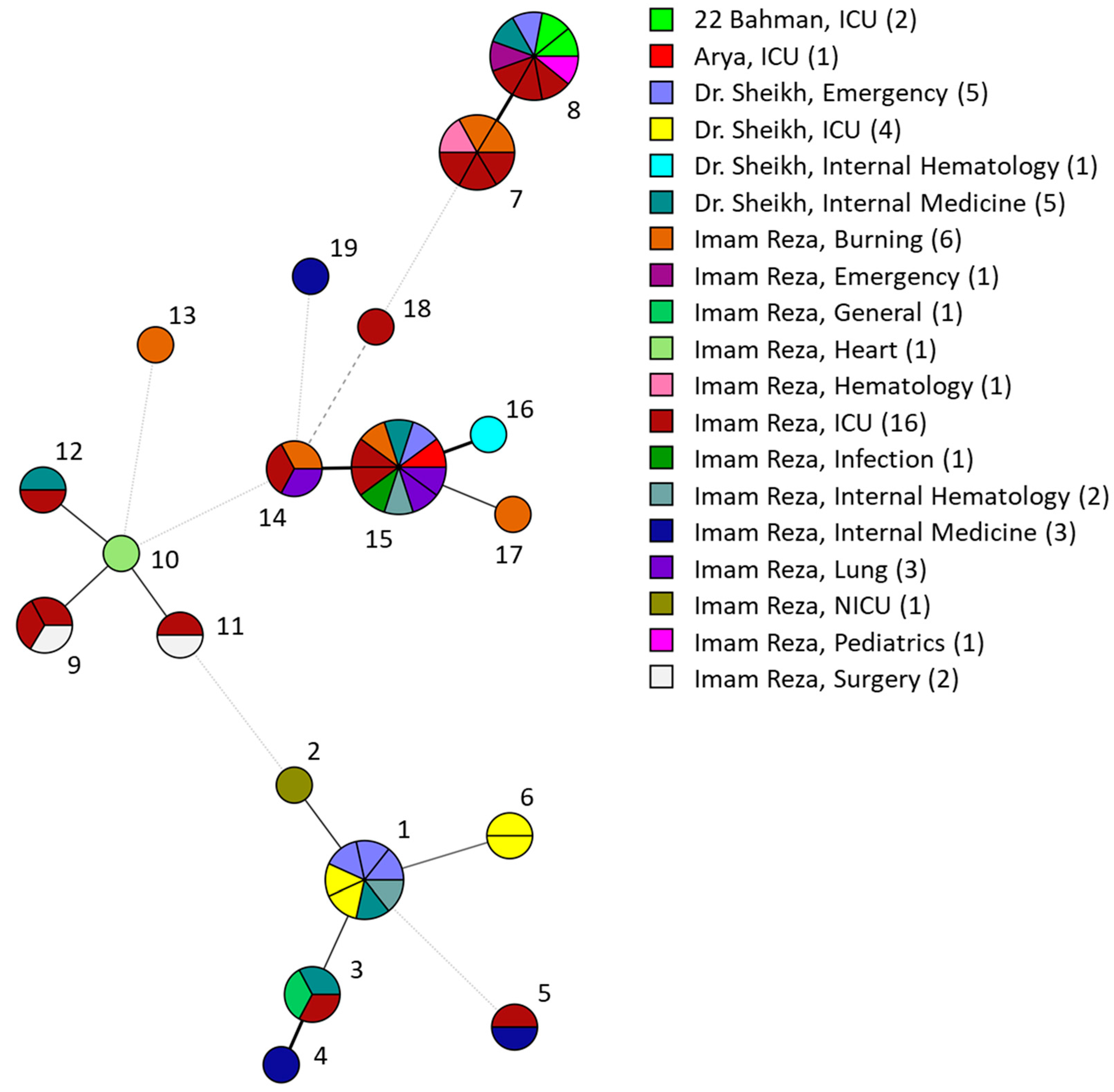
3.3. C. parapsilosis STR Genotyping Shows Clusters

To further investigate the high incidence of C. parapsilosis and the potential role of nosocomial transmission, we determined the phylogenetic relatedness between the 57 C. parapsilosis isolates by amplifying six microsatellite markers using multiplex PCR. The STR genotyping yielded 19 different genotypes, containing one to ten isolates (Figure S1). Isolates differed in four markers at most between each other. A total of seven clusters (≥3 isolates) were found, of which four spanned isolates from two or three hospitals (Figure 1). Many of these clusters were closely related to each other, like genotypes 7 to 8, and 14 to 16, differing only in the ploidy of one or two markers, respectively. Only the genotype 9 cluster was restricted to one hospital and not closely related (>2 markers difference) to any other cluster. Within all clusters, isolates originated from two to eight departments (Figure 2). For example, the largest cluster of ten isolates contained isolates from three hospitals, totaling eight different departments.

4. Discussion

4.1. Epidemiology

In the present study, we investigated the epidemiology of candidemia in five hospitals in Mashhad for a limited period of two years. The male-female ratio was overall comparable, which is in line with previous studies [6]. We found that C. parapsilosis (36%) was the leading candidemia agent, followed by C. albicans (33%). While the majority of Iranian studies identified C. albicans as most common species, a small nationwide study also found C. parapsilosis as most common [26,27]. Interestingly, some centers reported a C. glabrata frequency of 20–23% [28,29,30], which is higher than the 10% we found. Probably these differences in species distribution are to some extent due to different patient populations, as the average age of our patients was much lower. Previous epidemiological studies demonstrated that in elderly patients C. glabrata candidemia occurs more often, while pediatric or neonatal patients are more prone to C. albicans and C. parapsilosis candidemia episodes [3,6]. Nonetheless, the proportion of C. albicans in pediatric patients in our study (37%) was still much lower than the 57% found in the study from Kord et al., which was performed in neonatal and pediatric intensive care units in Tehran [29]. This difference might indicate a changing epidemiology, as the Tehran study was conducted between 2014 to 2016, while recent studies report more NAC yeasts [3].
Another factor influencing species distribution can be antifungal prophylaxis. Patients that underwent fluconazole prophylaxis are known to experience more C. krusei candidemia, as this species has intrinsic elevated MICs to this agent [31]. However, in the previously mentioned Iranian studies [27,28,29,30], the proportion of C. krusei is low, possibly attributable to the restricted use of antifungal prophylaxis. Additionally, whereas C. tropicalis is the most frequent species in some countries, the proportion in this and previous Iranian studies was relatively low (<15%) [27,28,29,30]. Among the other yeast species, we found C. lusitaniae and Meyerozyma guilliermondii (former Candida guilliermondii), which are known for intrinsic antifungal resistance, potentially limiting antifungal options that could result in treatment failure [32].
The overall mortality of 53% was a bit lower than worldwide estimations (64%), but higher than previous Iranian and European studies, which ranged from 28% to 48% [2,10,27,28,29,30]. We found that the mortality rate of C. albicans candidemia was lower when compared to other species, while C. albicans and C. tropicalis are considered as the Candida species with the highest virulence [6]. This is likely attributable to the antifungal treatment in our cohort, as most of patients with C. albicans were treated with one or more antifungals, while more than half of patients with C. parapsilosis and one third of patients with C. tropicalis was not treated with any antifungal. The high mortality rates of patients with C. glabrata and C. krusei, whom were also almost all treated with antifungals, might be explained by the usage of fluconazole despite both species are intrinsically resistant or display naturally elevated MICs for this drug [33]. These findings emphasize the importance of broad candidemia surveillance, rapid and accurate identification and adequate antifungal treatment guided by susceptibility results. Of note, all candidemia episodes were detected using blood cultures. Molecular methods were not employed for diagnosing candidemia in this study.

4.2. Resistance Investigation

With microbroth AFST, resistance was in general rarely observed in this study, especially for azoles and amphotericin B. Previous resistance investigations from Iran also demonstrated limited resistance [13,28], which could be due to the limited administration of antifungals drugs in routine clinical use in Iran. Nonetheless, antifungal resistance is globally increasing, warranting continued surveillance. Among the isolates with reduced susceptibility, one C. lusitaniae was resistant to fluconazole. While most strains are susceptible, this species is known to rapidly acquire antifungal resistance, which could result in treatment failure [34]. Furthermore, elevated MICs were observed for some C. albicans isolates for echinocandins, with a single isolate resistant for anidulafungin. Although resistance in yeasts against this antifungal class is rarely found, resistant isolates are often from patients who underwent echinocandin treatment, indicating the resistance is possibly therapy-induced [35,36]. In this study none of the patients from whom the C. albicans strains with elevated MICs were isolated, were treated with echinocandins. Although transmission of echinocandin-resistant isolates cannot be ruled out, it is rarely reported, making prior colonization with these echinocandin-resistant isolates more likely.

4.3. Outbreak Investigation with STR Genotyping

With STR genotyping, multiple clusters were found that frequently consisted of isolates from multiple hospitals. Within these clusters, isolates originated from multiple departments and hospitals, suggesting potential intra-hospital nosocomial transmission. This potential nosocomial transmission needs to be further investigated with a whole genome sequencing (WGS) single nucleotide polymorphism (SNP) analysis. We previously found for other yeast species that STR analyses cannot distinguish isolates differing less than 150 SNPs, which are not clonal but closely related nonetheless [37,38]. For C. parapsilosis, numerous nosocomial outbreaks have been reported, frequently caused by fluconazole-resistant strains [12,39,40]. In most reported outbreaks, the source is unknown, but some studies suggest the hands of health care workers or contaminated nosocomial surfaces and equipment [12,41]. Nonetheless, C. parapsilosis strains are known to persist despite infection control measures in healthcare settings [42,43]. Recently, transmission of fluconazole-resistant C. parapsilosis between two hospitals was reported in Canada, highlighting the need for adequate genomic surveillance [44].
Study limitations include the restriction of genotyping to C. parapsilosis, while potential nosocomial transmission of other species is also possible. In addition, to confirm the suspected clonal transmission within the hospitals, WGS analysis would have been required. Furthermore, the current study did not determine whether enforced hand hygiene followed by genotyping of C. parapsilosis candidemia episodes is effective to halt clonal transmission within these hospitals and to what extent. Finally, the current study was conducted over a period of two years, which only provides a limited view of the candidemia epidemiology in Iran. For a comprehensive overview, long-term follow-up studies should be conducted to determine whether the species composition further moves from C. albicans to NAC yeast species, as this can rapidly shift.
To conclude, the current study demonstrates a high proportion of NAC yeasts causing candidemia in Mashhad, Iran, with a high mortality rate. This emphasis the importance of adequate diagnosis and appropriate antifungal treatment based on AFST data. Additionally, the finding of multiple STR clusters might be caused by nosocomial transmission, which, if confirmed by WGS SNP analysis, would suggest the need for additional infection prevention measures.

Supplementary Materials

The following supporting information can be downloaded at: https://www.mdpi.com/article/10.3390/jof10070481/s1, Figure S1: UPGMA dendrogram of 57 C. parapsilosis isolates; Table S1: In vitro minimum inhibitory concentrations against common antifungals according to CLSI M27 guidelines for Candida isolates.

Author Contributions

Conceptualization, S.D. and T.d.G.; methodology, S.D. and B.S.; software, B.S.; validation, M.J.N., A.R. and H.Z.; formal analysis, S.D. and B.S.; investigation, M.J. and T.d.G.; resources, J.F.M. and C.L.-F.; data curation, E.F.J.M. and T.d.G.; writing—original draft preparation, S.D., M.J.N. and B.S.; writing—review and editing, A.R., H.Z., M.J., E.F.J.M., J.F.M., C.L.-F. and T.d.G.; visualization, B.S.; supervision, J.F.M., C.L.-F. and T.d.G.; project administration, S.D.; funding acquisition, S.D. All authors have read and agreed to the published version of the manuscript.

Funding

This research was funded by the Elite Researcher Grand Committee under award number 982822 and 4000347 from the National Institutes for Medical Research Development (NIMAD), Tehran, Iran.

Institutional Review Board Statement

The study was approved by the ethic committee of Mashhad University of Medical Sciences (ethical approval number IR.NIMAD.REC.1398.103).

Informed Consent Statement

Informed consent was obtained from all subjects involved in the study.

Data Availability Statement

The original contributions presented in the study are included in the article/Supplementary Material, further inquiries can be directed to the corresponding authors.

Conflicts of Interest

The authors declare no conflicts of interest. The funders had no role in the design of the study, in the collection, analyses, or interpretation of data; in the writing of the manuscript; or in the decision to publish the results.

References

  1. Lamoth, F.; Lockhart, S.R.; Berkow, E.L.; Calandra, T. Changes in the epidemiological landscape of invasive candidiasis. J. Antimicrob. Chemother. 2018, 73, i4–i13. [Google Scholar] [CrossRef] [PubMed]
  2. Denning, D.W. Global incidence and mortality of severe fungal disease. Lancet Infect. Dis. 2024, 23, S1473–S3099. [Google Scholar] [CrossRef] [PubMed]
  3. Khan, Z.; Ahmad, S.; Al-Sweih, N.; Mokaddas, E.; Al-Banwan, K.; Alfouzan, W.; Al-Obaid, I.; Al-Obaid, K.; Asadzadeh, M.; Jeragh, A.; et al. Changing trends in epidemiology and antifungal susceptibility patterns of six bloodstream Candida species isolates over a 12-year period in Kuwait. PLoS ONE 2019, 14, e0216250. [Google Scholar] [CrossRef] [PubMed]
  4. Astvad, K.M.T.; Johansen, H.K.; Røder, B.L.; Rosenvinge, F.S.; Knudsen, J.D.; Lemming, L.; Schønheyder, H.C.; Hare, R.K.; Kristensen, L.; Nielsen, L.; et al. Update from a 12-Year Nationwide Fungemia Surveillance: Increasing Intrinsic and Acquired Resistance Causes Concern. J. Clin. Microbiol. 2018, 56, e01564-17. [Google Scholar] [CrossRef] [PubMed]
  5. Lockhart, S.R.; Iqbal, N.; Cleveland, A.A.; Farley, M.M.; Harrison, L.H.; Bolden, C.B.; Baughman, W.; Stein, B.; Hollick, R.; Park, B.J.; et al. Species identification and antifungal susceptibility testing of Candida bloodstream isolates from population-based surveillance studies in two U.S. cities from 2008 to 2011. J. Clin. Microbiol. 2012, 50, 3435–3442. [Google Scholar] [CrossRef] [PubMed]
  6. Guinea, J. Global trends in the distribution of Candida species causing candidemia. Clin. Microbiol. Infect 2014, 20, 5–10. [Google Scholar] [CrossRef] [PubMed]
  7. Doi, A.M.; Pignatari, A.C.C.; Edmond, M.B.; Marra, A.R.; Camargo, L.F.A.; Siqueira, R.A.; da Mota, V.P.; Colombo, A.L. Epidemiology and Microbiologic Characterization of Nosocomial Candidemia from a Brazilian National Surveillance Program. PLoS ONE 2016, 11, e0146909. [Google Scholar] [CrossRef] [PubMed]
  8. Hirayama, T.; Miyazaki, T.; Ito, Y.; Wakayama, M.; Shibuya, K.; Yamashita, K.; Takazono, T.; Saijo, T.; Shimamura, S.; Yamamoto, K.; et al. Virulence assessment of six major pathogenic Candida species in the mouse model of invasive candidiasis caused by fungal translocation. Sci. Rep. 2020, 10, 3814. [Google Scholar] [CrossRef] [PubMed]
  9. Andes, D.R.; Safdar, N.; Baddley, J.W.; Playford, G.; Reboli, A.C.; Rex, J.H.; Sobel, J.D.; Pappas, P.G.; Kullberg, J.B.; Mycoses Study Group. Impact of treatment strategy on outcomes in patients with candidemia and other forms of invasive candidiasis: A patient-level quantitative review of randomized trials. Clin. Infect. Dis. 2012, 54, 1110–1122. [Google Scholar] [CrossRef]
  10. Hoenigl, M.; Salmanton-García, J.; Egger, M.; Gangneux, J.P.; Bicanic, T.; Arikan-Akdagli, S.; Alastruey-Izqueirdo, A.; Klimko, N.; Barac, A.; Özenci, V.; et al. Guideline adherence and survival of patients with candidaemia in Europe: Results from the ECMM Candida III multinational European observational cohort study. Lancet Infect. Dis. 2023, 23, 751–761. [Google Scholar] [CrossRef]
  11. Farmakiotis, D.; Kontoyiannis, D.P. Epidemiology of antifungal resistance in human pathogenic yeasts: Current viewpoint and practical recommendations for management. Int. J. Antimicrob. Agents 2017, 50, 318–324. [Google Scholar] [CrossRef] [PubMed]
  12. Daneshnia, F.; de Almeida Júnior, J.N.; Ilkit, M.; Lombardi, L.; Perry, A.M.; Gao, M.; Nobile, C.J.; Egger, M.; Perlin, D.S.; Zhai, B.; et al. Worldwide emergence of fluconazole-resistant Candida parapsilosis: Current framework and future research roadmap. Lancet Microbe 2023, 4, e470–e480. [Google Scholar] [CrossRef] [PubMed]
  13. Arastehfar, A.; Daneshnia, F.; Najafzadeh, M.J.; Hagen, F.; Mahmoudi, S.; Salehi, M.; Zarrinfar, H.; Namvar, Z.; Zareshahrabadi, Z.; Khodavaisy, S.; et al. Evaluation of Molecular Epidemiology, Clinical Characteristics, Antifungal Susceptibility Profiles, and Molecular Mechanisms of Antifungal Resistance of Iranian Candida parapsilosis Species Complex Blood Isolates. Front. Cell Infect. Microbiol. 2020, 10, 206. [Google Scholar] [CrossRef] [PubMed]
  14. Chakrabarti, A.; Sood, P.; Rudramurthy, S.M.; Chen, S.; Kaur, H.; Capoor, M.; Chhina, D.; Rao, R.; Eshwara, V.K.; Xess, I.; et al. Incidence, characteristics and outcome of ICU-acquired candidemia in India. Intensive Care Med. 2015, 41, 285–295. [Google Scholar] [CrossRef] [PubMed]
  15. Spruijtenburg, B.; Baqueiro, C.C.S.Z.; Colombo, A.L.; Meijer, E.F.J.; de Almeida, J.N., Jr.; Berrio, I.; Fernández, N.B.; Chaves, G.M.; Meis, J.F.; de Groot, T.; et al. Short Tandem Repeat Genotyping and Antifungal Susceptibility Testing of Latin American Candida tropicalis Isolates. J. Fungi 2023, 9, 207. [Google Scholar] [CrossRef] [PubMed]
  16. Ahangarkani, F.; Shokohi, T.; Rezai, M.S.; Ilkit, M.; Nesheli, H.M.; Karami, H.; Tamaddoni, A.; Alizadeh-Navaei, R.; Khodavaisy, S.; Meis, J.F.; et al. Epidemiological features of nosocomial candidaemia in neonates, infants and children: A multicentre study in Iran. Mycoses 2020, 63, 382–394. [Google Scholar] [CrossRef] [PubMed]
  17. Najafzadeh, M.J.; Shaban, T.; Zarrinfar, H.; Sedaghat, A.; Hosseinikargar, N.; Berenji, F.; Jalali, M.; Lackner, M.; James, J.E.; Ilkit, M.; et al. COVID-19 associated candidemia: From a shift in fungal epidemiology to a rise in azole drug resistance. Med. Mycol. 2024, 62, myae031. [Google Scholar] [CrossRef] [PubMed]
  18. Arastehfar, A.; Shaban, T.; Zarrinfar, H.; Roudbary, M.; Ghazanfari, M.; Hedayati, M.T.; Sedaghat, A.; Ilkit, M.; Najafzadeh, M.J.; Perlin, D.S. Candidemia among Iranian Patients with Severe COVID-19 Admitted to ICUs. J. Fungi 2021, 7, 280. [Google Scholar] [CrossRef] [PubMed]
  19. Arastehfar, A.; Wickes, B.L.; Ilkit, M.; Pincus, D.H.; Daneshnia, F.; Pan, W.; Fang, W.; Boekhout, T. Identification of Mycoses in Developing Countries. J. Fungi 2019, 5, 90. [Google Scholar] [CrossRef]
  20. Thomaz, D.Y.; de Almeida, J.N., Jr.; Lima, G.M.E.; Nunes, M.O.; Camargo, C.H.; Grenfell, R.C.; Benard, G.; Del Negro, G.M.B. An Azole-Resistant Candida parapsilosis Outbreak: Clonal Persistence in the Intensive Care Unit of a Brazilian Teaching Hospital. Front. Microbiol. 2018, 9, 2997. [Google Scholar] [CrossRef]
  21. Stielow, J.B.; Lévesque, C.A.; Seifert, K.A.; Meyer, W.; Iriny, L.; Smits, D.; Renfurm, R.; Verkley, G.J.M.; Groenewald, M.; Chaduli, D.; et al. One fungus, which genes? Development and assessment of universal primers for potential secondary fungal DNA barcodes. Persoonia 2015, 35, 242–263. [Google Scholar] [CrossRef]
  22. CLSI standards M27; Reference Method for Broth Dilution Antifungal Susceptibility Testing of Yeasts, 4th ed. Clinical and Laboratory Standards Institute: Wayne, PA, USA, 2017.
  23. CLSI supplement M57S; Epidemiological Cutoff Values for Antifungal Susceptibility Testing, 4th ed. Clinical and Laboratory Standards Institute: Wayne, PA, USA, 2022.
  24. de Groot, T.; Spruijtenburg, B.; Parnell, L.A.; Chow, N.A.; Meis, J.F. Optimization and Validation of Candida auris Short Tandem Repeat Analysis. Microbiol. Spectr. 2022, 10, e0264522. [Google Scholar] [CrossRef]
  25. Diab-Elschahawi, M.; Forstner, C.; Hagen, F.; Meis, J.F.; Lassnig, A.M.; Presterl, E.; Klaassen, C.H.W. Microsatellite genotyping clarified conspicuous accumulation of Candida parapsilosis at a cardiothoracic surgery intensive care unit. J. Clin. Microbiol. 2012, 50, 3422–3426. [Google Scholar] [CrossRef]
  26. Vaezi, A.; Fakhim, H.; Khodavaisy, S.; Alizadeh, A.; Nazeri, M.; Soleimani, A.; Boekhout, T.; Badali, H. Epidemiological and mycological characteristics of candidemia in Iran: A systematic review and meta-analysis. J. Mycol. Med. 2017, 27, 146–152. [Google Scholar] [CrossRef]
  27. Charsizadeh, A.; Mirhendi, H.; Nikmanesh, B.; Eshaghi, H.; Makimura, K. Microbial epidemiology of candidaemia in neonatal and paediatric intensive care units at the Children’s Medical Center, Tehran. Mycoses 2018, 61, 22–29. [Google Scholar] [CrossRef]
  28. Arastehfar, A.; Yazdanpanah, S.; Bakhtiari, M.; Fang, W.; Pan, W.; Mahmoudi, S.; Pakshir, K.; Daneshnia, F.; Boekhout, T.; Ilkit, M.; et al. Epidemiology of candidemia in Shiraz, southern Iran: A prospective multicenter study (2016–2018). Med. Mycol. 2021, 59, 422–430. [Google Scholar] [CrossRef]
  29. Kord, M.; Salehi, M.; Khodavaisy, S.; Hashemi, S.J.; Ghazvini, R.D.; Rezaei, S.; Maleki, A.; Elmimoghaddam, A.; Alijani, N.; Abdollahi, A.; et al. Epidemiology of yeast species causing bloodstream infection in Tehran, Iran (2015–2017); superiority of 21-plex PCR over the Vitek 2 system for yeast identification. J. Med. Microbiol. 2020, 69, 712–720. [Google Scholar] [CrossRef]
  30. Kord, M.; Salehi, M.; Hashemi, S.J.; Abdollahi, A.; Alijani, N.; Maleki, A.; Mahmoudi, S.; Ahmadikia, K.; Parsameher, N.; Moradi, M.; et al. Clinical, epidemiological, and mycological features of patients with candidemia: Experience in two tertiary referral centers in Iran. Curr. Med. Mycol. 2022, 8, 9–17. [Google Scholar] [CrossRef]
  31. Karakoyun, A.S.; Spruijtenburg, B.; Unal, N.; Meijer, E.F.J.; Sucu, M.; Hilmioğlu-Polat, S.; Meis, J.F.; de Groot, T.; Ilkit, M. Molecular typing and antifungal susceptibility profile of Candida krusei bloodstream isolates from Turkiye. Med. Mycol. 2024, 62, myae005. [Google Scholar] [CrossRef]
  32. Sharma, M.; Chakrabarti, A. Candidiasis and Other Emerging Yeasts. Curr. Fungal Infect. Rep. 2023, 17, 15–24. [Google Scholar] [CrossRef]
  33. Lass-Flörl, C.; Kanj, S.S.; Govender, N.P.; Thompson III, G.R.; Ostrosky-Zeichner, L.; Govrins, M.A. Invasive candidiasis. Nat. Rev. Dis. Primers 2024, 10, 20. [Google Scholar] [CrossRef]
  34. Asner, S.A.; Giulieri, S.; Diezi, M.; Marchetti, O.; Sanglard, D. Acquired Multidrug Antifungal Resistance in Candida lusitaniae during Therapy. Antimicrob. Agents Chemother. 2015, 59, 7715–7722. [Google Scholar] [CrossRef]
  35. Coste, A.T.; Kritikos, A.; Li, J.; Khanna, N.; Goldenberger, D.; Garzoni, C.; Zehnder, C.; Boggian, K.; Neofytos, D.; Riat, A.; et al. Emerging echinocandin-resistant Candida albicans and glabrata in Switzerland. Infection 2020, 48, 761–766. [Google Scholar] [CrossRef]
  36. Spruijtenburg, B.; Ahmad, S.; Asadzadeh, M.; Alfouzan, W.; Al-Obaid, I.; Mokaddas, E.; Meijer, E.F.J.; Meis, J.F.; de Groot, T. Whole genome sequencing analysis demonstrates therapy-induced echinocandin resistance in Candida auris isolates. Mycoses 2023, 66, 1079–1086. [Google Scholar] [CrossRef]
  37. Spruijtenburg, B.; Rudramurthy, S.M.; Meijer, E.F.J.; van Haren, M.H.I.; Kaur, H.; Chakrabarti, A.; Meis, J.F.; de Groot, T. Application of Novel Short Tandem Repeat Typing for Wickerhamomyces anomalus Reveals Simultaneous Outbreaks within a Single Hospital. Microorganisms 2023, 11, 1525. [Google Scholar] [CrossRef]
  38. Spruijtenburg, B.; Meijer, E.F.J.; Xiao, M.; Shawky, S.M.; Meis, J.F.; de Groot, T.; El-Kholy, M.A. Genotyping and susceptibility testing uncovers large azole-resistant Candida tropicalis clade in Alexandria, Egypt. J. Glob. Antimicrob. Resist 2023, 34, 99–105. [Google Scholar] [CrossRef]
  39. Alcoceba, E.; Gómez, A.; Lara-Esbrí, P.; Oliver, A.; Ferre Beltrán, A.; Ayestarán, I.; Muñoz, P.; Escribano, P.; Guinea, J. Fluconazole-resistant Candida parapsilosis clonally related genotypes: First report proving the presence of endemic isolates harbouring the Y132F ERG11 gene substitution in Spain. Clin. Microbiol. Infect 2022, 28, 1113–1119. [Google Scholar] [CrossRef]
  40. Misas, E.; Witt, L.S.; Farley, M.M.; Thomas, S.; Jenkins, E.N.; Gade, L.; Peterson, J.G.; Mesa Restrepo, A.; Fridkin, S.; Lockhart, S.R.; et al. Molecular and Epidemiological Investigation of Fluconazole-resistant Candida parapsilosis-Georgia, United States, 2021. Open Forum Infect Dis. 2024, 11, ofae264. [Google Scholar] [CrossRef]
  41. Guinea, J.; Mezquita, S.; Gómez, A.; Padilla, B.; Zamora, E.; Sánchez-Luna, M.; Sánchez-Carrillo, C.; Muñoz, P.; Escribano, P. Whole genome sequencing confirms Candida albicans and Candida parapsilosis microsatellite sporadic and persistent clones causing outbreaks of candidemia in neonates. Med. Mycol. 2022, 60, myab068. [Google Scholar] [CrossRef]
  42. De Carolis, E.; Posteraro, B.; Falasca, B.; Spruijtenburg, B.; Meis, J.F.; Sanguinetti, M. The Fourier-transform infrared spectroscopy-based method as a new typing tool for Candida parapsilosis clinical isolates. Microbiol. Spectr. 2023, 11, e0238823. [Google Scholar] [CrossRef]
  43. Díaz-García, J.; Gómez, A.; Alcalá, L.; Reigadas, E.; Sánchez-Carrillo, C.; Pérez-Ayala, A.; Gómez-García de la Pedrosa, E.; González-Romo, F.; Merino-Amador, P.; Soledad Cuétara, M.; et al. Evidence of Fluconazole-Resistant Candida parapsilosis Genotypes Spreading across Hospitals Located in Madrid, Spain and Harboring the Y132F ERG11p Substitution. Antimicrob. Agents Chemother. 2022, 66, e00710-22. [Google Scholar] [CrossRef]
  44. McTaggart, L.R.; Eshaghi, A.; Hota, S.; Poutanen, S.M.; Johnstone, J.; De Luca, D.G.; Bharat, A.; Patel, S.N.; Kus, J.V. First Canadian report of transmission of fluconazole-resistant Candida parapsilosis within two hospital networks confirmed by genomic analysis. J. Clin. Microbiol. 2024, 62, e0116123. [Google Scholar] [CrossRef]
Figure 1. Minimum spanning tree of 57 C. parapsilosis isolates marked after the hospital of origin. Branch lengths indicate relatedness according to STR markers with thick solid lines (variation in one marker), thin solid lines (variation in two markers), thin dashed lines (variation in three alleles) and thin dotted lines (variation in four or more markers). Isolates are colored after the hospital and the number of isolates per hospital is shown in the color key. Genotype numbers correspond with Figure S1.
Jof 10 00481 g001
Figure 2. Minimum spanning tree of 57 C. parapsilosis isolates marked after the hospital with ward of origin. Branch lengths indicate relatedness according to STR markers with thick solid lines (variation in one marker), thin solid lines (variation in two markers), thin dashed lines (variation in three alleles) and thin dotted lines (variation in four or more markers). Isolates are colored after the hospital and ward and the number of isolates per hospital with ward is shown in the color key. Genotype numbers correspond with Figure S1. ICU, intensive care unit; NICU, neonatal intensive care unit.
Jof 10 00481 g002
Table 1. Patients’ characteristics and epidemiology are divided between multiple Candida species. In parentheses, the percentages within each species are displayed.
CharacteristicOverall
(n = 160, 100%)
C. parapsilosis (n = 58, 36%)C. albicans (n = 52, 33%)C. tropicalis (n = 20, 13%)C. glabrata (n = 15, 10%)C. krusei
(n = 7, 4%)
Other Yeasts 1 (n = 8, 5%)
Age
<16 years63 (39)28 (48)23 (44)5 (25)3 (20)-4 (50)
≥16 years96 (60)30 (52)29 (56)15 (75)12 (80)7 (100)3 (38)
Unknown1 (1)-----1 (13)
Sex
Male83 (52)28 (48)33 (63)7 (35)8 (53)4 (57)3 (38)
Female77 (48)30 (52)19 (37)13 (65)7 (47)3 (43)5 (63)
Hospital
Emam Reza96 (60)41 (70)27 (52)8 (40)13 (87)4 (57)3 (38)
Doctor Sheikh42 (26)14 (24)20 (38)4 (20)1 (7)-3 (38)
Ghaem15 (9)-5 (10)5 (25)-3 (42)2 (25)
22 Bahman5 (3)2 (3)-2 (10)1 (7)--
Arya2 (1)1 (2)-1 (5)---
Wards
ICU79 (49)25 (43)30 (58)6 (30)9 (60)6 (86)3 (38)
Internal51 (32)24 (41)16 (30)3 (15)4 (27)-4 (50)
Emergency13 (8)5 (9)4 (8)1 (5)2 (13)1 (14)-
Surgery5 (3)3 (5)1 (2)1 (5)---
Other12 (8)1 (2)1 (2)9 (45)--1 (13)
Underlying conditions
Cardiovascular complications13 (8)5 (9)3 (6)4 (20)--1 (14)
Malignancy25 (16)8 (14)10 (19)4 (20)2 (13)1 (14)-
Diabetes35 (22)13 (22)9 (17)4 (20)5 (33)3 (43)1 (14)
Internal complications67 (42)26 (45)20 (38)8 (20)7 (47)2 (29)4 (57)
Cerebrospinal complications5 (3)1 (2)3 (6)--1 (14)-
None14 (9)5 (9)7 (13)-1 (7)-1 (14)
Antifungal treatment
Fluconazole48 (30)10 (17)16 (31)3 (15)12 (80)3 (43)4 (50)
Liposomal Amphotericin B40 (25)7 (12)20 (38)9 (45)1 (7)3 (43)-
Caspofungin3 (2)1 (2)1 (2)1 (5)---
Clotrimazole3 (2)-3 (6)----
Nystatin2 (1)2 (3)----
Fluconazole and nystatin2 (1)2 (3)-----
Fluconazole and amphotericin B7 (4)2 (3)3 (6)--1 (14)1 (13)
Fluconazole and caspofungin1 (1)-1 (2)----
Not treated54 (34)34 (59)8 (15)7 (35)2 (13)-3 (38)
Outcome
Died82 (51)33 (57)20 (39)12 (60)10 (67)5 (61)2 (25)
Survived74 (46)25 (43)31 (60)8 (40)5 (33)1 (14)4 (50)
Unknown4 (3)-1 (2)--1 (14)2 (25)
1 Other species comprise six Candida lusitaniae, one Candida dubliniensis, and one Meyerozyma guilliermondii. ICU, intensive care unit.
Table 2. In vitro antifungal susceptibility profile (µg/mL) of 131 Candida isolates, comprising 48 C. albicans, 43 C. parapsilosis, 16 C. tropicalis, 11 C. glabrata, 7 C. krusei, and 6 C. lusitaniae, according to CLSI M27 guidelines.
Antifungal DrugSpeciesRangeGMMIC50MIC90n Resistant/Non-WT (%)
Amphotericin BC. albicans0.125–40.5450.511 (2)
C. parapsilosis0.031–10.5000.510
C. tropicalis0.25–10.5690.510
C. glabrata0.5–10.730110
C. krusei111N/A0
C. lusitaniae0.5–10.6300.5N/A0
FluconazoleC. albicans0.125–40.3800.2510
C. parapsilosis0.125–40.4120.510
C. tropicalis0.25–10.595110
C. glabrata0.25–42.416440
C. krusei8–3217.66516N/AN/A
C. lusitaniae0.5–20.7070.5N/A1 (17)
VoriconazoleC. albicans0.032–0.1250.0360.0320.0640
C. parapsilosis0.032–0.1250.0350.0320.0640
C. tropicalis0.032–0.1250.0510.0640.1250
C. glabrata0.032–0.1250.0500.0640.1250
C. krusei0.064–0.250.1250.125N/A0
C. lusitaniae0.0320.0320.032N/AN/A
MicafunginC. albicans0.016–0.50.0220.0160.0640
C. parapsilosis0.016–10.2550.510
C. tropicalis0.016–0.0320.0170.0160.0320
C. glabrata0.0160.0160.0160.0160
C. krusei0.064–0.1250.1030.125N/A0
C. lusitaniae0.016–0.0640.0290.032N/A0
AnidulafunginC. albicans0.016–10.0270.0160.251 (2)
C. parapsilosis0.016–20.376110
C. tropicalis0.016–0.0640.0290.0320.0640
C. glabrata0.016–0.0640.0320.0320.0640
C. krusei0.032–0.1250.0700.064N/A0
C. lusitaniae0.032–0.0640.0570.064N/A0
GM, geometric mean; MIC, minimum inhibitory concentration; N/A, non-available.
Disclaimer/Publisher’s Note: The statements, opinions and data contained in all publications are solely those of the individual author(s) and contributor(s) and not of MDPI and/or the editor(s). MDPI and/or the editor(s) disclaim responsibility for any injury to people or property resulting from any ideas, methods, instructions or products referred to in the content.

