Article

Functional Characterization of the Transcription Factor Gene CgHox7 in Colletotrichum gloeosporioides, Which Is Responsible for Poplar Anthracnose

1
Beijing Key Laboratory for Forest Pest Control, College of Forestry, Beijing Forestry University, Beijing 100083, China
2
The Key Laboratory for Silviculture and Conservation of Ministry of Education, College of Forestry, Beijing Forestry University, Beijing 100083, China
*
Author to whom correspondence should be addressed.
These authors contributed equally to this work.
J. Fungi 2024, 10(7), 505; https://doi.org/10.3390/jof10070505
Submission received: 3 June 2024 / Revised: 16 July 2024 / Accepted: 18 July 2024 / Published: 21 July 2024

Abstract

:
Colletotrichum gloeosporioides is the main pathogen that causes poplar anthracnose. This hemibiotrophic fungus, which can severely decrease the economic benefits and ecological functions of poplar trees, infects the host by forming an appressorium. Hox7 is an important regulatory factor that functions downstream of the Pmk1 MAPK signaling pathway. In this study, we investigated the effect of deleting CgHox7 on C. gloeosporioides. The conidia of the ΔCgHox7 deletion mutant germinated on a GelBond membrane to form non-melanized hyphal structures, but were unable to form appressoria. The deletion of CgHox7 weakened the ability of hyphae to penetrate a cellophane membrane and resulted in decreased virulence on poplar leaves. Furthermore, deleting CgHox7 affected the oxidative stress response. In the initial stage of appressorium formation, the accumulation of reactive oxygen species differed between the ΔCgHox7 deletion mutant and the wild-type control. Moreover, CgHox7 expression was necessary for maintaining cell wall integrity. Considered together, these results indicate that CgHox7 is a transcription factor with crucial regulatory effects on appressorium formation and the pathogenicity of C. gloeosporioides.

1. Introduction

Poplar anthracnose is an important tree disease that is widely distributed across China. With the extensive cultivation of poplar trees in recent years, the associated increase in poplar anthracnose caused by Colletotrichum gloeosporioides has resulted in widespread leaf withering and significant economic losses, while also decreasing the beneficial effects of poplar trees on ecosystems [1,2,3]. C. gloeosporioides is a typical hemibiotrophic fungus. After adhering to the host leaf surface, one side of the conidium germinates, forming a germ tube that develops into a mature appressorium. Subsequently, the appressorium generates a strong turgor pressure and forms a penetration peg that ruptures the host cuticle [4]. The penetration peg differentiates into primary and secondary hyphae that kill mesophyll cells, leading to the development of necrotic lesions, which are a hallmark symptom of anthracnose [5].
Various pathways regulating the pathogenicity-related processes of C. gloeosporioides (e.g., conidial germination, appressorium formation, and penetration) have been identified, including the mitogen-activated protein kinase (Pmk1 MAPK), cyclic adenosine monophosphate (cAMP), and target of rapamycin (TOR) pathways [6,7,8]. These pathways form a complex regulatory network responsive to fungal growth and infection-related processes in diverse environments [9]. This network primarily involves the regulation of gene expression and cellular differentiation by transcription factors. For example, in C. gloeosporioides, the Pmk1 MAPK pathway genes CgSte50, CgSte12, CgSte11, CgSte7, and CgPmk1 have been functionally characterized. Deleting CgPmk1 leads to a complete lack of appressorium formation and a loss of pathogenicity [10], whereas deleting CgSte50, CgSte11, and CgSte7, which function upstream of the Pmk1 MAPK pathway, results in the formation of defective appressoria and a loss of pathogenicity [11]. The deletion of CgSte12, which functions downstream of the Pmk1 MAPK pathway, does not substantially alter vegetative growth and appressorium formation, but it affects pathogenicity [12].
Homeobox-domain-containing proteins, which are ubiquitous among animals, plants, and fungi, are characterized by their homeobox-DNA-binding motifs that enable them to function as transcription factors controlling the expression of multiple genes. Hox7, which is encoded by a homeobox gene family member, is conserved in animals, plants, and fungi, wherein it plays a crucial role in morphogenesis [13]. In Magnaporthe oryzae, MoHox7 is a critical regulatory factor that functions downstream of the Pmk1 MAPK signaling pathway and must be phosphorylated by Pmk1 to coordinate the expression of genes related to appressorium formation and development [14]. Deleting MoHox7 abolishes appressorium formation, with germ tubes elongating abnormally (several rounds of swelling and hooking), leading to decreased pathogenicity. MoHox7 regulates cell cycle progression and autophagy during appressorium development [2], effectively suppressing hyphal growth and appressorium differentiation [14]. Similarly, in Colletotrichum orbiculare, the homolog of Hox7 (i.e., Hox3) influences appressorium formation and affects pathogenicity on host leaves [13]. However, transcription factors in diverse pathogenic fungi regulate the metabolism of cellular substances, leading to the formation of various infection structures (e.g., attachment cells), resulting in differences in the development time and the appearance of symptoms on the infected host (e.g., lesions), making them species-specific and environment-specific factors influencing infections.
In this study, we identified and functionally characterized CgHox7 in C. gloeosporioides on the basis of analyses of phenotypes and gene expression. Our findings indicate that deleting CgHox7 results in decreased appressorium formation and virulence because of the associated changes to cell wall integrity, oxidative stress responses, and the accumulation of reactive oxygen species (ROS) during conidial germination. The study results provide a theoretical basis for developing strategies to control poplar anthracnose pathogens and for identifying potential fungicide targets.

2. Materials and Methods

2.1. Fungal Strains and Culture Conditions

A wild-type (WT) C. gloeosporioides strain CFCC80308 was isolated from Populus × beijingensis in Beijing, China [15] and used to generate the ΔCgHox7 and ΔCgHox7/HOX7 mutants. The WT and mutant strains were maintained on solid potato dextrose agar (PDA) medium at 25 °C to induce sporulation. Liquid complete medium (CM) was used for culturing mycelia, which were collected to extract DNA, whereas TB3 medium (3 g yeast extract, 3 g casamino acids, 20% sucrose, and 0.7% agar) was used to screen for deletion mutants.

2.2. Bioinformatics Analysis of CgHox7

The CgHox7 sequence (Protein ID: EVM0009219.1) of CFCC80308 strain (genome sequence data have been deposited in GenBank, accession numbers: PRJNA961031) was obtained by screening the C. gloeosporioides genome database (http://genome.jgi.doe.gov/Gloci1/Gloci1.home.html, accessed on 3 June 2023) using BLASTP, with the C. gloeosporioides Cg-14 Hox7 sequence (EQB44263-13294) serving as a query. The CgHox7 amino acid sequence was determined on the basis of the homology to amino acid sequences in other fungi, which was revealed by BLASTP. In addition, a phylogenetic tree was constructed using MEGA 6.0 along with full-length protein sequences and the neighbor-joining method (1000 bootstrap replicates). CgHox7 domains were predicted using the InterProScan tool (https://www.ebi.ac.uk/interpro, accessed on 3 June 2023).

2.3. Targeted Gene Knockout and Complementation

A published split-marker method [16] was used to construct a deletion cassette containing the hygromycin B resistance gene (Hph) to replace the native CgHox7 gene in WT C. gloeosporioides. To generate the CgHox7 deletion mutant, approximately 1.6 kb upstream and downstream flanking sequences were amplified by PCR using the primer pairs CgHox7-5Ffor/CgHox7-5Frev and CgHox7-3Ffor/CgHox7-3Frev. The Hph cassette was amplified by PCR using the primer pair hygromycinfor and hygromycinrev, which include an approximately 20 bp sequence that overlaps (M13F or M13R) the two flanking sequences amplified by PCR. After a fusion PCR, the upstream and downstream fragments were fused with two-thirds of the Hph cassette. The ΔCgHox7 mutant was produced via a PEG-mediated transformation as described previously [11]. The fusion fragments were mixed with the protoplasts of WT C.gloeosporioides in PEG buffer solution for 12–14 h to obtain the transformants. The resulting transformants were initially selected using TB3 medium supplemented with 300 μg/mL hygromycin B. Individual transformants successfully grown were recovered from the selective medium and cultured on the PDA medium for DNA extraction. The mutations were confirmed by performing a PCR analysis using the primers External-CgHox7for/External-CgHox7rev and Internal-CgHox7for/Internal-CgHox7rev.
The complementation mutant was generated by co-transforming ΔCgHox7 mutant protoplasts with the full-length CgHox7 coding sequence with the 1.6 kb upstream region and a phleomycin resistance cassette (amplified from the pBC-phleo vector provided by FGSC) according to a PEG-mediated transformation method, as described in Wang et al. [11]. Transformants were first selected using TB3 medium containing 200 μg/mL phleomycin, with mutations confirmed by PCR using the primers Internal-CgHox7for/Internal-CgHox7rev. All primers used in this study are listed in Table S1.

2.4. Conidial Germination and Appressorium Formation

Selected strains were cultured on solid PDA medium in plates for 5 days, after which the surface of the medium in each plate was washed repeatedly with sterile water to obtain a conidial suspension. A blood cell counting plate was used to adjust the conidial suspension concentration to 2 × 10⁴ conidia/mL. Equal volumes (30 μL) of conidial suspensions from each strain were added to the hydrophobic surface of a GelBond membrane for a 12-h culture at 25 °C in a constant temperature incubator (12.5 cm filter paper soaked in sterile water to maintain humidity). Samples were examined using an optical microscope and photographed after 12 h. All experiments were repeated three times.

2.5. Cellophane Membrane Penetration Assay

To conduct penetration assays, cellophane membranes were cut into 3 × 3 cm squares and autoclaved at 120 °C for 20 min. Sterile cellophane membranes were placed in the center of solid PDA medium in plates, after which hyphal blocks of each strain were placed on top of the cellophane membrane for 3 days. The cellophane membrane was removed and then the new colonies that grew on the PDA medium were examined and photographed 2 days later. The penetration assay was repeated three times.

2.6. Pathogenicity Assay

Leaves of the susceptible species Populus × beijingensis were used to evaluate the pathogenicity of the WT and mutant C. gloeosporioides strains. Current-year poplar branches were maintained in water for 2 weeks before leaves were collected. The surface of intact leaves was inoculated with equal volumes (30 μL) of conidial suspensions (2 × 10⁵ conidia/mL) or 5 mm hyphal blocks. The inoculated leaves were placed in Petri dishes containing moistened filter paper and sterile cotton wool. The Petri dishes were incubated at 25 °C in a constant temperature incubator. All leaves were examined for disease symptoms and photographed at approximately 8 days post-inoculation. Necrotic lesions were measured using quadrille paper. The pathogenicity assay was repeated three times.

2.7. Stress Sensitivity Assay

The WT, ΔCgHox7, and ΔCgHox7/HOX7 strains were grown on solid PDA medium for 4 days and then 5 mm hyphal blocks (collected at the colony edges) were used to inoculate fresh solid PDA medium containing 100 μg/mL Congo red (CR), 1.2 M NaCl, 1 M sorbitol, or 5 or 10 mM H2O2 (final concentrations) in plates. After a 4-day incubation in a constant temperature incubator set at 25 °C, each colony was photographed. The stress sensitivity assay was repeated three times.

2.8. ROS Staining Assay

The WT, ΔCgHox7, and ΔCgHox7/HOX7 strains were cultured on solid PDA medium in plates for 5 days, after which the medium surface was washed repeatedly with sterile water to obtain a spore suspension (2 × 10⁴ conidia/mL). The conidial suspensions (30 μL) were added to the hydrophobic surface of a GelBond membrane moistened with 12.5 cm filter paper soaked in sterile water for a 4- or 8-h culture at 25 °C. Next, 50 μL nitroblue tetrazolium (NBT) solution (0.05%) was added dropwise to the GelBond membrane for a 2-h staining at 25 °C in a constant temperature incubator. Samples were examined using an optical microscope and photographed. The ROS staining assay was repeated three times.

3. Results

3.1. Characterization and Deletion of CgHox7 in C. gloeosporioides

CgHox7 (Protein ID: EVM0009219.1) was identified by a BLASTP search of the C. gloeosporioides genome database (http://genome.jgi.doe.gov/Gloci1/Gloci1.home.html, accessed on 3 June 2023) using the C. gloeosporioides Cg-14 Hox7 sequence (EQB44263-13294) as a query. A phylogenetic analysis of CgHox7 and orthologs in other fungi revealed the close relationship between C. gloeosporioides and Colletotrichum asianum. A homeobox domain was detected in CgHox7 (Figure 1A). To functionally characterize CgHox7, it was replaced with Hph via PEG-mediated transformation (Figure 1B–E). The full-length CgHox7 sequence was reintroduced into the ΔCgHox7 mutant to produce the complementation mutant (Figure 1F).

3.2. CgHox7 Does Not Regulate Hyphal Vegetative Growth

To investigate the effect of CgHox7 on C. gloeosporioides vegetative growth, WT, ΔCgHox7, and ΔCgHox7/HOX7 strains were grown on solid PDA medium for 4 days. In terms of their colony diameters, there were no major differences in the vegetative growth of the three strains (Figure 2A). The PDA medium in plates was cut longitudinally to examine the growth status and aerial hyphae thickness of each strain (Figure 2A), which revealed a lack of significant differences among the three strains (Figure 2B). These findings suggest that CgHox7 does not affect C. gloeosporioides hyphal growth.

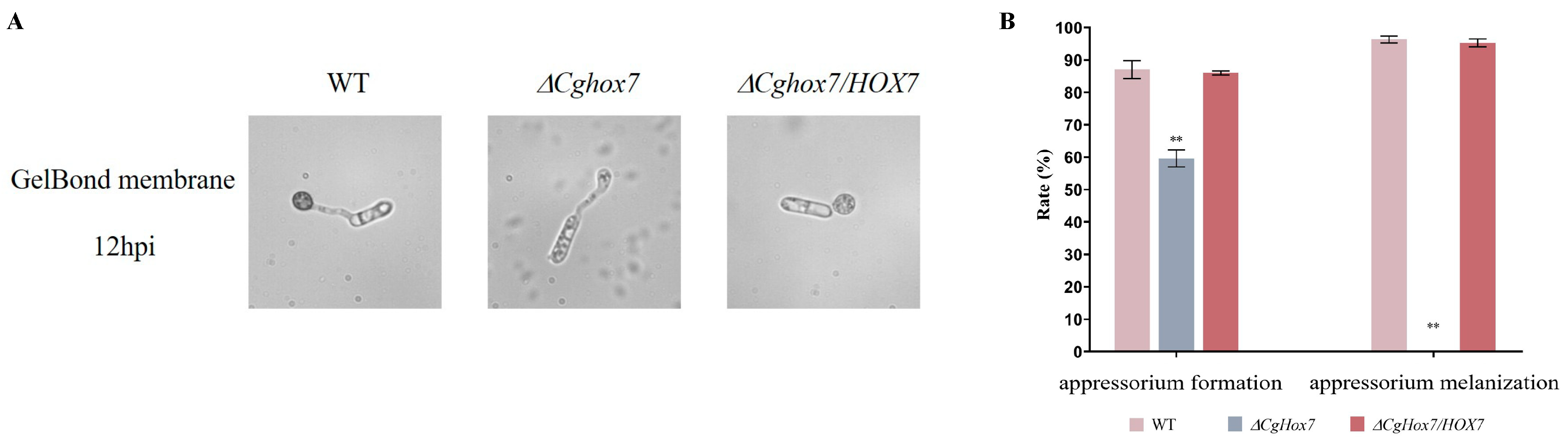
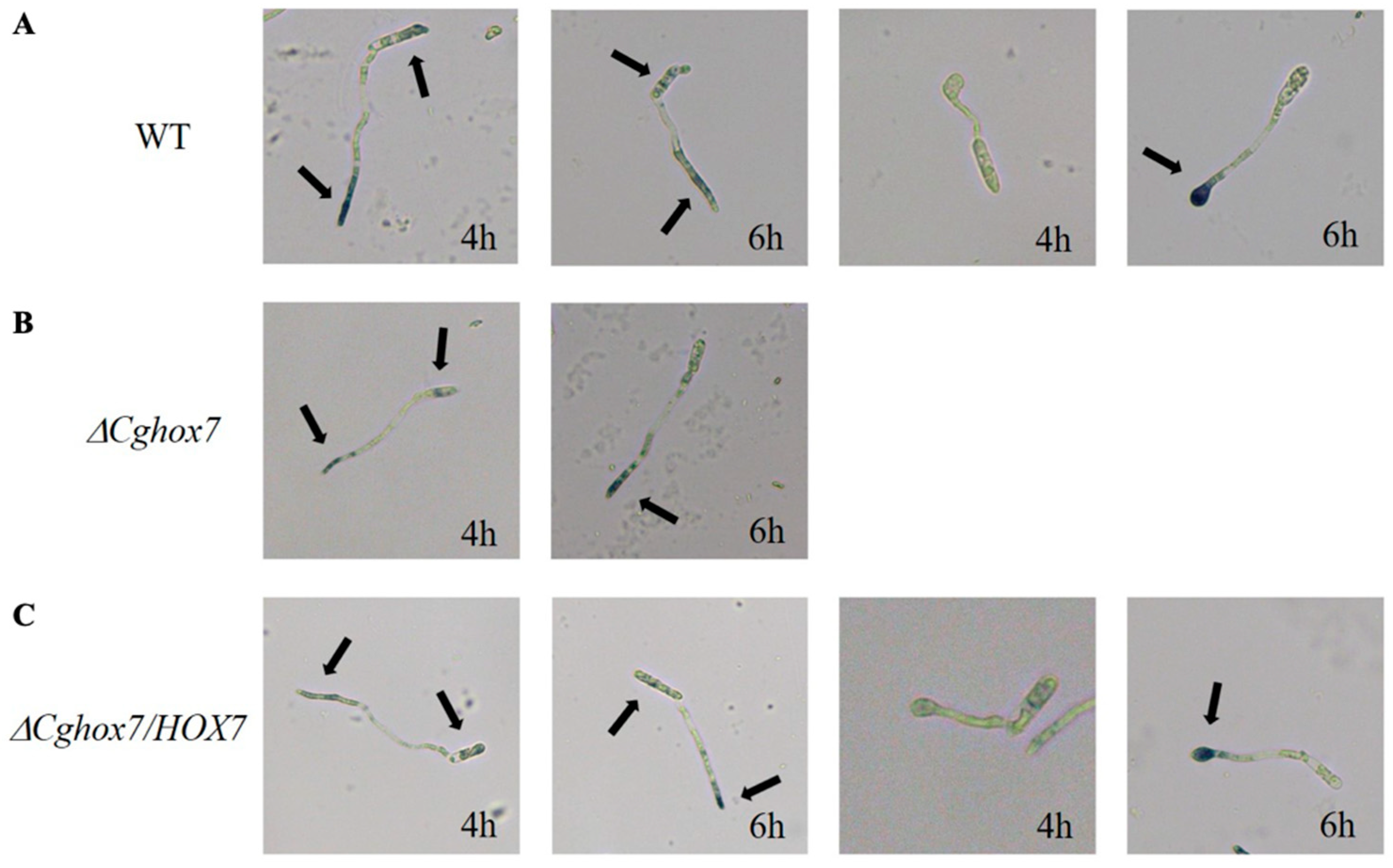
3.3. CgHox7 Is Essential for Appressorium Formation and Maturation

During an infection of susceptible plants, C. gloeosporioides forms an appressorium, which is a specialized infection structure. In this study, the WT strain formed numerous appressoria on the hydrophobic surface of a GelBond membrane (appressorium formation rate of 87%) at 12 h post-inoculation (hpi), with a melanization rate of 96.3%. The appressorium formation rate (86%) and melanization rate (95.2%) of the ΔCgHox7/HOX7 complementation strain did not differ significantly from the corresponding WT rates. In contrast, at 12 hpi, only some of the ΔCgHox7 mutant conidia germinated to form an appressorium (59.7%), while the rest generated slender hyphal structures (Figure 3A,B). The appressoria of the ΔCgHox7 mutant were significantly smaller than those of the WT strain and were not melanized, indicating that they were in the early appressorium formation stage (Figure 3B). These results imply that CgHox7 plays a crucial role in regulating appressorium formation and maturation.

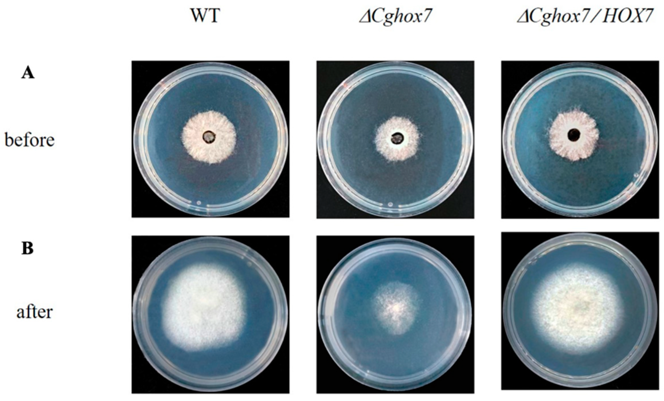
3.4. CgHox7 Regulates the Penetration Ability of Hyphae

To determine whether CgHox7 influences the penetration ability of hyphae, the surface of a cellophane membrane placed on top of solid PDA medium in plates was inoculated with the WT, ΔCgHox7, and ΔCgHox7/HOX7 strains and then incubated at 25 °C for 3 days (Figure 4A). The cellophane membrane was removed and the plates were maintained at 25 °C for an additional 2 days (Figure 4B). Although all three strains were able to penetrate the cellophane membrane, the WT control and the ΔCgHox7/HOX7 complementation strain grew significantly more than the ΔCgHox7 mutant on the PDA medium (Figure 4B). Therefore, CgHox7 is likely involved in regulating the ability of C. gloeosporioides mycelia to penetrate certain materials (e.g., plant host tissue).

3.5. Disruption of CgHox7 Results in a Loss of Virulence

To further investigate the potential effects of CgHox7 on pathogenicity, intact poplar leaves were inoculated with WT, ΔCgHox7, and ΔCgHox7/HOX7 conidial suspensions (2 × 10⁵/mL). At 2–4 days post-inoculation (dpi), necrotic lesions formed and spread at a steady rate on the leaves inoculated with the WT or ΔCgHox7/HOX7 conidial suspension, whereas lesions were undetectable on the leaves inoculated with the ΔCgHox7 conidial suspension (Figure 5A). The decreased virulence of the ΔCgHox7 mutant may be related to its weakened ability to penetrate the leaf surface. However, at 5 dpi, necrotic lesions were detected on the leaves inoculated with the ΔCgHox7 conidial suspension (Figure 5A,B). Intact leaves were also inoculated with WT, ΔCgHox7, and ΔCgHox7/HOX7 hyphal blocks. At 1–8 dpi, the lesions on the leaves inoculated with the ΔCgHox7 hyphal block expanded more slowly than the lesions on the leaves inoculated with the WT or ΔCgHox7/HOX7 hyphal block (Figure 5C,D), providing additional evidence of the decreased virulence of the ΔCgHox7 mutant. These results indicate that CgHox7 is required for the full virulence of C. gloeosporioides.

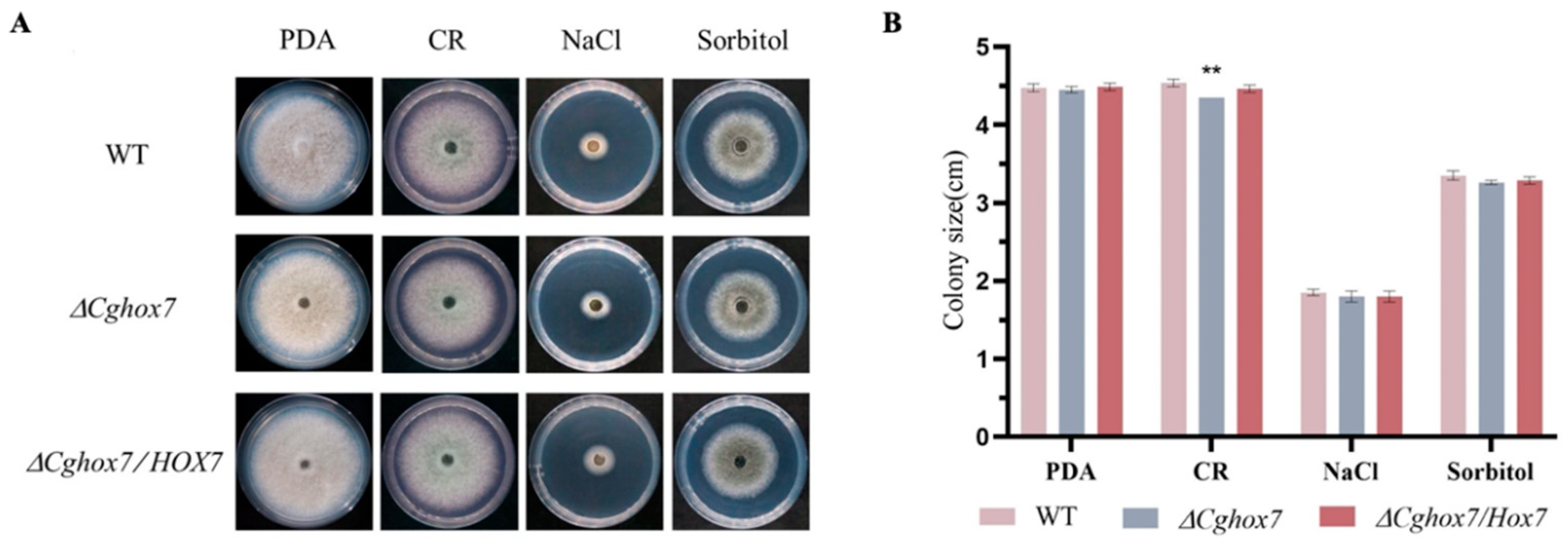
3.6. CgHox7 Is Required for Cell Wall Integrity

A cell wall inhibitor and osmotic stressors were used to determine whether CgHox7 contributes to abiotic stress responses. More specifically, PDA medium containing 100 μg/mL CR, 1.2 M NaCl, or 1 M sorbitol was inoculated with the WT, ΔCgHox7, or ΔCgHox7/HOX7 strain (Figure 6A). The growth rate of these three strains on medium containing 1.2 M NaCl or 1 M sorbitol was determined. There were no major differences in the inhibitory effects of 1.2 M NaCl or 1 M sorbitol on growth among the ΔCgHox7, WT, and ΔCgHox7/HOX7 strains (Figure 6B). Thus, CgHox7 may not be critical for the osmotic stress response of C. gloeosporioides. However, compared with the WT and ΔCgHox7/HOX7 strains, the ΔCgHox7 strain differed significantly in terms of vegetative growth on PDA medium supplemented with 100 μg/mL CR (Figure 6A,B). This result indicates that CgHox7 may be involved in maintaining cell wall integrity.

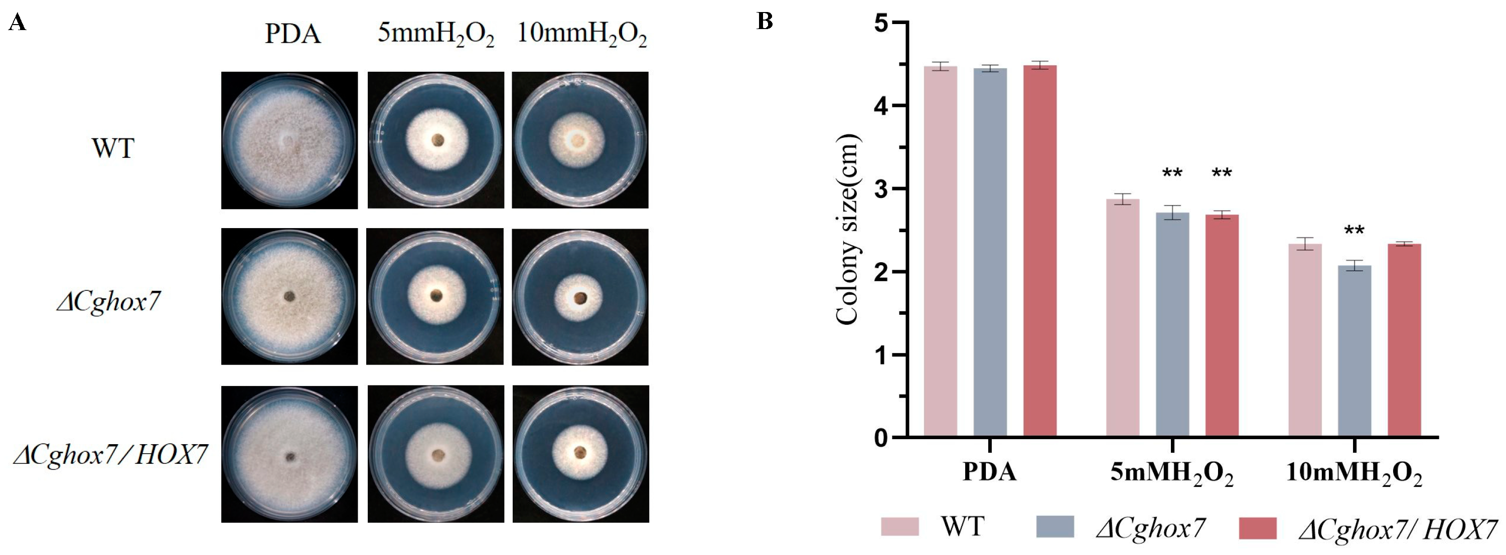
3.7. Deletion of CgHox7 Affects the Oxidative Stress Response

In plants, ROS can directly inhibit the growth of pathogens, thereby providing protection against infections [17]. The rapid accumulation of ROS at the infection site is one of the earliest responses to a pathogen attack. Therefore, a successful infection of a plant host by pathogenic fungi depends on an appropriate oxidative stress response. To determine whether CgHox7 is involved in the response to oxidative stress, we inoculated solid PDA medium containing 5 or 10 mM H2O2 with hyphal blocks of the WT, ΔCgHox7, and ΔCgHox7/HOX7 strains. At 4 dpi, the colony sizes of the three strains were recorded (Figure 7A). The 10 mM H2O2 treatments inhibited the growth of the ΔCgHox7 strain significantly more than the growth of the WT and ΔCgHox7/HOX7 strains (Figure 7B), possibly reflecting the importance of CgHox7 for the oxidative stress response of C. gloeosporioides.

3.8. ROS Accumulation during Conidial Germination Is Affected by the Deletion of CgHox7

Plant pathogenic fungi produce ROS during conidial germination and appressorium formation [18,19]. To assess whether deleting CgHox7 disrupts the accumulation of endogenous ROS during conidial germination, the initial stage of C. gloeosporioides appressorium formation was analyzed by NBT staining, which can reveal the presence of superoxide ions (detected as blue precipitates) [20]. At 4 hpi, endogenous ROS were mainly distributed at the anterior part of the conidia and germ tubes in the WT and ΔCgHox7/HOX7 strains. The WT and ΔCgHox7/HOX7 strains had partially formed appressoria at 4 hpi, but there was relatively little accumulation of ROS in the appressoria and conidia (Figure 8A,C). At the same time point, the ΔCgHox7 strain lacked appressoria. Moreover, ROS accumulated mainly at the anterior part of germ tubes, with a smaller amount within the conidia (Figure 3B). At 6 hpi, ROS also accumulated at the anterior part of WT conidia and germ tubes that had yet to form an appressorium. Most of the WT germ tubes eventually formed appressoria, which accumulated relatively large amounts of ROS (Figure 8A). Similar results were obtained for the ΔCgHox7/HOX7 complementation strain (Figure 8C). However, the ΔCgHox7 strain still lacked appressoria, with ROS mainly concentrated at the anterior part of germ tubes (Figure 8B). These observations indicate that the distribution of endogenous ROS is disrupted during the germination of ΔCgHox7 conidia.

4. Discussion

Homeobox transcription factors are highly conserved in eukaryotes. Notably, their functions have been systematically characterized in several fungi. For example, Hox7, which is a homeobox gene family member, encodes an important regulator of fungal morphology [21]. In the current study, we determined that CgHox7 in C. gloeosporioides is a crucial transcription factor for regulating appressorium formation, cellophane membrane penetration, pathogenicity, cell wall integrity, oxidative stress response, and ROS accumulation during conidial germination.
In M. oryzae, MoHox1–8 are transcription factors significantly affecting functional development and pathogenicity. Mutations to the genes encoding these transcription factors reportedly decrease vegetative growth (ΔMohox1 and ΔMohox6 mutants), disrupt conidial development (ΔMohox2 and ΔMohox4 mutants), or result in no observable phenotypic changes (ΔMohox3 and ΔMohox5 mutants); however, the ΔMohox7 mutant cannot produce appressoria and is non-virulent on intact leaves, but can still grow within plant cells, whereas the ΔMohox8 mutant can form appressoria, but is non-pathogenic because of a defective osmotic stress response [2]. In the present study, the ΔCgHox7 mutant was normal in terms of vegetative growth of hyphae and conidial development (Figure 2 and Figure 3), but the inability of its appressorium to form normally ultimately resulted in decreased penetration and virulence (Figure 4 and Figure 5).
The melanization of germinated conidia expressing HOX7 differs between C. gloeosporioides and M. oryzae. A previous study showed the M. oryzae ΔMohox7 mutant fails to develop appressoria, but forms germ tubes that elongate abnormally during several rounds of swelling and hooking [2]. The C. orbiculare ΔCohox3 mutant (deletion of a CgHox7 homolog) also lacks appressoria, but its conidia germinate to produce lightly pigmented structures that are smaller than WT appressoria and have elongated hyphae [13]. The conidia of the ΔCgHox7 mutant germinated on a GelBond membrane and host leaves, but only 59.7% formed an appressorium at 12 hpi; this appressorium formation rate was significantly lower than the corresponding rate for the WT strain. Moreover, the appressoria were smaller than the WT appressoria and were not melanized, which are characteristics associated with the early stage of appressorium formation (Figure 5). On the basis of this observation combined with the delayed pathogenicity and decreased virulence (i.e., relatively small lesions) of the ΔCgHox7 mutant (Figure 5), we speculate that the deletion of CgHox7 may have detrimental effects on appressorium formation and development.
Pmk1 MAPK, which is homologous to the yeast MAPKs Fus3/Kss1, is important for regulating the differentiation of the mature appressorium and invasive hyphae [22,23,24]. Successive phosphorylation of upstream MAPKKs (Ste11 and Ste7) mediates the activation of MAPK cascades in filamentous fungi, and subsequently activated Pmk1 MAPK can phosphorylate downstream substrates and regulate various functions [25,26]. Earlier research showed that the M. oryzae infection of plants mediated by appressoria is regulated by Pmk1 MAPK and hierarchical transcriptional networks dependent on the related transcription factors Hox7 and Mst12. MoHox7, which is directly targeted by Pmk1 MAPK, is the most critical transcription factor associated with M. oryzae appressorium morphogenesis. Its phosphorylation by Pmk1 MAPK regulates autophagy and cell cycle arrest to promote appressorium development [14]. Additionally, Mst12, which also functions downstream of Pmk1 MAPK, is responsible for regulating appressorium maturation and repolarization. The ΔMomst12 mutant is apparently normal in terms of vegetative growth, conidial germination, appressorium formation, and melanization; however, it is non-pathogenic on rice leaves and exhibits defective growth [27]. The C. gloeosporioides ΔCgste12 (MST12) mutant is phenotypically similar to the ΔMomst12 mutant [12]. Therefore, we speculate that there is also a hierarchical network of transcription factors (including Hox7 and Mst12) regulated by Pmk1 MAPK or additional transcription factors in C. gloeosporioides. Notably, the interaction between Hox7 and Mst12 remains unclear.
In this study, we also revealed the effect of CgHox7 on abiotic stress responses and ROS distribution during appressorium development. Fungus-specific Zn2Cys6 transcription factor genes are involved in regulating growth, asexual development, conidial germination, appressorium formation, pathogenicity, and stress responses [28]. Among these genes, FZC64 and FZC52 are directly regulated by Hox7 [14]. In M. oryzae, both of these genes contribute to multiple stress responses [28]. Therefore, the deletion of CgHox7 likely prevents the activation of Fzc64 and Fzc52. However, in our study, compared with the WT strain, the ΔCgHox7 mutant differed significantly in terms of cell wall integrity and oxidative stress response; it was also unaffected by the 1.2 M NaCl and 1 M sorbitol treatments (Figure 6 and Figure 7). We speculate that there are other transcription factors that control FZC64 and FZC52 expression or other genes associated with osmotic stress responses. Following an infection by pathogenic fungi, plants rapidly release ROS as a defense mechanism [17]. In addition, an oxidative burst at the infection site is one of the earliest plant defense responses. Therefore, the adaptation of pathogenic fungi to oxidants is critical for their virulence. The ΔCgHox7 mutant was adversely affected by oxidants, which may help to explain its decreased virulence (Figure 7). Interestingly, while infecting a plant host, pathogenic fungi also produce ROS derived from NADPH oxidase during conidial germination and appressorium formation. In eukaryotes, ROS are key signaling molecules that can modify signaling proteins to activate the MAPK pathway [29,30,31]. In our study, the deletion of CgHox7 weakened the ability of the mutant to form appressoria, suggesting that the production of endogenous ROS during conidial germination may have been disrupted. Unsurprisingly, at 6 hpi, the ΔCgHox7 mutant still lacked appressoria and ROS continued to accumulate in the anterior part of the germ tube; these characteristics differed significantly from those of the WT strain (Figure 8). Earlier research showed that the thioredoxin gene TRX2 affects intracellular ROS signaling in M. oryzae. Specifically, Trx2 functions upstream of the Pmk1 MAPK pathway; the deletion of the corresponding gene leads to a decrease in the phosphorylation levels of Pmk1 [5]. Therefore, we propose that there is a certain regulatory relationship between CgTRX2 or other unidentified redox-related genes and CgHox7, which may be associated with the observed changes in ROS accumulation in the ΔCgHox7 mutant. Future studies will need to elucidate the relationships among intracellular ROS, Pmk1 MAPK, and CgHox7 as well as the underlying molecular mechanisms (Figure S1).
In conclusion, our study revealed the importance of the transcription factor CgHox7 for C. gloeosporioides appressorium formation, appressorium melanization, penetration, pathogenicity, cell wall integrity, oxidative stress response, and ROS accumulation. Our results clarified the pleiotropic effects of CgHox7 in C. gloeosporioides and suggest that CgHox7 may be a key regulatory factor affecting Pmk1 MAPK.

Supplementary Materials

The following supporting information can be downloaded at: https://www.mdpi.com/article/10.3390/jof10070505/s1. Figure S1: The hypothetical model about Hox7 in Pmk1 MAPK of C. gloeosporioides; Table S1: Primers used in this study.

Author Contributions

F.M. conceived the project. F.M., Q.H. and F.L. designed the experiments. Q.H. and F.L. performed the experiments and analyzed the data. Q.H. and F.L. wrote the manuscript. FM revised the manuscript. All authors have read and agreed to the published version of the manuscript.

Funding

This work was supported by the National Natural Science Foundation of China (32201564), the Young Elite Scientist Sponsorship Program by BAST (BYESS2024298), and the Beijing Forestry University Undergraduate Training Programs for Innovation and Entrepreneurship (X202310022053).

Institutional Review Board Statement

Not applicable.

Informed Consent Statement

Not applicable.

Data Availability Statement

Data are contained within the article or Supplementary Materials.

Conflicts of Interest

The authors declare that the research was conducted in the absence of any commercial or financial relationships that could be construed as potential conflicts of interest.

References

  1. Flaishman, M.A.; Hwang, C.S.; Kolattukudy, P.E. Involvement of protein phosphorylation in the induction of appressorium formation in Colletotrichum gloeosporioides by its host surface wax and ethylene. Physiol. Mol. Plant Pathol. 1995, 47, 103–117. [Google Scholar] [CrossRef]
  2. Kim, S.; Park, S.Y.; Kim, K.S.; Rho, H.S.; Chi, M.H.; Choi, J.; Park, J.; Kong, S.; Park, J.; Goh, J.; et al. Homeobox transcription factors are required for conidiation and appressorium development in the rice blast fungus Magnaporthe oryzae. PLoS Genet. 2009, 5, e1000757. [Google Scholar] [CrossRef] [PubMed]
  3. Li, Z.; Liang, Y.M.; Tian, C.M. Characterization of the causal agent of poplar anthracnose occurring in the Beijing region. Mycotaxon 2012, 120, 277–286. [Google Scholar] [CrossRef]
  4. Kleemann, J.; Rincon-Rivera, L.J.; Takahara, H.; Neumann, U.; Van Themaat, E.V.; van Der Does, H.C.; Hacquard, S.; Stüber, K.; Will, I.; Schmalenbach, W.; et al. Sequential delivery of host-induced virulence effectors by appressoria and intracellular hyphae of the phytopathogen Colletotrichum higginsianum. PLoS Pathog. 2012, 8, e1002643. [Google Scholar] [CrossRef]
  5. Zhang, X.L.; Zhang, J.; He-Pu, H.Z.; Wang, X.L.; Tian, C.M. Histopathology study of poplar leaves infected by Colletotrichum gloeosporioides. J. Beijing For. Univ. 2018, 40, 101–109. [Google Scholar]
  6. Jiang, C.; Zhang, X.; Liu, H.; Xu, J.-R. Mitogen-activated protein kinase signaling in plant pathogenic fungi. PLoS Pathog. 2018, 14, e1006875. [Google Scholar] [CrossRef] [PubMed]
  7. Sakulkoo, W.; Osés-Ruiz, M.; Garcia, E.O.; Soanes, D.M.; Littlejohn, G.R.; Hacker, C.; Correia, A.; Valent, B.; Talbot, N.J. A single fungal MAP kinase controls plant cell-to-cell invasion by the rice blast fungus. Science 2018, 359, 1399–1403. [Google Scholar] [CrossRef] [PubMed]
  8. Marroquin-Guzman, M.; Wilson, R.A. GATA-dependent glutaminolysis drives appressorium formation in Magnaporthe oryzae by suppressing TOR inhibition of cAMP/PKA signaling. PLoS Pathog. 2015, 11, e1004851. [Google Scholar] [CrossRef]
  9. Huang, P.; Wang, J.; Li, Y.; Wang, Q.; Huang, Z.; Qian, H.; Liu, X.-H.; Lin, F.-C.; Lu, J. Transcription factors Vrf1 and Hox7 coordinately regulate appressorium maturation in the rice blast fungus Magnaporthe oryzae. Microbiol. Res. 2022, 263, 127141. [Google Scholar] [CrossRef]
  10. He, P.; Wang, Y.; Wang, X.; Zhang, X.; Tian, C. The mitogen-activated protein kinase CgMK1 governs appressorium formation, melanin synthesis, and plant infection of Colletotrichum gloeosporioides. Front. Microbiol. 2017, 8, 2216. [Google Scholar] [CrossRef]
  11. Wang, X.; Lu, D.; Tian, C. Mitogen-activated protein kinase cascade CgSte50-Ste11-Ste7-Mk1 regulates infection-related morphogenesis in the poplar anthracnose fungus Colletotrichum gloeosporioides. Microbiol. Res. 2021, 248, 126748. [Google Scholar] [CrossRef]
  12. Li, F.; Lu, D.; Meng, F.; Tian, C. Transcription factor CgSte12 regulates pathogenicity by affecting appressorium structural development in the anthracnose-causing fungus Colletotrichum gloeosporioides. Phytopathology 2024. [Google Scholar] [CrossRef] [PubMed]
  13. Yokoyama, A.; Izumitsu, K.; Sumita, T.; Tanaka, C.; Irie, T.; Suzuki, K. Homeobox transcription factor CoHox3 is essential for appressorium formation in the plant pathogenic fungus Colletotrichum orbiculare. Mycoscience 2018, 59, 353–362. [Google Scholar] [CrossRef]
  14. Osés-Ruiz, M.; Cruz-Mireles, N.; Martin-Urdiroz, M.; Soanes, D.M.; Eseola, A.B.; Tang, B.; Derbyshire, P.; Nielsen, M.; Cheema, J.; Were, V.; et al. Appressorium-mediated plant infection by Magnaporthe oryzae is regulated by a Pmk1-dependent hierarchical transcriptional network. Nat. Microbiol. 2021, 6, 1383–1397. [Google Scholar] [CrossRef] [PubMed]
  15. Meng, F.; Tian, C. Gene family expansion during the adaptation of Colletotrichum gloeosporioides to woody plants. J. Fungi 2023, 9, 1185. [Google Scholar] [CrossRef] [PubMed]
  16. Wilson, R.A.; Gibson, R.P.; Quispe, C.F.; Littlechild, J.A.; Talbot, N.J. An NADPH-dependent genetic switch regulates plant infection by the rice blast fungus. Proc. Natl. Acad. Sci. USA 2010, 107, 21902–21907. [Google Scholar] [CrossRef] [PubMed]
  17. Apostol, I.; Heinstein, P.F.; Low, P.S. Rapid stimulation of an oxidative burst during elicitation of cultured plant cells: Role in defense and signal transduction. Plant Physiol. 1989, 90, 109–116. [Google Scholar] [CrossRef]
  18. Egan, M.J.; Wang, Z.Y.; Jones, M.A.; Smirnoff, N.; Talbot, N.J. Generation of reactive oxygen species by fungal NADPH oxidases is required for rice blast disease. Proc Natl Acad Sci. 2007, 104, 11772. [Google Scholar] [CrossRef]
  19. Shinogi, T.; Suzuki, T.; Kurihara, T.; Narusaka, Y.; Park, P. Microscopic detection of reactive oxygen species generation in the compatible and incompatible interactions of Alternaria alternata Japanese pear pathotype and host plants. J Gen Plant Pathol. 2003, 69, 7–16. [Google Scholar] [CrossRef]
  20. Voisey, C.R.; Christensen, M.T.; Johnson, L.J.; Forester, N.T.; Gagic, M.; Bryan, G.T.; Johnson, R.D. cAMP signaling regulates synchronised growth of symbiotic Epichloë fungi with the host grass Lolium perenne. Front. Plant Sci. 2016, 7, 1546. [Google Scholar] [CrossRef]
  21. Arnaise, S.; Zickler, D.; Poisier, C.; Debuchy, R. pah1: A homeobox gene involved in hyphal morphology and microconidiogenesis in the filamentous ascomycete Podospora anserina. Mol. Microbiol. 2001, 39, 54–64. [Google Scholar] [CrossRef]
  22. Ruiz-Roldán, M.C.; Maier, F.J.; Schäfer, W. PTK1, a mitogen-activated-protein kinase gene, is required for conidiation, appressorium formation, and pathogenicity of Pyrenophora teres on barley. Mol. Plant-Microbe Interact. 2001, 14, 116–125. [Google Scholar] [CrossRef] [PubMed]
  23. Xu, J.R.; Hamer, J.E. MAP kinase and cAMP signaling regulate infection structure formation and pathogenic growth in the rice blast fungus Magnaporthe grisea. Genes Dev. 1996, 10, 2696–2706. [Google Scholar] [CrossRef] [PubMed]
  24. Gustin, M.C.; Albertyn, J.; Alexander, M.; Davenport, K. MAP kinase pathways in the yeast Saccharomyces cerevisiae. Microbiol. Mol. Biol. Rev. 1998, 62, 1264–1300. [Google Scholar] [CrossRef]
  25. Hamel, L.-P.; Nicole, M.-C.; Duplessis, S.; Ellis, B.E. Mitogen-activated protein kinase signaling in plant-interacting fungi: Distinct messages from conserved messengers. Plant Cell 2012, 24, 1327–1351. [Google Scholar] [CrossRef] [PubMed]
  26. Wilson, R.A.; Talbot, N.J. Under Pressure: Investigating the biology of plant infection by Magnaporthe oryzae. Nat. Rev. Microbiol. 2009, 7, 185–195. [Google Scholar] [CrossRef]
  27. Park, G.; Xue, C.; Zheng, L.; Lam, S.; Xu, J.-R. MST12 Regulates infectious growth but not appressorium formation in the rice blast fungus Magnaporthe grisea. Mol. Plant-Microbe Interact. 2002, 15, 183–192. [Google Scholar] [CrossRef] [PubMed]
  28. Lu, J.; Cao, H.; Zhang, L.; Huang, P.; Lin, F. Systematic analysis of Zn2Cys6 transcription factors required for development and pathogenicity by high-throughput gene knockout in the rice blast fungus. PLoS Pathog. 2014, 10, e1004432. [Google Scholar] [CrossRef] [PubMed]
  29. Kovtun, Y.; Chiu, W.-L.; Tena, G.; Sheen, J. Functional analysis of oxidative stress-activated mitogen-activated protein kinase cascade in plants. Proc. Natl. Acad. Sci. USA 2000, 97, 2940–2945. [Google Scholar] [CrossRef]
  30. McCubrey, J.A.; LaHair, M.M.; Franklin, R.A. Reactive oxygen species-induced activation of the MAP kinase signaling pathways. Antioxid. Redox Signal. 2006, 8, 1775–1789. [Google Scholar] [CrossRef]
  31. Apel, K.; Hirt, H. Reactive oxygen species: Metabolism, oxidative stress, and signal transduction. Annu. Rev. Plant Biol. 2004, 55, 373–399. [Google Scholar] [CrossRef] [PubMed]
Figure 1. Phylogenetic analysis of CgHox7 and orthologs and deletion of CgHox7. (A) A neighbor-joining phylogenetic tree was constructed according to the amino acid sequences of representative fungal homeodomain transcription factors and CgHox7; domains were predicted using the InterProScan 5.0 tool. The numbers at nodes represent the percentage of their occurrence in 10,000 bootstrap replicates. (B) Schematic diagram of the CgHox7 knockout vector constructed using the split-marker method. (C) Agarose gel electrophoresis analysis of upstream and downstream flanking sequences fused with two-thirds of the Hph cassette. (D) CgHox7 deletion mutant verified by a PCR amplification using internal primers. (E) CgHox7 deletion mutant verified by a PCR amplification using external primers. (F) CgHox7 complementary strain verified by a PCR amplification using internal primers.
Jof 10 00505 g001
Figure 2. Effect of deleting CgHox7 on C. gloeosporioides vegetative growth. (A) Colonies of WT, ΔCgHox7, and ΔCgHox7/HOX7 strains grown on PDA medium in plates at 25 °C for 4 days. The lower panel presents the front, back, and side of the colonies of the indicated strains. (B) Bar chart of the colony sizes of the strains presented in (A). Error bars represent the standard deviation.
Jof 10 00505 g002
Figure 3. Regulatory effects of CgHox7 on appressorium formation and maturation. (A) Conidia of the WT, ΔCgHox7, and ΔCgHox7/HOX7 strains germinated on the hydrophobic surface of a GelBond membrane. (B) Bar chart of the appressorium formation rate and appressorium melanization rate for the strains presented in (A). Data were analyzed using Duncan’s multiple range test. ** indicate statistically significant differences (p < 0.05).
Jof 10 00505 g003
Figure 4. Cellophane membrane penetration assay. Three strains were grown on cellophane membranes overlaid on solid PDA medium in plates for 3 days at 25 °C ((A) before). After the cellophane membrane was removed, the plates were incubated at 25 °C for 2 days ((B) after).
Jof 10 00505 g004
Figure 5. Pathogenicity assay on poplar leaves. (A) Intact leaves were inoculated with equal volumes (30 µL) of conidial suspensions (2 × 10⁵ conidia/mL) from WT, ΔCgHox7, and ΔCgHox7/HOX7 strains and then incubated at 25 °C under humid conditions. The inoculated leaves were photographed at 2–6 dpi. (B) Bar chart of the lesion sizes of leaves 6 days after being inoculated with WT, ΔCgHox7, and ΔCgHox7/HOX7 conidial suspensions. Error bars represent the standard deviation. Data were analyzed using Duncan’s multiple range test. ** indicate statistically significant differences (p < 0.05). (C) Intact leaves were inoculated with 5 mm hyphal blocks of WT, ΔCgHox7, and ΔCgHox7/HOX7 strains and then incubated at 25 °C under humid conditions. The inoculated leaves were photographed at 8 dpi. (D) Bar chart of the lesion sizes of leaves 8 days after being inoculated with WT, ΔCgHox7, and ΔCgHox7/HOX7 hyphal blocks. Error bars represent the standard deviation. Data were analyzed using Duncan’s multiple range test. ** indicate statistically significant differences (p < 0.05).
Jof 10 00505 g005
Figure 6. Responses to abiotic stresses. (A) Hyphal blocks of the WT, ΔCgHox7, and ΔCgHox7/HOX7 strains were used for the inoculation of solid PDA medium containing 100 μg/mL CR, 1.2 M NaCl, or 1 M sorbitol (final concentrations) in plates, which were photographed at 4 dpi. (B) Bar chart of the colony sizes of the strains on solid PDA medium and solid PDA medium containing 100 μg/mL CR, 1.2 M NaCl, or 1 M sorbitol. Error bars represent the standard deviation. Data were analyzed using Duncan’s multiple range test. ** indicate statistically significant differences (p < 0.05).
Jof 10 00505 g006
Figure 7. Response to oxidative stress induced by H2O2. (A) Hyphal blocks of the WT, ΔCgHox7, and ΔCgHox7/HOX7 strains were used for the inoculation of solid PDA medium or solid PDA medium containing 5 or 10 mM H2O2 in plates, which were photographed at 4 dpi. (B) Bar chart of the colony sizes of the strains under H2O2-induced oxidative stress conditions. Error bars represent the standard deviation. Data were analyzed using Duncan’s multiple range test. ** indicate statistically significant differences (p < 0.05).
Jof 10 00505 g007
Figure 8. Distribution of intracellular ROS during conidial germination. The hydrophobic surface of a GelBond membrane was inoculated with equal volumes (30 μL) of conidial suspensions (2 × 10⁴ conidia/mL) from (A) WT, (B) ΔCgHox7, and (C) ΔCgHox7/HOX7 strains. Water was replaced with 30 μL 0.05% (w/v) NBT dissolved in 0.05 M sodium phosphate, pH 7.5, at 4 and 6 hpi. After 2 h, the reaction was stopped by replacing the NBT solution. Black arrows indicate the accumulation of intracellular ROS (blue precipitate).
Jof 10 00505 g008
Disclaimer/Publisher’s Note: The statements, opinions and data contained in all publications are solely those of the individual author(s) and contributor(s) and not of MDPI and/or the editor(s). MDPI and/or the editor(s) disclaim responsibility for any injury to people or property resulting from any ideas, methods, instructions or products referred to in the content.

