Article

The Arrestin-like Protein palF Contributes to Growth, Sporulation, Spore Germination, Osmolarity, and Pathogenicity of Coniella vitis

1
Shandong Academy of Grape, Shandong Academy of Agricultural Sciences, Jinan 250100, China
2
School of Landscape and Ecological Engineering, Hebei University of Engineering, Handan 056038, China
*
Author to whom correspondence should be addressed.
These authors contributed equally to this work.
J. Fungi 2024, 10(7), 508; https://doi.org/10.3390/jof10070508
Submission received: 14 May 2024 / Revised: 19 July 2024 / Accepted: 19 July 2024 / Published: 22 July 2024

Abstract

:
Coniella vitis is a dominant phytopathogen of grape white rot in China, significantly impacting grape yield and quality. Previous studies showed that the growth and pathogenicity of C. vitis were affected by the environmental pH. Arrestin-like protein PalF plays a key role in mediating the activation of an intracellular-signaling cascade in response to alkaline ambient. However, it remains unclear whether palF affects the growth, development, and virulence of C. vitis during the sensing of environmental pH changes. In this study, we identified a homologous gene of PalF/Rim8 in C. vitis and constructed CvpalF-silenced strains via RNA interference. CvpalF-silenced strains exhibited impaired fungal growth at neutral/alkaline pH, accompanied by reduced pathogenicity compared to the wild-type (WT) and empty vector control (CK) strains. The distance between the hyphal branches was significantly increased in the CvpalF-silenced strains. Additionally, CvpalF-silenced strains showed increased sensitivity to NaCl, H2O2, and Congo red, and decreased sensitive to CaSO4. RT-qPCR analysis demonstrated that the expression level of genes related to pectinase and cellulase were significantly down-regulated in CvpalF-silenced strains compared to WT and CK strains. Moreover, the expression of PacC, PalA/B/C/F/H/I was directly or indirectly affected by silencing CvpalF. Additionally, the expression of genes related to plant cell wall-degrading enzymes, which are key virulence factors for plant pathogenic fungi, was regulated by CvpalF. Our results indicate the important roles of CvpalF in growth, osmotolerance, and pathogenicity in C. vitis.

1. Introduction

Grape white rot is a destructive disease in grape-growing regions worldwide, particularly in warm and rainy climates, leading to 20–30% production loss and 30–50% economic loss [1,2,3]. The phytopathogens responsible for the disease include Coniella diplodiella, C. diplodiopsis, C. fragariae, Pilidiella castaneicola, and C. vitis [1,4,5]. C. vitis has been reported as the dominant phytopathogen of grape white rot in China, causing typical symptoms on fruit, shoot, and leaves. Diseased fruit and leaves are covered with dense and black pycnidia [6,7,8]. Previous research has highlighted the acidic environment as a key aspect of the pathogenicity of C. vitis, with the ability to produce various organic acids and cell wall-degrading enzymes, which are crucial for its virulence [9].
Environmental pH is a key factor that influences the growth, development, secondary metabolism, and virulence of the phytopathogen. Fungal plant pathogens can successfully identify and invade hosts by responding quickly and effectively to environmental pH changes. The pathogenicity of Fusarium oxysporum on strawberry roots is higher in acidic soil than in neutral soil [10]. Penicillium expansum enhances its colonization ability on apples by sensing organic acids secreted by apples [11]. Colletotrichum gloeosporioides can increase its pathogenicity to avocados by secreting ammonia to alkalize the surrounding environment [12]. C. vitis exhibits rapid growth and heightened virulence in acidic environments, particularly around pH 3, compared to neutral or alkaline conditions [9]. Additionally, it is notable that the pH of table and wine grape leaves and fruits ranges from 2.79 to 3.77 [9,13]. Therefore, it is reasonable to speculate that pH is an important factor in the infection of grapes by C. vitis.
Phytopathogens have evolved various mechanisms to adapt to diverse environmental conditions, including the Pal/Rim–pH pathway. This pathway comprises several key proteins, including PalA/Rim20, PalB/Rim13, PalC/Rim23, PalF/Rim8, PalH/Rim21, PalI/Rim9, and the transcription factor PacC/Rim101 [14,15]. This Pal/Rim–pH pathway plays a crucial role in sensing and responding to changes in environmental pH. PacC modulates a wide range of phenotypes, including mycelial growth, spore germination, sporulation, and virulence. The loss of PePacC leads to a noticeable decrease in growth and conidiation of P. expansum and Trichothecium roseum, as well as reduced pathogenicity [16,17]. Under neutral and alkaline conditions, the ambient pH signal is sensed by PalH, which subsequently facilitates the ubiquitination of the arrestin PalF [18,19,20]. Ubiquitinated PalF triggers and activates the proteolysis of PacC/Rim101 in response to alkaline pH, resulting in the generation of PacC27 (kDa) by two successive proteolytic cleavages from PacC72 (kDa). PacC27 then localizes to the nucleus to modulate gene expression [19,21]. PalF/Rim8 can promote Pal/Rim–pH pathway activation in Saccharomyces cerevisiae, Aspergillus nidulans, and Candida albicans, but there are significant differences in PalF activity among these model organisms [22,23]. For example, Rim8 ubiquitination is pH-independent in S. cerevisiae but pH-dependent in A. nidulans [24]. However, it remains unclear whether palF affects the growth, development, and virulence of fungi in response to changes in environmental pH. Although the interaction between PalF and the C-terminal cytoplasmic tail of PalH highlights the role of PalF as a mediator of pH signaling within the cell, the role of PalF in the growth, development, and virulence of fungi needs further investigation.
Previous research identified CvpalF in C. vitis as a homologue of PalF/Rim8, with its expression down-regulated at alkaline pH [9]. The present study (i) compares the sequence identity and genetic relationship between CvpalF and those of other pathogenic fungi; (ii) silences the CvpalF gene via RNA interference; (iii) elucidates the effects of CvpalF on growth, development, and stress response under different pH conditions and pathogenicity; and (iv) demonstrates the regulation of CvpalF on genes in Pal-signaling pathways and other C. vitis virulence genes. These findings provide new insights into the pathogenic mechanism of C. vitis regulated by pH.

2. Materials and Methods

2.1. Strains, Culture Conditions, DNA Extraction, and Bioinformatics Analysis

C. vitis GP1 (strain no. 23888) is a highly virulent strain causing grape white rot, and presently preserved in the China General Microbiological Culture Collection Center [7]. The wild-type strain C. vitis GP1 and transformants generated in this study were cultured on potato dextrose agar (PDA, Difco, Detroit, MI, USA, pH = 6.5) plates at 28 °C in the dark for 3 days and potato dextrose broth (PDB) media at 28 °C, in the dark, and at 180 rpm.
For genomic DNA extraction, the fungal strains were grown on PDA (28 °C, darkness) for 3 days on the surface of cellophane membranes inoculated with 4-mm mycelial plugs. Genomic DNA was extracted with the DNeasy Plant Mini Kit (Qiagen, Hilden, Germany) according to the manufacturer’s instructions.
The genomic DNA of strain C. vitis GP1 was sequenced at Novogene Bioinformatics Technology Co., Ltd. (Nanjing, China) using a combination of third-generation PacBio and second-generation Illumina. The sequence data for CvpalF were extracted from the annotation results and are presented in Text S1. Homologous sequences of PalF protein from other fungal species were downloaded from the NCBI database. Multiple sequence alignment and phylogenetic tree construction were performed using MEGA 7.0 with the maximum likelihood clustering method. Structural domains of PalF proteins were predicted using the online biology tool in NCBI (https://www.ncbi.nlm.nih.gov/Structure/cdd/wrpsb.cgi?RID=XG0R7TS6016&mode=all accessed on 2 February 2024).

2.2. Construction of the CvpalF-Silenced Strains

2.2.1. Hygromycin Sensitivity

C. vitis GP1 was cultured on PDA for 3 days at 28 °C in the dark. Following this, mycelial plugs (5 mm diameter) were transferred to fresh PDA-containing hygromycin B (Roche) at concentrations of 0, 1, 5, 10, 50, 100, 200, and 300 μg/mL. The plates were incubated at 28 °C in the dark for 3 d. The diameters of the developed colonies were measured.

2.2.2. Construction of the CvpalF-Silencing Plasmid

pSlient-1 was used to construct the gene CvpalF-silencing vector. Two reverse-complementary CvpalF-silencing targets were amplified using the primers of palF1/2 and palF3/4 (Table 1). Firstly, the restriction endonucleases XhoI (New England Biolabs, Beijing, China) were used to digest at 37 °C and the CvpalF sense forward fragment was inserted into the restriction sites by one step of the Cloning kit (Vazyme, Nanjing, China). Secondly, the restriction endonucleases Kpn I (New England Biolabs, Beijing, China) was used to digest, and the CvpalF sense reverse-complementary fragment was inserted into the corresponding restriction site, yielding the final silencing vector pCvpalFi (Figure S1). Subsequently, the resultant constructs were introduced into E. coli DH5α, and positive clones were chosen for PCR identification with primers LFcheck 1F/R, LFcheck 2F/R (Table 1), and sequencing to ensure the validation of their accuracy.

2.2.3. Transformation Procedure

The spore of C. vitis GP1 was cultured in the yeast extract peptone dextrose (YEPD) media (with 10% grape juice) at 28 ℃ in the dark for 20 h with 180 rpm for preparation of protoplast according to the method described by Zou [25]. The silencing vector pCvpalFi was transferred into the C. vitis GP1 strain by PEG-mediated protoplast transformation [26,27]. Transformants were selected and cultured on PDA supplemented with hygromycin B (200 μg/mL) for three successive subcultures. After extraction using the EZ-DNAaway RNA Mini-Preps Kit (Sangon Biotech, Shanghai, China) according to the manufacturer’s protocol, CvpalF-silenced strains were confirmed by PCR amplification with LFcheck 1F/R, LFcheck 2F/R, and Fhyg/Rhyg primers (Table 1). The PCR cycling conditions were as follows: initial denaturation at 95 °C for 3 min; 34 cycles of denaturation at 95 °C for 30 s, annealing at 58 °C for 30 s, and extension at 72 °C for 1 min; followed by a final extension step at 72 °C for 10 min.

2.2.4. Gene-Expression Analysis

The wild-type (WT) strain C. vitis GP1, vector control (CK) strain, and CvpalF transformants CvpalFi-17 and CvpalFi-29 were cultured in PDB media of pH 3, 5, 7, and 9 for 3 days at 28 °C, in the dark, 180 rpm. The pH of the PDA medium was adjusted using 0.2 M Na2HPO4·12H2O, 0.1 M C6H8O·7H2O and NaOH as described previously [9]. Total RNA was extracted using EZ-10 DNAaway RNA Mini-Preps Kit (Sangon Biotech, Shanghai, China) according to the protocol. RNA samples (1 μg) were reverse transcribed using the HiScript III 1st Strand cDNA Synthesis Kit (+gDNA wiper, Vazyme, Nanjing, China). The primers of palF gene designed for quantitative real-time PCR (qRT-PCR) were listed in Table S1. qRT-PCR analysis was performed in a two-step method using Taq Pro Universal SYBR qPCR Master Mix (Vazyme, Nanjing, China) in a CFX-96 real-time PCR detection system (Bio-Rad, Hercules, CA, USA). The qRT-PCR mixture (20 μL) consisted of 10 μL of 2 × qPCR Master Mix, 0.4 μL of each primer (10 μM), and 2 μL of cDNA. The qRT-PCR conditions were as follows: 95 °C for 30 s, followed by 40 cycles of 95 °C for 3 s and 60 °C for 20 s. The actin gene was used as the reference gene and relative expression levels were measured using the 2(−ΔΔCt) analysis method [28]. All the experiments were performed with three technical replicates of three biological replicates.

2.3. Silenced Strains Characterization

2.3.1. Growth under Different pH

On Solid Media

Strains WT, CK, and CvpalF transformants CvpalFi-17 and CvpalFi-29 were cultured on PDA and amended to pH 3, 5, 7, and 9. The pH was adjusted as described by Yuan [9]. After 3 days of incubation (28 °C in the dark), the colony diameters were measured, and the colony edges were observed by a light microscope (Optika B-383PL, Ponteranica, Italy). The WT, CK, CvpalFi-17, and CvpalFi-29 strains were grown on PDA supplemented with a pH indicator (0.005% Bromothymol blue) at 28 °C, in the dark, after 3 days to observe the color of the PDA. The experiment was performed with three biological replicates and repeated three times.

In Liquid Media

Spore suspension of strains WT, CK, and CvpalF transformants (CvpalFi-17 and CvpalFi-29) was prepared as described previously. PDB (potato dextrose broth) (100 mL, pH = 6.5) inoculated with 1 mL spore suspension (1 × 10⁶ spore/mL) was cultured at 28 °C, darkness, at 180 rpm. After 3 days, the mycelia were harvested by filtering through four layers of cheesecloth, dried at 60 °C, and weighted with an analytical balance to determine the mycelia biomass. The pH of each cuture was measured every day with a pH meter (Sartorius, PB-10).

2.3.2. Spore Production and Vitality

Strains WT, CK, and CvpalF transformants (CvpalFi-17 and CvpalFi-29) were cultured on PDA at pH3, 5, 7, and 9 for 14 days at 28 °C in the dark. Spores were collected by washing each colony surface with 40 mL of 0.05% (v/v) Tween 80 (Sigma-Aldrich, Copenhagen, Denmark) in distilled water. Suspensions were filtered on miracloth (Calbiochem) into 50 mL tubes. The spore concentrations were determined by hemocytometer, and spore morphology was observed under light microscopy (Optika B-383PL, Ponteranica, Italy). Spore vitality was verified as proposed by Yuan et al. [9].

2.3.3. Pathogenicity

Mycelial plugs (5 mm in diameter) taken from 3-day-old C. vitis strains WT, CK, and CvpalF transformant (CvpalFi-17 and CvpalFi-29) colonies on PDA were used to inoculate detached grape leaves (cv. Red Globe). Detached grape leaves inoculated with PDA plugs served as a control. Inoculated leaves were incubated in a greenhouse at 28 °C, 90% RH, and under natural photoperiod. The spot diameters on the leaves were determined at 1, 2, and 3 dpi (days post inoculation). The reduced rate = [the lesion diameter of the WT strain—the lesion diameter of transformants]/the lesion diameter of WT strain × 100%). The experiment was repeated three times with three biological replicates.

2.3.4. Effects of Osmotic Stress

The effects of CvPalF on osmotic stress adaptability on PDA amended with KCl (1 M), NaCl (1 M), CaSO4 (1 M), MgSO4 (1 M), H2O2 (1 M), or Congo red (1 M). Plates were inoculated with a mycelial plug (5 mm in diameter) cut from 3-day-old C. vitis strains WT, CK, CvpalFi-17, and CvpalFi-29 cultures on PDA (28 °C, in the dark). After 7 days of incubation (28 ℃, in the dark) the colony diameter was measured. Inhibition rate = [the diameter of the WT strain—the diameter of transformants]/the diameter of WT strain × 100%.

2.3.5. Virlence Gene Expression

Conidia of C. vitis strains WT, CK, CvpalFi-17, and CvpalFi-29 were inoculated in 20 mL of PDB with a pH of 3 to obtain a final concentration of 3 × 10⁶ spores/mL. Flasks were incubated at 28 °C, in the dark, with shaking at 180 rpm for 24 h. Then, the cultures were shifted to PDB with pH values of 3 and pH 9 for further 15, 30, 60, or 120 min according to the method of Hervas–Aguilar et al. [29]. Subsequently, mycelia were harvested using four layers of cheesecloth. Total RNA was extracted and conducted as described by Yuan et al. [9] and Mushtaq et al. [30]. qRT-PCR was conducted using the primer pairs of the Pal-signaling pathway and other virulence genes listed in Table S1, and the rection conditions were the same as described in Section 2.2.4 in this study.

2.4. Statistical Analysis

All data in this study were analyzed by SPSS Version 22.0, and one-way ANOVA with Tukey’s test was used for multiple comparison analyses. Significance was accepted at p < 0.05 [31,32]. Each experiment was performed with three biological replicates and repeated three times, and the data were presented as averages ± SE.

3. Results

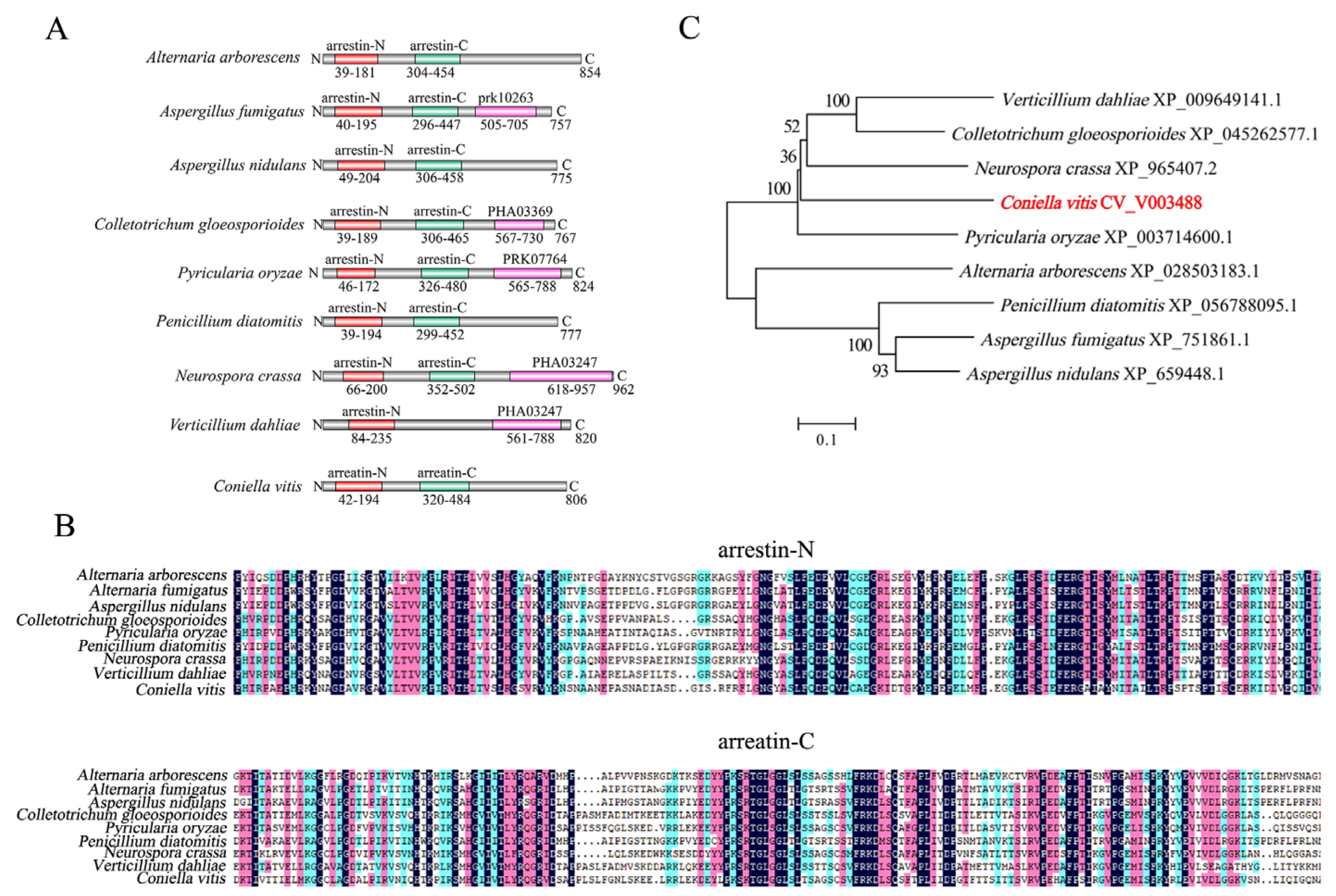
3.1. Bioinformatics Analysis of CvpalF

The CvpalF gene in the C. vitis GP1 genome was 2421 bp long and encoded a total of 806 amino acids (Text S1). The SMART structural domain analysis showed that CvpalF protein contains arrenstin-N and arrestin-C domains located at Positions 49–204 and 306–458, respectively (Figure 1A). These domains showed high conservation in Aspergillus nidulatus, Pyricularia oryzae, and Neurospra crassa (Figure 1B). The phylogenetic tree of PalF proteins (Figure 1C) showed that CvPalF had a relatively close evolutionary relationship with P. oryzae and N. crassa but a distant relationship with A. nidulatus, A. fumigatus, and Penicillium diatomitis.

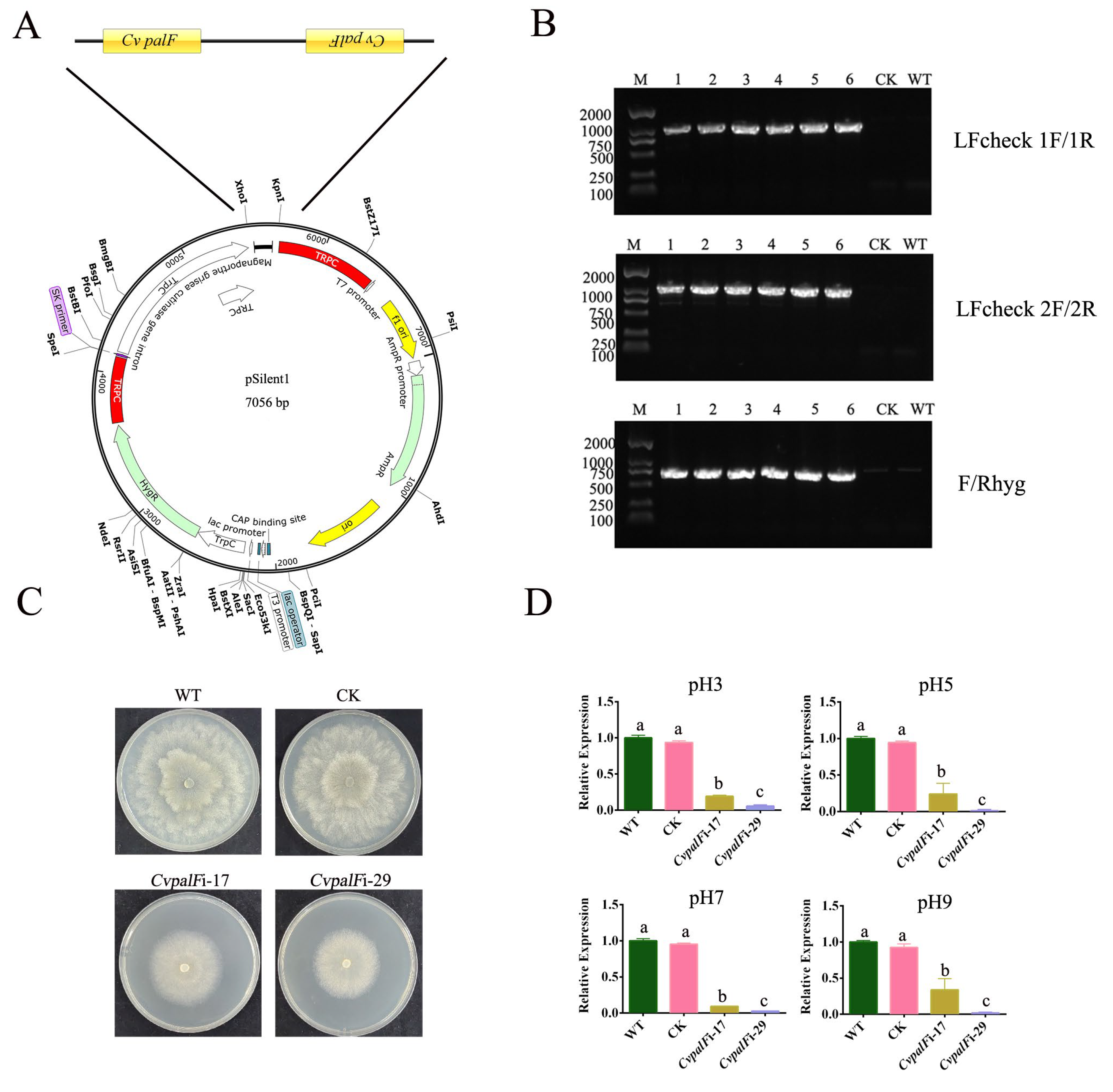
3.2. Construction of CvpalF-Silenced Strains

The pCvpalFi vector and the pSlient-1 vector used as a vacant vector control (Figure 2A, Figure S1) were inserted via PEG-mediated protoplast transformation into WT strain C. vitis GP1. Hygromycin B fully suppressed the growth of C. vitis GP1 at 100 μg/mL (Figure S2). To avoid false positives, Hygromycin B at 200 μg/mL was used for selecting transformants. Six transformants were randomly selected. PCR amplifications with primers LFcheck 1F/1R, LFcheck 2F/2R, and F/Rhyg confirm transformation, yielding bands of 1072, 1164, and 750 bp, respectively (Figure 2B). The expression level of the CvpalF gene was significantly reduced in the CvpalFi-17 and CvpalFi-29 transformants, with a gene-silencing efficiency of 76.17–98.93% (Figure 2C,D). Meanwhile, the expression level of the CvpalF gene was not significantly changed in CK and WT strains.

3.3. Silenced Strains Characterization

3.3.1. The Growth of CvpalF-Silenced Strains Under Different pH Values

CvpalF-silenced strains CvpalFi-17 and CvpalFi-29 exhibited impaired growth compared with WT and CK strains, with significant inhibition under alkaline conditions (Figure 3A). Compared to WT and CK strains, the growth inhibition (Figure 3B) of the two CvpalF silenced strains ranged from 28.48 to 46.81%. Additionally, the colony edges appeared shorter and sparser in CvpalFi-17 and CvpalFi-29 (Figure 3C).
The mycelial biomass of strains CvpalFi-17 and CvpalFi-29 was significantly reduced by the pH of PDB (Figure 3D). The mycelial biomasses of CvpalF-silenced strains CvpalFi-17 and CvpalFi-29 declined 20.86 and 14.75%, 65.71 and 66.94%, and 52.06 and 58.22% at pH 3, 5, and 7, respectively (Figure 3D). However, the mycelial weight of CvpalFi-17 and CvpalFi-29 had no significant reduction when grown in PDB at pH 9 (Figure 3D).

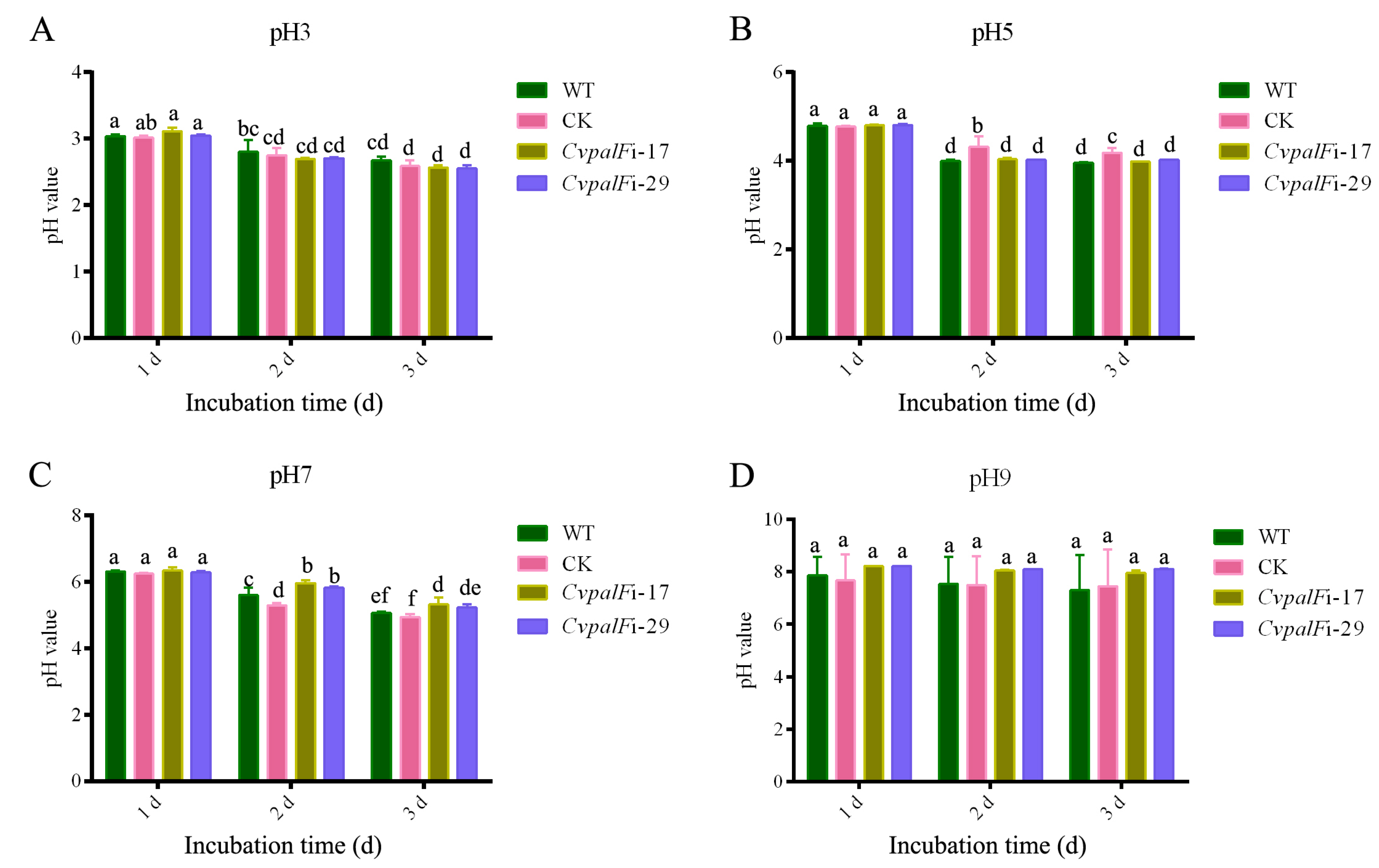
3.3.2. The pH Change of Liquid Media

The pH values of PDB decreased during the cultivation of WT strain, CK, and CvpalF silenced strains CvpalFi-17 and CvpalFi-29, with initial pH values of 3, 5, and 7 (Figure 4A–C). During cultivation with an initial pH of 7, the pH values of PDB for CvpalFi-17 and CvpalFi-29 were slightly higher than those for the WT (Figure 4C). However, there was no significant difference in the pH values of PDB between the WT and the two CvpalF-silenced strains when the initial pH was 9 (Figure 4D). Additionally, the WT demonstrated a stronger capacity to produce acid and decrease the environmental pH compared to CvpalFi-17 and CvpalFi-29 (Figure S3).

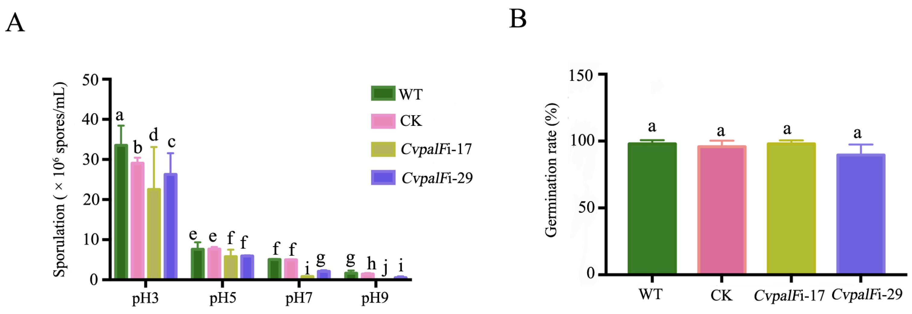
3.3.3. Sporulation and Spore Germination of CvpalF-Silenced Strains under Different pH Values

Sporulation of CvpalF-silenced strains CvpalFi-17 and CvpalFi-29 was reduced by 32.83% and 21.64%, respectively, compared to the WT and CK strains when incubated in PDA with pH 3 at 28 °C for 14 days. In pH 5, their sporulation was reduced by 24.34% and 21.71%, respectively. Moreover, the sporulation of strains CvpalFi-17 and CvpalFi-29 was further reduced by 84.23% and 58.62%, respectively, compared to the WT and CK strains when incubated in PDA media with pH 7. Particularly in alkaline environments, the sporulation ability of transformant CvpalFi-17 was completely lost, while the sporulation of Strain CvpalFi-29 was reduced by 68.61% (Figure 5A). However, CvpalF did not contribute to spore germination, regardless of the environmental pH. The rate of spore germination did not significantly differ between CvpalFi-17, CvpalFi-29, CK, and WT strains (Figure 5B).

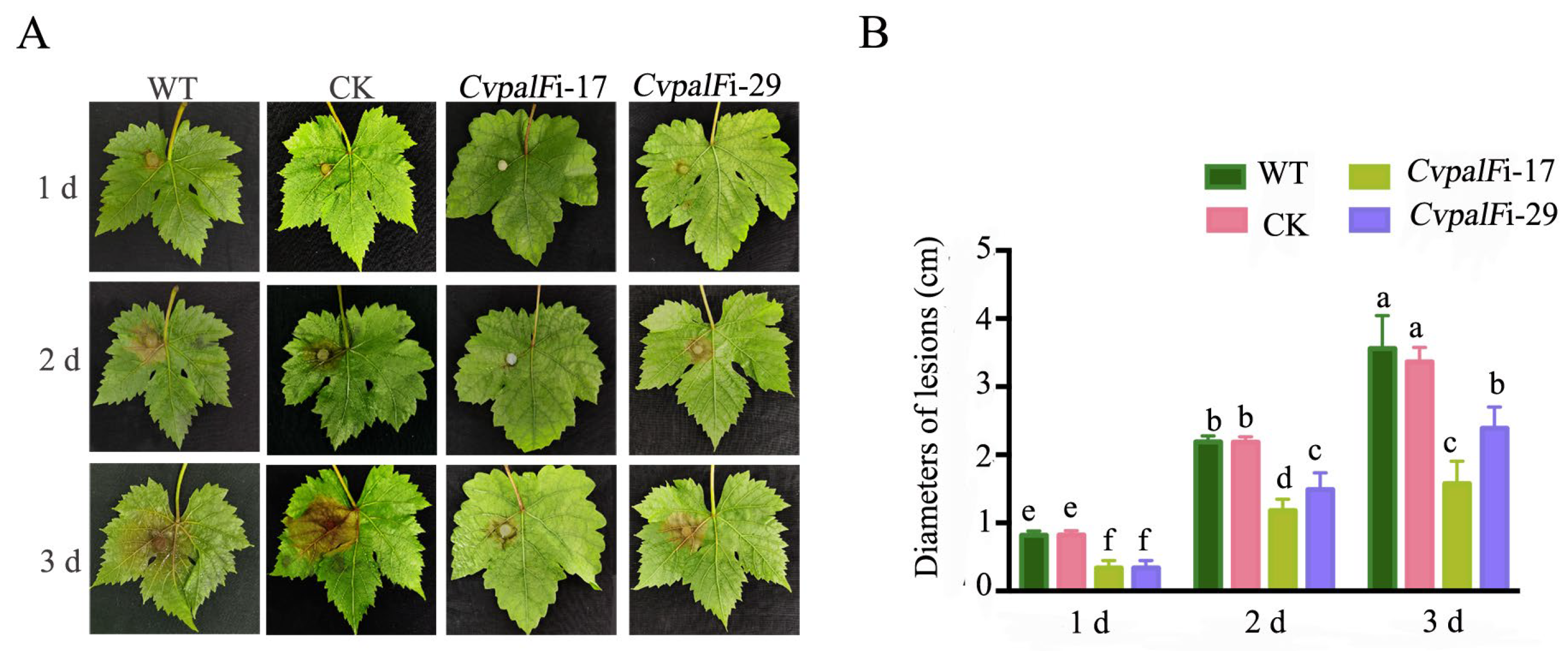
3.3.4. Pathogenicity of CvpalF-Silenced Strains

On detached leaves of cv. Red globe, CvpalF-silenced strains CvpalFi-17 and CvpalFi-29, developed brown spots much slower than Strains WT and CK. Compared with WT and CK, a reduction of spot diameter by 55.75 and 32.87% in strains CvpalFi-17 and CvpalFi-29, respectively (Figure 6A,B and Figure S4).

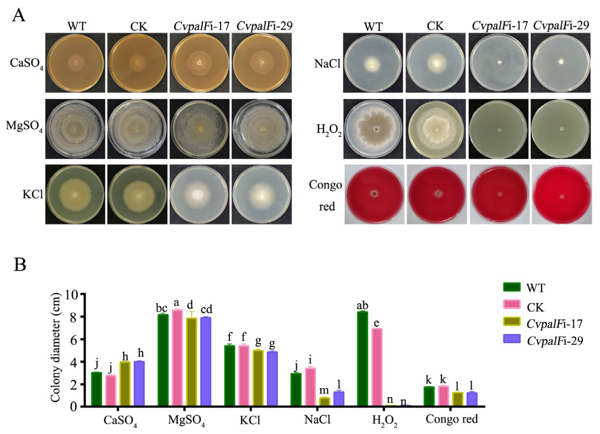
3.3.5. The Responses of CvpalF-Silenced Strains to Cell Wall Integrity, Oxidation, and Ionic Stress

Under cell wall integrity of Congo red and oxidative stress induced by H2O2, the growth of CvpalF-silenced strains CvpalFi-17 and CvpalFi-29 were partly inhibited (Figure 7). CvpalF-silenced strains CvpalFi-17 and CvpalFi-29 were notably more sensitive to 1 M NaCl and exhibited approximately 45–50% greater growth inhibition than the WT and CK strains. While CvpalF-silenced strains CvpalFi-17 and CvpalFi-29 were less sensitive to 1 M KCl and exhibited approximately only 9.81% growth inhibition than the WT and CK strains. Interestingly, CvpalF-silenced strains showed decreased sensitivity to 1 M CaSO4, and the growth of CvpalF-silenced strains was faster than the WT and CK strains. In the presence of 1 M MgSO4, no significant differences (p > 0.05) were found between the CvpalF-silenced strains, WT, and CK strains (Figure 7 and Figure S5).

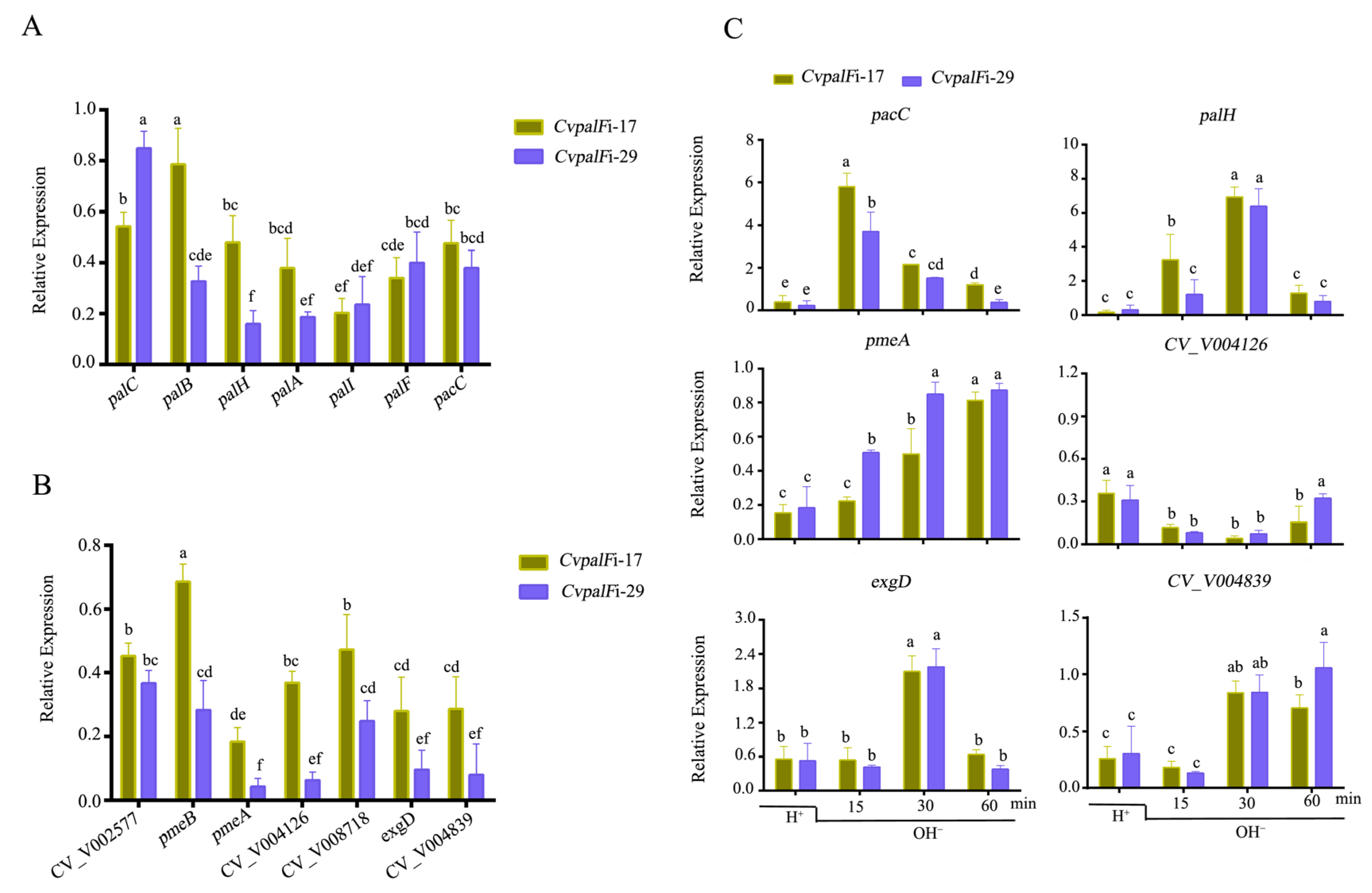
3.3.6. CvpalF Affected the Expression of Genes Related to Pal-Signaling Pathway and Plant Cell Wall-Degrading Enzyme

The expression levels of CvpalA, CvpalB, CvpalC, CvpalH, CvpalI, and CvpacC in CvpalF-silenced strains were significantly down-regulated by approximately 1.27–6.25 folds (Figure 8A). Additionally, genes related to plant cell wall-degrading enzymes, including CvpmeA, CvpmeB, CvpLL (pectin lyase-like protein), CV_V008718, CV_V004839, CV_V009031, and CV_V008957, were significantly down-regulated by approximately 1.46–23.08 folds (Figure 8B). The expression level of CvpacC increased by 3.69–5.80 folds after 15 min when the pH shifted from 3 to 9, while the expression level of CvpalH increased by 6.37–6.93 folds after 30 min under the same pH shift condition (Figure 8C). In addition, the expression level of CvpmeA was up-regulated in CvpalFi-17 and CvpalFi-29 strains when the environment pH shifted from 3 to 9, but it was lower than that in WT. The expression level of CvexgD was increased by 2.09–2.17 folds in CvpalFi-17 and CvpalFi-29 strains compared to WT at 30 min in the pH shift experiment. The expression levels of CV_V004126 and CV_V004839 were lower than that in WT, although the expression level of CV_V004839 increased by 0.84–0.85 folds after 30 min when pH shifted from 3 to 9 (Figure 8C).

4. Discussion

Grape white rot, caused by C. vitis, has been a major concern in grape production and the wine industry [4,33,34]. Unlike most plant pathogens, the host plants of C. vitis reported so far are grapes and Virginia Creeper (Parthenocissus quinquefolia) with a pH value of about 3 [35,36]. Previous studies show that C. vitis grows slowly and includes a loss or reduction of pathogenicity in neutral and alkaline environments [9]. The Pal/Rim 101 signal pathway regulates the adaption to changes in the environmental pH for the survival and development of fungi, and PalF was known to trigger the Pal/Rim 101 signal pathway in response to the change of the environmental pH [20]. In this study, a homologous gene of PalF/Rim8 was identified in C. vitis. The CvpalF-silenced strains CvpalFi-17 and CvpalFi-29 were also obtained using an RNAi approach with the fungal transformation vector pSilent-1 through the PEG-mediated protoplast transformation. The applied method was successful in both Ascomycota and Basidiomycota [37,38,39]. Furthermore, the role of CvpalF in growth, virulence, and response to the different pH conditions was elucidated. Compared with C. vitis WT and CK, CvpalF-silenced strains reduce linear growth and mycelia biomass production while worsening pathogenicity (Figure 3A and Figure 6).
Pathogenic fungi alkalize or acidify the host tissue during infection to ensure their normal growth, development, and pathogenicity [40]. PacC plays a crucial role in the Pal/Rim-signaling pathway. PacC contains three Cys2His2 zinc finger motifs. Under alkaline conditions, PacC72 undergoes a two-step proteolytic process to degrade into PacC27. PacC27 then enters the nucleus and binds to the 5′-GCCARG-3′ sequence in the promoter regions of pH-responsive genes, thereby regulating the expression of these genes [14]. B. cinerea secretes organic acids to acidify some of the host plant tissues during colonization, while the BcpacC deletion mutant revealed a decreased capacity to acidify their environment due to a decrease in the production of oxalic acid [13,41,42]. Previous research indicates that the expression of CvpacC is increased 2.77-fold under alkaline conditions compared to acidic conditions (unpublished), and over 200 genes related to carboxylic acid metabolism are differentially expressed [9]. In this study, we found that C. vitis decreased the environmental pH in vitro, and the capability of acidification of C. vitis in PDB media of pH 5 and pH 7 were slightly declined when CvpalF was silenced (Figure 4B,C). These results suggested that CvpalF sensed the environmental pH but cannot directly regulate the metabolism of organic acids to change the environmental pH. Whether CvpacC is responsible for the ability to secrete organic acids to acidify the environmental pH needs to be verified further.
Previous studies have shown pacC and five pal genes, excluding palI, have proved essential for cellular response to osmotic salts/agents, and their osmoregulative roles are all pH-dependent in many fungi [43,44]. PacC-silenced strains of Ganoderma lucidum were more sensitive to 0.3 M NaCl and KCl [45]. Fusarium oxysporum is an important and diverse soilborne plant pathogen infecting almost 150 plant species while FopacC transformants increased sensitivity to NaCl [46,47]. Fusarium graminearum (Fg), a destructive fungal pathogen of cereal crops, and FgPacC, which can directly bind and inhibit the HAT activity of FgGcn5, subsequently enhance the adaption of F. graminearum to host-derived high-iron stress during infection [26]. In this study, we found that CvpalFi-17 and CvpalFi-29 showed defects in cell walls and were more sensitive to 1 M NaCl compared to WT and CK strains. Our results suggested that palF in the Pal signal pathway was required for osmotolerance adaptation. The mechanisms that respond to ionic pressure in the environment in C. vitis need further elucidation, and whether CvPacC is involved in this process and its role remains to be explained. Additionally, reactive oxygen species burst is an early defense response in most plant hosts during the infection of pathogenic fungi [48,49]. CvpalF-silenced strains exhibited increased sensitivity to H2O2, which might contribute to decline in the virulence of grapes.
Plant cell walls serve as the first line of defense against pathogen infection [50]. Polygalacturonases play an important role in cell wall degrading, and pectin methylesterase (PME) contributes to this progress [51]. Previous studies have shown that the ability of pal/rim mutants to secrete extracellular enzymes is affected. For example, the expression levels of genes encoding chitin synthases (CHS) and glucan synthase (GLS) were reduced in the palB mutant of Ustilago maydis, which was accompanied by changes in the cell wall structure and chemical composition [52]. In Botrytis cinerea and F. graminearum, Bcpme1 and FgPme1 mutants showed lower virulence on host plant tissues, such as apple and grapevine for B. cinerea and wheat for F. graminearum [53,54]. In this study, we found that silencing CvpalF resulted in the decline of the expression levels of genes related to pectinase and cellulase (e.g., genes CvpmeA and CvpmeB) (Figure 7B). The results suggested that CvpalF is involved in regulating the production of cell wall-degrading enzymes in C. vitis, which is important for its virulence.

5. Conclusions

In this study, the role of arrestin-like protein CvpalF was revealed by constructing CvpalF-silenced strains via RNA interference. Our results suggested that CvpalF is required for the growth and sporulation of C. vitis in neutral/alkaline pH conditions. CvpalF-silenced strains exhibited impaired fungal growth and decreased sporulation capacity compared to the WT and CK strains. CvpalF is essential for virulence and adaptability to environmental stresses, such as osmotic stress, oxidative stress, and cell wall stress. In addition, the expression of PacC, PalA/B/C/F/H/I was directly or indirectly regulated by CvpalF. These results contribute to a better understanding of the mechanism underlying Pal pathway regulation in C. vitis.

Supplementary Materials

The following supporting information can be downloaded at: https://www.mdpi.com/article/10.3390/jof10070508/s1, Text S1. Sequences information of CvpalF gene. Table S1. List of primers used qPCR in this study. Figure S1. Schematic depiction of pSlient-1 plasmid. Figure S2. Sensitivity tests of C. vitis GP1 to hygromycin. Figure S3. Medium acidification. The WT, CK, CvpalFi-17, and CvpalFi-29 strains were grown on a solid complete medium supplemented with a pH indicator. The color change from green to yellow indicates acidification (pH < 6.0). Photos are representatives of three independent experiments 3 days after inoculation with a mycelial plug. Figure S4. Reduce rate of lesion diameters on grape after 3 days inoculated the indicated strains. Bars indicate standard error. Different letters indicate significant differences (p < 0.05). Figure S5. Inhibition rate of the CK and CvpalF-silenced strains to different environmental stresses.

Author Contributions

L.Y., Y.W. and X.Y. conceived and designed the experiments. X.Y., Z.C., T.L., X.H. and X.J. performed the experiments and analyzed the data. L.Y., Q.L. and Y.W. wrote the manuscript. X.Y., T.L., X.J., Q.L., C.W. and X.H. revised the manuscript. All authors have read and agreed to the published version of the manuscript.

Funding

This research was supported by the Shandong Provincial Natural Science Foundation (ZR2021QC131), and a Typical Case of Serving Rural Revitalization Model Construction project (2022DXAL0226). the Shandong Province Key Research and Development Plan (2022TZXD001102), the Innovation Project of Shandong Academy of Agricultural Sciences (CXGC2023F15, CXGC2023A41, CXGC2023A47, CXGC2024A07).

Institutional Review Board Statement

Not applicable.

Informed Consent Statement

Not applicable.

Data Availability Statement

Data are contained within the article and Supplementary Materials.

Conflicts of Interest

The authors declare no conflict of interest.

References

  1. Zhang, Y.; Yao, J.L.; Feng, H.; Jiang, J.; Fan, X.; Jia, Y.F.; Wang, R.; Liu, C. Identification of the defense-related gene VdWRKY53 from the wild grapevine Vitis davidii using RNA sequencing and ectopic expression analysis in Arabidopsis. Hereditas 2019, 156, 14. [Google Scholar] [CrossRef] [PubMed]
  2. Zhang, J.; Liang, L.; Xie, Y.; Zhao, Z.; Su, L.; Tang, Y.; Sun, B.; Lai, Y.; Li, H. Transcriptome and metabolome analyses reveal molecular responses of two pepper (Capsicum annuum L.) cultivars to cold stress. Front. Plant Sci. 2022, 13, 819630. [Google Scholar] [CrossRef] [PubMed]
  3. Ji, T.; Languasco, L.; Li, M.; Rossi, V. Effects of temperature and wetness duration on infection by Coniella diplodiella, the fungus causing white rot of grape berries. Plants 2021, 10, 1696. [Google Scholar] [CrossRef] [PubMed]
  4. Zhou, S.; Li, B. Genome sequence resource of Coniella vitis, a fungal pathogen causing grape white rot disease. Mol. Plant-Microbe Interact. 2020, 33, 787–789. [Google Scholar] [CrossRef] [PubMed]
  5. He, Z.; Cui, C.; Jiang, J. First report of white rot of grape caused by Pilidiella castaneicola in China. Plant Dis. 2017, 101, 1673. [Google Scholar] [CrossRef]
  6. Qin, J.; Li, B.; Zhou, S. A novel glycoside hydrolase 74 xyloglucanase CvGH74A is a virulence factor in Coniella vitis. J. Integr. Agric. 2020, 19, 2725–2735. [Google Scholar] [CrossRef]
  7. Wang, Q.; Sun, J.; Wang, W.; Ren, W.; Li, B.; Zhou, S. Baseline sensitivity of Coniella vitis to fluazinam and resistance risk assessment. Plant Pathol. 2023, 72, 807–818. [Google Scholar] [CrossRef]
  8. Chethana, K.W.T.; Zhou, Y.; Zhang, W.; Liu, M.; Xing, Q.K.; Li, X.H.; Yan, J.Y.; Chethana, K.W.T.; Hyde, K.D. Coniella vitis sp. nov. is the common pathogen of white rot in Chinese vineyards. Plant Dis. 2017, 101, 2123–2136. [Google Scholar] [CrossRef]
  9. Yuan, L.; Jiang, H.; Liu, Q.; Jiang, X.; Wei, Y.; Yin, X.; Li, T. Acidic environment favors the development and pathogenicity of the grape white rot fungus Coniella vitis. J. Integr. Agric. 2024, in press. [Google Scholar] [CrossRef]
  10. Gordon, T.; Stueven, M.; Pastrana, A.M.; Dennehy, C.; Daugovish, O. The effect of pH on spore germination, growth, and infection of strawberry roots by Fusarium oxysporum f. sp. fragariae, cause of fusarium wilt of strawberry. Plant Dis. 2019, 103, 697–704. [Google Scholar] [CrossRef]
  11. Barad, S.; Horowitz, S.B.; Moskovitch, O.; Lichter, A.; Sherman, A.; Prusky, D.A. Penicillium expansum glucose oxidase-encoding gene, GOX2, is essential for gluconic acid production and acidification during colonization of deciduous fruit. Mol. Plant Microbe Interact. 2012, 25, 779–788. [Google Scholar] [CrossRef] [PubMed]
  12. Miyara, I.; Shafran, H.; Davidzon, M.; Sherman, A.; Prusky, D. pH regulation of ammonia secretion by Colletotrichum gloeosporioides and its effect on appressorium formation and pathogenicity. Mol. Plant Microbe Interact. 2010, 23, 304–316. [Google Scholar] [CrossRef] [PubMed]
  13. Manteau, S.; Abouna, S.; Lambert, B.; Legendre, L. Differential regulation by ambient pH of putative virulence factor secretion by the phytopathogenic fungus Botrytis cinerea. FEMS Microbiol. Ecol. 2003, 43, 359–366. [Google Scholar] [CrossRef] [PubMed]
  14. Li, B.; Chen, Y.; Tian, S. Function of pH-dependent transcription factor PacC in regulating development, pathogenicity, and mycotoxin biosynthesis of phytopathogenic fungi. FEBS J. 2022, 289, 1723–1730. [Google Scholar] [CrossRef] [PubMed]
  15. Penalva, M.A.; Tilburn, J.; Bignell, E.; Arst, H.N. Ambient pH gene regulation in fungi: Making connections. Trends Microbiol. 2008, 16, 291–300. [Google Scholar] [CrossRef] [PubMed]
  16. Chen, Y.; Li, B.; Xu, X.; Zhang, Z.; Tian, S. The pH-responsive PacC transcription factor plays pivotal roles in virulence and patulin biosynthesis in Penicillium expansum. Environ. Microbiol. 2018, 20, 4063–4078. [Google Scholar] [CrossRef] [PubMed]
  17. Wang, B.; Han, Z.; Gong, D.; Xu, X.; Li, Y.; Sionov, E.; Prusky, D.; Bi, Y.; Zong, Y. The pH signalling transcription factor PacC modulate growth, development, stress response and pathogenicity of Trichothecium roseum. Environ. Microbiol. 2022, 24, 1608–1621. [Google Scholar] [CrossRef] [PubMed]
  18. Herranz, S.; Rodríguez, J.M.; Bussink, H.J.; Sánchez-Ferrero, J.C.; Arst, H.N., Jr.; Peñalva, M.A.; Vincent, O. Arrestin-related proteins mediate pH signaling in fungi. Proc. Natl. Acad. Sci. USA 2005, 102, 12141–12146. [Google Scholar] [CrossRef] [PubMed]
  19. Hervás-Aguilar, A.; Galindo, A.; Peñalva, M.A. Receptor-independent Ambient pH signaling by ubiquitin attachment to fungal arrestin-like PalF. J. Biol. Chem. 2010, 285, 18095–18102. [Google Scholar] [CrossRef]
  20. Selvig, K.; Alspaugh, J.A. pH Response Pathways in Fungi: Adapting to host-derived and environmental signals. Mycobiology 2011, 39, 249–256. [Google Scholar] [CrossRef]
  21. Li, L.; Hao, B.; Zhang, Y.; Ji, S.; Chou, G. Metabolite profiling and distribution of militarine in rats using UPLC-Q-TOF-MS/MS. Molecules 2020, 25, 1082. [Google Scholar] [CrossRef] [PubMed]
  22. Yan, H.; Fang, T.; Xu, H.; Jiang, L. The pH-sensing Rim101 pathway positively regulates the transcriptional expression of the calcium pump gene PMR1 to affect calcium sensitivity in budding yeast. Biochem. Biophys. Res. Commun. 2020, 532, 453–458. [Google Scholar] [CrossRef]
  23. Peñalva, M.A.; Lucena-Agell, D.; Arst, H.N., Jr. Liaison alcaline: Pals entice non-endosomal ESCRTs to the plasma membrane for pH signaling. Curr. Opin. Microbiol. 2014, 22, 49–59. [Google Scholar] [CrossRef]
  24. Gomez-Raja, J.; Davis, D.A. The β-arrestin-like protein Rim8 is hyperphosphorylated and complexes with Rim21 and Rim101 to promote adaptation to neutral-alkaline pH. Eukaryot. Cell 2012, 11, 683–693. [Google Scholar] [CrossRef] [PubMed]
  25. Zou, M.; Xin, B.; Sun, X.; Lin, R.; Lu, J.; Qi, J.; Xie, B.; Cheng, X. URA3 as a Selectable Marker for Disruption and Functional Assessment of PacC Gene in the Entomopathogenic Fungus Isaria javanica. J. Fungi 2023, 9, 92. [Google Scholar] [CrossRef]
  26. Gu, Q.; Wang, Y.; Zhao, X.; Yuan, B.; Zhang, M.; Tan, Z.; Zhang, X.; Chen, Y.; Wu, H.; Luo, Y.; et al. Inhibition of histone acetyltransferase GCN5 by a transcription factor FgPacC controls fungal adaption to host-derived iron stress. Nucleic Acids Res. 2022, 50, 6190–6210. [Google Scholar] [CrossRef] [PubMed]
  27. Yun, Y.; Liu, Z.; Zhang, J.; Shim, W.B.; Chen, Y.; Ma, Z. The MAPKK FgMkk1 of Fusarium graminearum regulates vegetative differentiation, multiple stress response, and virulence via the cell wall integrity and high-osmolarity glycerol signaling pathways. Environ. Microbiol. 2014, 16, 2023–2037. [Google Scholar] [CrossRef]
  28. Livak, K.J.; Schmittgen, T.D. Analysis of relative gene expression data using real-time quantitative PCR and the 2−ΔΔCT method. Methods 2001, 25, 402–408. [Google Scholar] [CrossRef]
  29. Hervás-Aguilar, A.; Rodríguez, J.M.; Tilburn, J.; Arst, H.N., Jr.; Peñalva, M.A. Evidence for the direct involvement of the proteasome in the proteolytic processing of the Aspergillus nidulans zinc finger transcription factor PacC. J. Biol. Chem. 2007, 282, 34735–34747. [Google Scholar] [CrossRef]
  30. Mushtaq, S.; Tayyeb, A.; Firdaus-E-Bareen. A comparison of total RNA extraction methods for RT-PCR based differential expression of genes from Trichoderma atrobrunneum. J. Microbiol. Methods. 2022, 200, 106535. [Google Scholar] [CrossRef]
  31. Kwak, S.K.; Kim, J. Transparency considerations for describing statistical analyses in research. Korean J. Anesthesiol. 2021, 74, 488–495. [Google Scholar] [CrossRef] [PubMed]
  32. Wei, K.; Zheng, Y.; Li, J.; Shen, F.; Zou, Z.; Fan, H.; Li, X.; Wu, C.; Yao, M. Microbial aerosol character istics in highly polluted and near-pristine environments featuring different climatic conditions. Sci. Bull. 2015, 60, 1439–1447. [Google Scholar] [CrossRef]
  33. Li, D.; Wan, Y.; Wang, Y.; He, P. Relatedness of resistance to anthracnose and to white rot in Chinese wild grapes. Vitis 2008, 47, 213–215. [Google Scholar]
  34. Liu, R.; Wang, Y.; Li, P.; Sun, L.; Jiang, J.; Fan, X.; Liu, C.; Zhang, Y. Genome assembly and transcriptome analysis of the fungus Coniella diplodiella during infection on grapevine (Vitis vinifera L.). Front. Microbiol. 2021, 11, 599150. [Google Scholar] [CrossRef] [PubMed]
  35. Yin, X.; Li, T.; Wei, Y.; Liu, Q.; Jiang, X.; Yuan, L. First report of Coniella vitis causing white rot on Virginia creeper (Parthenocissus quinquefolia [L.] Planch.) in China. Plant Dis. 2023, 107, 1244. [Google Scholar] [CrossRef] [PubMed]
  36. Yuan, L.; Yin, X.; Jiang, X.; Li, T.; Wei, Y. Identification of the pathogen Coniella vitis causing grape white rot in Shandong province. Acta Phytopathol. Sin. 2023, 53, 729–733. [Google Scholar]
  37. Fitzgerald, A.; Van Kan, J.A.; Plummer, K.M. Simultaneous silencing of multiple genes in the apple scab fungus, Venturia inaequalis, by expression of RNA with chimeric inverted repeats. Fungal Genet. Biol. 2004, 41, 963–971. [Google Scholar] [CrossRef] [PubMed]
  38. Hammond, T.M.; Keller, N.P. RNA silencing in Aspergillus nidulans is independent of RNA-dependent RNA polymerases. Genetics 2005, 169, 607–617. [Google Scholar] [CrossRef] [PubMed]
  39. Kadotani, N.; Nakayashiki, H.; Tosa, Y.; Mayama, S. RNA silencing in the phytopathogenic fungus Magnaporthe oryzae. Mol. Plant Microbe Interact. 2003, 16, 769–776. [Google Scholar] [CrossRef]
  40. Prusky, D.; Barad, S.; Ment, D.; Bi, F. The pH modulation by fungal secreted molecules: A mechanism affecting pathogenicity by postharvest pathogens. Isr. J. Plant Sci. 2016, 63, 22–30. [Google Scholar] [CrossRef]
  41. Li, H.; Chen, Y.; Zhang, Z.; Li, B.; Qin, G.; Tian, S. Pathogenic mechanisms and control strategies of Botrytis cinerea causing post-harvest decay in fruits and vegetables. Food Qual. Saf. 2018, 2, 111–119. [Google Scholar] [CrossRef]
  42. Rascle, C.; Dieryckx, C.; Dupuy, J.W.; Muszkieta, L.; Souibgui, E.; Droux, M.; Bruel, C.; Girard, V.; Poussereau, N. The pH regulator PacC: A host-dependent virulence factor in Botrytis cinerea. Environ. Microbiol. Rep. 2018, 10, 555–568. [Google Scholar] [CrossRef] [PubMed]
  43. Zhu, J.; Ying, S.H.; Feng, M.G. The Pal pathway required for ambient pH adaptation regulates growth, conidiation, and osmotolerance of Beauveria bassiana in a pH-dependent manner. Appl. Microbiol. Biotechnol. 2016, 100, 4423–4433. [Google Scholar] [CrossRef]
  44. Cervantes-Chávez, J.A.; Oetiz-Castellanos, L.; Tejeda-Sartorius, M.; Gold, S.; Ruiz-Herrera, J. Functional analysis of the pH responsive pathway Pal/Rim in the phytopathogenic basidiomycete Ustilago maydis. Fungal Genet. Biol. 2010, 47, 446–457. [Google Scholar]
  45. Wu, F.L.; Zhang, G.; Ren, A.; Dang, Z.H.; Shi, L.; Jiang, A.L.; Zhao, M.W. The pH-responsive transcription factor PacC regulates mycelial growth, fruiting body development, and ganoderic acid biosynthesis in Ganoderma lucidum. Mycologia 2016, 108, 1104–1113. [Google Scholar] [CrossRef] [PubMed]
  46. Zuriegat, Q.; Zheng, Y.; Liu, H.; Wang, Z.; Yun, Y. Current progress on pathogenicity-related transcription factors in Fusarium oxysporum. Mol. Plant Pathol. 2021, 22, 882–895. [Google Scholar] [CrossRef] [PubMed]
  47. O’Meara, T.R.; Norton, D.; Price, M.S.; Hay, C.; Clements, M.F.; Nichols, C.B.; Alspaugh, J.A. Interaction of Cryptococcus neoformans rim101 and protein kinase a regulates capsule. PLoS Pathog. 2010, 6, e1000776. [Google Scholar] [CrossRef]
  48. Torres, M.A.; Jones, J.D.; Dangl, J.L. Reactive Oxygen Species Signaling in Response to Pathogens. Plant Physiol. 2006, 141, 373–378. [Google Scholar] [CrossRef]
  49. Li, Z.; Xu, X.; Leng, X.; He, M.; Wang, J.; Cheng, S.; Wu, H. Roles of reactive oxygen species in cell signaling pathways and immune responses to viral infections. Arch. Virol. 2017, 162, 603–610. [Google Scholar] [CrossRef]
  50. Wan, J.; He, M.; Hou, Q.; Zou, L.; Yang, Y.; Wei, Y.; Chen, X. Cell wall associated immunity in plants. Stress Biol. 2021, 1, 3. [Google Scholar] [CrossRef]
  51. Tomassini, A.; Sella, L.; Raiola, A.; D’Ovidio, R.; Favaron, F. Characterization and expression of Fusarium graminearum endo- polygalacturonases in vitro and during wheat infection. Plant Pathol. 2009, 58, 556–564. [Google Scholar] [CrossRef]
  52. Fonseca-García, C.; León-Ramírez, C.G.; Ruiz-Herrera, J. The regulation of different metabolic pathways through the Pal/Rim pathway in Ustilago maydis. FEMS Yeast Res. 2012, 12, 547–556. [Google Scholar] [CrossRef] [PubMed]
  53. Valette-Collet, O.; Cimerman, A.; Reignault, P.; Levis, C.; Boccara, M. Disruption of Botrytis cinerea pectin methylesterase gene Bcpme1 reduces virulence on several host plants. Mol. Plant-Microbe Interact. 2003, 16, 360–367. [Google Scholar] [CrossRef] [PubMed]
  54. Sella, L.; Castiglioni, C.; Paccanaro, M.C.; Janni, M.; Schäfer, W.; D’Ovidio, R.; Favaron, F. Involvement of Fungal Pectin Methylesterase Activity in the Interaction Between Fusarium graminearum and Wheat. Mol. Plant Microbe Interact. 2016, 29, 258–267. [Google Scholar] [CrossRef] [PubMed]
Figure 1. Bioinformatic analysis of palF genes in different fungal species: protein domain (A), multiple sequence alignment (B), and phylogenetic tree (C).
Jof 10 00508 g001
Figure 2. Construction and assay of C. vitis transformants: (A) Recombinant pSilent-1 vector. (B) PCR profile of Hygromycin (Hyg) resistance gene amplified by Primers LFcheck 1F/1R, LFcheck 2F/2R), and Fhyg/Rhyg (Table 1). Lane M: 100–2000 bp DNA marker, Lanes 1–6: different C. vitis transformants, CK: C. vitis transformant with empty vector, WT: wild type C. vitis strain GP1. (C) Colony morphology of WT, CK, and C. vitis transformant strains CvpalFi-17 and CvpalFi-29 after 3 days on Potato Dextrose Agar at 28 ℃ in the dark. (D) Cvpal gene expression in C. vitis WT, CK, CvpalFi-17, and CvpalFi-29 cropped on potato dextrose broth under different initial pH at 28 ℃, for 1 day, in the dark. For each pH tested, the CvpalF relative expression in the WT strain was used to normalize the values. Data are the means of three experiments ± se. In each chart, different letters indicate significant differences (p < 0.05) according to Tukey’s test.
Jof 10 00508 g002
Figure 3. C. vitis GP1 (WT), GP1 empty vector transformant control (CK), and two CvpalF silenced strains (CvpalFi-17 and CvpalFi-29) growth on Potato Dextrose Agar (PDA) or in Potato Dextrose Broth (PDB) under pH 3, 5, 7, or 9: colony morphologies (A), diameter (B), and hyphae morphology (C) on PDA, mycelial biomass on PDB, scale bar = 50 μm. (D). For culture conditions, see the Material and Methods section. Histograms report an average of nine replicates ± SE. In each chart, different letters indicate significant differences (p < 0.05) according to Tukey’s test.
Jof 10 00508 g003
Figure 4. Variation of pH in Potato Dextrose Broth (PDB) during the growth of C. vitis GP1 (WT), GP1 empty vector transformant control (CK), and two CvpalF-silenced strains (CvpalFi-17 and CvpalFi-29). pH3, pH5, pH7, and pH9 indicate the initial pH of PDB. Values are the average of nine replicates ± SE. In each chart, different letters indicate significant differences according to Tukey’s test at p < 0.05. In each chart, different letters indicate significant differences (p < 0.05) according to Tukey’s test.
Jof 10 00508 g004
Figure 5. Effects of CvpalF on the sporulation and spore germination in C. vitis. (A) Sporulation of the C. vitis GP1 (WT), GP1 empty vector transformant control (CK), and two CvpalF-silenced strains (CvpalFi-17 and CvpalFi-29) from 14-day-old culture on Potato Dextrose Agar (PDA) at 28 °C in the dark. (B) Spore germination rate after 16 h of incubation (28 °C, in the dark) in 10% grape juice. Values are the average of nine replicates ± SE. In each chart, different letters indicate significant differences according to Tukey’s test at p < 0.05.
Jof 10 00508 g005
Figure 6. Pathogenicity test of C. vitis strain GP1 (WT), GP1 empty vector transformant (CK), and two CvpalF-silenced strains (CvpalFi-17 and CvpalFi-29) on Vitis vinifera cv Red globe on detached leaves: (A) symptoms and lesion diameters (B). Histograms report the average of nine replicates ± SE, different letters indicate significant differences (p < 0.05) according to Tukey’s test.
Jof 10 00508 g006
Figure 7. Response to different stresses of C.vitis GP1 (WT), GP1 empty vector transformant (CK), and two CvpalF-silenced strains (CvpalFi-17 and CvpalFi-29) growth for 7 days (28 ℃, in the dark) on Potato Dextrose Agar amended with 1M KCl, NaCl, CaSO4, MgSO4, H2O2, or Congo red: (A) colony morphologies and (B) colony diameter. Histograms report the average of nine replicates ± SE, different letters indicate significant differences (p < 0.05) according to Tukey’s test.
Jof 10 00508 g007
Figure 8. C. vitis GP1 (WT) and two CvpalF-silenced strains (CvpalFi-17 and CvpalFi-29) of gene expression: (A) genes related to Pal-signaling pathway, (B) plant cell wall-degrading enzymes, and (C) Virulence gene expression upon shifting from pH 3 to pH 9. Relative expression was normalized using actin as an internal control. The relative gene-expression levels in WT were taken as the value of 1. different letters indicate significant differences (p < 0.05) according to Tukey’s test. The primers used are listed in Table S1.
Jof 10 00508 g008
Table 1. Primers used in this study.
NameSequence (5′-3′)Fragments (bp)
palF1AGCATCGATACCGTCGACCACCGTTTGGAGGTTATTGTGGAC485
palF2AGCAAGCTTGTACGTACGAACTGCGGAGGAGGTATGTAGT
palF3TAAGTGGATCCGGGGCCCAGACCGTTTGGAGGTTATTGTGGAC485
palF4TCGCATGCTAAGGCCTGTGAACTGCGGAGGAGGTATGTAGT
LFcheck 1FATGAGCAAGCGGACGGAGTG1072
LFcheck 1RCTTGTCAGTCCCTTCCATTTATTT
LFcheck 2FACACACAGCCAGGGAACGG1164
LFcheck 2RGGAGCATTCACTAGGCAACCA
FhygGTCCTGCGGGTAAATAGCTG750
RhygATTTGTGTACGCCCGACAGT
palF-RTFGGGTTCCGGGAGAAATGATTAG102
palF-RTRGTCTTGCATTCTGTCCGATTTG
Disclaimer/Publisher’s Note: The statements, opinions and data contained in all publications are solely those of the individual author(s) and contributor(s) and not of MDPI and/or the editor(s). MDPI and/or the editor(s) disclaim responsibility for any injury to people or property resulting from any ideas, methods, instructions or products referred to in the content.

