Article

Aspergillus in the Indoor Air of Critical Areas of a Tertiary Hospital in Brazil

by
Michele Scardine Corrêa de Lemos
1,
Minoru German Higa Junior
2,
Anamaria Mello Miranda Paniago
1,
Marcia de Souza Carvalho Melhem
1,
Juliana Possato Fernandes Takahashi
3,
Wellington Santos Fava
1,4,
Fabio Antonio Venancio
1,
Nayara Moreno Martins
5 and
Marilene Rodrigues Chang
1,5,*
1
Graduate Program in Infectious and Parasitic Diseases, Faculty of Medicine, Federal University of Mato Grosso do Sul, Campo Grande 79070-900, MS, Brazil
2
Hospital Infection Control Commission, Maria Aparecida Pedrossian University Hospital, Federal University of Mato Grosso do Sul, Campo Grande 79070-900, MS, Brazil
3
Quantitative Pathology Unit, Adolfo Lutz Institute, Secretary of Health, São Paulo 01246-002, SP, Brazil
4
Laboratory of Infectious and Parasitic Diseases, Faculty of Medicine, Federal University of Mato Grosso do Sul, Campo Grande 79070-900, MS, Brazil
5
Microbiological Research Laboratory, Faculty of Pharmaceutical Sciences, Food and Nutrition, Federal University of Mato Grosso do Sul, Campo Grande 79070-900, MS, Brazil
*
Author to whom correspondence should be addressed.
J. Fungi 2024, 10(8), 538; https://doi.org/10.3390/jof10080538
Submission received: 23 April 2024 / Revised: 8 June 2024 / Accepted: 16 June 2024 / Published: 1 August 2024

Abstract

:
Airborne Aspergillus spp. are critical pathogens that cause nosocomial infections in hospitals. Despite their importance, little is known about the distribution of Aspergillus species in the indoor air of hospitals in Brazil. We investigated Aspergillus spp. in the indoor air of critical areas in a tertiary hospital in Brazil. Air samples (n = 238) were collected from the intensive care unit (ICU), medical clinic unit (MCU), and urgency and emergency unit (UEU) using an air sampler (100 L/min). Of the 324 Aspergillus isolates, 322 were identified using phenotypic methods, and 37 were identified using DNA sequencing. Aspergillus spp. was grouped into five sections: Fumigati (29.3%), Nidulantes (27.8%), Nigri (27.5%), Flavi (11.7%), and Terrei (3.1%). The predominant species identified via sequencing were Aspergillus sydowii (n = 9), Aspergillus flavus (n = 7), and Aspergilus fumigatus (n = 6). The number of Aspergillus spp. and their sections varied according to the collection day. A. fumigatus was isolated more frequently during winter and in the ICU. This study is the first to demonstrate the diversity of airborne Aspergillus (saprophytic, allergenic, toxigenic, and potentially pathogenic) strains in a hospital located in the Midwest region of Brazil. It contributes to the knowledge of the diversity of cryptic species in the hospital environment.

1. Introduction

An environment contaminated with fungal spores plays an important role in the colonization of patients, and can precede infections such as invasive aspergillosis, a potentially lethal condition if not diagnosed and treated promptly [1,2,3,4].
Aspergillus spp. is responsible for the second highest occurrence of invasive fungal infections in tertiary hospitals [5]. These opportunistic fungi can cause allergic reactions and serious pathologies, such as pneumonia, aspergilloma, and even disseminated infections, depending on the individual’s immunological condition [2,6,7,8]. The increases in incidence, resistance to antifungals, and mortality due to invasive aspergillosis are worrying trends [3,9,10,11,12].
Aspergillus species belonging to sections Nigri, Fumigati, and Flavi are most commonly described in hospital environments and clinical samples [2,3,5,7,13,14,15]. A. fumigatus is more prevalent than the other species, and an increase in antifungal-resistant isolates from clinical and environmental samples has been reported [3,7,11,12]. Despite its significance, little is known about the distribution of airborne Aspergillus species in Brazilian hospitals [1]. Aspergillus niger, A. fumigatus, and A. flavus were the most commonly found [14].
Approximately 62% of Aspergillus isolates in published Brazilian studies have not been identified at the species level [14]. This study describes for the first time the different sections and species of Aspergillus in the indoor air of critical hospital areas in the state of Mato Grosso do Sul, Brazil, contributing to our knowledge of the biodiversity of these microorganisms in hospital environments.

2. Materials and Methods

2.1. Collection Procedure

Air samples were collected every 15 days in January, February, March, August, and December 2021 between 2:00 pm and 4:00 pm from Campo Grande, the capital of Mato Grosso do Sul State, in the Midwest region of Brazil. University Hospital Maria Aparecida Pedrossian (UHMAP) is a public tertiary teaching hospital. Geographically, Campo Grande is located near the borders of Brazil, Paraguay, and Bolivia. The tropical climate is characterized by dry winters and humid summers, with heavy rainfall and high temperatures. Humidity, temperature, and rainfall information were obtained from Mato Grosso do Sul Weather and Climate Monitoring Center website (https://www.cemtec.ms.gov.br/bancodedados/2021-2), accessed on 21 July 2022 and corresponded to the days and times of collection.
Air collection was performed in the intensive care unit (ICU), in the medical clinic unit (MCU), and in the urgency and emergency unit (UEU). The MCU and the UEU do not have air conditioning. The ICU is equipped with wall air conditioners, which are periodically cleaned and maintained by a specialized company. None of the three units has a high efficiency air filtration system. Floors and surfaces are cleaned daily.
Of each hospitalization unit, nine air samples/day were collected using an air impactor (MiniCapt Microbial Air Sampler, Particle Measuring Systems, Boulder, CO, USA) adjusted to a volume of 100 L/min per 1 min on a plate containing Dichloran Rose-Bengal Chloramphenicol (DRBC) agar (Kasvi, Conda Laboratories, Madrid, Spain). Immediately after collection, petri dishes containing air samples were packed in individual plastic bags and transported to the laboratory in a closed box. The fungal colony plates were processed in a biological safety cabin to avoid possible sample contamination. The plates were then incubated at 30 °C for 72 h. In this study, fungal load corresponds to the number of filamentous fungi colony-forming units (CFU/m3) of a Petri dish.

2.2. Fungal Identification

The collection sites were positioned 1.5 m above the ground and air was collected at the same points determined during the first sampling.
Aspergillus isolates (n = 324) were identified at the section level by analyzing their macroscopic and microscopic characteristics [16]. Owing to financial constraints, only 37 isolates were randomly selected for molecular identification using polymerase chain reaction (PCR) and sequencing. Briefly, genomic DNA was extracted using a Biogene kit (Bioclin, Belo Horizonte, Brazil), following the manufacturer’s instructions. PCR was performed as previously described [17,18]. The isolated DNA fragments were amplified using primers targeting the β-tubulin and calmodulin regions (Table 1). The PCR products were purified using the ExoSap-IT Express kit (Applied Biosystes, Foster City, CA, USA) and sequenced using Bt2a/Bt2b primers in an ABI 3730xl System service provided by Macrogen facility (Seoul, South Korea). The sequences were checked for quality, and the forward and reverse sequences were concatenated using Geneious software (Geneious 7.1.3, Biomatters Ltd., Auckland, New Zealand). For species identification, nucleotide sequence comparisons were performed against the National Center for Biotechnology Information (NCBI) database using the Basic Local Alignment Search Tool (BLAST; GenBank accession OR758882-OR758901.

2.3. Statistical Analyses

ANOVA was used to evaluate whether there was a significant difference in the number of Aspergillus spp. collected per plate across collection days. The Pearson chi-square or the Fisher exact test (in cases where one of the cells had an expected value of ≤5) was applied to determine whether there was a difference in the type of Aspergillus collected from different hospital units and between seasons.
A Pearson correlation analysis was conducted to assess the influence of independent variables (humidity, temperature, and rainfall) on the dependent variable, total Aspergillus spp. The dataset comprises observations collected on different dates during the study period. Correlation coefficients (r) were used to measure the strength and direction of the relationships between independent and dependent variables. The p values were examined to determine the statistical significance of each correlation. Statistical analyses were performed using R (version 4.1.1) at the RStudio interface [21].

3. Results

Between January and December 2021, in the 238 air samples collected from the intensive care unit (ICU), Medical Clinic Unit (MCU), and the urgency and emergency unit (UEU), we observed the growth of 4300 filamentous fungi colonies.

3.1. Number of Colony-Forming Units and Identification of Aspergillus Sections and Species

3.1.1. Number of Filamentous Fungi Colony-Forming Units (CFU)

A total of 4295 filamentous fungi CFU were isolated from indoor air of UHMAP critical areas. The average number of filamentous fungal colonies isolated was 224 CFU/m3 and the average number per air sample ranged from 7.4 to 42.4 CFU, with variations depending on the day of collection. Table 2 shows the filamentous fungal load in hospital air according to the collection date. A significant difference was found between the fungal load and the day of collection (p < 0.001).
Of the total colonies obtained, 324 (7.5%) exhibited macro- and microscopic characteristics of Aspergillus spp. and were grouped into five sections: Fumigati (n = 95, 29.3%), Nidulantes (n = 90, 27.8%), Nigri (n = 89, 27.5%), Flavi (n = 38, 11.7%), and Terrei (n = 10, 3.1%). Two isolates (0.6%) could not be identified in this study.
The overall average number of Aspergillus spp. isolated per air sample ranged from 17 to 30 CFU/m3, depending on the day of collection. The CFU of Aspergillus colonies varied between the days of collection (p = 0.02). The daily distribution of Aspergillus sections is presented in Table 3.

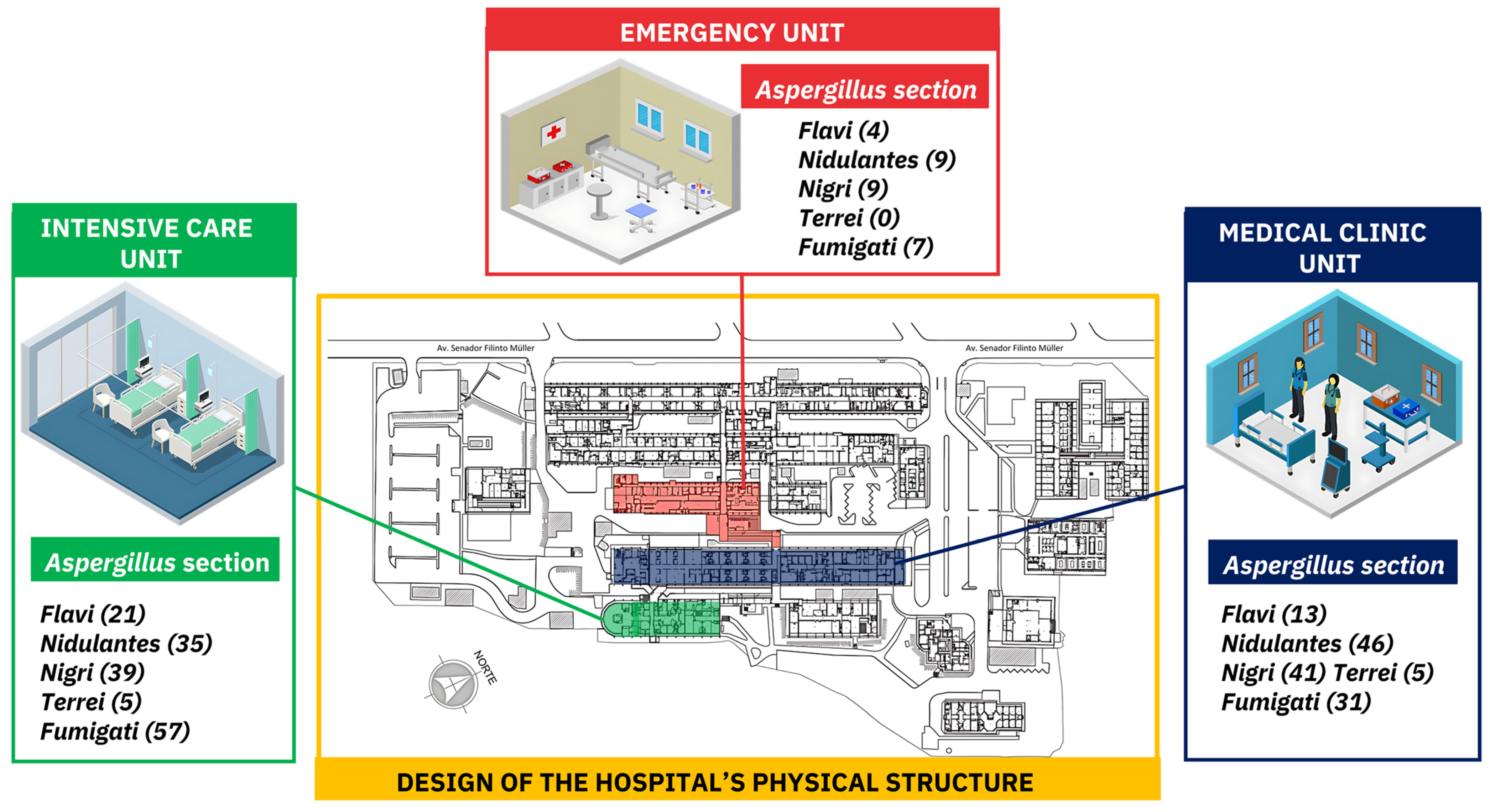
3.1.2. Aspergillus Section Per Hospitalization Unit

Analysis of the number of Aspergillus colonies per hospitalization unit revealed that the ICU had the highest load (157; 48.5%) of Aspergillus colonies dispersed in the air. Fumigati was the most frequent section of the ICU (36.3%, 57/157). Aspergillus from the Nidulantes section (46/138) was more prevalent in the MCU, followed by that in the Nigri section (41/138). Figure 1 shows the distribution of Aspergillus spp. sections per hospital unit.

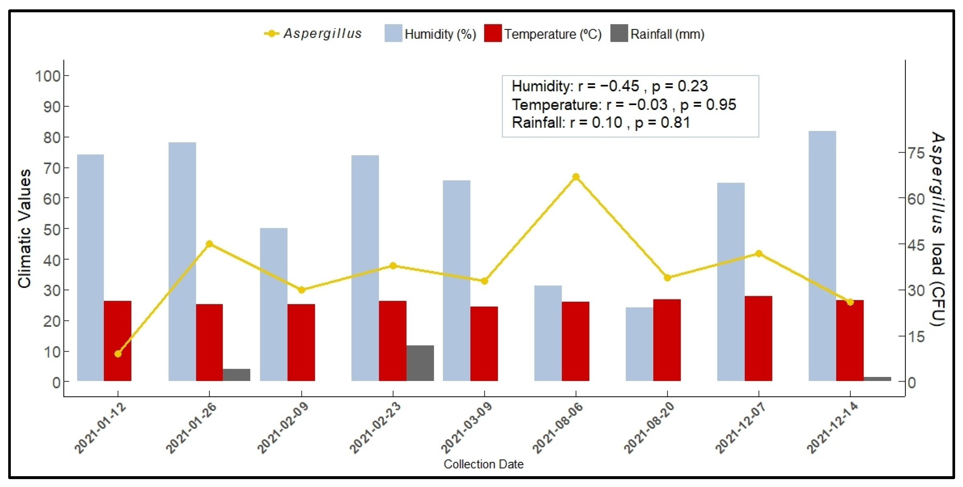
3.1.3. Aspergillus Load According to Section and Season

The variation in Aspergillus load according to section and season was significant (p = 0.001). Unfortunately, it was not possible to collect data during the autumn. Table 4 shows that Aspergillus species from the Fumigati section were more frequent in the winter (64, 67.4%). In contrast, species from the Nigri section (71; 79.8%) and Nidulantes (49; 54.4%) were isolated during the summer.
Figure 2 shows the Aspergillus spp load according to humidity, temperature, and rainfall on the collection dates. In winter, during periods of lower rainfall, we observed a larger load of Aspergillus in ambient air. Pearson correlation analysis indicated that humidity, temperature, and rainfall were not significantly correlated with the presence of Aspergillus spp. in indoor air.

4. Discussion

The results showed saprophytic, allergenic, toxigenic, and potentially pathogenic Aspergillus strains in the air of critical areas of the studied hospital. This study provides for the first time the molecular identification of Aspergillus species from the indoor air of critical hospital areas in the Midwest region of Brazil. Globally, few studies have evaluated the microbial quality of indoor air in hospitals (or hospital environments).
Despite their relevance, there are no specific regulations or guidelines that specify limits for fungi in the indoor air of hospitals [22]. According to the standards of the Brazilian Ministry of Health and the National Health Surveillance Agency, the Maximum Recommended Value for fungal contamination of indoor air is 750 CFU/m3 in indoor air [23]. Although the average number of filamentous fungal colonies found in environmental air in our study was within the limits established by Brazilian law, this value (224 CFU/m3) was higher than those previously described in other Brazilian hospitals. Pantoja et al. (2012) [24] described that, in air collected from the ICUs of three hospitals in northeastern Brazil, the number of fungal spores ranged from 43.75 to 73.67 CFU/m3. In the indoor air of three ICUs of two university hospitals in southern Brazil, the median overall fungal concentrations for ICU 1, 2, and 3 were 109.5, 134.3, and 55.7 CFUm3, respectively [1].
In this study, the source of air contamination by Aspergillus spores is uncertain. External and internal environmental factors and other predisposing conditions may be involved. In the medical clinic unit (MCU), and in the emergency unit (UEU) where there is no air conditioning and the windows are open, there is the possibility that the indoor Aspergillus can, in part, originate from an outdoor environment via the airflow. In these hospital units, the movement of people can also favor the dispersion of fungal conidia that grow on surfaces [25]. In the ICU, where the movement of people is quite restricted, and there is no direct access to the external environment, it is believed that the aerocontamination by Aspergillus is mainly due to internal sources. In enclosed environments, without a high efficiency air filtration system or with an improperly maintained air system, bacteria and fungi can spread and survive in the environment [26,27].
Our results are particularly relevant for hospital units to which immunosuppressed patients and those with chronic diseases are admitted, such as intensive care units and medical clinics. The results revealed that the average number of filamentous fungal colonies (including Aspergillus spp.) per plate varied significantly, depending on the day of collection (p < 0.05). Previous studies have shown that in hospitals, where many spores can circulate in the air, the risks of both infection and hospital infection outbreaks increase significantly [1,26,28].
The findings of this study prompted the Hospital Infection Control Commission to act, as fungi are recognized as indicators of indoor air quality. Support from senior hospital management was sought to implement routine air analysis. Additionally, meetings were held with the engineering team to discuss interventions for enhanced environmental control, such as installing barriers to prevent air infiltration from construction or renovation activities, and regular maintenance of air conditioning equipment to reduce the amount of filamentous fungal spores.
Other measures to minimize the number of filamentous fungal spores in the internal air, include vacuum cleaning, restricting the entry and propagation of microorganisms in the internal environments, and using air cleaning devices. To control air quality more effectively and reduce fungal colony counts, it may be necessary to install heating, ventilation, and air conditioning (HVAC) and high efficiency particulate air (HEPA) filtering systems [13,22].
One of the limitations of this study is that for logistical reasons, outdoor air samples could not be obtained, which could have provided more accurate data on the distribution of these fungi in the environment. Sample collections were consistently conducted simultaneously prior to routine cleaning of the site to minimize potential sources of interference. The higher number of colonies observed on specific days may have been influenced by various internal or external factors, such as the movement of patients, healthcare teams, visitors, and employees, as well as minor renovations within the hospital [13,29,30].
As observed in the present study, filamentous fungi present in the internal air of hospitals may aggravate asthma and allergic rhinitis and cause serious diseases in susceptible individuals [2,4,9,31,32,33,34].
The analysis of the microbial load of fungi in indoor air is challenging. In this study, on the last day of collection (14 December 2021), the number of filamentous fungi was higher in almost all sectors, indicating that the air in the hospital had a high load of filamentous fungi. Future studies should aim to correlate the genotypes of clinical and environmental isolates collected during the same period to determine the impact of high concentrations of airborne spores dispersed in the air.
Aspergillus from the Fumigati section is considered the main cause of invasive aspergillosis and was the most prevalent agent in this study. Unlike what has been described in other countries and other Brazilian regions, in the studied hospital, Aspergillus from the Nidulantes section was the second most frequent, with a similar quantity to species from the Nigri section, which tends to be the second most frequent section in the indoor air of hospitals [12,13,15]. Aspergillus belonging to the Fumigati and Nigri sections was prevalent in the indoor air of UHMAP, corroborating studies in Asia [13], Europe [12,15], Africa [25], and the American continent [1]. In Brazil, these fungi have been described in hospital environments in the Northeast [24,27,35,36], Midwest [37], Southeast [38], and South [1,39] regions. Most Brazilian studies have not identified Aspergillus at the species level, limiting our knowledge of the most prevalent species in hospitals in Brazil [14,26].
In the present study, significant differences were observed in the load of sections of Aspergillus spp., according to the sampling date. In the first quarter of 2021, isolates from the Nigri and Nidulantes sections were predominant. This period was characterized by heavy rainfall. High humidity may have contributed to the many Aspergillus isolates found in these sections [8]. Aspergillus species from the Nigri and Nidulantes sections are routinely recovered from hospital environments and can cause severe diseases [8,31,40]
Aspergillus from the Fumigati section was the most common species isolated from ICU patients (36.3%). This finding is crucial because immunocompromised patients are vulnerable to fatal respiratory infections caused by A. fumigatus [4,9,41]. Species from the Flavi section were also frequently found in the ICU. According to a recent study, A. flavus is the second most common Aspergillus species isolated from invasive aspergillosis patients [10]. The presence of Aspergillus isolates from the Fumigati and Flavi sections in the critical care units found in our study highlights the urgent need to implement measures to improve air quality, not only in the units studied but also in other hospital units.
Previous studies have shown that temperature and relative humidity positively correlate with the growth and reproduction of fungi in hospital environments [42,43,44]. On December 14, considered summer in Brazil, the largest load of filamentous fungi found may be due to high relative humidity, which on this day was greater than 80%. However, in our study, the environmental parameters evaluated (humidity, temperature, and rainfall) did not significantly influence the load of Aspergillus spp. in the internal air of the hospital. Similar to our results, Cho et al., 2018 [13] did not find a statistical correlation between Aspergillus sections in the internal air and temperature. This result may be related to the remarkable ability of some species, such as A. fumigatus, to adapt to the changing climate [45]. In fact, little is known about the variations between Aspergillus strains and their growth at different temperatures and how their geographical origin affects such variations [45]. Further studies are needed to verify how these environmental parameters correlate with the presence of different Aspergillus species.
The study hospital is located in a tropical climate characterized by dry winters and humid summers with heavy rain and high temperatures. Although we did not observe a correlation between the number of Aspergillus isolates in the air and the temperature, it was observed that during the winter, when there was less rain, more CFU of Aspergillus spp. were isolated on average per plate. One of the limitations of this study was the inability to collect air samples during the autumn owing to the COVID-19 pandemic. The impossibility of collecting samples in autumn influenced the total number of filamentous fungi, the number of Aspergillus (sections and species) isolated, and other results related to seasonality. However, we could not evaluate the impact of the lack of such data in the present study. The reason some Aspergillus species are more prevalent in summer and winter is not well established. Seasonal fluctuations in airborne fungal concentrations are influenced by various factors, with geoclimatic conditions being a significant contributor [13,42,45].
Statistical analyses revealed differences in Aspergillus sections and sampling periods in terms of fungal cell counts, indicating that seasonality might be related to an increase in certain species of the genus. This investigation revealed that species from the Fumigati section were isolated in greater numbers during periods of drought, particularly in the winter. This trend is consistent with the characteristics of A. fumigatus, which thrives under such conditions [46]. A. fumigatus spores are highly conducive to air dispersion owing to their small size and significantly higher hydrophobicity than those of other Aspergillus species, allowing them to remain airborne for longer periods [6]. A study conducted in the USA reported that the incidence of aspergillosis was associated with seasonal periods of low precipitation and high temperatures. According to the authors, hot and dry weather conditions facilitate greater dispersal of hydrophobic conidia, which are critical factors in the transmission of aspergillosis [44].
In this study, Aspergillus isolates were initially identified based on their macro- and microscopic characteristics, and almost all were classified into sections. However, phenotypic identification of Aspergillus fungi is challenging and has certain limitations. This is because these fungi share many species similarities and can exhibit morphological variations dependent on culture medium or incubation temperature. Moreover, phenotypic identification requires analysts to have a certain level of experience [16].
The results of this study help fill a gap in the knowledge of Aspergillus species present in the internal air of hospitals in Brazil. Figure 3 shows the distribution of Aspergillus species identified in the indoor air of hospitals in only three of the five regions of Brazil: Northeast [27,36], Midwest [37], and Southeast [38,47]. Despite their high sensitivity and specificity, molecular techniques are of moderate complexity, require specialized technical knowledge, and have high capital costs [48], which makes it challenging to implement the routine of hospital laboratories from low income regions.
Molecular identification revealed cryptic species (which are difficult to distinguish by morphological identification and exhibit distinctive molecular characteristics), including A. sydowii (n = 9), A. tubingensis (n = 4), A. pseudotamarii (n = 2), A. luchuensis (n = 1), A. uvarum (n = 1), A. japonicus (n = 1), and A. pseudocaelatus (n = 1) in the hospital’s indoor air. A. sydowii (Nidulantes section) and A. tubingensis (Nigri section) are among the most common Aspergillus cryptic species in human pathology [3,49,50]. A. sydowii is known to cause allergies and aggravate asthma, and is rarely involved in endophthalmitis, aspergilloma, and invasive pulmonary aspergillosis [34,50]. A. tubingensis is commonly found in indoor air and has been described as an agent of otitis, keratitis, and lower respiratory tract infections in hospitalized patients [3,7]. Nosocomial infections caused by these agents are difficult to treat because of their reduced sensitivity to amphotericin B and intrinsic resistance to azoles [3,10,49,50]. A. luchensis, another important member of the Aspergillus species belonging to the Nigri section, can cause invasive aspergillosis [51]. A. pseudotamarii, an aflatoxigenic member of the Aspergillus section Flavi, has been identified as a mycotic keratitis agent [52]. A. japonicus is one of the main allergenic fungi in India and is considered an agent of pulmonary aspergillosis in patients with COVID-19 [3,53,54]. A. pseudocaelatus is a toxigenic fungus that produces aflatoxins in various crops [55]. Improvements in microbiology laboratories and the widespread use of molecular diagnostic tools will facilitate more precise species descriptions [56].

5. Conclusions

Aspergillus fungi, including species from the Fumigati, Nidulantes, Nigri, and Flavi sections, were found in the indoor air of a tertiary referral hospital for infectious diseases in the mid-western region of Brazil.
Isolates from the Fumigati section were more prevalent during the winter months; however, additional research is needed to establish the seasonality of these fungi.
The presence of A. fumigatus in intensive care units increases the risk of aspergillosis caused by these pathogens, and emphasizes the need for measures to improve air quality in the hospital environment.

Author Contributions

Conceptualization, M.S.C.d.L. and M.R.C.; Formal analysis, W.S.F. and F.A.V.; Funding acquisition, M.R.C.; Investigation, M.S.C.d.L.; Methodology, M.S.C.d.L., J.P.F.T., W.S.F. and N.M.M.; Project administration, M.G.H.J. and M.R.C.; Resources, A.M.M.P., M.d.S.C.M. and M.R.C.; Supervision, M.G.H.J., A.M.M.P. and M.d.S.C.M.; Writing—original draft, M.G.H.J., A.M.M.P. and M.R.C.; Writing—review & editing, M.S.C.d.L., W.S.F., F.A.V. and M.R.C. All authors have read and agreed to the published version of the manuscript.

Funding

This research was funded by the Coordenação de Aperfeiçoamento de Pessoal de Nível Superior (CAPES), Fundação de Apoio ao Desenvolvimento do Ensino, Ciência e Tecnologia do Estado de Mato Grosso do Sul (grant number TO: 048/2018, SIAFIC number 029008) and Federal University of Mato Grosso do Sul (UFMS). National Council for Scientific and Technological Development (CNPq), 312910/2020-7 and 422757/2021-7.

Institutional Review Board Statement

Not applicable.

Data Availability Statement

Data are contained within the article.

Acknowledgments

The authors thank to the Federal University of Mato Grosso do Sul and the team of health professionals from the Intensive and Semi-Intensive Care Unit, Medical Clinic Unit, and Urgency and Emergency Unit of the Maria Aparecida Pedrossian University Hospital—EBSERH) for their support in this study.

Conflicts of Interest

The authors declare no conflicts of interest.

References

  1. Boff, C.; Zoppas, B.C.D.A.; Aquino, V.R.; Kuplich, N.M.; Miron, D.; Pasqualotto, A.C. The indoor air as a potential determinant of the frequency of invasive aspergillosis in intensive care. Mycoses 2013, 56, 527–531. [Google Scholar] [CrossRef] [PubMed]
  2. Cho, S.Y.; Lee, D.G.; Choi, J.K.; Lee, H.J.; Kim, S.H.; Park, S.H.; Choi, S.M.; Choi, J.H.; Yoo, J.H.; Park, Y.J.; et al. Characteristics of culture-positive invasive pulmonary aspergillosis in patients with hematologic diseases. Medicine 2017, 96, e8841. [Google Scholar] [CrossRef] [PubMed]
  3. Cho, S.Y.; Lee, D.G.; Kim, W.B.; Chun, H.S.; Park, C.; Myong, J.-P.; Park, Y.-J.; Choi, J.-K.; Lee, H.-J.; Kim, S.-H.; et al. Epidemiology and antifungal susceptibility profile of Aspergillus species: Comparison between environmental and clinical isolates from patients with hematologic malignancies. J. Clin. Microbiol. 2019, 57, e02023-18. [Google Scholar] [CrossRef] [PubMed]
  4. Loeffert, S.T.; Melloul, E.; Gustin, M.-P.; Hénaff, L.; Guillot, C.; Dupont, D.; Wallon, M.; Cassier, P.; Dananché, C.; Bénet, T.; et al. Investigation of the relationships between clinical and environmental isolates of Aspergillus fumigatus by multiple-locus variable number tandem repeat analysis during major demolition work in a french hospital. Clin. Infect. Dis. 2019, 68, 321–329. [Google Scholar] [CrossRef] [PubMed]
  5. Diba, K.; Jangi, F.; Makhdoomi, K.; Moshiri, N.; Mansouri, F. Aspergillus diversity in the environments of nosocomial infection cases at a university hospital. J. Med. Life 2019, 12, 128–132. [Google Scholar] [CrossRef] [PubMed]
  6. Martinez-Herrera, E.O.; Frias-De-Leon, M.G.; Duarte-Escalante, E.; Calderon-Ezquerro, M.d.C.; Jimenez-Martinez, M.d.C.; Acosta-Altamirano, G.; Rivera-Becerril, F.; Toriello, C.; Reyes-Montes, M.d.R. Fungal diversity and Aspergillus in hospital environments. Ann. Agric. Environ. Med. 2016, 23, 264–269. [Google Scholar] [CrossRef] [PubMed]
  7. Xu, Y.; Chen, M.; Zhu, J.; Gerrits van den Ende, B.; Chen, A.J.; Al-Hatmi, A.M.S.; Li, L.; Zhang, Q.; Xu, J.; Liao, W.; et al. Aspergillus species in lower respiratory tract of hospitalized patients from Shanghai, China: Species Diversity and Emerging Azole Resistance. Infect. Drug Resist. 2020, 13, 4663–4672. [Google Scholar] [CrossRef] [PubMed]
  8. Géry, A.; Rioult, J.-P.; Heutte, N.; Séguin, V.; Bonhomme, J.; Garon, D. First characterization and description of Aspergillus series Versicolores in French Bioaerosols. J. Fungi 2021, 7, 676. [Google Scholar] [CrossRef] [PubMed]
  9. Rhodes, J.; Abdolrasouli, A.; Dunne, K.; Sewell, T.R.; Zhang, Y.; Ballard, E.; Brackin, A.P.; van Rhijn, N.; Chown, H.; Tsitsopoulou, A.; et al. Population genomics confirms acquisition of drug-resistant Aspergillus fumigatus infection by humans from the environment. Nat. Microbiol. 2022, 7, 663–674. [Google Scholar] [CrossRef] [PubMed]
  10. Stemler, J.; Többen, C.; Lass-Flörl, C.; Steinmann, J.; Ackermann, K.; Rath, P.-M.; Simon, M.; Cornely, O.A.; Koehler, P. Diagnosis and Treatment of invasive aspergillosis caused by non-fumigatus Aspergillus spp. J. Fungi 2023, 9, 500. [Google Scholar] [CrossRef] [PubMed]
  11. Shishodia, S.K.; Tiwari, S.; Shankar, J. Resistance Mechanism and Proteins in Aspergillus species against antifungal agents. Mycology 2019, 10, 151–165. [Google Scholar] [CrossRef] [PubMed]
  12. Wirmann, L.; Ross, B.; Reimann, O.; Steinmann, J.; Rath, P.-M. Airborne Aspergillus fumigatus spore concentration during demolition of a building on a hospital site, and patient risk determination for invasive aspergillosis including azole resistance. J. Hosp. Infec. 2018, 100, e91–e97. [Google Scholar] [CrossRef] [PubMed]
  13. Cho, S.-Y.; Myong, J.-P.; Kim, W.-B.; Park, C.; Lee, S.J.; Lee, S.H.; Lee, D.-G. Profiles of environmental mold: Indoor and outdoor air sampling in a hematology hospital in Seoul, south Korea. Int. J. Environ. Res. Public Health 2018, 15, 2560. [Google Scholar] [CrossRef] [PubMed]
  14. Andrade Júnior, F.P.D.; Barbosa, V.; Medeiros, C.; Cruz, J.; Filho, A. Presença de Aspergillus em hospitais brasileiros: Uma revisão integrativa. J. Med. Health Promot. 2019, 4, 1242–1253. [Google Scholar]
  15. Loeffert, S.T.; Melloul, E.; Dananché, C.; Hénaff, L.; Bénet, T.; Cassier, P.; Dupont, D.; Guillot, J.; Botterel, F.; Wallon, M.; et al. Monitoring of Clinical strains and environmental fungal aerocontamination to prevent invasive aspergillosis infections in hospital during large deconstruction work: A protocol study. BMJ Open 2017, 7, e018109. [Google Scholar] [CrossRef] [PubMed]
  16. Walsh, T.J.; Hayden, R.T.; Larone, D.H. Larone’s Medically Important Fungi: A Guide to Identification; Wiley: Hoboken, NY, USA, 2018. [Google Scholar]
  17. Ferrer, C.; Colom, F.; Frasés, S.; Mulet, E.; Abad, J.L.; Alió, J.L. Detection and identification of fungal pathogens by PCR and by ITS2 and 5.8S Ribosomal DNA Typing in Ocular Infections. J. Clin. Microbiol. 2001, 39, 2873–2879. [Google Scholar] [CrossRef] [PubMed]
  18. Sepúlveda, V.E.; Márquez, R.; Turissini, D.A.; Goldman, W.E.; Matute, D.R. Genome sequences reveal cryptic speciation in the human pathogen Histoplasma capsulatum. mBio 2017, 8, e01339-17. [Google Scholar] [CrossRef] [PubMed]
  19. Glass, N.L.; Donaldson, G.C. Development of primer sets designed for use with the PCR to amplify conserved genes from filamentous ascomycetes. Appl. Environ. Microbiol. 1995, 61, 1323–1330. [Google Scholar] [CrossRef] [PubMed]
  20. O’Donnell, K.; Nirenberg, H.I.; Aoki, T.; Cigelnik, E. A Multigene phylogeny of the Gibberella fujikuroi species complex: Detection of additional phylogenetically distinct species. Mycoscience 2000, 41, 61–78. [Google Scholar] [CrossRef]
  21. R Core Team. R: A Language and Environment for Statistical Computing; R Foundation for Statistical Computing: Vienna, Austria, 2021. [Google Scholar]
  22. Božić, J.; Ilić, P.; Ilić, S. Indoor Air Quality in the Hospital: The influence of heating, ventilating and conditioning systems. Braz. Arch. Biol. Technol. 2019, 62, e19180295. [Google Scholar] [CrossRef]
  23. Brazil. Ministério da Saúde. Available online: https://bvsms.saude.gov.br/bvs/saudelegis/anvisa/2003/rdc0009_16_01_2003.html (accessed on 12 November 2023).
  24. Pantoja, L.D.M.; Couto, M.S.; Leitão Junior, N.P.; Sousa, B.L.; Mourão, C.I.; Paixão, G.V. Fungal biodiversity of air in hospitals in the city of Fortaleza, Ceará, Brazil. Rev. Bras. Promoç. Saúde 2012, 25, 192–196. [Google Scholar] [CrossRef]
  25. Gheith, S.; Ranque, S.; Bannour, W.; Ben Youssef, Y.; Khelif, A.; Ben Said, M.; Njah, M.; Saghrouni, F. Hospital environment fungal contamination and aspergillosis risk in acute leukaemia patients in Sousse (Tunisia). Mycoses 2015, 58, 337–342. [Google Scholar] [CrossRef] [PubMed]
  26. Belizario, J.A.; Lopes, L.G.; Pires, R.H. Fungi in the indoor air of critical hospital areas: A review. Aerobiologia 2021, 37, 379–394. [Google Scholar] [CrossRef] [PubMed]
  27. Souza, A.K.P.; Nascimento, J.P.M.; Araújo, M.A.D.S.; Pedrosa, K.P.D.S.; Tenorio, B.M.; Pires, L.L.S.; Lima, G.B.C.; Barboza, R.I.D.S.; Silva Filho, E.A. Airborne Fungi in neonatal intensive care unit of a public hospital in Brazil. Int. J. Curr. Microbiol. Appl. Sci. 2019, 8, 1210–1219. [Google Scholar] [CrossRef]
  28. Hassan, A.; Zeeshan, M. Microbiological indoor air quality of hospital buildings with different ventilation systems, cleaning frequencies and occupancy levels. Atmos. Pol. Res. 2022, 13, 101382. [Google Scholar] [CrossRef]
  29. Sivagnanasundaram, P.; Amarasekara, R.W.K.; Madegedara, R.M.D.; Ekanayake, A.; Magana-Arachchi, D.N. Assessment of airborne bacterial and fungal communities in selected areas of teaching hospital, Kandy, Sri Lanka. BioMed. Res. Int. 2019, 2019, e7393926. [Google Scholar] [CrossRef] [PubMed]
  30. Abbasi, F.; Samaei, M.R. The effect of temperature on airborne filamentous fungi in the indoor and outdoor space of a hospital. Environ. Sci. Pollut. Int. Res. 2019, 26, 16868–16876. [Google Scholar] [CrossRef] [PubMed]
  31. Balajee, S.A.; Kano, R.; Baddley, J.W.; Moser, S.A.; Marr, K.A.; Alexander, B.D.; Andes, D.; Kontoyiannis, D.P.; Perrone, G.; Peterson, S.; et al. Molecular identification of Aspergillus species collected for the transplant-associated infection surveillance network. J. Clin. Microbiol. 2009, 47, 3138–3141. [Google Scholar] [CrossRef] [PubMed]
  32. Kluge, S.; Strauß, R.; Kochanek, M.; Weigand, M.A.; Rohde, H.; Lahmer, T. Aspergillosis: Emerging risk groups in critically ill patients. Med. Mycol. 2022, 60, myab064. [Google Scholar] [CrossRef] [PubMed]
  33. Park, J.H.; Ryu, S.H.; Lee, J.Y.; Kim, H.J.; Kwak, S.H.; Jung, J.; Lee, J.; Sung, H.; Kim, S.-H. Airborne fungal spores and invasive aspergillosis in hematologic units in a tertiary hospital during construction: A prospective cohort study. Antimicrob. Resist. Infect. Control. 2019, 8, 88. [Google Scholar] [CrossRef] [PubMed]
  34. Reboux, G.; Rocchi, S.; Laboissière, A.; Ammari, H.; Bochaton, M.; Gardin, G.; Rame, J.-M.; Millon, L. Survey of 1012 Moldy dwellings by culture fungal analysis: Threshold proposal for asthmatic patient management. Indoor Air 2019, 29, 5–16. [Google Scholar] [CrossRef]
  35. Mobin, M.; do Amparo, M. Fungus microbiota in air conditioners in intensive care units in Teresina, Piauí. Rev. Soc. Bras. Med. Trop. 2006, 39, 556–559. [Google Scholar] [CrossRef] [PubMed]
  36. Calumby, R.J.N.; Silva, J.A.; da Silva, D.P.; de Farias Moreira, R.T.; dos Santos Araujo, M.A.; de Almeida, L.M.; Grillo, L.A.M.; Alvino, V. Isolamento e identificação da microbiota fúngica anemófila em unidade de terapia intensiva/isolation and identification of anemophilic fungal microbiota in an intensive care unit. Braz. J. Dev. 2019, 5, 19708–19722. [Google Scholar] [CrossRef]
  37. De Oliveira, M.T.; Batista, N.K.R.; Gil, E.d.S.; Silva, M.R.R.; Costa, C.R.; Bara, M.T.F.; Torres, I.M.S. Risks associated with pathogenic fungi isolated from surgical centers, intensive care units, and materials sterilization centers in hospitals. Risks associated with pathogenic fungi isolated from critical hospital areas. Med. Mycol. 2020, 58, 881–886. [Google Scholar] [CrossRef] [PubMed]
  38. Barreiros, G.; Akiti, T.; Magalhães, A.C.G.; Nouér, S.A.; Nucci, M. Effect of the implosion and demolition of a hospital building on the concentration of fungi in the air. Mycoses 2015, 58, 707–713. [Google Scholar] [CrossRef] [PubMed]
  39. Gonçalves, C.L.; Mota, F.V.; Ferreira, G.F.; Mendes, J.F.; Pereira, E.C.; Freitas, C.H.; Vieira, J.N.; Villarreal, J.P.; Nascente, P.S. Airborne fungi in an intensive care unit. Braz. J. Biol. 2017, 78, 265–270. [Google Scholar] [CrossRef] [PubMed]
  40. Gits-Muselli, M.; Hamane, S.; Verillaud, B.; Cherpin, E.; Denis, B.; Bondeelle, L.; Touratier, S.; Alanio, A.; Garcia-Hermoso, D.; Bretagne, S. Different repartition of the cryptic species of black Aspergilli According to the anatomical sites in human infections, in a French university hospital. Med. Mycol. 2021, 59, 985–992. [Google Scholar] [CrossRef] [PubMed]
  41. Demuyser, T.; De Cock, E.; Sermijn, E. Airborne Aspergillus fumigatus contamination in an intensive care unit: Detection, management and control. J. Infect. Public Health 2019, 12, 904–906. [Google Scholar] [CrossRef] [PubMed]
  42. Mori, T.; Kikuchi, T.; Kato, J.; Koda, Y.; Sakurai, M.; Kikumi, O.; Inose, R.; Murata, M.; Hasegawa, N.; Nakayama, H.; et al. Seasonal Changes in indoor airborne fungal concentration in a hematology ward. J. Infec. Chemother. 2020, 26, 363–366. [Google Scholar] [CrossRef] [PubMed]
  43. Shelton, B.G.; Kirkland, K.H.; Flanders, W.D.; Morris, G.K. Profiles of airborne fungi in buildings and outdoor environments in the United States. Appl. Environ. Microbiol. 2002, 68, 1743–1753. [Google Scholar] [CrossRef] [PubMed]
  44. Panackal, A.A.; Li, H.; Kontoyiannis, D.P.; Mori, M.; Perego, C.A.; Boeckh, M.; Marr, K.A. Geoclimatic influences on invasive aspergillosis after hematopoietic stem cell transplantation. Clin. Infect. Dis. 2010, 50, 1588–1597. [Google Scholar] [CrossRef] [PubMed]
  45. Korfanty, G.; Heifetz, E.; Xu, J. Assessing thermal adaptation of a global sample of Aspergillus fumigatus: Implications for climate change effects. Front. Public Health. 2023, 11, 1059238. [Google Scholar] [CrossRef] [PubMed] [PubMed Central]
  46. O’Gorman, C.M.; Fuller, H.T.; Dyer, P.S. Discovery of a sexual cycle in the opportunistic fungal pathogen Aspergillus fumigatus. Nature 2009, 457, 471–474. [Google Scholar] [CrossRef] [PubMed]
  47. Teixeira, A.B.A.; Silva, M.; Lyra, L.; Luz, E.A.; Uno, J.; Takada, H.; Miyaji, M.; Nishimura, K.; Schreiber, A.Z. Antifungal susceptibility and pathogenic potential of environmental isolated filamentous fungi compared with colonizing agents in immunocompromised patients. Mycopathologia 2005, 160, 129–135. [Google Scholar] [CrossRef] [PubMed]
  48. Wickes, B.L.; Wiederhold, N.P. Molecular diagnostics in medical mycology. Nat. Commun. 2018, 9, 5135. [Google Scholar] [CrossRef] [PubMed]
  49. Géry, A.; Séguin, V.; Eldin de Pécoulas, P.; Bonhomme, J.; Garon, D. Aspergilli series Versicolores: Importance of species identification in the clinical setting. Crit. Rev. Microbiol. 2022, 49, 485–498. [Google Scholar] [CrossRef] [PubMed]
  50. Negri, C.E.; Gonçalves, S.S.; Xafranski, H.; Bergamasco, M.D.; Aquino, V.R.; Castro, P.T.O.; Colombo, A.L. Cryptic and rare Aspergillus species in Brazil: Prevalence in clinical samples and in vitro susceptibility to triazoles. J. Clin. Microbiol. 2014, 52, 3633–3640. [Google Scholar] [CrossRef] [PubMed]
  51. Wang, Q.; Li, Y.; Li, Y.; Osherov, N.; Goldman, G.H.; Verweij, P.E.; Zheng, B.; Li, R.; Chen, W.; Liang, T.; et al. Triazole-Resistant Aspergillus Luchuensis, an Industrially Important Black Aspergillus spp. Used in fermentation in East Asia, isolated from the patient with invasive pulmonary aspergillosis in China. Emerg. Microbes Infect. 2022, 11, 1435–1438. [Google Scholar] [CrossRef] [PubMed]
  52. Baranyi, N.; Kocsubé, S.; Szekeres, A.; Raghavan, A.; Narendran, V.; Vágvölgyi, C.; Panneer Selvam, K.; Babu Singh, Y.R.; Kredics, L.; Varga, J.; et al. Keratitis caused by Aspergillus pseudotamarii. Med. Mycol. Case Rep. 2013, 2, 91–94. [Google Scholar] [CrossRef] [PubMed]
  53. Singh, A.B.; Kumar, P. Common environmental allergens causing respiratory allergy in India. Indian J. Pediatr. 2002, 69, 245–250. [Google Scholar] [CrossRef]
  54. Kariyawasam, R.M.; Dingle, T.C.; Kula, B.E.; Vandermeer, B.; Sligl, W.I.; Schwartz, I.S. Defining COVID-19–associated pulmonary aspergillosis: Systematic review and meta-analysis. Clin. Microbiol. Infect. 2022, 28, 920–927. [Google Scholar] [CrossRef] [PubMed]
  55. Katsurayama, A.M.; Martins, L.M.; Iamanaka, B.T.; Fungaro, M.H.P.; Silva, J.J.; Frisvad, J.C.; Pitt, J.I.; Taniwaki, M.H. Occurrence of Aspergillus section Flavi and aflatoxins in brazilian rice: From field to market. Int. J. Food Microbiol. 2018, 266, 213–221. [Google Scholar] [CrossRef] [PubMed]
  56. Houbraken, J.; Kocsubé, S.; Visagie, C.M.; Yilmaz, N.; Wang, X.-C.; Meijer, M.; Kraak, B.; Hubka, V.; Bensch, K.; Samson, R.A.; et al. Classification of Aspergillus, Penicillium, Talaromyces and related genera (eurotiales): An overview of families, genera, subgenera, sections, series and species. Stud. Mycol. 2020, 95, 5–169. [Google Scholar] [CrossRef] [PubMed]
Figure 1. Aspergillus distribution according to section and collection site. UHMAP Campo Grande-MS, January–December 2021.
Jof 10 00538 g001
Figure 2. Aspergillus spp. load according to humidity, temperature, and rainfall on the date of collection. Campo Grande-MS, 2021. Note: Humidity (%) represents the percentage of humidity recorded on each collection date. Temperature (°C): Indicates the temperature in degrees Celsius observed on each collection date. Rainfall (mm): The amount of precipitation in millimeters measured on each collection date. Aspergillus: Presents the load of Aspergillus spp. on each collection date. Explanatory Note: This grouped bar chart illustrates the climatic variations and load of Aspergillus on nine different collection dates throughout 2021. Each group of bars represents measurements of humidity, temperature, and precipitation (rainfall) associated with a specific date. The above legend describes the climatic variables represented by distinct colors, and the gold color indicates the load of Aspergillus. The heights of the bars represent the values of each variable, whereas the secondary axis on the right displays the load of Aspergillus. Source: Mato Grosso do Sul State Weather and Climate Monitoring Center (CEMTEC/SEMAGRO). The Pearson correlation coefficient was used for correlation analysis.
Jof 10 00538 g002
Figure 3. Distribution of species of Aspergillus identified in indoor air of Brazilian hospitals.
Jof 10 00538 g003
Table 1. Primer used in DNA amplification and sequencing for molecular identification of Aspergillus species.
PrimerSequence (5′-3′)Reference
β-tubulin 2aGGTAACCAAATCGGTGCTGCTTTCGlass and Donaldson, 1995 [19]
β-tubulin 2bACCCTCAGTGTAGTGACCCTTGGCGlass and Donaldson, 1995 [19]
Calmodulin 1GARTWCAAGGAGGCCTTCTCO’Donnell et al., 2000 [20]
Calmodulin 2aTTTTTGCATCATGAGTTGGACO’Donnell et al., 2000 [20]
Calmodulin 11ACCATGATGGCGCGCAAGO’Donnell et al., 2000 [20]
Calmodulin 22TCCTTCATCTTGCGCGCCO’Donnell et al., 2000 [20]
Table 2. Comparison of filamentous fungal load according to collection date. UHMAP—Campo Grande, 2021.
CFU/m3 of Filamentous
Fungi/100 L/min
DateN (%)Mean per Plate (SD)p Value *
12 January253 (6.04)9.3 (6.6)
26 January448 (10.46)16.7 (9.2)
9 February202 (4.98)7.4 (5.0)
23 February325 (7.55)12.5 (9.2)
9 March431 (10.33)15.9 (7.8)<0.001
6 August442 (10.80)17.0 (6.7)
20 August498 (12.22)19.1 (8.9)
7 December593 (11.86)18.8 (7.6)
14 December1103 (25.73)42.4 (12.5)
Note: SD = standard deviation; * ANOVA. CFU = Colony Forming Unit.
Table 3. Distribution of 324 Aspergillus species grouped by section, per day of collection. UHMAP—Campo Grande-MS, 2021.
DateFlavi
N (%)
Nidulantes N (%)Nigri
N (%)
Terrei
N (%)
Fumigati
N (%)
Indeterminate
N (%)
Total of Aspergillus
N (%)
p Value
12 January0 (0.0)0 (0.0)5 (55.5)0 (0.0)4 (44.4)0 (0.0)9 (2.8)<0.001
26 January8 (17.8)5 (11.1)26 (57.8)4 (8.9)2 (4.4)0 (0.0)45 (13.9)
9 February0 (0.0)26 (86.7)4 (13.3)0 (0.0)0 (0.0)0 (0.0)30 (9.3)
23 February1 (2.6)18 (47.4)18 (47.4)1 (2.6)0 (0.0)0 (0.0)38 (11.7)
9 March11 (33.3)0 (0.0)18 (54.5)2 (6.1)0 (0.0)2 (6.1)33 (10.2)
6 August1 (1.5)13 (27.6)4 (6.0)1 (1.5)48 (71.6)0 (0.0)67 (20.7)
20 August9 (26.4)3 (8.8)5 (14.7)1 (2.9)16 (47.1)0 (0.0)34 (10.5)
7 December5 (11.9)13 (30.9)5 (11.9)1 (2.4)18 (42.9)0 (0.0)42 (13.0)
14 December3 (11.5)12 (46.2)4 (15.4)0 (0.0)7 (26.9)0 (0.0)26 (8.0)
Total38 (11.7)90 (27.8)89 (27.4)10 (3.1)95 (29.3)2 (0.6)324 (100)
Table 4. Aspergillus sections scattered in the indoor air during the UHMAP season (Campo Grande-MS, 2021).
Aspergillus spp.
(Section)
WINTER
N (%)
SPRING
N (%)
SUMMER
N (%)
Total of Aspergillus
N (%)
p Value
Flavi10 (26.3)8 (21.0)20 (52.6)38 (11.7)<0.001
Nidulantes16 (17.8)25 (27.8)49 (54.4)90 (27.8)
Nigri9 (10.1)9 (10.1)71 (79.8)89 (27.4)
Terrei2 (20.0)1 (10.0)7 (70.0)10 (3.1)
Fumigati64 (67.4)25 (26.3)6 (6.3)95 (29.3)
Indeterminate0 (0.0)0 (0.0)2 (100.0)2 (0.6)
Total101 (31.2)68 (21.0)155 (47.8)324 (100)
Disclaimer/Publisher’s Note: The statements, opinions and data contained in all publications are solely those of the individual author(s) and contributor(s) and not of MDPI and/or the editor(s). MDPI and/or the editor(s) disclaim responsibility for any injury to people or property resulting from any ideas, methods, instructions or products referred to in the content.

