Article

The Catalase Gene MrCat1 Contributes to Oxidative Stress Tolerance, Microsclerotia Formation, and Virulence in the Entomopathogenic Fungus Metarhizium rileyi

1
College of Plant Protection, Southwest University, Chongqing 400716, China
2
Southeast Chongqing Academy of Agricultural Sciences, Chongqing 408000, China
*
Author to whom correspondence should be addressed.
J. Fungi 2024, 10(8), 543; https://doi.org/10.3390/jof10080543 (registering DOI)
Submission received: 21 June 2024 / Revised: 30 July 2024 / Accepted: 1 August 2024 / Published: 2 August 2024

Abstract

:
Catalases play a crucial role in the metabolism of reactive oxygen species (ROS) by converting H2O2 into molecular oxygen and water. They also contribute to virulence and fungal responses to various stresses. Previously, the MrCat1-deletion mutant (ΔMrCat1) was generated using the split-marker method in Metarhizium rileyi. In this study, the Cat1 gene was identified, and its function was evaluated. Under normal culture conditions, there were no significant differences in colony growth or dimorphic switching between ΔMrCat1 and the wild-type (WT) strains. However, under oxidative stress, the colony growth was inhibited, and the yeast–hyphal transition was suppressed in the ΔMrCat1 strain. Hyperosmotic stress did not differ significantly between the two strains. In the ΔMrCat1 strain, microsclerotia (MS) formation was delayed, resulting in less uniform MS size and a 76% decrease in MS yield compared to the WT strain. Moreover, the ΔMrCat1 strain exhibited diminished virulence. Gene expression analysis revealed up-regulation of ΔMrCat1, MrCat2, MrCat4, and MrAox in the ΔMrCat1 strain. These findings indicate that the MrCat1 gene in M. rileyi is essential for oxidative stress tolerance, MS formation, and virulence.

1. Introduction

Metarhizium rileyi is a valuable entomopathogenic fungus known for its ability to infect lepidopterous pests, particularly Noctuidae spp., making it a potential candidate for insect biocontrol [1,2,3,4,5]. However, the sporulation of M. rileyi is dependent on specific conditions such as light stimulation and a maltose carbon source, which limits its commercial potential. To overcome this limitation, researchers have successfully induced microsclerotia (MS) formation in M. rileyi using liquid-amended media (AM) [6]. These MS are specialized, hyperpigmented hyphal aggregations with a diameter of 50–600 μm. Compared to M. rileyi conidia, MS have higher production efficiency and exhibit longer persistence in the field. Other entomopathogenic fungi like Metarhizium brunneum [7] and Metarhizium anisopliae [8] also produce MS and have been used in insect biocontrol.
In filamentous fungi, studies have shown that reactive oxygen species (ROS) play a critical role in various aspects of cell physiology, cellular differentiation, cell signaling, and defense against pathogens [9]. In the case of M. rileyi, comparative transcriptome analysis has been conducted to investigate the molecular mechanisms underlying MS development. This analysis revealed that oxidative stress is a key factor in MS development [10]. Several genes involved in MS development have been identified in M. rileyi. For example, the small GTPase RacA and Cdc42, along with their regulatory factors Cdc24 and Bem1, regulate MS formation by controlling ROS generation [11,12]. Additionally, the NADH:flavin oxidoreductase/NADH oxidase gene (Nox) and alternative oxidase (Aox) genes are involved in MS formation by regulating intracellular H2O2 concentrations [13,14,15]. Defects in the transmembrane sensor genes Sho1 and Sln1 [16], as well as the deletion of two MAPK genes, Hog1 and Slt2, have also been found to inhibit MS formation [17].
Catalases are crucial enzymes in the metabolism of ROS. They, along with superoxide dismutases (SODs) and catalases, convert superoxide and H2O2 into water and molecular oxygen [18,19,20,21]. Previous studies have highlighted the important adaptive role of catalases in response to environmental stress. For example, in Beauveria bassiana, five catalase genes (CatA, CatB, CatP, CatC, and CatD) have been implicated in the regulation of virulence and tolerance to oxidative stress, high temperatures, and UV-B radiation [22]. Overexpression of the Cat1 gene in M. anisopliae has been shown to enhance resistance to exogenous H2O2, reduce germination time, and increase virulence [23]. In contrast, Neurospora crassa exhibited defects in the survival of conidia under oxidative and light-induced stress [24], while Cat3 mutants exhibited growth and differentiation defects [25].
In Aspergillus oryzae, the CatB gene has been implicated in the detoxification of oxidative stress [26]. In the phytopathogenic fungus Claviceps purpurea, catalase has been shown to suppress the host defense system [27]. Furthermore, peroxisomal catalases may be involved in insect hydrocarbon catabolism [28]. These results collectively demonstrate the importance of fungal catalase genes in detoxification, cellular differentiation, and catabolism. However, there have been no reports on the involvement of catalase genes in MS formation. In this study, we isolated genomic DNA and cDNA of the Cat1 gene in M. rileyi and investigated its role in oxidative stress tolerance, MS formation, and virulence through gene deletion experiments.

2. Materials and Methods

2.1. Strains and Growth Conditions

The M. rileyi strain Nr01 utilized in this study was obtained from the Engineering Research Center of Fungal Insecticides located in Chongqing, China. The mutant strain ΔMrCat1, lacking the MrCat1 gene, was previously generated and confirmed by a study [29]. Both the wild-type (WT) and ΔMrCat1 strains were cultured on solid SMAY media containing 40 g L−1 maltose, 15 g L−1 yeast extract powder, and 10 g L−1 peptone. Blastospores were collected during the early stages of growth, while conidia were collected after spore production by suspending the colonies in a sterile 0.05% Tween-80 solution and filtering them through lens wiping paper. The resulting blastospores of the WT and ΔMrCat1 strains were washed and suspended in sterile phosphate-buffered saline (PBS) using a process involving three rounds of centrifugation. The M. rileyi spores were then cultured in liquid AM medium at 28 °C with continuous shaking at 250 rpm, following a described protocol [6].

2.2. Cloning the MrCat1 Gene of M. rileyi

The partial MrCat1 sequence in this study was obtained from transcriptome data reported by Song [10]. To obtain the full cDNA and genomic sequences, the utilization of the fusion primer and nested integrated PCR (FPNI-PCR) method described by Wang [30]. The amino acid sequence was deduced by performing BlastX searches in GenBank. Signal peptide prediction was carried out by signalP 6.0, a web-based tool available at http://www.cbs.dtu.dk/serbices.signal/ (accessed on 21 February 2023). The conserved domain of MrCat1 was predicted using the SMART web resource, available at http://smart.embl.de/ (accessed on 21 February 2023). The obtained sequences were aligned using MUSCLE with default settings, and an unrooted phylogenetic tree was generated using the Maximum Likelihood method in MEGA-X (https://www.megasoftware.net/) (accessed on 25 February 2023) with a 1000 replicates bootstrap test, as detailed by Kumar [31].

2.3. Gene Expression Patterns of M. rileyi WT and ΔMrCat1 Strains during MS Development

The transcription levels of MrCat1, MrCat2, MrCat4, and MrAox were quantified using real-time quantitative PCR (RT-qPCR) at different time points during the development of MS. The WT and ΔMrCat1 strains were introduced into flasks containing 100 mL of liquid AM, along with 0.5 mL of conidia suspension (1 × 10⁸ sp mL−1). The flasks were then incubated with agitation at 28 °C and 250 rpm for 1.5–7 days. Samples were collected at specific time intervals corresponding to different stages of development: germinating spores (1.5–2 days), yeast-like cells (2.5–3 days), MS initiation and hyphal period (3.5–4 days), MS formation (4.5–5 days), MS maturation (5.5–6 days), and secondary mycelial growth (6.5–7 days). After centrifugation and two washes with sterile distilled water, total RNA was extracted using TRIzol® reagent (Invitrogen, Carlsbad, CA, USA). First-strand cDNA was then synthesized following the manufacturer’s protocol using SuperScript II Reverse Transcriptase (Invitrogen, Carlsbad, CA, USA). RT-qPCR was performed using SYBR® Green II mix (TaKaRa, Shiga, Japan) according to the manufacturer’s instructions. The primers used to assess the expression levels of MrCat1, MrCat2, MrCat4, and MrAox are detailed in Table A1. Each sample was prepared in triplicate, and each reaction was carried out three times. The MrTef and MrTub genes were employed as reference genes for normalization.

2.4. H2O2 Sensitivity of WT and ΔMrCat1 Strains

To assess the sensitivity to H2O2 in the WT and ΔMrCat1 strains, the filter paper method was employed. Conidial suspensions (1 × 10⁶ sp mL−1) of the WT and ΔMrCat1 strains were spread onto plates. On the center of each plate, a 4-mm filter paper containing 800, 1600, or 3200 mM H2O2 was placed. For colony morphology evaluation, conidial suspensions (1 × 10⁷, 1 × 10⁶, and 1 × 10⁵ sp mL−1) of the WT and ΔMrCat1 strains were pipetted onto SMAY plates and SMAY plates supplemented with 2 mM H2O2. All treatments were then incubated at 25 °C.

2.5. Hyperosmolarity Tolerance of WT and ΔMrCat1 Strains

To evaluate the tolerance of the WT and ΔMrCat1 strains to NaCl, KCl, LiCl, and sorbitol, solid SMAY media was used. Three concentrations of conidial suspensions (1 × 10⁷, 1 × 10⁶, and 1 × 10⁵ sp mL−1) were pipetted onto plates for the evaluation of colony morphology.

2.6. MS Formation of WT and ΔMrCat1 Strains

To examine the MS formation capacities of the WT and ΔMrCat1 strains, a conidial suspension (1 × 10⁸ sp mL−1) was inoculated into 100 mL of liquid AM. The inoculated cultures were then incubated at 28 °C with shaking at 250 rpm for 3.5–6.0 days. During the incubation period, the formation of MS yield was monitored, and the yield of MS was assessed. Additionally, the morphologies of the MS structures were observed using a microscope.

2.7. Virulence Assays

To determine the virulence of the WT and ΔMrCat1 strains, third-instar larvae of Prodenia litura were used in the experiment. The larvae were immersed in three different concentrations of conidial suspensions: 2.5 × 10⁷ sp mL−1, 5 × 10⁷ sp mL−1, and 1 × 10⁸ sp mL−1. Control experiments were also conducted using a 0.05% Tween-80 solution without the fungus. After treatment, the insects were incubated at 26 °C, and mortality was recorded every 24 h. The median lethal time (LT50) values were calculated using the minimum squares method, and the median lethal concentration (LC50) values were calculated using the linearized regression model (LRM) method. Each treatment was repeated three times, with 15 larvae per replicate, to ensure reliable and statistically significant results.

2.8. Data Statistical Analysis

To assess variations in the relative expression levels of the genes among the samples, a variance analysis was conducted. Following the variance analysis, multiple comparisons were performed using Duncan’s least significant range (LSR) tests in the SPSS statistical software (SPSS 16.0, SPSS Inc., Armonk, NY, USA). If a column lacks a common superscript letter, it indicates a significant difference compared to other columns (p < 0.05). On the other hand, columns labeled with the same letter were not found to be significantly different at the 5% level, based on Duncan’s multiple range test.

3. Results

3.1. Features of MrCat1 in M. rileyi

The MrCat1 gene was identified from the expressed sequence tag (EST) of the Cat1 gene isolated from the transcriptome library of M. rileyi. It has a full-length sequence of 2561 base pairs (bp) and encodes 716 amino acid residues. Comparison of cDNA and genomic sequences revealed the presence of four introns in MrCat1. Similar to other fungal genes, it contains a signal peptide for secretion and exhibits a typical catalase structural domain (Figure 1A), indicating that it is likely an exocrine protein. The predication molecular weight of the MrCat1 is 79.085 KDa, and its isoelectric point (pI) is 5.92. A phylogenetic analysis using MEGA-X software revealed that MrCat1 is closely associated with Metarhizium spp. (Figure 1B). The deduced amino acid sequence of MrCat1 showed similarities with catalase in Metarhizium acridum (78.85% identity) [32] and catalaseB in Purpureocillium lilacinum (69.18% identity) [33], suggesting functional similarities with these enzymes.
To assess the expression of MrCat1 during MS development, RT-qPCR analysis was performed. The results showed that MrCat1 was expressed at all stages of MS development. It exhibited significantly up-regulated during MS initiation (84 h, 3.6-fold), MS formation (120 h, 5.4-fold), MS maturation (144 h, 4.4-fold), and the early stage of secondary mycelial growth (156 h, 4.1-fold) compared to the germinating spore period (36 h). These findings suggest that the MrCat1 may play a potential role in hyphal growth and MS formation (Figure 1C).

3.2. Deletion of MrCat1 Impaired the Tolerance to H2O2 in M. rileyi

To investigate the role of MrCat1 in H2O2 metabolism, a deletion mutant of MrCat1 was created in M. rileyi using the split-marker method [29]. Both the WT and ΔMrCat1 conidia were grown on SMAY plates supplemented with 2 mM H2O2 at a temperature of 25 °C for 7 days. Notably, there were no noticeable differences in morphology between the colonies of the WT and ΔMrCat1 strains. However, a delay in the dimorphic switch was observed in the ΔMrCat1 strain grown on SMAY plates with 2 mM H2O2. Specifically, the dimorphic switch was delayed by approximately 1 and 2 days compared to the WT strain (Figure 2A). In H2O2 inhibition assays, the ΔMrCat1 strain exhibited larger inhibition zones compared to the WT strain. The size of the inhibition zones for the ΔMrCat1 strain grown at H2O2 concentrations of 800, 1600, and 3200 mM were 1.7, 1.3, and 1.1 times larger, respectively, than those of the WT strain under the same conditions (Figure 2B). Variance analysis revealed that the diameter of the inhibition zones for the ΔMrCat1 strain grown at an H2O2 concentration of 800 mM was significantly different from the WT strain (p < 0.01).

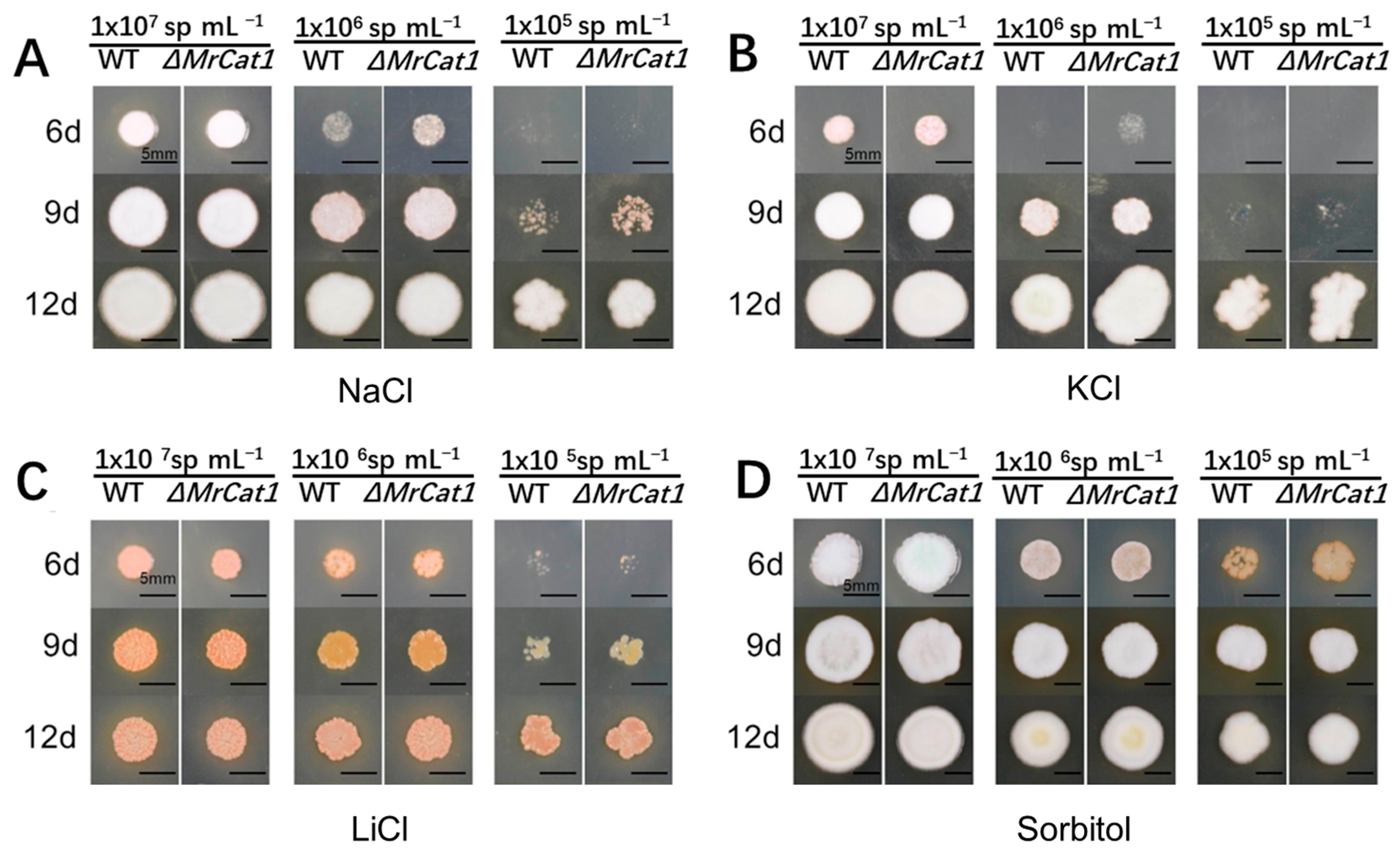
3.3. Deletion of MrCat1 Did Not Affect the Hyperosmotic Tolerance of M. rileyi

Both the WT and ΔMrCat1 conidia were cultivated on SMAY plates supplemented with different hyperosmotic conditions, including 1 M NaCl, 1 M KCl, 0.04 M LiCl, and 1 M sorbitol. The cultivation period lasted for 18 days at a temperature of 25 °C. The growth rate of both strains, WT and ΔMrCat1, was significantly hindered by the presence of hyperosmotic conditions, except for the presence of sorbitol. The growth of ΔMrCat1 strain exhibited a promotion at the beginning of the inoculation period (6d and 9d) compared with the WT strain (Figure 3A–D). However, despite the differences in growth rate, there were no discernible differences in colony morphologies between the two strains (Figure 3A–D).

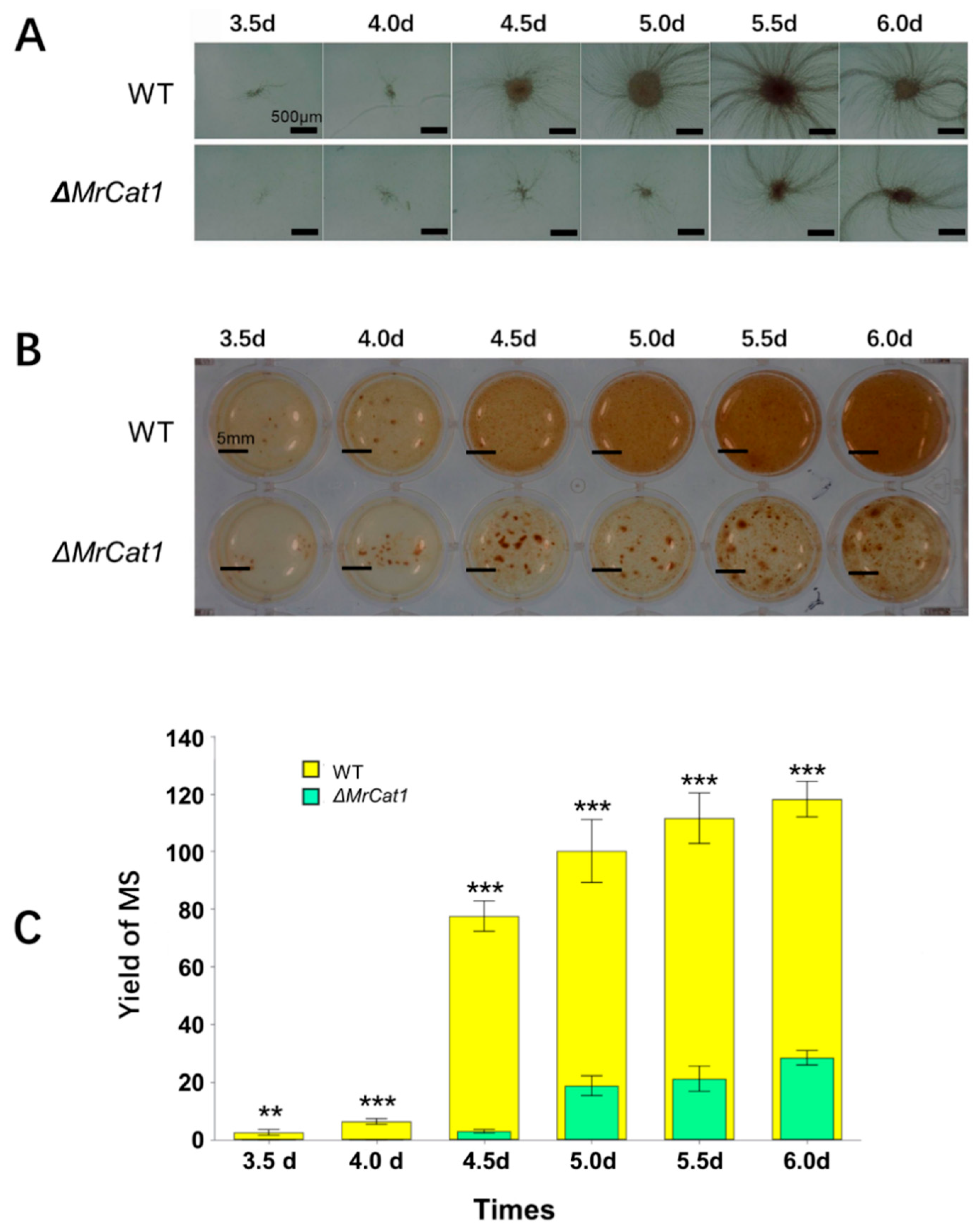
3.4. Deletion of MrCat1 Affected MS Development of M. rileyi

Both the WT and ΔMrCat1 conidia were introduced into a liquid AM medium and subjected to agitation. The WT strains exhibited visible MS formation after a period of 3.5–4 days, while the ΔMrCat1 strain experienced a delay in MS formation, occurring after 4.5–5 days. Furthermore, the fermentation broth of the ΔMrCat1 strain exhibited lower viscosity and a lighter pigment compared to the WT strain (Figure 4A,B). Additionally, the size distribution of WT MS displayed greater uniformity, ranging from 70 to 300 μm, compared to the ΔMrCat1 MS, which ranged from 50 to 1500 μm. Moreover, the MS yield of the ΔMrCat1 strain was diminished by approximately 76% compared to the WT strain (Figure 4C).

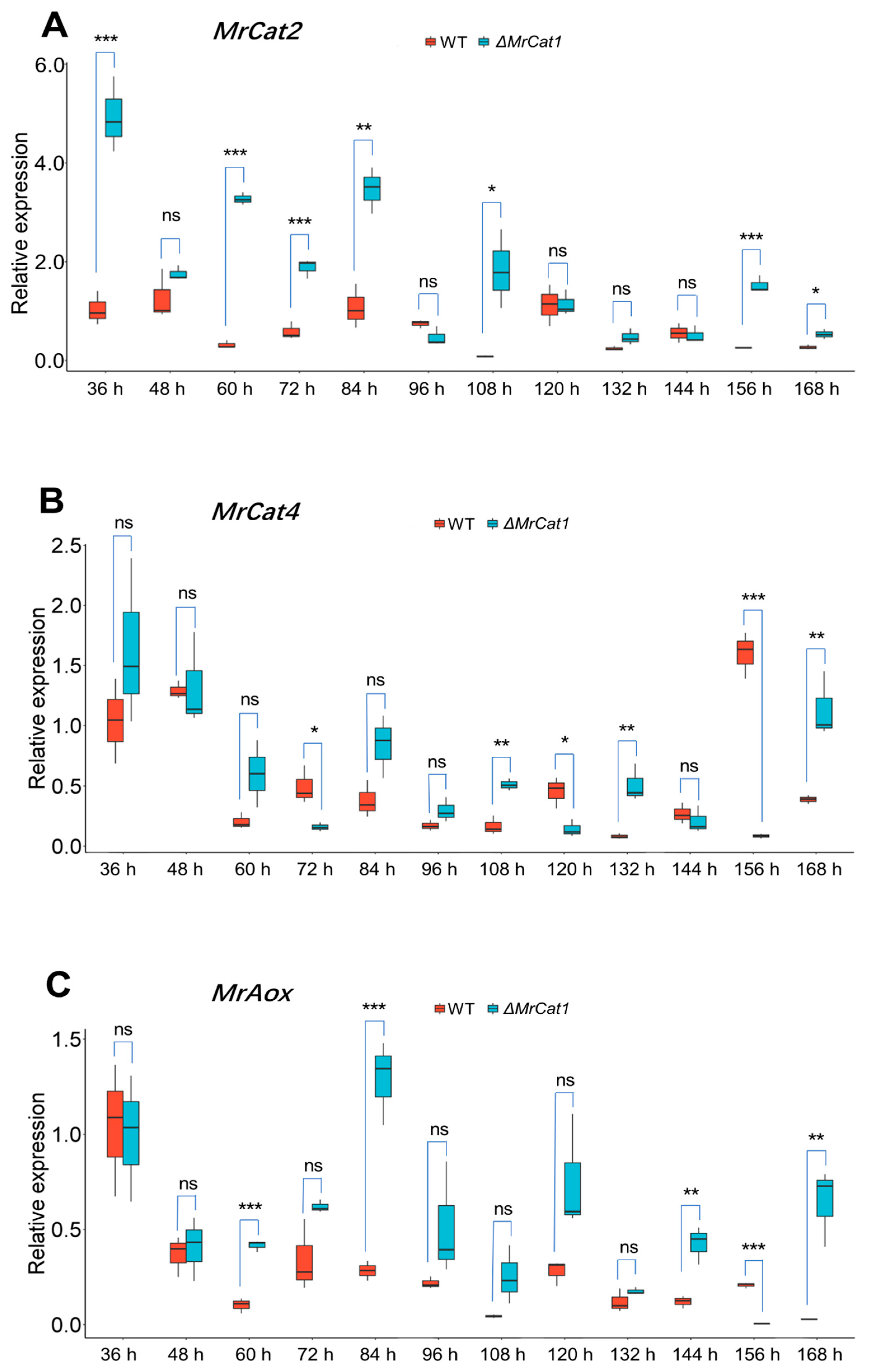
3.5. Deletion of MrCat1 Resulted in Enhancing the Expression of MrCat2, MrCat4, and MrAox during MS Development of M. rileyi

The expressions of MrCat2, MrCat4, and MrAox genes were analyzed during different stages of MS development, including spore germinating, MS initiation, formation stage, and secondary mycelial growth phase, in both the WT and ΔMrCat1 strains of M. rileyi. Remarkably, the transcription levels of the MrCat2 and MrAox were consistently higher in the ΔMrCat1 strain compared to the WT strain (Figure 5A,C). Specifically, for MrCat2, the transcription levels were higher during the spore germinating to MS initiation period (36 h–84 h) and exhibited higher expression in the early period of MS formation (108 h) and early period of secondary mycelial growth (156 h). The most significant compensation occurred at 60 h, where MrCat2 was up-regulated by 10.2-fold (Figure 5A).
Similarly to MrAox, the transcription levels were higher from the yeast-like cell period to MS maturation (60 h–144 h) and were up-regulated in the later period of secondary mycelial growth (168 h). The most significant compensation occurred at 168 h, where MrAox was up-regulated by 22.1-fold. However, transcription levels of the MrCat4 gene were prominent during the secondary mycelial growth phase and exhibited irregular compensation between the WT and ΔMrCat1 strains. Taken together, the results indicate that MrCat2, MrCat4, and MrAox genes exhibit significantly higher expression in the ΔMrCat1 strain compared to the WT strain during the MS initiation period (84 h), which is a key time point for MS initiation (Figure 5A–C). This suggests that in M. rileyi, MrCat1, MrCat2, and MrAox may have shared functions during MS formation.

3.6. Deletion of MrCat1 Reduced the Virulence of M. rileyi

To examine the effects of MrCat1 deletion on the pathogenicity of M. rileyi, insect bioassays were conducted to infect P. litura larvae. The survival rates and LT50 were determined and compared between the WT and ΔMrCat1 strains. After infection, it was observed that the survival rates of P. litura larvae infected by the ΔMrCat1 strain were significantly higher than those infected by the WT strain at the same concentration (2.5 × 10⁷ sp mL−1, 5.0 × 10⁷ sp mL−1, 1.0 × 10⁸ sp mL−1) (Figure 6A). Moreover, the mortality of the ΔMrCat1 stain at the concentration of 2.5 × 10⁷ sp mL−1 and 5.0 × 10⁷ sp mL−1 was less than 50% at the time point of 9 d. However, at the concentration of 1.0 × 10⁸ sp mL−1, the LT50 was 6.81 ± 0.52 days. In comparison, the LT50 for the WT at the concentrations of 2.5 × 10⁷ sp mL−1, 5.0 × 10⁷ sp mL−1, and 1.0 × 10⁸ sp mL−1 were 7.29 ± 0.57 days, 6.56 ± 0.33 days, and 5.57 ± 0.27 days, respectively. The LT50 of the ΔMrCat1 strain was prolonged 1.2 times compared with the WT strain at a concentration of 1.0 × 10⁸ sp mL−1, which exhibited a significant difference (p < 0.05; Figure 6B). The LC50 of ΔMrCat1 and WT at the time point of 8 d were 3.077 × 10⁷ sp mL−1 and 9.831 × 10⁶ sp mL−1, respectively. However, there was no noticeable difference in the morphology of muscardine cadavers between the WT and the ΔMrCat1 strains. Furthermore, the insect cadavers were fully covered by fungal spores or mycelia from both the WT and ΔMrCat1 strains after insect death (Figure 6C). For more detailed information about the bioassay, please refer to Table A2.

4. Discussion

In this study, a catalase gene (MrCat1) was identified and isolated from M. rileyi. This gene showed up-regulation during MS formation. Sequence analysis revealed that MrCat1 shares sequence similarity with monofunctional catalases found in other entomopathogenic fungi such as M. anisopliae, B. bassiana, and Magnaporte grisea. Previous studies have demonstrated the involvement of catalases in virulence and stress response. In these fungi, catalases are also known to play a role in combating oxidative stress, heat stress, hyperosmotic stress, and UV-B radiation in B. bassiana, Cryptococcus neoformans, and M. grisea [22,34,35].
In this study, no significant differences were observed in colony growth or dimorphic switching between yeast and hyphae on solid media when comparing the ΔMrCat1 mutants and the WT strain. However, under oxidative stress conditions, both colony growth and dimorphic switching were impaired in the ΔMrCat1 mutants compared to the WT strain. Additionally, the tolerance of conidia to H2O2 showed notable variation. These findings align with the documented role of Cat1 in reactive oxygen species (ROS) metabolism in B. bassiana [22], M. anisopliae [23], and M. grisea [35]. Deletion of the MrCat1 analog MgCatB in M. grisea resulted in accelerated hyphal growth but also caused paler pigmentation, reduced biomass, fragile conidia and appressoria, poor sporulation, and impaired melanization [35]. However, in B. bassiana and M. rileyi, the deletion of MrCat1 did not lead to these phenotypic changes [22]. The studies have suggested that MrCat1 in M. rileyi is involved in oxidative stress response and plays a role in colony growth, dimorphic switching, and conidial tolerance to H2O2. Understanding the specific mechanisms of MrCat1 and their interactions with microsclerotia (MS) is crucial for future research in this field.
The present study also found that MrCat1 in M. rileyi is significantly up-regulated during the development of mycelial sclerotia (MS) and the early stage of secondary mycelial growth. Deleting MrCat1 resulted in a 76% reduction in MS production compared to the WT strain, as well as irregular MS morphology. These findings indicate that Cat1 is involved in both reactive oxygen species (ROS) metabolism and MS formation in M. rileyi. Previous studies have established that oxidative stress plays a role in inducing sclerotium differentiation [36,37,38,39], with optimal concentrations of H2O2 promoting MS formation while high concentrations inhibiting it [13]. In the WT M. rileyi, gene expression analysis of MrCat1, MrCat2, and MrCat4 during the development of MS revealed that these three catalase genes are up-regulated during sclerotial initiation and MS formation, indicating higher H2O2 concentrations during this process. The ΔMrCat1 mutants displayed deficiencies in both MS proliferation and morphology.
The expression levels of ΔMrCat1 and MrCat2 mutants were found to be higher from the spore germinating period to MS initiation (36 to 84 h) compared to the WT strain. This indicates that MrCat2 is up-regulated as a compensatory mechanism in response to the deletion of MrCat1 during the early stages of MS formation. This observation is similar to what has been reported in B. bassiana, where the disruption of one of the five catalases led to the up-regulation of one or more other catalase genes. This suggests that some of the five catalases were functionally complementary, leading to complicated phenotypic changes [22], which is consistent with the results of the present study. However, the compensatory expression pattern of MrCat4 in the ΔMrCat1 mutants is irregular due to the absence of MrCat1. In contrast, in M. grisea, mutants lacking MgCatB do not show overexpression of other catalase or catalase-related antioxidant genes [35]. These findings suggest that the function of MrCat1 is analogous to that of BbCatB, but the compensatory mechanisms may differ between different fungal species.
Interestingly, in the ΔMrCat1 mutants, it was observed that another gene involved in ROS metabolism, MrAox, exhibited over-expression during the yeast-like cell period to MS maturation (60 h–144 h). Previous studies have shown that Aox is involved in the metabolism of ROS generated through energy production and metabolism in fungi [40,41]. In M. rileyi, defects in MrAox have been shown to impair ROS metabolism and MS formation [14]. These findings indicate that reactive oxygen species (ROS) accumulate during microsclerotia (MS) formation. It has been established that catalases are important enzymes for scavenging ROS, as they can convert H2O2 to water and molecular oxygen. In Beauveria bassiana, the five catalases play distinct roles in regulating tolerance to oxidation. In the current study, it was found that MrAox was up-regulated to compensate for the deletion of MrCat1. This suggests that Aox may function similarly to catalases by regulating ROS metabolism during MS formation. Catalase genes have been identified as essential virulence factors in entomogenous fungi [22,23] and phytopathogenic fungi [35]. In line with previous research, the deletion of MrCat1 in this study led to a notable decrease in virulence. However, further comprehensive studies are necessary to elucidate the underlying mechanism. Additionally, it is important to conduct further investigations to determine the potential involvement of MrCat2 or MrCat4 in virulence and MS formation.

5. Conclusions

The deletion of the MrCat1 gene had an impact on ROS metabolism and MS development in M. rileyi. MrCat2 and MrCat4 were up-regulated as compensatory mechanisms in the absence of MrCat1. Additionally, the up-regulation of MrAox in response to MrCat1 deletion, which has not been previously reported, suggests its involvement in compensating for the loss of MrCat1 in M. rileyi.

Author Contributions

Conceptualization, Y.S.; methodology, Y.S.; data curation, Y.S.; writing—original draft preparation, Y.S.; validation, X.W., Y.L. and H.J.; visualization, X.W.; formal analysis, Y.L.; investigation, G.T.; writing—review and editing, H.L.; supervision, H.L.; project administration, H.L. All authors have read and agreed to the published version of the manuscript.

Funding

This research received no external funding.

Institutional Review Board Statement

Not applicable.

Informed Consent Statement

Not applicable.

Data Availability Statement

The original contributions presented in the study are included in the article; further inquiries can be directed to the corresponding author.

Conflicts of Interest

The authors declare no conflicts of interest.

Appendix A

Table A1. Primers used for RT-qPCR in this work.
PrimersSequence 5′-3′
RT-tub-FGGCAAGGTCGCTATGAAG
RT-tub-RCTGGATGGAGGTAGAGTTAC
RT-tef-FGTCATCGTCCTCAACCATC
RT-tef-RCAGTCTCAACAGCCTTACC
RT-Cat1-FTTCGTCAACGAAGAGGGTGA
RT-Cat1-RGTGGAAGTCGGAGTTCTTGC
RT-Cat2-FACTGGAAGCTCAACAACCCT
RT-Cat2-RGCGCATCTGGACATTCTTTA
RT-Cat4-FGGCGTCCCTGATATTCCTGA
RT-Cat4-RGGCGATGATATCCGTGTGTG
RT-Aox-FAATCCCACTACATCCGTGGT
RT-Aox-RGCTCTCGAGAAAGACGAACC
Table A2. LT50s of the wild-type and ΔMrCat1 strains against P. litura larvae.
StrainsConcentrations of Conidial SuspensionsLT50 (d)
WT2.5 × 107 sp mL−17.29 ± 0.57
5.0 × 10⁷ sp mL−16.56 ± 0.33
1.0 × 10⁸ sp mL−15.57 ± 0.27
ΔMrCat12.5 × 107 sp mL−1>9 *
5.0 × 107 sp mL−1>9 *
1.0 × 108 sp mL−16.81 ± 0.52
* Mortality less than 50% at the time point of 9 d, accurate LT50 could not be determined in this case.

References

  1. Boucias, D.G.; Tigano, M.S.; Sosa-Gomez, D.R.; Glare, T.R.; Inglis, P.W. Genotypic properties of the entomopathogenic fungus Nomuraea rileyi. Biol. Control 2000, 19, 124–138. [Google Scholar] [CrossRef]
  2. Chen, H.; Yin, Y.P.; Li, Y.; Mahmud, M.S.; Wang, Z.K. Identification and analysis of gene differentially expressed in the Spodoptera litura fat body in response to the biocontrol fungus, Nomuraea rileyi. Comp. Biochem. Physiol. Part B Biochem. Mol. Biol. 2012, 163, 203–210. [Google Scholar] [CrossRef]
  3. Devi, P.S.; Prasad, Y.G.; Chowdary, D.A.; Rao, L.M.; Balakrishnan, K. Identification of virulent isolates of the entomopathogenic fungus Nomuraea rileyi (F) Samson for the management of Helicoverpa armigera and Spodoptera litura (identification of virulent isolates of N. rileyi). Mycopathologia 2003, 156, 356–373. [Google Scholar]
  4. Supakdamrongkul, P.; Bhumiratana, A.; Wiwat, C. Characterization of an extracellular lipase from the biocontrol fungus, Nomuraea rileyi MJ, and its toxicity toward Spodoptera litura. J. Invertebr. Pathol. 2010, 105, 228–235. [Google Scholar] [CrossRef]
  5. Vega-Aquino, P.; Sanchez-Peña, S.; Blanco, C.A. Activity of oil-formulated conidia of the fungal entomopathogens Nomuraea rileyi and Isaria tenuipes against lepidopterous larvae. J. Invertebr. Pathol. 2010, 103, 145–149. [Google Scholar] [CrossRef] [PubMed]
  6. Yin, Y.P.; Shan, H.; Song, Z.Y.; Wang, Z.K. Microsclerotia artificial inductions of Nomuraea rileyi CQNr01. China Agric. Sci. 2012, 4523, 4801–4807. (In Chinese) [Google Scholar]
  7. Jackson, M.A.; Jaronski, S.T. Development of pilot-scale fermentation and stabilisation processes for the production of microsclerotia of the entomopathogenic fungus Metarhizium brunneum strain F52. Biocontrol Sci. Technol. 2012, 22, 915–930. [Google Scholar] [CrossRef]
  8. Jaronski, S.T.; Jackson, M.A. Efficacy of Metarhizium anisopliae microsclerotial granules. Biocontrol Sci. Technol. 2008, 18, 849–863. [Google Scholar] [CrossRef]
  9. Kawasaki, L.; Aguirre, J. Multiple catalase genes are differentially regulated in Aspergillus nidulans. J. Bacteriol. 2001, 183, 1434–1440. [Google Scholar] [CrossRef]
  10. Song, Z.Y.; Yin, Y.P.; Jiang, S.S.; Liu, J.J.; Chen, H.; Wang, Z.K. Comparative transcriptome analysis of microsclerotia development in Nomuraea rileyi. BMC Genom. 2013, 141, 411. [Google Scholar] [CrossRef]
  11. Jiang, S.S.; Yin, Y.P.; Song, Z.Y.; Zhou, G.L.; Wang, Z.K. Raca and cdc42 regulate polarized growth and microsclerotium formation in the dimorphic fungus Nomuraea rileyi. Res. Microbiol. 2014, 1653, 233–242. [Google Scholar] [CrossRef] [PubMed]
  12. Song, Z.Y.; Wei, J.; Yin, Y.P.; Wang, Z.K. Polarity Proteins Mrcdc24 and Mrbem1 Required for Hypha Growth and Microsclerotia Formation in Metarhizium Rileyi. Biocontrol Sci. Techn. 2016, 26, 733–745. [Google Scholar] [CrossRef]
  13. Liu, J.J.; Yin, Y.P.; Song, Z.Y.; Li, Y.; Jiang, S.S.; Shao, C.W.; Wang, Z.K. NADH: Flavin oxidoreductase/NADH oxidase and ROS regulate microsclerotium development in Nomuraea rileyi. World J. Microbiol. Biotechnol. 2014, 30, 1927–1935. [Google Scholar] [CrossRef] [PubMed]
  14. Zhou, G.L.; Song, Z.Y.; Yin, Y.P.; Jiang, W.; Wang, Z.K. Involvement of alternative oxidase in the regulation of hypha growth and microsclerotia formation in Nomuraea rileyi CQNr01. World J. Microbiol. Biotechnol. 2015, 31, 1343–1352. [Google Scholar] [CrossRef] [PubMed]
  15. Fan, L.Q.; Li, B.J.; Wang, J.H.; Li, X.X.; Ma, F.L.; Du, F.; Li, H.L.; Lin, Y.L. Multifunctional regulation of NADPH oxidase in growth, microsclerotia formation and virulence in Metarhizium rileyi. Biotechnol. Lett. 2023, 45, 1441–1455. [Google Scholar] [CrossRef]
  16. Song, Z.Y.; Shen, L.; Yin, Y.P.; Tan, W.Y.; Shao, C.W.; Xu, J.; Wang, Z.K. Role of two Nomuraea rileyi transmembrane sensors sho1p and sln1p in adaptation to stress due to changing culture conditions during microsclerotia development. World J. Microbiol. Biotechnol. 2015, 31, 477–485. [Google Scholar] [CrossRef] [PubMed]
  17. Song, Z.Y.; Zhong, Q.; Yin, Y.P.; Shen, L.; Li, Y.; Wang, Z.K. The high osmotic response and cell wall integrity pathways cooperate to regulate morphology, microsclerotia development, and virulence in Metarhizium rileyi. Sci. Rep. 2016, 6, 38765. [Google Scholar] [CrossRef] [PubMed]
  18. Polidoros, A.N.; Mylona, P.V.; Scandalios, J.G. Transgenic tobacco plants expressing the maize Cat2 gene have altered catalase levels that affect plant–pathogen interactions and resistance to oxidative stress. Transgenic Res. 2001, 10, 555–569. [Google Scholar] [CrossRef]
  19. Alscher, R.G.; Erturk, N.; Heath, L.S. Role of superoxide dismutases SODs in controlling oxidative stress in plants. J. Exp. Bot. 2002, 53, 1331–1341. [Google Scholar] [CrossRef]
  20. Schouten, A.; Tenberge, K.B.; Vermeer, J.; Stewart, J.; Wagemakers, L.; Williamson, B.; van Kan, J.A. Functional analysis of an extracellular catalase of Botrytis cinerea. Mol. Plant Pathol. 2002, 3, 227–238. [Google Scholar] [CrossRef]
  21. Kwok, L.Y.; Schlüter, D.; Clayton, C.; Clayton, C.; Soldati, D. The antioxidant system in Toxoplasma gondii and the role of cytosolic catalase in defense against oxidative injury. Mol. Microbiol. 2004, 51, 47–61. [Google Scholar] [CrossRef] [PubMed]
  22. Wang, Z.L.; Zhang, L.B.; Ying, S.H.; Feng, M.G. Catalases play differentiated roles in the adaptation of a fungal entomopathogen to environmental stresses. Environ. Microbiol. 2013, 152, 409–418. [Google Scholar] [CrossRef] [PubMed]
  23. Morales Hernandez, C.E.; Padilla Guerrero, I.E.; Gonzalez Hernandez, G.A.; Salazar Solis, E.; Torres Guzman, J.C. Catalase overexpression reduces the germination time and increases the pathogenicity of the fungus Metarhizium anisopliae. Appl. Microbiol. Biotechnol. 2010, 87, 1033–1044. [Google Scholar] [CrossRef] [PubMed]
  24. Wang, N.Y.; Yoshida, Y.; Hasunuma, K. Loss of Catalase-1 (Cat-1) results in decreased conidial viability enhanced by exposure to light in Neurospora crassa. Mol. Genet. Genom. 2007, 277, 13–22. [Google Scholar] [CrossRef] [PubMed]
  25. Michán, S.; Lledías, F.; Hansberg, W. Asexual development is increased in Neurospora crassa cat-3-null mutant strains. Eukaryot. Cell 2003, 2, 798–808. [Google Scholar] [CrossRef] [PubMed]
  26. Hisada, H.; Hata, Y.; Kawato, A.; Abe, Y.; Akita, O. Cloning and expression analysis of two catalase genes from Aspergillus oryzae. J. Biosci. Bioeng. 2005, 99, 562–568. [Google Scholar] [CrossRef] [PubMed]
  27. Garre, V.; Tenberge, K.B.; Eising, R. Secretion of a fungal extracellular catalase by Claviceps purpurea during infection of rye: Putative role in pathogenicity and suppression of host defense. Phytopathology 1998, 88, 744–753. [Google Scholar] [CrossRef] [PubMed]
  28. Pedrini, N.; Juárez, M.P.; Crespo, R.; de Alaniz, M.J. Clues on the role of Beauveria bassiana catalases in alkane degradation events. Mycologia 2006, 98, 528–534. [Google Scholar] [CrossRef] [PubMed]
  29. Su, Y.; Wang, Z.K.; Shao, C.W.; Luo, Y.L.; Wang, L.; Yin, Y.P. An efficient gene disruption method using a positive-negative split-selection marker and Agrobacterium tumefaciens-mediated transformation for Nomuraea rileyi. World J. Microbiol. Biotechnol. 2018, 34, 26. [Google Scholar] [CrossRef]
  30. Wang, Z.; Ye, S.F.; Li, J.J.; Zheng, B.; Bao, M.Z.; Ning, G.G. Fusion primer and nested integrated PCR FPNI-PCR: A new high-efficiency strategy for rapid chromosome walking or flanking sequence cloning. BMC Biotechnol. 2011, 11, 109. [Google Scholar] [CrossRef]
  31. Kumar, S.; Stecher, G.; Li, M.; Knyaz, C.; Tamura, K. MEGA X: Molecular Evolutionary Genetics Analysis across Computing Platforms. Mol. Biol. Evol. 2018, 35, 1547–1549. [Google Scholar] [CrossRef] [PubMed]
  32. Gao, Q.; Jin, K.; Ying, S.H.; Zhang, Y.J.; Xiao, G.H.; Shang, Y.F.; Duan, Z.B.; Hu, X.; Xie, X.Q.; Zhou, G.; et al. Genome sequencing and comparative transcriptomics of the model entomopathogenic fungi Metarhizium anisopliae and M. acridum. PLoS Genet. 2011, 7, e1001264. [Google Scholar] [CrossRef] [PubMed]
  33. Wang, G.; Liu, Z.G.; Lin, R.M.; Li, E.F.; Mao, Z.C.; Ling, J.; Yang, Y.H.; Yin, W.B.; Xie, B.Y. Biosynthesis of Antibiotic Leucinostatins in Bio-control Fungus Purpureocillium lilacinum and Their Inhibition on Phytophthora Revealed by Genome Mining. PLoS Pathog. 2016, 12, e1005685. [Google Scholar] [CrossRef] [PubMed]
  34. Giles, S.S.; Stajich, J.E.; Nichols, C.; Gerrald, Q.D.; Alspaugh, J.A.; Dietrich, F.; Perfect, J.R. The Cryptococcus neoformans catalase gene family and its role in antioxidant defense. Eukaryot. Cell 2006, 5, 1447–1459. [Google Scholar] [CrossRef] [PubMed]
  35. Skamnioti, P.; Henderson, C.; Zhang, Z.; Robinson, Z.; Gurr, S.J. A novel role for catalase B in the maintenance of fungal cell-wall integrity during host invasion in the rice blast fungus Magnaporthe grisea. Mol. Plant-Microbe Interact. 2007, 20, 568–580. [Google Scholar] [CrossRef] [PubMed]
  36. Georgiou, C.D.; Petropoulou, K.P. The role of ascorbic acid role in the differentiation of sclerotia in sclerotinia minor. Mycopathologia 2002, 154, 71–77. [Google Scholar] [CrossRef] [PubMed]
  37. Georgiou, C.D.; Zees, A. Lipofuscins and sclerotial differentiation in phytopathogenic fungi. Mycopathologia 2002, 153, 203–208. [Google Scholar] [CrossRef] [PubMed]
  38. Georgiou, C.D.; Patsoukis, N.; Papapostolou, I.; Zervoudakis, G. Sclerotial metamorphosis in filamentous fungi is induced by oxidative stress. Integr. Comp. Biol. 2006, 46, 691–712. [Google Scholar] [CrossRef]
  39. Patsoukis, N.; Georgiou, C.D. Thiol redox state and oxidative stress affect sclerotial differentiation of the phytopathogenic fungi sclerotium rolfsii and sclerotinia sclerotiorum. J. Appl. Microbiol. 2008, 104, 42–50. [Google Scholar] [CrossRef]
  40. Hattori, T.; Honda, Y.; Kino, K.; Kirimura, K. Expression of alternative oxidase gene (aox1) at the stage of single-cell conidium in citric acid-producing Aspergillus niger. J. Biosci. Bioeng. 2008, 105, 55–57. [Google Scholar] [CrossRef]
  41. Helmerhorst, E.J.; Stan, M.; Murphy, M.P.; Sherman, F.; Oppenheim, F.G. The concomitant expression and availability of conventional and alternative, cyanide-insensitive, respiratory pathways in Candida albicans. Mitochondrion 2005, 5, 200–211. [Google Scholar] [CrossRef] [PubMed]
Figure 1. Features of MrCat1 in Metarhizium rileyi. (A) The schematic of the protein primary structure encoded by the MrCat1 gene, with the small red box indicating the signal peptide structure. (B) A phylogenetic tree inferred from Cat1 DNA sequence alignment using the neighbor-joining (NJ). The aligned sequences of Cat1 are from Metarhizium rileyi (OAA38085.1) Metarhizium acridum CQMa 102 (XM_007811861.1), Metarhizium majus ARSEF 297 (XM_014726471.1), Purpureocillium lilacinum (XM_018322662.1), Fusarium graminearum PH-1 (XM_011328072.1), Fusarium pseudograminearum CS3096 (XM_009262196.1), Thermothelomyces thermophila ATCC 42464 (XM_003662984.1), Colletotrichum higginsianum IMI 349063 (XM_018298034.1), Colletotrichum fioriniae PJ7 (XM_007598278.1), Colletotrichum graminicola M1.001 (XM_008092892.1), Verticillium alfalfae VaMs.102, (XM_003001071.1), Verticillium dahliae VdLs.17 (XM_009658953.1), Metarhizium robertsii ARSEF 23 (XM_007823877.1), and Beauveria bassiana (JX050139.1). (C) RT-qPCR analysis to assess the expression of MrCat1. Total RNA was isolated during different periods of MS development from the WT strain. Results are mean relative expression ± SD. The letters (a, b, c) indicated a significant difference compared to a column lacking a common superscript letter (p < 0.05). Conversely, columns labeled with the same letter were not significantly different at the 5% level.
Jof 10 00543 g001
Figure 2. Deletion of MrCat1 impaired the tolerance to H2O2 in M. rileyi. (A) The colony morphology of the wild-type (WT) strain and the ΔMrCat1 mutant strain observed on SMAY plates or SMAY plates supplemented with 2 mM H2O2 for 7 days. Three concentrations of conidial suspensions (1 × 10⁷, 1 × 10⁶, and 1 × 10⁵ sp mL−1) were pipetted onto the plates. (B) The diameter of H2O2 inhibition zones measured at H2O2 concentrations of 800, 1600, and 3200 mM. Values are presented as the relative mean ± SD from five independent assays. Standard error bars indicate variation in measurements. * p < 0.05, ** p < 0.01, compared with WT grown at the same concentration of H2O2.
Jof 10 00543 g002
Figure 3. Deletion of MrCat1 did not affect the hyperosmotic tolerance of M. rileyi. The colony morphology of the WT and ΔMrCat1 strains observed on SMAY plates supplemented with (A) 1 M NaCl, (B) 1 M KCl, (C) 0.04 M LiCl, and (D) 1 M sorbitol for 6, 9, and 12 days, respectively.
Jof 10 00543 g003
Figure 4. Deletion of MrCat1 affected the MS development of M. rileyi. Microscopic images (A) and morphology (B) of the WT and ΔMrCat1 strains grown in AM media from day 3.5–6. (C) Numbers of MS generated by the WT and ΔMrCat1 strains from day 3.5–6. Values are presented as relative mean ± SD from three independent assays. Standard error bars indicate variation in measurements. ** p < 0.01, *** p < 0.001, compared with the WT strain at the same time point.
Jof 10 00543 g004
Figure 5. Deletion of MrCat1 resulted in enhancing the expression of MrCat2, MrCat4, and MrAox during the MS formation of M. rileyi. RT-qPCR analysis of MrCat2 (A), MrCat4 (B), and MrAox (C) expression. Total RNA was isolated during MS development from the WT and ΔMrCat1 strains. Results are mean relative expression ± SD. * p < 0.05, ** p < 0.01, *** p < 0.001, ‘ns’ represented no significant difference, compared between WT and ΔMrCat1 at the same time point.
Jof 10 00543 g005
Figure 6. Deletion of MrCat1 reduced the virulence of M. rileyi. (A) The survival rates of P. litura larvae infected by the WT and ΔMrCat1 strains measured at different concentrations of spore suspensions (C1: 2.5 × 10⁷ sp mL−1, C2: 5.0 × 10⁷ sp mL−1, C3: 1.0 × 10⁸ sp mL−1). (B) LT50 of the WT and ΔMrCat1 strains against P. litura larvae under spore concentration of 1 × 10⁸ sp mL−1. * p < 0.05, compared between WT and ΔMrCat1. (C) The morphology of muscardine cadavers infected by WT and ΔMrCat1. The P. litura larvae were killed by WT and ΔMrCat1 for 5, 10, and 15 days.
Jof 10 00543 g006
Disclaimer/Publisher’s Note: The statements, opinions and data contained in all publications are solely those of the individual author(s) and contributor(s) and not of MDPI and/or the editor(s). MDPI and/or the editor(s) disclaim responsibility for any injury to people or property resulting from any ideas, methods, instructions or products referred to in the content.

