Article

Phenotypic and Genotypic Characterization of Resistance and Virulence Markers in Candida spp. Isolated from Community-Acquired Infections in Bucharest, and the Impact of AgNPs on the Highly Resistant Isolates

by
Viorica Maria Corbu
1,2,†,
Ana-Maria Georgescu
1,*,†,
Ioana Cristina Marinas
2,†,
Radu Pericleanu
1,
Denisa Vasilica Mogos
1,
Andreea Ștefania Dumbravă
1,2,
Liliana Marinescu
3,
Ionut Pecete
4,
Tatiana Vassu-Dimov
1,
Ilda Czobor Barbu
1,2,
Ortansa Csutak
1,2,
Denisa Ficai
3,5 and
Irina Gheorghe-Barbu
1,2,†
1
Faculty of Biology, University of Bucharest, Intrarea Portocalelor No. 1-3, 060101 Bucharest, Romania
2
The Research Institute of the University of Bucharest (ICUB), 050095 Bucharest, Romania
3
Faculty of Chemical Engineering and Biotechnologies, National University of Science and Technology Politechnica of Bucharest, 060042 Bucharest, Romania
4
Central Reference Synevo-Medicover Laboratory, 021408 Bucharest, Romania
5
Academy of Romanian Scientists, 3 Ilfov Street, 050045 Bucharest, Romania
*
Author to whom correspondence should be addressed.
These authors contributed equally to this work.
J. Fungi 2024, 10(8), 563; https://doi.org/10.3390/jof10080563
Submission received: 15 July 2024 / Revised: 4 August 2024 / Accepted: 7 August 2024 / Published: 9 August 2024
(This article belongs to the Special Issue Fungal Biofilms, 2nd Edition)

Abstract

:
Background: This study aimed to determine, at the phenotypic and molecular levels, resistance and virulence markers in Candida spp. isolated from community-acquired infections in Bucharest outpatients during 2021, and to demonstrate the efficiency of alternative solutions against them based on silver nanoparticles (AgNPs). Methods: A total of 62 Candida spp. strains were isolated from dermatomycoses and identified using chromogenic culture media and MALDI-TOF MS, and then investigated for their antimicrobial resistance and virulence markers (VMs), as well as for metabolic enzymes using enzymatic tests for the expression of soluble virulence factors, their biofilm formation and adherence capacity on HeLa cells, and PCR assays for the detection of virulence markers and the antimicrobial activity of alternative solutions based on AgNPs. Results: Of the total of 62 strains, 45.16% were Candida parapsilosis; 29.03% Candida albicans; 9.67% Candida guilliermondii; 3.22% Candida lusitaniae, Candia pararugosa, and Candida tropicalis; and 1.66% Candida kefyr, Candida famata, Candida haemulonii, and Candida metapsilosis. Aesculin hydrolysis, caseinase, and amylase production were detected in the analyzed strains. The strains exhibited different indices of adherence to HeLa cells and were positive in decreasing frequency order for the LIP1, HWP1, and ALS1,3 genes (C. tropicalis/C. albicans). An inhibitory effect on microbial growth, adherence capacity, and on the production of virulence factors was obtained using AgNPs. Conclusions: The obtained results in C. albicans and Candida non-albicans circulating in Bucharest outpatients were characterized by moderate-to-high potential to produce VMs, necessitating epidemiological surveillance measures to minimize the chances of severe invasive infections.

1. Introduction

Around 150 species belong to the genus Candida, with only a few regarded as human pathogens, and with most being part of the normal human microbiome, present on the oral mucosa as well as in the gastrointestinal, urinary, and genital systems. However, host defense alterations can lead to superficial infections in immunocompetent individuals or systemic infections in immunocompromised patients [1,2]. Candidemia is the fourth most common cause of hospital-acquired bloodstream infections in the United States [1,3] and ranks seventh in Europe, with mortality rates between 22 and 75% [1]. Candida spp. cause a wide range of infections globally, from superficial to severe systemic illnesses [4]. In Eastern Europe, the incidence of candidiasis is concerning due to population density, healthcare infrastructure, and antimicrobial usage. Understanding the characteristics of Candida isolates from community-acquired infections is crucial for assessing antifungal susceptibility and informing treatment strategies. Furthermore, in Bucharest (the capital city of Romania), limited information exists on the characterization of Candida isolates from community-acquired infections such as dermatomycoses.
Recently, in Romania, high levels of antibiotic resistance have been reported in ESKAPE pathogens (Enterococcus faecium, Staphylococcus aureus, Klebsiella pneumoniae, Acinetobacter baumannii, Pseudomonas aeruginosa, Enterobacter spp.), indicating that there is a major risk for the Romanian population to also be affected by fungal infections. Normally, antibiotics are used to treat bacterial infections by killing or inhibiting the growth of bacteria. However, their use can inadvertently impact fungal infections, particularly by disrupting the natural balance of microorganisms in the body. Antibiotics can eliminate beneficial bacteria that compete with fungi like Candida, creating an environment where these fungi can thrive and proliferate. This imbalance can lead to an overgrowth of fungi, resulting in infections such as candidiasis. Therefore, while antibiotics are essential for combating bacterial diseases, their usage can inadvertently contribute to the proliferation of fungal infections by disturbing the microbial ecosystem [5,6].
Despite C. albicans being the primary species responsible for infections [7], there has been an increase in Candida non-albicans (NAC) species such as Candida glabrata, Candida tropicalis, Candida krusei, Candida parapsilosis, Candida kefyr, and Candida dubliniensis [1,7,8]. The pathogenicity of Candida spp. involves virulence traits like adherence, biofilm formation, secretion of hydrolytic enzymes, yeast-to-hyphae transition, and immune evasion [9]. Adherence to host cells is a critical first step in the development of infection, with Candida spp. also adhering to medical device surfaces, leading to biofilm formation [7,10]. Cell wall proteins like adhesins (ALS and EPA) and hyphal wall protein (HWP1) play crucial roles in adherence and biofilm development [7].
Hydrolytic enzymes are linked to adherence and host cell damage [11]. Secreted aspartyl proteases (SAPs) degrade multiple human proteins, while phospholipases hydrolyze cell membrane phospholipids, leading to tissue invasion. Hemolysins facilitate iron acquisition, vital for fungal survival [10]. Lipases, particularly LIP1 and LIP4, contribute to virulence through host cell adherence, tissue invasion, and lipid utilization [12,13,14].
Antifungal resistance mechanisms in Candida spp. include modifications of cell wall components and efflux pump overexpression. Echinocandins target cell wall synthesis, but FKS gene mutations confer resistance [15,16]. Azole resistance involves multiple mechanisms, such as efflux pump overexpression and enzyme mutations [15]. Antifungal resistance is a global issue, necessitating surveillance to guide therapy decisions [17,18]. Candida biofilms contribute to resistance and immune evasion, emphasizing the need for genotypic characterization to understand resistance and virulence mechanisms [19,20,21].
Combining phenotypic and molecular methods provides a robust framework for understanding antifungal resistance and virulence in Candida spp. isolates.
Phenotypic methods (chromogenic culture media; disk diffusion; epsilometer test; broth microdilution; VITEK 2; biofilm and hyphal formation assays) are essential for determining the resistance and virulence profiles and guiding clinical treatment decisions [22,23,24,25,26,27,28,29,30,31,32]. Molecular methods provide precise and detailed insights into the genetic and functional mechanisms underlying antimicrobial resistance and virulence in Candida spp. isolates and are based on the following: conventional PCR; real-time PCR; Sanger sequencing, used to sequence specific genes that confer resistance, such as ERG11, FKS1, and FKS2, which are associated with azole and echinocandin resistance, respectively; next-generation sequencing, which provides comprehensive analysis of the entire genome; proteomics (MALDI-TOF mass spectrometry for the identification of fungal species and resistance profiles based on protein spectra); molecular typing methods based on multilocus sequence typing, used for epidemiological studies; and whole-genome sequencing, which provides detailed genetic information about the pathogen, allowing for the detection of all possible resistance genes and mutations, particularly useful for epidemiological studies and understanding the genetic basis of resistance; genetic manipulation techniques such as CRISPR-Cas9, used to create gene knockouts to study the function of virulence genes; or RNA interference (RNAi), which silences specific genes to observe changes in virulence traits [33,34,35]. Integrating phenotypic and molecular methods provides a comprehensive understanding of fungal pathogens’ resistance and virulence, which is crucial for developing effective treatments and improving the diagnosis, treatment, and control of infections.
While there are multiple strategies to control Candida infections, their effectiveness can be compromised by factors related to the pathogen (e.g., exhibiting different virulence and susceptibility levels), the host (e.g., the individual’s immune status, comorbidities, and overall health), the environment (humidity and warmth), and patient compliance with prescribed treatments and lifestyle modifications. Ongoing research and individualized treatment plans are essential to improve outcomes in managing Candida infections [36,37]. One possible strategy is represented by metallic nanoparticles, among which silver nanoparticles (AgNPs) are effective against Candida, interfering with virulence factors and biofilm formation [38,39,40,41]. AgNPs inhibit extracellular enzyme production and hemolysin activity, which are crucial for pathogenesis [42,43]. They also disrupt biofilms, enhancing susceptibility to antifungal treatment [44,45].
This study aims to investigate resistance and virulence markers in C. albicans and NAC provided by the Central Reference Synevo-Medicover, and to evaluate the effectiveness of AgNPs as an alternative treatment.

2. Materials and Methods

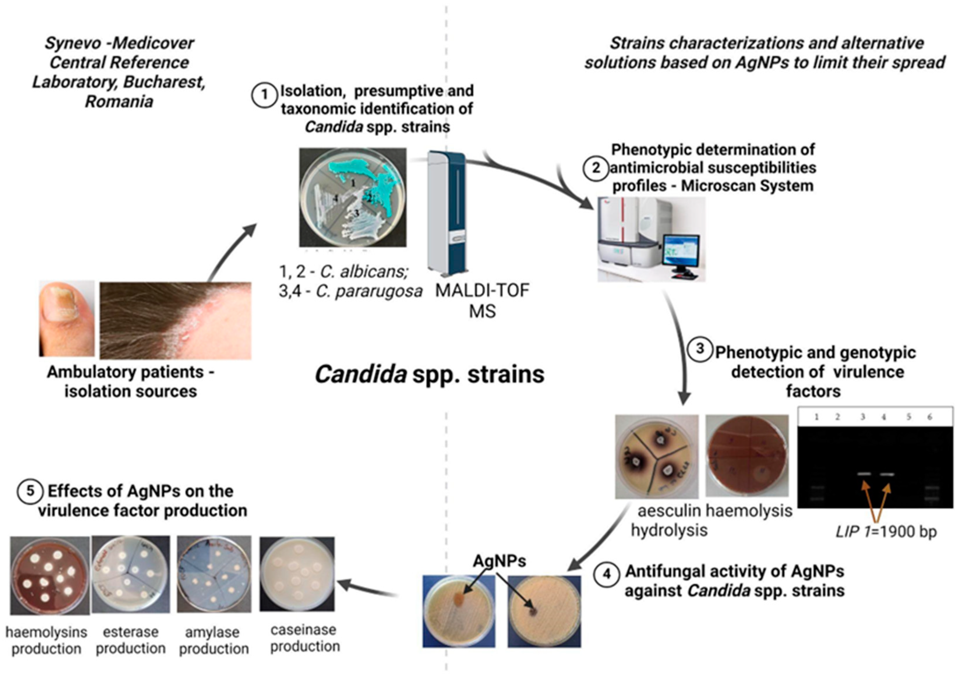
2.1. Strains Identification and Analysis

A total of 62 Candida spp. strains were provided by the Central Reference Synevo-Medicover (Bucharest, Romania) and previously isolated from cutaneous infections represented by nail fragments, interdigital scales, and scales from ambulatory patients during 2021 (Supplementary Table S1) The isolates’ identification was performed based on presumptive characteristics on chromogenic culture media using CHROMagarTM Candida (CHROMagar, Paris, France), and taxonomic identification was carried out using MALDI-TOF MS MBT Smart, with MSP 96 target polished steel BC (Bruker system, Berlin, Germany) (see Figure 1).
The investigated Candida spp. strains were maintained in a Revco LegaciTM Refrigeration System (Copeland, UK) at −80 °C on Sabouraud (Sab, Merck, Darmstadt, Germany) medium supplemented with 20% glycerol. Before their use, the Candida strains were cultivated for 24 h at 37 °C on Sab medium to gain insights into their behavior, survival mechanisms, and pathogenic potential in the human body.

2.2. Antibiotic Susceptibility Testing

The antifungal resistance patterns of Candida spp. strains were evaluated using Integral System Yeast Plus (Liofilchem, Roseto degli Abruzzi, Italy). This system assesses the growth inhibition of strains in media containing different antifungals and a growth indicator. Susceptibility profiles were determined by observing the color changes after inoculating a standardized suspension (1 McFarland) from the tested strains into sterile water and incubating at 37 °C for 48 h. The color indicators were red for sensitive, orange for intermediate, and yellow for resistant. The antifungals tested, and their specific concentrations, were as follows: nystatin (1.25 µg/mL), amphotericin B (2 µg/mL), flucytosine (16 µg/mL), econazole (2 µg/mL), ketoconazole (0.5 µg/mL), clotrimazole (1 µg/mL), miconazole (2 µg/mL), itraconazole (1 µg/mL), voriconazole (2 µg/mL), and fluconazole (64 µg/mL).

2.3. Soluble Virulence Factors: Enzyme and Organic Acid Production

The soluble virulence factors, i.e., metabolic enzymes (amylase, lipase, gelatinase, hemolysins, aesculin hydrolysis, caseinases, and lecithinase) in Candida spp. were assessed on specific solid culture media as previously described, on Sab medium supplemented with 5% sheep blood (hemolysin production revealed by the colorless area around the culture after incubation for 24 h at 37 °C), 2.5% yolk agar (lecithinase activity: a clear zone around the inoculated culture after incubation for 24 h at 37 °C), 1% Tween 80 (Sigma-Aldrich, Burlington, MA, USA; lipase production: a precipitation zone around the spot after incubation for 24 h at 37 °C), 15% soluble casein (caseinase activity: a white precipitate surrounding the culture after incubation for 24 h at 37 °C), 3% gelatine (Merck, Germany; gelatin hydrolysis: a colorless zone around the culture after incubation for 24 h at 37 °C), 1% starch (amylase activity: yellow ring around the culture, while the rest of the plate will be blue after adding Lugol’s solution; Sigma-Aldrich, USA), and Fe3+ citrate (aesculin hydrolysis: a black precipitate around the culture after incubation for 24 h at 37 °C) [46,47,48].

2.4. Biofilm Formation Capacity

The quantification of biofilm formation capacity was assessed as described by Stepanović et al. For this, 20 μL of a 1-McFarland suspension from 24 h Candida spp. cultures grown at 37 °C on Sab medium was inoculated onto Roswell Park Memorial Institute 1640 Medium (RPMI 1640) to a final volume of 120 μL in 96-well microtiter plates. After incubating for 24 h at 37 °C, the plates were washed three times with phosphate-buffered saline (PBS). To facilitate fixation, the cells were treated with methanol for 5 min. Staining was then conducted using a 1% solution of crystal violet (Sigma, USA) for 15 min. Following three additional PBS washes, the cells were resuspended in 33% acetic acid solution. Absorbance (OD) values were measured at 490 nm using the Thermo Scientific Multiskan FC spectrophotometer. All samples were analyzed in triplicate, and the relative biofilm-forming capacity of each strain was assessed as previously described in [49]. The reference strains of C. albicans (ATCC 10231) and C. parapsilosis (ATCC 22019) purchased from the American Type Culture Collection (ATCC, Washington, DC, USA) were used as positive controls for biofilm producers.

2.5. Investigation of Adherence Capacity on HeLa Cells

A modified Cravioto method was used to assess the adherence capacity of the selected Candida spp. strains to HeLa cells (ATCC CCL-2, USA), as previously described [50]. HeLa cells were cultured in 6-well plates with Dulbecco’s Modified Eagle’s Medium (Sigma-Aldrich, USA) and antibiotics for 24 h until 80% confluence. After washing the wells thrice with PBS, we prepared the fungal suspension by incubating yeast in SAB glucose broth at 37 °C for 48 h. The yeast cells were centrifuged, washed, and resuspended to a density of 107 CFU/mL. We then added 1 mL of this suspension to each well and incubated the plates at 37 °C for 2 h to facilitate yeast adherence. Post-adherence, the cells were fixed with methanol and stained with Giemsa solution (Sigma, Darmstadt, Germany). After drying, the samples were examined under a microscope, using a wet objective (×1000 magnification), and photographed. The adherence patterns to eukaryotic cells were defined as follows: localized adherence, characterized by clusters of Candida cells to specific areas on the surface of HeLa cells; aggregative adherence, characterized by microorganisms’ adhesion to the HeLa cell surface in overlapping structures; and diffuse adherence, characterized by a widespread attachment of Candida cells, covering the entire surface of the HeLa cell. The adherence index (AIn) and the average number of fungal cells per HeLa cell were calculated.

2.6. Genotypic Investigation of Virulence Markers Involved in Adherence, Lipolytic Activity, or Genes Encoding β-1,3-glucan Synthase

Simplex and multiplex PCR were used to identify genes associated with adherence (ALS1, ALS3, HWP1) and lipolytic activity (LIP1, LIP4). DNA templates were extracted using a protocol developed by Csutak et al. (2014) [51] and amplified using specific primers and programs outlined in Supplementary Tables S2 and S3. The presence of these genes was verified through gel electrophoresis. Positive strains were used as positive controls (C. albicans ATCC 10231 for ALS1 and 3, and C. tropicalis CMGB165 for LIP1 and 4).

2.7. Evaluation of Antimicrobial and Anti-Biofilm Activity, and the Influence of AgNP Solutions (AgNPs) on Metabolic Activity

2.7.1. Synthesis and Characterization of AgNPs

Approximately 0.888 g of PVP K 30 was dissolved in 80 mL of PEG 400, agitated, and heated to 80 °C until the solution turned clear. At this temperature, 2 mL of 0.5 M AgNO3 was quickly added, producing a dark yellow hue. Stirring continued at 80 °C until the mixture had turned dark brown. The solution was then transferred to a Teflon vat and heated to 220 °C under 1 bar pressure for 2 h. After cooling, the reddish-brown liquid was removed from the vat, yielding 1 mg/mL nanoparticle solution. The silver nitrate (AgNO3), sodium borohydride (NaBH4), polyvinylpyrrolidone (PVP), and polyethylene glycol (PEG 400) were acquired from Roth, hydrogen peroxide (30% H2O2) from Silal Trading, trisodium citrate (Na3C6H5O7) from Alfa Aesar, and 1-butanol from Merck [38]. All reagents and solvents were used without further purification.
For the characterization of AgNPs, FTIR, SEM, TEM, DLS, and XRD methods were used as described by Corbu et al. in 2023 [52].
Before any experiments involving the AgNPs solution, an ultrasonic bath (Daihan Scientific, Seoul, Republic of Korea) set to a frequency range between 20 and 40 kHz for 10 min was used in order to obtain an homogeneous dispersion.

2.7.2. Qualitative Antimicrobial Activity

A modified diffusion method was conducted to perform a rapid screening regarding the antimicrobial activity of AgNPs against a total of 62 strains of Candida spp. From 24 h cultures 1-McFarland microbial suspensions were prepared in sterile distilled water, and then each suspension was evenly spread on Sab medium. Subsequently, 10 μL of 1 mg/mL AgNPs was spotted over. The plates were incubated at 37 °C for 24 h, and at the end of the incubation time the diameters of the growth inhibition zones were measured and converted into arbitrary units: 0 for no inhibition, 1 for a growth inhibition zone diameter up to 10 mm, and 2 for a growth inhibition zone diameter of 11–20 mm [50,53,54].

2.7.3. Quantitative Antimicrobial Activity

A total of 9 strains selected as being sensitive to AgNPs and as representative for each Candida species were used in order to determine the minimal inhibitory concentration of AgNPs. The quantitative antimicrobial activity was assessed in RPMI 1640 broth medium (American Biorganics, Buffalo, NY, USA) by performing serial twofold microdilutions of AgNPs in 100 μL of the medium (ranging between 500 and 0.97 µg/mL). This broth was inoculated in the next step with 20 μL of a 1-McFarland suspension from 24 h cultures grown at 37 °C on Sab medium. The growth was monitored using a BioTek Synergy HTX multi-reader (Santa Clara, CA, USA) by determining the optical density at 600 nm. The positive (untreated cultures) and negative controls (sterility control) were included, and the minimum inhibitory concentration (MIC) values were determined after incubation for 24 h at 37 °C as the last concentration for which no growth was recorded.

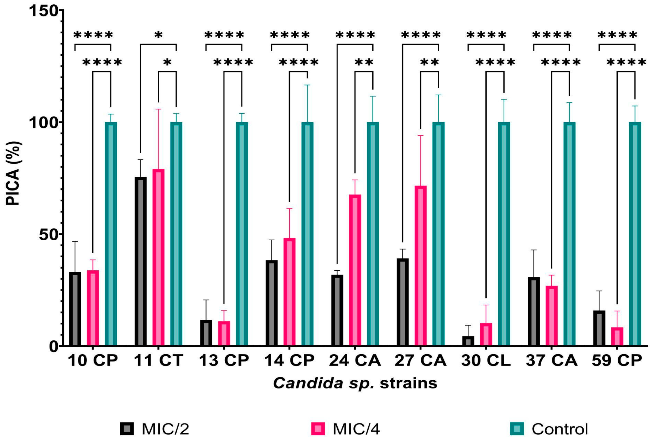
2.7.4. The Influence of AgNPs on Adherence Capacity

Candida spp. strains treated with sub-inhibitory concentrations of AgNPs (MIC/2 and MIC/4) were assessed for their ability to produce soluble virulence factors (hemolysins, amylase, lipase, caseinase, and aesculin hydrolysis) using 10 μL of 1-McFarland suspensions from strains cultured for 24 h at 37 °C on Sab medium [untreated cultures (serving as growth controls) and cultures treated with AgNPs]. After incubating for 24 h at 37 °C, the influence on virulence factor production was determined, based on previously described relations [46,55]. For the determination of the percentage inhibition of Candida adherence (PICA%), we used the same protocol as for the biofilm formation capacity (Section 2.4) and the following relationship: PICA% = (As-Ablank)*100/(Ac-Ablank), where As is the absorbance value of samples treated with sub-inhibitory concentrations of materials at 490 nm, and Ac is the absorbance value of the control (microbial strain not treated with materials) at 490 nm, as previously revealed in [46,55].

2.7.5. The Influence of AgNPs on the Viability of Microorganisms in Biofilms

To assess the viability of microbial cells in biofilms, a colorimetric method based on the reduction of a tetrazolium salt was used, as described in [56]. After incubation in microtiter plates, the microbial suspension was removed, and 150 μL of PBS and 50 μL of MTT 0.3% were added for 2 h at 37 °C. After removing the MTT solution, 150 μL of DMSO and 25 μL of 0.1 M glycine buffer (pH 10.2) were added. The absorbance was measured at 550 nm. For the determination of the percentage inhibition of Candida viability (PICV%), the equation used was PICV% = (As-Ablank)*100/(Ac-Ablank), where As is the absorbance value of samples treated with sub-inhibitory concentrations of materials, and Ac is the absorbance value of the control (microbial strain not treated with materials).

2.8. Extracellular Nitric Oxide Quantification

The extracellular nitric oxide (NO) was measured using previously published techniques, with certain modifications [57]. The NO release was determined by a spectrophotometric assay using the Griess reagent. After the incubation period, the microbial suspension was centrifuged at 6739 g for 10 min. The supernatant was mixed with 50 μL of 2% sulfanilamide (Sigma-Aldrich, Germany) in 5% (v/v) H3PO4 and 50 μL of 0.1% N-(1-naphthyl)-ethylenediamine aqueous solution (Sigma-Aldrich, Germany). After 30 min, the azo dye was measured at λ = 540 nm. To quantify the nitric oxide, a calibration curve was developed using NaNO2 (Sigma-Aldrich Chemie GmbH, Darmstadt, Germany) at concentrations ranging from 1 to 100 μM (R2 = 0.9971).

2.9. Statistical Analysis

Data collected and analyzed in triplicate were presented as means ± SD, and statistical analysis was conducted using GraphPad Prism v10 (GraphPad Software, San Diego, CA, USA). For assessing adherence and virulence factor inhibition, a two-way ANOVA was applied. Dunnett’s multiple comparisons test was used to adjust for multiple comparisons in evaluating the effects of AgNPs at sub-inhibitory concentrations on Candida adherence to inert substrata and the inhibition of virulence factor production, comparing control values with MIC/2 and MIC/4 values for each isolation source. Additionally, extracellular nitric oxide content was analyzed using two-way ANOVA, with Tukey’s method for multiple comparisons corrections using a single pooled variance, comparing samples to strain controls. Correlations among extracellular nitric oxide content, adherence, and virulence factor inhibition were determined using Pearson correlation. The threshold for statistical significance was established at p < 0.05.

3. Results

3.1. Phenotypic and Genotypic Features of Resistance and Virulence Markers in Candida spp. Isolates

In this study, 62 strains of Candida spp. were previously isolated from dermatomycoses and identified using chromogenic CHROMagarTM Candida media and by mass spectrophotometry (MALDI-TOF MS), in the following decreasing frequency order: 45.16% C. parapsilosis; 29.03% C. albicans; 9.67% C. guilliermondii; 3.22% C. pararugosa, C. tropicalis, and Candida lusitaniae; 1.66% Candida haemulonii, Candida famata, Candida metapsilosis, and Candida kefyr.
The analyzed strains were isolated mostly from women (62.90% of cases) aged between 15 and 77 years. Also, the main source of isolation from the ambulatory patients treated in Bucharest was the right hand nail (40.32%), followed by the right foot (22.58%), left foot (16.12%), left hand nail (14.51%), and scalp and face (3.22%) (see Table 1).
The antifungal susceptibility profiles determined using Integral System Yeast Plus demonstrated the following patterns, by species: C. parapsilosis isolates were resistant to amphotericin B (29%), clotrimazole (24%), nystatin (18%), econazole (11%), miconazole (7%), and itraconazole (4%); C. albicans to amphotericin B (22%), clotrimazole (27%), nystatin (17%), econazole (11%), and miconazole (6%); C. tropicalis to clotrimazole, econazole, and nystatin (50/50/%); C. guilliermondii to nystatin (17%); C. lusitaniae to nystatin and amphotericin B (100%); and C. haemulonii to clotrimazole and econazole (100%) (see Table 1). At the opposite side, C. parapsilosis strains were sensitive only to ketoconazole, voriconazole, and fluconazole; C. albicans to flucytosine, ketoconazole, itraconazole, voriconazole, and fluconazole; C. tropicalis to amphotericin B, flucytosine, ketoconazole, miconazole, itraconazole, voriconazole, and fluconazole; C. guilliermondii to all antifungals except nystatin; C. krusei to all antifungals tested; along C. metapsilosis, C. pararugosa, C. famata, and C. haemulonii to all except econazole and clotrimazole; and C. lusitaniae to all antifungals except amphotericin B and nystatin.
Virulence factors tested on specific culture media supplemented with various nutrients revealed that Candida spp. strains were positive for aesculin hydrolysis (93.35%), caseinase (79.03%), amylase (35.48%), hemolysins (30.65%), and lipase production (20.79%). The virulence factors’ distribution by species and isolation sources was as follows: C. albicans, isolated from feet, hand nails, and face scales was positive for aesculin hydrolysis, hemolysins, amylase, and lipase; C parapsilosis from feet, left hand nails, and scalps was positive for aesculin hydrolysis, caseinase, hemolysins, amylase, and lipase; C. tropicalis, isolated from left feet, was positive for aesculin hydrolysis, caseinase, hemolysins, and amylase; C. guilliermondii from left feet, hand nails, and scalps was positive for aesculin hydrolysis, caseinase, hemolysins, amylase, and lipase; C. lusitaniae from right hand nails was positive for aesculin hydrolysis, caseinase hemolysins, amylase, and lipase; C. krusei from left hand nails was positive for aesculin hydrolysis and amylase; C. metapsilosis from right hand nails was positive for aesculin hydrolysis and caseinase; C. famata from left feet was positive for caseinase; and C. pararugosa isolated from left and right hand nails was positive for aesculin hydrolysis (see Table 2).
Based on their biofilm formation capacities, the studied strains of C. albicans and NAC were classified into the following categories: 20.64% did not produce biofilms (NP), while 37.09% were categorized as weak biofilm producers (W), 1.61% as moderate biofilm producers (M), and 30.64% as strong biofilm producers (Table 3).
The Candida spp. strains included in our study were also investigated for their adherence patterns to eukaryotic cells (HeLa cells). It was found that most of the strains presented all investigated adherence patterns, in the following decreasing order: diffuse (47.8%), localized (29.7%), and aggregative (22.5%). The adherence index (AIn) ranged from 26% to 50%, indicating that the data correspond to the high frequency of diffuse adherence pattern identification (see Table 3).
Genotypic assays were performed for the targeted identification of virulence markers such as the ALS1, ALS3, HWP1, LIP1, and LIP4 genes. The results showed that the strains were positive i for the following genes, in decreasing order of frequency: LIP1 (100% of C. tropicalis), HWP1, and ALS3 (82.35% of C. albicans) (see Table 2).

3.2. Antimicrobial and Anti-Biofilm Activity, and the Impact of AgNP Solutions on the Adherence Capacity and Metabolic Activity

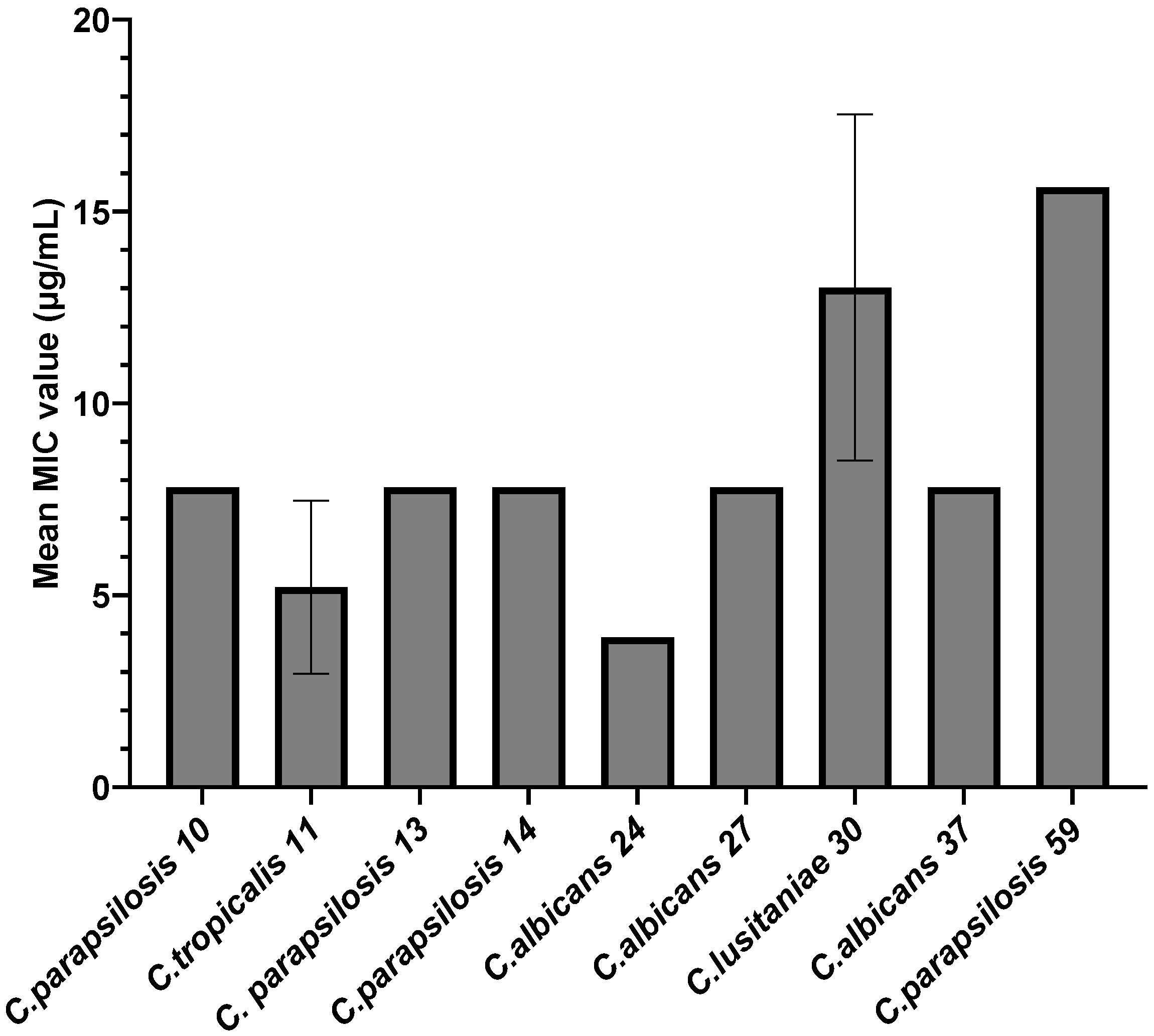
3.2.1. Qualitative and Quantitative Antimicrobial Activity of AgNPs against Candida spp. Strains

The qualitative analysis of AgNPs showed that most of the Candida spp. strains analyzed in this study were identified as having arbitrary unit values of 0 (AU0; 66.12%), 1 (AU1; 19.35%), and 2 (AU2; 14.51%). In correlation with the isolation sources, it was observed that most strains of Candida spp. that presented arbitrary units (AU) = 2 and AU = 1 originated from the right hand nail (four and six strains, respectively), followed by the right foot nail (three and five strains, respectively). However, AU = 0 was the predominant identified value for the qualitative screening of AgNPs (see Figure 2).
However, only strains that showed 2 arbitrary units were selected, as these were the most sensitive to the AgNPs tested.
In the case of the strains for which, following the qualitative testing, an inhibition zone equivalent to 2 arbitrary units was obtained (n = 9), the minimum inhibitory concentration values were also determined. According to Figure 3, the inhibitory effect of AgNPs depends on the species to which the tested strain belongs. Thus, the average MIC values obtained ranged from 3.90 µg/mL to 15.62 µg/mL, with the most sensitive strain being C. albicans 24 (MIC = 3.90 µg/mL), followed by C. tropicalis 11 (MIC = 5.20 µg/mL), while the most resistant strains were C. parapsilosis 59 (MIC = 15.62 µg/mL) and C. lusitaniae 30 (MIC = 13.02 µg/mL). For subsequent tests, MIC/2 and MIC/4 were considered, with fractions of the MIC value being determined for each strain.

3.2.2. Anti-Biofilm Activity of AgNPs

The selected Candida strains also presented the ability to develop biofilms on the surface of inert substrata. Thus, we also tested the impact of sub-inhibitory concentrations of AgNPs on their ability to adhere to polypropylene surfaces. According to Figure 4, their adherence ability was significantly reduced at both MIC/2 and MIC/4, the best results being recorded for the C. lusitaniae 30 strain, where a reduction of up to 95% in adhesion capacity was observed when it was exposed to MIC/2 AgNPs (PICA% MIC/2 = 4.41%; PICA% MIC/4 = 10.57%). Similar results were obtained for the C. parapsilosis 13 (PICA% MIC/2 = 11.66%; PICA% MIC/4 = 11.09%) and C. parapsilosis 59 strains (PICA% MIC/2 = 15.89%; PICA% MIC/4 = 8.36%). For the other C. parapsilosis strains, the effect was diminished, but the PICA% values remained below 50% when exposed to MIC/2 AgNP concentrations, indicating high efficiency in preventing biofilm formation. Although C. tropicalis 11’s growth was strongly inhibited by the presence of AgNPs, the effect of AgNPs on its ability to adhere was limited (PICA% values of up to 75% for MIC/2 and MIC/4). This indicates that, in the case of this strain, the presence of AgNPs induces a generalized state of stress, against which special metabolic pathways are activated, including those associated with adherence. Similarly, for the two C. albicans strains, AgNPs reduced their ability to adhere, with the PICA% being 30.814% and 31.89%, respectively, when exposed to MIC/2.
Although the crystal violet (CV) test serves as an indicator of the biomass attached to a biofilm without revealing the metabolic state of the cells, the MTT test, a tetrazole salt, highlights the presence of metabolic active cells, thus serving as an indicator of the breathing of living cells [58,59]. In biofilms, the MTT test can be used as an indicator of the viable cells attached, while CV colors both viable and nonviable cells that are attached to the inert substrate [60]. The results of the MTT test confirmed that AgNPs significantly inhibited the metabolic activity of biofilms formed by all Candida species in this work (p < 0.0001) at MIC/2 and MIC/4. The addition of AgNPs prevented the initial attachment of cells, reducing not only the biomass (as indicated by the CV test) but also the metabolic activity of the cells, which resulted in 0.36 ± 0.07% PICV for C. albicans strain 37 and 0.10 ± 0.0% PICV for C. albicans strain 27 (Table 4). For all strains, the metabolic activity was inhibited as the AgNP concentration increased.

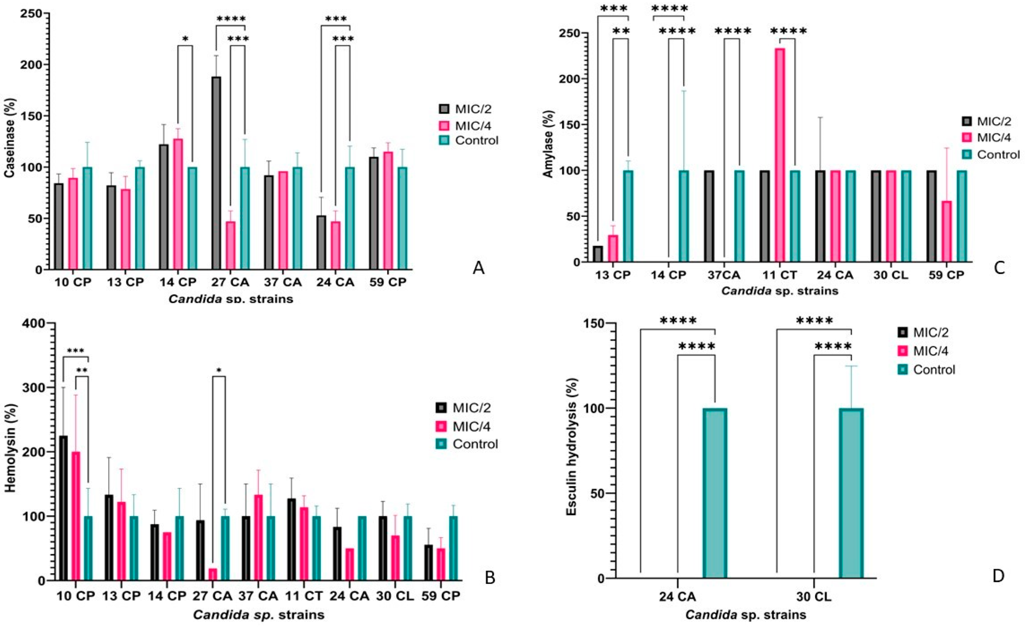
3.2.3. The Impact of AgNPs on Metabolic Activity in Selected Candida spp. Isolates

Of the tested Candida strains, 79.03% were able to hydrolyze casein, and 93.55% hydrolyzed aesculin. Smaller percentages of strains were able to secrete amylase (35.48%), hemolysins (30.65%), and lipases (20.97%). No Candida strain was able to produce gelatinases, DNase, or lecithinase. Under these conditions, the strains that presented the capacity to secrete the most varied spectrum of soluble virulence factors and the greatest sensitivity to the antimicrobial action of AgNPs were tested in order to establish the effects of AgNPs on the release of hydrolytic enzymes linked to virulence and pathogenicity. For caseinase production, the best results were obtained in the C. albicans 24 strain, for which the secretion capacity was reduced by up to 50% compared to the untreated strain. A similar result was obtained for the C. albicans 27 strain treated with MIC/4 AgNPs (47.05%), but when the concentration of AgNPs was increased the effect was the opposite, stimulating the secretion of caseinase in the culture medium (Figure 5A).
C. albicans 24 and C. lusitaniae 30 were also tested in the presence of sub-inhibitory concentrations of AgNPs for their ability to hydrolyze aesculin and, according to Figure 5D, their ability was completely inhibited at both MIC/2 and MIC/4 concentrations.
Amylase production was completely inhibited for the C. parapsilosis 14 and C. albicans 37 strains when treated with MIC/4 AgNPs. Good results were also obtained for the C. parapsilosis 13 strain, the production being reduced by almost 80% in the presence of MIC/2. On the other hand, in the case of C. tropicalis 11, it seems that the presence of sub-inhibitory concentrations of AgNPs had a stimulatory effect (Figure 5C). When tested for esterase production, its ability to hydrolyze Tween 80 was completely inhibited in the presence of MIC/4 AgNPs.
Although nine Candida strains were tested for hemolysin production in the presence of sub-inhibitory concentrations of AgNPs, only in the case of C. parapsilosis 10 was a significant stimulatory effect observed (Figure 5B).

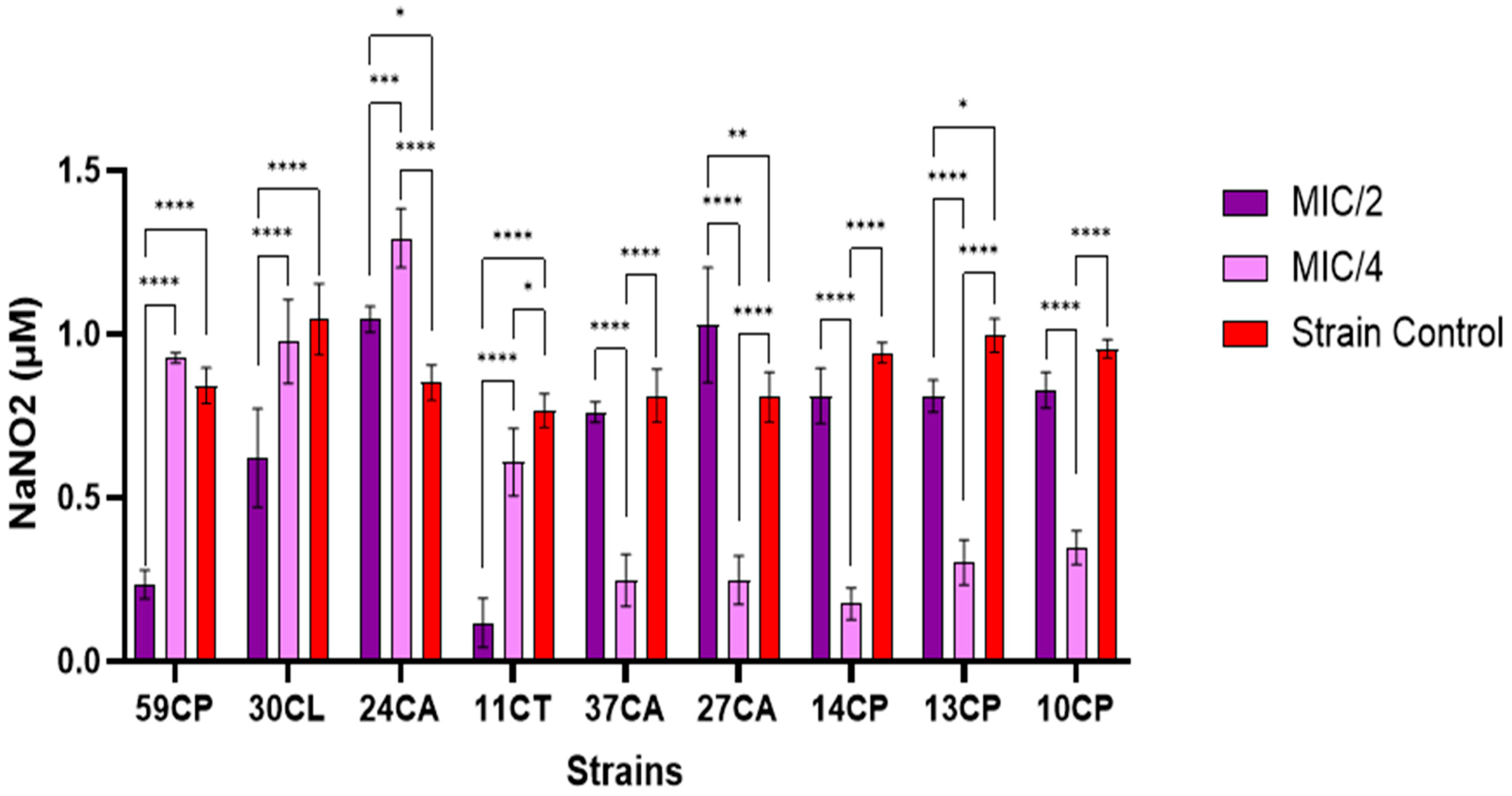
3.2.4. Extracellular Nitric Oxide Content

Exogenous NO at sublethal doses may induce microorganisms within biofilms to switch from sessile behavior to free-swimming planktonic behavior. NO, at non-lethal levels, has also been shown to increase the susceptibility of various biofilms to antimicrobial treatments [61]. From Figure 6, it can be observed that the concentration of extracellular NO increased with the increase in the AgNPs’ concentration in most C. parapsilosis strains, except for 59 CP, which exhibited a higher MIC value compared to other strains. In the case of C. albicans strains, it was highlighted that lower sample concentrations generated higher extracellular NO. Among the strains tested at MIC/2, it can be seen that only strains 27CA (p < 0.01) and 24CA (p < 0.05) showed a significantly higher extracellular NO concentration than the strain control (Figure 7). A significant decrease in extracellular NO concentration was observed compared to the strain control at MIC/4 values for variants 37CA, 27CP, 14CP, 13CP, and 10CP (p < 0.0001), which may indicate that the microorganisms used a variety of enzymes and complex processes to recognize, collect, and convert NO into less reactive molecules. Due to its high reactivity, NO can be at inappropriate concentrations and is thus rapidly transformed by microorganisms through the process of aerobic denitrification and oxidation [62].
The anti-biofilm activity of exogenous NO released by low-molecular-weight donors (e.g., AgNP) is dose-dependent; eradication concentrations also depend on the microbial species and potential genetic modifications [55]. The relationship between extracellular NO concentration produced at MIC/2 levels and microbial adhesion revealed that the higher the NO content, the stronger the microbial inhibition of adherence. The Pearson correlation showed that one variable increases as the other grows (r > 0), the correlations being better for the strains of C. albicans (r = 0.56) than for C. parapsilosis (r = 0.46) (Figure 7A,B). In the case of C. albicans, it can be observed that the correlations between NO vs. hemolytic activity (r = −0.82) and NO vs. MIC value (r = −0.55) are inversely proportional, i.e., the higher the extracellular NO content, the lower the hemolysis and MIC value, while the correlations between inhibition of adhesion vs. caseinase activity (r = 0.92), MIC values vs. caseinase activity (r = 0.72), and MIC values vs. hemolytic activity (r = 0.93) are directly proportional, which suggests that the greater the concentration of AgNPs, the higher the caseinase activity and hemolysis, and the better the inhibition of microbial adherence.
In the case of C. parapsilosis (Figure 7B), the following correlations can be highlighted: PICA vs. caseinase activity (r = −0.82), PICA vs. hemolysis (r = 0.51), caseinase activity vs. hemolysis (r = 0.82), NO vs. hemolysis (r = −0.62), and sample concentration vs. hemolysis (r = 0.62).

4. Discussion

Our obtained results highlighted a high diversity of species responsible for dermatomycoses in the Bucharest community, as follows, in decreasing frequency order: C. parapsilosis, C. albicans, C. guilliermondii, C. pararugosa, C. tropicalis, C. lusitaniae, C. haemulonii, C. famata, C. metapsilosis, and C. kefyr. Depending on the identified species, strains exhibited different resistance profiles, e.g., C. albicans, C. parapsilosis, C. guilliermondii, and C. lusitaniae were resistant to nystatin and amphotericin B, while C. albicans, C. parapsilosis, C. tropicalis, and C. haemulonii were resistant to clotrimazole and econazole. Genotypic assays for the identification of the virulence markers responsible for adherence (ALS1, ALS3, and HWP1) and lipolytic activity (LIP1 and LIP4) showed that most of the tested strains belonging to C. albicans were positive for ALS1 and HWP1 genes, and LIP1 in the case of C. tropicalis.
The studied strains showed varying ability to form biofilms, differentiated by species as follows: C. parapsilosis was categorized as a strong, weak, and moderate biofilm producer in decreasing frequencies of 38.88%, 33.33%, and 5.55%, respectively; C. albicans was divided into weak and strong biofilm producers (35.71%/32.14%, respectively); C. guilliermondii was predominantly a weak biofilm producer (50%); C. lusitaniae had strong biofilm-producing strains (100%); C. tropicalis had an equal distribution between weak and strong biofilm formation (50%/50%); and C. krusei and C. parrarugosa were primarily weak biofilm producers (100% and 50%, respectively), excluding those strains that were not biofilm producers. Our results suggest that the most sensitive strains to AgNPs were C. albicans 24 and C. tropicalis 11, and at the opposite side were C. parapsilosis 59 and C. lusitaniae 30. Due to the weak-to-strong biofilm formation capacity of the investigated Candida strains, the impact of sub-inhibitory concentrations of AgNPs on their ability to adhere to inert substrata was further investigated, and it was found that the adherence capacity for the most resistant strains was significantly reduced at both MIC/2 and MIC/4 values (e.g., for C. lusitaniae 30, a reduction of up to 95% in the adherence capacity was observed after the treatment with MIC/2 AgNPs, followed by C. parapsilosis 13 and C. parapsilosis 59).
Strains were positive for aesculin hydrolysis, caseinase, amylase, hemolysins, and lipase production and showed different biofilm formation capacities. Furthermore, results for the adherence capacity of Candida spp. strains to eukaryotic cells showed that a high diffuse adherence frequency was obtained. The influence of AgNPs on the secretion of hydrolytic enzymes responsible for virulence and pathogenicity demonstrated that the treatment of microbial cultures with sub-inhibitory concentrations of AgNPs caused stimulatory or inhibitory effects that varied depending on the species, the tested enzyme, or the substrate.
Previously, Christofidou et al., in a comparative study including Romanian and Greek Candida spp. isolates, showed that cutaneous candidiasis is caused mostly by C. albicans, followed by C. parapsilosis, Candida glabrata, C. tropicalis, C. krusei, C. guilliermondii, and other Candida spp. resistant to ketoconazole and itraconazole [63]. As in our study, which included Candida spp. strains recovered from mucocutaneous candidiasis, several other authors have shown a high prevalence of NAC in different clinical sources as a consequence of immunocompromised status, prolonged antifungal therapy, or other administered therapies [64,65,66,67]. In Romania, Sadik et al. (2019) [50], Najee et al. (2018) [68], and Sadik et al. (2017) [69] showed a wide array of adherence genes in Romanian C. albicans with varying ability to form biofilms on inert substrata and adherence patterns to eukaryotic cells, with strains isolated from different sources in hospitalized patients aged 20 to 85 and 25 to 89 years [50,68,69].
Several other studies have also highlighted the hemolysin production, adherence markers, and biofilm capacity of C. albicans and NAC, especially from intra-hospital infection settings, e.g., Singh et al. (2020) [70]. In 2019, Sadeghi et al. showed that C. albicans strains prevalent in cases of cutaneous candidiasis in outpatient settings in Tehran, Iran, indicated a change in the disease’s epidemiological patterns, contrasting with our findings. Their research also identified the presence of other NAC, e.g., C. parapsilosis, C. tropicalis, and C. guilliermondii—species that were also detected in our study, which are capable of biofilm formation and exhibit phospholipase activity [71]. Mello et al., in 2020, reported the prevalence of C. albicans, followed by C. parapsilosis complex and C. krusei biofilm-producing strains—protease and phospholipase producers isolated from dermatomycosis in Sao Paulo, Brazil, during 2016 and 2017, underscoring the importance of recognizing different fungal species involved in dermatomycosis cases, as well as understanding their pathogenicity and susceptibility to antifungals for effective preventive and therapeutic actions against them [72].
In addition to the classic solutions for treating cutaneous candidiasis based on the use of traditional antifungals (fluconazole, miconazole, clotrimazole, econazole, nystatin, micafungin, anidulafungin, and caspofungin) or topical medications based on potassium permanganate, gentian violet solution, or aluminum acetate solution [73], AgNPs could be a promising tool for treating dermatomycosis, due to their known antifungal activity via different mechanisms, including ion release, oxidative and nitrosative stress, damage to membranes and cell walls, inhibition of biochemical processes, reduction in ATP levels, and dysfunction in DNA, proteins, and mitochondria [63,64], as supported by several studies [74,75]. AgNPs’ effects on human cells are complex and depend on several factors, including their size, coating, concentration, and the specific types of human cells they interact with. In terms of size, smaller NPs tend to have a higher cytotoxic effect due to their greater surface area–volume ratio, which facilitates more interactions with cellular components. However, certain coatings can mitigate these effects by providing a barrier that reduces cellular uptake and toxicity [76] Concerning human cell types, different cells exhibit varying levels of sensitivity to AgNPs. For instance, human hepatoma cells (HepG2) and human lung epithelial cells have shown different responses to AgNP exposure, with some studies indicating that specific coatings or modifications can reduce cytotoxicity while maintaining antimicrobial efficacy [77,78]. AgNPs can induce apoptosis and necrosis in human osteoblast cells through oxidative damage and nitric oxide signaling pathways [77,78].
Despite their potential cytotoxic effects, AgNPs are being explored for their therapeutic benefits, such as antiviral and antibacterial properties. Studies have demonstrated that AgNPs can inhibit the replication of viruses and bacteria at non-cytotoxic concentrations, suggesting their potential use in medical applications without significant harm to human cells [76].
Comparable to our findings, high antifungal activity of AgNPs has been demonstrated in C. albicans and NAC isolates recovered from different isolation sources—including skin swabs, nails, oral swabs, cervical swabs, urine, sputum, endotracheal aspirates—that are resistant to fluconazole, ketoconazole, clotrimazole, itraconazole, amphotericin B, and nystatin [79]. The anti-biofilm properties of biogenic AgNPs against reference strains of C. albicans, as well as the inhibition of yeast-to-hyphal transition, increased the membrane permeabilization, ROS production, and oxidative stress, and finally led to apoptosis [80]. Also, the influence of AgNPs against C. albicans biofilms was explored, emphasizing the pressing demand for new antifungal treatments due to the biofilms’ resistance to conventional antifungal agents. This study revealed that AgNPs exhibit potent inhibitory effects on biofilm formation and against pre-formed biofilms of C. albicans. Furthermore, our findings underscore the potential of AgNPs as a promising treatment strategy against C. albicans infections, demonstrating that AgNPs target both growth stages by disrupting the cell membrane structure of the fungal cells. AgNPs show significant antifungal activity, suggesting their role as instrumental in combating biofilm-associated candidiasis, offering a new avenue for antifungal intervention [39]. Several other studies have demonstrated the antimicrobial activity of AgNPs against C. albicans, C. dubliniensis, and C. guilliermondii through qualitative and quantitative assays, as well as the capacity of uninvestigated NPs to disrupt the cell membrane/wall based on microscopic examination [81]. Furthermore, Vazquez-Muñoz demonstrated the fungicidal effect of AgNPs and their ability to reduce cell viability when combined with fluconazole. Ultrastructural analysis revealed the presence of AgNPs both outside and inside the cells, suggesting a potential mechanism of action. Their study also discussed the potential clinical implications of using AgNPs in combination with antifungals and highlighted the need for further research to understand their safety and effectiveness [82].
The effectiveness of AgNPs in combating fungal infections in plants and humans was also demonstrated, with a specific emphasis on Candida spp., highlighting that AgNPs effectively inhibit the growth of Candida spp. and influence the production of virulence factors, e.g., biofilm formation. Furthermore, research supports the notion that AgNPs not only reduce yeast cell proliferation but also impact various virulence determinants, including biofilm maturation [83]. In 2022, the fungicidal properties of AgNPs were demonstrated against resistant strains of C. albicans and NAC, isolated from both outpatients and inpatients in Egypt. The authors proposed that AgNPs could serve as a viable alternative to traditional antifungal agents [84]. Moreover, the increased hemolysin production in the presence of AgNPs described in this study was observed previously, enhancing the pathogenicity of these fungi, possibly due to interactions with fungal cell membranes or metabolic processes [85]. Comparative research shows that both C. albicans and NAC strains respond similarly to NP exposure [86]. Further studies suggest that NPs not only compromise the structural integrity of Candida biofilms but also stimulate hemolysin production, impacting the biofilm’s resilience and offensive capabilities [39,87]. These findings point to a potential for using silver nanoparticles to modulate fungal virulence, although more research is needed to explore the mechanisms and therapeutic possibilities.
Furthermore, AgNPs act as carrier systems for different antifungal agents, e.g., the synergistic interaction of AgNPs and fluconazole was demonstrated in C. albicans planktonic cells [88], with a decrease in ergosterol levels and cell membrane disruption by downregulating the ERG1, ERG11, and ERG25 genes, and cellular damage as a consequence of the ROS production [89]. Several other studies have highlighted the synergistic interactions of AgNPs functionalized with poly(methacrylic acid) and fluconazole in fluconazole-resistant C. albicans [90], as well as the synergistic effects of biogenic AgNPs derived from Anabaena variabilis and fluconazole against C. albicans planktonic cells [80], or the antifungal effects and the impact on biochemical processes of green-synthesized silver–copper nanoparticles against Candida strains [91].
This study’s findings might be limited by its small sample size and specific demographic focus, having analyzed only 62 Candida spp. strains previously isolated from Bucharest outpatients (2021). This narrow geographic and temporal scope may not capture the full diversity and potential variations in antimicrobial resistance and virulence traits of Candida strains found in other regions or over different periods. Such limitations could hinder the broader applicability of the results to other settings or over time, as treatment practices and environmental factors evolve.
The use of qualitative (adapted disk diffusion method) and quantitative (serial twofold microdilutions) assessments might not fully capture the dynamics of nanoparticle–fungal interactions, particularly if the nanoparticles form aggregates or interact differentially with the assay components. Additionally, this study’s reliance on in vitro assays to predict clinical efficacy could be a limitation, as these conditions do not fully replicate the complex environment of a human infection, including immune responses and the presence of other microbiota.
These limitations suggest that, while this study provides valuable insights into the potential of AgNPs against Candida infections, the results should be interpreted with caution, considering the need for further characterization of the nanoparticles and validation of the findings in more clinically relevant models.
Considering the limited number of studies that have examined the relationship between biofilm production by dermatomycosis isolates and their consequences on pathogenesis of infections, our study concentrated on the phenotypic and genotypic traits of 62 Candida spp. previously isolated from community-acquired infections in the Central Reference Synevo-Medicover Laboratory, Bucharest, Romania, as well as on the efficiency of alternative solutions based on AgNPs to limit the spread of virulent Candida spp. strains. This study has significant implications for both practice and policy. The findings enhance our understanding of the characteristics and behaviors of these strains, which is crucial for developing targeted treatment strategies. Moreover, this study’s exploration of alternative solutions based on AgNPs presents a promising tool for limiting the spread of virulent Candida strains. This approach could potentially improve treatment outcomes and reduce the reliance on traditional antifungal agents, which often face resistance issues. Therefore, this study supports the need for integrating advanced diagnostic methods and innovative treatments into clinical practice and informing policy decisions aimed at controlling Candida infections more effectively. Future research should focus on the following: understanding how these Candida spp. develop resistance to conventional antifungal agents, as well as how AgNPs can overcome these mechanisms; evaluating the safety and efficacy of AgNPs in treating Candida infections, ensuring that they are a viable alternative to current therapies; exploring the most effective ways to administer AgNPs, whether through topical, systemic, or combined therapies; investigating the long-term impacts of AgNP use on both patients and microbial communities, to ensure that there are no adverse effects; comparing the effectiveness of AgNPs with other emerging antifungal agents; and providing data to support the development of guidelines and policies for the clinical use of AgNPs, ensuring that they are appropriately integrated into treatment protocols. Overall, this study highlights the need for continued research into alternative solutions based on AgNPs to address the growing challenge of antifungal resistance in Candida spp.

5. Conclusions

Considering the scarcity of research on the virulence of Candida spp. in Romanian dermatomycosis cases, this study employed a multifaceted approach.
This study represents the first characterization of Candida spp. strains isolated from dermatomycosis in ambulatory patients from Bucharest, the capital city of Romania, and provides valuable insights into the prevalence, diversity, and antifungal susceptibility of these pathogens. This study found that a variety of Candida species are responsible for dermatomycosis in this urban population, highlighting the importance of accurate identification for effective treatment. Notably, our findings indicate a concerning trend of antifungal resistance among certain strains, underscoring the need for ongoing surveillance and tailored therapeutic strategies. This initial characterization serves as a crucial step towards understanding the epidemiology of Candida infections in Bucharest, ultimately contributing to better clinical management and control measures in the region. Furthermore, the obtained results constitute a preliminary study suggesting the potential use of AgNPs as therapeutic alternatives to combat Candida spp. strains causing dermatomycosis.

Supplementary Materials

The following supporting information can be downloaded at: https://www.mdpi.com/article/10.3390/jof10080563/s1, Table S1: Candida albicans and Candida non-albicans strains investigated in the study; Table S2: Primers for virulence genes detection; Table S3: Amplification programs for virulence genes detection; Table S4: The AgNPs’ MIC values and the corresponding MIC/2 and MIC/4 for PICA determination in selected Candida spp. strains; Table S5: The influence of AgNPs sub-inhibitory concentrations (MIC/2) on the virulence factor production in selected Candida spp. strains; Table S6: The influence of AgNPs sub-inhibitory concentrations (MIC/4) on the virulence factor production in selected Candida spp. strains; Figure S1: Electrophoresis gel for LIP1 gene (1900 bp) gene detection; Figure S2: Electrophoresis gel for LIP4 gene (2300 bp) gene detection; Figure S3: Electrophoresis gel for HWP1 gene (570 bp) gene detection; Figure S4: Electrophoresis gel for ALS1 gene (320 bp) gene detection; Figure S5: Electrophoresis gel for ALS3 gene (185 bp) gene detection.

Author Contributions

Conceptualization, I.G.-B., V.M.C., I.C.M. and A.-M.G.; methodology, I.G.-B., V.M.C., A.Ș.D., L.M., I.P., A.-M.G., I.C.M., I.C.B., R.P., D.V.M. and D.F.; software, V.M.C., I.C.B. and I.C.M.; validation, I.G.-B., L.M., I.C.M., T.V.-D., O.C., D.F. and V.M.C.; formal analysis, V.M.C., O.C.; I.C.M., I.C.B. and I.G.-B.; investigation, I.G.-B., V.M.C., A.-M.G., A.Ș.D., R.P., D.V.M., I.P. and I.C.B.; resources, I.G.-B. and D.F.; data curation, V.M.C., T.V.-D. and I.G.-B.; writing—original draft preparation, I.G.-B., V.M.C., I.C.M., R.P., D.V.M., A.Ș.D., L.M., I.P. and A.-M.G.; writing—review and editing, I.G.-B., O.C., T.V.-D. and D.F.; project administration, I.G.-B. All authors have read and agreed to the published version of the manuscript.

Funding

This study was supported by PN-III-P2-2.1-PED-2021-2526 (736 PED/2022) research projects awarded by the Romanian Executive Agency for Higher Education, Research, Development, and Innovation.

Institutional Review Board Statement

The study was of was approved by the Institutional Review Board of Synevo Romania who provided the Candida isolates used in the analysis.

Informed Consent Statement

Informed consent was obtained with the approval of the Medical Director of Synevo-Medicover Central Reference Laboratory from Bucharest, Romania, and all of the necessary steps were taken to protect patients’ privacy and confidentiality.

Data Availability Statement

AgNP samples and previous characterization, Candida spp. isolates, and datasets pertaining to the results can be obtained from the authors.

Conflicts of Interest

The funders had no role in the design of the study; in the collection, analyses, or interpretation of data; in the writing of the manuscript; or in the decision to publish the results.

References

  1. Barantsevich, N.; Barantsevich, E. Diagnosis and Treatment of Invasive Candidiasis. Antibiotics 2022, 11, 718. [Google Scholar] [CrossRef]
  2. Freitas, C.G.; Felipe, M.S. Candida albicans and Antifungal Peptides. Infect. Dis. Ther. 2023, 12, 2631–2648. [Google Scholar] [CrossRef]
  3. Ferngren, G.; Yu, D.; Unalan-Altintop, T.; Dinnétz, P.; Özenci, V. Epidemiological patterns of candidaemia: A comprehensive analysis over a decade. Mycoses 2024, 64, e13729. [Google Scholar] [CrossRef]
  4. Richardson, J.P.; Moyes, D.L. Adaptive immune responses to Candida albicans infection. Virulence 2015, 6, 327–337. [Google Scholar] [CrossRef]
  5. Patangia, D.V.; Anthony Ryan, C.; Dempsey, E.; Paul Ross, R.; Stanton, C. Impact of antibiotics on the human microbiome and consequences for host health. Microbiologyopen 2022, 11, e1260. [Google Scholar] [CrossRef]
  6. Esaiassen, E.; Fjalstad, J.W.; Juvet, L.K.; van den Anker, J.N.; Klingenberg, C. Antibiotic exposure in neonates and early adverse outcomes: A systematic review and meta-analysis. J. Antimicrob. Chemother. 2017, 72, 1858–1870. [Google Scholar] [CrossRef]
  7. Czechowicz, P.; Nowicka, J.; Gościniak, G. Virulence Factors of Candida spp. and Host Immune Response Important in the Pathogenesis of Vulvovaginal Candidiasis. Int. J. Mol. Sci. 2022, 23, 5895. [Google Scholar] [CrossRef]
  8. Xiao, Z.; Wang, Q.; Zhu, F.; An, Y. Epidemiology, species distribution, antifungal susceptibility and mortality risk factors of candidemia among critically ill patients: A retrospective study from 2011 to 2017 in a teaching hospital in China. Antimicrob. Resist. Infect. Control 2019, 8, 89. [Google Scholar] [CrossRef]
  9. Saiprom, N.; Wongsuk, T.; Oonanant, W.; Sukphopetch, P.; Chantratita, N.; Boonsilp, S. Characterization of Virulence Factors in Candida Species Causing Candidemia in a Tertiary Care Hospital in Bangkok, Thailand. J. Fungi 2023, 9, 353. [Google Scholar] [CrossRef]
  10. Yang, Y.L. Virulence factors of Candida species. J. Microbiol. Immunol. Infect. 2003, 36, 223–228. [Google Scholar]
  11. Silva, S.; Negri, M.; Henriques, M.; Oliveira, R.; Williams, D.W.; Azeredo, J. Adherence and biofilm formation of non-Candida albicans Candida species. Trends Microbiol. 2011, 19, 241–247. [Google Scholar] [CrossRef]
  12. Naglik, J.R.; Challacombe, S.J.; Hube, B. Candida albicans secreted aspartyl proteinases in virulence and pathogenesis. Microbiol. Mol. Biol. Rev. 2003, 67, 400–428. [Google Scholar] [CrossRef] [PubMed]
  13. Ghannoum, M.A. Potential role of phospholipases in virulence and fungal pathogenesis. Clin. Microbiol. Rev. 2000, 13, 122–143. [Google Scholar] [CrossRef]
  14. Tournu, H.; Van Dijck, P. Candida biofilms and the host: Models and new concepts for eradication. Int. J. Microbiol. 2012, 2012, 845352. [Google Scholar] [CrossRef]
  15. Maubon, D.; Garnaud, C.; Calandra, T.; Sanglard, D.; Corne, M. Resistance of Candida spp. to antifungal drugs in the ICU: Where are we now? Intensive Care Med. 2014, 40, 1241–1255. [Google Scholar] [CrossRef] [PubMed]
  16. Perlin, D.S. Mechanisms of echinocandin antifungal drug resistance. Ann. New York Acad. Sci. 2015, 1354, 1–11. [Google Scholar] [CrossRef]
  17. Pfaller, M.A.; Diekema, D.J. Epidemiology of invasive candidiasis: A persistent public health problem. Clin. Microbiol. Rev. 2007, 20, 133–163. [Google Scholar] [CrossRef] [PubMed]
  18. Arendrup, M.C.; Patterson, T.F. Multidrug-resistant Candida: Epidemiology, molecular mechanisms, and treatment. J. Infect. Dis. 2017, 216 (Suppl. S3), S445–S451. [Google Scholar] [CrossRef]
  19. Nobile, C.J.; Johnson, A.D. Candida albicans Biofilm Development. Annu. Rev. Microbiol. 2015, 69, 71–92. [Google Scholar] [CrossRef]
  20. El-Batal, A.I.; Nada, H.G.; El-Behery, R.R.; Gobara, M.; El-Sayyad, G.S. Nystatin-mediated bismuth oxide nano-drug synthesis using gamma rays for increasing the antimicrobial and antibiofilm activities against some pathogenic bacteria and Candida species. RSC Advances 2020, 10, 9274–9289. [Google Scholar] [CrossRef]
  21. Ford, C.B.; Funt, J.M.; Abbey, D.; Issi, L.; Guiducci, C.; Martinez, D.A.; Regev, A. The evolution of drug resistance in clinical isolates of Candida albicans. eLife 2015, 4, e00662. [Google Scholar] [CrossRef] [PubMed]
  22. Clinical and Laboratory Standards Institute (CLSI). Method for Antifungal Disk Diffusion Susceptibility Testing of Yeasts; Approved Guideline—Second Edition; CLSI document M44-A2; Clinical and Laboratory Standards Institute: Wayne, PA, USA, 2009. [Google Scholar]
  23. Wanjare, S.; Suryawanshi, R.; Bhadade, A.; Mehta, P. Utility of Chromagar Medium for Antifungal Susceptibility Testing of Candida Species Against Fluconazole and Voriconazole in Resource Constrained Settings. Bombay Hosp. J. 2015, 57, 147–152. [Google Scholar]
  24. Siopi, M.; Pachoulis, I.; Leventaki, S.; Spruijtenburg, B.; Meis, J.F.; Pournaras, S.; Vrioni, G.; Tsakris, A.; Meletiadis, J. Evaluation of the Vitek 2 system for antifungal susceptibility testing of Candida auris using a representative international panel of clinical isolates: Overestimation of amphotericin B resistance and underestimation of fluconazole resistance. J. Clin. Microbiol. 2024, 62, e01528-23. [Google Scholar] [CrossRef]
  25. Espinel-Ingroff, A. Comparison of Etest with the CLSI broth microdilution method for antifungal susceptibility testing of common and emerging Candida spp. J. Clin. Microbiol. 2007, 45, 2197–2203. [Google Scholar]
  26. Whaley, S.G.; Berkow, E.L.; Rybak, J.M.; Nishimoto, A.T.; Barker, K.S.; Rogers, P.D. Azole Antifungal Resistance in Candida albicans and Emerging Non-albicans Candida Species. Front Microbiol. 2017, 7, 2173. [Google Scholar] [CrossRef] [PubMed] [PubMed Central]
  27. Sanglard, D.; Odds, F.C. Resistance of Candida species to antifungal agents: Molecular mechanisms and clinical consequences. Lancet Infect. Dis. 2002, 2, 73–85. [Google Scholar] [CrossRef]
  28. Marichal, P.; Koymans, L.; Willemsens, S.; Bellens, D.; Verhasselt, P.; Luyten, W.; Borgers, M.; Ramaekers, F.C.; Odds, F.C.; Vanden Bossche, H. Contribution of mutations in the cytochrome P450 14α-demethylase (Erg11p, Cyp51p) to azole resistance in Candida albicans. Microbiology 1997, 143, 1291–1298. [Google Scholar] [CrossRef]
  29. Arendrup, M.C.; Garcia-Effron, G.; Lass-Flörl, C.; Lopez, A.G.; Rodriguez-Tudela, J.L.; Cuenca-Estrella, M.; Perlin, D.S. Echinocandin susceptibility testing of Candida species: Comparison of results from disk diffusion and Etest with CLSI MIC data. J. Clin. Microbiol. 2010, 48, 1592–1599. [Google Scholar]
  30. Chandra, J.; Kuhn, D.M.; Mukherjee, P.K.; Hoyer, L.L.; McCormick, T.; Ghannoum, M.A. Biofilm formation by the fungal pathogen Candida. J. Bacteriol. 2001, 183, 5385–5394. [Google Scholar] [CrossRef] [PubMed]
  31. Calderone, R.A.; Fonzi, W.A. Virulence factors of Candida albicans. Trends Microbiol. 2001, 9, 327–335. [Google Scholar] [CrossRef]
  32. Buchan, B.W.; Ledeboer, N.A. Advances in identification of clinical yeast isolates by use of matrix-assisted laser desorption ionization-time of flight mass spectrometry. J. Clin. Microbiol. 2013, 51, 1359–1366. [Google Scholar] [CrossRef] [PubMed]
  33. Tavanti, A.; Davidson, A.D.; Johnson, E.M.; Maiden, M.C.; Shaw, D.J.; Gow, N.A.; Odds, F.C. Multilocus sequence typing for differentiation of strains of Candida tropicalis. J. Clin. Microbiol. 2004, 42, 2929–2938. [Google Scholar] [CrossRef] [PubMed]
  34. Pham, D.; Sivalingam, V.; Tang, H.M.; Montgomery, J.M.; Chen, S.C.-A.; Halliday, C.L. Molecular Diagnostics for Invasive Fungal Diseases: Current and Future Approaches. J. Fungi 2024, 10, 447. [Google Scholar] [CrossRef]
  35. Tosiano, M.A.; Lanni, F.; Mitchell, A.P.; McManus, C.J. Roles of P-body factors in Candida albicans filamentation and stress response. Cold Spring Harb. Lab. 2024. [Google Scholar] [CrossRef]
  36. Costa-de-Oliveira, S.; Rodrigues, A.G. Candida albicans Antifungal Resistance and Tolerance in Bloodstream Infections: The Triad Yeast-Host-Antifungal. Microorganisms 2020, 8, 154. [Google Scholar] [CrossRef]
  37. Czajka, K.M.; Venkataraman, K.; Brabant-Kirwan, D.; Santi, S.A.; Verschoor, C.; Appanna, V.D.; Singh, R.; Saunders, D.P.; Tharmalingam, S. Molecular Mechanisms Associated with Antifungal Resistance in Pathogenic Candida Species. Cells 2023, 12, 2655. [Google Scholar] [CrossRef]
  38. Czernel, G.; Bloch, D.; Matwijczuk, A.; Cieśla, J.; Kędzierska-Matysek, M.; Florek, M.; Gagoś, M. Biodirected Synthesis of Silver Nanoparticles Using Aqueous Honey Solutions and Evaluation of Their Antifungal Activity against Pathogenic Candida spp. Int. J. Mol. Sci. 2021, 22, 7715. [Google Scholar] [CrossRef]
  39. Lara, H.H.; Romero-Urbina, D.G.; Pierce, C.; Lopez-Ribot, J.L.; Arellano-Jiménez, M.J.; Jose-Yacaman, M. Effect of silver nanoparticles on Candida albicans biofilms: An ultrastructural study. J. Nanobiotech. 2015, 13, 91. [Google Scholar] [CrossRef]
  40. AlJindan, R.; AlEraky, D.M. Silver Nanoparticles: A Promising Antifungal Agent against the Growth and Biofilm Formation of the Emergent Candida auris. J. Fungi 2022, 8, 744. [Google Scholar] [CrossRef] [PubMed] [PubMed Central]
  41. Alghofaily, M.; Alfraih, J.; Alsaud, A.; Almazrua, N.; Sumague, T.S.; Auda, S.H.; Alsalleeh, F. The Effectiveness of Silver Nanoparticles Mixed with Calcium Hydroxide against Candida albicans: An Ex Vivo Analysis. Microorganisms 2024, 12, 289. [Google Scholar] [CrossRef]
  42. Kim, K.J.; Sung, W.S.; Suh, B.K.; Moon, S.K.; Choi, J.S.; Kim, J.G.; Lee, D.G. Antifungal activity and mode of action of silver nano-particles on Candida albicans. Biometals 2009, 22, 235–242. [Google Scholar] [CrossRef] [PubMed]
  43. Lara, H.H.; Ayala-Núñez, N.V.; Ixtepan-Turrent, L.; Rodríguez-Padilla, C. Mode of antiviral action of silver nanoparticles against HIV-1. J. Nanobiotech. 2010, 8, 1. [Google Scholar] [CrossRef] [PubMed]
  44. Monteiro, D.R.; Silva, S.; Negri, M.; Gorup, L.F.; de Camargo, E.R.; Oliveira, R.; Henriques, M. Silver nanoparticles: Influence of stabilizing agent and diameter on antifungal activity against Candida albicans and Candida glabrata biofilms. Lett. Appl. Microbiol. 2013, 58, 450–456. [Google Scholar] [CrossRef] [PubMed]
  45. Panáček, A.; Kvítek, L.; Smékalová, M.; Večeřová, R.; Kolář, M.; Röderová, M.; Zbořil, R. Bacterial resistance to silver nanoparticles and how to overcome it. Nat. Nanotechnol. 2018, 13, 65–71. [Google Scholar] [CrossRef] [PubMed]
  46. Corbu, V.M.; Dumbravă, A.Ş.; Marinescu, L.; Motelica, L.; Chircov, C.; Surdu, A.V.; Gheorghe-Barbu, I.; Pecete, I.; Balotescu, I.; Popa, M.; et al. Alternative mitigating solutions based on inorganic nanoparticles for the preservation of cultural heritage. Front. Mater. 2023, 10, 1272869. [Google Scholar] [CrossRef]
  47. Truşcă, B.S.; Gheorghe-Barbu, I.; Manea, M.; Ianculescu, E.; Barbu, I.C.; Măruțescu, L.G.; Dițu, L.-M.; Chifiriuc, M.-C.; Lazăr, V. Snapshot of Phenotypic and Molecular Virulence and Resistance Profiles in Multidrug-Resistant Strains Isolated in a Tertiary Hospital in Romania. Pathogens 2023, 12, 609. [Google Scholar] [CrossRef] [PubMed]
  48. Lazar, V.; Herlea, V.; Cernat, R.; Bulai, D.; Balotescu, M.; Moraru, M. Microbiologie Generala Manual de Lucrari Practice; Editura Universitatii Bucuresti: Bucharest, Romania, 2004. [Google Scholar]
  49. Stepanović, S.; Vuković, D.; Hola, V.; Bonaventura, G.D.; Djukić, S.; Ćirković, I.; Ruzicka, F. Quantification of Biofilm in Microtiter Plates: Overview of Testing Conditions and Practical Recommendations for Assessment of Biofilm Production by Staphylococci. J. Pathol. Microbiol. Immunol. 2007, 115, 891–899. [Google Scholar] [CrossRef] [PubMed]
  50. Sadik, O.; Ditu, L.M.; Gheorghe, I.; Holban, A.M.; Curutiu, C.; Gradisteanu Parcalabioru, G.; Avram, I.; Banu, O.; Chifiriuc, M.C. Phenotypic and genotypic evaluation of adherence and biofilm development in Candida albicans respiratory tract isolates from hospitalized patients. Rev. Română Med. Lab. 2019, 27, 73–84. [Google Scholar] [CrossRef]
  51. Csutak, O.; Stoica, I.; Vassu, T. Molecular identification and antimicrobial activity of two new Kluyveromyces lodderae and Saccharomyces cerevisiae strains. Biointerface Res. Appl. Chem. 2014, 4, 873–878. [Google Scholar]
  52. Corbu, V.M.; Gheorghe, I.; Marinaș, I.C.; Geană, E.I.; Moza, M.I.; Csutak, O.; Chifiriuc, M.C. Demonstration of Allium sativum Inhibitory Effect on Biodeteriogenic Microbial Strain Growth, Biofilm Development, and Enzymatic and Organic Acid Production. Molecules 2021, 26, 7195. [Google Scholar] [CrossRef]
  53. Muthamil, S.; Devi, V.A.; Balasubramaniam, B.; Balamurugan, K.; Pandian, S.K. Green synthesized silver nanoparticles demonstrating enhanced In Vitro and In Vivo antibiofilm activity against Candida spp. J. Basic Microbiol. 2018, 58, 343–357. [Google Scholar] [CrossRef]
  54. Dhabalia, D.; Ukkund, S.J.; Syed, U.T.; Uddin, W.; Kabir, M.A. Antifungal activity of biosynthesized silver nanoparticles from Candida albicans on the strain lacking the CNP41 gene. Mater. Res. Express 2020, 7, 125401. [Google Scholar] [CrossRef]
  55. Gheorghe-Barbu, I.; Corbu, V.M.; Vrancianu, C.O.; Marinas, I.C.; Popa, M.; Dumbravă, A.Ș.; Niță-Lazăr, M.; Pecete, I.; Muntean, A.A.; Popa, M.I.; et al. Phenotypic and Genotypic Characterization of Recently Isolated Multidrug-Resistant Acinetobacter baumannii Clinical and Aquatic Strains and Demonstration of Silver Nanoparticle Potency. Microorganisms 2023, 11, 2439. [Google Scholar] [CrossRef] [PubMed]
  56. Trafny, E.A.; Lewandowski, R.; Zawistowska-Marciniak, I.; Stępińska, M. Use of MTT assay for determination of the biofilm formation capacity of microorganisms in metalworking fluids. World J. Microbiol. Biotechnol. 2013, 29, 1635–1643. [Google Scholar] [CrossRef]
  57. Quinteros, M.A.; Cano Aristizábal, V.; Dalmasso, P.R.; Paraje, M.G.; Páez, P.L. Oxidative stress generation of silver nanoparticles in three bacterial genera and its relationship with the antimicrobial activity. Toxicol. Vitr. 2016, 36, 216–223. [Google Scholar] [CrossRef] [PubMed]
  58. Jadhav, S.; Shah, R.; Bhave, M.; Palombo, E.A. Inhibitory activity of yarrow essential oil on Listeria planktonic cells and biofilms. Food Control 2013, 29, 125–130. [Google Scholar] [CrossRef]
  59. Shahina, Z.; Ndlovu, E.; Persaud, O.; Sultana, T.; Dahms, T.E.S. Candida albicans Reactive Oxygen Species (ROS)-Dependent Lethality and ROS-Independent Hyphal and Biofilm Inhibition by Eugenol and Citral. Microbiol. Spectr. 2022, 10, e0318322. [Google Scholar] [CrossRef] [PubMed] [PubMed Central]
  60. Brocca, S.; Grandori, R.; Longhi, S.; Uversky, V. Liquid–Liquid Phase Separation by Intrinsically Disordered Protein Regions of Viruses: Roles in Viral Life Cycle and Control of Virus–Host Interactions. Int. J. Mol. Sci. 2020, 21, 9045. [Google Scholar] [CrossRef] [PubMed]
  61. Barraud, N.; Hassett, D.J.; Hwang, S.; Rice, S.A.; Kjelleberg, S.; Webb, J.S. Involvement of Nitric Oxide in Biofilm Dispersal of Pseudomonas aeruginosa. J. Bacteriol. 2006, 188, 7344–7353. [Google Scholar] [CrossRef]
  62. Hu, Z.; Wessels, H.J.C.T.; van Alen, T.; Jetten, M.S.; Kartal, B. Nitric oxide-dependent anaerobic ammonium oxidation. Nat. Commun. 2019, 10, 1244. [Google Scholar] [CrossRef]
  63. Christofidou, M.; Spiliopoulou, A.; Vamvacopoulou, S.; Dimitracopoulos, G.; Anastassiou, E.; Junie, L.M.; Costache, C.; Colosi, I.; Ciobanca, P.T. Species distribution and antifungal susceptibility of Candida isolates collected from hospitalized patients in Romania and Greece. Sci. Parasitol. 2008, 1, 61–67. [Google Scholar]
  64. Taei, M.; Chadeganipour, M.; Mohammadi, R. An alarming rise of non-albicans Candida species and uncommon yeasts in the clinical samples; a combination of various molecular techniques for identification of etiologic agents. BMC Res. Notes 2019, 12, 779. [Google Scholar] [CrossRef]
  65. Das, K.H.; Mangayarkarasi, V.; Sen, M. Antifungal resistant in non-albicans Candida species are emerging as a threat to antenatal women with vulvovaginal candidiasis. Biomed. Pharmacol. J. 2019, 12, 1369–1378. [Google Scholar] [CrossRef]
  66. Liu, F.; Zhong, L.; Zhou, F.; Zheng, C.; Zhang, K.; Cai, J.; Zhou, H.; Tang, K.; Dong, Z.; Cui, W.; et al. Clinical features, strain distribution, antifungal resistance and prognosis of patients with non-albicans candidemia: A retrospective observational study. Infect. Drug Resist. 2021, 14, 3233–3246. [Google Scholar] [CrossRef]
  67. Aydın Kurç, M.; Kaya, A.D.; Erfan, G.; Albayrak, Ş. Distribution and antifungal susceptibility profiles of candida species isolated from dermatomycosis patients. J. Health Sci. Med. JHSM 2024, 7, 290–295. [Google Scholar] [CrossRef]
  68. Najee, H.; Gheorghe, I.; Pircalabioru, G.G.; Chifiriuc, M.C.; Lazar, V. Genotypic characterization of adhesion and biofilm development genes in Candida albicans strains isolated from different clinical specimens. Biointerface Res. Appl. Chem. 2018, 8, 3590–3593. [Google Scholar]
  69. Sadik, O.; Ditu, L.M.; Gheorghe, I.; Pircalabioru, G.G.; Bleotu, C.; Banu, O.; Merezeanu, N.; Lupu, A.C.; Lazar, V.; Chifiriuc, M.C. Adherence and Biofilm Formation in Candida albicans Strains Isolated from Different Infection Sites in Hospitalized Patients. Rev. Chim. 2017, 68, 2832–2835. [Google Scholar] [CrossRef]
  70. Singh, D.P.; Kumar Verma, R.; Sarswat, S.; Saraswat, S. Non-Candida albicans Candida species: Virulence factors and species identification in India. Curr. Med. Mycol. 2021, 7, 8–13. [Google Scholar] [CrossRef] [PubMed]
  71. Sadeghi, G.; Ebrahimi-Rad, M.; Shams-Ghahfarokhi, M.; Jahanshiri, Z.; Ardakani, E.M.; Eslamifar, A.; Mousavi, S.F.; Razzaghi-Abyaneh, M. Cutaneous candidiasis in Tehran-Iran: From epidemiology to multilocus sequence types, virulence factors and antifungal susceptibility of etiologic Candida species. Iran J. Microbiol. 2019, 11, 267–279. [Google Scholar] [CrossRef]
  72. Mello, V.G.; Escudeiro, H.; Weckwerth, A.C.V.B.; Andrade, M.I.; Fusaro, A.E.; de Moraes, E.B.; Baptista, I.M.F.D. Virulence Factors and Antifungal Susceptibility in Candida Species Isolated from Dermatomycosis Patients. Mycopathologia 2020, 186, 71–80. [Google Scholar] [CrossRef]
  73. Nenoff, P.; Krüger, C.; Paasch, U.; Ginter-Hanselmayer, G. Mycology—An update Part 3: Dermatomycoses: Topical and systemic therapy. J. Dtsch. Dermatol. Ges. 2015, 13, 387–410. [Google Scholar] [CrossRef]
  74. Hassan, A.; Mansour, K.; Mahmoud, H. Biosynthesis of silver nanoparticles(Ag-Nps) (a model of metals) by Candida albicans and its antifungal activity on Some fungal pathogens (Trichophyton mentagrophytes and Candida albicans). N. Y. Sci. J. 2013, 6, 27–34. [Google Scholar]
  75. Choi, J.S.; Lee, J.W.; Shin, U.C.; Lee, M.W.; Kim, D.J.; Kim, S.W. Inhibitory activity of silver nanoparticles synthesized using Lycopersicon esculentum against biofilm formation in Candida species. Nanomaterials 2019, 9, 1512. [Google Scholar] [CrossRef]
  76. Gherasim, O.; Puiu, R.A.; Bîrcă, A.C.; Burdușel, A.-C.; Grumezescu, A.M. An Updated Review on Silver Nanoparticles in Biomedicine. Nanomaterials 2020, 10, 2318. [Google Scholar] [CrossRef]
  77. Rohde, M.M.; Snyder, C.M.; Sloop, J.; Solst, S.R.; Donati, G.L.; Spitz, D.R.; Furdui, C.M.; Singh, R. The mechanism of cell death induced by silver nanoparticles is distinct from silver cations. Part. Fibre Toxicol. 2021, 18, 37. [Google Scholar] [CrossRef]
  78. Zielinska, E.; Tukaj, C.; Radomski, M.W.; Inkielewicz-Stepniak, I. Molecular Mechanism of Silver Nanoparticles-Induced Human Osteoblast Cell Death: Protective Effect of Inducible Nitric Oxide Synthase Inhibitor. PLoS ONE 2016, 11, e0164137. [Google Scholar] [CrossRef]
  79. Perween, N.; Khan, H.M.; Fatima, N. Silver nanoparticles: An upcoming therapeutic agent for the resistant Candida infections. J. Microbiol. Exp. 2019, 7, 49–54. [Google Scholar] [CrossRef]
  80. Ahamad, I.; Bano, F.; Anwer, R.; Srivastava, P.; Kumar, R.; Fatma, T. Antibiofilm Activities of Biogenic Silver Nanoparticles Against Candida albicans. Front. Microbiol. 2022, 12, 741493. [Google Scholar] [CrossRef] [PubMed]
  81. Masoumizadeh, M.; Madani, M.; Fatahian, S.; Shakib, P. Effect of Silver Nanoparticles (AgNPs) on Candida albicans, Candida dubliniensis and Candida guilliermondii. Curr. Drug Ther. 2022, 17, 50–55. [Google Scholar] [CrossRef]
  82. Vazquez-Muñoz, R.; Avalos-Borja, M.; Castro-Longoria, E. Ultrastructural Analysis of Candida albicans When Exposed to Silver Nanoparticles. PLoS ONE 2014, 9, e108876. [Google Scholar] [CrossRef]
  83. Górka, K.; Kubiński, K. Antifungal Activity against Human and Plant Mycopathogens, and Green Synthesis of Silver Nanoparticles Exhibiting Such Activity. Appl. Sci. 2024, 14, 115. [Google Scholar] [CrossRef]
  84. Elnagar Rasha, M.; Elshaer, M.; Abd El-Raouf, M.A. Silver Nanoparticles Display Inhibitory Effect against Drug-Resistant Pathogenic Candida Isolates from Different Clinical Specimens. Egypt. J. Bot. 2023, 63, 305–314. [Google Scholar] [CrossRef]
  85. Zhou, L.; Zhao, X.; Li, M.; Lu, Y.; Ai, C.; Jiang, C.; Liu, Y.; Pan, Z.; Shi, J. Antifungal activity of silver nanoparticles synthesized by iturin against Candida albicans In vitro and In Vivo. Appl. Microbiol. Biotechnol. 2021, 105, 3759–3770. [Google Scholar] [CrossRef]
  86. Rossoni, R.D.; Barbosa, J.O.; Vilela, S.F.; Jorge, A.O.; Junqueira, J.C. Comparison of the hemolytic activity between C. albicans and non-albicans Candida species. Braz. Oral Res. 2013, 27, 484–489. [Google Scholar] [CrossRef]
  87. Monteiro, D.R.; Silva, S.; Negri, M.; Gorup, L.F.; de Camargo, E.R.; Oliveira, R.; Barbosa, D.B.; Henriques, M. Silver colloidal nanoparticles: Effect on matrix composition and structure of Candida albicans and Candida glabrata biofilms. J. Appl. Microbiol. 2013, 114, 1175–1183. [Google Scholar] [CrossRef]
  88. Jia, D.; Sun, W. Silver nanoparticles offer a synergistic effect with fluconazole against fluconazole-resistant Candida albicans by abrogating drug efflux pumps and increasing endogenous ROS. Infect. Genet. Evol. 2021, 93, 104937. [Google Scholar] [CrossRef]
  89. Slavin, Y.N.; Bach, H. Mechanisms of Antifungal Properties of Metal Nanoparticles. Nanomaterials 2022, 12, 4470. [Google Scholar] [CrossRef]
  90. Falcão, C.M.C.; Andrade, A.; Holanda, V.N.; de Figueiredo, R.C.B.Q.; Ximenes, E.A.; Gomes, A.S.L. Activity of poly(methacrylic acid)-silver nanoparticles on fluconazole-resistant Candida albicans strains: Synergistic and cytotoxic effects. J. Appl. Microbiol. 2022, 132, 4300–4309. [Google Scholar] [CrossRef]
  91. Jalal, M.; Ansari, M.A.; Alzohairy, M.A.; Ali, S.G.; Khan, H.M.; Almatroudi, A.; Siddiqui, M.I. Anticandidal activity of biosynthesized silver nanoparticles: Effect on growth, cell morphology, and key virulence attributes of Candida species. Int. J. Nanomed. 2019, 14, 4667–4679. [Google Scholar] [CrossRef]
Figure 1. The experimental design (created with Biorender.com; accessed on 11 June 2024).
Jof 10 00563 g001
Figure 2. The distribution of arbitrary units by isolation sources of Candida spp. strains. Legend: AU1 = 1 arbitrary unit, AU2 = 2 arbitrary units, and AU0 = 0 arbitrary units.
Jof 10 00563 g002
Figure 3. Average MIC values for Candida spp. strains.
Jof 10 00563 g003
Figure 4. Adherence inhibition percentage (PICA%) values for AgNPs compared to the Candida spp. strains (* p < 0.05, ** p < 0.001, **** p < 0.0001) (Dunnett’s multiple comparisons test).
Jof 10 00563 g004
Figure 5. AgNPs’ effects on Candida spp. strains’ virulence factors (* p < 0.05, ** p < 0.001, *** p < 0.001, **** p < 0.0001) (Dunnett’s multiple comparisons test). (A) Caseinase production of Candida sp. strains, (B) Hemolysis production of Candida sp. strains, (C) Amylase production of Candida sp. strains, (D) Esculin Hydrolysis production of Candida sp. strains.
Jof 10 00563 g005
Figure 6. Extracellular NO content determined by the Griess reaction for AgNPs in the presence of C. albicans strains (Tukey’s method, * p < 0.05, ** p < 0.01, *** p < 0.001, **** p < 0.0001).
Jof 10 00563 g006
Figure 7. Pearson correlation among extracellular NO content, adhesion inhibition percentage (PICA%), caseinase activity (%), amylase activity (%), and hemolysin (%) for C. albicans (A) and C. parapsilosis (B).
Jof 10 00563 g007
Table 1. Phenotypic characterization of Candida spp. ambulatory strains.
Species/Strain NumberIsolation SourcesPatient AgesSexResistance Profiles
C. albicans (n = 18)Left and right foot, right and left hand nail, face scales26–73 yearsMale and femaleAmphotericin B (CMI > 2 µg/mL)
(22%), clotrimazole (CMI > 1 µg/mL) (27%), nystatin (CMI > 1.25 µg/mL) (17%), econazole (CMI > 2 µg/mL) (11%),
miconazole (CMI > 2 µg/mL) (6%),
C. parapsilosis
(n = 28)
Left and right foot, right and left hand nail, scalp25–77 yearsMale and femaleAmphotericin B (CMI > 2 µg/mL)
(29%),
clotrimazole (CMI > 1 µg/mL) (24%), nystatin (CMI > 1.25 µg/mL) (18%), econazole (CMI > 2 µg/mL) (11%), miconazole (CMI > 2 µg/mL) (7%), itraconazole (CMI > 1 µg/mL) (4%),
C. tropicalis
(n = 2)
Left foot31–72 yearsMale and femaleClotrimazole (CMI > 1 µg/mL), econazole (CMI > 2 µg/mL) and nystatin (CMI > 1.25 µg/mL) (50/50/%)
C. guilliermondii
(n = 6)
Left foot, right and left hand nail, scalp42–75 yearsMale and femaleNystatin (17%)
(CMI > 1.25 µg/mL)
C. lusitaniae
(n = 2)
Right hand nail42 to 54 yearsFemaleNystatin (CMI > 1.25 µg/mL) and amphotericin B (CMI > 2 µg/mL) (100%)
C. krusei
(n = 1)
Left hand nail55 yearsFemale-
C. metapsilosis (n = 1)Right hand nail31 yearsFemale-
C. haemulonii (n = 1)Left hand nail47 yearsFemaleClotrimazole (CMI > 1 µg/mL) and econazole (CMI > 2 µg/mL) (100%)
C. famata (n = 1)Left foot45 yearsFemale-
C. pararugosa (n = 2)Left and right hand nail15–28 yearsFemale-
“-“ states that no resistance profile was determined.
Table 2. Phenotypic and genotypic virulence markers of Candida spp. strains.
Species/Strain NumberVirulence Factors/Metabolic
Enzymes
Genotypic Traits
VMs, Adherence,
Lipolytic Activity
C. albicans (n = 18)Aesculin hydrolysis, hemolysins, amylase, lipaseALS1, ALS3, HWP1
C. parapsilosis (n = 28)Aesculin hydrolysis, caseinase hemolysins, amylase, lipaseN/A
C. tropicalis (n = 2)Aesculin hydrolysis, caseinase hemolysins, amylaseLIP 1
C. guilliermondii (n = 6)Aesculin hydrolysis, caseinase hemolysins, amylase, lipaseN/A
C. lusitaniae (n = 2)Aesculin hydrolysis, caseinase hemolysins, amylase, lipaseN/A
C. krusei (n = 1)Aesculin hydrolysis, amylaseN/A
C. metapsilosis (n = 1)Aesculin hydrolysis, caseinaseN/A
C. haemulonii (n = 1)-N/A
C. famata (n = 1)CaseinaseN/A
C. pararugosa (n = 2)Aesculin hydrolysisN/A
“N/A”—not assessed, “-“ states that no enzyme was detected.
Table 3. Biofilm and adherence capacity of Candida spp. strains.
Species/Strain NumberBiofilm Formation
Capacity on Inert
Substratum
Adherence Pattern to HeLa CellsAIn
C. albicans (n = 18)NP(4)Diffuse adherence39.5%
W (6)
M (1)
S (7)
C. parapsilosis (n = 28)NP (9)Localized adherence50%
W (10)
S (9)
C. tropicalis (n = 2)W(1)Aggregative adherence35%
S (1)
C. guilliermondii (n = 6)NP (3)Localized adherence28.5%
W (3)
C. lusitaniae (n = 2)S (2)Not testedNot tested
C. krusei (n = 1)W (1)Localized adherence26%
C. metapsilosis (n = 1)NP (1)Not testedNot tested
C. haemulonii (n = 1)NP (1)Not testedNot tested
C. famata (n = 1)NP (1)Not testedNot tested
C. pararugosa (n = 2)NP(1)Not testedNot tested
W(1)
NP—biofilm non-producers; W—weak, M—moderate, S—strong biofilm-producing strains AIn—adherence index; VMs—virulence markers.
Table 4. The AgNPs’ MIC values and the corresponding MIC/2 and MIC/4 for PICV (%) determination in selected Candida spp. strains.
StrainMIC/2 (μg/mL)PICV%p-ValueMIC/4 (μg/mL)PICV%p-Value
10 C. parapsilosis3.9038.98 ± 0.51%<0.00011.9545.88 ± 1.37%<0.0001
11 C. tropicalis2.6018.72 ± 0.88%<0.00011.3075.26 ± 2.15%<0.0001
13 C. parapsilosis3.9044.95 ± 5.67%<0.00013.9065.23 ± 2.49%<0.0001
14 C. parapsilosis3.906.43 ± 0.23%<0.00011.9554.80 ± 1.51%<0.0001
24 C. albicans1.9510.84 ± 0.34%<0.00010.9713.95 ± 0.28%<0.0001
27 C. albicans3.900.10 ± 0.02%<0.00011.9546.82 ± 2.50%<0.0001
30 C. lusitaniae6.510.39 ± 0.03%<0.00013.2566.69 ± 8.88%<0.0001
37 C. albicans3.900.36 ± 0.07%<0.00011.952.98 ± 0.07%<0.0001
59 C. parapsilosis7.8122.53 ± 2.30%<0.00013.9043.02 ± 0.81%<0.0001
Disclaimer/Publisher’s Note: The statements, opinions and data contained in all publications are solely those of the individual author(s) and contributor(s) and not of MDPI and/or the editor(s). MDPI and/or the editor(s) disclaim responsibility for any injury to people or property resulting from any ideas, methods, instructions or products referred to in the content.

