Article

Diversity and Pathogenicity of Six Diaporthe Species from Juglans regia in China

1
The Key Laboratory of Efficient Production of Forest Resources, Beijing Forestry University, Beijing 100083, China
2
The Key Laboratory for Silviculture and Conservation of the Ministry of Education, Beijing Forestry University, Beijing 100083, China
*
Author to whom correspondence should be addressed.
J. Fungi 2024, 10(8), 583; https://doi.org/10.3390/jof10080583
Submission received: 2 July 2024 / Revised: 8 August 2024 / Accepted: 14 August 2024 / Published: 16 August 2024
(This article belongs to the Section Fungal Evolution, Biodiversity and Systematics)

Abstract

:
Walnut (Juglans regia L.) is cultivated extensively in China for its substantial economic potential as a woody oil species. However, many diseases caused by Diaporthe greatly affect the health of Juglans regia trees. The present study revealed the presence of Diaporthe species from Juglans regia. A total of six species of Diaporthe were isolated from twigs of Juglans regia in three provinces in China, including two known species (Diaporthe gammata and D. tibetensis) and four novel species (D. chaotianensis, D. olivacea, D. shangluoensis and D. shangrilaensis). Phylogenetic relationships of the new species were determined by multilocus phylogenetic analyses based on partial sequences of the internal transcribed spacer (ITS) region, calmodulin (cal) gene, histone H3 (his3) gene, translation elongation factor 1-α (tef1-α) gene and β-tubulin (tub2) gene. Pathogenicity tests indicated that all Diaporthe species obtained in this study were confirmed as pathogens of Juglans regia. This study deepens the understanding of species associated with several disease symptoms in Juglans regia and provides useful information for effective disease control.

1. Introduction

The walnut tree (Juglans regia L.), a perennial deciduous species, stands out as an economically significant hardwood tree cultivated worldwide for its nutritious nuts and valuable timber. Leading in global production are the United States and China, accounting for 30% (611,280 tons) and 43% (0.88 million tons) of the total fruit production worldwide, respectively [1]. Walnut seeds are also a high-density source of nutrients, particularly rich in proteins and essential fatty acids. The production of walnut seeds has increased rapidly worldwide in recent years. However, various diseases affect the condition of walnuts, thereby diminishing their economic potential. For example, twelve species of genera Botryosphaeria, Diaporthe, Diplodia, Dothiorella, Lasiodiplodia and Neofusicoccum cause cankers and blights of Juglans regia [2]; species of genera Colletotrichum and Fusarium cause serious leaf-spot disease [3,4]; and species of genera Diaporthe and Neofusicoccum can also cause fruit blight disease [5].
The genus Diaporthe (syn. Phomopsis) was established by Nitschke (1870) [6] with D. eres as the type species. Species of this genus are distributed global world with a wide host range and can occur as plant pathogens, endophytes and saprobes [7,8,9,10,11,12,13,14,15,16,17]. Over 1200 species epithets of Diaporthe have been recorded in the Index Fungorum (https://indexfungorum.org/; accessed on 1 July 2024). The sexual morph of Diaporthe generally has immersed ascomata and erumpent pseudostroma with elongated perithecial necks. Asci are unitunicate and sessile producing hyaline ascospores [7]. The asexual morph of Diaporthe can be identified by ostiolate conidiomata, cylindrical phialides and three types (alpha, beta and gamma) of conidia. All three types of conidia are aseptate and hyaline, while alpha conidia are fusiform, usually biguttulate; beta conidia are filiform, straight or more often hamate, and lack guttules; and gamma conidia are fusiform to subcylindrical, and multiguttulate [7,10].
The identification of Diaporthe has traditionally relied mainly on host associations and morphological characteristics such as the shape and size of ascomata, asci, ascospores, conidiomata, conidia and conidiophores [7,18,19,20,21]. The initial concept of Diaporthe species was founded on the premise of host specificity [19], which has given rise to the designation of nearly 2000 species names for Diaporthe and Phomopsis. Nonetheless, the validity of identifying species within this genus based solely on host associations and morphological features is contentious. Previous studies have shown that the morphological characters of many Diaporthe species are not always stable, as they may vary with the environment [10,18,22]. Recent studies demonstrated that most Diaporthe species could be found on diverse hosts and could co-occur on the same host or lesion with different life patterns [20,21,23,24]. Therefore, identification and description of species based on host association and morphological characters are not reliable within Diaporthe [10,25,26]. Currently, a polyphasic taxonomic approach combining phylogenetic and morphological analyses is widely employed [10,13,17,25,26,27,28]. Five genetic sequences (ITS, cal, his3, tef1-α and tub2) are widely used for phylogenetic analyses [27,28,29].
To date, a total of 26 Diaporthe species have been recorded to infect Juglans regia trees (https://nt.ars-grin.gov/fungaldatabases, accessed on 1 July 2024), of which eight are found in China: D. amygdali, D. biguttulata, D. dejiangensis, D. eres, D. juglandigena, D. shennongjiaensis, D. tibetensis and D. tongrensis [12,30,31,32,33,34]. Among these species, only D. amygdali has been confirmed as a causal agent of twig canker disease in Juglans regia [30]. Consequently, there is a clear necessity for additional research into the diversity and pathogenicity of Diaporthe species isolated from Juglans regia in China.
During the investigation of pathogens causing tree cankers or dieback diseases in Shaanxi, Sichuan and Yunnan provinces of China, branches with typical canker symptoms were collected and subsequently identified combining modern taxonomic concepts (Figure 1). Therefore, the objectives of the present study were to (i) identify Diaporthe taxa associated with dieback diseases of Juglans regia collected in this study and (ii) test the pathogenicity of species collected on Juglans regia.

2. Materials and Methods

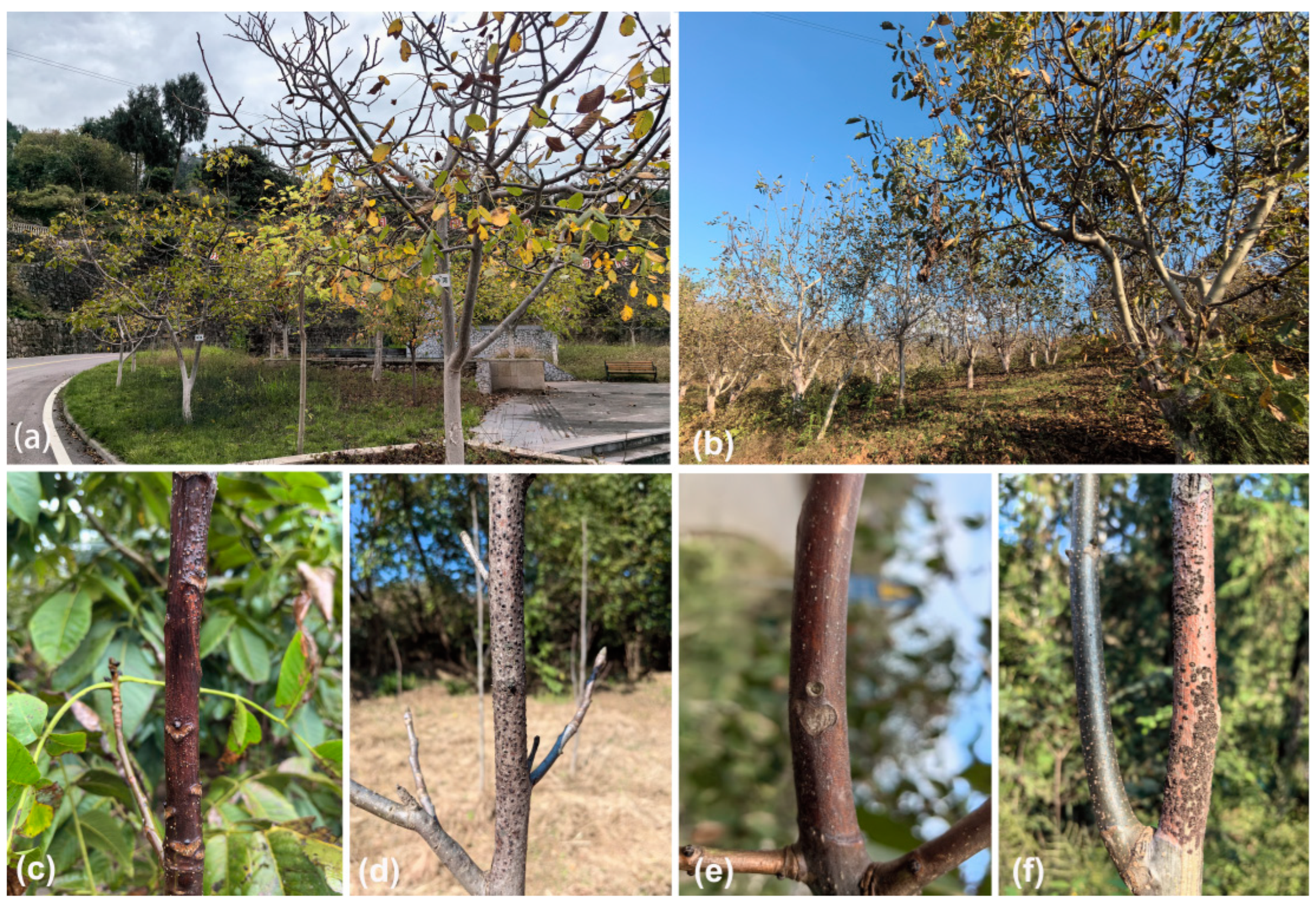
2.1. Sampling and Isolation

During the survey conducted in 2022 and 2023, 122 diseased (branches and twigs with canker symptoms) Juglans regia branch samples were collected from five Juglans regia plantations in Shaanxi, Sichuan and Yunnan in China. Approximately 15–25 Juglans regia trees were sampled from each site, and cankered tissues were collected from a single branch of each tree showing symptoms typical of branch canker and dieback for detailed examination and fungal isolation. In total, 45 samples were collected. A total of 31 Diaporthe isolates were obtained from 45 specimens by removing a mucoid spore mass from conidiomata and/or ascomata, spreading the suspension over the surface with potato dextrose agar (PDA) (200 g potatoes, 20 g glucose and 20 g agar/L water) in a Petri dish and incubating at 25 °C for up to 24 h. Hyphal tips were removed to a new PDA plate twice to obtain a pure culture. Specimens were deposited in the Museum of Beijing Forestry University (BJFC). Axenic cultures are maintained in the China Forestry Culture Collection Centre (CFCC).

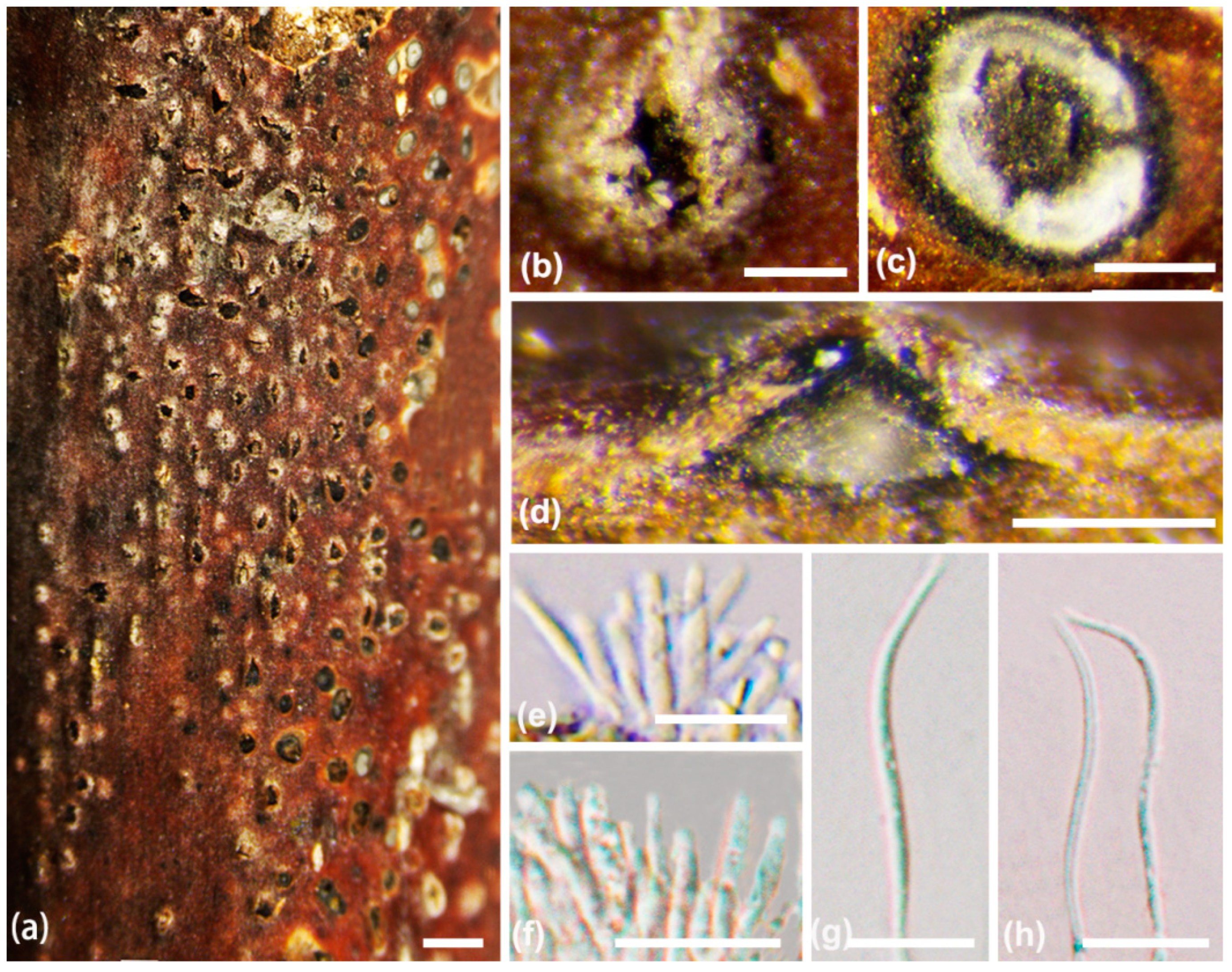
2.2. Morphological Analyses

Species identification was based on morphological characteristics of the ascomata or conidiomata formed on infected host materials. Macromorphological features (structure and size of conidiomata, ascomata, ectostromatic disc and ostioles) were photographed using a Leica stereomicroscope (M205 FA) (Leica Microsystems, Wetzlar, Germany). Micromorphological features (conidiophores, conidiogenous cells, asci and conidia/ascospores) were photographed using a Nikon Eclipse 80i microscope (Nikon Corporation, Tokyo, Japan), equipped with a Nikon digital sight DS-Ri2 high-resolution colour camera with differential interference contrast. Over 20 conidiomata were sectioned and 50 conidia were selected randomly to measure their lengths and widths. Colony diameters were measured and the colony colours described after 3 days and 14 days according to the colour charts of Rayner (1970) [35].

2.3. DNA Extraction, PCR Amplification and Sequencing

Mycelium used for DNA extraction was grown on PDA for three days and obtained from the cellophane surface by scraping. The genomic DNA was extracted from axenic cultures using the modified CTAB method [36]. Sequences were amplified by PCR from the ITS, cal, his3, tef1-α and tub2 genetic regions. The PCR mixtures for all genes included 10 μL Mix (Promega, Madison, MI, USA), 7 μL double deionized water, 1 μL of each primer and 1 μL template DNA. All primers and PCR conditions are listed in Table 1. PCR products were electrophoresed in 1% agarose gel, and the DNA was sequenced by the Sino Geno Max Biotechnology Company Limited (Beijing, China). DNA sequences generated by the forward and reverse primers combination were used to obtain consensus sequences using Seqman v. 7.1.0 (DNASTAR Inc., Madison, WI, USA).

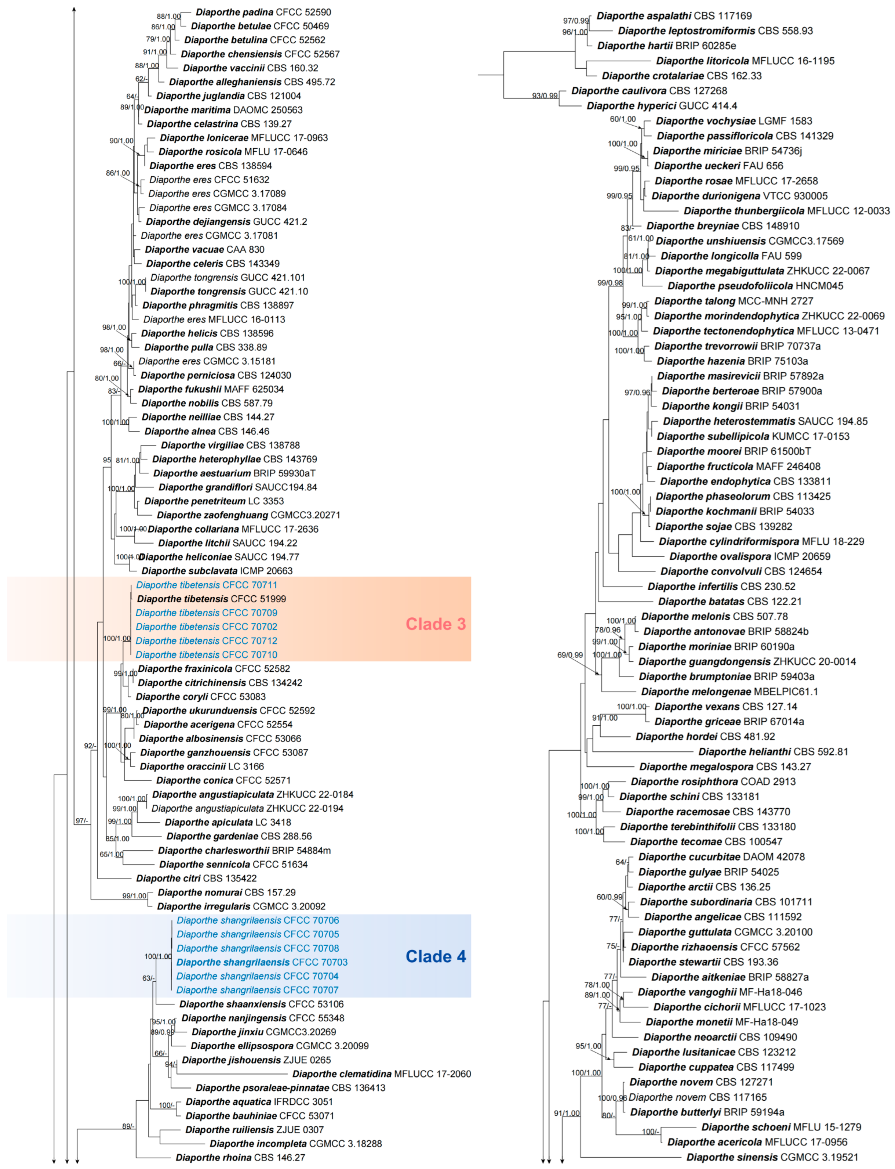
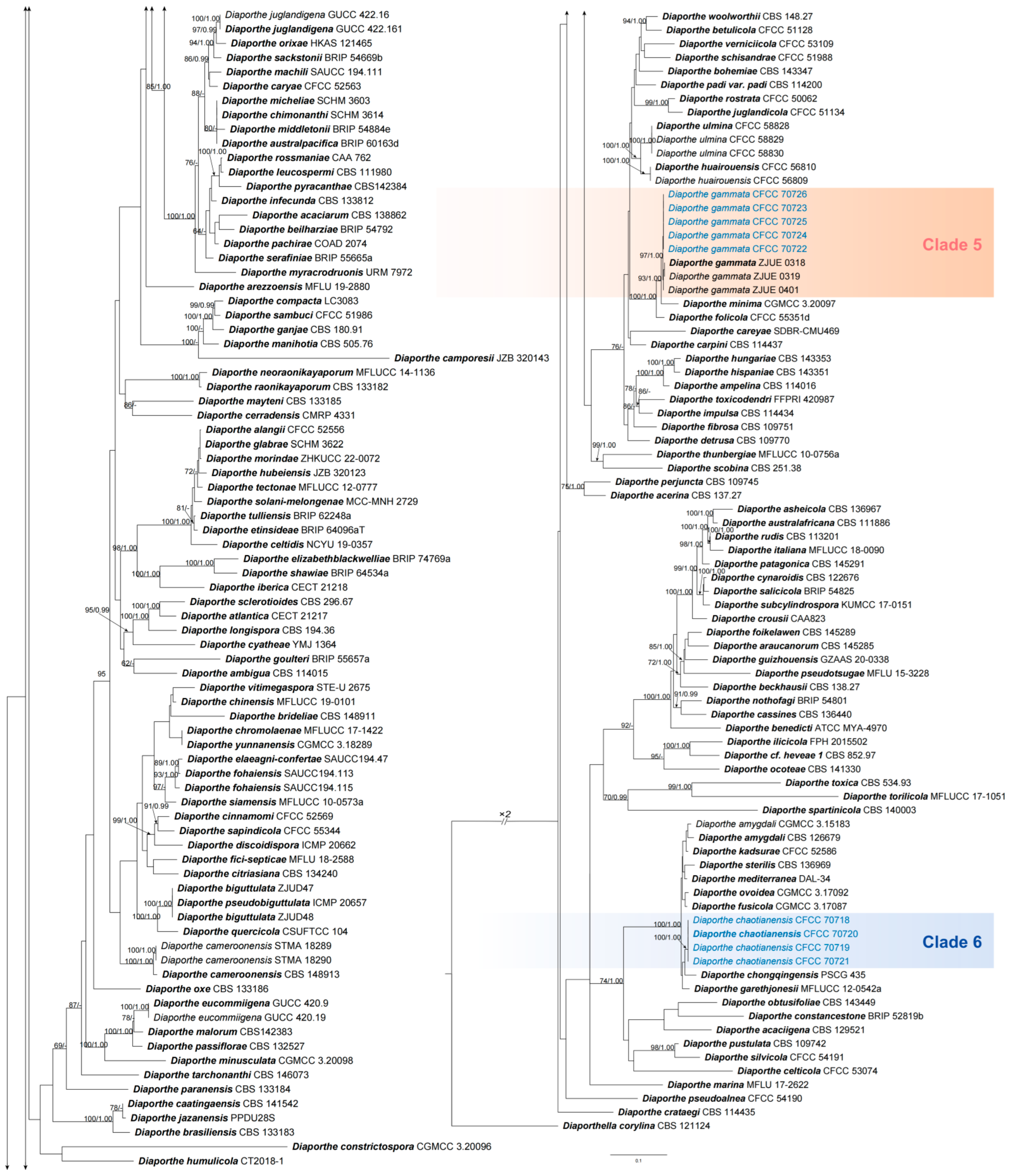
2.4. Phylogenetic Analyses

The sequences obtained from this study were analyzed with the NCBIs GenBank nucleotide datasets. Alignments based on ITS, cal, his3, tef1-α and tub2 sequence data, including sequences obtained from this study and those downloaded from GenBank (Supplementary Table S1), were first aligned using MAFFT v. 6 [37] and edited manually using MEGA v. 6.0 [38]. Diaporthella corylina (CBS 121124) was used as the outgroup in polygenic Diaporthe analyses. Phylogenetic analyses were performed with PhyML v. 3.0 for the maximum likelihood (ML) method [39] and MrBayes v. 3.1.2 for the Bayesian inference (BI) method [40].
The best-fit evolutionary models for each partitioned locus were estimated by MrModeltest v. 2.3 following the Akaike information criterion (AIC) in ML and BI analyses [41]. For ML analysis, RAxML-NG was used [42]. Bootstrap supports were estimated with 100 pseudoreplicates and the appropriate models for each gene. BI analyses were completed using a Markov chain Monte Carlo (MCMC) algorithm with Bayesian posterior probabilities (BPPs) [43]. Trees were sampled every 100th generation after two MCMC chains were run from random trees for 10 million generations, which stopped when the average standard deviation of split frequencies fell below 0.01. For the burn-in phase of each analysis, the first 25% of the trees were discarded and the remaining trees were assessed to calculate BPP [43]. FigTree v. 1.3.1 [44] was used to show phylograms. The sequence data of 31 isolates were deposited in GenBank; their accession numbers, together with those of the other species used for the analysis, are listed in Table S1. The multilocus sequence alignment was deposited in TreeBASE (www.treebase.org, accessed on 1 July 2024; study ID 31531).

2.5. Pathogenicity Test

Three representative isolates from each identified Diaporthe species were selected for pathogenicity testing in this study (D. chaotianensis: CFCC 70718–70720; D. gammata: CFCC 70722–70724; D. olivacea: CFCC 70713, 70715, 70716; D. shangluoensis: CFCC 70728, 70729, 70731; D. shangrilaensis: CFCC 70703, 70705, 70706; D. tibetensis: CFCC 70702, 70710, 70711). The pathogenicity tests were conducted on 2–year–old Juglans regia trees 1.3 m high and 1.5 cm thick which were planted in the field at a nursery of the Forest Protection Lab (Beijing, China). After the leaves grew, they were inoculated under natural conditions to determine pathogenicity during early April 2024 (mean air temperature = 15 °C). Sterilized 5 mm diameter inoculation rings were used to scald the bark surface of each branch to a depth of 2 mm. Agar plugs of the same size were removed from 6-day-old colonies of selected isolates, inserted into the wounds, sealed with moistened cotton wool and protected with parafilm. Six replications were made for each isolate. One plant per isolate was used as the negative control, and an equal number of plants inoculated with PDA agar plugs without colonies were used as the positive control. After one week, the parafilm and cotton wool were removed. These inoculated plants were maintained in the field. Fourteen days after inoculation, the lengths of the spots on the bark surface were measured from the inoculation point upwards and downwards using a digital calliper and then averaged. All spots from the experimental and control groups were reisolated to verify that the morphological characteristics and DNA sequences were consistent with the original isolates, thus fulfilling the Koch hypothesis. Differences in lesion length between isolates were analyzed by one-way analysis of variance (ANOVA) followed by least significant difference (LSD) tests. Statistical analysis was carried out by SPSS v. 20.0 and considered as significant at p < 0.05.
Table 1. Genes used in this study with PCR primers, primer DNA sequence, optimal annealing temperature and corresponding references.
LocusPCR PrimersPCR: Thermal Cycles: (Annealing Temp. in Bold)References of
Primers Used
ITSITS1(95 °C: 30 s, 51 °C: 30 s, 72 °C: 1 min) × 35 cycles[45]
ITS4
calCAL228F(95 °C: 15 s, 54 °C: 20 s, 72 °C: 1 min) × 35 cycles[46]
CAL737R
his3CYLH3F(95 °C: 30 s, 58 °C: 30 s, 72 °C: 1 min) × 35 cycles[47,48]
H3-1b
tef1-α728F(95 °C: 15 s, 55 °C: 20 s, 72 °C: 1 min) × 35 cycles[46]
1567R
tub2T1(95 °C: 30 s, 55 °C: 30 s, 72 °C: 1min) × 35 cycles[48]
Bt2b

3. Results

3.1. Phylogeny

Each gene region and the concatenated sequences of five genetic regions (ITS, cal, his3, tef1-α and tub2) were analyzed to infer the interspecific relationships within Diaporthe. The topological structures derived from each gene region were found to be consistent with those of the combined dataset (Figure 2, Figures S1–S5). The combined sequences dataset comprised 481 isolates (480 ingroup taxa including 31 new isolates in this study and one outgroup taxa, Diaporthella corylina CBS 121124). The sequence fragments were 3954 characters including gaps (548 for ITS, 952 for cal, 663 for his3, 785 for tef1-α and 1006 for tub2). The topologies resulting from ML and BI analyses of the concatenated dataset were similar. ML bootstraps (ML BS ≥ 60%) and Bayesian posterior probabilities (BPP ≥ 0.95) have been shown above the branches (Figure 2). For ML analysis, the matrix had 3044 distinct alignment patterns. The model parameters were as follows: A = 0.217335, C = 0.286382, G = 0.258070, T = 0.218908: substitution rates: AC = 0.866066, AG = 3.129585, AT = 0.966855, CG = 0.698073, CT = 3.794326, GT = 1.000000; gamma distribution shape parameter α = 0.538403; and likelihood value of ln: −124,733.462770.
The current 31 isolates clustered in six clades representing six species. Two represented known species (D. gammata and D. tibetensis) and four new clades. Isolates in clades 1, 2, 4 and 6 were separated from all other species and were also highly supported (ML/BI = 100/1) (Figure 2), representing four novel species (D. chaotianensis, D. olivacea, D. shangluoensis and D. shangrilaensis), which are detailed in the following sections.

3.2. Taxonomy

Diaporthe chaotianensis A.L. Jia and X.L. Fan, sp. nov. (Figure 3)
MycoBank: MB 854191
Etymology: Named after the place where the fungus was isolated, Chaotian District, Guangyuan City.
Typification: China, Sichuan Province, Guangyuan City: Chaotian District, Zhongzi Town, 32°41′14″ N, 106°02′23″ E, from branches of Juglans regia, 10 October 2023, Y.X. Li, L. Lin and X.L. Fan (holotype BJFC-S2345, ex-holotype living culture CFCC 70720); 32°41′20″ N, 106°02′42″ E (paratype BJFC-S2346, ex-paratype living culture CFCC 70718).
Description: Sexual morph not observed. Asexual morph: conidiomata pycnidial, conical, immersed in bark, scattered, erumpent through the surface, with a solitary locule. Ectostromatic disc black to white, with one ostiole per disc, ovoid to circular. Locule undivided, 185–670 μm (av. = 370 μm, n = 20) diam. Conidiophores cylindrical, attenuate towards the apex, hyaline, phialidic, unbranched, slightly curved, 8–15 × 1–1.2 μm (av. = 11 ± 1.8 × 1.6 ± 0.3 μm, n = 50). Conidiogenous cells phialidic, hyaline, cylindrical, tapered towards the apex, 6–19 × 1–2 μm (av. = 10 ± 3.3 × 1.4 ± 0.3 µm, n = 50). Beta conidia hyaline, aseptate, filiform, straight or curved, tapering towards both ends, 21–32 × 1–2 μm (av. = 26 ± 3 × 1.4 ± 0.3 µm, n = 50). Alpha conidia are not observed.
Cultural characteristics: Colonies initially white, grown to 63 mm after 3 days, compact at the centre and sparse at the surroundings, becoming honey after 7 days. Colonies are flat with a uniform texture, lacking aerial mycelium. Conidiomata were sparse, black, distributed irregularly (Figure 4a).
Additional materials examined: China, Sichuan Province, Guangyuan City: Chaotian District, Zhongzi Town, 32°41′21″ N, 106°02′25″ E, from branches of Juglans regia, 10 October 2023, Y.X. Li, L. Lin and X.L. Fan (BJFC-S2347, living culture CFCC 70719, 70721).
Notes: Four isolates represent the D. chaotianensis cluster in a clade distinct from other species of Diaporthe known from DNA sequence data. Phylogenetically, D. chaotianensis is most closely related to D. chongqingensis in phylogenetic trees of the combined dataset and tef1-α loci (Figure 2, Figure S4), but can be distinguished from D. chongqingensis based on phylogeny in ITS, cal, his3 and tub2 loci (15/506 for ITS, 3/558 for cal, 1/600 for his3 and 11/641 for tub2). In the phylogenetic tree of ITS loci, D. chaotianensis forms an independent lineage closely related to D. fusicola and D. kadsurae (Figure S1), but can be distinguished from these species based on alignments of the separate loci (differs from D. fusicola in: 5/523 for ITS, 6/600 for cal, 11/677 for tef1-α and 5/632 for tub2; D. kadsurae: 5/523 for ITS, 3/483 for his3, 11/677 for tef1-α and 6/857 for tub2). In the phylogenetic trees of cal and his3 loci, D. chaotianensis is closely related to D. amygdali, D. mediterranea and D. sterilis (Figures S2 and S3), but can be distinguished from these species based on alignments of the separate loci (differs from D. amygdali in: 11/523 for ITS, 2/600 for cal, 3/483 for his3, 13/677 for tef1-α and 15/935 for tub2; D. mediterranea: 10/523 for ITS, 3/600 for cal, 3/483 for his3, 11/677 for tef1-α and 7/935 for tub2; D. sterilis: 10/523 for ITS, 3/600 for cal, 19/483 for his3, 11/677 for tef1-α and 6/857 for tub2). In the phylogenetic tree of tub2 loci, D. chaotianensis is closely related to D. garethjonesii (Figure S5), but can be distinguished from D. chongqingensis based on phylogeny in ITS, cal, tef1-α and tub2 loci (11/523 for ITS, 8/653 for cal, 10/677 for tef1-α and 2/631 for tub2). Morphologically, D. chaotianensis differs from these species based on morphology. Conidiophores of D. chaotianensis are longer than D. chongqingensis (8–15 × 1–1.2 μm vs. 6.5–12.5 × 2–6 μm) [16], but smaller than D. fusicola (8–15 × 1–1.2 μm vs. 11–24.1 × 1.6–2.9 μm) [49] and narrower than D. kadsurae (8–15 × 1–1.2 μm vs. 7–11 × 1.8–2.9 μm) [12]. Conidiogenous cells of D. chaotianensis are smaller than D. chongqingensis (6–19 × 1–2 μm vs. 14–26 × 1.5–2.5 μm) [16]. Furthermore, D. chaotianensis differs from D. amygdali, D. fusicola, D. kadsurae and D. mediterranea in the production of beta conidia, which is not observed in these species [12,49,50]. Diaporthe chaotianensis differs from D. garethjonesii in its smaller conidia (21–32 × 1–2 μm vs. 40–50 × 3–4 μm) [51], and mainly differs from D. sterilis in its capacity to produce beta conidia, because all isolates representing D. sterilis could not be induced to sporulate on any of the culture media used by Lombard et al. [52], when this new Diaporthe species collected from Vaccinium corymbosum was described [52].
Diaporthe gammata X.E. Xiao, Crous and H.Y. Li, Persoonia 51: 243 (2023) (Figure 5)
Description: Sexual morph not observed. Asexual morph: conidiomata pycnidial, immersed in the bark, erumpent through bark surface, with a single locule. Ectostromatic disc brown to black, with one ostiole per disc, ovoid to circular. Locule undivided, (280–)380–600(–650) μm (av. = 480 μm, n = 20) diam. Conidiophores cylindrical to subcylindrical, hyaline, smooth, aseptate, 5.6–12 × 1–2.6 μm (av. = 8.7 ± 1.8 × 1.8 ± 0.5 μm, n = 50). Conidiogenous cells hyaline, smooth, phialidic, cylindrical, straight or slightly curved, tapered towards the apex, (5–)6–13(–16) × 1–3.5 μm (av. = 8 ± 1.5 × 2 ± 0.5 μm, n = 50). Alpha conidia hyaline, aseptate, fusoid to ovoid, hyaline, aseptate, tapering towards both ends, usually one guttulate at each end, rarely three guttulate, 5.5–9.5 × 1.5–3.7 μm (av. = 7.4 ± 1.2 × 2.5 ± 0.4 μm, n = 50). Beta conidia hyaline, aseptate, filiform, straight or curved, tapering towards both ends, 18–28 × 1.2–1.8 μm (av. = 23 ± 4 × 1.5 ± 0.2 μm, n = 50). Gamma conidia hyaline, aseptate, fusoid to obclavate, multi-guttulate, apex rounded, base rounded to slightly acutate, 6.8–10.6 × 1.8–3 μm (av. = 9 ± 1.3 × 2.4 ± 0.4 μm, n = 50).
Cultural characteristics: Colonies initially white, felty with a thicker texture, aerial mycelium lacking, growing up to 36 mm after 3 days, turning grey olivaceous at the centre, becoming dark brick on the surface and sepia to black in the reverse after 12 days (Figure 4b). Conidiomata are distributed randomly at the marginal area.
Materials examined: China, Sichuan Province, Guangyuan City: Chaotian District, Walnut Culture Square, 32°40′58″ N, 106°02′08″ E, from branches of Juglans regia, 10 October 2023, L. Lin and X.L. Fan (BJFC-S2348, living cultures CFCC 70722 and CFCC 70724); Guangyuan City: Chaotian District, Walnut Culture Square, 32°40′41″ N, 106°02′16″ E, from branches of Juglans regia, 10 October 2023, L. Lin and X.L. Fan (BJFC–S2349, living cultures CFCC 70723, 70725); Guangyuan City: Chaotian District, Walnut Culture Square, 32°40′49″ N, 106°02′23″ E, from branches of Juglans regia, 10 October 2023, L Lin and X.L. Fan (BJFC-S2350, living culture CFCC 70726).
Notes: Diaporthe gammata was first introduced by Xiao et al. (2023) [53] and was isolated from Citrus reticulata in Chongqing Municipality, China. It was named for the presence of gamma conidia. Phylogenetically, five isolates form a fully supported clade that are close to D. gammata in combined datasets and five gene loci trees (Figure 2, Figures S1–S5). Morphologically, three types of conidia were observed in the present study, which is consistent with the description of D. gammata [53]. Therefore, five isolates collected in this study are identified as D. gammata. Additionally, this is the first report of D. gammata being responsible for Juglans regia shoot canker.
Diaporthe olivacea A.L. Jia and X.L. Fan, sp. nov. (Figure 6)
MycoBank: MB 854192
Etymology: Named after the colour of cultural characteristics, olivaceous buff.
Typification: China, Sichuan Province, Guangyuan City: Lizhou District, National Highway 212, 32°26′20″ N, 105°38′26″ E, from branches of Juglans regia, 11 October 2023, L. Lin and X.L. Fan (holotype BJFC-S2351, ex-holotype living culture CFCC 70713). 32°26′25″ N, 105°38′32″ E (paratype BJFC-S2352, ex-paratype living culture CFCC 70715).
Description: Sexual morph not observed. Asexual morph: conidiomata pycnidial, immersed in the bark, erumpent through bark surface, with a single locule. Ectostromatic disc black to white, with one ostiole per disc, ovoid to circular. Locule undivided, 400–970 μm (av. = 700 μm, n = 20) diam. Conidiophores hyaline, smooth, 1–2(–3) septate, densely aggregated, cylindrical, 10.8–17.5 × 1–2 μm (av. = 13.4 ± 2 × 1.5 ± 0.3 μm, n = 50). Conidiogenous cells hyaline, smooth, phialidic, cylindrical, tapered towards the apex, 7–18.5 × 1.3–2 μm (av. = 11.5 ± 3 × 1.6 ± 0.2 μm, n = 50). Alpha conidia broadly fusiform to obovoid, hyaline, apex rounded or acute, base acutate, biguttulate to multiguttulate, aseptate, 5.5–9.5 × 1.5–3.7 μm (av. = 7.4 ± 1.2 × 2.5 ± 0.4 μm, n = 50). Beta conidia hyaline, aseptate, filiform, straight or curved, tapering towards both ends, 14–22 × 1.3–2.2 μm (av. = 19 ± 2.5 × 1.7 ± 0.2 μm, n = 50). Gamma conidia are not observed.
Cultural characteristics: Colonies covering dish after 7 days in the dark at 25 °C, on the PDA surface with fluffy white aerial mycelium, being hazel on the surface, olivaceous buff in the reverse after 10 days (Figure 4c). Colony margin is regular. Conidiomata are sparse, irregularly distributed over the agar surface after 30 days.
Additional materials examined: China, Sichuan Province, Guangyuan City: Lizhou District, National Highway 212, 32°26′28″ N, 105°38′18″ E, from branches of Juglans regia, 11 Oct. 2023, L. Lin and X.L. Fan (BJFC-S2353, living cultures CFCC 70714, 70716); 32°26′31″ N, 105°38′22″ E (BJFC-S2354, living culture CFCC 70717).
Notes: Phylogenetic analysis combined five gene loci, and each locus showed that all the isolates of D. olivacea clustered together in a highly supported clade (ML/BI = 100/1) (Clade 1) and displayed a close relationship, but they were clearly differentiated from D. musigena, D. schimae and D. taoicola (Figure 2, Figures S3–S5). Based on alignments of the separate loci, D. olivacea differs from D. musigena in ITS, cal, his3, tef1-α and tub2 loci (17/529 for ITS, 35/651 for cal, 8/586 for his3, 16/691 for tef1-α and 25/969 for tub2); differs from D. schimae in ITS, cal, his3, tef1-α and tub2 loci (10/518 for ITS, 29/678 for cal, 2/580 for his3, 4/691 for tef1-α and 16/969 for tub2); and differs from D. taoicola in ITS, tef1-α and tub2 loci (11/520 for ITS, 8/677 for tef1-α and 9/748 for tub2). Morphologically, D. olivacea differs from D. musigena in having smaller conidiophores (10.8–17.5 × 1–2 μm vs. 15–40 × 1.5–2.5 μm) and bigger conidiogenous cells (7–18.5 × 1.3–2 μm vs. 2–5 × 0.5–1 μm) [54]. Moreover, D. musigena differs from D. olivacea in the production of gamma conidia, which is not observed in D. olivacea [55]. Diaporthe olivacea differs from D. schimae in having smaller alpha conidia (5.5–9.5 × 1.5–3.7 μm vs. 8–8.5 × 2.5–3 μm) and smaller beta conidia (14–22 × 1.3–2.2 μm vs. 27.5–38.5 × 1–1.5 μm) [14], and differs from D. taoicola in having smaller alpha conidia (5.5–9.5 × 1.5–3.7 μm vs. 7–9 × 2–3 μm), smaller beta conidia (14–22 × 1.3–2.2 μm vs. 20–25 × 1.5–2 μm) and narrower conidiophores (10.8–17.5 × 1–2 μm vs. 10–25 × 2–3 μm) [11].
Diaporthe shangluoensis A.L. Jia and X.L. Fan, sp. nov. (Figure 7)
MycoBank: MB 854193
Etymology: Named after the place where the fungus was isolated, Shangluo City.
Typification: China, Shaanxi Province, Shangluo City: Shangzhou District, Lijia Plateau Bridge, 33°46′40″ N, 110°06′13″ E, from branches of Juglans regia, 13 October 2023, L. Lin and X.L. Fan (holotype BJFC-S2355, ex-holotype living culture CFCC 70728); 33°46′36″ N, 110°06′05″ E (paratype BJFC-S2356, ex-paratype living culture CFCC 70731).
Description: Sexual morph not observed. Asexual morph: conidiomata pycnidial, immersed in the bark, erumpent through bark surface, with a single locule. Ectostromatic disc white to black, with one ostiole per disc, ovoid to circular. Locule undivided, 140–300(–460) μm (av. = 210 μm, n = 20) diam. Conidiophores hyaline, smooth, 1-septate, unbranched, cylindrical, 4.5–9.5 × 1–1.6 μm (av. = 7.5 ± 1.5 × 1.3 ± 0.2 μm, n = 50). Conidiogenous cells hyaline, phialidic, cylindrical, straight or slightly curved, tapered towards the apex, 5–11.5 × 1.5–2.8 μm (av. = 7.5 ± 2 × 1.8 ± 0.3 μm, n = 50). Alpha conidia hyaline, aseptate, fusoid to ovoid, tapering towards both ends, one guttulate at each end, 5.8–8.3 × 2–3 μm (av. = 7 ± 0.5 × 2.7 ± 0.3 μm, n = 50). Beta conidia hyaline, aseptate, filiform, straight or curved, sinuous at one end, tapering towards both ends, eguttulate, 29.5–38 × 1–2 μm, av. = 32.3 ± 2.6 × 1.4 ± 0.1 μm, (n = 50). Gamma conidia are not observed.
Culture characteristics: Cultures on PDA incubated at 25 °C in darkness, colony originally flat after 3 days, becoming hazel after 7−10 days (Figure 4d). Colonies flat with a uniform texture, lacking aerial mycelium, margin regular. Conidiomata sparse, irregularly distributed over agar surface after 30 days.
Additional materials examined: CHINA, Shaanxi Province, Shangluo City: Shangzhou District, Lijia Plateau Bridge, 33°46′42″ N, 110°06′06″ E, from branches of Juglans regia, 13 October 2023, L. Lin and X.L. Fan (BJFC-S2357, living cultures CFCC 70729 and 70730); 33°46′36″ N, 110°06′25″ E (BJFC-S2358, living cultures CFCC 70727 and 70732).
Notes: Diaporthe shangluoensis was isolated from Juglans regia collected in Shaanxi, China. Six isolates are phylogenetically separated from all other available isolates included in this study. Diaporthe shangluoensis is most closely related to D. guangxiensis, D. hainanensis, D. pandanicola, D. viciae and D. viniferae (Figure 2, Figures S1–S5), but differentiated from them in ITS (13 different unique fixed alleles by D. guangxiensis, 11 by D. hainanensis, 28 by D. pandanicola, 8 by D. viciae and 28 by D. viniferae), cal loci (6 different unique fixed alleles by D. guangxiensis and 12 by D. viniferae), his3 loci (4 different unique fixed alleles by D. viciae), tef1-α loci (20 different unique fixed alleles by D. guangxiensis, 21 by D. hannanensis, 20 by D. viciae and 21 by D. viniferae) and tub2 loci (25 different unique fixed alleles by D. guangxiensis, 10 by D. hainanensis, 8 by D. pandanicola, 12 by D. viciae and 27 by D. viniferae). Moreover, D. shangluoensis differs from D. guangxiensis, D. hainanensis and D. viniferae in having bigger beta conidia (29.5–38 × 1–2 vs. 20–32 × 1–1.5 μm) [55], (29.5–38 × 1–2 vs. 23–25 × 1.1 μm) [56], (29.5–38 × 1–2 vs. 23–35 × 1–1.5 μm) [55]. Alpha conidia are smaller than in D. viciae (5.8–8.3 × 2–3 vs. 7–10 × 2–4 μm) [57]. Conidiogenous cells are smaller than in D. hainanensis (5–11.5 × 1.5–2.8 vs. 14.5–20 × 1.4–1.8 μm) [56].
Diaporthe shangrilaensis A.L. Jia and X.L. Fan, sp. nov. (Figure 8)
MycoBank: MB 854194
Etymology: Named after the place where the fungus was isolated, Shangri-La City.
Typification: China, Yunnan Province, Diqing Tibetan Autonomous Prefecture, Shangri-La City: Sanba Naxi Township, 27°34′18″ N, 100°1′19″ E, from branches of Juglans regia, 9 August 2022, L. Lin (holotype BJFC-S2359, ex-holotype living culture CFCC 70703); 27°34′26″ N, 100°1′5″ E (paratype BJFC-S2360, ex-paratype living culture CFCC 70706).
Description: Sexual morph not observed. Asexual morph: conidiomata pycnidial, immersed in the bark, erumpent through bark surface, with a single locule. Ectostromatic disc brown to black, with one ostiole per disc, ovoid to circular. Locule undivided, (70–)110–390(–460) μm (av. = 230 μm, n = 20) diam. Conidiophores hyaline, smooth, aseptate, densely aggregated, cylindrical, straight, 15–20 × 1–2.3 μm (av. = 16 ± 1 × 1.5 ± 0.5 μm, n = 50). Conidiogenous cells hyaline, smooth, branched, phialidic, cylindrical, straight or slightly curved, 4.3–8.6 × 1–2.8 μm (av. = 6 ± 1.2 × 1.8 ± 0.6 μm, n = 50). Alpha conidia hyaline, aseptate, ellipsoidal to cylindrical, obtusely rounded at both ends, usually eguttulate, rarely one guttule at each end, 6–8.8 × 2–3.8 μm (av. = 7.6 ± 0.6 × 2.8 ± 0.4 μm, n = 50). Beta conidia and gamma conidia are not observed.
Culture characteristics: Cultures on PDA incubated at 25 °C in darkness, colony originally flat with a white felty aerial mycelium after 3 days, becoming a white compact aerial mycelium at the centre with a smoke-grey aerial mycelium at the margin after 7–10 days (Figure 4e), margin irregular. Conidiomata are sparse, irregularly distributed over the agar surface after 30 days.
Additional materials examined: China, Yunnan Province, Diqing Tibetan Autonomous Prefecture, Shangri-La City: Sanba Naxi Township, 27°34′33″ N, 100°1′21″ E, from branches of Juglans regia, 9 August 2022, L. Lin (BJFC-S2361, living cultures CFCC 70704 and 70705); 27°34′27″ N, 100°1′16″ E (BJFC-S2362, living cultures CFCC 70707 and 70708).
Notes: Based on the combining five gene loci and each locus individually, six isolates representing D. shangrilaensis form an independent clade and are phylogenetically distinct from D. shaanxiensis in a well-supported clade (ML/BI= 100/1) (Figure 2, Figures S1–S5). Diaporthe shangrilaensis can be distinguished from D. shaanxiensis based on the differences in ITS, cal, his3 and tef1-α loci (18/532 for ITS, 33/702 for cal, 25/565 for his3 and 15/778 for tef1-α). In addition, D. shangrilaensis differs from D. shaanxiensis in having smaller locules (110–390 μm vs. 526–765 μm) and smaller conidiogenous cells (4.3–8.6 × 1–2.8 μm vs. 14.5–17 × 1–1.5 μm) [13].
Diaporthe tibetensis C.M. Tian, Qin Yang & X.L. Fan, Mycological Progress 17(7): 847 (2018) (Figure 9)
Description: Conidiomata pycnidial, scattered or serried, immersed in bark, discoid to conical, erumpent slightly through the bark surface at maturity, with single locule. Ectostromatic disc brown to black, ovoid to circular. Locule undivided, (370–)420–700(−780) μm (av. = 580 μm, n = 20) diam. Conidiophores hyaline, smooth, aseptate, densely aggregated, cylindrical, 5–20 × 1.5–4 μm. Conidiogenous cells hyaline, smooth, branched, phialidic, cylindrical, tapering towards apex, straight or slightly curved, (1.5–)2–5(–7) × (1–)0.5–1.3(–2) μm (av. = 3.5 ± 0.8 × 1 ± 0.2 μm, n = 50). Alpha conidia abundant in twigs, hyaline, aseptate, ellipsoidal or oval, 0–3–guttulate, 5.8–8 × 2–5 μm (av. = 6.8 ± 0.6 × 2.6 ± 0.5 μm, n = 50). Beta conidia hyaline, aseptate, filiform, straight or curved, tapering towards one end, (8–)10–20(–25) × (0.5–)0.7–1.2(–1.6) μm (av. = 15.5 ± 4.6 × 1 ± 0.3 μm, n = 50). Gamma conidia are not observed.
Cultural characteristics: Cultures on PDA incubated at 25 °C in darkness, initially white, irregular. Colony originally flat with a white felty aerial mycelium after 7 days, becoming olivaceous to isabelline with a smoke-grey aerial mycelium after 14 days (Figure 4f), margin irregular, conidiomata sparse, irregularly distributed over agar surface after 30 days.
Materials examined: China, Yunnan Province, Diqing Tibetan Autonomous Prefecture, Shangri-La City: Sanba Naxi Township, 27°34′14″ N, 100°1′35″ E, from branches of Juglans regia, 9 August 2022, L. Lin (BJFC-S2363 living cultures CFCC 70710 and 70711); 27°34′16″ N, 100°1′33″ E (BJFC-S2364 living cultures CFCC 70709 and 70712); 27°34′25″ N, 100°1′36″ E (BJFC-S2365 living culture CFCC 70702).
Notes: Diaporthe tibetensis was introduced by Fan et al. (2018) [31] as causing canker disease on Juglans regia in Tibet Autonomous Region, China. This species can be distinguished from D. citrichinensis and D. oraccinii by its shorter conidiogenous cells and larger alpha conidia [31]. In this study, five isolates are grouped together with D. tibetensis in combined datasets and five gene loci trees (Figure 2, Figures S1–S5) (ML/BI = 100/1) (Clade 3). Therefore, they are identified as Diaporthe tibetensis. Additionally, the current study observed beta conidia of this species.

3.3. Analysis of Pathogenicity Test

For pathogenicity tests via Juglans regia shoot inoculations, the results showed that all the tested Diaporthe isolates could induce discoloured and necrotic lesions 14 d post inoculation (Figure 10; Table 2). Sunken cankers were obvious on the stems and produced brown lesions upward and downward from the point of inoculation. At the same time, no lesions were observed on the branches of the control (Figure 10(g1,g2)). Koch’s postulates were fulfilled and confirmed that all the tested Diaporthe species in this study are pathogens of Juglans regia. Diaporthe Shangrilaensis was shown to be the most aggressive species: isolates CFCC 70703, 70705, 70706 of D. shangrilaensis caused larger lesions (Figure 10(e1,e2)), followed by the isolates CFCC 70713, 70715, 70716 of D. olivacea (Figure 10(c1,c2)), and the remaining isolates CFCC 70728, 70729, 70731 of D. shangluoensis induced smaller lesions (Figure 10(d1,d2)). Isolates of D. gammata caused only slight discolouration around the inoculation points (Figure 10(b1,b2)). In contrast, the remaining Diaporthe isolates induced very limited lesions (5 < mean lesion length < 6 mm). Although the difference in mean lesion length between D. chaotianensis and D. tibetensis was not significant, the disease incidence of D. chaotianensis (89%) was higher than that of D. tibetensis (56%). Therefore, D. chaotianensis was found to be more virulent than D. tibetensis. Diaporthe gammata was significantly lower than the other five species in lesion length, with canker length averaging 9.1 ± 0.6 mm. ANOVA revealed significant (p < 0.05) differences among the treatment means in all six species (Table 2).

4. Discussion

The current study reveals the diversity and pathogenicity of Diaporthe species from Juglans regia in China. Six Diaporthe species were identified from the collected specimens in this study (Diaporthe chaotianensis, D. gammata, D. olivacea, D. shangluoensis D. shangrilaensis and D. tibetensis). Among these, D. chaotianensis, D. olivacea, D. shangluoensis and D. shangrilaensis were described as four novel species and D. gammata was reported for the first time in Juglans regia trees. These findings suggest a higher level of diversity among Diaporthe species responsible for cankers on Juglans regia than has been previously recognized. Moreover, Koch’s postulates confirmed that those species were pathogens of Juglans regia.
The taxonomy of Diaporthe species is increasingly attracting attention from researchers. Most research focuses on the identification and descriptions of novel species and new host records, as well as on the regulation of pathogenicity in important Diaporthe species, indicating that genus Diaporthe has a high potential for rapid evolution [14]. Previous studies have revealed that Diaporthe species have high genetic diversity on a single host. For example, Gao et al. (2016) [58] reported nine species of Diaporthe isolated from Camellia in China, and Wan et al. (2023) [59] revealed three new Diaporthe species on Acer palmatum in China [58,59]. This study collected an extensive number of Diaporthe isolates from areas of Juglans regia cultivation to reveal the genetic diversity of Diaporthe species. Previous studies have reported the presence of Diaporthe species in Juglans regia, and the results of this study further support these findings. Indeed, more novel species will likely be found in the future because several species of Diaporthe have a wide host range and can move between hosts among geographic regions. For example, D. gammata was originally reported on Citrus in Chongqing Municipality [53], but the fungus has also been found on Juglans regia in this study.
The current pathogenicity tests showed that D. chaotianensis, D. gammata, D. olivacea, D. shangluoensis, D. shangrilaensis and D. tibetensis are pathogens, which could pose threats to the Juglans regia industry in China. Furthermore, D. shangrilaensis, D. olivacea and D. shangluoensis are more aggressive among the six species, and this should be considered in the development of disease control measures. Among these species, Diaporthe shangrilaensis demonstrated greater aggressiveness, evidenced by its more potent virulence and the larger lesions it induced. To clarify the reproductive characteristics of this pathogen, additional quantitative experimental analysis is warranted. The virulence of Diaporthe species could be affected by environmental factors such as moisture content, rainfall intensity and temperature [60,61]. Manawasinghe et al. (2018) [15] found that environmental factors could alter the life mode of the fungi, from endophytic or saprophytic to pathogenic, thus enabling the colonization of new hosts. Diaporthe tibetensis was first reported by Fan et al. (2018) [31] on Juglans regia in Tibet Autonomous Region, and was also found on Juglans regia branches in Yunnan in this study, where the climates are quite different from the place where the pathogenicity tests were conducted. The pathogenicity tests showed that the majority of these species obtained in this study are weakly aggressive or non-aggressive to Juglans regia branches (Figure 10(f1,f2); Table 2). This may be because the experimental conditions were different from those in the natural environment and because of differences in climate between the north and south of China. This implies that D. tibetensis may become highly aggressively pathogenic to Juglans regia under favourable environmental conditions, which need to be determined by further research in order to prevent Juglans regia cankers.
This study provides novel information on the ability of those species to cause disease in Juglans regia. In addition, many studies have reported that most Diaporthe species had a wide host range. For example, Diaporthe eres could cause shoot blight, leaf necrosis and branch canker on different hosts [62,63,64,65,66]. Diaporthe sojae was confirmed as the pathogen of fungal diseases on pears, sunflowers, honeybush, kiwi fruit and soybeans [16,67,68,69,70]. Those studies indicated that species obtained in this study may be capable of infecting other plants. In this study, pathogenicity tests were conducted exclusively on Juglans regia. Therefore, it would be recommended that pathogenicity tests be conducted on other plants in future studies. In addition, among the eight species from Juglans regia in China previously reported, only D. amygdali was confirmed as a pathogen causing twig canker disease in Juglans regia [30]. The present study confirms that D. tibetensis is also a causal agent on Juglans regia. The pathogenicity of most reported species has not been tested, and their potential economic impact on Juglans regia remains unknown. Thus, the pathogenicity of Diaporthe species from Juglans regia needs to be studied further.
In conclusion, this study has focused on the diversity and pathogenicity of Diaporthe species from Juglans regia in China. Six species were isolated, and pathogenicity tests indicated that they exhibited varying degrees of pathogenicity. The current findings establish a foundational understanding crucial for enhancing disease management strategies. As for canker disease in Juglans regia trees, future studies should focus on the most widespread and aggressive pathogens reported here, and prevention and control measures should be investigated to mitigate the impact of canker diseases.

Supplementary Materials

The following supporting information can be downloaded at: https://www.mdpi.com/article/10.3390/jof10080583/s1, Table S1: Strains and their GenBank accession numbers used in the molecular phylogenetic analyses of Diaporthe. Figure S1: Phylogenetic tree of Diaporthe based on ITS loci. Figure S2: Phylogenetic tree of Diaporthe based on cal loci. Figure S3: Phylogenetic tree of Diaporthe based on his3 loci. Figure S4: Phylogenetic tree of Diaporthe based on tef1-α loci. Figure S5: Phylogenetic tree of Diaporthe based on tub2 loci.

Author Contributions

Conceptualization, A.J. and X.F.; methodology, A.J. and L.L.; software, A.J. and Y.L.; validation, A.J., L.L. and X.F.; formal analysis, A.J.; investigation, L.L., Y.L. and X.F.; resources, X.F.; data curation, A.J. and X.F.; writing—original draft preparation, A.J.; writing—review and editing, L.L. and X.F.; visualization, A.J.; supervision, X.F.; funding acquisition, X.F. All authors have read and agreed to the published version of the manuscript.

Funding

This research was funded by the National Key R & D Program of China (2023YFD1401302), National Science and Technology Fundamental Resources Investigation Program of China (2021FY100900).

Institutional Review Board Statement

Not applicable.

Informed Consent Statement

Not applicable.

Data Availability Statement

All sequence data are available in NCBI GenBank following the accession numbers in the manuscript.

Acknowledgments

The authors would like to thank those who provided assistance and advice for this study.

Conflicts of Interest

The authors declare no conflicts of interest.

References

  1. López-Larrinaga, F.; Mellado-Bermejo, E.; Valadares de Queirós, R.; Figueras-Lorenzo, M.; Gomes-Pires, J. Las nuevas plantaciones de nogal en Espana. Fruticultura 2017, 64, 45–59. [Google Scholar]
  2. Chen, S.F.; Moran, D.P.; Hasey, J.K.; Anderson, K.; Michailides, T.J. Phylogeny, morphology, distribution, and pathogenicity of Botryosphaeriaceae and Diaporthaceae from English walnut in California. Plant Dis. 2014, 98, 636–652. [Google Scholar] [CrossRef] [PubMed]
  3. Wang, Q.H.; Fan, K.; Li, D.W.; Han, C.M.; Qu, Y.Y.; Qi, Y.K.; Wu, X.Q. Identification, Virulence and Fungicide Sensitivity of Colletotrichum gloeosporioides s.s. Responsible for Walnut Anthracnose Disease in China. Plant Dis. 2020, 104, 1358–1368. [Google Scholar] [CrossRef]
  4. Wang, Z.Y.; Liu, Y.; Yan, A.H.; Wang, Z.G. The First Report of Juglans regia Leaf Spot Caused by Fusarium proliferatum in China. Plant Dis. 2022, 106, 2265. [Google Scholar] [CrossRef]
  5. Agustí-Brisach, C.; Moral, J.; Felts, D.; Trapero, A.; Michailides, T.J. Interaction Between Diaporthe rhusicola and Neofusicoccum mediterraneum Causing Branch Dieback and Fruit Blight of English Walnut in California, and the Effect of Pruning Wounds on the Infection. Plant Dis. 2019, 103, 1196–1205. [Google Scholar] [CrossRef]
  6. Nitschke, T. Pyrenomycetes Germanici; Eduard Trewendt: Breslau, Germany, 1870. [Google Scholar]
  7. Udayanga, D.; Liu, X.Z.; McKenzie, E.H.C.; Chukeatirote, E.; Bahkali, A.H.A.; Hyde, K.D. The genus Phomopsis: Biology, applications, species concepts and names of common phytopathogens. Fungal Divers. 2011, 50, 189–225. [Google Scholar] [CrossRef]
  8. Udayanga, D.; Liu, X.Z.; Crus, P.W.; McKenzie, E.H.C.; Chukeatirote, E.; Hyde, K.D. A multi-locus phylogenetic evaluation of Diaporthe (Phomopsis). Fungal Divers. 2012, 56, 157–171. [Google Scholar] [CrossRef]
  9. Udayanga, D.; Castlebury, L.A.; Rossman, A.Y.; Chukeatirote, E.; Hyde, K.D. The Diaporthe sojae species complex, phylogenetic re-assessment of pathogens associated with soybean, cucurbits and other field crops. Fungal Biol. 2015, 119, 383–407. [Google Scholar] [CrossRef] [PubMed]
  10. Gomes, R.R.; Glienke, C.; Videira, S.I.R.; Lombard, L.; Groenewald, J.Z.; Crous, P.W. Diaporthe: A genus of endophytic, saprobic and plant pathogenic fungi. Persoonia 2013, 31, 1–41. [Google Scholar] [CrossRef]
  11. Dissanayake, A.J.; Camporesi, E.; Hyde, K.D.; Wei, Z.; Yan, J.Y.; Li, X.H. Molecular phylogenetic analysis reveals seven new Diaporthe species from ltaly. Mycosphere 2017, 8, 853–877. [Google Scholar] [CrossRef]
  12. Yang, Q.; Fan, X.L.; Guamaccia, V.; Tian, C.M. High diversity of Diaporthe species associated with dieback diseases in China, with twelve new species described. MycoKeys 2018, 39, 97–149. [Google Scholar] [CrossRef] [PubMed]
  13. Yang, Q.; Jiang, N.; Tian, C.M. Three new Diaporthe species from Shaanxi Province, China. MycoKeys 2020, 67, 1–18. [Google Scholar] [CrossRef] [PubMed]
  14. Yang, Q.; Jiang, N.; Tian, C.M. New species and records of Diaporthe from Jiangxi Province, China. MycoKeys 2021, 77, 41–64. [Google Scholar] [CrossRef] [PubMed]
  15. Manawasinghe, I.S.; Zhang, W.; Li, X.; Zhao, W.; Chethana, K.W.T.; Xu, J.; Chen, Z.; Dissanayaka, A.J.; Mugnai, L.; Úrbez-Torres, J.R.; et al. Novel microsatellite markers reveal multiple origins of Botryosphaeria dothidea causing the Chinese grapevine trunk disease. Fungal Ecol. 2018, 33, 134–142. [Google Scholar] [CrossRef]
  16. Guo, Y.S.; Crous, P.W.; Bai, Q.; Fu, M.; Yang, M.M.; Wang, X.H.; Du, Y.M.; Hong, N.; Xu, W.X.; Wang, G.P. High diversity of Diaporthe species associated with pear shoot canker in China. Persoonia 2020, 45, 132–162. [Google Scholar] [CrossRef] [PubMed]
  17. Bai, Y.K.; Lin, L.; Pan, M.; Fan, X.L. Studies of Diaporthe (Diaporthaceae, Diaporthales) species associated with plant cankers in Beijing, China, with three new species described. Mycokeys 2023, 98, 59–86. [Google Scholar] [CrossRef] [PubMed]
  18. Wehmeyer, L.E. The Genus Diaporthe Nitschke and Its Segregates; Scientific Series; University of Michigan Studies: Ann Arbor, MI, USA, 1933. [Google Scholar]
  19. Uecker, F.A. A world list of Phomopsis names with notes on nomenclature, morphology and biology. Contributions from the U.S. Nationa Fungus Collection. Mycol. Mem. 1988, 13, 9–12. [Google Scholar]
  20. Rehner, S.A.; Uecker, F.A. Nuclear ribosomal internal transcribed spacer phylogeny and host diversity in the coelomycete Phomopsis. Can. J. Bot. 1994, 72, 1666–1674. [Google Scholar] [CrossRef]
  21. Mostert, L.; Crous, P.W.; Kang, J.C.; Phillips, A.J.L. Species of Phomopsis and a Libertella sp. occurring on grapevines with specific reference to South Africa: Morphological, cultural, molecular and pathological characterization. Mycologia 2001, 93, 146–167. [Google Scholar] [CrossRef]
  22. Nitimargi, N.M. Studies in the genera Cytosporina, Phomopsis and Diaporthe. VII. Chemical factors influencing sporing characters. Ann. Bot. 1935, 49, 19–40. [Google Scholar] [CrossRef]
  23. Guarnaccia, V.; Crous, P.W. Emerging citrus diseases in Europe caused by Diaporthe spp. IMA Fungus 2017, 8, 317–334. [Google Scholar] [CrossRef] [PubMed]
  24. Guarnaccia, V.; Groenewald, J.Z.; Woodhall, J.; Armengol, J.; Cinelli, T.; Eichmeier, A.; Ezra, D.; Fontaine, F.; Gramaje, D.; Gutierrez-Aguirregabiria, A.; et al. Diaporthe diversity and pathogenicity revealed from a broad survey of grapevine diseases in Europe. Persoonia 2018, 40, 135–153. [Google Scholar] [CrossRef] [PubMed]
  25. Udayanga, D.; Castlebury, L.A.; Rossman, A.Y.; Chukeatirote, E.; Hyde, K.D. Insights into the genus Diaporthe: Phylogenetic species delimitation in the D. eres species complex. Fungal Divers. 2014, 67, 203–229. [Google Scholar] [CrossRef]
  26. Udayanga, D.; Castlebury, L.A.; Rossman, A.Y.; Hyde, K.D. Species limits in Diaporthe: Molecular re-assessment of D. citri, D. cytosporella, D. foeniculina and D. rudis. Persoonia 2014, 32, 83–101. [Google Scholar] [CrossRef] [PubMed]
  27. Gao, H.; Pan, M.; Tian, C.M.; Fan, X.L. Cytospora and Diaporthe species associated with hazelnut canker and dieback in Beijing, China. Front. Cell. Infect. Microbiol. 2021, 11, 664366. [Google Scholar] [CrossRef] [PubMed]
  28. Jiang, N.; Voglmayr, H.; Piao, C.G.; Li, Y. Two new species of Diaporthe (Diaporthaceae, Diaporthales) associated with tree cankers in the Netherlands. MycoKeys 2021, 85, 31–56. [Google Scholar] [CrossRef]
  29. Dissanayake, A.J.; Chen, Y.Y.; Liu, J.K. Unravelling Diaporthe species associated with woody hosts from karst formations (Guizhou) in China. J. Fungi 2020, 6, 251. [Google Scholar] [CrossRef]
  30. Meng, L.; Yu, C.L.; Wang, C.X.; Li, G.F. First Report of Diaporthe amygdali Causing Walnut Twig Canker in Shandong Province of China. Plant Dis. 2018, 102, 1859. [Google Scholar] [CrossRef]
  31. Fan, X.L.; Yang, Q.; Bezerra, J.D.P.; Alvarez, L.V.; Tian, C.M. Diaporthe from walnut tree (Juglans regia) in China, with insight of the Diaporthe eres complex. Mycol. Pap. 2018, 17, 841–853. [Google Scholar] [CrossRef]
  32. Zhu, H.Y.; Pan, M.; Bonthond, G.; Tian, C.M.; Fan, X.L. Diaporthalean fungi associated with canker and dieback of trees from Mount Dongling in Beijing, China. MycoKeys 2019, 59, 67–94. [Google Scholar] [CrossRef] [PubMed]
  33. Zhou, H.; Hou, C.L. Three new species of Diaporthe from China based on morphological characters and DNA sequence data analyses. Phytotaxa 2019, 422, 157–174. [Google Scholar] [CrossRef]
  34. Wang, S.Y.; McKenzie, E.H.C.; Phillips, A.J.L.; Li, Y.; Wang, Y. Taxonomy and Multigene Phylogeny of Diaporthales in Guizhou Province, China. J. Fungi 2022, 8, 1301. [Google Scholar] [CrossRef] [PubMed]
  35. Rayner, R.W. A Mycological Colour Chart; Commonwealth Mycological Institute: London, UK, 1970. [Google Scholar]
  36. Doyle, J.J.; Doyle, J.L. Isolation of plant DNA from fresh tissue. Focus 1990, 12, 3–15. [Google Scholar]
  37. Katoh, K.; Standley, D.M. MAFFT multiple sequence alignment software version 7: Improvements in performance and usability. Mol. Biol. Evol. 2013, 304, 772–780. [Google Scholar] [CrossRef]
  38. Tamura, K.; Stecher, G.; Peterson, D.; Filipski, A.; Kumar, S. MEGA6: Molecular evolutionary genetics analysis version 6.0. Mol. Biol. Evol. 2013, 30, 2725–2729. [Google Scholar] [CrossRef] [PubMed]
  39. Guindon, S.; Dufayard, J.F.; Lefort, V.; Anisimova, M.; Hordijk, W.; Gascuel, O. New algorithms and methods to estimate maximum-likelihood phylogenies: Assessing the performance of PhyML 3.0. Syst. Biol. 2010, 59, 307–321. [Google Scholar] [CrossRef] [PubMed]
  40. Ronquist, F.; Huelsenbeck, J.P. MrBayes 3: Bayesian phylogenetic inference under mixed models. Bioinformatics 2003, 19, 1572–1574. [Google Scholar] [CrossRef] [PubMed]
  41. Posada, D.; Crandall, K.A. Modeltest: Testing the model of DNA substitution. Bioinformatics 1998, 14, 817–818. [Google Scholar] [CrossRef] [PubMed]
  42. Kozlov, A.M.; Darriba, D.; Flouri, T.; Morel, B.; Stamatakis, A. RAxML-NG: A fast, scalable, and user-friendly tool for maximum likelihood phylogenetic inference. Bioinformatics 2019, 35, 4453–4455. [Google Scholar] [CrossRef]
  43. Rannala, B.; Yang, Z. Probability distribution of molecular evolutionary trees: A new method of phylogenetic inference. J. Mol. Evol. 1996, 43, 304–311. [Google Scholar] [CrossRef]
  44. Rambaut, A.; Drummond, A. FigTree, version 1.3.1; Institute of Evolutionary Biology: Edinburgh, UK, 2010. [Google Scholar]
  45. White, T.J.; Bruns, T.; Lee, S.; Taylor, J. Amplification and direct sequencing of fungal ribosomal, RNA genes for phylogenetics. PCR Protoc. Guide Methods Appl. 1990, 18, 315–322. [Google Scholar]
  46. Carbone, I.; Kohn, L.M. A method for designing primer sets for speciation studies in filamentous ascomycetes. Mycologia 1999, 3, 553–556. [Google Scholar] [CrossRef]
  47. Crous, P.W.; Groenewald, J.Z.; Risède, J.M.; Simoneau, P.; Hywel-Jones, N.L. Calonectria species and their Cylindrocladium anamorphs: Species with sphaeropedunculate vesicles. Stud. Mycol. 2004, 50, 415–430. [Google Scholar]
  48. Glass, N.L.; Donaldson, G.C. Development of primer sets designed for use with the PCR to amplify conserved genes from filamentous ascomycetes. Appl. Environ. Microbiol. 1995, 61, 1323–1330. [Google Scholar] [CrossRef] [PubMed]
  49. Gao, Y.; Su, Y.; Sun, W.; Cai, L. Diaporthe species occurring on Lithocarpus glabra in China, with descriptions of five new species. Fungal Biol. 2015, 119, 295–309. [Google Scholar] [CrossRef] [PubMed]
  50. León, M.; Berbegal, M.; Rodríguez-Reina, J.M.; Elena, G.; Abad-campos, P.; Ramón-Albalat, A.; Olmo, D.; Vicent, A.; Luque, J.; Miarnau, X.; et al. Identification and Characterization of Diaporthe spp. Associated with Twig Cankers and Shoot Blight of Almonds in Spain. Agronomy 2020, 10, 1062. [Google Scholar] [CrossRef]
  51. Hyde, K.D.; Hongsanan, S.; Jeewon, R.; Bhat, D.J.; McKenzie, E.H.C.; Jones, E.B.G.; Phookamsak, R.; Ariyawansa, H.A.; Boonmee, S.; Zhao, Q.; et al. Fungal diversity notes 367–492, taxonomic and phylogenetic contributions to fungal taxa. Fungal Divers. 2016, 80, 1–270. [Google Scholar] [CrossRef]
  52. Lombard, L.; Leeuwen, G.C.; Guarnaccia, V.; Polizzi, G.; Rijswick, P.C.; Rosendahl, K.C.; Gabler, J.; Crous, P.W. Diaporthe species associated with Vaccinium, with specific reference to Europe. Phytopathol. Mediterr. 2014, 53, 287–299. [Google Scholar]
  53. Xiao, X.E.; Liu, Y.D.; Zheng, F.; Xiong, T.; Zeng, Y.T.; Wang, W.; Zheng, X.L.; Wu, Q.; Xu, J.P.; Crous, P.W.; et al. High species diversity in Diaporthe associated with citrus diseases in China. Persoonia 2023, 51, 229–256. [Google Scholar] [CrossRef]
  54. Crous, P.W.; Groenewald, J.Z.; Shivas, R.G.; Edwards, J.; Seifert, K.A.; Alfenas, A.C.; Alfenas, R.F.; Burgess, T.I.; Carnegie, A.J.; Hardy, G.E.; et al. Fungal Planet description sheets: 69–91. Persoonia 2011, 26, 108–156. [Google Scholar] [CrossRef]
  55. Manawasinghe, I.S.; Dissanayake, A.J.; Li, X.; Liu, M.; Wanasinghe, D.N.; Xu, J.; Zhao, W.; Zhang, W.; Zhou, Y.; Hyde, K.D.; et al. High Genetic Diversity and Species Complexity of Diaporthe Associated with Grapevine Dieback in China. Front. Microbiol. 2019, 10, 1936. [Google Scholar] [CrossRef]
  56. Liu, H.Y.; Luo, D.; Huang, H.L.; Yang, Q. Two new species of Diaporthe (Diaporthaceae, Diaporthales) associated with Camellia oleifera leaf spot disease in Hainan Province, China. MycoKeys 2024, 102, 225–243. [Google Scholar] [CrossRef]
  57. Abeywickrama, P.; Qian, N.; Jayawardena, R.; Li, Y.; Zhang, W.; Guo, K.; Zhang, L.; Zhang, G.; Yan, J.; Li, X.Q.; et al. Endophytic fungi in green manure crops; friends or foe? Mycosphere 2023, 14, 1–106. [Google Scholar] [CrossRef]
  58. Gao, Y.; Liu, F.; Cai, L. Unravelling Diaporthe species associated with Camellia. Syst. Biodivers. 2016, 14, 102–117. [Google Scholar] [CrossRef]
  59. Wan, Y.; Li, D.W.; Si, Y.Z.; Li, M.; Huang, L.; Zhu, L.H. Three New Species of Diaporthe Causing Leaf Blight on Acer palmatum in China. Plant Dis. 2023, 107, 849–860. [Google Scholar] [CrossRef] [PubMed]
  60. Battilani, P.; Chiusa, G.; Arciuolo, R.; Somenzi, M.; Fontana, M.; Castello, G.; Spigolon, N. Diaporthe as the main cause of hazelnut defects in the Caucasus region. Phytopathol. Mediterr. 2018, 57, 320–333. [Google Scholar]
  61. Mondal, S.N.; Agostini, J.P.; Zhang, L.; Timmer, L.W. Factors affecting pycnidium production of Diaporthe citri on detached citrus twigs. Plant Dis. 2004, 88, 379–382. [Google Scholar] [CrossRef]
  62. Anagnostakis, S.L. Diaporthe eres (Phomopsis oblonga) as a pathogen of butternut (Juglans cinerea) in Connecticut. Plant Dis. 2007, 91, 1198. [Google Scholar] [CrossRef] [PubMed]
  63. Bastide, F.; Serandat, I.; Gombert, J.; Laurent, E.; Morel, E.; Kolopp, J.; Guillermin, P.L.; Hamon, B.; Simoneau, P.; Bermyer, R.; et al. Characterization of fungal pathogens (Diaporthe angelicae and D. eres) responsible for umbel browning and stem necrosis on carrot in France. Plant Pathol. 2017, 66, 239–253. [Google Scholar] [CrossRef]
  64. Li, D.; Zhang, H.; Song, Q.; Liu, J.; Yang, Q.; Luan, F.; Li, D. First report of Diaporthe eres causing branch canker on Cinnamomum camphora (Camphor Tree) in Jiangxi Province. China. Plant Dis. 2021, 105, 1563. [Google Scholar] [CrossRef]
  65. Meepagala, K.M.; Briscoe, W.E.; Techen, N.; Johnson, R.D.; Clausen, B.M.; Duke, S.O. Isolation of a phytotoxic isocoumarin from Diaporthe eres infected Hedera helix (English ivy) and synthesis of its phytotoxic analogs. Pest Manag. Sci. 2018, 74, 37–45. [Google Scholar] [CrossRef] [PubMed]
  66. Tao, H.; Wang, H.; Huang, S.X.; Zhang, Y.; Zhang, Z.H.; Liu, w.; Shi, N.X.; Zhu, F.; Ji, Z.L.; Chen, X.R. Identification and characterization of Diaporthe eres causing leaf blight disease on the medicinal herb Polygonatum sibiricum. J. Gen. Plant Pathol. 2020, 86, 468–476. [Google Scholar] [CrossRef]
  67. Du, Y.M.; Wang, X.H.; Guo, Y.S.; Xiao, F.; Peng, Y.H.; Hong, N.; Wang, G.P. Biological and molecular characterization of seven Diaporthe species associated with kiwifruit shoot blight and leaf spot in China. Phytopathol. Mediterr. 2021, 60, 177–198. [Google Scholar] [CrossRef]
  68. Smit, L.; Langenhoven, W.E.; Petersen, Y. Diaporthe species associated with dieback on Cyclopia (honeybush). Eur. J. Plant Pathol. 2021, 161, 565–578. [Google Scholar] [CrossRef]
  69. Zambelli, A.; Mancebo, M.F.; Bazzalo, M.E.; Reid, R.J.; Sanchez, M.C.; Kontz, B.J.; Mathew, F.M. Six species of Diaporthe associated with Phomopsis stem canker of sunflower in southern pampean region of Argentina. Plant Health Prog. 2021, 22, 136–142. [Google Scholar] [CrossRef]
  70. Zhao, X.; Li, K.; Zheng, S.; Yang, J.; Chen, C.; Zheng, X.; Wang, Y.; Ye, W. Diaporthe diversity and pathogenicity revealed from a broad survey of soybean stem blight in China. Plant Dis. 2022, 11, 2892–2903. [Google Scholar] [CrossRef]
Figure 1. Canker and dieback diseases caused by Diaporthe species in Juglans regia. (a,b) Disease of the Juglans regia caused by Diaporthe in the field. (cf) The trees infected by Diaporthe.
Jof 10 00583 g001
Figure 2. Phylogenetic tree of Diaporthe based on multiple gene loci (ITS, cal, his3, tef1-α and tub2) derived from ML analysis. ML bootstrap support values above 60% and Bayesian posterior probabilities above 0.95 are shown near nodes. Ex-type isolates are in bold. Strains in the current study are in blue. Isolates in this study are highlighted in two different colours. Clade 1–2, 4, 6 represent novel species. Clade 3, 5 represent known species.
Jof 10 00583 g002aJof 10 00583 g002bJof 10 00583 g002c
Figure 3. Diaporthe chaotianensis (BJFC-S2345). (ac) Habit of conidiomata on twig; (d) longitudinal section through a conidioma; (e,f) conidiophores and conidiogenous cell; (g,h) beta conidia. Scale bars: 2 mm (a); 500 µm (b,c); 200 µm (d); 10 µm (eh).
Jof 10 00583 g003
Figure 4. Colonies of Diaporthe species on potato dextrose agar (PDA). (a) D. chaotianensis; (b) D. gammata; (c) D. olivacea; (d) D. shangluoensis; (e) D. shangrilaensis; (f) D. tibetensis. (af) Colonies on potato dextrose agar after 7 days (left) and 10 days (right).
Jof 10 00583 g004
Figure 5. Diaporthe gammata (BJFC-S2348). (ac) Habit of conidiomata on twig; (d) longitudinal section through a conidioma; (e) conidiophores and conidiogenous cell; (f) alpha conidia; (g) gamma conidia; (h,i) beta conidia. Scale bars: 2 mm (a); 500 µm (b,c); 200 µm (d); 10 µm (ei).
Jof 10 00583 g005
Figure 6. Diaporthe olivacea (BJFC-S2351). (ac) Habit of conidiomata on twig; (d) longitudinal section through a conidioma; (e) conidiophores and conidiogenous cell; (f) alpha conidia; (g,h) beta conidia. Scale bars: 2 mm (a); 500 µm (b,c); 200 µm (d); 10 µm (eh).
Jof 10 00583 g006
Figure 7. Diaporthe shangluoensis (BJFC-S2355); (ac) Habit of conidiomata on twig; (d) Longitudinal section through a conidioma; (e) Conidiophores and conidiogenous cell; (f) Alpha conidia; (gi) Beta conidia. Scalbars: 2 mm (a); 500 µm (b,c); 200 µm (d); 10 µm (ei).
Jof 10 00583 g007
Figure 8. Diaporthe shangrilaensis (BJFC-S2359). (ac) Habit of conidiomata on twig; (d) longitudinal section through a conidioma; (e) conidiophores and conidiogenous cell; (f) alpha conidia. Scale bars: 2 mm (a); 500 µm (b,c); 200 µm (d); 10 µm (e,f).
Jof 10 00583 g008
Figure 9. Diaporthe tibetensis (BJFC-S2363). (ac) Habit of conidiomata on twig; (d) longitudinal section through a conidioma; (e) conidiophores and conidiogenous cell; (f) alpha conidia; (g,h) beta conidia. Scale bars: 2 mm (a); 500 µm (b,c); 200 µm (d); 10 µm (eh).
Jof 10 00583 g009
Figure 10. Lesions resulting from inoculation of Diaporthe species onto Juglans regia, and wound response on the negative control. Disease symptoms inoculated with (a1,a2) D. chaotianensis (CFCC 70720); (b1,b2) D. gammata (CFCC 70722); (c1,c2) D. olivacea (CFCC 70713); (d1,d2) D. shangluoensis (CFCC 70728); (e1,e2) D. shangrilaensis (CFCC 70703); (f1,f2) D. tibetensis (CFCC 70702). (g1,g2) Blank control. (1) after 1 week, lesions on the bark; (2) after 2 weeks, lesions beneath the bark.
Jof 10 00583 g010
Table 2. Disease incidence and lesion size on Juglans regia branches one month after inoculation with isolates of Diaporthe species.
SpeciesIsolateDisease
Incidence (%)
Lesion Size
(mm)
Diaporthe chaotianensisCFCC 70718–707208913.6 ± 0.8 d
Diaporthe gammataCFCC 70722–70724399.1 ± 0.6 e
Diaporthe olivaceaCFCC 70713, 70715, 707165621.4 ± 1.1 b
Diaporthe shangluoensisCFCC 70728, 70729, 707316117.6 ± 1.7 c
Diaporthe shangrilaensisCFCC 70703, 70705, 707069431.3 ± 1.5 a
Diaporthe tibetensisCFCC 70702, 70710, 707115613.3 ± 0.5 d
ControlNoncolonized potato dextrose agar plug06.5 ± 0.2 f
Different lowercase letters indicate significant differences among six fungal species (LSD test, p < 0.05).
Disclaimer/Publisher’s Note: The statements, opinions and data contained in all publications are solely those of the individual author(s) and contributor(s) and not of MDPI and/or the editor(s). MDPI and/or the editor(s) disclaim responsibility for any injury to people or property resulting from any ideas, methods, instructions or products referred to in the content.

