Article

Metarhizium caribense sp. nov., a Novel Species of Entomopathogenic Metarhizium Fungi Associated with Weevils Impairing Coffee, Sugar Cane and Sweet Potato Cultivation

by
Yamilé Baró Robaina
1,*,
Christina Schuster
2,
Rafael F. Castañeda-Ruiz
1,
Yohana Gato Cárdenas
1,
María Elena Márquez Gutiérrez
1,
Amaia Ponce de la Cal
1 and
Andreas Leclerque
2,*
1
Plant Health Research Institute (INISAV), 110 Str. 514, Havana 11600, Cuba
2
Department of Biology, Technische Universität Darmstadt (TUDa), Schnittspahnstraße 10, 64287 Darmstadt, Germany
*
Authors to whom correspondence should be addressed.
J. Fungi 2024, 10(9), 612; https://doi.org/10.3390/jof10090612
Submission received: 19 July 2024 / Revised: 6 August 2024 / Accepted: 25 August 2024 / Published: 28 August 2024
(This article belongs to the Section Fungal Evolution, Biodiversity and Systematics)

Abstract

:
(1) Background: Insect pathogenic fungi of the genus Metarhizium are under study and in application as highly solicited, more eco-system friendly substitutes for chemical insecticides in many countries and in different agricultural contexts. In Cuba and Florida, Metarhizium strains have previously been isolated from economically important coffee and sugar cane pests. (2) Methods: Unambiguous species delineation within the Metarhizium anisopliae species complex is methodologically challenging. Recently, a species-discriminating PCR approach has been developed based on ribosomal intergenic spacer (rIGS) sequences that covered the prominent four “PARB” species within the complex. This approach is combined here with further genetic markers and is extended to a further species. (3) Results: Metarhizium isolates from Cuba, found to be more naturally associated with the coffee berry borer, Hypothenemus hampei, were morphologically, microscopically and molecular taxonomically characterized. Multilocus sequence analysis based on 5TEF, MzIGS3 and rIGS markers delineated these weevil-associated strains from all previously established Metarhizium species. (4) Conclusions: The isolates under study represent a new fungal taxon proposed to be designated Metarhizium caribense. The rIGS-based species-discriminating diagnostic PCR is a suitable tool for the identification of new Metarhizium species and can be productively combined to approaches using further genetic markers.

1. Introduction

The taxonomic genus Metarhizium Sorokin (Hypocreales; Clavicipitaceae) comprises fungal pathogens of a wide range of insect hosts and is globally one of the most widely studied fungi for biological control. Metarhizium fungi are widespread in nature and may fulfill diverse functions in ecosystems [1]. Their efficacy and biocontrol potential has been successfully exploited to develop microbial insecticides [2].
Traditionally, species identification within the genus Metarhizium has been based on phenotypic characters such as the morphology and dimensions of conidia, conidiophores and phialides. However, these morphological criteria alone are not sufficient to identify fungal isolates at the species level. Several studies [3,4,5] have underlined the importance of molecular taxonomy to consistently differentiate between Metarhizium species. During the past two decades, a multilocus sequence analysis (MLSA) scheme, formed as taxonomic markers of partial sequences of the genes encoding translation elongation factor 1 alpha (EF1A) and the RNA polymerase II subunits 1 and 2 (RPB1 and RPB2, respectively), together with the intron-rich 5′-region of the translation elongation factor 1 alpha gene, referred to as 5TEF, have gained particular relevance in Metarhizium systematics [6,7]. As a result of systematic studies employing these genetic markers, the boundaries of the genus Metarhizium were redefined [4,8] and numerous new Metarhizium species were introduced [5,6,9,10,11,12,13]. By now, a total of 111 Metarhizium species have been described (Index Fungorum). In the currently accepted internal structure of the genus, two subsets of taxa clustered into species complexes referred to as Metarhizium anisopliae sensu lato and Metarhizium flavoviride sensu lato. Within the M. anisopliae complex, a tight cluster of species informally termed the “PARB clade”, in accordance with the species epithets of the four originally included taxa, namely M. pinghaense, M. anisopliae, M. robertsii and M. brunneum, comprises a group of fungi of high relevance for both fundamental research and insect biocontrol [14]. Recently, the species Metarhizium humberi has been described as a fifth taxon within the PARB clade [15,16,17].
In order to improve species delineation within these species complexes, seven intergenic regions of nuclear genomes were identified as additional markers across the M. anisopliae complex [18]. One of these sequences, termed MzIGS3 or DUF895, has been successfully employed in both species delineation [18,19] and diversity studies [15,20]. Moreover, the intergenic spacer sequence of the ribosomal RNA operon (rIGS) has been evaluated as a molecular taxonomic marker for Metarhizium fungi [18,19,21,22]. Recently, an rIGS sequence-based diagnostic PCR protocol for the sequencing and independent delineation of Metarhizium PARB clade species has been established [19].
In several genera of entomopathogenic fungi, including Metarhizium, 28S rRNA genes have been found to be interrupted by self-splicing group-I introns at four conserved positions [23,24,25]. Sequence variation and polymorphism present at these “hot spots” have been exploited in the molecular identification of these fungi [26,27,28,29], including Metarhizium [21,30]
In Cuba, systematic studies in microbial control involving Metarhizium strains started about four decades ago, targeting economically important pests, including the sweet potato weevil, Cylas formicarius Fabricius (Coleoptera: Brentidae). This insect is one of the main pests in sweet potato cultivation in Cuba and numerous other countries worldwide. Available microbial control methods are not sufficient to satisfy growers’ demand, and a screening program for new fungal isolates that hold potential for the development of innovative bio-insecticides is in continuous implementation. As part of these R&D activities, a set of Metarhizium isolates from Cuba had been previously investigated. Five of the isolates were assigned to the species M. anisopliae sensu stricto and one to M. robertsii [31]. However, for three further isolates that had previously been found pathogenic to adults of C. formicarius [32], conclusive species-level assignment appeared difficult.
The purpose of the present study is to conclusively identify these potential biocontrol strains at the species-level. Consequently, our investigations have led us to propose the introduction of a new species within the genus Metarhizium.

2. Materials and Methods

2.1. Fungal Strains and Isolates

Three fungal isolates from the INISAV culture collection of micro-organisms were used in this study, which originate from insect and soil samples from different environments in Cuba (Table 1). Isolates were recovered from sterile mineral oil preservation and grown on Sabouraud dextrose agar (SDA) at 26 °C for approximately 15 days. Single-spore cultures were prepared using the method described by Inglis et al. [33]. Reference strains for taxonomic studies were provided by the ARS Collection of Entomopathogenic Fungal Cultures (ARSEF) in Beltsville, Maryland, U.S.A., and by the Quesada-Moraga laboratory at University of Cordoba, Spain (Supplementary Table S1).

2.2. Morphological and Cultural Characterization of Fungal Isolates

For morphological and cultural characterization, a conidial suspension at 107 conidia per ml was prepared for each fungal isolate. Fungal suspension (0.1 mL) was spread on a Petri dish with SDA. Petri dishes were incubated in the dark at 26 °C for 72 h. A mycelium disc of 5 mm diameter from 3-day-old cultures was transferred to the center of a 90 mm Petri dish containing potato dextrose agar (PDA), Sabouraud dextrose agar (SDA) or complete medium (MC: 0.4 g/L KH2PO4, 1.4 g/L Na2HPO4, 0.6 g/L MgSO4, 1.0 g/L KCl, 0.7 g/L NH4NO3, 10 g/L glucose, 5 g/L yeast extract, 15 g/L agar). Five replicates were prepared for each isolate and incubated under the conditions described above. The growth of the isolates was observed from 72 h to 14 days post-inoculation. Cultural characteristics of fungal growth as colony diameter, color, border and texture were determined.
For microscopic characterization, fungi were grown in a synthetic low-nutrient agar (SNA) culture medium (1 g/L K2HPO4, 1 g/L KNO3, 0.5 g/L MgSO4∙7H2O, 0.5 g/L KCl, 0.2 g/L glucose, 0.2 g/L sucrose, 20 g/L agar) at 26 ± 1 °C in darkness for 5 days. Fungal mycelium was mounted in a glass slide with 90% lactate acid solution and 0.01% lactophenol cotton blue and observed with an optical phase–contrast microscope model Nikon Eclipse 80i (Nikon, Tokyo, Japan) using a 40× magnifying microscope objective. The conidial morphology was described, and the conidial dimensions (length × width) were determined. Fungal structures were photographed with a Nikon DS-Fi1 Camera (Nikon, Tokyo, Japan).
Metarhizium fungi were morphologically identified according to the taxonomic descriptions by Driver et al. [3,6].

2.3. DNA Extraction

For DNA extraction, fungal isolates were grown at 25 °C on YPG agar (2 g/L yeast extract, 10 g/L peptone, 20 g/L glucose) containing 25 µg/mL of tetracycline for 1–2 weeks. Approximately 100 mg of mycelium were transferred to a screw-capped 2 ml microcentrifuge tube containing Lysing Matrix C (MP Biomedicals, Santa Ana, CA, USA). Samples were frozen in liquid nitrogen and processed for 30–60 s at intermediate speed in a Minilys homogenizer (Bertin technologies, Montigny-le-Bretonneux, France). DNA was extracted from homogenized samples using the DNeasy Plant kit (Qiagen, Venlo, The Netherlands) according to the standard protocol as provided by the manufacturer. Purified DNA was eluted in 100 µL EB buffer (10 mM Tris-Cl, pH = 8.5) and stored at −20 °C. DNA concentrations were determined using a NanoDrop One device (Thermo Scientific, Waltham, MA, USA).

2.4. Preparative PCR Amplification and Marker Sequence Determination

All PCR reactions performed in this study were run in a T-One thermocycler (Biometra, Göttingen, Germany) using 0.025 U/µL GoTaq polymerase (Promega, Fitchburg, MA, USA) with dNTP and oligonucleotide primer concentrations of 200 µM and 500 nM, respectively. Preparative PCR for DNA sequence determination was performed as 50 µL reactions using a generalized PCR protocol consisting of one initial denaturation step of 95 °C for 2 min, 35 cycles of 30 sec at 95 °C, 30 s at the primer-specific annealing temperature and a 72 °C elongation step of amplicon-specific time, followed by a 5 min final elongation step at 72 °C. Using this protocol, the following marker sequences were amplified from extracted fungal DNA: EF1A (using primer pair EF1A-983F/EF1A-2218R), RPB1 (RPB1Af/RPB1Cr), RPB2 (RPB2-5f/RPB2-7r), 5TEF (EF1T/EF2T), MzIGS3 (MzIGS3-1F/MzIGS3-4R), rIGS-ID800 (Migs1-F1/Migs850-R1), the complete rIGS sequence (Migs1-F2/Migs1200R and Migs1100F/Migs2000R), and the group-I intron insertion region of the 28S rRNA gene (28Si-I29F/28Si-E24R). Primer sequences as well as reaction-specific parameters are indicated in Supplementary Table S2. Formation of a single PCR product of expected apparent size was routinely controlled through horizontal electrophoresis in 1× TAE buffer (40 mM TRIS, 20 mM acetic acid, 1 mM EDTA, pH 8.3) of a 5 µL sample using 1% agarose gels stained with 5 µL/100 mL Roti Gelstain (Carl Roth, Karlsruhe, Germany). PCR products were purified using the Qiaquick PCR purification kit (Qiagen, Venlo, The Netherlands) according to the standard protocol provided by the manufacturer and comprising the final elution in 50 µL of EB buffer (10 mM Tris·Cl, pH 8.5).
Sanger sequencing of PCR products was performed externally (by Starseq, Mainz, Germany or MicroSynth, Göttingen, Germany) using respective PCR primers and additional sequencing primers as indicated in Supplementary Table S2. Raw sequence data were combined into a single consensus sequence for each fungal specimen and marker using version 11 of the MEGA software package [34]. Sequences determined were submitted to the GenBank database under the accession numbers indicated in Supplementary Table S1. Homologous GenBank database entries were searched for using the BlastN software tool, version 2.15.0 [35,36].

2.5. Phylogenetic Reconstruction

Nucleotide sequences were aligned using the CLUSTAL W function [37] as implemented in version 11 of the MEGA software package [34]; for comprehensive analyses, concatenations of marker sequences were aligned in a marker-by-marker approach. The Tree-Puzzle version 5.2 software [38] was used to estimate data-set-specific parameters, such as nucleotide frequencies, the percentage of invariable sites, the transition/transversion ratio and the alpha-parameter for the gamma-distribution-based correction of rate heterogeneity among sites. Two algorithms were used to reconstruct Metarhizium phylogenies: (i) a p-distance matrix-based neighbor joining (NJ) method, as implemented in MEGA 11, and (ii) the maximum likelihood (ML) method, as implemented in version 3.1 of the PhyML software tool [39], using the Hasegawa–Kishino–Yano model of nucleotide substitution [40] under the assumption of a gamma-distribution-based model of rate heterogeneity [41], allowing for eight rate categories. For both reconstruction methods, alignment gaps and missing data were treated using a pairwise deletion setting, and tree topology confidence limits were explored in non-parametric bootstrap analyses over 1000 pseudo-replicates.

2.6. Diagnostic PCR Amplification for Species Discrimination

Prior to use as diagnostic PCR templates, extracted DNA samples were diluted in sterile 1× TE buffer (10 mM TRIS, 1 mM EDTA, pH 8.0) to a concentration of 3 ng/µL. Diagnostic PCRs were typically performed as 20 µL reactions containing 100 pg/µL of extracted genomic DNA template. The diagnostic PCR protocol established for rIGS-based species-discrimination consisted of one initial denaturation step of 95 °C for 2 min, 35 cycles of denaturation at 95 °C for 30 s, annealing at 60 °C for 30 s and elongation at 72 °C for 45 s, followed by a 2 min final elongation step at 72 °C. Diagnostic PCR primer pairs employed are listed in Supplementary Table S3. The rationale for the design of a new diagnostic primer pair specific for M. caribense was the same as that detailed in [19]. For identification of long 28Si4 intron variants, discriminating forward primers were combined to the same reverse primer, 28Si-E24Rshort (Supplementary Table S3). Diagnostic PCR results were controlled through horizontal gel electrophoresis typically using 10 µL samples on 1.5% agarose gels.

3. Results

3.1. Molecular Taxonomic Identification Based on Protein-Encoding Gene Sequences

For the three Cuban isolates investigated, consistent consensus sequences were obtained for the molecular taxonomic markers used. Comparisons with reference sequences gave rise to alignments comprising lengths of 629 bp (5TEF), 882 bp (EF1A), 552 bp (RPB1), and 1026 bp (RPB2), respectively. Pairwise sequence similarities between the marker sequences from strains LBM-30, LBM-41 and LBM-42 were 100% for both 5TEF and EF1A and 99.8–100% for both RPB1 and RPB2.
When used as a query in unfiltered BlastN searches across the GenBank database, the 5TEF sequence from isolate LBM-41 identified as best hit a single identical entry (i.e., displaying 100% similarity at 100% sequence coverage) assigned to the M. pinghaense strain ARSEF 5197. Moreover, eight second-best hits at >99.8% similarity (100% coverage) were identified, followed by a plethora of entries at <99.7% similarity. The group of second-best hits comprised 5TEF sequences assigned to the species M. pinghaense, including strains ARSEF 1448, ARSEF 2162, ARSEF 3180 and ARSEF 4290. GenBank searches using the EF1A, RPB1 and RPB2 marker sequences as query did not give rise to differentiated outcomes defining a best hit or a group of best hits.
In the phylogenetic tree reconstructed from an alignment of the 5TEF sequences from the isolates under study with the identified best and second-best hit entries and 5TEF sequences from reference strains representing the five Metarhizium PARB clade species (Supplementary Figure S1), isolates LBM-30, LBM-41 and LBM-42, together with the identified entries were located within the clade comprising all M. pinghaense reference strains. However, this M. pinghaense clade received low (79%) bootstrap support at its root and lacked well supported sub-clades.
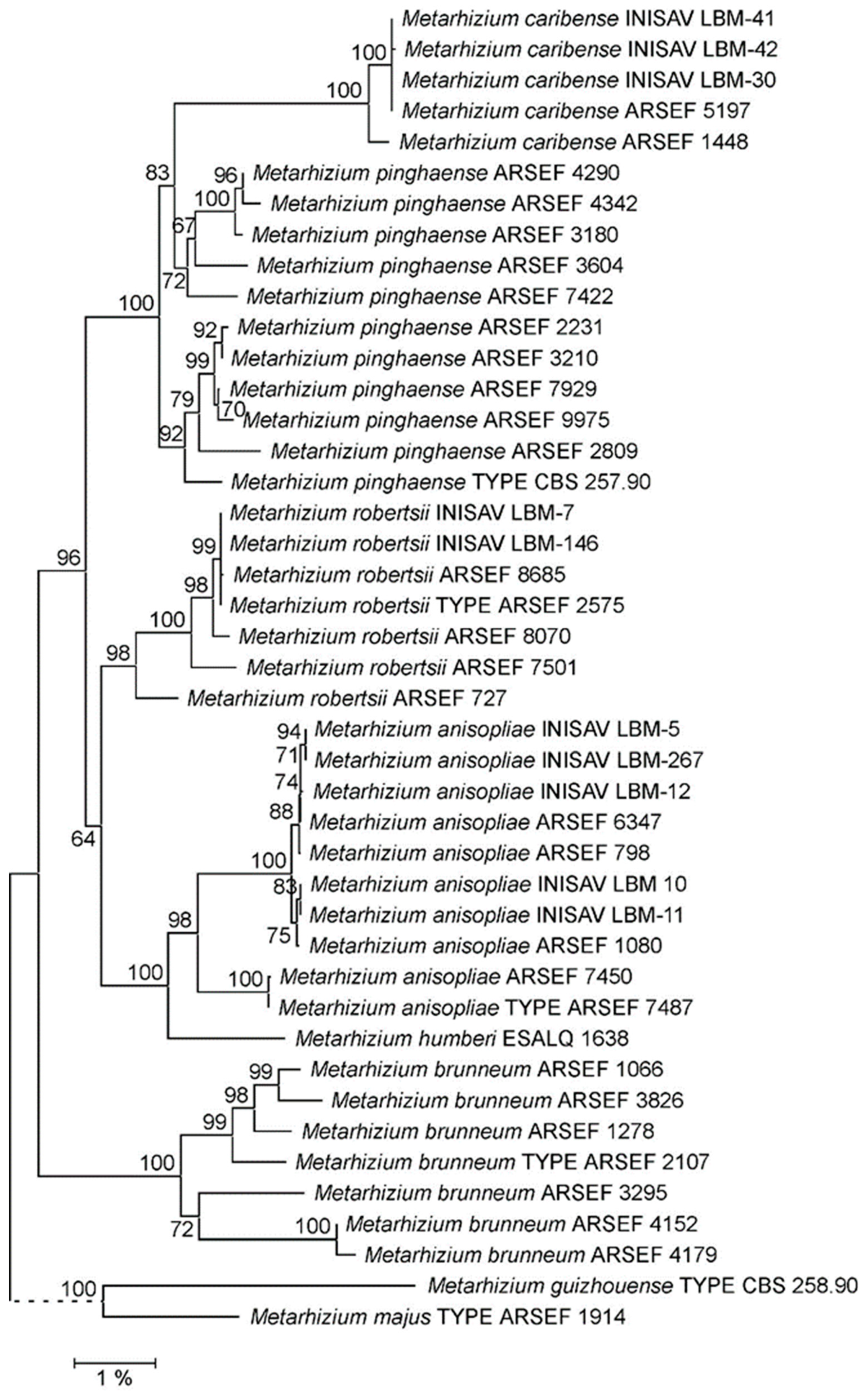
In contrast, in the NJ phylogeny reconstructed from concatenated EF1A, RPB1 and RPB2 marker sequences (Figure 1), isolates LBM-30, LBM-41 and LBM-42, together with strains ARSEF 1448 and ARSEF 5197, but not ARSEF 3180 and ARSEF 4290, formed a 99% bootstrap-supported sub-clade within an overall ill-supported M. pinghaense clade. In the corresponding ML phylogeny (Supplementary Figure S2) this sub-clade appeared even in a sister-clade position with respect to the presumed M. pinghaense clade. However, as has been observed previously [19], PARB clade species were insufficiently resolved in phylogenies reconstructed from EF1A–RPB1–RPB2 concatenations.
On the basis of the above analyses, isolates LBM-30, LBM-41 and LBM-42 were provisionally identified as Metarhizium pinghaense sensu lato. As the presumably most closely related available specimens, strain ARSEF 1448, originally isolated from the burrowing bug, Scaptocoris castanea, in Goiás (Brazil), and strain ARSEF 5197, isolated from the sugarcane rootstalk borer weevil, Diaprepes abbreviata in Florida (USA), were included in the downstream analyses.

3.2. Molecular Taxonomic Identification Based on Intergenic Spacer (IGS) Sequences

Differences in ribosomal intergenic spacer (rIGS) sequences have previously been developed into a diagnostic PCR tool discriminating between Metarhizium PARB clade species [19]. When probed with the corresponding diagnostic primer pairs (Supplementary Table S3), the five strains under study gave negative results not only for M. anisopliae, M. brunneum and M. robertsii, but also for M. pinghaense, which is an outcome in conflict with the provisional species level assignment (Supplementary Figure S3). In order to conclusively identify the strains at the taxonomic species level, further genetic markers were investigated.
Amplification and sequencing of the rIGS-ID800 element that had previously been established as molecular marker for Metarhizium PARB clade species identification [19], gave rise to an identical marker sequence comprising a length of 785 bp from strains LBM-30, LBM-41, LBM-42 and ARSEF 5197, whereas rIGS-ID800 from ARSEF 1448 (788 bp) was determined to be 98.3% similar. In the NJ phylogeny reconstructed from rIGS-ID800 marker sequences (Supplementary Figure S4), these five strains formed a 100% bootstrap supported sub-clade developing out of the supposed M. pinghaense clade. It has been observed previously [19] that rIGS-ID800, when used as single marker, supported the monophyly of the further PARB species (99–100% support in Supplementary Figure S4), whereas M. pinghaense was represented by an ill-supported (58%), apparently loose association of several sub-clades, consistent with the taxon M. pinghaense being polyphyletic.
Moreover, PCR amplification of the molecular–taxonomic MzIGS3 (or DUF895) marker from the three Cuban strains under study gave rise to almost identical (i.e., >99.9% pairwise similarities) consensus sequences comprising a length of 1012 bp. When used as query for an unfiltered BlastN search across the GenBank database, the MzIGS3 marker from strain LBM-41 identified 163 entries of at least 96.0% similarity (at not less than 90% coverage) comprising the bulk of MzIGS3 database entries assigned to the species M. pinghaense. MzIGS3, from ARSEF 5197, was identified as unique identical best hit (displaying 100% similarity at 100% coverage), followed by the DUF895 sequence from ARSEF 1448 as single second-best hit of 99.7% (100%) and a plethora of entries at <99.0% sequence similarity. In the NJ phylogeny reconstructed from these identified MzIGS3 sequences (Supplementary Figure S5), strains LBM-30, LBM-41 and LBM-42 together with ARSEF 1448 and ARSEF 5197 formed an optimally supported sub-clade within the M. pinghaense clade; no further DUF895 sequences from the GenBank database were part of this clade. However, it is clear from Supplementary Figure S5 that—as has been stated previously [20]—when used as single marker, MzIGS3 does not sufficiently resolve the species M. anisopliae, M. humberi and M. robertsii.
In order to obtain a conclusive species assignment for the Cuban isolates, NJ and ML phylogenies were reconstructed from the concatenation of non-coding markers 5TEF, MzIGS3 and rIGS-ID800 (Figure 2, Supplementary Figure S6). In both phylogenies, PARB species clades appeared solidly supported. However, the presumed M. pinghaense clade, receiving optimal (100%) bootstrap support at its root in both the NJ and ML tree, comprised a long-branched and, by itself, 100% bootstrap-supported sub-clade made up of strains LBM-30, LBM-41, LBM-42, ARSEF 1448 and ARSEF 5197. However, bootstrap support for inclusion of this sub-clade into the M. pinghaense clade was only about 80% in both trees. Both the NJ and ML tree topologies were, therefore, prima facie consistent with both the five strains under study forming (i) a sub-species of the species M. pinghaense (sensu stricto) or (ii) an independent species within an M. pinghaense (sensu lato) species complex.
A comparison of pairwise sequence similarities was employed to evaluate this topological finding within the context of Metarhizium PARB species delineation. When calculating pairwise sequence similarity percentages for the 5TEF—MzIGS3—rIGS-ID800 marker concatenation from a p-distance matrix (Supplementary Table S4), sequences from strains LBM-30, LBM-41, LBM-42 and ARSEF 5197 were almost identical (99.9–100% similarity) and very similar to that of strain ARSEF 1448 (99.4–99.5%). Employed as a representative of these five tightly clustering strains, strain ARSEF 5197 displayed 96.1% to 96.5% pairwise sequence similarity to the nomenclatural-type strain and further reference strains of the species M. pinghaense. These values are in the same range as the inter-specific pairwise similarities between the type and reference strains of the PARB species M. pinghaense, M. anisopliae and M. robertsii (96.2–96.9%), but are considerably lower than intra-specific pairwise similarities between the M. pinghaense type and further reference strains (97.8–99.1%). Taken together, pairwise sequence similarity analysis indicates that the phylogenetic relationship of strain ARSEF 5197 and the four further strains under study to the nomenclatural-type strain of the species M. pinghaense are inter- rather than intra-specific.

3.3. rIGS-Based Species-Discriminating Diagnostic PCR

In order to extend the previously developed rIGS-based species-discriminating diagnostic PCR tool to the presumed new Metarhizium species, complete rIGS sequences comprising lengths of between 1593 bp and 1602 bp were determined for strains LBM-30, LBM-41, LBM-42, ARSEF 1448 and ARSEF 5197 and were carefully compared with those of Metarhizium PARB clade reference strains. Species-specific sequence features, such as InDels or SNPs, were identified and pairs of oligonucleotide primers of varying length comprising these specific sequence features were designed (Supplementary Figure S7) and validated under pre-established diagnostic PCR parameters for functionality and discriminative power. Following this rationale gave rise to the definition of a pair of species-discriminating diagnostic PCR primers for the new species: mcar-IDF1 5′-GGACTTGGCATATTTGCTTGAATTG and mcar-IDR1 5′-TCTTATATACCCACCAACTACCTTG (Supplementary Table S3). When employed with the previously established diagnostic PCR protocol, the primer pair unambiguously discriminated between the five strains under study, on the one hand, and the Metarhizium PARB species-type strains together with further M. pinghaense reference strains, on the other hand, amplifying partial rIGS sequences exclusively from cognate genomic DNA templates (Figure 3).

3.4. 28S Ribosomal RNA Gene Group-I Intron Analysis

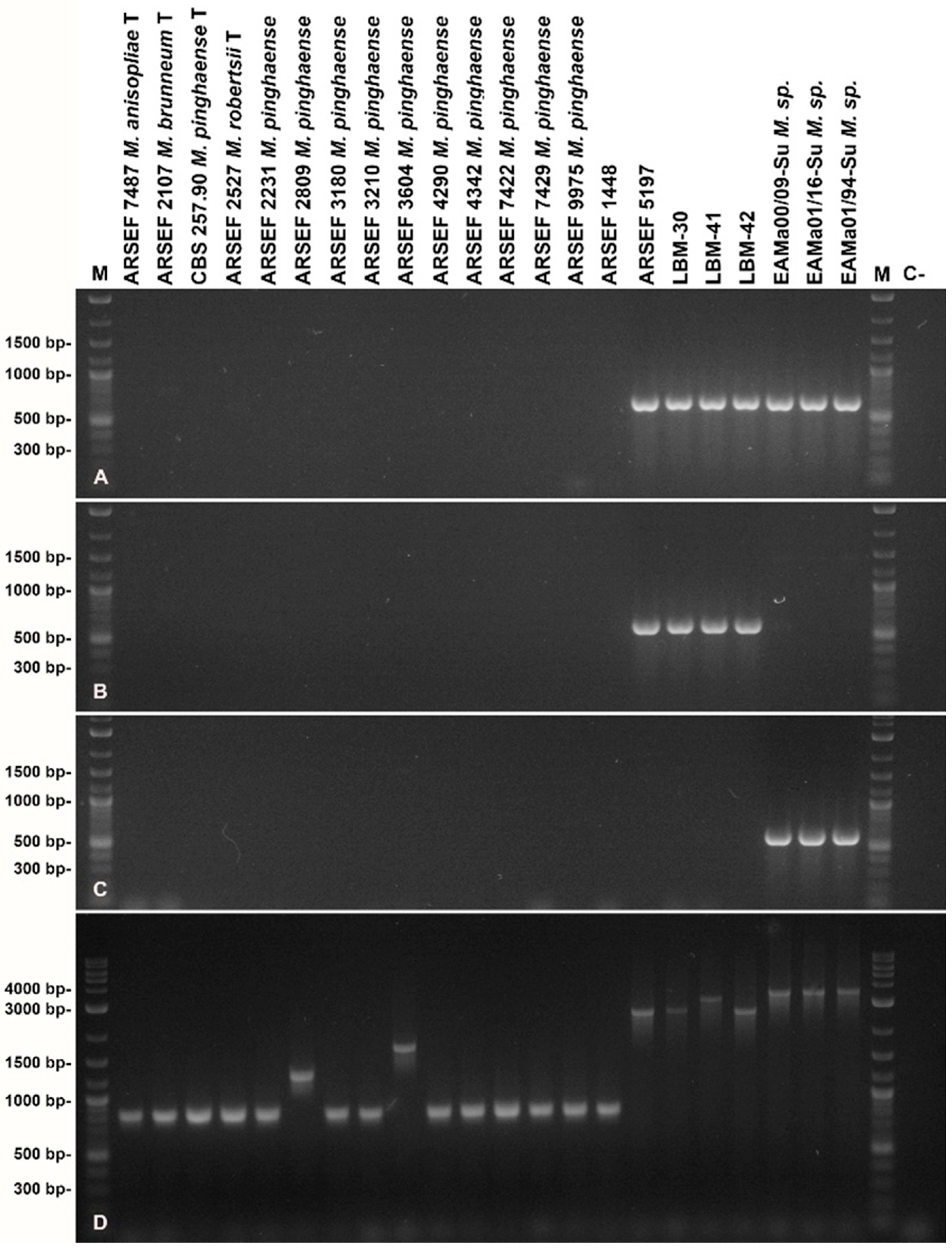
Amplification of the 28S rRNA gene intron insertion region from fungal isolates was consistent with the presence of copies of self-splicing group-I introns in strains LBM-30, LBM-41, LBM-42 and ARSEF 5197, but not in ARSEF 1448 (Figure 4D). Sequencing of the PCR products revealed the presence of an identical intron, termed 28Si4, comprising 1751 bp in insertion position 4 of the 28S rRNA gene of strains LBM-30, LBM-41, LBM-42 and ARSEF 5197, but not ARSEF 1448. In addition, LBM-41 was found to carry a second intron of 438 bp in position 1. None of the Metarhizium PARB species-type strains and further M. pinghaense reference strains analyzed carried a similar long intron in the 28S rRNA intron region. Though strains ARSEF 2809 and ARSEF 3604 carried smaller introns comprising app. 400 bp of unrelated sequence in positions 1 and 4, further reference strains did not carry 28S rRNA introns.
When used as query for unfiltered BlastN searches across the GenBank database, the 28Si4 sequence from ARSEF 5197 identified two highly similar (98.5% similarity at 100% sequence coverage) intron sequences residing in position 4 of the 28S rRNA gene of Metarhizium isolates from different locations in Spain that had previously been assigned to the Metarhizium anisopliae complex [30]. However, by means of phylogenetic reconstruction using 5TEF and concatenated EF1A-RPB1-RPB2 markers, the Spanish isolates EAMa 00/09-Su, EAMa 01/16-Su and EAMa 01/94-Su, carrying a copy of the long 28S rRNA intron, were found to be more closely related to M. majus and M. guizhouense than to the PARB clade species (Figure 1; Supplementary Figures S1 and S2). The 28Si4 introns identified in these isolates were almost identical to each other (99.9–100% similarity). No further sequence entries of high similarity to the 28Si4 intron from ARSEF 5197 were identified in the GenBank database.
Given that the length of autocatalytic group-I introns disrupting fungal rRNA genes usually ranges from app. 300 bp to 500 bp [24,25], the >1700 bp-long 28Si4 introns identified might be expected to have increased in length by acquisition of homing endonuclease genes [42,43]. A search for open reading frames (ORFs) along both directions of the 28Si4 sequence revealed a single, potentially protein-encoding ORF, comprising 1077 bp in collinear orientation with respect to the 28S rRNA gene. A similar ORF encoding a protein of 97% deduced amino acid sequence identity was carried by the homologous introns from Spanish Metarhizium isolates. A search for homologous GenBank entries identified, at a low similarity level, several hypothetical fungal proteins of unknown function. However, no similarities to any of the four types of homing endonucleases and their characteristic motifs [42] became obvious from this analysis.
In depth comparative analysis of 28Si4 intron sequences (Supplementary Figure S8) enabled the design of diagnostic PCR primers (i) Meta28Si4long-F1 for the identification of related long 28Si4 introns in Metarhizium and potentially other fungi, and (ii) Mcar28Si4long-F1 and Mmaj28Si4long-F1 for the discrimination of the two intron variants present in strains LBM-30, LBM-41, LBM-42 and ARSEF 5197, on the one hand, and the Spanish M. majus related strains, on the other (Supplementary Table S3). Application of these primers in respective diagnostic PCR analyses gave rise to the expected results (Figure 4A–C).

3.5. Morphological and Cultural Characterization of Fungal Isolates

In general, the fungal isolates under study produced cottony colonies with white mycelium that became colored with the development of conidia varying from yellow, olive green, greenish to dark green. The conidiation was slight or formed halos or concentric rings. The colony reverse was colorless or ochreous. Phenotypic differences in colony appearance were observed in different culture media (Figure 5). Conidiophores were broadly branched (like candelabra) and phialides were organized in compact palisades. The conidial shape was cylindrical to ellipsoidal. Morphological characteristics of individual strains are described in Table 2 (Supplementary Figure S9).

4. Taxonomic Description

Metarhizium caribense Y. Baró, C. Schuster, R.F. Castañeda and A. Leclerque, sp. nov.
Index Fungorum IF 901982.
Etymology: Latin, caribense refers to the Caribbean, the original locality of the majority of strains isolated.
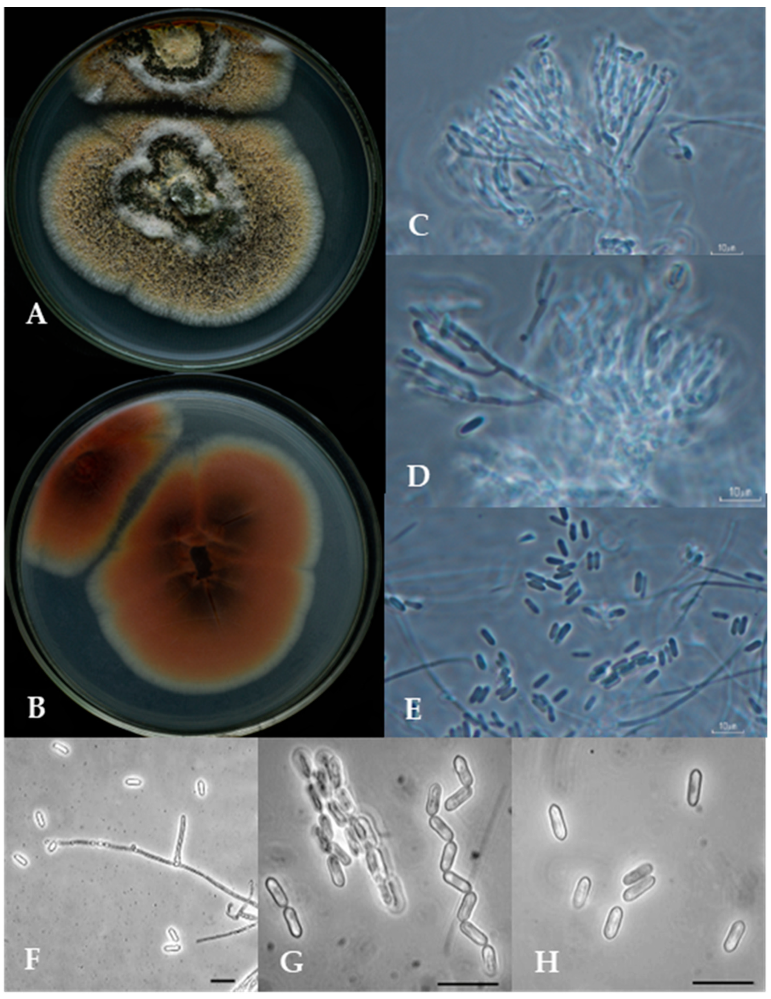
Morphological characteristics: Colonies on SNA after 10 days at 25 ± 2 °C, mycelium mostly immersed, citrine [21K], reverse. Conidiomata sporodochia, columnar, chandelier-like, scattered, arising from vegetative hyphae. Mycelium immersed, composed of branched, hyaline, smooth-walled hyphae. Conidiophores macronematous, densely fasciculate, slightly penicillate branched at the apex, smooth, hyaline. Conidiogenous cells monophialidic, terminal in branches, cylindrical, discrete, smooth, forming a penicillate cluster. Conidia basocatenate, cylindrical, unicellular, smooth-walled, hyaline to olivaceous to green, dry, 5.08–7.04 × 1.22–2.39 µm (Figure 6).
Holotype: Ex-type metabolically inactive culture: deposited as ARSEF 5197 in the USDA-ARS Collection of Entomopathogenic Fungal Cultures (Ithaca, New York, NY, USA) by M. Browning, 20 May 1996; collected by C.W. McCoy.
Host type: Diaprepes abbreviata (Coleoptera: Curculionidae).
Type locality: Florida (USA).
Additional specimens examined as follows: strain INISAV LBM-30, La Habana province (Cuba), isolated with the Galleria bait method from soil sample; strains INISAV LBM-41 and LBM-42, Granma province (Cuba), isolated from Hypothenemus hampei (Coleoptera: Curculionidae); strain ARSEF 1448, Goiatuba (Goiás, Brazil), isolated from Scaptores castanea (Hemiptera: Cydnidae) (Supplementary Figure S10).
M. caribense cannot be distinguished from most closely related taxa based on conidial and phialidic morphologies alone.
Genetic characteristics: Species-discriminating diagnostic PCR employing the primer pair mcar-IDF1 (5′-GGACTTGGCATATTTGCTTGAATTG) and mcar-IDR1 (5′-TCTTATATACCCACCAACTACCTTG) amplifies a partial sequence of the ribosomal intergenic spacer (rIGS) giving rise to a product of app. 350 bp apparent size exclusively from genomic DNA of M. caribense.

5. Discussion

Three fungal strains, designated LBM-30, LBM-41 and LBM-42, that had previously been isolated in Cuba within the framework of a monitoring program aiming towards the identification of new microbial control agents, were investigated in this study. Using morphological taxonomic criteria together with genetic markers (EF1A, RPB1, RPB2) established for the systematics of Hypocreales fungi, the strains were demonstrated to belong to the genus Metarhizium and to be most closely related to the two reference strains, ARSEF 1448 and ARSEF 5197, that had previously been assigned to the PARB clade species M. pinghaense. However, species assignment remained inconclusive at this level of analysis, and LBM-30, LBM-41 and LBM-42 were provisionally designated as M. pinghaense sensu lato.
In depth analysis of the systematic position of these fungi using genetic markers (5TEF, MzIGS3, rIGS) established for the M. anisopliae species complex or the PARB clade, demonstrated that the three Cuban isolates, together with strains ARSEF 1448 and ARSEF 5197, formed a statistically well-supported sub-clade within the PARB clade, but apart from further well-supported Metarhizium PARB species. Pairwise sequence distance analysis motivated the proposal of a new Metarhizium species termed M. caribense, with reference strain ARSEF 5197 being proposed as nomenclatural type of the new taxon. Application of diagnostic PCR using species-specific primer pairs lent further support to this interpretation of the molecular taxonomic data.
M. caribense reference strains ARSEF 5197 and ARSEF 1448 have been studied previously under different perspectives. A broad study of Jin et al. [44] evaluated the pathogenicity against nymphs of the brown planthopper, Nilaparvata lugens, of 35 Metarhizium strains, including ARSEF 5197, with the latter showing only moderate virulence (about 43%) against this pest. Leadmon et al. [45] attributed the virulence of several Metarhizium strains to the secretion of ergot alkaloids in infected insects; however, the strain ARSEF 5197 included in this survey was found to be negative for ergot alkaloids. The authors highlight the importance of these metabolites in pathogenesis, indicating a positive correlation between Metarhizium ergot alkaloid accumulation and insect colonization. In addition to ergot alkaloids, Metarhizium fungi produce other molecules, such as destruxins (DTXs), that contribute to insecticidal activity. Golo et al. [46] evaluated the potential correlation of virulence and DTX production of a set of 20 Metarhizium strains comprising ARSEF 1448. The latter was found to be highly virulent to Tenebrio molitor but did not produce any of the assessed destruxin types, A, B or E.
Previous research had demonstrated the pathogenicity of the three Cuban isolates under study for adults of the sweet potato weevil [32]. Strain LBM-30 has been shown to produce and secrete hydrolytic enzymes as chitinases, lipases, proteases, amylases and caseinases that are generally supposed to contribute to the high virulence of entomopathogenic fungi [32]. Moreover, the strain displays appropriate parameters of conidiation, conidial yield and germination as well as growth in different culture media, making it a potential target for biocontrol agent development. Concomitant research has identified a second Cuban strain, M. anisopliae LBM-267, as a potential microbial control agent targeting the sweet potato weevil [31]. C. formicarius is the most destructive pest affecting the tropical and subtropical production of sweet potatoes (Ipomoea batatas) both in the field and during storage. The larval and adult stages of the weevil have a significant economic impact, causing yield or crop losses up to 100% [47,48]. Currently, control of C. formicarius relies mainly on chemical insecticides. However, due to negative side effects of agrochemicals and the pest’s soil inhabiting lifestyle, the use of insect pathogenic fungi as part of integrated pest management programs is highly solicited and increases each year. In particular, Metarhizium and Beauveria fungi have been used for biological control of this pest.
Future research directions for bio-insecticide developments for the control of the sweet potato weevil will focus on formulation, field experiments and risk assessment with both M. caribense LBM-30 and M. anisopliae LBM-267, including evaluation of possible synergistic effects of a binary product comprising both strains together.
At the methodological level, the present study has demonstrated the potential of the ribosomal intergenic spacer (rIGS) sequence, both as single marker and in combination with 5TEF and MzIGS3 sequences, for species delineation within the Metarhizium PARB clade. The recently developed rIGS-based species-discriminating PCR approach covering the original four PARB clade species [19] and a further PCR tool based on the presence of self-splicing group-I introns in fungal 28S rRNA genes [27,28,29] have been extended to M. caribense, enabling the future sequencing-independent identification of further strains belonging to this new species.

6. Conclusions

Within the PARB clade of the taxonomic genus Metarhizium, a new entomopathogenic fungal species was introduced as consequence of an in-depth multigene phylogenetic analysis of three fungal isolates from Cuba, together with two previously described Metarhizium strains from Florida and Brazil. The species epithet “caribense”, referring to four of these isolates’ geographic origin, was proposed for the new species, and strain ARSEF 5197 was described as a nomenclatural-type strain of the new taxon. A previously established rIGS-based diagnostic PCR tool has been extended to the new species and has been shown to enable sequencing- independent species discrimination within the Metarhizium anisopliae complex.

Supplementary Materials

The following supporting information can be downloaded at: https://www.mdpi.com/article/10.3390/jof10090612/s1. Supplementary Figure S1: Neighbor-Joining (NJ) phylogeny of Metarhizium fungi as reconstructed from 5TEF marker sequences. Supplementary Figure S2: Maximum Likelihood (ML) phylogeny of Metarhizium fungi as reconstructed from a concatenation of EF1A, RPB1 and RPB2 marker sequences. Supplementary Figure S3: Agarose gel electrophoresis of diagnostic PCRs using Metarhizium PARB species-discriminating primer pairs. Supplementary Figure S4: Neighbor-Joining (NJ) phylogeny of Metarhizium fungi as reconstructed from rIGS-ID800 marker sequences. Supplementary Figure S5: Neighbor-Joining (NJ) phylogeny of Metarhizium fungi as reconstructed from MzIGS3 (DUF895) marker sequences. Supplementary Figure S6: Maximum likelihood (ML) phylogeny of Metarhizium fungi as reconstructed from a concatenation of 5TEF, DUF895 (MzIGS3) and rIGS-ID800 marker sequences. Supplementary Figure S7: Alignment of ribosomal intergenic spacer (rIGS) sequences used for species-discriminating primer design. Supplementary Figure S8: Alignment of 28Si4 intron sequences used for diagnostic primer design. Supplementary Figure S9: Colony growth of Metarhizium caribense strain ARSEF 5197 on SNA medium. Supplementary Figure S10: Morphological characteristics of Metarhizium caribense strain ARSEF 1448. Supplementary Table S1: Fungal strains and GenBank accession numbers of marker sequences referred to in this study. Supplementary Table S2: Preparative PCR and sequencing primers used, and reaction-specific parameters applied in this study. Supplementary Table S3: Diagnostic PCR primers used in this study. Supplementary Table S4: Pairwise sequence similarity percentages of concatenated 5TEF–MzIGS3–rIGSID800 sequences as calculated from a p-distance matrix. Refs. [49,50,51,52,53,54,55] are cited in the Supplementary Materials.

Author Contributions

Conceptualization, A.L.; methodology, C.S. and Y.B.R.; validation, C.S. and Y.B.R.; formal analysis, A.P.d.l.C.; investigation, C.S., Y.B.R., Y.G.C. and A.L.; data curation, C.S., Y.B.R., Y.G.C. and R.F.C.-R.; writing—original draft preparation, C.S., Y.B.R. and A.L.; writing—review and editing, R.F.C.-R. and A.P.d.l.C.; project administration, C.S., Y.B.R., M.E.M.G. and A.L.; funding acquisition, Y.B.R., M.E.M.G. and A.L. All authors have read and agreed to the published version of the manuscript.

Funding

This research has received financial support from the German Ministry of Education and Research under research grants 031B0766 (BMBF/PT Jülich), project BioEconomy International–FUNGI 4 FOOD, and 01DN14020 (BMBF/DLR), project CUBAgroBio, as well as from the Cuban research projects: Empleo de nuevos aislados de los entomopatógenos Beauveria y Metarhizium spp. para el desarrollo de bioproductos para el control de plagas agrícolas financed by the Ministerio de Ciencia Tecnología y Medio Ambiente (CITMA), under research grant PSAV P131LH003047 and Desarrollo de procedimientos para la evaluación de la efectividad de bioplaguicidas y contribuir al manejo integrado de plagas, financed from the Ministry of Agriculture, under research grant PSSAV PS223MY003-111. Y.B.R. acknowledges receipt of a research scholarship by the German Academic Exchange Service (DAAD), grant ID 57314019, financed by funding from the German Foreign Office. The APC was partially funded by the Deutsche Forschungsgemeinschaft (DFG) and the Open Access Publishing Fund of Technical University of Darmstadt.

Institutional Review Board Statement

Not applicable.

Informed Consent Statement

Not applicable.

Data Availability Statement

Sequence data analyzed in this study are publicly available from the GenBank database (https://www.ncbi.nlm.nih.gov; accessed 1 July 2024) under nucleotide sequence accession numbers listed in Supplementary Table S1 to this study.

Acknowledgments

The authors owe Kathryn E. Bushley from the ARSEF collection and Enrique Quesada-Moraga from Universidad de Cordoba a debt of gratitude for making their fungal strains available. Moreover, the authors are grateful to Enrique Ponce Grijuela and Yaremis Ulloa Martín from INISAV for excellent technical support.

Conflicts of Interest

The authors declare no conflicts of interest. The funders had no role in the design of the study; in the collection, analyses, or interpretation of data; in the writing of the manuscript; or in the decision to publish the results.

References

  1. Holz, S.; Celeste, P.d.A.; Héros, J.M.; Nascimento de Souza, P.H.; Yenjit, R.; Garcia, C.; Borges, D.; Delalibera, I., Jr.; Zhi-Yuan, C.; Pascholati, S.F. The potential of using Metarhizium anisopliae and Metarhizium humberi to control the Asian soybean rust caused by Phakopsora pachyrhizi. Biocontrol Sci. Technol. 2023, 33, 366–382. [Google Scholar] [CrossRef]
  2. Aw, K.M.S.; Hue, S.M. Mode of Infection of Metarhizium spp. Fungus and their Potential as Biological Control Agents. J. Fungi 2017, 3, 30. [Google Scholar] [CrossRef]
  3. Driver, F.; Milner, R.J.; Trueman, J.W.H. A taxonomic revision of Metarhizium based on a phylogenetic analysis of rDNA sequence data. Mycol. Res. 2000, 104, 134–150. [Google Scholar] [CrossRef]
  4. Kepler, R.M.; Humber, R.A.; Bischoff, J.F.; Rehner, S.A. Clarification of generic and species boundaries for Metarhizium and related fungi through multigene phylogenetics. Mycologia 2014, 106, 811–829. [Google Scholar] [CrossRef]
  5. Mongkolsamrit, S.; Khonsanit, A.; Thanakitpipattana, D.; Tasanathai, K.; Noisripoom, W.; Lamlertthon, S.; Himaman, W.; Houbraken, J.; Samson, R.A.; Luangsa-Ard, J. Revisiting Metarhizium and the description of new species from Thailand. Stud. Mycol. 2020, 95, 171–251. [Google Scholar] [CrossRef]
  6. Bischoff, J.F.; Rehner, S.A.; Humber, R.A. A multilocus phylogeny of the Metarhizium anisopliae lineage. Mycologia 2009, 101, 512–530. [Google Scholar] [CrossRef]
  7. Sung, G.H.; Sung, J.M.; Hywel-Jones, N.L.; Spatafora, J.W. A multi-gene phylogeny of Clavicipitaceae (Ascomycota, Fungi): Identification of localized incongruence using a combinational bootstrap approach. Mol. Phylogenet. Evol. 2007, 44, 1204–1223. [Google Scholar] [CrossRef]
  8. Sung, G.H.; Hywel-Jones, N.L.; Sung, J.M.; Luangsa-Ard, J.J.; Shrestha, B.; Spatafora, J.W. Phylogenetic classification of Cordyceps and the clavicipitaceous fungi. Stud. Mycol. 2007, 57, 5–59. [Google Scholar] [CrossRef]
  9. Bischoff, J.F.; Rehner, S.A.; Humber, R.A. Metarhizium frigidum sp. nov.: A cryptic species of M. anisopliae and a member of the M. flavoviride complex. Mycologia 2006, 98, 737–745. [Google Scholar] [CrossRef]
  10. Kepler, R.M.; Sung, G.H.; Ban, S.; Nakagiri, A.; Chen, M.J.; Huang, B.; Li, Z.; Spatafora, J.W. New teleomorph combinations in the entomopathogenic genus Metacordyceps. Mycologia 2012, 104, 182–197. [Google Scholar] [CrossRef]
  11. Montalva, C.; Collier, K.; Rocha, L.F.; Inglis, P.W.; Lopes, R.B.; Luz, C.; Humber, R.A. A natural fungal infection of a sylvatic cockroach with Metarhizium blattodeae sp. nov., a member of the M. flavoviride species complex. Fungal Biol. 2016, 120, 655–665. [Google Scholar] [CrossRef]
  12. Lopes, R.B.; Souza, D.A.; Rocha, L.F.N.; Montalva, C.; Luz, C.; Humber, R.A.; Faria, M. Metarhizium alvesii sp. nov.: A new member of the Metarhizium anisopliae species complex. J. Invertebr. Pathol. 2018, 151, 165–168. [Google Scholar] [CrossRef]
  13. Gutierrez, A.C.; Leclerque, A.; Manfrino, R.G.; Luz, C.; Ferrari, W.A.O.; Barneche, J.; García, J.J.; López Lastra, C.C. Natural occurrence in Argentina of a new fungal pathogen of cockroaches, Metarhizium argentinense sp. nov. Fungal Biol. 2019, 123, 364–372. [Google Scholar] [CrossRef]
  14. Rehner, S.A.; Kepler, R.M. Species limits, phylogeography and reproductive mode in the Metarhizium anisopliae complex. J. Invertebr. Pathol. 2017, 148, 60–66. [Google Scholar] [CrossRef]
  15. Rezende, J.M.; Zanardo, A.B.R.; da Silva Lopes, M.; Delalibera, I., Jr.; Rehner, S.A. Phylogenetic diversity of Brazilian Metarhizium associated with sugarcane agriculture. BioControl 2015, 60, 495–505. [Google Scholar] [CrossRef]
  16. Luz, C.; Rocha, L.F.N.; Montalva, C.; Souza, D.A.; Botelho, A.B.R.Z.; Lopes, R.B.; Faria, M.; Delalibera, I., Jr. Metarhizium humberi sp. nov. (Hypocreales: Clavicipitaceae), a new member of the PARB clade in the Metarhizium anisopliae complex from Latin America. J. Invertebr. Pathol. 2019, 166, 107216. [Google Scholar] [CrossRef]
  17. Iwanicki, N.S.A.; Botelho, A.B.R.Z.; Klingen, I.; Júnior, I.D.; Rossmann, S.; Lysøe, E. Genomic signatures and insights into host niche adaptation of the entomopathogenic fungus Metarhizium humberi. G3 2022, 12, jkab416. [Google Scholar] [CrossRef]
  18. Kepler, R.M.; Rehner, S.A. Genome-assisted development of nuclear intergenic sequence markers for entomopathogenic fungi of the Metarhizium anisopliae species complex. Mol. Ecol. Resour. 2013, 13, 210–217. [Google Scholar] [CrossRef]
  19. Schuster, C.; Baró Robaina, Y.; Ben Gharsa, H.; Bobushova, S.; Manfrino, R.G.; Gutierrez, A.C.; Lopez Lastra, C.C.; Doolotkeldieva, T.; Leclerque, A. Species Discrimination within the Metarhizium PARB Clade: Ribosomal Intergenic Spacer (rIGS)-Based Diagnostic PCR and Single Marker Taxonomy. J. Fungi 2023, 9, 996. [Google Scholar] [CrossRef]
  20. Riguetti, Z.B.A.B.; Alves-Pereira, A.; Colonhez, P.R.; Zucchi, M.I.; Delalibera, I., Jr. Metarhizium species in soil from Brazilian biomes: A study of diversity, distribution, and association with natural and agricultural environments. Fungal Ecol. 2019, 41, 289–300. [Google Scholar] [CrossRef]
  21. Pantou, M.P.; Mavridou, A.; Typas, M.A. IGS sequence variation, group-I introns and the complete nuclear ribosomal DNA of the entomopathogenic fungus Metarhizium: Excellent tools for isolate detection and phylogenetic analysis. Fung. Genet. Biol. 2003, 38, 159–174. [Google Scholar] [CrossRef]
  22. Mayerhofer, J.; Lutz, A.; Dennert, F.; Rehner, S.A.; Kepler, R.M.; Widmer, F.; Enkerli, J. A species-specific multiplexed PCR amplicon assay for distinguishing between Metarhizium anisopliae, M. brunneum, M. pingshaense and M. robertsii. J. Invertebr. Pathol. 2019, 161, 23–28. [Google Scholar] [CrossRef]
  23. Neuvéglise, C.; Brygoo, Y. Identification of group-1 intron in the 28s rDNA of the entomopathogenic fungus Beauveria brongniartii. Curr. Genet. 1994, 27, 38–45. [Google Scholar] [CrossRef]
  24. Mavridou, A.; Cannone, J.; Typas, M.A. Identification of group-I introns at three different positions within the 28S rDNA gene of the entomopathogenic fungus Metarhizium anisopliae var. anisopliae. Fungal Genet. Biol. 2000, 31, 79–90. [Google Scholar] [CrossRef]
  25. Wang, C.; Li, Z.; Typas, M.A.; Butt, T.M. Nuclear large subunit rDNA group I intron distribution in a population of Beauveria bassiana strains: Phylogenetic implications. Mycol. Res. 2003, 107, 1189–1200. [Google Scholar] [CrossRef]
  26. Neuveglise, C.; Brygoo, Y.; Riba, G. 28S rDNA group-I introns: A powerful tool for identifying strains of Beauveria brongniartii. Mol. Ecol. 1997, 6, 373–381. [Google Scholar] [CrossRef]
  27. Fatu, A.C.; Fatu, V.; Andrei, A.M.; Ciornei, C.; Lupastean, D.; Leclerque, A. Strain-specific PCR-based diagnosis for Beauveria brongniartii biocontrol strains. IOBC/wprs Bull. 2011, 66, 213–216. [Google Scholar]
  28. Schuster, C.; Saar, K.; Manfrino, R.; Aguilera Sammaritano, J.; Tornesello Galván, J.; García, J.J.; López Lastra, C.C.; Leclerque, A. Group-I intron-based strain-specific diagnosis of entomopathogenic Lecanicillium fungi for aphid biocontrol. IOBC/wprs Bull. 2016, 113, 53–56. [Google Scholar]
  29. Manfrino, R.G.; Schuster, C.; Saar, K.; López, L.C.; Leclerque, A. Genetic characterization, pathogenicity and benomyl susceptibility of Lecanicillium fungal isolates from Argentina. J. Appl. Entomol. 2019, 143, 204–213. [Google Scholar] [CrossRef]
  30. Márquez, M.; Iturriaga, E.A.; Quesada-Moraga, E.; Santiago-Alvarez, C.; Monte, E.; Hermosa, R. Detection of potentially valuable polymorphisms in four group I intron insertion sites at the 3’-end of the LSU rDNA genes in biocontrol isolates of Metarhizium anisopliae. BMC Microbiol. 2006, 6, 77. [Google Scholar] [CrossRef]
  31. Baró, Y.; Schuster, C.; Gato, Y.; Márquez, M.E.; Leclerque, A. Characterization, identification and virulence of Metarhizium species from Cuba to control the sweet potato weevil, Cylas formicarius Fabricius (Coleoptera: Brentidae). J. Appl. Microbiol. 2022, 132, 3705–3716. [Google Scholar] [CrossRef]
  32. Gato, Y.; Márquez, M.E.; Baró Robaina, Y.; Calle, J. Detección de enzimas extracelulares en cepas cubanas del complejo Metarhizium anisopliae con acción entomopatogénica contra Cylas formicarius Fabricius (Coleoptera: Brentidae). Actu Biol. 2017, 39, 71–78. [Google Scholar] [CrossRef]
  33. Inglis, D.; Enkerli, J.; Goettel, M. Laboratory techniques used for entomopathogenic fungi: Hypocreales. In Manual of Techniques in Invertebrate Pathology, 2nd ed.; Lacey, L.A., Ed.; Academic Press: London, UK, 2012; pp. 189–243. [Google Scholar]
  34. Tamura, K.; Stecher, G.; Kumar, S. MEGA11: Molecular Evolutionary Genetics Analysis version 11. Mol. Biol. Evol. 2021, 38, 3022–3027. [Google Scholar] [CrossRef]
  35. Altschul, S.F.; Madden, T.L.; Schäffer, A.A.; Zhang, J.; Zhang, Z.; Miller, W.; Lipman, D.J. Grapped BLAST and PSI-BLAST: A new generation of protein database search programs. Nucleis Acids Res. 1997, 2, 3389–3402. [Google Scholar] [CrossRef]
  36. Zhang, Z.; Schwartz, S.; Wagner, L.; Miller, W. A greedy algorithm for aligning DNA sequences. J. Comput. Biol. 2000, 7, 203–214. [Google Scholar] [CrossRef]
  37. Thompson, J.D.; Higgins, D.G.; Gibson, T.J. CLUSTAL W: Improving the sensitivity of progressive multiple sequence alignment through sequence weighting, position-specific gap penalties and weight matrix choice. Nucleic Acids Res. 1994, 22, 4673–4680. [Google Scholar] [CrossRef]
  38. Schmidt, H.A.; Strimmer, K.; Vingron, M.; von Haeseler, A. Tree-Puzzle: Maximum likelihood phylogenetic analysis using quartets and parallel computing. Bioinformatics 2002, 18, 502–504. [Google Scholar] [CrossRef]
  39. Guindon, S.; Gascuel, O. A simple, fast, and accurate algorithm to estimate large phylogenies by maximum likelihood. Syst. Biol. 2003, 52, 696–704. [Google Scholar] [CrossRef]
  40. Hasegawa, M.; Kishino, H.; Yano, T.A. Dating of the human–ape splitting by a molecular clock of mitochondrial DNA. J. Mol. Evol. 1985, 22, 160–174. [Google Scholar] [CrossRef]
  41. Yang, Z. Maximum-Likelihood estimation of phylogeny from DNA sequences when substitution rates differ over sites. Mol. Biol. Evol. 1993, 10, 1396–1401. [Google Scholar]
  42. Haugen, P.; Simon, D.M.; Bhattacharya, D. The natural history of group I introns. Trends Genet. 2005, 21, 111–119. [Google Scholar] [CrossRef]
  43. Gogarten, J.P.; Hilario, E. Inteins, introns, and homing endonucleases: Recent revelations about the life cycle of parasitic genetic elements. BMC Evol. Biol. 2006, 6, 94. [Google Scholar] [CrossRef]
  44. Jin, S.F.; Feng, M.G.; Chen, J.Q. Selection of global Metarhizium isolates for the control of the rice pest Nilaparvata lugens (Homoptera: Delphacidae). Pest. Manag. Sci. 2008, 64, 1008–1014. [Google Scholar] [CrossRef]
  45. Leadmon, C.E.; Sampson, J.K.; Maust, M.D.; Macias, A.M.; Rehner, S.A.; Kasson, M.T.; Panaccione, D.G. Several Metarhizium species produce ergot alkaloids in a condition-specific manner. Appl. Environ. Microbiol. 2020, 86, e00373-20. [Google Scholar] [CrossRef]
  46. Golo, P.S.; Gardner, D.R.; Grilley, M.M.; Takemoto, J.Y.; Krasnoff, S.B.; Pires, M.S.; Fernandes, E.K.K.; Bittencourt, V.R.E.P.; Roberts, D.W. Production of Destruxins from Metarhizium spp. Fungi in Artificial Medium and in Endophytically Colonized Cowpea Plants. PLoS ONE 2014, 9, e104946. [Google Scholar] [CrossRef]
  47. Muniappan, R.; Shepard, B.M.; Carner, G.R.; Ooi, P.A.C. Arthropod Pests of Horticultural Crops in Tropical Asia; CABI: Wallingford, UK, 2012; pp. 52–134. [Google Scholar]
  48. Reddy, P.P. Plant Protection in Tropical Root and Tuber Crops; Springer: New Delhi, India, 2015; pp. 87–98. [Google Scholar]
  49. Kepler, R.M.; Chen, Y.; Kilcrease, J.; Shao, J.; Rehner, S.A. Independent origins of diploidy in Metarhizium. Mycologia 2016, 108, 1091–1103. [Google Scholar]
  50. Rehner, S.A. Genetic structure of Metarhizium species in western USA: Finite populations composed of divergent clonal lineages with limited evidence for recent recombination. J. Invertebr. Pathol. 2020, 177, 107491. [Google Scholar] [CrossRef]
  51. Rocha, L.F.; Inglis, P.W.; Humber, R.A.; Kipnis, A.; Luz, C. Occurrence of Metarhizium spp. in Central Brazilian soils. J. Basic. Microbiol. 2013, 53, 251–259. [Google Scholar] [CrossRef]
  52. Rehner, S.A.; Buckley, E.A. Beauveria phylogeny inferred from nuclear ITS and EF1-alpha sequences: Evidence for cryptic diversification and links to cordyceps teleomorphs. Mycologia 2005, 97, 84–98. [Google Scholar]
  53. Stiller, J.W.; Hall, B.D. The origin of red algae: Implications for plastid evolution. Proc. Natl. Acad. Sci. USA 1997, 94, 4520–4525. [Google Scholar] [CrossRef]
  54. Liu, Y.J.; Whelen, S.; Hall, B.D. Phylogenetic relationships among ascomycetes: Evidence from an RNA polymerse II subunit. Mol. Biol. Evol. 1999, 16, 1799–1808. [Google Scholar] [CrossRef]
  55. Goetsch, L.; Eckert, A.J.; Hall, B.D. The molecular systematics of Rhododendron (Ericaceae): A phylogeny based upon RPB2 gene sequences. Syst. Bot. 2005, 30, 616–626. [Google Scholar] [CrossRef]
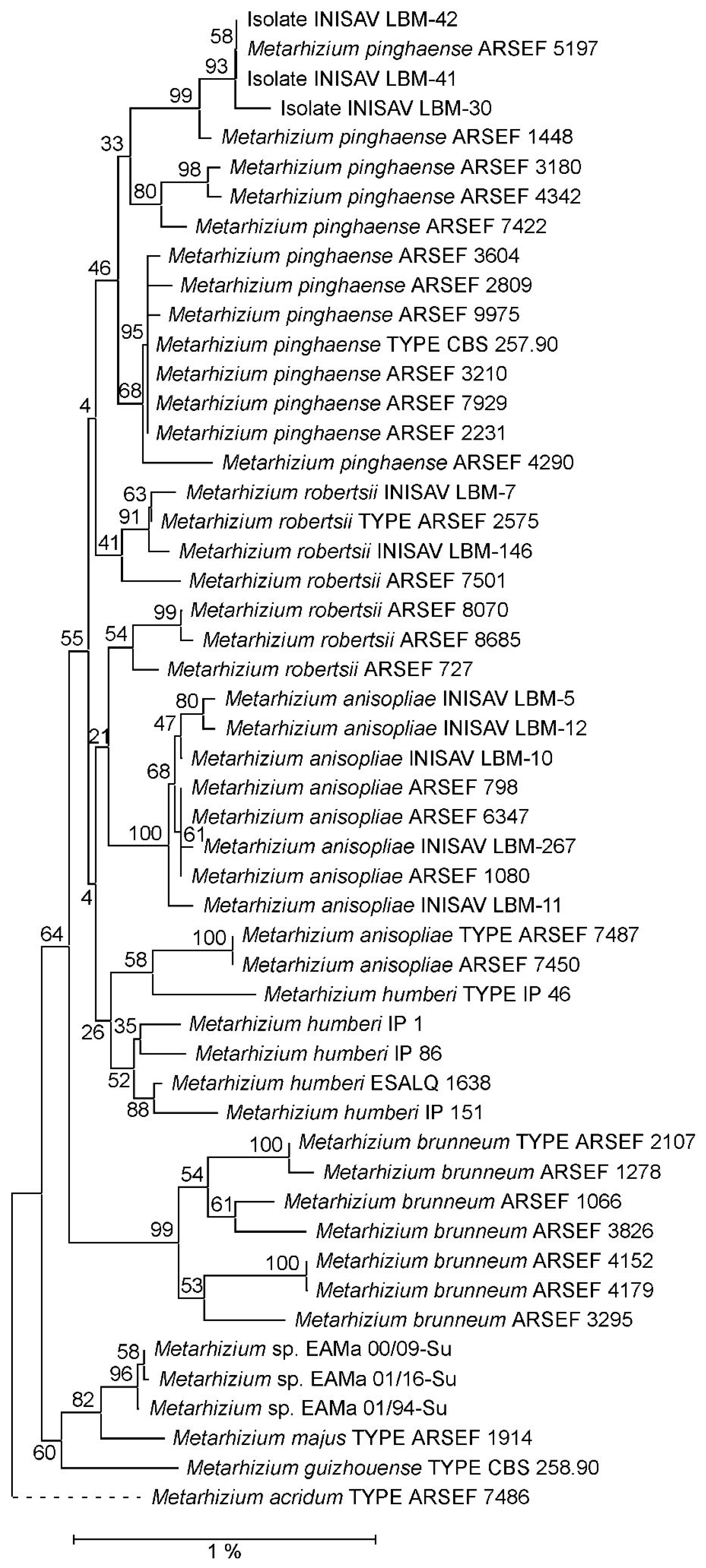
Figure 1. Neighbor joining (NJ) phylogeny of Metarhizium fungi as reconstructed from a concatenation of EF1A, RPB1 and RPB2 marker sequences. Terminal branches are labelled by genus, species and strain designations; “TYPE” denotes the nomenclatural-type strain of a species. Numbers on branches indicate bootstrap support percentages. The size bar corresponds to 1% sequence divergence; branches drawn as dashed lines are not to scale. A concatenation of the orthologous sequences from the M. acridum type strain has been used as outgroup.
Jof 10 00612 g001
Figure 2. Neighbor joining (NJ) phylogeny of Metarhizium fungi as reconstructed from a concatenation of 5TEF, DUF895 (MzIGS3) and rIGS-ID800 marker sequences. Terminal branches are labelled by genus, species and strain designations; “TYPE” denotes the nomenclatural-type strain of a species. Numbers on branches indicate bootstrap support percentages. The size bar corresponds to 1% sequence divergence; branches drawn as dashed lines are not to scale. Concatenations of the orthologous sequences from the M. majus- and M. guizhouense-type strains have been used as outgroup.
Jof 10 00612 g002
Figure 3. Agarose gel electrophoresis of diagnostic PCRs using primer pair mcar-IDF1/mcar-IDR1 (Supplementary Table S3) specific for the species Metarhizium caribense. The length of relevant signals in the size standard is indicated at the left margin. Lane labels on top of the picture designate the Metarhizium species, strain or isolate; “T” indicates nomenclatural type strains, “M” denotes the size standard, and “C-” denotes negative (no template) control.
Jof 10 00612 g003
Figure 4. Agarose gel electrophoresis of diagnostic PCR amplification of 28Si4 intron sequences using primer pairs Meta28Si4long-F1/28Si-E24Rshort (A), Mcar28Si4long-F1/28Si-E24Rshort (B), and Mmaj28Si4long-F1/28Si-E24Rshort (C) (Supplementary Table S3) as well as the complete 28S rRNA gene intron insertion region (D). The length of relevant signals in the size standard is indicated at the left margin. Lane labels on top of the picture designate the Metarhizium species, strain or isolate; “T” indicates nomenclatural type strains, “M” denotes the size standard, and “C-” denotes negative (no template) control.
Jof 10 00612 g004
Figure 5. Morphological characteristics of strains LBM-30, LBM-41, LBM-42, ARSEF 1448 and ARSEF 5197 on different culture media, after 14 days of growth at 26 ± 1 °C. Sabouraud dextrose agar (A,D,G,J,M); complete medium (B,E,H,K,N); potato dextrose agar (C,F,I,L,O).
Jof 10 00612 g005
Figure 6. Morphological characteristics of the proposed Metarhizium caribense type strain ARSEF 5197. Colony on SDA. (A) anverse, (B) reverse (C) conidiophore, (D) phialides, and (EH) conidia. All scale bars correspond to 10 µm.
Jof 10 00612 g006
Table 1. Fungal isolates from the INISAV culture collection investigated in this study.
Strain
Designation
Source of IsolationGeographic Origin
LBM-30Soil using Galleria mellonella (L.) (Lepidoptera: Pyralidae) bait methodLa Habana, Cuba
LBM-41Coffee berry borer, Hypothenemus hampei Ferrari (Coleoptera: Curculionidae)Granma, Cuba
LBM-42Coffee berry borer, Hypothenemus hampei Ferrari (Coleoptera: Curculionidae)Granma, Cuba
Table 2. Morphological characteristics of fungal strains studied when grown on SDA.
IsolateColony
Characteristics
Conidial
Size a
Conidial Shape
LBM-30Cottony texture, abundant mycelia growth and wooly in the central area, with a white mycelial margin. Abundant conidiation yellow to olive green in color and crusty appearance, with regular border. Conidiophores broadly branched (chandelier-like)5.00–8.50 (6.60) ×
2.00–3.00 (2.44)
Cylindrical
LBM-41White colonies to greenish olivaceous, forming halos or concentric rings with conidia maturation. Abundant conidiation in central area forming small pustules, with white mycelial margin. Ochreous pigment diffusing into medium. Conidiophores broadly branched (chandelier-like). 4.88–7.32 (5.66) ×
2.44–3.66 (2.71).
Cylindrical to ellipsoidal
LBM-42White to dark olivaceous green with conidia maturation, colonies with white mycelial margin. Diffuse conidiation forming small pustules. Ochreous pigment diffusing into medium4.88–7.32 (6.93) ×
2.43–2.44 (2.44).
Cylindrical to ellipsoidal
ARSEF 1448Colonies were white and became yellow to olive green with a white mycelial margin. Colony reverses were yellow. Conidiophores were broadly branched, like a candelabrum arrangement. Phialides were cylindrical and organized in compact palisades.4.91–7.19 (5.99) ×
1.29–2.56 (1.83).
Cylindrical
ARSEF 5197Colonies initially appeared white and became yellow to dark olive green with white mycelial margin. Colony reverses were ochreous. Conidiophores were broadly branched, like a candelabrum arrangement. Phialides were cylindrical and organized in compact palisades5.08–7.04 (5.78) ×
1.22–2.39 (1.82)
Cylindrical
a All measurements in μm, length × width. Average values are indicated in brackets.
Disclaimer/Publisher’s Note: The statements, opinions and data contained in all publications are solely those of the individual author(s) and contributor(s) and not of MDPI and/or the editor(s). MDPI and/or the editor(s) disclaim responsibility for any injury to people or property resulting from any ideas, methods, instructions or products referred to in the content.