Article

A Genomic Sequence Resource of Diaporthe mahothocarpus GZU-Y2 Causing Leaf Spot Blight in Camellia oleifera

1
College of Forestry, Guizhou University, Huaxi District, Guiyang 550025, China
2
School of Biological Sciences, Guizhou Education University, Wudang District, Guiyang 550018, China
*
Authors to whom correspondence should be addressed.
J. Fungi 2024, 10(9), 630; https://doi.org/10.3390/jof10090630
Submission received: 18 April 2024 / Revised: 9 August 2024 / Accepted: 2 September 2024 / Published: 3 September 2024

Abstract

:
Diaporthe mahothocarpus GZU-Y2, a new pathogen responsible for leaf spot blight disease, leads to significant damage and economic losses in some Camellia oleifera plantations. The current study annotated the genome of the D. mahothocarpus isolate GZU-Y2 to advance our knowledge of the pathogen and facilitate improved disease management of leaf spot blight. The initial Pacbio-Illumina hybrid draft genome for GZU-Y2 resulted in a high-quality assembly with 62 contigs, characterized by an N50 length of 7.07 Mb. The complete genome of isolate GZU-Y2 was 58.97 Mbp, with a GC content of 50.65%. Importantly, the assembly exhibits remarkable integrity, with 97.93% of complete BUSCO validating genome completeness. The prediction results showed that a total of 15,918 protein-coding genes were annotated using multiple bioinformatics databases. The genome assembly and annotation resource reported here will be useful for the further study of fungal infection mechanisms and pathogen–host interaction.

1. Introduction

Camellia oleifera C.Abel., a woody tree species that produces edible oil, is unique to China and is one of the four major woody oil tree species in the world. C. oleifera has an extremely wide distribution range in China, from the coastal hills in the southeast to the Yunnan-Guizhou Plateau in the west. In recent years, the C. oleifera industry in Guizhou Province has developed significantly, but due to the relatively weak research on pest control, the harm of pests and diseases on C. oleifera has become increasingly serious. Among them, leaf spot blight of C. oleifera caused by Diaporthe spp. is one of the most important diseases, which results in significant leaf and fruit loss, ultimately affecting the yield and quality of the oil [1,2]. The disease initially manifests as light yellow, nearly round, or irregular spots on the leaf margins. These spots later turn dark brown, with the appearance of light brown and many dark black spots. The boundary between the diseased and healthy parts is distinct. As the disease progresses, the leaf blade eventually dies and turns gray. Diaporthe spp. associated with leaf blight of C. oleifera were identified and characterized as D. camelliae oleiferae, D. hunanensis, D. hubeiensis, and D. sojae [3,4]. We previously identified the pathogen isolate in Guizhou Province as D. mahothocarpus GZU-Y2, belonging to the D. eres species complex [5].
The genus Diaporthe is a significant group of plant pathogenic fungi that belongs to the Diaporthaceae family, with a broad host range and geographical distribution [6]. The MycoBank database currently records 1346 Diaporthe species and over 1057 species of its asexual form, Phomopsis, which are found worldwide, particularly in tropical and subtropical ecosystems. They are commonly found in plants as phytopathogenic, endophytic, or saprophytic fungi [7]. For example, D. ampelina is a significant pathogen responsible for grapevine twig blight and leaf spot, which infects shoots and leaves of grapes, causing up to 30% yield loss in temperate regions [8]. D. citri could cause citrus rot in all major citrus production areas except in Europe [9,10]. Furthermore, D. helianthi was initially reported in the former Yugoslavia, which primarily causes sunflower stem ulcer disease [11,12]. In addition, due to its biological and chemical diversity, Diaporthe spp. is also a rich source of active natural products. It has been found that they can produce a variety of metabolites with novel structures, including polyketides, alkaloids, terpenoids, and anthraquinones, which exhibit significant anti-tumor, antimicrobial, and antioxidant activities [13].
High-throughput whole genome sequencing is an effective method for gaining a comprehensive understanding of strain-related properties at the gene level [14]. Thus, the present study involved sequencing, predicting assembly, and annotating high-quality genome sequences of D. mahothocarpus, aiming to provide a systematic analysis of its pathogenicity and interaction mechanisms with the host at the molecular level.

2. Materials and Methods

2.1. Fungal Material and Culture Conditions

Diaporthe mahothocarpus GZU-Y2 was isolated from leaf-spot-blight diseased leaves of Camellia oleifera, preserved at 4 °C on potato dextrose agar (PDA) medium in the Forest Pathology Laboratory of the College of Forestry, Guizhou University (Guiyang, China). It was cultured on PDA at 28 °C for 7 days before DNA extraction.

2.2. DNA Extraction

The mycelium of D. mahothocarpus GZU-Y2 was obtained from cultures grown in 100 mL of fresh potato dextrose broth (PDB) at 28 °C for 2 days. Mycelium was filtered through sterile filter paper and ground to a powder in liquid nitrogen. Genomic DNA was then extracted by SDS-based DNA extraction using Omega Fungal DNA Kit D3390-02 (Omega Bio-Tek, Inc., Norcross, GA, USA). The extracted DNA was separated by 1% agarose gel electrophoresis, stained with ethidium bromide (0.1 mg/mL), detected by UV transmission, and quantified by Qubit.

2.3. Genome Sequencing and Assembly

The total DNA was sequenced using the PacBio Sequel II single-molecule real-time (SMRT) sequencing platform from Beijing PacMark Biotechnology Co., Ltd. (Beijing, China). The low-quality reads were filtered, and the remaining reads were assembled into a gap-free isoform using SMRT Link v5.0.1. Segments were spliced based on the overlapping regions between reads, first splicing them into longer contiguous sequences (contigs), and then contigs were spliced into longer scaffolds that were allowed to contain gap sequences (gaps) by eliminating errors and gaps in the scaffolds and localizing these scaffolds to chromosomes. The circular consensus sequencing (CCS) reads were assembled using Hifiasm 0.12-r304 (Use the parameter -t 16 followed by some of the default parameters: -k 51 -w 51 -f 37 -D 5.0 -N 100 -r 3 -a 4 -m 10,000,000 -p 100,000 -n 3) software. Then the assembled genes were further corrected by Pilon 1.22 (Uses parameters -mindepth 0.1 -changes -fix bases) software using the transcriptome sequencing data to obtain a final genome with higher accuracy [15,16].

2.4. Phylogenetic Analysis

D. mahothocarpus GZU-Y2 was allowed to grow using bwa with selected other strains (D. amygdali CAA958, D. caulivora D57, D. aspalathi MS-SSC91, D. destruens F3, D. batatatis CRI 302-4, D. longicolla TWH P74, Phomopsis vexans PV4, D. ampelina DA912, D. velutina CJ32, D. citri Q7, D. citri NFHF-8-4, D. citri ZJUD14, D. capsici GY-Z16, D. vaccinii CBS 11857, D. eres CBS 160.32) were compared to the genome. Notably, the annotations of these used fungal genomes are publicly available. The three conditions are equal to 0 to filter out the pure heterozygotes, and then we used the vt normalized pair and normalized to REF/ALT. Finally, we used PhyML (version 20120412) (parameter: -m GTR -f m -v e -a e -o tlr -b 100) to build the tree and used the itol online tool for the drawing of the pictures.

2.5. Genome Prediction

LTR_FINDER v1.05 [17], MITE-Hunter (http://target.iplantcollaborative.org/mite_hunter.html, accessed on 17 April 2024) [18], RepeatScout v1.0.5 [19], and PILER-DF v2.4 [20] were used to construct a repeat sequence database. This database was merged with the Repbase database to create the final database [21]. RepeatMasker v4.0.6 software was then used to predict the repeat sequences [22]. Gene structure prediction was mainly achieved through ab initio prediction, homologous protein prediction, and transcriptome data prediction. The three prediction results were then integrated. Genscan (http://hollywood.mit.edu/GENSCAN.html, accessed on 17 April 2024) [23], Augustus v2.4 [24], GlimmerHMM v3.0.4 [25], GeneID v1.4 [26], and SNAP version 2006-07-28 [27] were used to make de novo predictions. GeMoMa v1.3.1 [28] was used for homologous protein prediction. Hisat2 v2.0.4 [29] and Stringtie v1.2.3 [30] were used to perform assembly based on reference transcripts [31] and TransDecoder v2.0 for Unigene sequence prediction. Finally, EVM v1.1.1 was used to integrate the prediction results, and PASA v2.0.2 was used to modify [32]. Transfer RNA (tRNA) and ribosome RNA (rRNA) genes were predicted using tRNAscan-SE v1.3.1 [33] and Infernal v1.1.1 [34], respectively. The whole genomes were scanned using GenBlastA v1.0.1 (use parameters -P blast -pg tblastn -p T -e 1e-5 -g T -f F -a 0.5 -r 10 -c 0.5 -s 0) after masking predicted functional genes. Putative candidates were then analyzed for non-mature and frame-shift mutations using GeneWise v2.2.0 (Use the parameter -both -pseudo). The secondary metabolism gene cluster was predicted using antiSMASH v6.0.0 [35,36].

2.6. Gene Function Annotation

The proteins were predicted and then compared using blast (e-value: 1 × 10⁵) against Nr, Swiss-Prot (SWISS-PROT protein knowledgebase, http://www.expasy.org/sprot/, accessed on 17 April 2024) [37], TrEMBL (http://www.ebi.ac.uk/embl/index.html, accessed on 17 April 2024), KEGG (https://www.genome.jp/kegg/, accessed on 17 April 2024) [38], and KOG (https://ftp.ncbi.nlm.nih.gov/pub/COG/KOG/, accessed on 17 April 2024) [39]. GO annotation was performed using Blast2go 2.5 (-annot with some of the default parameters in the property file: Annotation.goweight = 5, Blast.hitDescPosition = 5), while Pfam annotation was performed using hmmer 3.0 (-E 0.00001 -domE 0.00001 -noali -acc -notextw) [40]. Additionally, pathogenicity can be investigated by blasting [41,42] against CAZy, TCDB, PHI, CYPED, and DFVF databases and to predict the abundance of BGC. For subcellular localization, the subcellular localization information is one of the key features of protein function research; secretory proteins were detected by SignalP 4.0 (-f long -g png followed by some of the default parameters -s best -c 70), and after transmembrane proteins were filtered by TMHMM 2.0c (Direct access to protein sequences), the candidate secretory proteins can be obtained [43,44,45]. EffectorP 2.0 (-i pep.fa -o Effector.result -E Effector.pep) was used to further analyze the secreted protein to predict the effector protein [46,47,48].

2.7. Data Availability

This Whole Genome Shotgun project has been deposited at DDBJ/ENA/GenBank under the accession JBBYIC000000000 (BioProject PRJNA1098494; BioSample SAMN40910135). The version described in this paper is version JBBYIC 010000000, which includes the sequencing data and the annotation file.

3. Results

3.1. Genome Assembly and Genomic Characteristics

The whole genome sequencing of D. mahothocarpus GZU-Y2 generated more than 6.92 G CCS reads with 117.34×-fold genome coverage. After removing duplicates and low-quality reads, the predicted genome size is 58.97 Mbp with 50.65% GC content (Table 1). The overall assembly genomic characteristics for GZU-Y2 are summed up in Table 1. In brief, the genome of GZU-Y2 consisted of 62 contigs in total, with a contig length of 58,973,678 bp, an N50 length of 7,066,871 bp, and no gaps. The completeness of the genome assembly of GZU-Y2 was 97.93%, assessed using BUSCO (Benchmarking Universal Single-Copy Homologous Genes) v2.0 software with a single-copy orthologous gene library. Moreover, 97.24%, 0.69%, 0, and 2.07% of the BUSCOs were single-copy, duplicated, fragmented, and missed, respectively.
Moreover, the prediction of repetitive sequences for D. mahothocarpus GZU-Y2 resulted in 1,901,760 bp, with a repetitive sequence ratio of 3.22%. Notably, this value is likely an underestimate, as we have filtered out alleles shorter than 1 kb, which may contain additional repetitive sequences. The statistical results of the proportions of different types of repetitive sequence elements are shown in Table 2. The analysis revealed that 246 Class I retrotransposons and 153 Class II DNA transposons were among the disrupted repeat sequences (DRs). Among them, Class I retrotransposons comprised 20 LINEs, 98 LTRs, 57 PLEs, 11 TRIMs, and 60 Unknown elements, while Class II DNA retrotransposons included 5 Helitrons, 45 MITEs, 85 TIRs, 18 Unknown elements, 58 Potential Host Genes, and 738 SSRs.

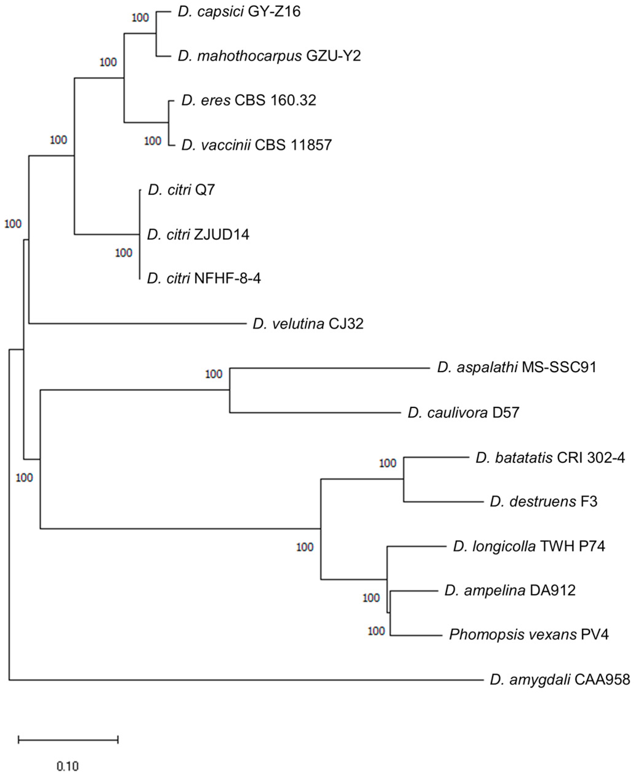
3.2. Phylogenetic Analysis

By using snippy in snippy 4.6.0 for D. amygdali CAA958, D. caulivora D57, D. aspalathi MS-SSC91, D. destruens F3, D. batatatis CRI 302-4, D. longicolla TWH P74, Phomopsis vexans PV4, D. ampelina DA912, D. velutina CJ32, D. citri Q7, D. citri NFHF-8-4, D. citri ZJUD14, D. capsici GY-Z16, D. vaccinii CBS 11857, D. eres CBS 160.32 and D. mahothocarpus GZU-Y2 were analyzed separately, snippy-core analyses of the other strains and D. mahothocarpus GZU-Y2 as a whole were performed to obtain the results of each strain and the results of the overall analyses are shown in Figure 1.

3.3. Gene Prediction

3.3.1. Prediction of Protein-Coding Genes

The gene structure prediction of the D. mahothocarpus GZU-Y2 was based on de novo, homology, and transcriptome evidence. The results of the three types of predictions were integrated using EVM v1.1.1. Then, PASA v2.0.2 was used to modify the final 15,918 predicted genes, among which the number of genes supported by homology prediction and transcriptome prediction accounted for 97.61%, indicating a high quality of prediction (Figure 2). As seen from Table 3, the total length of the predicted genes was 33,450,242 bp, with an average length of 2101.41 bp. The number of CDS, introns, and exons are 46,210, 31,029, and 46,947, respectively.

3.3.2. Prediction of Non-Coding RNA

Non-coding RNAs, which do not encode proteins, encompass a variety of RNAs with known functions, such as microRNAs, rRNAs, and tRNAs. Different strategies were employed to predict non-coding RNAs based on their structural characteristics, resulting in the prediction of 127 rRNAs, 361 tRNAs, and 89 other ncRNAs (Table 1).

3.3.3. Prediction of Pseudogenes

The protein sequences predicted and obtained from the Swiss-Prot database were used to identify homologous gene sequences on the genome using GenBlastA software. Subsequently, the GeneWise software was used to identify premature termination codons and code-shifting mutations in the gene sequences, resulting in the identification of pseudogenes. A total number of 4 pseudogenes were predicted. The total length of the predicted pseudogene sequences was 282 bp, with an average length of 70.5 bp.

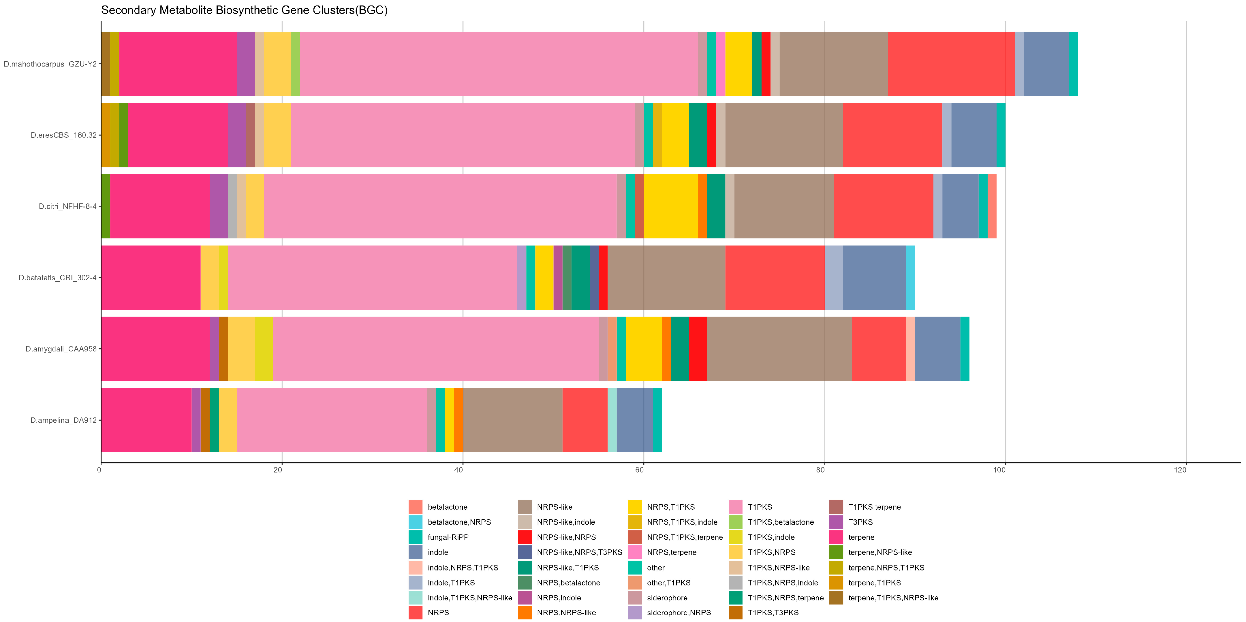
3.3.4. Prediction of Gene Cluster

The detection of D. mahothocarpus GZU-Y2 gene clusters was performed using an-tiSMASH v6.0.0, which predicted a total of 108 gene clusters with a combined length of 4,403,413 bp and an average length of 40,772 bp. Among them, there are 46 Type I polyketide synthases (T1PKSs) genes, 26 Non-ribosomal peptide synthetase (NRPs) genes, 5 indole genes, and 13 terpene genes. The specific information of gene clusters with alignment similarity greater than 80% is shown in Table 4. Among the genomes analyzed, D. mahothocarpus GZU-Y2 contained the highest number of BGCs (n = 108), followed by D. eres CBS 160.32 (n = 100), and the least was D. ampelina DA912 (n = 62). All the genomes analyzed were rich in T1PKS (terpene), NRPS (terpene), and terpene. Importantly, in D. mahothocarpus GZU-Y2, a gene cluster T1PKS (betalactone) was found, which was different and unique from the other genomes, and the results are shown in Figure 3. With its chemical diversity, betalactone is a good class of natural products with potential biological activities.

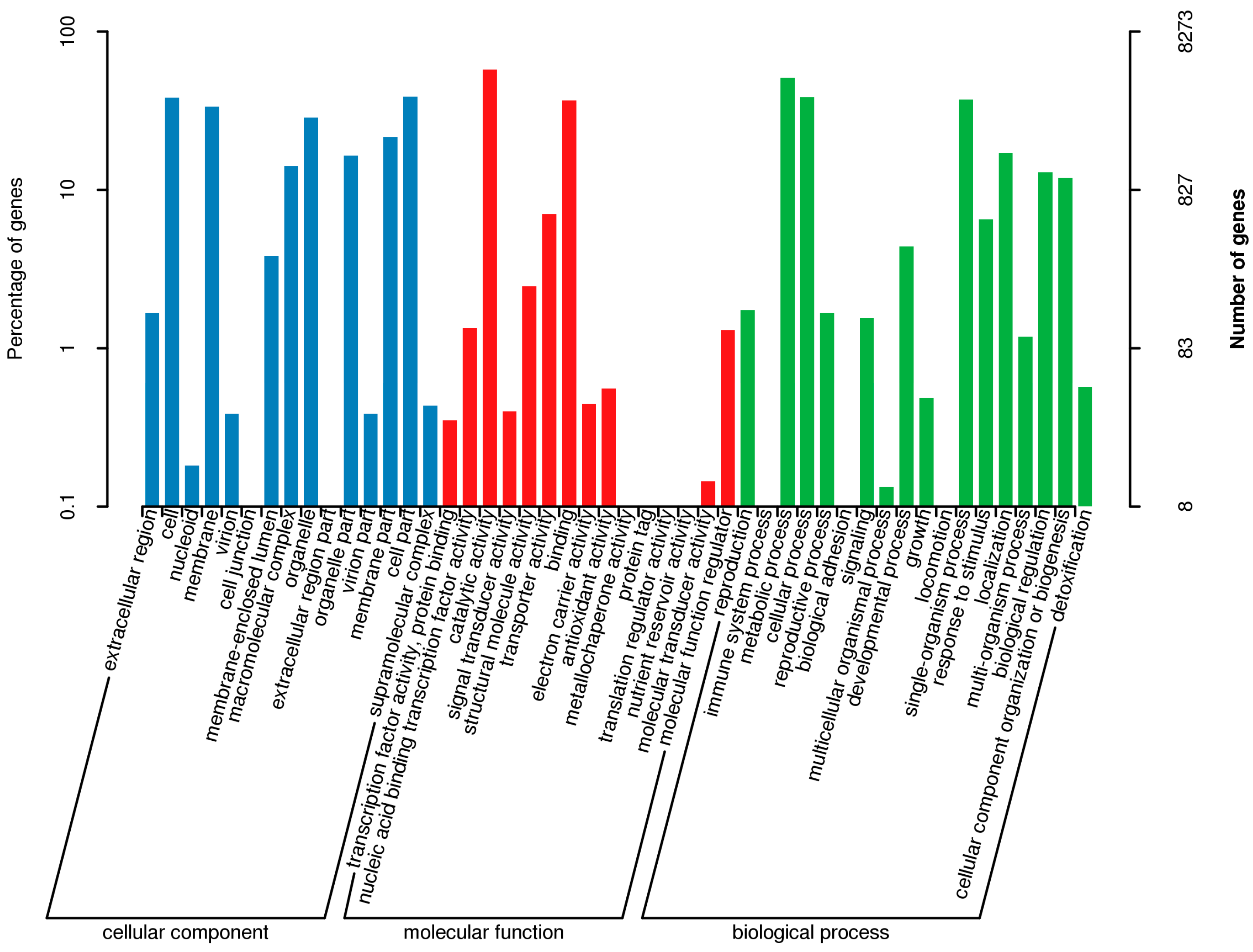
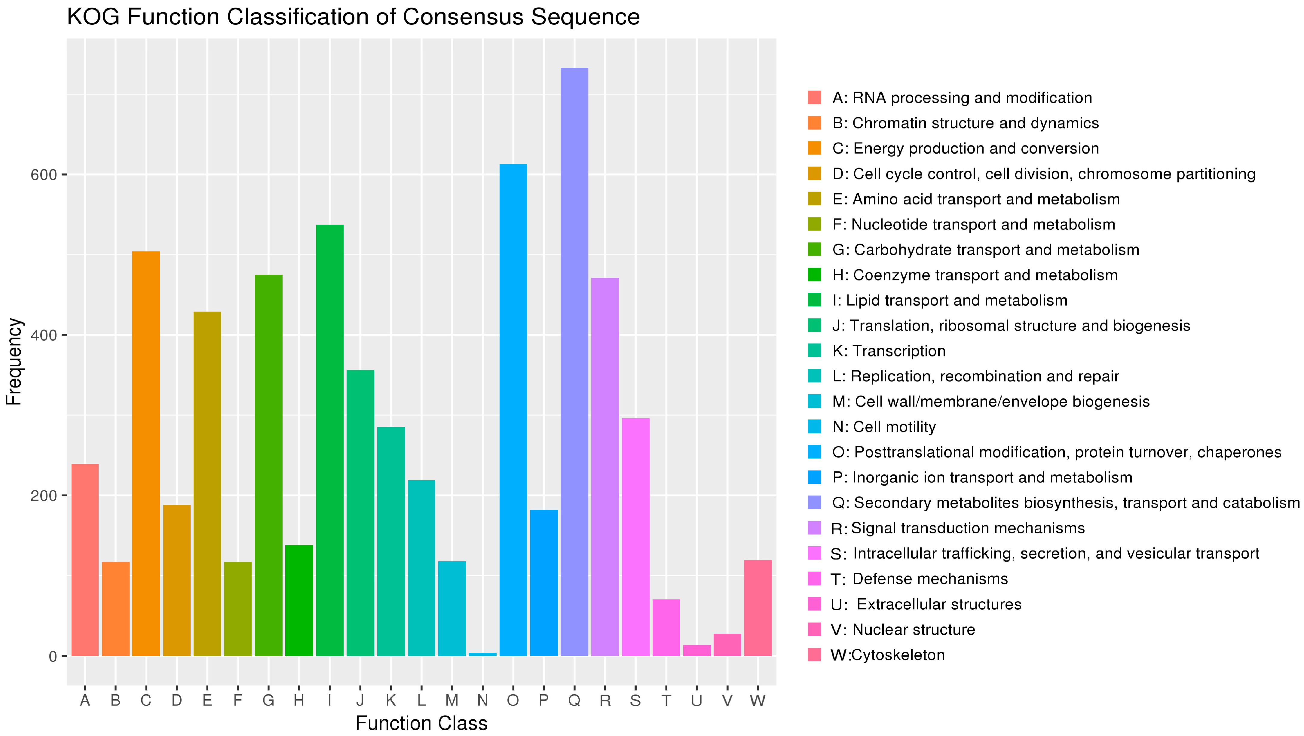
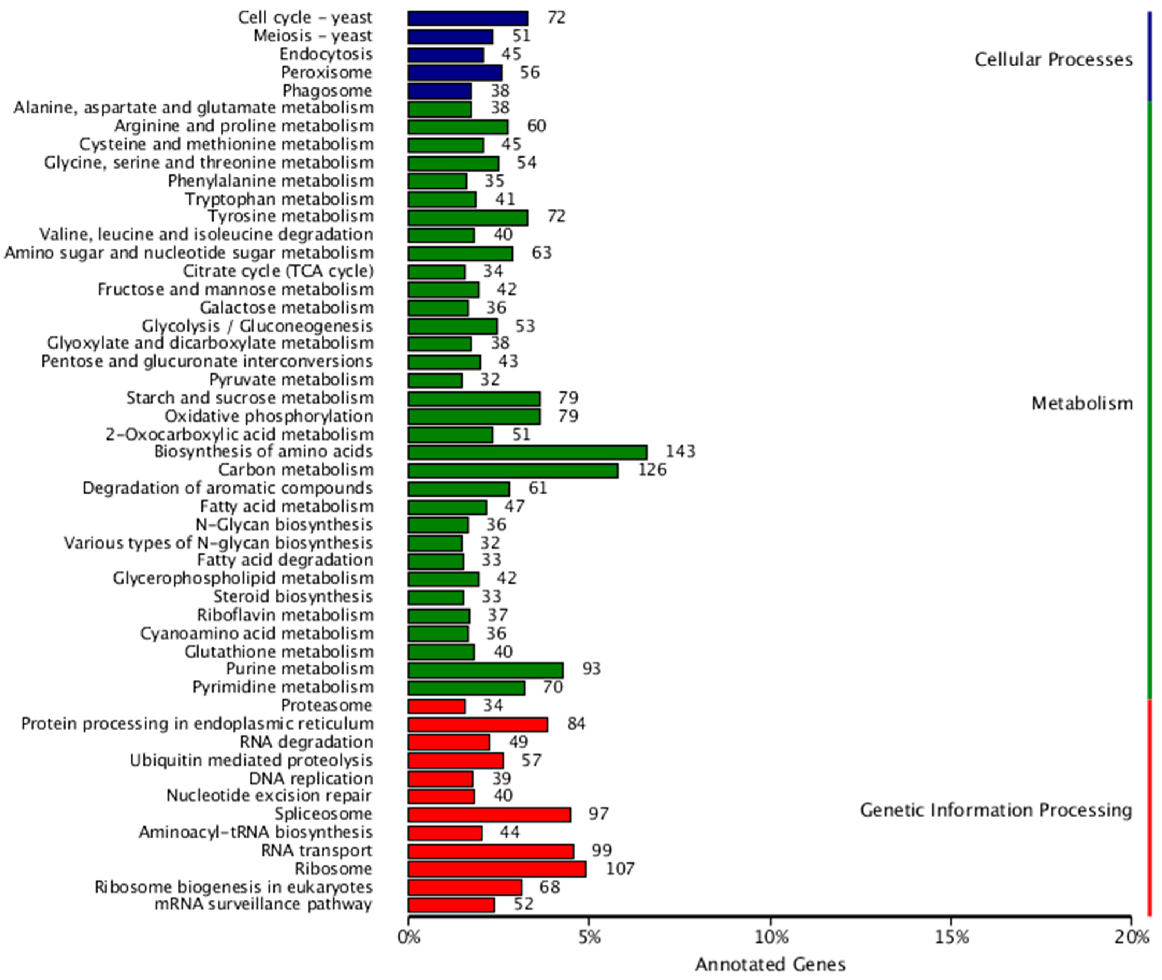
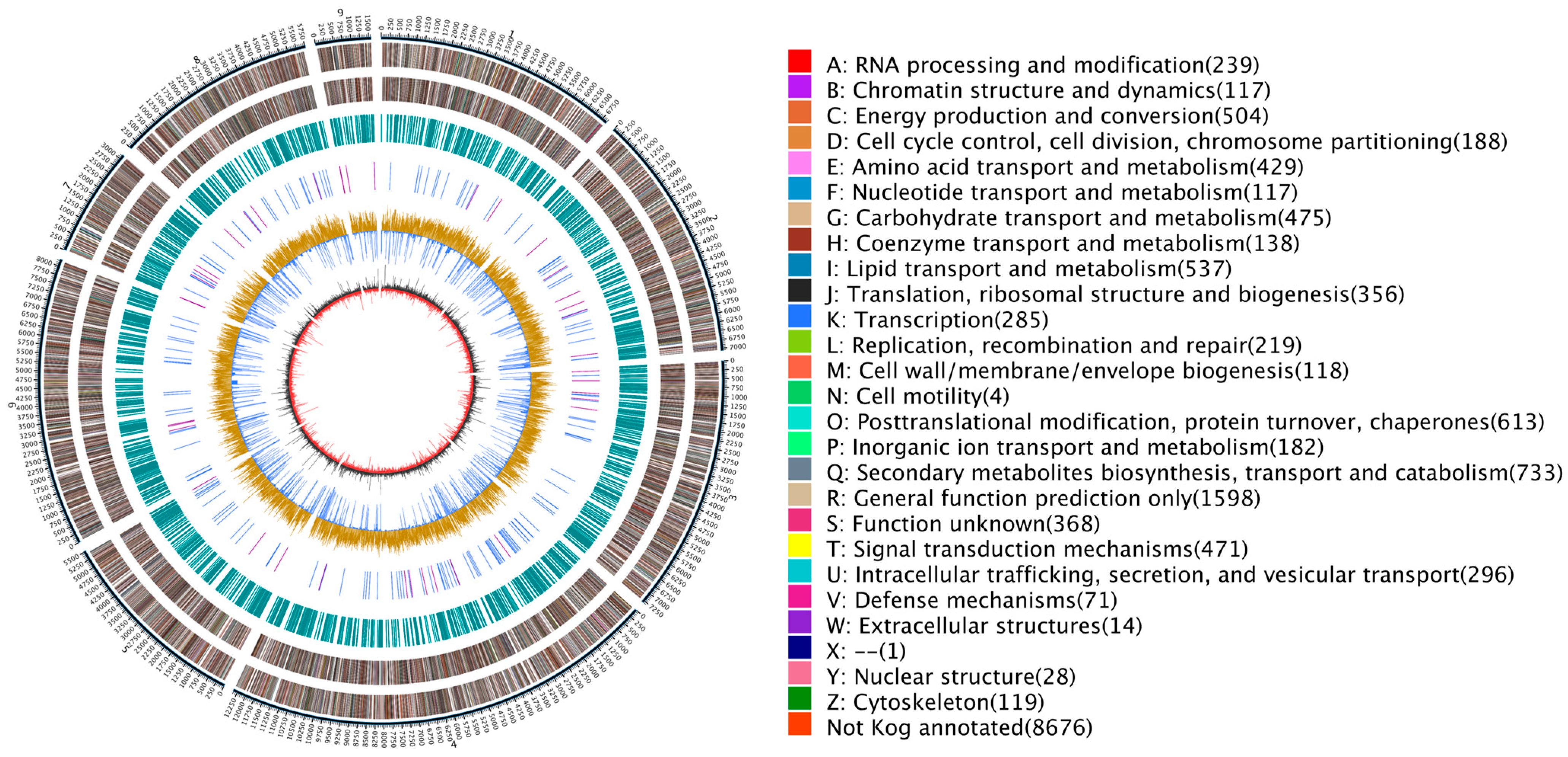
3.4. Gene Annotation

In the present study, 14,564 genes were annotated in the NCBI Nr database.
The GO database categorizes gene functions into cellular components, molecular functions, and biological processes. A gene can be annotated multiple times through the GO program. The results of the GO database annotation of the D. mahothocarpus GZU-Y2 genome for the enrichment of genes for each secondary function of GO in the context of the total genes, reflecting the status of each secondary function in this context (Figure 4).
The KOG database is a collection of immediate homologous protein clusters from eukaryotic organisms. It is used to infer sequence functions through comparison and classification. The number of genes in different functional classes reflects the metabolic or physiological bias in the corresponding period and environment. This can be scientifically interpreted to determine the distribution of research subjects in each functional class (Figure 5).
The KEGG database is a large knowledge base for systematically analyzing gene functions and linking genomic and functional information. The results showed that a total of 3750 genes were annotated, which were categorized into 3 major classes and 50 subclasses (Figure 6).

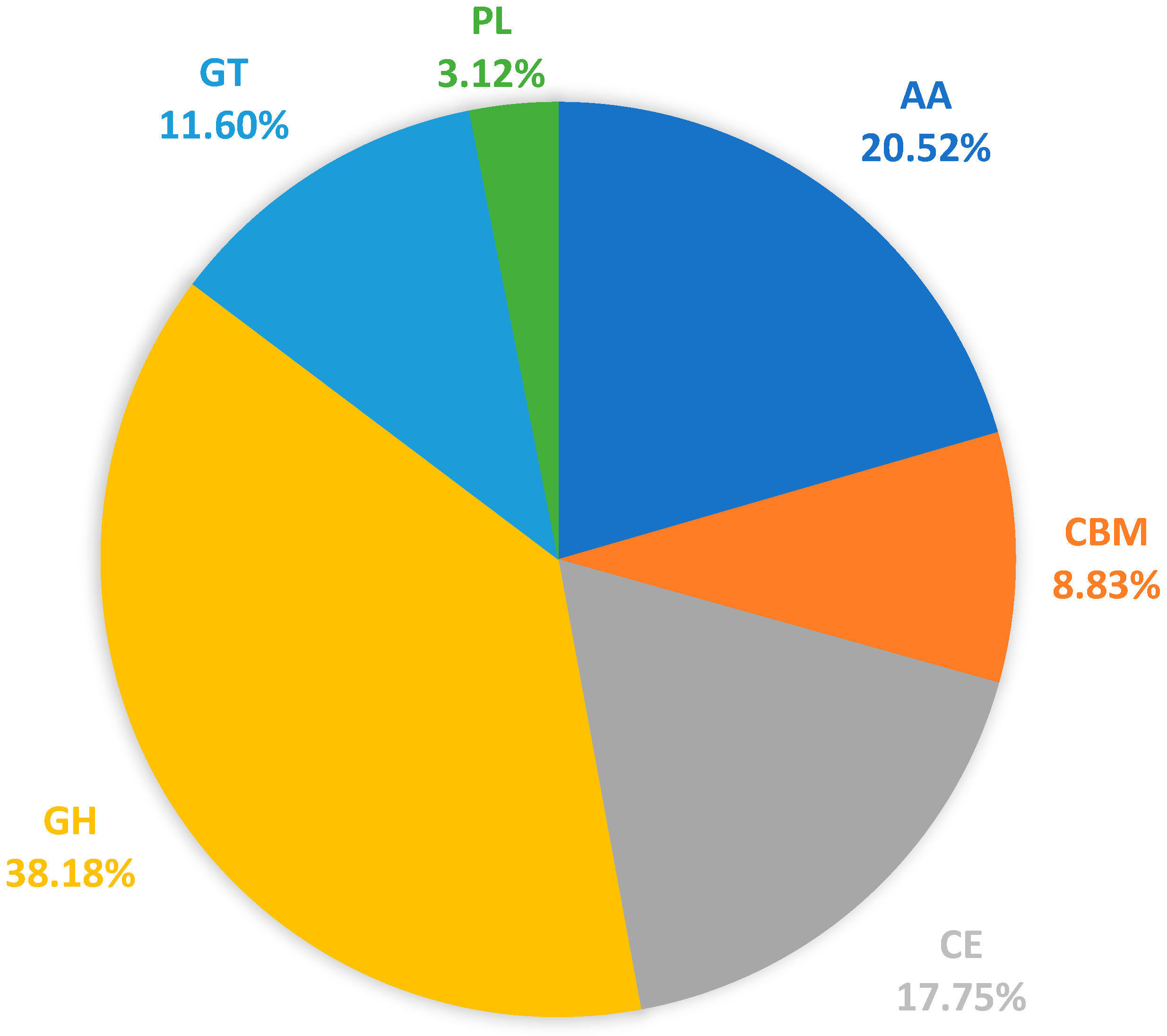
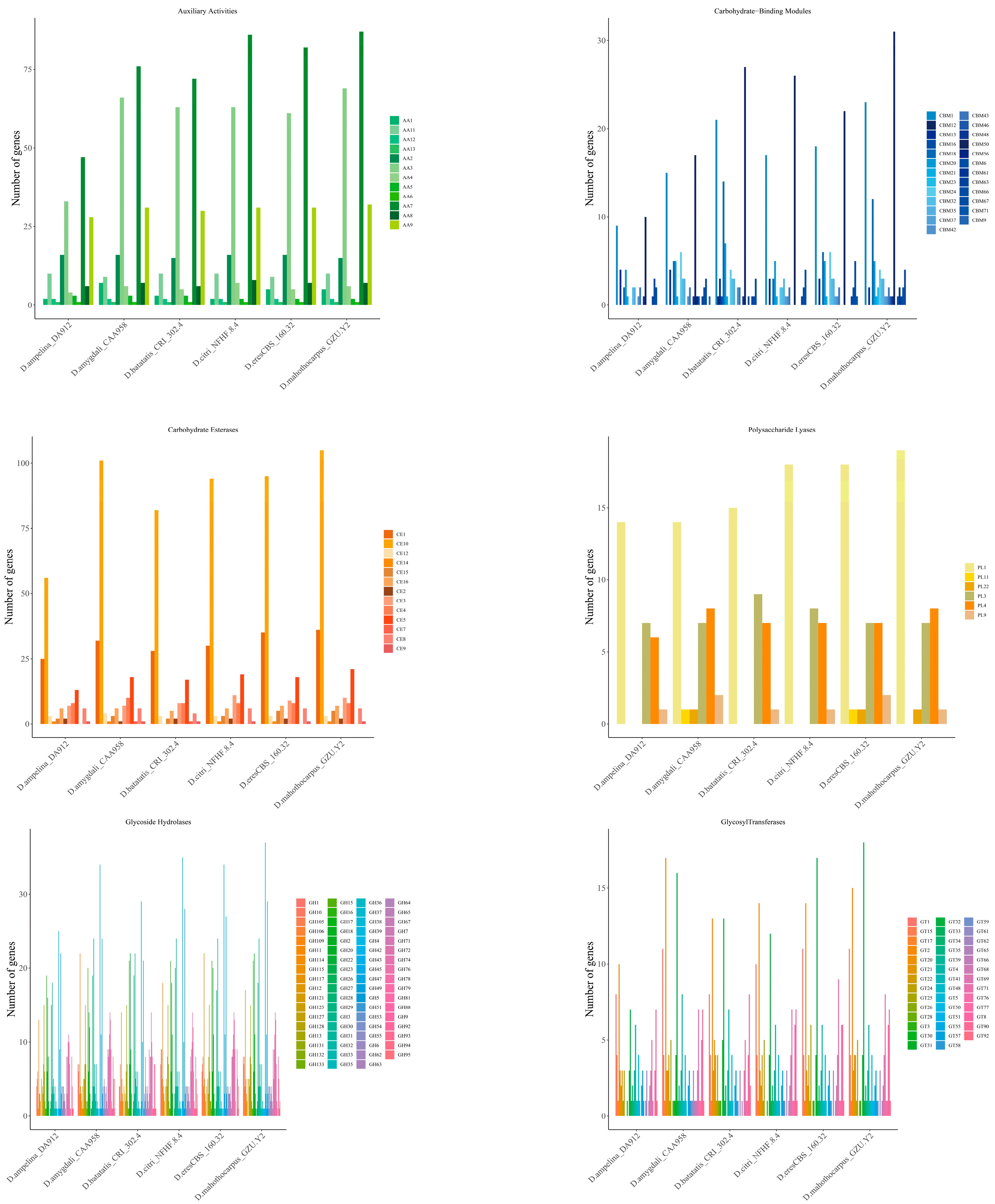
3.5. Carbohydrate-Active Enzymes (CAZymes)

The CAZymes have a crucial role in breaking down complex carbohydrates and plant pathogenic fungi. Certain species of CAZymes are responsible for obtaining nutrients from plants and play a role in the infection and colonization process [49,50]. In D. mahothocarpus GZU-Y2, a total of 1155 CAZyme genes were identified, including 441 GHs, 134 GTs, 36 PLs, 205 CEs, 237 AAs and 102 CBMs (Figure 7). A total of 682 genes were identified to be involved in plant cell wall hydrolases, including GHs, PLs, and CEs. These enzymes are crucial for the successful penetration and infection of plant hosts by fungi. Overall, the number of CAZymes varied among species, with D. mahothocarpus GZU-Y2 and D. eres CBS 160.32 being the most abundant. Of all the categories of CAZymes detected, GHs and AAs were the two most predicted proteins, with AA3, AA7, AA9, CBM1, CBM50, CE10, CE1, GH109, GH16, GH18, GH3, GH43, GH5, GT2, GT32, and PL1 having an abundance in the different species analyzed. Among them, D. mahothocarpus GZU-Y2 had the highest content of CAZymes, including AA3, AA7, AA9, CBM1, CBM50, CE10, GH43 and PL1 families. It was followed by D. eres CBS 160.32, including AA3, AA7, CBM1, CBM50, CE10, GH16, GH43, GT32, and PL1 family, and the results are shown in Figure 8.

3.6. Pathogenic System Analysis

The Transporter Classification (TC) System, developed by TCDB, a database that classifies membrane transporter proteins, predicts a total of 135 genes related to transporter proteins in D. mahothocarpus GZU-Y2. The Pathogen–Host Interaction Database (PHI) was used to predict potential pathogen-active proteins. A total of 4879 genes were predicted to play a role in pathogen–host interactions [51].
Cytochrome P450 (CYP450) is a large family of proteins that use heme iron as a coenzyme [52]. They catalyze the oxidation of a wide range of substrates and are involved in the metabolism of endogenous and exogenous substances. In D. mahothocarpus GZU-Y2, a total of 909 CYP450 genes were predicted.
The fungal secondary metabolites, including toxins, are believed to contribute to the pathogenicity of numerous plant pathogenic fungi and are referred to as potential virulence factors [53]. To analyze the virulence-related genes of D. mahothocarpus GZU-Y2, putative proteins were compared with the Database of Fungal Virulence Factors (DFVF), which identified a total of 3371 genes as fungus-independent factors.

3.7. Analysis of Protein Subcellular Localization

Protein subcellular localization analysis predicted a total of 1919 signal peptides. Additionally, 3467 transmembrane proteins, 1431 secreted proteins, and 164 effector proteins were predicted (Table 1). The specific information about 46 annotated effector proteins (hypothetical proteins not included) is listed in Table 5.

3.8. Comparative Analysis

As can be seen in Table 6, we comparatively analyzed six additional fungal genomes (D. eres CBS 160.32, D. aspalathi MS-SSC91, D. citri Q7, D. citri ZJUD14, D. citri NFHF-8-4 and D. capsici GY-Z16). Annotations of these genomes are publicly available and have been used to compare genome size, GC content, BUSCO Completeness, and number of CA-Zymes. The main reasons for selecting these species were their importance as plant pathogens and the availability of their annotations in public databases and published works [54]. Overall, the genomic characteristics of the analyzed species varied in terms of genome size, GC content, BUSCO Completeness, and number of CA-Zymes. The number of predicted genes ranged from 14,425 (D. capsici GY-Z16) to 16,499 (D. eres CBS 160.32), and BUSCO analysis verified assembly completeness and analyzed differences in the number of CA-Zymes. This analysis showed differences between disease genomes of different plants, such as Camellia oleifera, blueberry [55], soybean [56], citrus [57,58], and walnut [59], caused by Diaporthe species.

4. Discussion

The distribution of plant diseases caused by Diaporthe is global, with a wide range of reported hosts, including Camelliaceae, Leguminosae, Walnutaceae, Rosaceae, Lacertaceae, and Vitaceae. C. oleifera is a very important woody edible oilseed tree species, and D. mahothocarpus GZU-Y2 is an important pathogen on C. oleifera, but its genome has not been previously characterized. In the previous study, we isolated and identified D. mahothocarpus GZU-Y2, the strain responsible for leaf blight disease in C. oleifera, from infected leaves. Here, in the present study, we sequenced, assembled, and predicted the D. mahothocarpus GZU-Y2 genome. Moreover, the high quality of the D. mahothocarpus genome sequence was demonstrated by the Contig N50 of 7,066,871 bp. Based on the results of genome assembly and annotation, the genome landscape is shown in Figure 9.
By comparing the genome of D. mahothocarpus GZU-Y2 with other genomic data, it was found that there were differences in gene structure, especially in genome size and number of coding sequences, which were significantly smaller in D. mahothocarpus GZU-Y2 than in D. eres CBS 160.32 and D. citri Q7. The reasons for this result may be the different number of genes and gene duplication events in Diaporthe species. And there were also differences in BUSCO Completeness as well as GC Content in different genomes; the results are shown in Table 6 [55,56,57,58,59,60]. Based on the Carbohydrate-Active enZYmes Database (CAZymes), we investigated CAZymes in the D. mahothocarpus GZU-Y2 genome in more detail.
It was found that the genome of D. mahothocarpus GZU-Y2 was found to contain 441 Glycoside Hydrolases (GHs), 134 Glycosyl Transferases (GTs), 36 Polysaccharide Lysylases (PLs), 205 Carbohydrate Esterases (CEs), 237 Auxiliary Active enzymes (AAs), and 102 Carbohydrate-binding related enzymes (CBMs), as predicted. This strain has a high capacity to disrupt plant cell walls during infection, as indicated by multiple GHs, GTs, and PLs. Plant cell hydrolases of phytopathogenic fungi are key causative factors in breaking down cell walls and establishing infection and nutrient growth [61]. Interestingly, the number of CAZymes number is greater than D. eres CBS 160.32 and D. capsici GY-Z16 and less than D. citri Q7 and D. citri ZJUD14, shown in Table 6. The amplification of relevant cell wall degrading enzymes in the genome of D. mahothocarpus GZU-Y2 is likely to be an important factor in the infection of C. oleifera [62]. The combination of multiple AAs and CBMs is also expected to have a significant impact on D. mahothocarpus GZU-Y2 [63,64].
Furthermore, by annotating the genome of D. mahothocarpus GZU-Y2, we predicted a total of 909 CYP450 genes for this strain. It was found that CYP450 genes can be involved in many important cellular pathways, including primary and secondary metabolism, toxin production, and detoxification, and can catalyze oxidation reactions of a variety of substrates and participate in the metabolism of endogenous and exogenous substances. The identification of these genes provides research and development of biocides specific to D. mahothocarpus GZU-Y2 certain support [65]. Additionally, 4879 PHI genes were predicted to be involved in pathogen-host interactions, while 3371 genes were identified as fungal-independent factors. These findings provide insights into the mechanisms of infection and aspects of the pathogenicity of many phytopathogenic fungi [66,67]. The protein subcellular localization analysis predicted a total of 1919 signal peptide genes, 3467 transmembrane proteins, 1431 secreted proteins, and 164 effector proteins. The study predicted 135 genes associated with the transporter protein TCDB and found that the expression and changes of these genes were linked to the development of the disease.

5. Conclusions

Generally, a genomic resource of D. mahothocarpus GZU-Y2 causing C. oleifera leaf blight was provided in the present study, which is also compared with other related genomes. We found that the genome of D. mahothocarpus GZU-Y2 has many plant cell wall degrading enzymes, a virulence and protein secretion system associated with infestation, and focused on annotating and analyzing genes related to pathogenesis. Several related virulence factors can manipulate the host response and induce plant cell death, thereby favoring colonization by the pathogen. The rich results of sequencing and analyzing the whole genome of D. mahothocarpus GZU-Y2 of C. oleifera leaf blight provide an opportunity to understand further the characteristics of C. oleifera leaf blight fungi and a new starting point and idea for researching new control measures. Therefore, future studies using Dual RNA sequencing technology to clarify both host and pathogen transcriptomes may provide better insight into the process of pathogen infection and host defense mechanisms.

Author Contributions

X.S.: Investigation; Data statistical analysis; Writing—original draft; Y.Z.: Investigation; Data statistical analysis; J.Y.: Data statistical analysis; Supervision; Writing—review and editing; Funding acquisition; Y.C.: Supervision; Writing—review and editing. All authors have read and agreed to the published version of the manuscript.

Funding

This research was funded by the Guizhou Provincial Department of Education Higher Education Science Research Project-Youth Project (QJJ[2022]259), Guizhou Provincial Basic Research Program (Natural Science) (grant number QKHJC-ZK [2023]YB117), and Natural Science Special (Special Post) Research Fund of Guizhou University (grant number [2022]02).

Institutional Review Board Statement

Not applicable.

Informed Consent Statement

Not applicable.

Data Availability Statement

All data generated and analyzed in this study are included in this article. This Whole Genome Shotgun project has been deposited at GenBank under the accession JBBYIC000000000. The raw sequencing data and the assembly reported in this paper are associated with NCBI BioProject PRJNA1098494 and BioSample SAMN40910135 within the GenBank. The version described in this paper is version JBBYIC 010000000.

Acknowledgments

The authors thank Guizhou University for providing the research facilities.

Conflicts of Interest

The authors declare no conflicts of interest.

References

  1. Dissanayake, A.J.; Liu, M.; Zhang, W.; Chen, Z.; Udayanga, D.; Chukeatirote, E.; Li, X.; Yan, J.; Hyde, K.D. Morphological and molecular characterisation of Diaporthe species associated with grapevine trunk disease in China. Fungal Biol. 2015, 119, 283–294. [Google Scholar] [CrossRef] [PubMed]
  2. Zhou, J.H. Research on plant source agents for major diseases of oil tea. Zhongnan Univ. For. Sci. Technol. 2011, 19, 78–87. [Google Scholar]
  3. Xiao, Y.; Huo, G.; Liu, L.; Yang, C.; Cui, C. First report of postharvest fruit rot disease of yellow peach caused by Diaporthe eres in China. Plant Dis. 2022, 106, 1983. [Google Scholar] [CrossRef]
  4. Yang, Q.; Tang, J.; Zhou, G.Y. Characterization of Diaporthe species on Camellia oleifera in Hunan Province, with descriptions of two new species. MycoKeys 2021, 84, 15–33. [Google Scholar] [CrossRef] [PubMed]
  5. Shi, X.; Zhang, Y.; Wang, X.; Pan, Y.; Yu, C.; Yang, J. First Report of Leaf Spot Blight of Camellia oleifera Caused by Diaporthe mahothocarpus in China. Plant Dis. 2024, 108, 516. [Google Scholar] [CrossRef]
  6. Udayanga, D.; Liu, X.; Crous, P.W.; McKenzie, E.H.; Chukeatirote, E.; Hyde, K.D. A muli-locus phylogenetic evaluation of Diaporthe (Phomopsi). Fungal Divers. 2012, 56, 157–171. [Google Scholar] [CrossRef]
  7. Santos, J.M.; Correia, V.G.; Phillips, A.J. Primers for mating type dagnosis in Diaporthe and Phomopsis: Their use in teleomorph induction in vitro and biological species definition. Fungal Biol. 2010, 114, 255–270. [Google Scholar] [CrossRef]
  8. Erincik, O.; Madden, L.V. Effect of growth stage on susceptibility of grape berry and rachis tissues to infection by Phomopsis viticola. Plant Dis. 2001, 85, 517–520. [Google Scholar] [CrossRef]
  9. Guarnaccia, V.; Crous, P.W. Emerging citrus diseases in Europe caused by species of Diaporthe. IMA Fungus 2017, 8, 317–334. [Google Scholar] [CrossRef]
  10. Mondal, S.N. Saprophytic colonization of citrus twigs by Diaporthe citri and factors affecting pycnidial production and conidial survivalc. Plant Dis. 2007, 91, 387–392. [Google Scholar] [CrossRef]
  11. Thompson, S.; Tan, Y.; Young, A.; Neate, S.; Aitken, E.; Shivas, R. Stem cankers on sunflower (Helianthus annuus) in Australia reveal a complex of pathogenic Diaporthe (Phomopsis) species. Persoonia 2011, 27, 80–89. [Google Scholar] [CrossRef] [PubMed]
  12. Guarnaccia, V.; Groenewald, J.Z.; Woodhall, J.; Armengol, J.; Cinelli, T.; Eichmeier, A.; Ezra, D.; Fontaine, F.; Gramaje, D.; Gutierrez-Aguirregabiria, A.; et al. Diaporthe diversity and pathogenicity revealed from a broad survey of grapevine diseases in Europe. Persoonia 2018, 40, 135–153. [Google Scholar] [CrossRef]
  13. Udayanga, D.; Castlebury, L.A.; Rossman, A.Y.; Chukeatirote, E.; Hyde, K.D. The Diaporthe sojae species complex: Phylogenetic re-assessment of pathogens associated with soybean, cueurbits and other field crops. Fungal Biol. 2015, 119, 383–407. [Google Scholar] [CrossRef]
  14. Fu, F.-F.; Hao, Z.; Wang, P.; Lu, Y.; Xue, L.-J.; Wei, G.; Tian, Y.; Hu, B.; Xu, H.; Shi, J.; et al. Genome Sequence and Comparative Analysis of Colletotrichum gloeosporioides Isolated from Liriodendron Leaves. Phytopathology 2020, 110, 1260–1269. [Google Scholar] [CrossRef] [PubMed]
  15. Cheng, H.; Concepcion, G.T.; Feng, X.; Zhang, H.; Li, H. Haplotype-resolved de novo assembly using phased assembly graphs with hifiasm. Nat. Methods 2021, 18, 170–175. [Google Scholar] [CrossRef] [PubMed]
  16. Walker, B.J.; Abeel, T.; Shea, T.; Priest, M.; Abouelliel, A.; Sakthikumar, S.; Cuomo, C.A.; Zeng, Q.; Wortman, J.; Young, S.K.; et al. Pilon: An Integrated Tool for Comprehensive Microbial Variant Detection and Genome Assembly Improvement. PLoS ONE 2014, 9, e112963. [Google Scholar] [CrossRef]
  17. Xu, Z.; Wang, H. LTR_FINDER: An efficient tool for the prediction of full-length LTR retrotransposons. Nucleic Acids Res. 2007, 35, W265–W268. [Google Scholar] [CrossRef]
  18. Han, Y.; Wessler, S.R. MITE-Hunter: A program for discovering miniature inverted-repeat transposable elements from genomic sequences. Nucleic Acids Res. 2010, 38, e199. [Google Scholar] [CrossRef]
  19. Price, A.L.; Jones, N.C.; Pevzner, P.A. De novo identification of repeat families in large genomes. Bioinformatics 2005, 21, i351–i358. [Google Scholar] [CrossRef]
  20. Edgar, R.C.; Myers, E.W. PILER: Identification and classification of genomic repeats. Bioinformatics 2005, 21, i152–i158. [Google Scholar] [CrossRef]
  21. Wicker, T.; Sabot, F.; Hua-Van, A.; Bennetzen, J.L.; Capy, P.; Chalhoub, B.; Flavell, A.; Leroy, P.; Morgante, M.; Panaud, O. A unified classification system for eukaryotic transposable elements. Nat. Rev. Genet. 2007, 8, 973–982. [Google Scholar] [CrossRef] [PubMed]
  22. Chen, N. Using Repeat Masker to Identify Repetitive Elements in Genomic Sequences. In Current Protocols in Bioinformatics; Baxevanis, A.D., Ed.; Wiley: Hoboken, NJ, USA, 2004; Chapter 4: Unit 4.10. [Google Scholar]
  23. Burge, C.; Karlin, S. Prediction of complete gene structures in human genomic DNA. J. Mol. Biol. 1997, 268, 78–94. [Google Scholar] [CrossRef] [PubMed]
  24. Stanke, M.; Waack, S. Gene prediction with a hidden Markov model and a new intron submodel. Bioinformatics 2003, 19, ii225. [Google Scholar] [CrossRef]
  25. Majoros, W.H.; Pertea, M.; Salzberg, S.L. TigrScan and GlimmerHMM: Two open source ab initio eukaryotic gene-finders. Bioinformatics 2004, 20, 2878–2879. [Google Scholar] [CrossRef]
  26. Blanco, E.; Parra, G.; Guigó, R. Using geneid to identify genes. Curr. Protoc. Bioinform. 2007, 18, 4.3.1–4.3.28. [Google Scholar] [CrossRef]
  27. Korf, I. Gene finding in novel genomes. BMC Bioinform. 2004, 5, 59. [Google Scholar] [CrossRef]
  28. Keilwagen, J.; Wenk, M.; Erickson, J.L.; Schattat, M.H.; Grau, J.; Hartung, F. Using intron position conservation for homology-based gene prediction. Nucleic Acids Res. 2016, 44, e89. [Google Scholar] [CrossRef]
  29. Pertea, M.; Kim, D.; Pertea, G.M.; Leek, J.T.; Salzberg, S.L. Transcript-level expression analysis of RNA-seq experiments with HISAT, StringTie and Ballgown. Nat. Protoc. 2016, 11, 1650. [Google Scholar] [CrossRef] [PubMed]
  30. Campbell, M.A.; Haas, B.J.; Hamilton, J.P.; Mount, S.M.; Buell, C.R. Comprehensive analysis of alternative splicing in rice and comparative analyses with Arabidopsis. BMC Genom. 2006, 7, 327. [Google Scholar] [CrossRef]
  31. Haas, B.J.; Salzberg, S.L.; Zhu, W.; Pertea, M.; Allen, J.E.; Orvis, J.; White, O.; Buell, C.R.; Wortman, J.R. Automated eukaryotic gene structure annotation using EVidenceModeler and the Program to Assemble Spliced Alignments. Genome Biol. 2008, 9, R7. [Google Scholar] [CrossRef]
  32. Blin, K.; Shaw, S.; Kloosterman, A.M.; Charlop-Powers, Z.; Van Wezel, G.P.; Medema, M.H.; Weber, T. antiSMASH 6.0: Improving cluster detection and comparison capabilities. Nucleic Acids Res. 2021, 49, W29–W35. [Google Scholar] [CrossRef]
  33. Shi, X.L.; Yang, J.; Zhang, Y.; Qin, P.; Zhou, H.Y.; Chen, Y.Z. The photoactivated antifungal activity and possible mode of action of sodium pheophorbide a on Diaporthe mahothocarpus causing leaf spot blight in Camellia oleifera. Front. Microbiol. 2024, 15, 1403478. [Google Scholar] [CrossRef]
  34. Petersen, T.N.; Brunak, S.; von Heijne, G.; Nielsen, H. SignalP 4.0: Discriminating signal peptides from transmembrane regions. Nat. Methods 2011, 8, 785–786. [Google Scholar] [CrossRef] [PubMed]
  35. Krogh, A.; Larsson, B.; Von Heijne, G.; Sonnhammer, E.L. Predicting transmembrane protein topology with a hidden Markov model: Application to complete genomes. J. Mol. Biol. 2001, 305, 567–580. [Google Scholar] [CrossRef] [PubMed]
  36. Sperschneider, J.; Gardiner, D.M.; Dodds, P.N.; Tini, F.; Covarelli, L.; Singh, K.B.; Manners, J.M.; Taylor, J.M. EffectorP: Predicting Fungal Effector Proteins from Secretomes Using Machine Learning. New Phytol. 2015, 210, 743–761. [Google Scholar] [CrossRef]
  37. Boeckmann, B.; Bairoch, A.; Apweiler, R.; Blatter, M.C.; Estreicher, A.; Gasteiger, E.; Martin, M.J.; Michoud, K.; O’Donovan, C.; Phan, I.; et al. The SWISS-PROT protein knowledgebase and its supplement TrEMBL in 2003. Nucleic Acids Res. 2003, 31, 365–370. [Google Scholar] [CrossRef]
  38. Kanehisa, M.; Goto, S.; Kawashima, S.; Okuno, Y.; Hattori, M. The KEGG resource for deciphering the genome. Nucleic Acids Res. 2004, 32 (Suppl. 1), D277–D280. [Google Scholar] [CrossRef] [PubMed]
  39. Tatusov, R.L.; Galperin, M.Y.; Natale, D.A.; Koonin, E.V. The COG database: A tool for genome-scale analysis of protein functions and evolution. Nucleic Acids Res. 2000, 28, 33–36. [Google Scholar] [CrossRef]
  40. Deng, Y.Y.; Li, J.Q.; Wu, S.F.; Zhu, Y.P.; Chen, Y.W.; He, F.C. Integrated nr database in protein annotation system and its localization. Comput. Eng. 2006, 32, 71–74. [Google Scholar]
  41. Altschul, S.F.; Madden, T.L.; Schäffer, A.A.; Zhang, J.; Zhang, Z.; Miller, W.; Lipman, D.J. Gapped BLAST and PSI-BLAST: A new generation of protein database search programs. Nucleic Acids Res. 1997, 25, 3389–3402. [Google Scholar] [CrossRef]
  42. Conesa, A.; Götz, S.; García-Gómez, J.M.; Terol, J.; Talón, M.; Robles, M. Blast2GO: A universal tool for annotation, visualization and analysis in functional genomics research. Bioinformatics 2005, 21, 3674–3676. [Google Scholar] [CrossRef] [PubMed]
  43. Ashburner, M.; Ball, C.A.; Blake, J.A.; Botstein, D.; Butler, H.; Cherry, J.M.; Davis, A.P.; Dolinski, K.; Dwight, S.S.; Janan, T.E.; et al. Gene Ontology: Tool for the unification of biology. Nat. Genet. 2000, 25, 25–29. [Google Scholar] [CrossRef] [PubMed]
  44. Eddy, S.R. Profile hidden Markov models. Bioinformatics 1998, 14, 755–763. [Google Scholar] [CrossRef]
  45. Finn, R.D.; Coggill, P.; Eberhardt, R.Y.; Eddy, S.R.; Mistry, J.; Mitchell, A.L.; Potter, S.C.; Punta, M.; Qureshi, M.; Sangrador-Vegas, A.; et al. The Pfam protein families database: Towards a more sustainable future. Nucleic Acids Res. 2016, 44, D279–D285. [Google Scholar] [CrossRef] [PubMed]
  46. Saier, M.H., Jr.; Tran, C.V.; Barabote, R.D. TCDB: The Transporter Classification Database for membrane transport protein analyses and information. Nucleic Acids Res. 2006, 34 (Suppl. 1), D181–D186. [Google Scholar] [CrossRef]
  47. Winnenburg, R.; Baldwin, T.K.; Urban, M.; Rawlings, C.; Köhler, J.; Hammond-Kosack, K.E. PHI-base: A new database for pathogen host interactions. Nucleic Acids Res. 2006, 34 (Suppl. 1), D459–D464. [Google Scholar] [CrossRef]
  48. Cantarel, B.L.; Coutinho, P.M.; Rancurel, C.; Bernard, T.; Lombard, V.; Henrissat, B. The Carbohydrate-Active EnZymes database (CAZy): An expert resource for glycogenomics. Nucleic Acids Res. 2009, 37 (Suppl. 1), D233–D238. [Google Scholar] [CrossRef]
  49. Rafiei, V.; Velez, H.; Tzelepis, G. The Role of Glycoside Hydrolases in Phytopathogenic Fungi and Oomycetes Virulence. Int. J. Mol. Sci. 2021, 22, 9359. [Google Scholar] [CrossRef]
  50. Kubicek, C.P.; Starr, T.L.; Glass, N.L. Plant cell wall-degrading enzymes and their secretion in plant-pathogenic fungi. Annu. Rev. Phytopathol. 2014, 52, 427–451. [Google Scholar] [CrossRef]
  51. Urban, M.; Cuzick, A.; Seager, J.; Wood, V.; Rutherford, K.; Venkatesh, S.Y.; De Silva, N.; Martinez, M.C.; Pedro, H.; Yates, A.D.; et al. PHI-base: The pathogen-host interactions database. Nucleic Acids Res. 2019, 48, D613–D620. [Google Scholar] [CrossRef]
  52. Liu, J.; Wei, Y.; Yin, Y.; Zhu, K.; Liu, Y.; Ding, H.; Lei, J.; Zhu, W.; Zhou, Y. Effects of Mixed Decomposition of Pinus sylvestris var. mongolica and Morus alba Litter on Microbial Diversity. Microorganisms 2022, 10, 1117. [Google Scholar] [PubMed]
  53. Chandrasekaran, M.; Thangavelu, B.; Chun, S.C.; Sathiyabama, M. Proteases from phytopathogenic fungi and their importance in phytopathogenicity. J. Gen. Plant Pathol. 2016, 82, 233–239. [Google Scholar] [CrossRef]
  54. Garcia, J.F.; Lawrence, D.P.; Morales-Cruz, A.; Travadon, R.; Minio, A.; Hernandez-Martinez, R.; Rolshausen, P.E.; Baumgartner, K.; Cantu, D. Phylogenomics of Plant-Associated Botryosphaeriaceae Species. Front. Microbiol. 2021, 12, 652802. [Google Scholar] [CrossRef] [PubMed]
  55. Hilário, S.; Gonçalves, M.F.; Fidalgo, C.; Tacão, M.; Alves, A. Genome Analyses of Two Blueberry Pathogens: Diaporthe amygdali CAA958 and n CBS 160.32. J. Fungi 2022, 8, 804. [Google Scholar] [CrossRef]
  56. Li, S.; Song, Q.; Martins, A.M.; Cregan, P. Draft genome sequence of Diaporthe aspalathi isolate MS-SSC91, a fungus causing stem canker in soybean. Genom. Data 2016, 7, 262–263. [Google Scholar] [CrossRef]
  57. Gai, Y.; Xiong, T.; Xiao, X.; Li, P.; Zeng, Y.; Li, L.; Riely, B.K.; Li, H. The Genome Sequence of the Citrus Melanose Pathogen Diaporthe citri and Two Citrus related Diaporthe species. Phytopathology 2020, 111, 779–783. [Google Scholar] [CrossRef]
  58. Liu, X.Y.; Chaisiri, C.; Lin, Y.; Yin, W.X.; Luo, C.X. Whole-genome sequence of Diaporthe citri isolate NFHF-8-4, the causal agent of citrus melanose. Mol. Plant Microbe Interact. 2021, 34, 845–847. [Google Scholar] [CrossRef]
  59. Fang, X.; Qin, K.; Li, S.; Han, S.; Zhu, T. Whole genome sequence of Diaporthe capsici, a new pathogen of walnut blight. Genomics 2020, 112, 3751–3761. [Google Scholar] [CrossRef]
  60. Baroncelli, R.; Amby, D.B.; Zapparata, A.; Sarrocco, S.; Vannacci, G.; Le Floch, G.; Harrison, R.J.; Holub, E.; Sukno, S.A.; Sreenivasaprasad, S.; et al. Gene family expansions and contractions are associated with host range in plant pathogens of the genus Colletotrichum. BMC Genom. 2016, 17, 555. [Google Scholar] [CrossRef]
  61. Kong, L.; Chen, J.; Dong, K.; Shafik, K.; Xu, W. Genomic analysis of Colletotrichum camelliae responsible for tea brown blight disease. BMC Genom. 2023, 24, 528. [Google Scholar] [CrossRef]
  62. Mena, E.; Garaycochea, S.; Stewart, S.; Montesano, M.; Ponce De León, I. Comparative genomics of plant pathogenic Diaporthe species and transcriptomics of Diaporthe caulivora during host infection reveal insights into pathogenic strategies of the genus. BMC Genom. 2022, 23, 175. [Google Scholar] [CrossRef] [PubMed]
  63. Liu, D.-M.; Huang, Y.-Y.; Liang, M.-H. Analysis of the probiotic characteristics and adaptability of Lactiplantibacillus plantarum DMDL 9010 to gastrointestinal environment by complete genome sequencing and corresponding phenotypes. LWT 2022, 158, 113129. [Google Scholar] [CrossRef]
  64. Bradley, E.L.; Ökmen, B.; Doehlemann, G.; Henrissat, B.; Bradshaw, R.E.; Mesarich, C.H. Secreted Glycoside Hydrolase Proteins as Effectors and Invasion Patterns of Plant-Associated Fungi and Oomycetes. Front. Plant Sci. 2022, 13, 853106. [Google Scholar] [CrossRef] [PubMed]
  65. Črešnar, B.; Petrič, Š. Cytochrome P450 enzymes in the fungal kingdom. Biochim. Biophys. Acta 2011, 1814, 29–35. [Google Scholar] [CrossRef]
  66. Mair, W.J.; Deng, W.; Mullins, J.G.; West, S.; Wang, P.; Besharat, N.; Ellwood, S.R.; Oliver, R.P.; Oliver, R.P.; Lopez-Ruiz, F.J.; et al. Demethylase inhibitor fungicide resistance in Pyrenophora teres f. sp. Teres associated with target site modification and inducible overexpression of CYP51. Front. Microbiol. 2016, 7, 1279. [Google Scholar]
  67. Agrawal, Y.; Khatri, I.; Subramanian, S.; Shenoy, B.D. Genome Sequence, Comparative Analysis, and Evolutionary Insights into Chitinases of Entomopathogenic Fungus Hirsutella thompsonii. Genome Biol. Evol. 2015, 7, 916–930. [Google Scholar] [CrossRef]
Figure 1. Phylogenetic tree constructed based on SNPs. Bar = 0.10.
Jof 10 00630 g001
Figure 2. Integrated genes of D. mahothocarpus GZU-Y2 derived from three prediction methods.
Jof 10 00630 g002
Figure 3. Cluster of biosynthetic genes identified in the analyzed Diaporthe genome.
Jof 10 00630 g003
Figure 4. GO annotation of D. mahothocarpus GZU-Y2. The GO classification is presented with horizontal coordinates, while the percentage (left) and numbers (right) of genes are shown on the vertical axis.
Jof 10 00630 g004
Figure 5. KOG annotation of D. mahothocarpus GZU-Y2. The horizontal axis represents the function class, and the vertical axis is the number of genes.
Jof 10 00630 g005
Figure 6. KEGG classification of D. mahothocarpus GZU-Y2. The left vertical axis is the KEGG level 3 classification, and the right is the KEGG level 1 classification. The horizontal axis is the percentage of annotated genes, and the label is the number of genes.
Jof 10 00630 g006
Figure 7. Predicted carbohydrate-active enzymes of D. mahothocarpus GZU-Y2. AA, auxiliary activities; GH, glycoside hydrolases; GT, glycosyl transferases; PL, polysaccharide lyases; CE, carbohydrate esterases; CBM, carbohydrate-binding modules.
Jof 10 00630 g007
Figure 8. Number of predicted genes encoding for the most abundant carbohydrate-active enzyme families in all genomes of the analyzed Diaporthe species.
Jof 10 00630 g008
Figure 9. Genomic landscape of D. mahothocarpus GZU-Y2 visualized using Circos software (version 0.69-9). The outermost circle represents the location coordinates of the contig 1–9 of the genome sequence.
Jof 10 00630 g009
Table 1. Genomic characteristics of D. mahothocarpus GZU-Y2.
FeaturesValues
Reads features
(PacBio)
Total read number (G)6.92040043
SeqNum809,585
SumBase (G)6.92040043
N50Len9545
MeanLen8548
MaxLen45,409
Genome featuresPredicted genome size (Mbp)58.97
Complete BUSCOs (%)97.93
Complete and single-copy BUSCOs (%)97.24
Complete and duplicated BUSCOs (%)0.69
Fragmented BUSCOs (%)0
Missing BUSCOs (%)2.07
Total Lineage BUSCOs290
GC content (%)50.65
Contig Length (bp)58,973,678
Contig Number62
Contig N50 (bp)7,066,871
Contig N90 (bp)5,516,847
Gaps Number0
Repeat sequence (%)3.22
Protein-coding genes15,918
Number of non-coding RNA577
Pseudogene number4
Protein Sequence and Transporter Protein Classification Database (TCDB)135
Pathogen host interactive genes4879
Cytochrome p450 Engineering Database909
Fungal virulence factors3371
Carbohydrate-active enzymes1058
Signal peptide1919
Transmembrane protein3467
Secreted protein1431
Effector protein164
Table 2. Statistics of repeated sequence prediction.
TypeNumberLength (bp)Percentage (%)
ClassI246593,2291.01
ClassI/LINE2013370.00
ClassI/LTR/Copia70106,1540.18
ClassI/LTR/Gypsy2838480.01
ClassI/PLE|LARD57209,8440.36
ClassI/TRIM1137830.01
ClassI/Unknown60268,3240.45
ClassII15316,2420.03
ClassII/Helitron53810.00
ClassII/MITE4590750.02
ClassII/TIR8559740.01
ClassII/Unknown1811830.00
PotentialHostGene58480,7450.82
SSR738354,0920.60
Unknown1693571,3620.97
Total11951,901,7603.22
Table 3. Basic information statistics of the predicted genes.
FeaturesValues
Number of protein-coding genes15,918
Total length of protein-coding genes33,450,242
Average length of protein-coding genes2101.41
Total exon length29,284,507
Average length of exons623.78
Number of exons46,947
Average number of exons per gene2.95
Total length of CDS23,415,588
Average length of CDS506.72
Number of CDS46,210
Average number of CDSs per gene2.9
Total length of intron4,165,735
Average length of intron134.25
Number of introns31,029
Average number of introns per gene 1.95
Table 4. Characteristics of the representative gene clusters.
Scaffold IDGene
Cluster
StartEndLength (bp)TypeMost Similar
Known Cluster
Predicted Core Structure(s) *Similarity
(%)
ptg000001lr1c1141,995181,33239,338NRPSα-acorenolJof 10 00630 i001100
r1c3280,374326,69146,318T1PKSmonascorubrinJof 10 00630 i002100
r1c102,330,4462,375,71245,267T1PKSalternariolJof 10 00630 i003100
r1c156,578,0086,611,15633,149TerpenekoraiolNA100
ptg000006lr6c126,311,9616,332,17420,214IndolesespendoleNA83
ptg000007lr7c32,657,1442,703,63346,490T1PKSwortmanamide A/
wortmanamide B
Jof 10 00630 i00483
ptg000008lr8c31,389,0851,431,96842,884T1PKS(-)-MelleinJof 10 00630 i005100
* Predicted core structure(s) is a rough prediction of core scaffold based on assumed PKS/NRPS colinearity; tailoring reactions are not taken into account.
Table 5. The information of the annotated effector proteins in the genome of D. mahothocarpus GZU-Y2.
Gene IDPredicted Effector ProteinsEffector Probability
Dmahothocarpusptg000001lG002370.1Probable endo-beta-1,4-glucanase D (Precursor)0.691
Dmahothocarpusptg000001lG005880.1Putative ec86 protein0.739
Dmahothocarpusptg000001lG007160.1Acetylxylan esterase 2 (Precursor)0.787
Dmahothocarpusptg000002lG000540.1CFEM domain-containing protein0.713
Dmahothocarpusptg000002lG002080.1Protein CAP220.558
Dmahothocarpusptg000002lG003240.1Probable pectate lyase E (Precursor)0.847
Dmahothocarpusptg000003lG000940.1Putative bys1 domain protein0.641
Dmahothocarpusptg000003lG003280.1Lysine-specific metallo-endopeptidase0.812
Dmahothocarpusptg000003lG006090.1Putative sterigmatocystin biosynthesis peroxidase stcC0.681
Dmahothocarpusptg000003lG006750.1Cryparin (Precursor)0.625
Dmahothocarpusptg000003lG008560.1Putative sterigmatocystin biosynthesis peroxidase stcC0.573
Dmahothocarpusptg000003lG008870.1Phosphatidylglycerol/phosphatidylinositol transfer protein0.787
Dmahothocarpusptg000003lG011640.1CFEM domain0.647
Dmahothocarpusptg000003lG016500.1Putative transmembrane emp24 domain-containing protein 9 protein0.650
Dmahothocarpusptg000003lG017280.1Deoxyribonuclease NucA/NucB0.710
Dmahothocarpusptg000003lG019450.1Pectate lyase plyB (Precursor)0.626
Dmahothocarpusptg000004lG000540.1Putative carbohydrate-binding-like protein0.558
Dmahothocarpusptg000004lG006290.1short chain dehydrogenase0.551
Dmahothocarpusptg000004lG006480.1Pyranose dehydrogenase 30.697
Dmahothocarpusptg000004lG014360.1Necrosis-inducing protein (NPP1)0.650
Dmahothocarpusptg000004lG018200.1Probable glutamine amidotransferase SNO10.552
Dmahothocarpusptg000004lG021380.1Pathogen effector; putative necrosis-inducing factor0.617
Dmahothocarpusptg000004lG022680.1CoA binding domain0.646
Dmahothocarpusptg000004lG024050.1CVNH domain0.886
Dmahothocarpusptg000004lG033200.1Fungal fucose-specific lectin0.618
Dmahothocarpusptg000005lG001070.1Acetylxylan esterase-like protein0.552
Dmahothocarpusptg000005lG005760.1Cysteine-rich secretory protein family0.737
Dmahothocarpusptg000005lG007250.1Parallel beta-helix repeat protein0.726
Dmahothocarpusptg000005lG008360.1Cerato-ulmin (Precursor)0.699
Dmahothocarpusptg000005lG012990.1Putative exo-beta-glucanase protein0.639
Dmahothocarpusptg000005lG014470.1Pectate lyase F0.838
Dmahothocarpusptg000006lG000360.1IDI-2 precursor0.814
Dmahothocarpusptg000006lG004760.1Short chain dehydrogenase0.862
Dmahothocarpusptg000006lG004870.1Galactan endo-beta-1,3-galactanase (Precursor)0.597
Dmahothocarpusptg000006lG006270.1Aromatic peroxygenase0.654
Dmahothocarpusptg000006lG008080.1Phospholipase A20.570
Dmahothocarpusptg000006lG015010.1Pathogen effector; putative necrosis-inducing factor0.729
Dmahothocarpusptg000006lG022730.1Chloroperoxidase-like protein0.703
Dmahothocarpusptg000007lG001170.1chitin deacetylase0.586
Dmahothocarpusptg000007lG001450.1Necrosis-inducing protein (NPP1)0.652
Dmahothocarpusptg000007lG002990.1Putative endo-beta-1,4-glucanase D0.713
Dmahothocarpusptg000007lG003330.1Pectate lyase D0.68
Dmahothocarpusptg000007lG006760.1Ribonuclease clavin (Precursor)0.901
Dmahothocarpusptg000008lG001670.1Hydrophobic surface binding protein A0.840
Dmahothocarpusptg000008lG012160.1Putative barwin-like endoglucanase protein0.682
Dmahothocarpusptg000008lG014260.1Putative chitin binding protein0.657
Table 6. Comparison of genomic features among different Diaporthe species.
SpeciesStrainHostNext-Generation SequencingBUSCO
Completeness (%)
Genome Size
(Mb)
GC
Content
(%)
Predicted GenesCAZymesReferences
D. mahothocarpusGZU-Y2Camellia oleiferaPacbio Sequel II97.9358.9750.6515,9181058This study
D. eresCBS 160.32BlueberryIllumina HiSeq98.4060.8047.6016,499859[55]
D. aspalathiMS-SSC91SoybeanIllumina HiSeq 200097.6055.0051.0014,962ND[56]
D. citriQ7CitrusIllumina HiSeq98.5063.6147.4815,4221624[57]
D. citriZJUD14CitrusIllumina HiSeq98.6052.0652.7614,9911581[57]
D. citriNFHF-8-4CitrusIllumina HiSeq97.3057.0046.7215,921ND[58]
D. capsiciGY-Z16WalnutPacBio Sequel98.4057.6051.3014,425843[59]
ND, no data.
Disclaimer/Publisher’s Note: The statements, opinions and data contained in all publications are solely those of the individual author(s) and contributor(s) and not of MDPI and/or the editor(s). MDPI and/or the editor(s) disclaim responsibility for any injury to people or property resulting from any ideas, methods, instructions or products referred to in the content.

