Article

Fungal-Infected Weeds: A Potential Source of Leaf Spot Disease in Rubber Trees from Southern Thailand

by
Narit Thaochan
1,
Chaninun Pornsuriya
1,
Thanunchanok Chairin
1,
Kodeeyah Thoawan
1,
Putarak Chomnunti
2,*,† and
Anurag Sunpapao
1,*,†
1
Agricultural Innovation and Management Division (Pest Management), Faculty of Natural Resources, Prince of Songkla University, Hat Yai 90110, Thailand
2
School of Science, Mae Fah Luang University, Chiang Rai 507100, Thailand
*
Authors to whom correspondence should be addressed.
These authors contributed equally to this work.
J. Fungi 2025, 11(3), 220; https://doi.org/10.3390/jof11030220
Submission received: 30 January 2025 / Revised: 9 March 2025 / Accepted: 11 March 2025 / Published: 14 March 2025
(This article belongs to the Special Issue Current Trends in Mycological Research in Southeast Asia)

Abstract

:
The rubber tree (Hevea brasiliensis) is an economically important crop in Thailand. Severe defoliation caused by emerging diseases has been reported to substantially reduce rubber yields during the leaf fall phase. The classical disease dispersal patterns of fungi in rubber tree plantations might be derived from weeds in adjacent fields. However, this hypothesis remains untested. Therefore, in this study, we collected and isolated fungi from symptomatic weed samples in rubber tree plantations in Krabi Province in southern Thailand. We found that Parameria sp. were dominant, showing the development of conidiomata on leaves. A total of 25 symptomatic Parameria sp. leaves were collected and tested for their pathogenicity on rubber tree leaves. The tests produced six fungal isolates, WC001, WC002, WL001, WL002, WN001, and WN002, that caused spots on the rubber tree leaves similar to those observed on the weeds. Morphological characterization revealed that fungal isolates WC001 and WC002 were Colletotrichum sp., WL001 and WL002 were Lasiodiplodia sp., and WN001 and WN002 were Neopestalotiopsis sp. Multigene phylogenetic analyses of combined act, gapdh, ITS, and tub2 regions identified WC001 and WC002 as Colletotrichum siamense, while analyses of ITS, tub2, and tef1-α regions identified WL001 and WL002 as Lasiodiplodia brasiliensis and WN001 and WN002 as Neopestalotiopsis cubana. The occurrence of fungal diseases in rubber trees is significantly associated with leafy weeds in and around rubber tree plantations that could constitute reservoirs of fungal pathogens. The strategies used to control weeds have to be further considered in the future.

1. Introduction

The rubber tree (Hevea brasiliensis), belonging to family Euphorbiaceae, is an important flowering plant. It was originally located in the Amazon basin and is an economically important crop in the southwestern and northeastern regions of Thailand due to the milky latex it produces [1]. Rubber trees are mainly grown in Southeast Asia, especially Indonesia, Malaysia, Vietnam, and Thailand [2]. However, the climate change negatively impacts agricultural production, and altered temperature and precipitation patterns have been reported to shift pathogen distributions and increase disease incidence [3]. The rubber tree faces several diseases that can affect the natural rubber quality. Recent publications have reported increased incidences of fungal pathogen-associated diseases worldwide, particularly in tropical and subtropical regions. For instance, rubber tree leaf blight caused by Neofusicoccum ribis has been reported in Peninsular Malaysia [4], and rubber tree black spot disease caused by Alternaria alternata has been reported in China [5]. Indeed, leaf fall disease has been reported and linked to the fungus Corynespora cassiicola. This fungus is one of the most damaging illnesses and seriously impairs rubber tree plantations [6].
Pathogens affecting rubber trees employ diverse mechanisms that compromise tree health and productivity through systematic disruption of physiological processes. For instance, leaf pathogens such as Corynespora cassiicola and Colletotrichum gloeosporioides target the photosynthetic apparatus by penetrating leaf tissue and causing necrotic lesions. This reduces the tree’s photosynthetic capacity by 20–45%, severely limiting carbon assimilation and subsequent latex production [7]. Repeated defoliation events can deplete stored carbohydrate reserves, weakening trees and making them susceptible to secondary infections. Root pathogens, particularly Rigidoporus microporus causing white root disease and Ganoderma philippii causing red root disease, colonize and degrade woody root tissues through enzymatic breakdown of lignin and cellulose. This impairs water and nutrient uptake while destroying structural integrity [8]. As the infection progresses, the vascular system becomes compromised, often resulting in complete tree death within 6–12 months of symptom appearance. Panel diseases like bark necrosis and dry bark rot directly attack the latex vessel system. Phytophthora palmivora invades bark tissue through tapping wounds, causing localized tissue death and disrupting latex flow in affected areas. In severe cases, these infections can spread vertically along the trunk, rendering entire tapping panels unproductive for months or permanently [9]. Furthermore, many pathogens produce phytotoxins that trigger premature leaf senescence and inhibit cellular functions even in tissues not directly colonized. The Pestalotiopsis species, for example, produces toxins that interfere with stomatal regulation and membrane integrity, affecting tree-wide metabolic functions [10]. Climate variables significantly modulate disease progression, with high humidity (>85%) and temperatures between 25 and 30 °C generally accelerating pathogen development cycles and increasing infection severity [11].
Powdery mildew caused by Oidium heveae and South American Leaf Blight (SALB) caused by Microcyclus ulei, though the latter is not yet present in Asia, represent significant threats to production [12]. These diseases can reduce latex yield by 30–40% annually and, in severe cases, cause complete plantation failure requiring costly replanting operations [13]. The economic impact extends beyond direct yield losses. Treatment costs, including fungicides and labor for application, add significantly to production expenses. A study by [14] estimated that disease management accounts for approximately 15–20% of total rubber plantation operational costs in Southeast Asia. Furthermore, the premature death of trees can reduce the economic lifespan of plantations from the expected 25–30 years to as few as 15 years, dramatically altering return-on-investment calculations for growers [15].
Thailand’s rubber industry, which employs over one million people and generates annual export revenues exceeding USD 6 billion, faces a potential economic loss of USD 450–700 million annually due to pathogen-related issues [16]. These losses are expected to intensify with climate change, as altered precipitation patterns and increasing temperatures may favor pathogen proliferation and expand their geographical range [17]. The socioeconomic implications are particularly acute for smallholder farmers, who manage approximately 85% of Thailand’s rubber plantations. Without adequate resources for disease management, smallholders experience disproportionate economic hardship when outbreaks occur [18].
In Thailand, the total area of rubber tree plantations is approximately 3.2 million hectares, primarily in the tropical southern parts of Thailand. The weather in these areas with high temperatures (28–35 °C) and high relative humidity (>80%) favors pathogen germination and the spread of diseases. Recent studies have reported a new disease affecting rubber tree leaves in southern Thailand, associated with Calonectria foliicola [19], Colletotrichum siamense [20], Lasiodiplodia chonburiensis, and L. theobromae [21] as well as Neopestalotiopsis cubana and N. formicarum [22]. However, we found that some weeds in rubber plantations displayed leaf diseases like those observed in rubber trees. We hypothesized that some fungal pathogens infecting weeds in rubber tree plantations may act as inoculum sources that may cause diseases in rubber trees. Therefore, this research aims to isolate and identify fungal pathogens from weeds in rubber tree plantations and inoculate rubber tree leaves with the fungal pathogens to observe any incidence of disease.

2. Materials and Methods

2.1. Sample Collection and Fungal Isolation

A total of 25 symptomatic weed leaves from a rubber tree plantation were obtained from October to December 2021, during outbreaks of leaf fall and leaf spot diseases in Krabi Province, southern Thailand (7.909619, 99.163163). Infected samples were stored in plastic bags (20 × 20 cm) and then transported to the Plant Pathology Laboratory at the Faculty of Natural Resources, Prince of Songkla University, Hat Yai, Thailand. Fungi were isolated and characterized by a tissue transplanting method. Small pieces (0.5 × 0.5 cm) of healthy and infected tissues were cut, surface-disinfected in 0.5% sodium hypochlorite (NaClO) for 30 sec, and subsequently excess NaClO was removed with sterilized distilled water (DW) for 3 min twice. The samples were briefly dried on sterilized filter paper, then directly placed onto water agar (WA), and incubated at an ambient temperature (28 ± 2 °C) for three days. Hyphal tips were cut and transferred to potato dextrose agar (PDA) for morphological observations [23,24,25].

2.2. Morphology Study

Fungi were isolated from rubber tree leaves that showed disease symptoms, cultured on PDA, and incubated at an ambient temperature (28 ± 2 °C) with natural light to observe growth rates. Pure cultures were obtained after 2 cycles of purification. The mycelia were mounted on slides in water using a sterile needle. Their morphological characteristics were assessed with high-performance objectives under a Leica S8AP0 stereomicroscope (Leica Microsystems, Wetzlar, Germany) and a DM750 compound microscope (Leica Microsystems, Wetzlar, Germany).

2.3. Molecular Identification

The cultivated fungal isolates on PDA were incubated at an ambient temperature for 2 days to obtain the young mycelia. The extraction of DNA from fungal mycelia were assessed by a mini-preparation method [26]. We assessed the DNA quality via 1% agarose gel electrophoresis. PCR amplification was conducted using the BIO-RAD T100 Thermal Cycler (Bio-Rad Laboratories, Hercules, CA, USA). Specific regions of the internal transcribed spacer (ITS), β-tubulin 2 (tub2), and translation elongation factor 1-α (tef1-α) were amplified using the primer pairs ITS1/ITS4 [27], Bt2a/Bt2b [28], and EF1-728F/EF1-986R [29], respectively. They are designed for fungi in the genera Lasiodiplodia and Neopestalotiopsis. Portions of ITS, tub2, glyceraldehyde-3-phosphate dehydrogenase (gapdh), and actin (act) were, respectively, amplified using ITS5/ITS4 [27], Bt2a/Bt2b, GDF1/GDR1 [30], and ACT-512F/ACT-783R [29] primer pairs for the Colletotrichum genus. The PCR reaction mixture contained 2 μL DNA template, 20 pmol of each primer, 2 μL OneTaq® PCR master mix with buffer (Biolabs, New England, MA, USA), and nuclease-free water. PCR amplification with an initial denaturation at 94 °C for 30 s was used, followed by 30 cycles of denaturation at 94 °C for 30 s, annealing at 55 °C for 60 s, extension at 72 °C for 1 min, and finally analyzed with a final extension at 72 °C for 5 min [31]. The stained portions of the PCR products were performed using Novel Juice (GeneDirect, Taoyuan, Taiwan) and observed through 1% agarose gel electrophoresis. We used the Macrogen Sequencing Service (Macrogen, Seoul, Republic of Korea), utilizing the same primers employed during the PCR amplification to detect the sequencing of PCR products. The DNA sequences of the fungal isolates in this study were subjected to a Blastn search to compare them with known sequences in the NCBI (National Center for Biotechnology Information) database. DNA sequences were aligned using MEGA version 10 [32]. The generated sequences in this study were aligned with the known sequences in the NCBI (National Center for Biotechnology Information) database using MAFFT v. 7.0 online servers (http://mafft.cbrc.jp/alignment/server/index.html, accessed on 2 March 2024) and manually adjusted as necessary in MEGA X. Phylogenetic analysis of the combined ITS, tub2, and tef1-α DNA sequences for Lasiodiplodia and Neopestalotiopsis, as well as the combined act, gapdh, ITS, and tub2 sequences for Colletotrichum, was performed employing the maximum likelihood (ML), maximum parsimony (MP), and Bayesian inference (BI). Maximum likelihood trees were constructed using MEGA X based on the K2 + G evolution model. Maximum parsimonious trees were obtained using the heuristic search option with 1000 random additions of sequences and tree bisection and reconnection (TBR) as the branch-swapping algorithm of MEGA X. The Bayesian tree was generated using MrBayes ver. 3.2.7. Two parallel Markov chain Monte Carlo (MCMC) runs were performed for 1,000,000 generations and were sampled every 100 generations. The initial 1000 generations were discarded as burn-in, and the remaining trees were used to calculate the Bayesian inference posterior probability (BPP) values. The phylogenetic trees were visualized using FigTree v1.4.0 (http://tree.bio.ed.ac.uk/software/figtree/, accessed on 8 March 2024).

2.4. Pathogenicity Test

To determine whether fungi isolated from Parameria sp. could cause disease on rubber tree leaves, a pathogenicity test by agar plug with fungal mycelia was conducted on healthy leaves. Fungal isolations were performed and cultured on PDA for 7 days at ambient temperature and natural light (12:12 h). Rubber tree leaves (RRIM600) were pierced with fine needles, and agar plugs containing fungal mycelia were directly placed on the wounds. Agar plugs without fungal mycelia were used as the control. The experiment was carried out in three replicates and was repeated twice. Inoculated leaves were kept humid in a moist box (90–95% RH), incubated at 28 ± 2 °C with natural light, and lesion development was observed daily.

3. Results

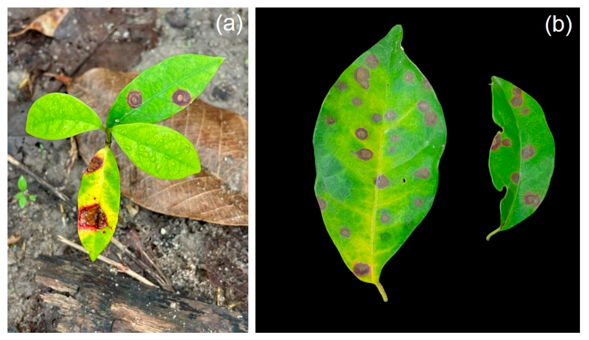
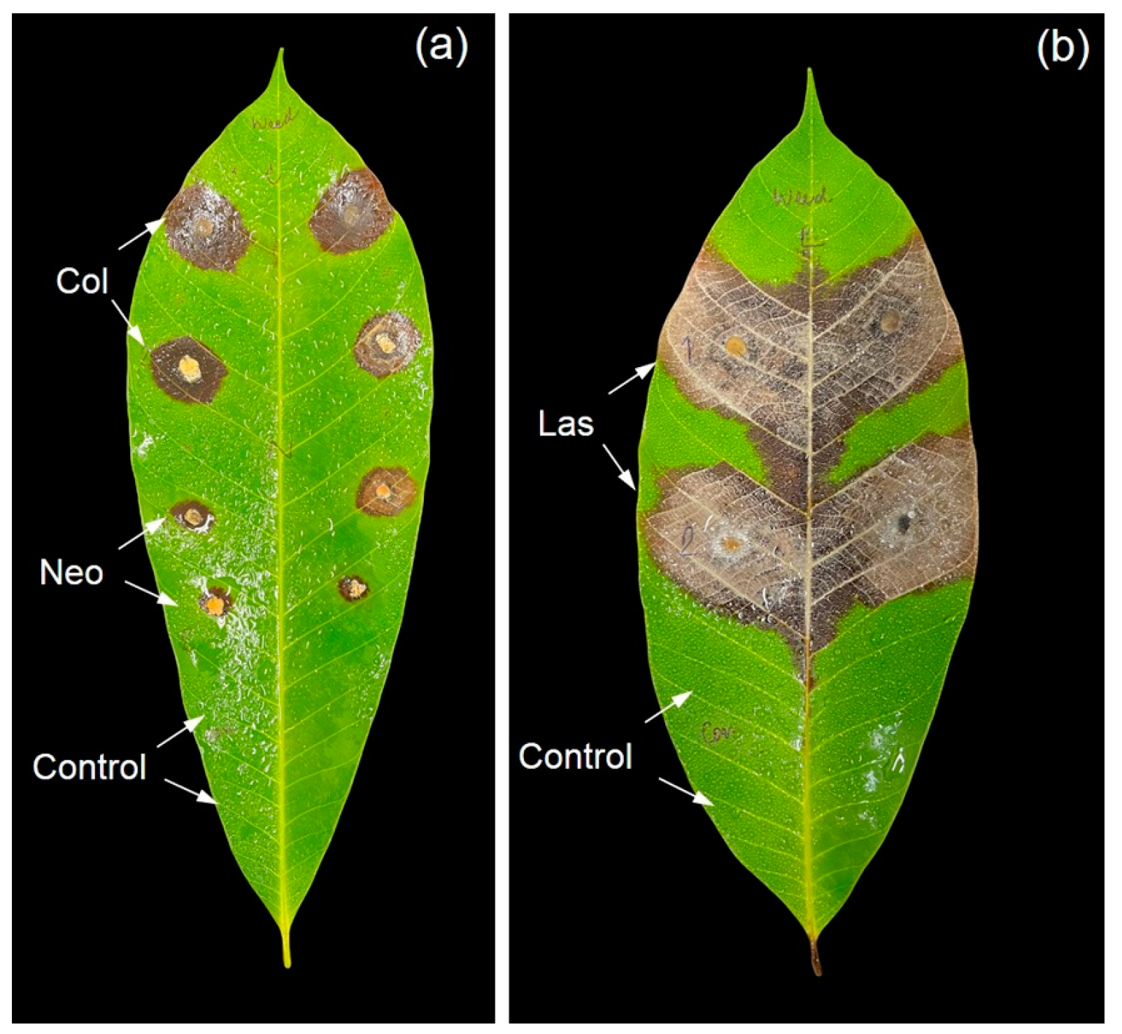
3.1. Symptom Recognition

The leaves of infected Parameria sp. presented similar features to the classic features of infected rubber tree leaves (Figure 1). Spots were initially uniform, small, and circular, light brown to dark brown in color, and later expanded, forming dark irregular margins (Figure 1). Several formed irregular branched pycnidia and some old lesions showed dense pycnidia.

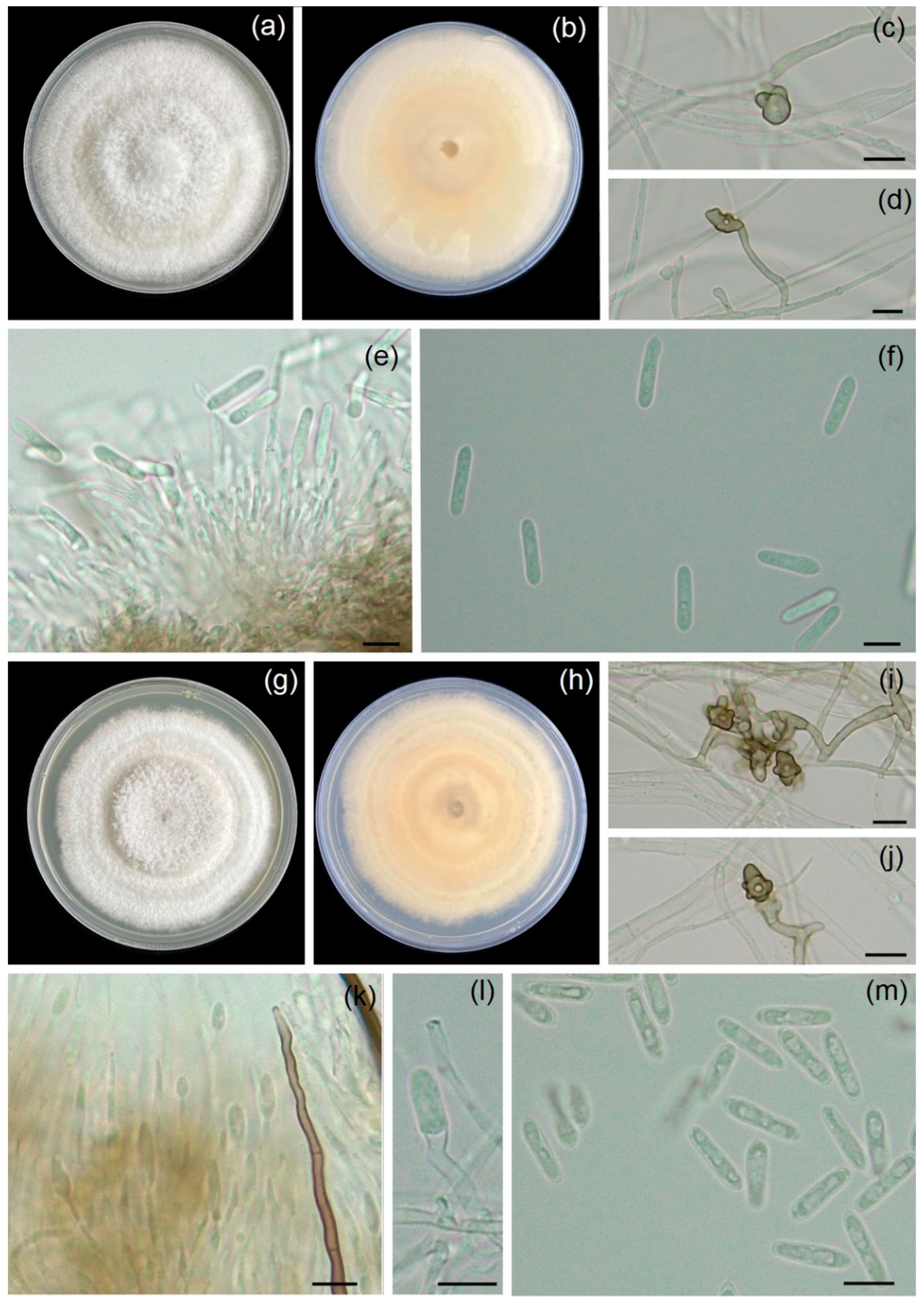
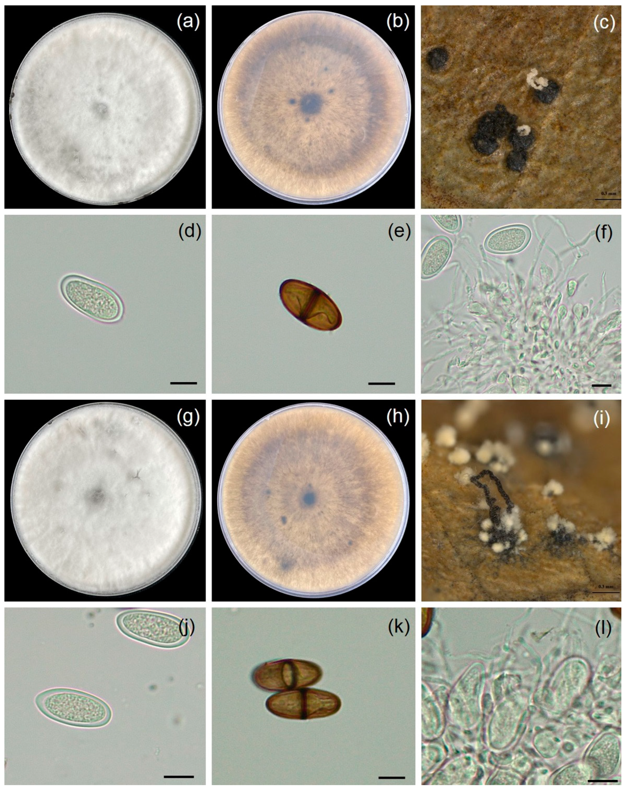
3.2. Morphology Study

The six fungal isolates were classified from morphological characteristics into three fungal genera, namely Colletotrichum (WC001 and WC002), Lasiodiplodia (WL001 and WL002), and Neopestalotiopsis (WN001 and WN002). The occurrence of fungal isolates (WC001 and WC002) in PDA covered an area of 7.8 cm in the Petri dish within 7 days, demonstrating a growth rate of 11.1 mm/day. Colonies exhibited a cottony material, pale, or white–gray color (Figure 2). The conidiomata presented a sticky pink to orange coloration and was darker in the center of the dish. The conidiophores were elongated and hyaline with openings. The conidia were cylindrical with round ends, non-septate and smooth, and 5.1–7.9 (6.4 ± 0.8) μm wide and 19.2–27.9 (24.6 ± 2.6) μm long ( x ¯ = 6.4 × 24.6 μm, n = 20). Based on their morphologies, fungal isolates WC001 and WC002 were identified as Colletotrichum sp. [23].
Fungal isolates WL001 and WL002 were grown in PDA, covering 8.2 cm of the Petri dish within 7 days, indicating a growth rate of 11.7 mm/day at ambient temperature. Colonies initially appeared as white to pale greenish gray and changed into a dark with age (Figure 3). Pycnidia developed on leaf surfaces. They were solitary or aggregate, globose to subglobose to irregular in shape, and brown to dark brown in color, depending on the isolate. Paraphyses were hyaline and septate. Both immature and mature conidia were exhibited as sub-ovoid to ellipsoid with round apices. Immature conidia were aseptate, double-layered, hyaline, unicellular, and 11.8–14.2 μm. Mature conidia were dark brown, septate, 24.1–29.9 (27.8 ± 1.8) μm long, and 13.4–15.6 (14.5 ± 0.8) μm wide ( x ¯ = 27.8 × 14.5 μm, n = 20). Hence, the fungal isolates (WL001 and WL002) were classified as Lasiodiplodia sp. based on morphological characteristics [24].
Fungal isolates WN001 and WN002 were grown in PDA and covered 6.5 cm of the Petri dish within 7 days, indicating a growth rate of 9.3 mm/day at ambient temperature. Colonies were white to pale brown on PDA with lobate edges, and conidiomata presented as black slimy conidial masses embedded in mycelia (Figure 4). Conidia were fusoid to ellipsoid, slightly curved, 20.5–31.4 (24.9 ± 3.1) μm long, and 5.1–6.7 (6.1 ± 0.5) μm wide ( x ¯ = 24.9 × 6.1 μm, n = 20), comprising five cells. The three median cells were versicolored, apical cells were conical, and hyaline with two to three apical appendages 12.1–27.9 (20.8 ± 3.7) μm long. The basal cells were conical hyaline with one basal appendage 4.2–6.8 (4.9 ± 0.9) μm ( x ¯ = 20.8 × 4.9 μm, n = 30) long. According to our morphological characteristics, it was noted that the fungal isolates (WN001 and WN002) were identified as Neopestalotiopsis sp. [25].
As mentioned above, there are six fungal isolates stored in the culture collection of Pest Management, Faculty of Natural Resources, Prince of Songkla University, Thailand, under the accession numbers PSU-WC001, PSU-WC002, PSU-WL001, PSU-WL002, PSU-WN001, and PSU-WN002.

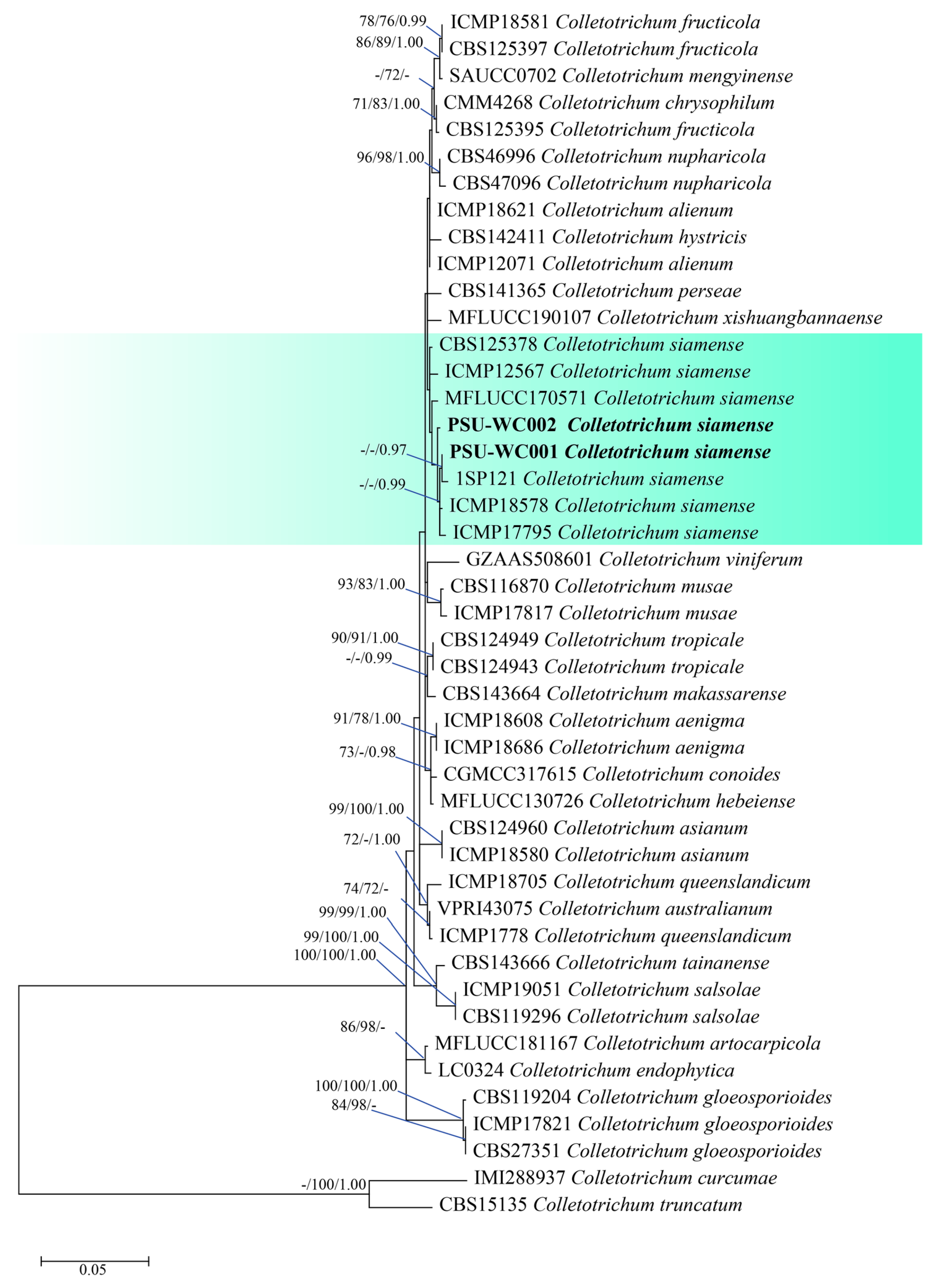
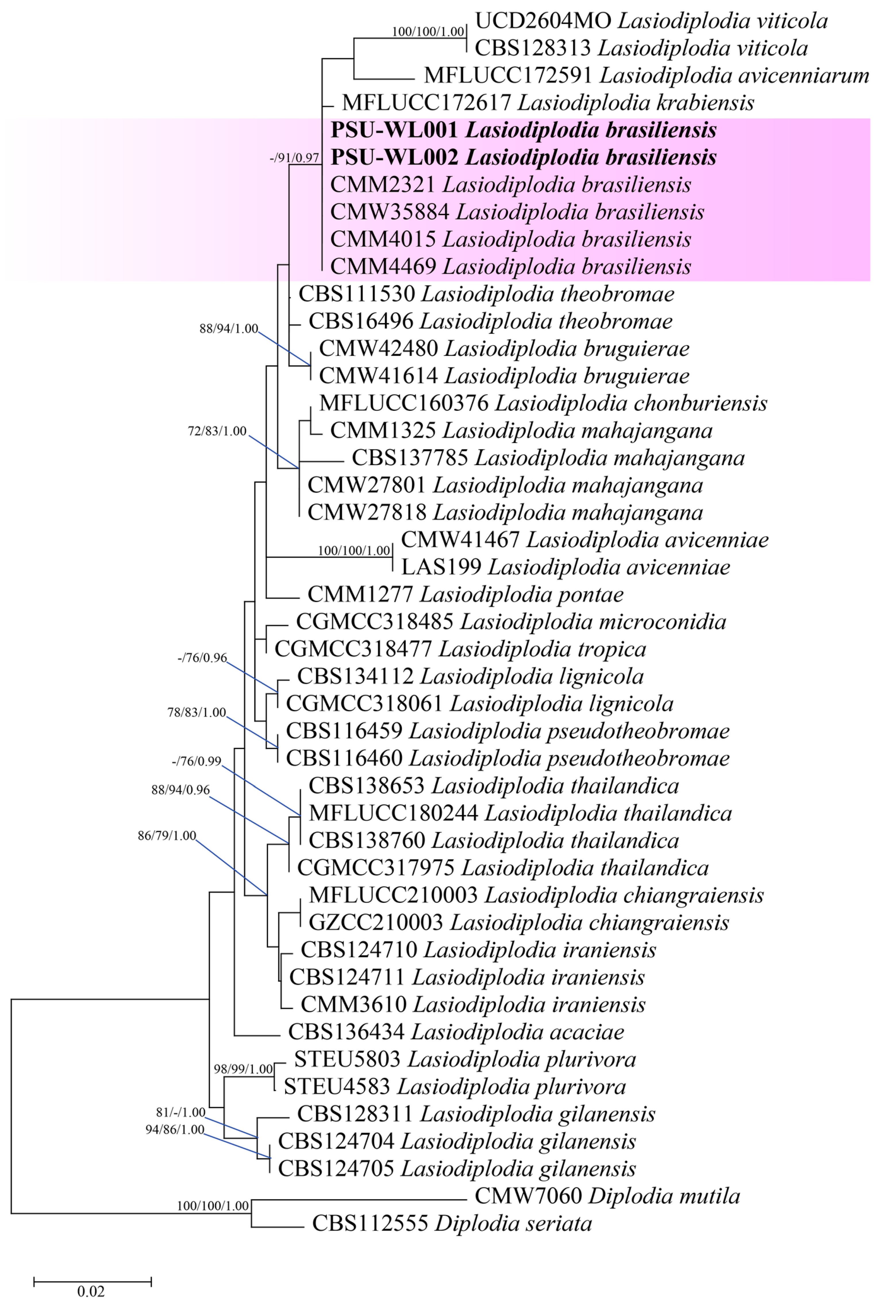
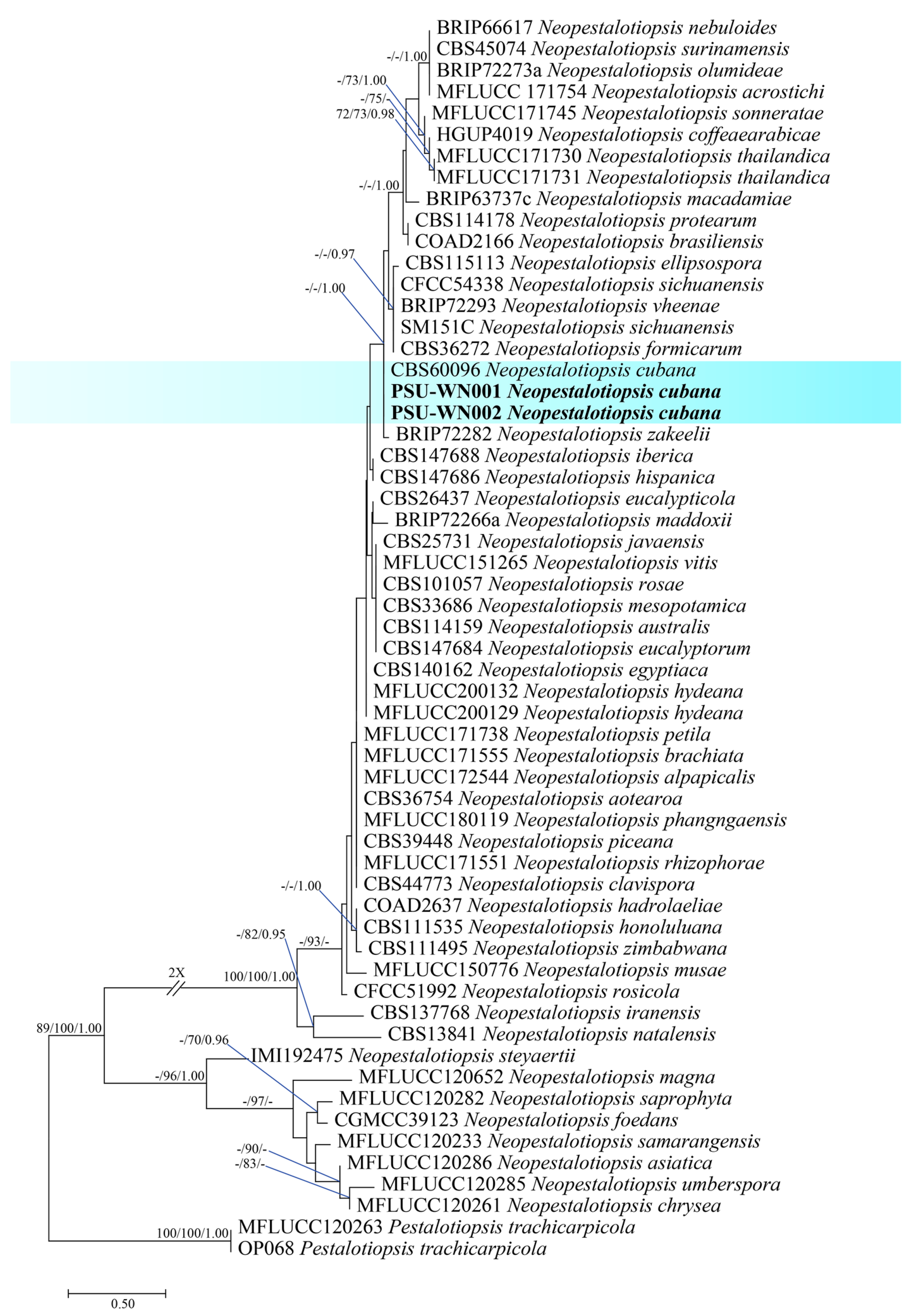
3.3. Molecular Identification

To identify the six fungal isolates at the species level, the ITS, tub2, and tef1-α portions of PSU-WL001, PSU-WL002, PSU-WN001, and PSU-WN002 were sequenced, and the act, gapdh, ITS, and tub2 portions of PSU-WC001 and PSU-WC002 were sequenced. The Blastn search results for PSU-WC001 and PSU-WC002 warranted the genus Colletotrichum. A phylogenetic tree was composed of two isolates and 43 reference sequences from Colletotrichum sp. C. curcumae and C. truncatum, while the tree with single loci and the concatenated data set (act, gapdh, ITS, and tub2) were demonstrated. The alignment used 217 bases for atc, 238 bases for gapdh, 448 bases for ITS, and 366 bases for tub2. PSU-WC001 and PSU-WC002 were grouped together with C. siamense (Figure 5). The partial DNA sequences of act, gapdh, ITS, and tub2 were submitted in GenBank via the accession numbers (Table S1).
The Blastn search of PSU-WL001 and PSU-WL002 showed they belonged to the genus Lasidiplodia and had a 99% sequence similarity to L. brasiliensis. The phylogenetic tree between the combined DNA sequences of ITS (487 bases), tub2 (379 bases), and tef1-α (280 bases) and the sequences of two isolates was aligned with those of 41 references of Lasiodiplodia and two outgroups (Diplodia mutila and D. seriata). The analysis of maximum likelihood showed that the phylogenetic placement of two isolates was closely associated with an isolate of L. brasilensis (Figure 6). It highlights the partial DNA sequences for ITS, tub2, and tef1-α, which are submitted to GenBank, with the corresponding accession numbers listed in Table S2.
The Blastn search of PSU-WN001 and PSU-WN002 revealed they belonged to the genus Neopestalotiopsis. The alignment composed of 427 bases for ITS, 390 bases for tub2, and 476 bases for tef1-α. The sequences of the two isolates obtained in the present study were aligned with the sequences of Neopestalotiopsis and an outgroup (Pestalotiopsis trachycarpicola). PSU-WN001 and PSU-WN002 grouped together with N. cubana (Figure 7). The partial DNA sequences of act, gapdh, ITS, and tub2 are submitted in GenBank with the accession numbers in Table S3.

3.4. Pathogenicity Test

After 7 days, the symptoms and signs of leaf disease began to appear on the inoculated rubber tree leaves (Figure 8). The fungal pathogens were re-isolated from the inoculated plants to observe their morphological examinations. The pathogenic morphology remained consistent with the characteristics noted before entering the inoculation process.

4. Discussion

Fungal pathogens were isolated from weeds (Parameria sp.) found in rubber tree plantations in southern Thailand. Six isolates (PSU-WC001, PSU-WC002, PSU-WL001, PSU-WL002, PSU-WN001, and PSU-WN002) effectively infected rubber tree leaves that showed leaf spot and leaf blight symptoms similar to those observed in previous studies [19,20,21,22]. The pathogens were identified from morphological characteristics and molecular alignments of multiple DNA sequences as C. siamense (PSU-WC001 and PSU-WC002), L. brasiliensis (PSU-WL001 and PSU-WL002), and N. cubana (PSU-WN001 and PSU-WN002). The results of both characterization methods were in agreement with previous reports that the information of morphology and molecular studies is to successfully identify fungal pathogens.
Colletotrichum siamense belongs to the C. gloeosporioides species complex and caused diseases in a number of plant species, including anthracnose in avocado [33], anthracnose in papaya [34], and leaf blotch in Viburnum odoratissimum [35]. Lasiodiplodia brasiliensis has been documented to cause root rot in watermelon [36] and root rot in melon plants [37]. N. cubana has been reported to cause leaf blight in Ixora chinensis [38] and leaf fall in H. brasiliensis [22]. Although both C. siamense and N. cubana have been previously reported to cause leaf spot and leaf fall disease in southern parts of Thailand, our findings showed L. brasilensis, for the first time, in weeds in the region and that it was able to cause leaf spot disease on rubber leaves. However, certain phytopathogenic fungi are able to infect some weeds and can be used as biological weed control agents. For instance, C. graminicola and Exserohilum fusiforme have been used to control Echinochloa crus-galli and Echinochloa spp. [39], and the application of Alternaria sp. has successfully controlled Rumex dentatus [40].
This study found that N. cubana from the weed Parameria sp. can cause disease in rubber trees, which is consistent with the previous report by Pornsuriya et al. [22], who identified this fungus as the cause of rubber leaf fall disease in southern Thailand. However, there have been no reports of N. cubana infecting rubber trees in other Southeast Asian countries, such as Malaysia, Indonesia, or Vietnam. Despite this, other species of Neopestalotiopsis have been reported to cause damage to rubber trees, such as N. clavispora in Malaysia [41] and N. rosae in China [42]. This indicates that fungi in the genus Neopestalotiopsis play a significant role in rubber tree diseases in this region.
In southern parts of Thailand, increased incidences of a number of diseases caused by fungi in the phylum Ascomycota have been recently reported in several plant species. In this study, three fungal pathogens (C. siamense, L. brasiliensis, and N. cunbana) in the phylum Ascomycota were isolated from Parameria sp., which are weeds commonly found in rubber plantations. Recently, leaf fall and leaf spot have been observed in rubber trees in these areas too [19,20,21,22]. It is possible that some weeds in rubber tree plantations may be a source of plant pathogenic fungi that infect the trees and may be associated with leaf disease in rubber trees.
To fulfill Koch’s postulates, the same pathogen should be inoculated onto the same host species by pathogenicity test. However, we did not directly inoculate pathogens found in this study onto weeds because we could not prepare healthy weed plants (no commercial seed available) for inoculation. We strongly wanted to observe whether fungi in weeds could cause leaf disease on rubber leaves. Therefore, we substituted inoculated fungal pathogens isolated from weeds to rubber tree leaves. Although weeds are considered a significant plant pest in crop production, their environmentally friendly management is crucial for agriculture to avoid yield losses. Our findings revealed at least three fungal species that could infect weeds and cause diseases, which may be useful for the biological control of weeds in rubber tree plantations. However, at least two of the fungal pathogens identified in this study, C. siamense and N. cubana, have been reported to cause leaf spot disease and leaf fall disease, respectively. Our results revealed that fungal strains isolated from weeds can infect rubber trees and cause leaf disease. This basic knowledge can prove useful in future surveillance and disease management strategies.
In southern Thailand, Colletotrichum siamense, Lasiodiplodia brasiliensis, and Neopestalotiopsis cubana have been identified as key fungal pathogens affecting rubber tree plantations. The infection process of these fungi likely follows a cycle involving alternative hosts, such as weeds, and environmental factors conducive to disease spread. C. siamense belongs to the C. gloeosporioides species complex and has been reported to cause anthracnose in multiple plant species, including rubber trees [20]. The pathogen spreads through conidia dispersed by rain splash and wind, allowing rapid colonization of host plants. L. brasiliensis, known to cause root rot in melon plants [37], was first identified in weeds within rubber plantations in this study, indicating its potential role as a latent pathogen in the ecosystem. This fungus produces pycnidia containing spores that can be disseminated by insects or rain, leading to infections in rubber trees. N. cubana, previously reported as the causal agent of leaf fall disease in rubber trees in Thailand [22], exhibits a similar infection pattern. The fungus produces multicellular conidia that attach to leaf surfaces, penetrate through stomata or wounds, and cause necrotic lesions. While N. cubana has been confirmed in Thailand, it has not yet been reported in other Southeast Asian rubber plantations, such as in Malaysia or Vietnam. However, closely related species like N. rosae have been associated with rubber tree diseases in China [42]. The presence of these fungi on both rubber trees and surrounding weeds suggests a potential reservoir for pathogen survival and transmission. Future studies should explore whether infected weeds serve as primary inoculum sources and investigate integrated disease management strategies to mitigate fungal spread in rubber plantations.

Supplementary Materials

The following supporting information can be downloaded at https://www.mdpi.com/article/10.3390/jof11030220/s1: Table S1: Collection details and GenBank accession numbers of Colletotrichum isolates in this study; Table S2: Collection details and GenBank accession numbers of Lasiodiplodia isolates in this study; and Table S3: Collection details and GenBank accession numbers of Neopestalotiopsis isolates in this study.

Author Contributions

Conceptualization, N.T., P.C. and A.S.; methodology, N.T. and K.T.; software, C.P.; validation, T.C., N.T. and A.S.; formal analysis, C.P.; investigation, A.S.; resources, N.T.; data curation, C.P.; writing—original draft preparation, N.T.; writing—review and editing, P.C. and A.S.; visualization, A.S.; supervision, A.S.; project administration, A.S.; funding acquisition, N.T. All authors have read and agreed to the published version of the manuscript.

Funding

This research was funded by the National Science, Research and Innovation Fund (NSRF), Prince of Songkla University (Grant No. NAT6505003M, NAT6505003a, NAT6601012M and NAT6601012a), and Thailand Science Research and Innovation (TSRl) grant No. 672A01014 from the Fundamental Fund for Basic Research, National Research Council of Thailand.

Institutional Review Board Statement

Not applicable.

Informed Consent Statement

Not applicable.

Data Availability Statement

The original contributions presented in this study are included in the article/Supplementary Materials. Further inquiries can be directed to the corresponding authors.

Acknowledgments

This work was partially supported by the Center of Excellence in Agricultural and Natural Resources Biotechnology (CoE-ANRB), Phase 3, Prince of Songkla University.

Conflicts of Interest

The authors declare no conflicts of interest.

References

  1. Hayashi, Y. Production of natural rubber from Para rubber tree. Plant Biotechnol. 2009, 26, 67–70. [Google Scholar] [CrossRef]
  2. Li, Z.; Fox, J.M. Mapping rubber tree growth in mainland Southeast Asia using time-series MODIS 250 m NDVI and statistical data. Appl. Geogr. 2012, 32, 420–432. [Google Scholar] [CrossRef]
  3. Ghini, R.; Bettiol, W.; Hamada, E. Diseases in tropical and plantation crops as affected by climate changes: Current knowledge and perspectives. Plant Pathol. 2011, 60, 122–132. [Google Scholar] [CrossRef]
  4. Ngobisa, A.N.; Abidin, M.Z.; Wong, M.; Noordin, M.W. Neofusicoccum ribis associated with leaf blight on rubber (Hevea brasiliensis) in Peninsular Malaysia. Plant Pathol. J. 2013, 29, 10. [Google Scholar] [CrossRef]
  5. Cai, Z.; Liu, Y.; Li, G.; Wang, Y.; Zhou, M. First report of Alternaria alternata causing black leaf spot of rubber tree in China. Plant Dis. 2015, 99, 290. [Google Scholar] [CrossRef] [PubMed]
  6. Pu, J.; Zhang, X.; Qi, Y.; Xie, Y.; Zhang, H.; Zhang, H. First record of Corynespora leaf fall disease of Hevea rubber tree in China. Australas. Plant Dis. Notes 2007, 2, 35–36. [Google Scholar]
  7. Hashim, I.; Chee, K.H.; Duncan, E.J. Damage assessment and yield losses due to Corynespora leaf fall disease in mature rubber plantations. J. Rubber Res. 2019, 22, 217–226. [Google Scholar]
  8. Ogbebor, N.O.; Omorusi, V.I.; Orumwense, K. Biochemical mechanisms of rubber tree root degradation by Rigidoporus microporus and implications for control strategies. For. Pathol. 2022, 52, e12715. [Google Scholar]
  9. Sreelatha, S.; Joseph, K. Anatomical and biochemical alterations in rubber tree bark following Phytophthora infection and their relation to latex yield reduction. Trees 2021, 35, 145–157. [Google Scholar]
  10. Wong, M.Y.; Zulkifli, Y.; Sunderasan, E. Phytotoxins produced by Pestalotiopsis species in rubber plantations: Detection, characterization and significance. Eur. J. Plant Pathol. 2020, 156, 1151–1163. [Google Scholar]
  11. Liyanage, K.K.; Khan, S.; Brooks, S. Climate-dependent development of rubber tree pathogens: Implications for disease management under environmental change. Plant Pathol. 2018, 67, 1026–1038. [Google Scholar]
  12. Liyanage, A.S.; Peries, O.S.; Liyanage, N.I.S. Major diseases of rubber and their management. In Natural Rubber: Biology, Cultivation and Technology; Elsevier Science Publishers: Amsterdam, The Netherlands, 2016; pp. 213–238. [Google Scholar]
  13. Jayasinghe, C.K. Rubber tree diseases: Economic impact and management strategies. Mycosphere 2020, 11, 795–817. [Google Scholar]
  14. Norhayati, M.; Nair, H.K.M.; Wong, M.Y. Economic assessment of fungicide application in rubber plantations in Malaysia. J. Plant. Crops 2019, 47, 182–191. [Google Scholar]
  15. Vongkhamheng, C.; Phimmavong, S.; Koutrakis, E. Economic impacts of white root disease on smallholder rubber plantations in Northern Laos. Forests 2022, 13, 512–526. [Google Scholar]
  16. European Forest Institute and Rubber Authority of Thailand. Briefing—Thailand’s Natural Rubber Producers Are Preparing for New Market Requirements. Available online: https://efi.int/sites/default/files/files/publication-bank/2024/Briefing%20-%20Thailand’s%20natural%20rubber%20producers%20are%20preparing%20for%20new%20market%20requirements.pdf (accessed on 28 February 2025).
  17. Puengcharoen, S.; Sirisomboon, P.; Duangpatra, J. Climate change effects on rubber tree diseases in Thailand: A review and future perspectives. Plant Pathol. 2021, 70, 1265–1280. [Google Scholar]
  18. Chambon, B.; Ruf, F.; Kongmanee, C.; Angthong, S. Can the cocoa industry solve the rubber smallholders’ crisis? A proposal from Thailand. For. Policy Econ. 2018, 85, 132–141. [Google Scholar]
  19. Thaochan, N.; Pornsuriya, C.; Chairin, T.; Chomnunti, P.; Sunpapao, A. Morphological and molecular characterization of Calonectria foliicola associated with leaf blight on rubber tree (Hevea brasiliensis) in Thailand. J. Fungi 2022, 8, 986. [Google Scholar] [CrossRef]
  20. Athinuwat, D.; Ruangwong, O.-U.; Harishchandra, D.L.; Latehnuering, F.; Sunpapao, A. Morphology and molecular characterization of Colltetotrichum siamense associated with leaf spot disease of rubber tree (Hevea brasiliensis) in southern Thailand. Physiol. Mol. Plant Pathol. 2024, 130, 102248. [Google Scholar] [CrossRef]
  21. Pornsuriya, C.; Thaochan, N.; Chairin, T.; Sunpapao, A. Morphological and phylogenetic evidences reveal Lasiodiplodia chonburiensis and L. theobromae associated with leaf blight in Hevea brasiliensis in Southern Thailand. Diversity 2023, 15, 961. [Google Scholar] [CrossRef]
  22. Pornsuriya, C.; Chairin, T.; Thaochan, N.; Sunpapao, A. Identification and characterization of Neopestalotiopsis fungi associated with a novel leaf fall disease of rubber trees (Hevea brasiliensis) in Thailand. J. Phytopathol. 2020, 168, 416–427. [Google Scholar] [CrossRef]
  23. Anderson, J.M.; Aitken, E.A.B.; Dann, E.K.; Coates, L.M. Morphological and molecular diversity of Colletotrichum spp. causing pepper spot and anthracnose of lychee (Litchi chinensis) in Australia. Plant Pathol. 2013, 62, 279–288. [Google Scholar] [CrossRef]
  24. Coutinho, I.B.L.; Freire, F.C.O.; Lima, C.S.; Lima, J.S.; Gonçalves, F.J.T.; Machado, A.R.; Silva, A.M.S.; Cardoso, J.E. Diversity of genus Lasiodiplodia associated with perennial tropical fruit plants in northeastern Brazil. Plant Pathol. 2017, 66, 90–104. [Google Scholar] [CrossRef]
  25. Darapanit, A.; Boonyuen, N.; Leesutthiphonchai, W.; Nuankaew, S.; Piasai, O. Identification, pathogenicity and effects of plant extracts on Neopestalotiopsis and Pseudopestalotiopsis causing fruit diseases. Sci. Rep. 2021, 11, 22606. [Google Scholar] [CrossRef]
  26. Saitoh, K.-i.; Togashi, K.; Arie, T.; Teraoka, T. A simple method for a mini-preparation of fungal DNA. J. Gen. Plant Pathol. 2006, 72, 348–350. [Google Scholar] [CrossRef]
  27. White, T. Amplification and direct sequencing of fungal ribosomal RNA genes for phylogenetics. In PCR Protocols: A Guide to Methods and Applications; Academic Press, Inc.: Cambridge, MA, USA, 1990. [Google Scholar]
  28. Glass, N.L.; Donaldson, G.C. Development of primer sets designed for use with the PCR to amplify conserved genes from filamentous ascomycetes. Appl. Environ. Microbiol. 1995, 61, 1323–1330. [Google Scholar] [CrossRef] [PubMed]
  29. Carbone, I.; Kohn, L.M. A method for designing primer sets for speciation studies in filamentous ascomycetes. Mycologia 1999, 91, 553–556. [Google Scholar] [CrossRef]
  30. Guerber, J.C.; Liu, B.; Correll, J.C.; Johnston, P.R. Characterization of diversity in Colletotrichum acutatum sensu lato by sequence analysis of two gene introns, mtDNA and intron RFLPs, and mating compatibility. Mycologia 2003, 95, 872–895. [Google Scholar] [CrossRef]
  31. Pornsuriya, C.; Ito, S.I.; Sunpapao, A. First report of leaf spot on lettuce caused by Curvularia aeria. J. Gen. Plant Pathol. 2018, 84, 296–299. [Google Scholar] [CrossRef]
  32. Kumar, S.; Stecher, G.; Li, M.; Knyaz, C.; Tamura, K. MEGA X: Molecular evolutionary genetics analysis across computing platforms. Mol. Biol. Evol. 2018, 35, 1547–1549. [Google Scholar] [CrossRef]
  33. Li, M.; Feng, W.; Yang, J.; Gao, Z.; Zhang, Z.; Zhang, W.; Wang, S.; Wang, W.; Gong, D.; Hu, M. First report of anthracnose caused by Colletotrichum siamense on avocado fruits in China. Crop Prot. 2022, 155, 105922. [Google Scholar] [CrossRef]
  34. Pacheco-Esteva, M.C.; Soto-Castro, D.; Vásquez-López, A.; Tovar-Pedraza, J.M. First report of Colletotrichum siamense causing papaya anthracnose in Mexico. J. Plant Pathol. 2022, 104, 1175. [Google Scholar] [CrossRef]
  35. Li, H.; Liao, Y.-C.-Z.; Wan, Y.; Li, D.-W.; Zhu, L.-H. Colletotrichum siamense, a novel causal agent of Viburnum odoratissimum leaf blotch and its sensitivity to fungicides. J. Fungi 2023, 9, 882. [Google Scholar] [CrossRef] [PubMed]
  36. Alves, C.P.S.S.; Negreiros, A.M.P.; Barros, A.P.O.; Souza, D.M.S.; Michereff, S.J.; Sales, R., Jr.; Correia, K.C. First report of Lasiodiplodia brasiliensis causing root rot on watermelon in Brazil. Plant Dis. 2023, 107, 573. [Google Scholar] [CrossRef] [PubMed]
  37. Maia da Silva, R.; Ambrósio, M.M.d.Q.; Neto, J.A.d.S.; Silva, J.L.d.S.; da Costa, T.E.; Figueiredo, F.R.A.; Barroso, K.A.; da Silva, W.; Holanda, I.S.A. First report of Lasiodiplodia brasiliensis causing root rot in melon plants in northeastern Brazil. Plant Dis. 2024, 108, 219. [Google Scholar] [CrossRef]
  38. Khoo, Y.W.; Tan, H.T.; Khaw, Y.S.; Li, S.-F.; Chong, K.P. First report of Neopestalotiopsis cubana causing leaf blight on Ixora chinensis in Malaysia. Plant Dis. 2022, 106, 2747. [Google Scholar] [CrossRef]
  39. Johanson, D.; Wyse, D.; Janes, K. Controlling weeds phytopathogen. Weed Tech. 2003, 10, 621–624. [Google Scholar] [CrossRef]
  40. Iffat Siddiqui, I.S.; Rukhsana Bajwa, R.B.; Arshad Javaid, A.J. Field evaluation of Alternaria alternata as mycoherbicide for the management of Rumex dentatus L. Philipp. Agric. Sci. 2010, 93, 116–120. [Google Scholar]
  41. Ngobisa, A.I.C.N.; Zainal Abidin, M.A.; Wong, M.Y.; Noordin, W.D. Characterisation and pathogenicity of Pestalotiopsis species associated with leaf disease of rubber (Hevea brasiliensis). Arch. Phytopathol. Plant Protect. 2015, 48, 448–457. [Google Scholar]
  42. Zhao, F.; Wang, D.; Tao, Z.; Fan, Z.; Guo, H.; Zhang, H.; Huang, L. First report of Neopestalotiopsis rosae causing leaf spot of rubber tree in China. Plant Dis. 2019, 103, 2947. [Google Scholar]
Figure 1. Symptomatic leaf samples from the weed Parameria sp.: brown lesions expanded, forming dark brown margins (a) and dark brown to green lesions (b).
Jof 11 00220 g001
Figure 2. Microphotographs showing the morphological characteristics of Colletotrichum sp. including WC001 and WC002: top view of colony on PDA (a,g); bottom view of colony on PDA (b,h); appressoria (c,d,i,j); conidiophores and conidia (e,k,l); and conidia (f,m). Bars (cf,im) = 10 µm.
Jof 11 00220 g002
Figure 3. Microphotographs showing the morphological characteristics of Lasiodiplodia sp. (WL001 and WL002): top view of colony on PDA (a,g); bottom view of colony on PDA (b,h); pycnidia (c,i); immature conidia (d,j); mature conidia (e,k); and conidiogenous cells and paraphyses (f,l). Bars (df,il) = 10 µm.
Jof 11 00220 g003
Figure 4. Microphotographs showing the morphological characteristics of Neopestalotiopsis sp. isolates WN001 and WN002: top view of colony on PDA (a,g); bottom view of colony on PDA (b,h); conidiomata with black slimy exudate (c,i); and conidia with versicolored median cells (df,jl). Bars (df,jl) = 10 µm.
Jof 11 00220 g004
Figure 5. The phylogenetic tree with the combined ITS, gapdh, act, and tub2 sequences. The phylogenetic relationships between Colletotrichum species in the Colletotrichum gloeosporioides species complex. Bootstrap support values above 70% and Bayesian posterior values above 0.95 are shown at each node (ML/MP/PP). Colletotrichum truncatum CBS 15135 and Colletotrichum curcumae IMI 288937 were used as outgroups. The green background indicates C. siamense and strains generated in this study are indicated in bold.
Jof 11 00220 g005
Figure 6. The phylogenetic tree with combined ITS, tef1-α, and tub2 sequences. The phylogenetic relationships between Lasiodiplodia species. Bootstrap support values above 70% and Bayesian posterior values above 0.95 are shown at each node (ML/MP/PP). Diplodia mutila CMW7060 and Diplodia seriata CBS112555 were used as outgroups. The purple background indicates Lasiodiplodia brasiliensis and strains generated in this study are indicated in bold.
Jof 11 00220 g006
Figure 7. The phylogenetic tree was generated by maximum likelihood analysis based on combined ITS, tef1-α, and tub2 sequences. The phylogenetic relationships between Neopestalotiopsis species. Bootstrap support values above 70% and Bayesian posterior values above 0.95 are shown at each node (ML/MP/PP). Pestalotiopsis trachicarpicola CMW7060 and OP068 were used as outgroups. The blue background indicates Neopestalotiopsis cubana and strains generated in this study are indicated in bold.
Jof 11 00220 g007
Figure 8. Pathogenicity test of fungal isolates on healthy rubber leaves: leaves were inoculated with Colletotrichum sp. (Col, WC001 [upper] and WC002 [lower]), Neopestalotiopsis sp. (Neo, WN001 [upper] and WN002 [lower]), control uninoculated, (a) and with Lasiodiplodia sp. (WL001 [upper] and WL002 [lower]) and control (b).
Jof 11 00220 g008
Disclaimer/Publisher’s Note: The statements, opinions and data contained in all publications are solely those of the individual author(s) and contributor(s) and not of MDPI and/or the editor(s). MDPI and/or the editor(s) disclaim responsibility for any injury to people or property resulting from any ideas, methods, instructions or products referred to in the content.

