Article

Molecular Mechanism of Aflatoxin B1 Synthesis Related AfVerB Regulating the Development, AFB1 Biosyntheis and Virulence of Aspergillus flavus Mainly Through Its CYP Domain

1
Key Laboratory of Pathogenic Fungi and Mycotoxins of Fujian Province, Key Laboratory of Biopesticide and Chemical Biology of Education Ministry, Proteomic Research Center, School of Life Sciences, Fujian Agriculture and Forestry University, Fuzhou 350002, China
2
College of Animal Sciences, Fujian Agriculture and Forestry University, Fuzhou 350002, China
3
State Key Laboratory of Pathogen and Biosecurity, Institute of Microbiology and Epidemiology, Academy of Military Medical Sciences (AMMS), Beijing 100071, China
*
Authors to whom correspondence should be addressed.
J. Fungi 2025, 11(4), 293; https://doi.org/10.3390/jof11040293
Submission received: 26 January 2025 / Revised: 25 March 2025 / Accepted: 26 March 2025 / Published: 9 April 2025

Abstract

:
Aspergillus flavus and its secondary metabolites aflatoxins pose a significant threat to the health of humans, animals, and plants. Therefore, there is an urgent need to control A. flavus contamination. AfverB plays a key role in the aflatoxin gene cluster; however, its function and mechanism in fungal development and virulence remain poorly understood. In this study, we constructed afVerB gene deletion mutants (∆afVerB−1 and ∆afVerB−2) and two CYP domain mutants (afVerB∆D1 and afVerB∆D2) through homologous recombination. Phenotype analysis revealed that, via its two CYP domains, AfVerB is deeply involved in fungal morphogenesis and aflatoxin synthesis. Insect and crop colonization models revealed that AfVerB plays a key role in the fungus’s ability to infect hosts, and stress experiments discovered that AfVerB plays a significant role in the response to various environmental stresses, which explains why AfVerB is a key factor in fungal infection to some extent. RT-qPCR analysis demonstrated that AfVerB performs its bio-function through corresponding regulatory factors. We ultimately discovered that AfVerB is deeply involved in cell membrane stress stability, thereby participating in the regulation of fungal drug resistance (sensitive to AMB and resistant to VOR in this study). The CYP domain of AfVerB, particularly its second CYP domain, is crucial for the execution of its biological functions. This study elucidated the regulatory mechanisms by which AfVerB regulates fungal pathogenicity and aflatoxin biosynthesis, providing potential strategies for controlling A. flavus and its aflatoxin contamination.

1. Introduction

Aspergillus spp. Are widely distributed all over the world, and a number of species produce mycotoxins, acting as pathogens that seriously jeopardize the health of plants and animals [1]. In addition, after Aspergillus fumigatus, A. flavus is the second most common cause of human aspergillosis [2]. Aflatoxin B1 (AFB1) is the most toxic and prevalent aflatoxin, and it has been classified as a group 1 carcinogen by the International Agency for Research on Cancer [3]. In recent years, research on aflatoxin synthesis has received much attention in attempts to reduce their impact on agricultural economies and human health [4]. Despite a series of advances in this field, our understanding of the molecular regulatory mechanisms of aflatoxin biosynthesis by A. flavus is still limited. Therefore, there is an urgent need to reveal the regulatory mechanisms on aflatoxin biosynthesis and fungal virulence in A. flavus.
Aflatoxins are toxic and carcinogenic secondary metabolites derived from polyketide compounds, and their synthesis mainly involves at least 27 enzymes, which constitute a 70 kb gene cluster in the fungal genome, modulated by two transcriptionally regulators, AflR and AflS [5,6,7]. In A. flavus, AflR regulates fungal development, nucleus formation, and toxin synthesis [8,9]. AflR and AflS form a complex, in which AflS does not exhibit DNA-binding ability but reduces the DNA-binding affinity of AflR [10]. The deletion of the aflN gene from the gene cluster results in blocked toxin synthesis and is associated with fungal responses to environmental oxidative stress and reactive oxygen species in cells [11]. In the cluster, aflX encodes an enzyme involved in the conversion of Versicolorin A to Demethylsterigmatocystin, aflO regulates aflatoxin synthesis and fungal pathogenicity through lysine acetylation, and aflQ regulates aflatoxin production through its m6A modification [5,12,13]. It has been shown that aflatoxin synthesis is also dependent on cytochrome p450 monooxygenase [14]. AfVerB is involved in the transformation of norsolorinic acid to Averufin (AVN), which is crucial for the synthesis of aflatoxin. It is also involved in methylation modification and mRNA stability modulation, and RNA-Seq and RT-qPCR have revealed that AfVerB takes part in aflatoxin synthesis regulated by the methyltransferase AflIme4 [15,16,17]. However, the biological function of AfVerB in fungal virulence and aflatoxin biosynthesis remains unclear.
Currently, the primary antifungal agents employed in the treatment of invasive aspergillosis are polyenes, azoles, and echinocandins. Among these, voriconazole and amphotericin B play crucial roles in the prevention and clinical management of invasive aspergillosis [18,19]. Previous research has indicated that azoles and other fungicides can effectively suppress the incidence of diseases and the production of mycotoxins caused by fungi from the genera Aspergillus [20,21]. In the context of A. flavus, a robust correlation has recently been unveiled between azole resistance and genes associated with the aflatoxin biosynthesis gene cluster [22]. Nevertheless, the biological functions of AfVerB in modulating the drug resistance of A. flavus remain to be elucidated.
In this study, we investigated the biological functions of AfVerB in fungal development, toxin biosynthesis, pathogenicity, and responses to diverse environmental stresses and drug resistance by constructing AfVerB mutants and key domain mutants through crop and insect models. This work provides theoretical support for elucidating the significance of this gene in the control of A. flavus contamination.

2. Materials and Methods

2.1. The A. flavus Strains and Media Used in This Study

The A. flavus strains used in this study are listed in Table S1. The media used for the incubation of fungal strains in this study are listed in Table S2.

2.2. The Construction of A. flavus Strains

All fungal mutants in this article were constructed through homologous recombination [23]. afVerB knockout strain (∆afVerB) was constructed as shown in Figure S1A. Three primer pairs (afVerB-AF and afVerB-AR, afVerB-BF and afVerB-BR, and pyrG-F and pyrG-R) were used in the amplification of the upstream region (1901 bp), the downstream region (1811 bp), and the selective marker pyrG, respectively. The fusion of the above three fragments was performed with primers afVerB -NF and afVerB -NR. Finally, the ∆afVerB strain was obtained by the introduction of the fused fragment into CA14 PTS by polyethylene glycol-mediated transformation. Three pairs of primers (afVerB-AF and P801, P1020 and afVerB-BR, and afVerB-OF and afVerB-OR) were used to verify the ∆afVerB strain by amplifying the upstream homology arm fragment (AP), the downstream homology arm (BP), and open reading frames (ORFs). Transformants with AP and BP but without ORF fragments were confirmed as ∆afVerB strains. The domains of AfVerB were predicted by NCBI’s CCD database (https://www.ncbi.nlm.nih.gov/Structure/cdd/cdd.shtml, accessed on 28 August 2024), and the construction of domain deletion strains followed the protocol used for ∆afVerB preparation, as shown in Figure S1B,C.

2.3. Phenotype Analysis

The phenotype of the mutants was observed according to the previous study [24]. WT, ∆afVerB−1, ∆afVerB−2, afVerB∆D1, and afVerB∆D2 strains were incubated in the dark on PDA, YES, and GMM media at 37 °C. After 4 d, the diameter of each colony was measured. Conidia from each sample were collected and washed with a spore elution solution (0.05% Tween-20 and 7% DMSO). The collected conidia were analyzed and counted with a hemocytometer under a light microscope (Leica, Heerbrugg, Germany). For sclerotia analysis, CM medium was prepared to induce mycorrhizal formation, and 104 spore suspensions were inoculated and incubated in the dark at 37 °C. After 7 d of incubation, conidia and hyphae were washed away by spraying with 75% ethanol, and the sclerotia were counted under a light microscope (Leica, Heidelberg, Germany). The experiment was performed in three replicates.

2.4. Analysis of AFB1 Production

Aflatoxin yield was monitored according to the previous protocol with minor modifications [25]. Spores (10 μL, 107 conidia/mL) of WT, ∆afVerB−1, ∆afVerB−2, afVerB∆D1, and afVerB∆D2 fungal strains were inoculated in 10 mL of PDB and incubated in the dark at 29 °C for 7 d. The bottom 4 mL of the medium was mixed with an equal amount of methylene chloride, and the mixtures were shaken at 29 °C for 1 h at 150 rpm. Then, the lower 3 mL of liquid was transferred into a 5 mL centrifuge tube and dried in a fume hood. Then, aflatoxins were re-dissolved with 100 μL of methylene chloride, and 10 μL of the solution was analyzed by thin-layer chromatography (TLC) (Haiyang Chemical, Qingdao, China). The results were recorded using a UV gel imaging system (Beijing Oriental Science and Technology Development Ltd., Beijing, China). The experiment was performed in three replicates.

2.5. Crop Colonization Analysis

The research was conducted based on the previous methods [26]. Six peanut and corn kernels of uniform size and good growth were sterilized by soaking in 0.05% sodium hypochlorite for 3 min, followed by a thorough rinsing with alcohol and sterile water, and then inoculated with the spore suspensions (107 conidia/mL) of WT, ∆afVerB−1, ∆afVerB−2, afVerB∆D1, and afVerB∆D2 strains. The inoculated kernels were incubated at 29 °C for 6 d. The infested host kernels were then collected in a 50 mL centrifuge tube, vortexed in 10 mL of ddH2O, and 1 mL of spore suspension was aspirated to count the number of spores with a hemocytometer. Then, 10 mL of dichloromethane was added to the centrifuge tube to extract the aflatoxins, and the extracted aflatoxins were analyzed by thin-layer chromatography. The experiment was peformed in three replicates.

2.6. Stress Analysis

The fungal susceptibility reaction to various stresses was analyzed based on the previous methods [27]. To evaluate the role of AfVerB in the fungal resistance against environmental stresses, fungal spores (107) were inoculated on PDA under a variety of inhibitors (including the cell membrane inhibitor SDS, osmotic stress NaCl, cell wall inhibitor CR, the DNA damaging agent MMS, and oxidative stress MSB). The diameters of fungal colonies were determined at the 3rd d after inoculation. Finally, the inhibition rate was calculated according to the following formula: inhibition rate = (diameter of colony without inhibitor—diameter of colony with inhibitor)/diameter of colony without inhibitor. The experiment was performed in three replicates.

2.7. Insect Infection Model

The role of AfVerB in the fungal infection of wax borers (Galleria mellonella) was assayed according to the previously described method [28]. Ten G.mellonella were selected for each experimental group. Each G mellonella was injected with 5 μL of spore suspension (107 conidia/mL). G.mellonella larvae injected with saline served as the control group. The injected wax borers were incubated in a dark at room temperature, and the survival rate was recorded for the next 5 d. The dead larvae were collected and placed at 29 °C for 7 d, after which conidiation ability and AFB1 yield were analyzed according to the aforementioned protocols. The experiment was performed in three replicates.

2.8. Agar Spotting Assays

The role of AfVerB in fungal drug resistance was analyzed according to a previous protocol [29]. To test the role of AfVerB in fungal drug resistance, the sensitivity of WT, ΔafVerB−1 and ΔafVerB−2 strains to AMB and VOR was analyzed, Müller-Hinton medium was supplemented with 0.67 μg/mL AMB and 0.25 μg/mL VOR. Then, 1 μL portions of conidial suspensions (2 × 10⁶, 2 × 10⁵, 2 × 10⁴, 2 × 10³ conidia/mL) of the indicated fungal strains were spotted onto the plates with corresponding media, then grown at 37 °C for 48 h, and the results were observed and documented. The experiment was repeated three times.

2.9. RT-qPCR Assays

Fungal spores (106/mL) were cultured in PDB for 48 h. Mycelium was ground into powder with liquid nitrogen, and each 0.1 g of mycelium powder was lysed in 1 mL of TRIzol Total RNA Extraction Reagent (TakaRa Bio, Inc., Kusatsu, Japan) for 30 min. Total RNA was then extracted according to a previous protocol with a few modifications [25]. The first-strand cDNA was synthesized using the HiScript III enzyme mixture (Vazyme Biotechnology, Nanjing, China) according to the manufacturer’s instructions. RT-qPCR analyses were performed with the Quantstudio1+PCR system (Applied Biosystems, Inc., Walthman, MA, USA). The primer sequences used for RT-qPCR in this study are shown in Table S3. The expression level of β-tubulin was used as the internal reference. The relative expression levels of the target genes were calculated following the formula 2−∆∆Ct. All experiments were repeated three times.

2.10. Statistical Analysis

All data in this study are expressed as mean ± standard deviation. Statistical analysis was performed using GraphPad Prism 8.0.2 and the one-way ANOVA with Tukey’s test. A difference was considered to be statistically significant when p < 0.05. Error bars indicate standard error from at least three repetitions.

3. Results

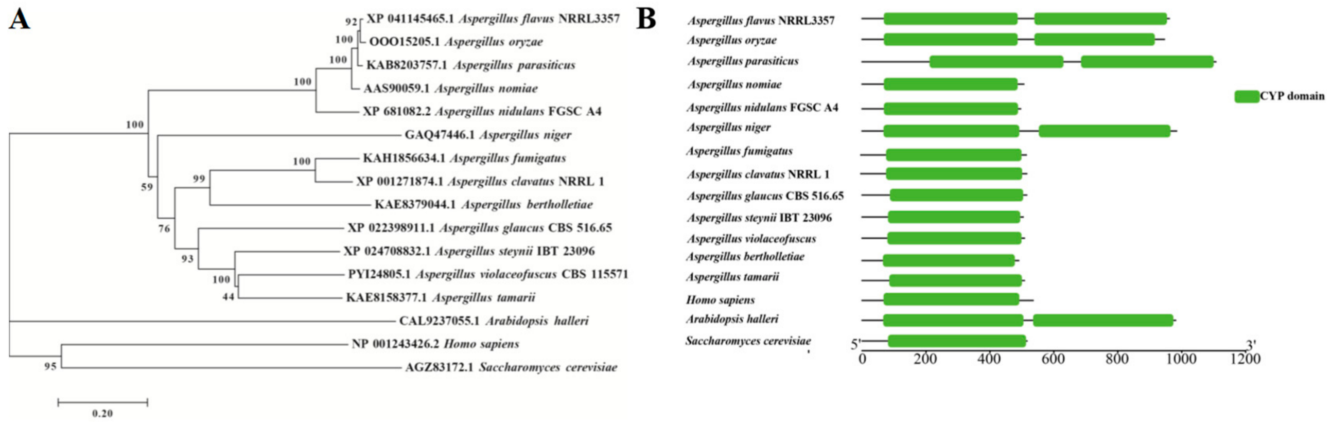
3.1. Bioinformatics Analysis and Strain Construction

To reveal the potential biological functions of AfVerB in A. flavus, the gene encoding afVerB (XP 041145465.1) was downloaded from NCBI (http://www.ncbi.nlm.nih.gov, accessed on 28 August 2024). The orthologs of afVerB were identified by BLAST in NCBI from 15 other species (A. bertholletiae, A. clavatus, A. glaucus, A. steynii, A. tamarii, A. violaceofuscus, A. nidulans, A. niger, A. nomiae, A. parasiticus, A. oryzae, A. fumigatus, S. cerevisiae, H. sapiens, and A. halleri). The evolutionary relationships of these homologous genes were constructed by MEGA 7.0 (Figure 1A), which showed that AfVerB is more closely related to A. oryzae VerB, and VerB is conserved among Aspergillus spp. The domains of VerB were predicted through CCD (https://www.ncbi.nlm.nih.gov/Structure/cdd/cdd.shtml, accessed on 28 August 2024) and visualized by tbtools (Toolbox for Biologists v2.149), which found that they all contain the conserved cyp domains. All the species of Aspergillus listed in the evolutionary tree bear a CYP domain. Among them, the VerB proteins of A. flavus, A. oryzae, A. niger, and A. parasiticus contain two CYP domains (Figure 1B). The above results suggest that AfVerB is conserved and may play important roles in the development and virulence of A. flavus.
The afVerB deletion mutant, including ∆afVerB−1, ∆afVerB−2, were constructed according to the protocol shown in Figure S1A. The transformants were verified through diagnostic PCR, and the results showed that both AP and BP fragments could be amplified from ∆afVerB−1 and ∆afVerB−2, but no ORF fragment was detected in both mutants (Figure S2A,B), which reflected that afVerB had been successfully deleted from both mutants. Further, the expression level of afVerB was further monitored by RT-qPCR, and the results showed that afVerB could not be detected from both ∆afVerB-1 and ∆afVerB-2 mutants, which meant that both afVerB deletion mutants were constructed (Figure S2E). The domain deletion strains (afVerB∆D1 and afVerB∆D2) were constructed and tested following the above method (Figure S2C,D), and were further confirmed through sequencing by Fuzhou Sunya Biotechnology Co., Ltd. (Fuzhou, China) (Figure S2F,G).

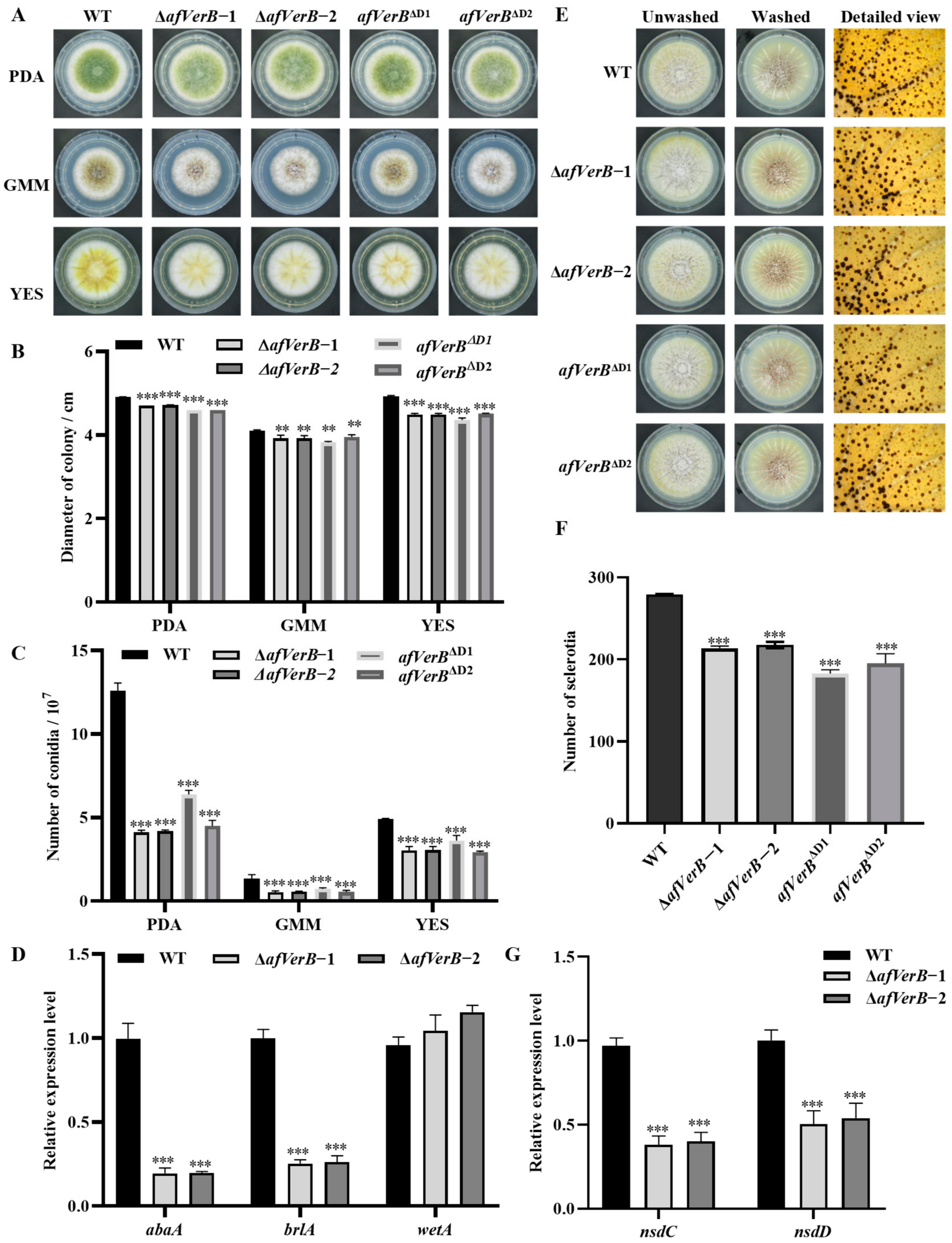
3.2. AfVerB Is Deeply Involved in Fungal Growth and Conidiation Through Its Both CYP Domains

In this study, in order to investigate the effect of VerB on the growth of A. flavus as well as conidia production, the spore suspension of WT, ∆afVerB−1, ∆afVerB−2, afVerB∆D1, and afVerB∆D2 strains were diluted to the same concentration and then cultured on PDA, YES, and GMM media for 4 d in dark at 37 °C. Photographs were taken and the diameter of each colony was measured. The number of spores was counted by a hemocytometer. The results showed that the colonial diameters of ∆afVerB−1, ∆afVerB−2, afVerB∆D1, and afVerB∆D2 were significantly smaller compared to that of the WT strain (Figure 2A,B), and the conidia number of ∆afVerB−1, ∆afVerB−2, afVerB∆D1, and afVerB∆D2 was significantly lower (Figure 2A,C). Further RT-qPCR showed that the deletion of AfVerB reduced conidia production by downregulating the expression levels of conidiation-related transcriptional factors abaA and brlA (Figure 2D). The above results suggest that AfVerB plays a crucial role in A. flavus conidiation.

3.3. AfVerB Is Involved in the Formation of Sclerotia via Both CYP Domains

Sclerotia is a kind of resistance structure formed by A. flavus to resist harsh external environments. In order to investigate the effect of afVerB on sclerotia formation, the spore suspension of WT, ∆afVerB−1, ∆afVerB−2, afVerB∆D1, and afVerB∆D2 strains were diluted to the same concentration and then incubated on CM medium at 37 °C in the dark for 7 d. The results showed that the sclerotia number of ∆afVerB−1, ∆afVerB−2, afVerB∆D1, and afVerB∆D2 was significantly reduced compared to the WT strain (Figure 2E,F). In order to reveal the potential pathway through which AfVerB regulates the formation of sclerotia, we examined the expression levels of transcriptional factors nsdC and nsdD genes by RT-qPCR. The results indicated that the relative expression levels of nsdC and nsdD were significantly reduced in both ∆afVerB−1 and ∆afVerB−2 compared with the WT strain (Figure 2G). The above results reveals that AfVerB plays an important role in sclerotia formation.

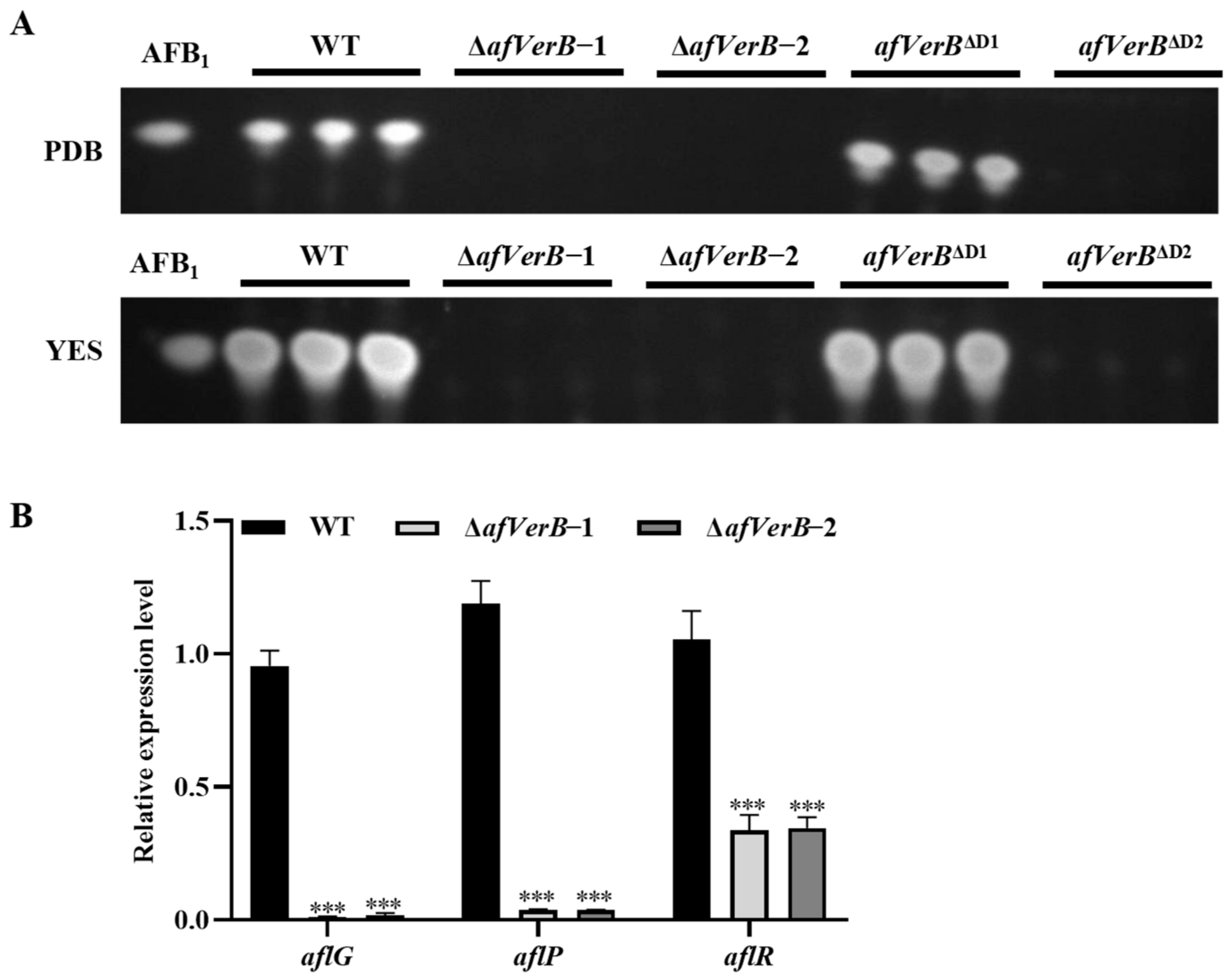
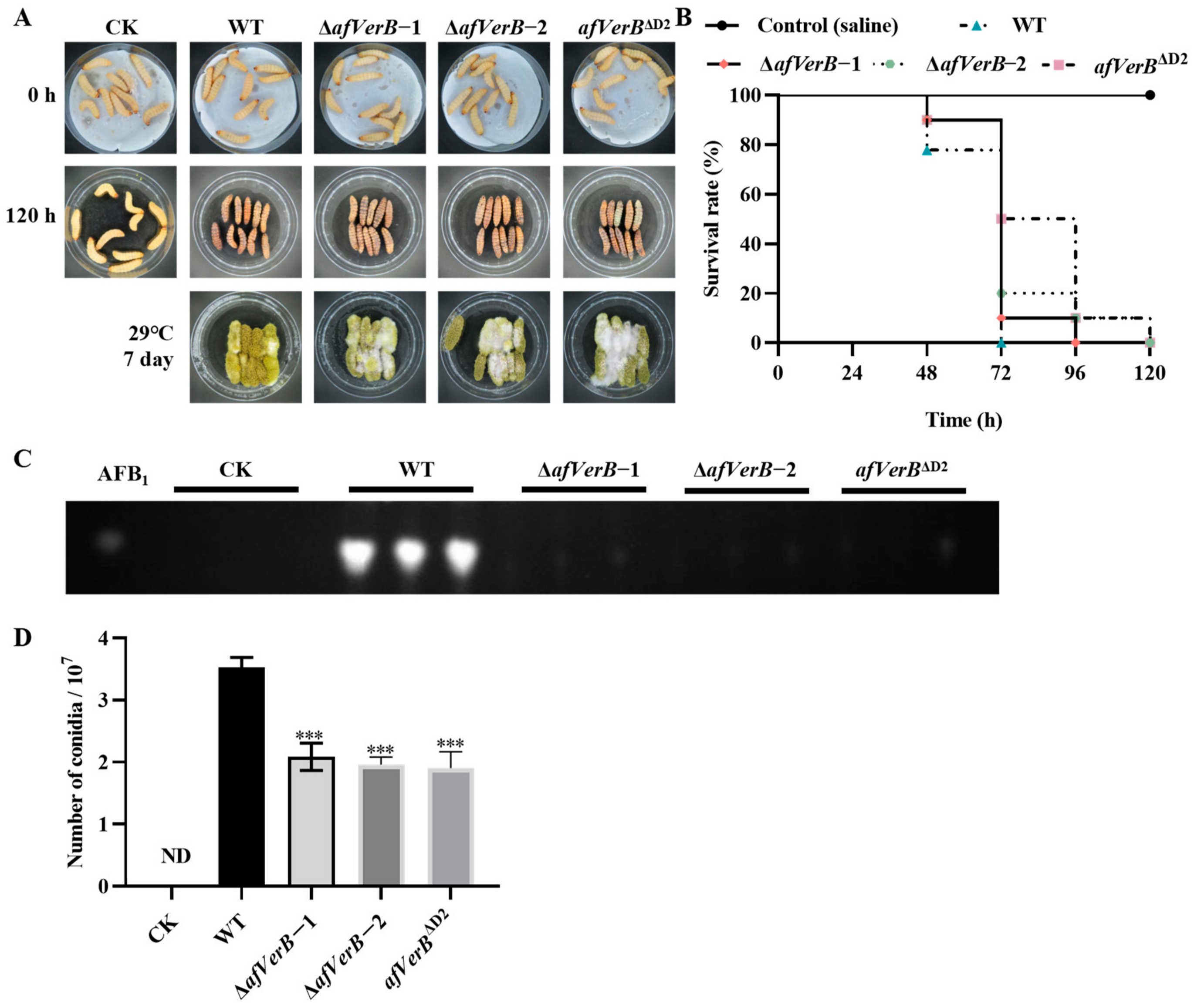
3.4. AfVerB Affects AFB1 Biosynthesis Through Its Second CYP Domain

In order to investigate the effect of afVerB on AFB1 biosynthesis, the spores of WT, ∆afVerB−1, ∆afVerB−2, afVerB∆D1, and afVerB∆D2 strains were diluted to the same concentration and incubated on PDB and YES for 7 d in the dark at 29 °C. The production of AFB1 from the above fungal strains was analyzed by TLC. It was found that AFB1 could not be detected in ∆afVerB−1, ∆afVerB−2, and afVerB∆D2, suggesting that afVerB is indispensable for aflatoxin biosynthesis, and that its second CYP domain is the key domain in this regulation process (Figure 3A). Further RT-qPCR analysis of the expression levels of genes in the aflatoxin gene cluster showed that aflR, aflG, and aflP were all significantly down-regulated in both ∆afVerB−1 and ∆afVerB−2 strains compared to the WT strain (Figure 3B). These results suggested that, through its second CYP domain, AfVerB plays a key role in AFB1 biosynthesis by regulating the aflatoxin gene cluster.

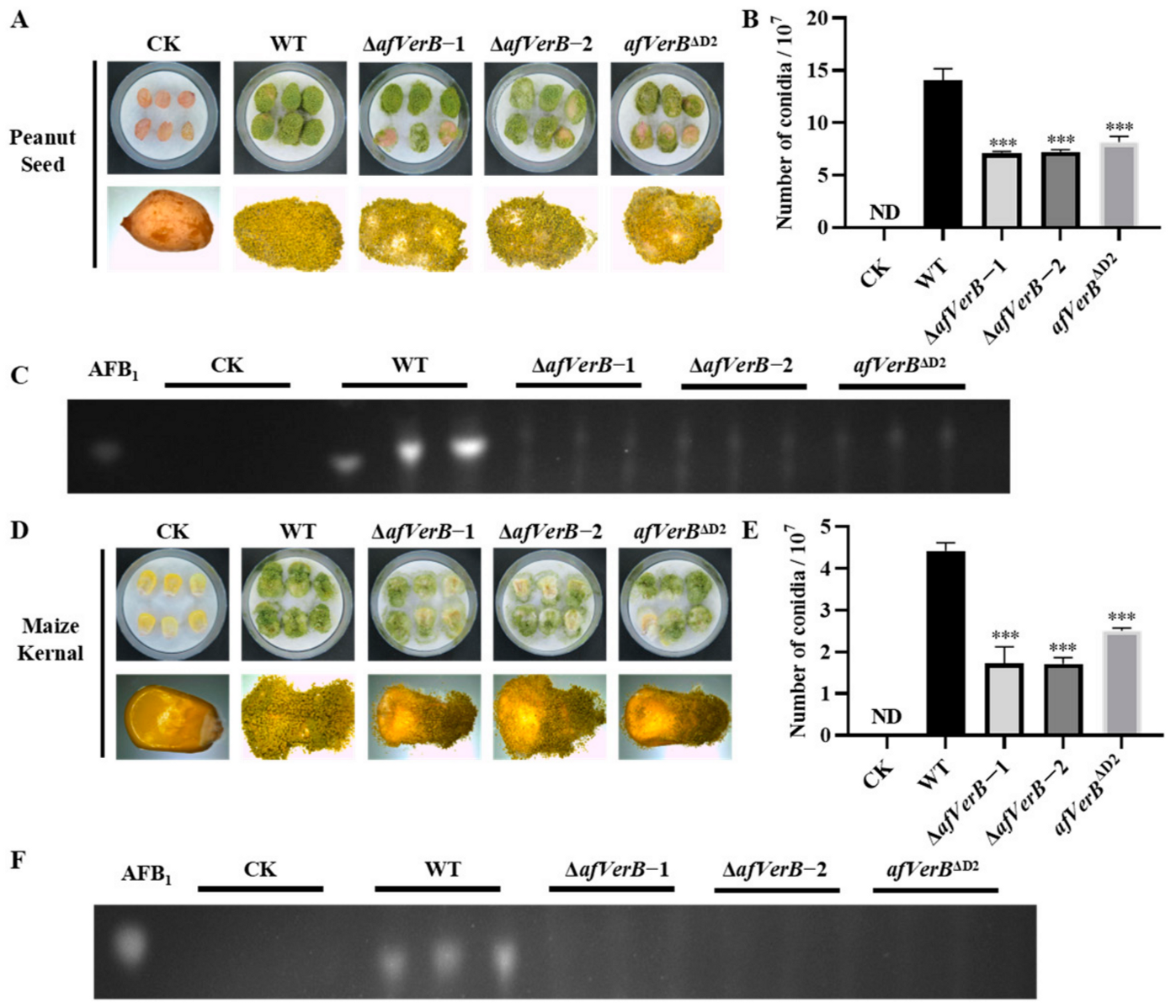
3.5. AfVerB Is Deeply Involved in the Colonization of Crop Grains by A. flavus

In order to elucidate the role of the afVerB in the colonization of A. flavus on the kernel of peanut and corn, grains of both corn and peanut were inoculated with the spore suspension of the above fungal strains and subsequently incubated in dark at 29°C for 5 d.The spores produced on peanut and corn kernels were then calculated, and the results showed that significantly fewer spores were produced on peanut kernels inoculated with ∆afVerB−1, ∆afVerB−2, and afVerB∆D2 than in the WT strain group (Figure 4A,B). Further TLC analyses showed that little AFB1 was detected from corn and peanut kernels in the ∆afVerB−1, ∆afVerB−2, and afVerB∆D2 groups, which was dramatically less than that of the WT group (Figure 4C). Similar results were observed in the corn kernels (Figure 4D–F). The above results suggest that the absence of AfVerB or its second CYP domain significantly reduces the ability of A. flavus to colonize crop kernels.

3.6. AfVerB Is Involved in Fungal Virulence to G. Mellonella

To investigate the impact of AfVerB on the pathogenicity of A. flavus to animals, wax moth (G. mellonella) larvae were selected as the animal host. A 5 μL spore suspension (107/mL) of WT, ∆afVerB−1, ∆afVerB−2, and afVerB∆D2 strains were injected into the larvae, respectively. After 120 hours of incubation and observation, it was found that all larvae in the WT group died at 72 h, while the survival rate of larvae colonized with spore suspensions of ∆afVerB-1 andafVerB-2 strains was at least 10% higher than that of the WT group at 72 h, and the afVerB∆D2 group even reached 50% higher than the WT group (Figure 5A,B). These results reflect that AfVerB, especially its second CYP domain, deeply participates in fungal virulence against wax moth larvae. The dead larvae were collected and cultured at 29 °C for 7 d, and then AFB1 was extracted from the dead larvae, and TLC analysis revealed that no AFB1 was detected in the ∆afVerB−1, ∆afVerB−2, and afVerB∆D2 groups, while large amounts of AFB1 were detected in the WT group (Figure 5C). The spore numbers were counted, and the results showed that the spore numbers in the ∆afVerB−1, ∆afVerB−2, and afVerB∆D2 groups were significantly reduced compared to the WT group (Figure 5D). The above results indicate that AfVerB plays a crucial role in fungal virulence in insects and significantly enhances the capacity of aflatoxin biosynthesis in A. flavus.

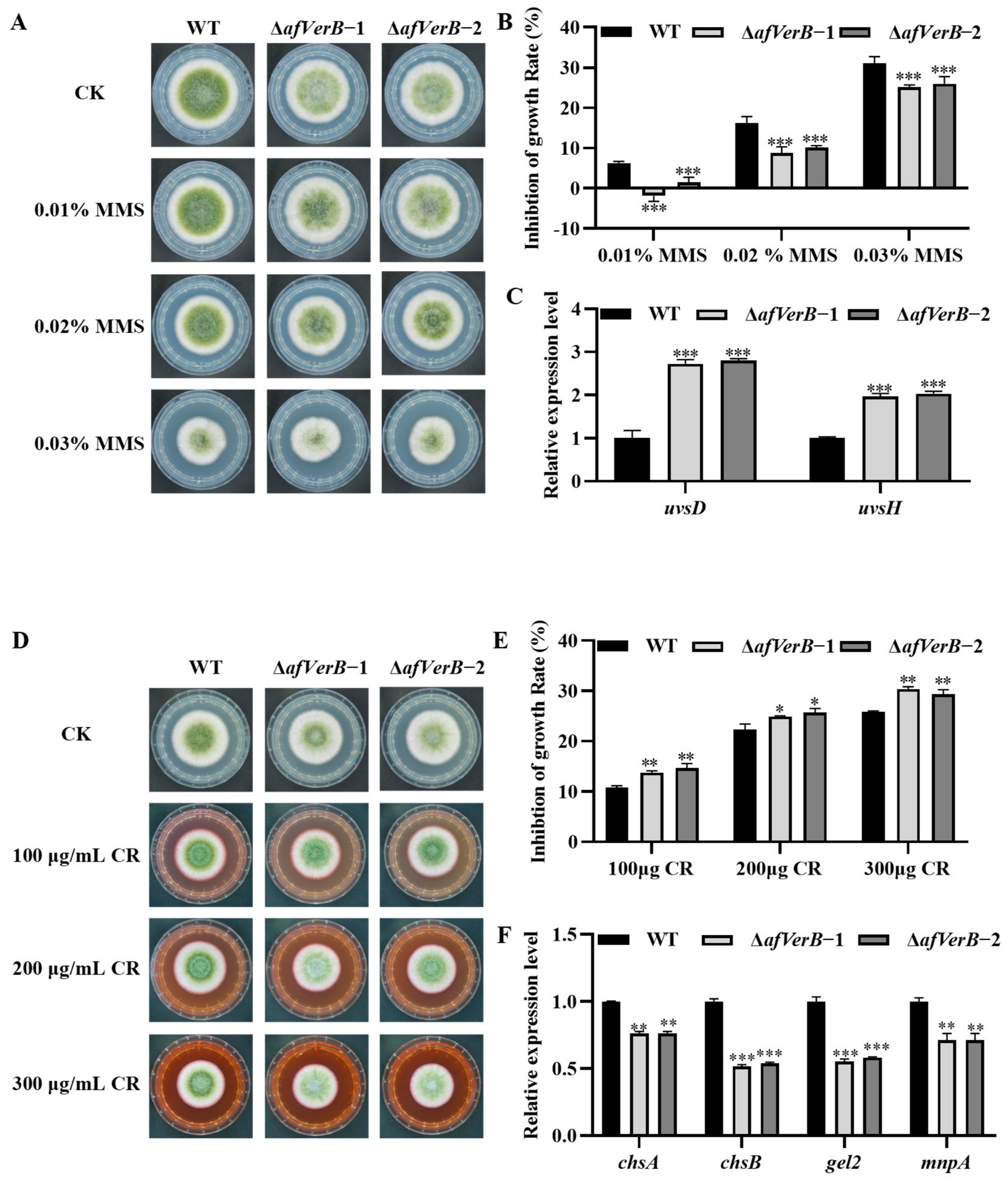
3.7. AfVerB Plays a Critical Role in the Response of A. flavus to Stresses

In order to explore the bio-functions of AfVerB in the response of A. flavus to various environmental stresses, the inhibitory effects of inhibitors, including KCl (mediated osmotic stress), CR (induced cell wall stress), MSB (mediated oxidative stress), and MMS (mediated DNA damage stress), was evaluated by adding them to PDA. The spore suspension (107/mL) of WT, ∆afVerB−1, and ∆afVerB−2 strains was inoculated onto the medium and incubated for 4 d. The results showed that MMS (0.01%, 0.02%, and 0.03%) significantly promoted the growth of ∆afVerB strains compared to the WT strain (Figure 6A,B). Further RT-qPCR suggested that AfVerB inhibits fungal growth under MMS-mediated DNA damage stress by decreasing the expression of uvsd and uvsh (Figure 6C).
Under CR-mediated cell wall stress (100, 200, and 300 μg/mL), ∆afVerB−1 and ∆afVerB−2 strains were significantly inhibited compared to the WT strain (Figure 6D,E). RT-qPCR analysis implied that AfVerB may improve fungal growth under CR-mediated cell wall stress by enhancing the expression of chsA, chsB, gel2, and mnpA (Figure 6F).
To assess the role of AfVerB in the response of A. flavus to osmotic stress, WT, ∆afVerB−1, and ∆afVerB−2 strains were inoculated onto PDA containing various concentrations of KCl (0.5, 1.0, and 1.5 M) for 4 d. The results showed that the growth inhibition rates of ∆afVerB-1 and ∆afVerB-2 strains were significantly increased compared to that of the WT strain. RT-qPCR suggested that AfVerB may enhance fungal growth under KCl-mediated osmotic stress by improving the expression of skn7, sskA, and sskB (Figure S3A–C). Oxidative stress mediated by MSB was also evaluated, and the results showed that AfVerB may decrease fungal growth under MSB-mediated oxidative stress by suppressing the expression of catA and catB (Figure S3D–F). The above findings indicate that AfVerB plays a key role in the response of A. flavus to various environmental stresses.

3.8. AfVerB Is Involved in the Response of A. flavus to Antifungal Drugs

In order to investigate the bio-function of afVerB in fungal drug resistance, the spore suspension of WT, ∆afVerB−1, and ∆afVerB−2 strains was diluted to 2 × 10⁶, 2 × 10⁵, 2 × 10⁴, and 2 × 10³ conidia/mL, and inoculated on MHA medium containing final concentrations of 0.67 ng/μL AMB and 0.25 ng/μL VOR, respectively. It was found that the ∆afVerB−1 and ∆afVerB−2 strains were more resistant to AMB but more sensitive to VOR compared to the WT strain (Figure 7A). Further RT-qPCR analysis was conducted on the drug efflux genes (atrC, atrF, and mdr1) and ergosterol biosynthesis-related genes (erg6). The results showed that atrC, atrD mdr1, and erg6 expression levels were significantly down-regulated in the absence of AfVerB. These results suggest that AfVerB enhances VOR resistance by increasing drug efflux through upregulating the expression of mdr1, atrC, and atrF, while promoting fungal susceptibility to AMB by increasing ergosterol synthesis via upregulating the expression of erg6 (Figure 7B).
Ergosterol is a key component of the fungal cell membrane [30]. Antifungal agents bind to ergosterol in the fungal cell membrane, thereby increasing the permeability of the cell membrane, which inhibits fungal growth and proliferation [31,32]. In light of these findings, we speculated that AfVerB plays an important role in keeping the dynamic equilibrium of fungal plasma membrane. Therefore, further research on the bio-function of AfVerB in cell membrane homeostasis is warranted. WT, ∆afVerB−1, and ∆afVerB−2 strains were inoculated on PDA under SDS-mediated cell membrane stress (0.01% and 0.02%), and the results showed that the growth inhibition rates of ∆afVerB−1 and ∆afVerB−2 strains were significantly increased compared to that of the WT strain (Figure 7C,D). RT-qPCR analysis implied that AfVerB may be involved in the maintenance of cell membrane stability by enhancing the expression of flba and rho1 (Figure 7E). The above results suggest that AfVerB is a key regulator in maintaining plasma membrane stability and may be one of the important potential candidates in regulating fungal drug sensitivity in the fight against drug-resistant invasive Aspergillosis.

4. Discussions

AfVerB is a pivotal gene in the biosynthetic pathway of aflatoxin, encoding an enzyme that participates in the synthesis of aflatoxin [33]. AfVerB is involved in the conversion from Versicolorin A to Demethylsterigmatocystin, which is crucial for the subsequent synthesis of aflatoxin [15,16,17,34]. Affinin and the ethanol extract from Heliopsis longipes can significantly inhibit the biosynthesis of aflatoxin in A. parasiticus through downregulating multiple genes in the aflatoxin gene cluster, including afVerB [35]. Given the severe impact of aflatoxin on crops and human health, it is imperative to investigate the role of the toxin-related gene afVerB in the growth, development, AFB1 synthesis, sclerotial formation, and pathogenicity of A. flavus. However, the regulatory role of afVerB in the phenotype, secondary metabolism, and pathogenicity of A. flavus has not been deeply reported. Therefore, this study was designed to unveil the bio-function of AfVerB in A. flavus.
In the analysis of the growth and development of A. flavus, we found that the deletion of AfVerB significantly affected fungal growth rate and conidiation, and both conserved CYP domains were significantly involved in the process (Figure 2A,C). A similar phenotype was observed in the colonization of peanuts and corn (Figure 4A,B,D,E), as well as in the colonization of wax moths (Figure 5A). In Aspergillus species and related fungi, the formation of conidiospores is primarily regulated by the central developmental pathway composed of abaA, brlA, and wetA [36]. brlA begins to be expressed in the early stage of conidiophore development, controlling the formation of conidiophores [37]. abaA functions in the middle stage of conidial development by activating the expression of the downstream gene wetA, thereby ensuring normal conidial development [38]. wetA, in the late stage of conidial development, regulates the synthesis of the conidial cell wall, conidial and hyphal development, and the formation of pigments, ensuring the normal maturation of conidia [39]. In this study, RT-qPCR revealed that AfVerB upregulates the expression levels of the transcription factors abaA and brlA, indicating that, through its CYP domains, AfVerB is involved in the regulation of the early and middle stage of conidia formation (including conidiophore and phialide formation), but does not participate in wetA mediated late stage of conidia development (Figure 2D).
Sclerotia is a structure of Aspergillus species’ adaptation to adverse environmental conditions, and it is also the structure responsible for the sexual reproduction of A. flavus [40,41]. In this study, the deletion of afVerB significantly impacted the formation of sclerotia in A. flavus. Similarly, the deletion of either conserved domain markedly reduced the number of sclerotia (Figure 2E,F). nsdC and nsdD are key regulatory factors in sclerotia formation [42]. The deletion of AfVerB or its two CYP domains resulted in the downregulation of transcriptional factors nsdC and nsdD (Figure 2G). The above results reflect that through positively regulating both nsdC and nsdD, AfVerB, mainly through its CYP domains, enhances sclerotia formation and may promote genetic variation in A. flavus by facilitating sexual reproduction, thereby enhancing the environmental adaptability of this pathogenic fungus.
This study revealed that the ability to synthesize aflatoxins was lost in different carbon source media after the deletion of afVerB or its second CYP domain (Figure 3A). Further RT-qPCR analysis revealed that AfVerB dramatically inhibits the production of AFB1 by downregulating the expression of the aflatoxin gene cluster, including aflR, aflG, and aflP (Figure 3B). AflR is the initial transcriptional factor that activates aflatoxin synthesis, and as the important regulator in the aflatoxin gene cluster, the activity of aflP and aflG are regulated by aflR [43,44]. In the aflatoxin gene cluster, afVerB and aflG are closely related, and the ability of aflG to participate in aflatoxin biosynthesis is lost when the afVerB gene is deleted [45]. Additionally, studies have shown that the CYP domain plays an important role in the biosynthesis of aflatoxins [46,47]. This study found that the deletion of the first CYP domain (afVerBD1) does not affect the synthesis of aflatoxins, while the deletion of its second CYP domain (afVerBD2) results in the total loss of toxin synthesis ability (Figure 3A). It inferred that the second CYP domain of AfVerB plays a key role in aflatoxin biosynthesis regulation. The same phenotype was observed in the colonization of peanuts and corn, as well as in the colonization of immune-competent G. mellonella [48]. These results suggest that AfVerB regulates the synthesis of afltoxins in A.flavus through its second CYP domain.
External conditions have a certain impact on the growth and development of Aspergillus [49]. The capacity of pathogenic fungi to cope with various environmental stresses is closely related to their ability to infect hosts. Therefore, it is important to explore the role of AfVerB in fungal sensitivity against various stresses. We found that AfVerB is involved in the osmotic stress mediated by KCl (Figure S3), the oxidative stress mediated by MSB (Figure S3), the DNA damage stress mediated by MMS (Figure 6), and the cell wall stress mediated by CR (Figure 6). The absence of skn7 leads to increased sensitivity of cells to high osmotic pressure, and skn7 regulates the Hog1 MAPK pathway to respond to high osmotic pressure in S. cerevisiae [50]. SskA and SskB are high osmotic regulators involved in the response to osmotic stress and the regulation of cell growth [50,51,52]. We have discovered that AfVerB exerts a significant influence on osmotic stress response by upregulating the expression levels of skn7, sskA, and sskB (Figure S3C), thereby playing a crucial role in maintaining osmotic homeostasis. In the context of DNA damage stress mediated by MMS, the genes uvsd and uvsh are involved in the nucleotide excision repair (NER) pathway, which is essential for DNA repair, genomic stability, and cellular survival [53,54]. Our findings indicated that AfVerB downregulates the expression of uvsd and uvsh, and the growth inhibition rate of MMS on the afVerB-deficient strains (∆afVerB−1, and ∆afVerB−2) is significantly lower than that of the wild-type (WT) strain (Figure 6A–C). This suggests that AfVerB is involved in the DNA damage stress response by downregulating the activity of uvsd and uvsh. Genes catA and catB are two important catalase genes that play key roles in the antioxidant defense and secondary metabolism of A. flavus [55]. We observed that AfVerB downregulates the expression of catA and catB under the stress of MSB, and the growth inhibition rate of the afVerB-deficient strain (∆afVerB-1, and ∆afVerB-2) is significantly lower than that of the WT strain (Figure S3D–F). This indicated that AfVerB is involved in the MSB-mediated oxidative stress response by downregulating the activity of catA and catB. Genes chsA and chsB are chitin synthase genes that play important roles in cell wall integrity [56,57]. The gel2 gene encodes β (1-3) glucan synthase, which actively participates in fungal cell wall biosynthesis, while mnpA is crucial for cell wall integrity and developmental patterning [58,59]. We found that AfVerB upregulates the expression levels of chsA, chsB, gel2, and mnpA under CR-mediated cell wall stress, and the growth inhibition rate of the afVerB-deficient (∆afVerB−1, and ∆afVerB−2) strain is significantly higher than that of the WT strain (Figure 6D–F). These results demonstrate that AfVerB plays a vital role in maintaining cell wall stability by up-regulating the expression levels of chsA, chsB, gel2, and mnpA.
VOR primarily inhibits the 14α-demethylation of fungal cytochrome P450, blocking ergosterol synthesis and disrupting fungal cell membrane stability, leading to the leakage of cellular contents and thereby inhibiting fungal growth and reproduction [31]. AMB destroys the integrity of the fungal cell membrane through multiple mechanisms, increasing intracellular oxidative damage, and effectively inhibiting fungal growth and reproduction [60]. The role of AfVerB in fungal sensitivity to antifungal drugs was explored in this study. Compared with the WT, we found that the mutants lacking AfVerB were more sensitive to VOR, but more resistant to AMB. Mitochondrial-deficient strains of A. fumigatus have developed resistance to itraconazole, mainly due to the high expression of the drug efflux pump gene mdr1 [61]. ATP-binding cassette (ABC) transporters are major contributors to antifungal drug resistance in pathogenic fungi [62]. In A. nidulans, two novel ABC transporter-encoding genes atrC and atrD were identified, and evidence was found that atrD is involved in multidrug transport and antibiotic production [63]. In A. fumigatus, the C2H2 transcription factor ZfpA can significantly upregulate the ABC transporter atrF, thereby enhancing its resistance to azole drugs [64]. Erg6 plays a crucial role in the ergosterol biosynthesis pathway. In relevant studies, it has been found that in AMB-resistant strains, the mRNA levels of erg6 are decreased, making it a potential target for reducing AMB sensitivity [65,66]. In this study, RT-qPCR analysis revealed that AfVerB confers fungal resistance to VOR by upregulating the expression of atrC, atrF, and mdr1, while enhancing fungal sensitivity to AMB by upregulating the expression of the erg6 gene (Figure 7A,B). Therefore, our results suggest that AfVerB is involved in the regulation of fungal resistance to VOR and fungal sensitivity to AMB by modulating the integrity of the fungal cell membrane.
The key role of AfVerB in the sensitivity of A. flavus to VOR and AMB reflected that AfVerB is deeply involved in the stability of the plasma membrane. To verify the above viewpoint, the role of AfVerB in the fungal response against SDS-mediated plasma membrane stress was explored. The results showed that AfVerB is a very important element in maintaining the homeostasis of the plasma membrane (Figure 7C,D). SDS-mediated stress triggers a kinase cascade response known as the cell wall integrity (CWI) pathway, which responds to disturbances in the cell wall and membrane to maintain cellular integrity [67]. G proteins, as key regulatory proteins in the CWI pathway, act in concert with flbA to modulate fungal morphogenesis [68]. The deletion of the rho1 gene leads to the rupture of the plasma membrane, cytoplasmic leakage, and ultimately cell necrosis [69]. In this study, further signaling pathway analysis through RT-qPCR revealed that AfVerB maintains plasma membrane stability by upregulating the expression levels of flbA and rho1 under SDS-mediated cell membrane stress (Figure 7E). Additionally, RT-qPCR analysis revealed that afVerB is downregulated by the chromatin remodeling factor (CRF) AflArp5 and AflArp8, and upregulated by the CRF Aflarp9 and AflRsc1 (Figure S4). Based on the above findings, we speculated that AfVerB is deeply involved in maintaining the stability of the cell membrane, playing a key role in the fungal response to antifungal drugs. In view of the above deduction, AfVerB may serve as a potential target in the fungal drug-resistance field.
In summary, we have explored and elucidated the biological functions of AfVerB, including its role in fungal morphogenesis, mycotoxin biosynthesis, regulation of fungal virulence, and response to a series of environmental stresses, as well as its involvement in fungal drug resistance. We also revealed that its CYP domains, especially the second CYP domain, play a crucial role in the process of AfVerB implementing its bio-functions. These findings shed light on the regulatory mechanisms of AfVerB in fungal virulence and secondary metabolism. However, how the methyltransferase AflIme4 regulates the action of AfVerB remains unknown. Further investigation into the role of AflIme4-mediated mRNA methylation in the biological function of AfVerB would reveal the complicated regulatory mechanism of fungal virulence and secondary metabolism at the epigenetic level. This study provides new insights into the development of potential strategies against pathogenic filamentous fungi in the future.

Supplementary Materials

The following supporting information can be downloaded at: https://www.mdpi.com/article/10.3390/jof11040293/s1. Table S1: Strains used in this study; Table S2: The media used in this study; Table S3:Primers used in this study; Figure S1: The scheme of fungal strain construction; Figure S2: The construction of AfVerB mutants and the sequencing results of CYP domain deletion strains; Figure S3:The role of AfVerB in fungal response to osmotic and oxidative stresses. Figure S4:The impact of Afverb on chromatin remodeling factors.

Author Contributions

Conceptualization, K.Y. and C.Y.; Methodology, K.Y., S.Z., D.W., D.M., C.Y., W.F. and Z.Z.; Software, S.Y.; Validation, K.Y., S.Z., D.M., Y.Y. (Yanfang Yao), M.S. and S.Y.; Formal analysis, K.Y., S.Z. and Y.Y. (Yanfang Yao); Investigation, D.W., D.M., M.S. and W.X.; Resources, W.X.; Data curation, K.Y. and D.W.; Writing—original draft, K.Y.; Writing—review & editing, Z.Z.; Visualization, Y.Y. (Yanling Yang); Supervision, Y.Y. (Yanling Yang); Project administration, J.Y. and Z.Z.; Funding acquisition, J.Y. and Z.Z. All authors have read and agreed to the published version of the manuscript.

Funding

This work was funded by the grants of the National Natural Science Foundation of China (No. 32470207), the grants of the State Key Laboratory of Pathogen and Biosecurity (SKLPBS2434), the Nature Science Foundation of Fujian Province (No. 2021J02026, No. 2022J01594), and the Science and Technology Innovation Special Fund Project of Fujian Agriculture and Forestry University (KFB24088, KFB22048XA).

Institutional Review Board Statement

Not applicable.

Informed Consent Statement

Not applicable.

Data Availability Statement

The authors confirm that the data supporting the findings of this study are available within the article and its Supplementary Materials.

Acknowledgments

We especially thank Shihua Wang for providing instrument support and related guidance, and Xiuna Wang, Yu Wang, and Xinyi Nie for their support in instrument maintenance and reagent ordering.

Conflicts of Interest

The authors declare no conflict of interest.

References

  1. Samson, R.A.; Visagie, C.M.; Houbraken, J.; Hong, S.B.; Hubka, V.; Klaassen, C.H.; Perrone, G.; Seifert, K.A.; Susca, A.; Tanney, J.B.; et al. Phylogeny, identification and nomenclature of the genus Aspergillus. Stud. Mycol. 2014, 78, 141–173. [Google Scholar] [CrossRef] [PubMed]
  2. Rudramurthy, S.M.; Paul, R.A.; Chakrabarti, A.; Mouton, J.W.; Meis, J.F. Invasive Aspergillosis by Aspergillus flavus: Epidemiology, Diagnosis, Antifungal Resistance, and Management. J. Fungi 2019, 5, 55. [Google Scholar] [CrossRef] [PubMed]
  3. Ortega-Beltran, A.; Moral, J.; Picot, A.; Puckett, R.D.; Cotty, P.J.; Michailides, T.J. Atoxigenic Aspergillus flavus Isolates Endemic to Almond, Fig, and Pistachio Orchards in California with Potential to Reduce Aflatoxin Contamination in these Crops. Plant Dis. 2019, 103, 905–912. [Google Scholar] [CrossRef] [PubMed]
  4. Shan, X.; Williams, W.P. Toward elucidation of genetic and functional genetic mechanisms in corn host resistance to Aspergillus flavus infection and aflatoxin contamination. Front. Microbiol. 2014, 5, 364. [Google Scholar] [CrossRef]
  5. Cary, J.W.; Ehrlich, K.C.; Bland, J.M.; Montalbano, B.G. The aflatoxin biosynthesis cluster gene, aflX, encodes an oxidoreductase involved in conversion of versicolorin A to demethylsterigmatocystin. Appl. Environ. Microbiol. 2006, 72, 1096–1101. [Google Scholar] [CrossRef] [PubMed]
  6. Bhatnagar, D.; Cary, J.W.; Ehrlich, K.; Yu, J.; Cleveland, T.E. Understanding the genetics of regulation of aflatoxin production and Aspergillus flavus development. Mycopathologia 2006, 162, 155–166. [Google Scholar] [CrossRef] [PubMed]
  7. Chang, P.K. Lack of interaction between AFLR and AFLJ contributes to nonaflatoxigenicity of Aspergillus sojae. J. Biotechnol. 2004, 107, 245–253. [Google Scholar] [CrossRef]
  8. Wang, P.; Xu, J.; Chang, P.K.; Liu, Z.; Kong, Q. New Insights of Transcriptional Regulator AflR in Aspergillus flavus Physiology. Microbiol. Spectr. 2022, 10, e0079121. [Google Scholar] [CrossRef] [PubMed]
  9. Kong, Q.; Chang, P.K.; Li, C.; Hu, Z.; Zheng, M.; Sun, Q.; Shan, S. Identification of AflR Binding Sites in the Genome of Aspergillus flavus by ChIP-Seq. J. Fungi 2020, 6, 52. [Google Scholar] [CrossRef]
  10. Abbas, A.; Prajapati, R.K.; Aalto-Setala, E.; Baykov, A.A.; Malinen, A.M. Aflatoxin biosynthesis regulators AflR and AflS: DNA binding affinity, stoichiometry, and kinetics. Biochem. J. 2024, 481, 805–821. [Google Scholar] [CrossRef]
  11. Jia, K.; Yan, L.; Jia, Y.; Xu, S.; Yan, Z.; Wang, S. aflN Is Involved in the Biosynthesis of Aflatoxin and Conidiation in Aspergillus flavus. Toxins 2021, 13, 831. [Google Scholar] [CrossRef] [PubMed]
  12. Yang, G.; Yue, Y.; Ren, S.; Yang, M.; Zhang, Y.; Cao, X.; Wang, Y.; Zhang, J.; Ge, F.; Wang, S. Lysine acetylation contributes to development, aflatoxin biosynthesis and pathogenicity in Aspergillus flavus. Environ. Microbiol. 2019, 21, 4792–4807. [Google Scholar] [CrossRef] [PubMed]
  13. Yang, C.; Wu, D.; Lin, H.; Ma, D.; Fu, W.; Yao, Y.; Pan, X.; Wang, S.; Zhuang, Z. Role of RNA Modifications, Especially m6A, in Aflatoxin Biosynthesis of Aspergillus flavus. J. Agric. Food Chem. 2024, 72, 726–741. [Google Scholar] [CrossRef]
  14. Ehrlich, K.C.; Chang, P.K.; Yu, J.; Cotty, P.J. Aflatoxin biosynthesis cluster gene cypA is required for G aflatoxin formation. Appl. Environ. Microbiol. 2004, 70, 6518–6524. [Google Scholar] [CrossRef] [PubMed]
  15. Yabe, K.; Nakamura, Y.; Nakajima, H.; Ando, Y.; Hamasaki, T. Enzymatic conversion of norsolorinic acid to averufin in aflatoxin biosynthesis. Appl. Environ. Microbiol. 1991, 57, 1340–1345. [Google Scholar] [CrossRef]
  16. Liang, L.; Wang, X.; Lan, H.; Wei, S.; Lei, Y.; Zhang, S.; Zhai, H.; Hu, Y.; Lv, Y. Comprehensive analysis of aflatoxin B(1) biosynthesis in Aspergillus flavus via transcriptome-wide m(6)A methylome response to cycloleucine. J. Hazard. Mater. 2024, 461, 132677. [Google Scholar] [CrossRef]
  17. Liang, L.; Wang, X.; Wei, S.; Lei, Y.; Zhang, S.; Zhai, H.; Hu, Y.; Lv, Y. m(6)A methyltransferase AflIme4 orchestrates mycelial growth, development and aflatoxin B(1) biosynthesis in Aspergillus flavus. Microbiol. Res. 2024, 283, 127710. [Google Scholar] [CrossRef] [PubMed]
  18. Cavassin, F.B.; Baú-Carneiro, J.L.; Vilas-Boas, R.R.; Queiroz-Telles, F. Sixty years of Amphotericin B: An Overview of the Main Antifungal Agent Used to Treat Invasive Fungal Infections. Infect. Dis. Ther. 2021, 10, 115–147. [Google Scholar] [CrossRef]
  19. Patterson, T.F.; Thompson, G.R., 3rd; Denning, D.W.; Fishman, J.A.; Hadley, S.; Herbrecht, R.; Kontoyiannis, D.P.; Marr, K.A.; Morrison, V.A.; Nguyen, M.H.; et al. Practice Guidelines for the Diagnosis and Management of Aspergillosis: 2016 Update by the Infectious Diseases Society of America. Clin. Infect. Dis. 2016, 63, e1–e60. [Google Scholar] [CrossRef] [PubMed]
  20. Markoglou, A.N.; Doukas, E.G.; Ziogas, B.N. Phenylpyrrole-resistance and aflatoxin production in Aspergillus parasiticus Speare. Int. J. Food Microbiol. 2008, 127, 268–275. [Google Scholar] [CrossRef]
  21. Markoglou, A.N.; Doukas, E.G.; Malandrakis, A.A. Effect of anilinopyrimidine resistance on aflatoxin production and fitness parameters in Aspergillus parasiticus Speare. Int. J. Food Microbiol. 2011, 146, 130–136. [Google Scholar] [CrossRef] [PubMed]
  22. Doukas, E.G.; Markoglou, A.N.; Vontas, J.G.; Ziogas, B.N. Effect of DMI-resistance mechanisms on cross-resistance patterns, fitness parameters and aflatoxin production in Aspergillus parasiticus Speare. Fungal Genet. Biol. 2012, 49, 792–801. [Google Scholar] [CrossRef] [PubMed]
  23. Zhuang, Z.; Pan, X.; Zhang, M.; Liu, Y.; Huang, C.; Li, Y.; Hao, L.; Wang, S. Set2 family regulates mycotoxin metabolism and virulence via H3K36 methylation in pathogenic fungus Aspergillus flavus. Virulence 2022, 13, 1358–1378. [Google Scholar] [CrossRef]
  24. Wu, D.; Yang, C.; Yao, Y.; Ma, D.; Lin, H.; Hao, L.; Xin, W.; Ye, K.; Sun, M.; Hu, Y.; et al. SntB triggers the antioxidant pathways to regulate development and aflatoxin biosynthesis in Aspergillus flavus. eLife 2024, 13, RP94743. [Google Scholar] [CrossRef]
  25. Huang, Z.; Wu, D.; Yang, S.; Fu, W.; Ma, D.; Yao, Y.; Lin, H.; Yuan, J.; Yang, Y.; Zhuang, Z. Regulation of Fungal Morphogenesis and Pathogenicity of Aspergillus flavus by Hexokinase AfHxk1 through Its Domain Hexokinase_2. J. Fungi 2023, 9, 1077. [Google Scholar] [CrossRef] [PubMed]
  26. Hao, L.; Zhang, M.; Yang, C.; Pan, X.; Wu, D.; Lin, H.; Ma, D.; Yao, Y.; Fu, W.; Chang, J.; et al. The epigenetic regulator Set9 harmonizes fungal development, secondary metabolism, and colonization capacity of Aspergillus flavus. Int. J. Food Microbiol. 2023, 403, 110298. [Google Scholar] [CrossRef]
  27. Pan, X.; Hao, L.; Yang, C.; Lin, H.; Wu, D.; Chen, X.; Zhang, M.; Ma, D.; Wang, Y.; Fu, W.; et al. SWD1 epigenetically chords fungal morphogenesis, aflatoxin biosynthesis, metabolism, and virulence of Aspergillus flavus. J. Hazard. Mater. 2023, 455, 131542. [Google Scholar] [CrossRef]
  28. Long, N.; Orasch, T.; Zhang, S.; Gao, L.; Xu, X.; Hortschansky, P.; Ye, J.; Zhang, F.; Xu, K.; Gsaller, F.; et al. The Zn2Cys6-type transcription factor LeuB cross-links regulation of leucine biosynthesis and iron acquisition in Aspergillus fumigatus. PLoS Genet. 2018, 14, e1007762. [Google Scholar] [CrossRef]
  29. Du, W.; Zhai, P.; Wang, T.; Bromley, M.J.; Zhang, Y.; Lu, L. The C2H2 Transcription Factor SltA Contributes to Azole Resistance by Coregulating the Expression of the Drug Target Erg11A and the Drug Efflux Pump Mdr1 in Aspergillus fumigatus. Antimicrob. Agents Chemother. 2021, 65, e01839-20. [Google Scholar] [CrossRef]
  30. Choy, H.L.; Gaylord, E.A.; Doering, T.L. Ergosterol distribution controls surface structure formation and fungal pathogenicity. mBio 2023, 14, e0135323. [Google Scholar] [CrossRef]
  31. Perez-Cantero, A.; Lopez-Fernandez, L.; Guarro, J.; Capilla, J. Azole resistance mechanisms in Aspergillus: Update and recent advances. Int. J. Antimicrob. Agents 2020, 55, 105807. [Google Scholar] [CrossRef]
  32. Wang, X.; Mohammad, I.S.; Fan, L.; Zhao, Z.; Nurunnabi, M.; Sallam, M.A.; Wu, J.; Chen, Z.; Yin, L.; He, W. Delivery strategies of amphotericin B for invasive fungal infections. Acta Pharm. Sin. B 2021, 11, 2585–2604. [Google Scholar] [CrossRef] [PubMed]
  33. Carbone, I.; Jakobek, J.L.; Ramirez-Prado, J.H.; Horn, B.W. Recombination, balancing selection and adaptive evolution in the aflatoxin gene cluster of Aspergillus parasiticus. Mol. Ecol. 2007, 16, 4401–4417. [Google Scholar] [CrossRef] [PubMed]
  34. Schamann, A.; Soukup, S.T.; Geisen, R.; Kulling, S.; Schmidt-Heydt, M. Comparative analysis of the genomes and aflatoxin production patterns of three species within the Aspergillus section Flavi reveals an undescribed chemotype and habitat-specific genetic traits. Commun. Biol. 2024, 7, 1134. [Google Scholar] [CrossRef]
  35. Buitimea-Cantua, G.V.; Magana-Barajas, E.; Buitimea-Cantua, N.E.; Leija Gutierrez, H.M.; Del Refugio Rocha-Pizana, M.; Rosas-Burgos, E.C.; Hernandez-Morales, A.; Molina-Torres, J. Down-regulation of aflatoxin biosynthetic genes in Aspergillus parasiticus by Heliopsis longipes roots and affinin for reduction of aflatoxin production. J. Environ. Sci. Health B 2021, 56, 899–908. [Google Scholar] [CrossRef] [PubMed]
  36. Zhang, A.X.; Mouhoumed, A.Z.; Tong, S.M.; Ying, S.H.; Feng, M.G. BrlA and AbaA Govern Virulence-Required Dimorphic Switch, Conidiation, and Pathogenicity in a Fungal Insect Pathogen. mSystems 2019, 4, e00140-19. [Google Scholar] [CrossRef]
  37. Prade, R.A.; Timberlake, W.E. The Aspergillus nidulans brlA regulatory locus consists of overlapping transcription units that are individually required for conidiophore development. EMBO J. 1993, 12, 2439–2447. [Google Scholar] [CrossRef] [PubMed]
  38. Mead, M.E.; Borowsky, A.T.; Joehnk, B.; Steenwyk, J.L.; Shen, X.X.; Sil, A.; Rokas, A. Recurrent Loss of abaA, a Master Regulator of Asexual Development in Filamentous Fungi, Correlates with Changes in Genomic and Morphological Traits. Genome Biol. Evol. 2020, 12, 1119–1130. [Google Scholar] [CrossRef]
  39. Wu, M.Y.; Mead, M.E.; Kim, S.C.; Rokas, A.; Yu, J.H. WetA bridges cellular and chemical development in Aspergillus flavus. PLoS ONE 2017, 12, e0179571. [Google Scholar] [CrossRef]
  40. Chang, P.K.; Cary, J.W.; Lebar, M.D. Biosynthesis of conidial and sclerotial pigments in Aspergillus species. Appl. Microbiol. Biotechnol. 2020, 104, 2277–2286. [Google Scholar] [CrossRef]
  41. Horn, B.W.; Gell, R.M.; Singh, R.; Sorensen, R.B.; Carbone, I. Sexual Reproduction in Aspergillus flavus Sclerotia: Acquisition of Novel Alleles from Soil Populations and Uniparental Mitochondrial Inheritance. PLoS ONE 2016, 11, e0146169. [Google Scholar] [CrossRef]
  42. Alves de Castro, P.; Valero, C.; Chiaratto, J.; Colabardini, A.C.; Pardeshi, L.; Pereira Silva, L.; Almeida, F.; Campos Rocha, M.; Nascimento Silva, R.; Malavazi, I.; et al. Novel Biological Functions of the NsdC Transcription Factor in Aspergillus fumigatus. mBio 2021, 12, e03102–e03120. [Google Scholar] [CrossRef] [PubMed]
  43. Liang, Y.; Kong, Q.; Yao, Y.; Xu, S.; Xie, X. Fusion expression and anti-Aspergillus flavus activity of a novel inhibitory protein DN-AflR. Int. J. Food Microbiol. 2019, 290, 184–192. [Google Scholar] [CrossRef] [PubMed]
  44. Abdel-Hadi, A.; Schmidt-Heydt, M.; Parra, R.; Geisen, R.; Magan, N. A systems approach to model the relationship between aflatoxin gene cluster expression, environmental factors, growth and toxin production by Aspergillus flavus. J. R. Soc. Interface 2012, 9, 757–767. [Google Scholar] [CrossRef]
  45. Carbone, I.; Ramirez-Prado, J.H.; Jakobek, J.L.; Horn, B.W. Gene duplication, modularity and adaptation in the evolution of the aflatoxin gene cluster. BMC Evol. Biol. 2007, 7, 111. [Google Scholar] [CrossRef]
  46. Wang-Buhler, J.L.; Lee, S.J.; Chung, W.G.; Stevens, J.F.; Tseng, H.P.; Hseu, T.H.; Hu, C.H.; Westerfield, M.; Yang, Y.H.; Miranda, C.L.; et al. CYP2K6 from zebrafish (Danio rerio): Cloning, mapping, developmental/tissue expression, and aflatoxin B1 activation by baculovirus expressed enzyme. Comp. Biochem. Physiol. C Toxicol. Pharmacol. 2005, 140, 207–219. [Google Scholar] [CrossRef] [PubMed]
  47. Ates, M.B.; Ortatatli, M. Corrigendum to ‘Phase-1 bioactivation mechanisms of aflatoxin through AhR, CAR and PXR nuclear receptors and the interactions with Nigella sativa seeds and thymoquinone in broilers’ [Ecotoxicol. Environ. Saf. 208 (2021) 11174]. Ecotoxicol. Environ. Saf. 2021, 210, 111898. [Google Scholar] [CrossRef]
  48. Zhang, Y.; Wang, Y.; Fan, J.; Zhu, G.; Lu, L. Aspergillus fumigatus Elongator complex subunit 3 affects hyphal growth, adhesion and virulence through wobble uridine tRNA modification. PLoS Pathog. 2022, 18, e1010976. [Google Scholar] [CrossRef]
  49. Abarca, M.L.; Bragulat, M.R.; Castella, G.; Cabanes, F.J. Impact of some environmental factors on growth and ochratoxin A production by Aspergillus niger and Aspergillus welwitschiae. Int. J. Food Microbiol. 2019, 291, 10–16. [Google Scholar] [CrossRef]
  50. Raitt, D.C.; Johnson, A.L.; Erkine, A.M.; Makino, K.; Morgan, B.; Gross, D.S.; Johnston, L.H. The Skn7 response regulator of Saccharomyces cerevisiae interacts with Hsf1 in vivo and is required for the induction of heat shock genes by oxidative stress. Mol. Biol. Cell 2000, 11, 2335–2347. [Google Scholar] [CrossRef]
  51. Vargas-Perez, I.; Sanchez, O.; Kawasaki, L.; Georgellis, D.; Aguirre, J. Response regulators SrrA and SskA are central components of a phosphorelay system involved in stress signal transduction and asexual sporulation in Aspergillus nidulans. Eukaryot. Cell 2007, 6, 1570–1583. [Google Scholar] [CrossRef] [PubMed]
  52. Furukawa, K.; Hoshi, Y.; Maeda, T.; Nakajima, T.; Abe, K. Aspergillus nidulans HOG pathway is activated only by two-component signalling pathway in response to osmotic stress. Mol. Microbiol. 2005, 56, 1246–1261. [Google Scholar] [CrossRef]
  53. Babudri, N.; Politi, M.G. Different action of MMS and EMS in UV-sensitive strains of Aspergillus nidulans. Mutat. Res. 1989, 217, 211–217. [Google Scholar] [CrossRef]
  54. Yoon, J.H.; Lee, B.J.; Kang, H.S. The Aspergillus uvsH gene encodes a product homologous to yeast RAD18 and Neurospora UVS-2. Mol. Gen. Genet. 1995, 248, 174–181. [Google Scholar] [CrossRef] [PubMed]
  55. Lv, Y.; Yang, H.; Wang, J.; Wei, S.; Zhai, H.; Zhang, S.; Hu, Y. Afper1 contributes to cell development and aflatoxin biosynthesis in Aspergillus flavus. Int. J. Food Microbiol. 2022, 377, 109828. [Google Scholar] [CrossRef]
  56. Zhu, Y.; Liu, T.; Wang, Y.; Chen, G.; Fang, X.; Zhou, G.; Wang, J. ChsA, a Class II Chitin Synthase, Contributes to Asexual Conidiation, Mycelial Morphology, Cell Wall Integrity, and the Production of Enzymes and Organic Acids in Aspergillus niger. J. Fungi 2023, 9, 801. [Google Scholar] [CrossRef] [PubMed]
  57. Muller, C.; McIntyre, M.; Hansen, K.; Nielsen, J. Metabolic engineering of the morphology of Aspergillus oryzae by altering chitin synthesis. Appl. Environ. Microbiol. 2002, 68, 1827–1836. [Google Scholar] [CrossRef] [PubMed]
  58. Zhao, W.; Lu, Y.; Ouyang, H.; Zhou, H.; Yan, J.; Du, T.; Jin, C. N-Glycosylation of Gel1 or Gel2 is vital for cell wall beta-glucan synthesis in Aspergillus fumigatus. Glycobiology 2013, 23, 955–968. [Google Scholar] [CrossRef] [PubMed]
  59. Jeong, H.Y.; Kim, H.; Han, D.M.; Jahng, K.Y.; Chae, K.S. Expression of the mnpA gene that encodes the mannoprotein of Aspergillus nidulans is dependent on fadA and flbA as well as veA. Fungal Genet. Biol. 2003, 38, 228–236. [Google Scholar] [CrossRef]
  60. Godet, C.; Goudet, V.; Laurent, F.; Le Moal, G.; Gounant, V.; Frat, J.P.; Cateau, E.; Roblot, F.; Cadranel, J. Nebulised liposomal amphotericin B for Aspergillus lung diseases: Case series and literature review. Mycoses 2015, 58, 173–180. [Google Scholar] [CrossRef] [PubMed]
  61. Li, Y.; Zhang, Y.; Zhang, C.; Wang, H.; Wei, X.; Chen, P.; Lu, L. Mitochondrial dysfunctions trigger the calcium signaling-dependent fungal multidrug resistance. Proc. Natl. Acad. Sci. USA 2020, 117, 1711–1721. [Google Scholar] [CrossRef] [PubMed]
  62. Buechel, E.R.; Pinkett, H.W. Transcription factors and ABC transporters: From pleiotropic drug resistance to cellular signaling in yeast. FEBS Lett. 2020, 594, 3943–3964. [Google Scholar] [CrossRef]
  63. Andrade, A.C.; Van Nistelrooy, J.G.; Peery, R.B.; Skatrud, P.L.; De Waard, M.A. The role of ABC transporters from Aspergillus nidulans in protection against cytotoxic agents and in antibiotic production. Mol. Gen. Genet. 2000, 263, 966–977. [Google Scholar] [CrossRef]
  64. Li, Y.; Dai, M.; Lu, L.; Zhang, Y. The C(2)H(2)-Type Transcription Factor ZfpA, Coordinately with CrzA, Affects Azole Susceptibility by Regulating the Multidrug Transporter Gene atrF in Aspergillus fumigatus. Microbiol. Spectr. 2023, 11, e0032523. [Google Scholar] [CrossRef]
  65. Lotfali, E.; Ghajari, A.; Kordbacheh, P.; Zaini, F.; Mirhendi, H.; Mohammadi, R.; Noorbakhsh, F.; Rezaie, S. Regulation of ERG3, ERG6, and ERG11 Genes in Antifungal-Resistant isolates of Candida parapsilosis. Iran. Biomed. J. 2017, 21, 275–281. [Google Scholar] [CrossRef] [PubMed]
  66. Ahmad, S.; Joseph, L.; Parker, J.E.; Asadzadeh, M.; Kelly, S.L.; Meis, J.F.; Khan, Z. ERG6 and ERG2 Are Major Targets Conferring Reduced Susceptibility to Amphotericin B in Clinical Candida glabrata Isolates in Kuwait. Antimicrob. Agents Chemother. 2019, 63, e01900-18. [Google Scholar] [CrossRef]
  67. Schroeder, L.; Ikui, A.E. Tryptophan confers resistance to SDS-associated cell membrane stress in Saccharomyces cerevisiae. PLoS ONE 2019, 14, e0199484. [Google Scholar] [CrossRef]
  68. Park, H.S.; Kim, M.J.; Yu, J.H.; Shin, K.S. Heterotrimeric G-protein signalers and RGSs in Aspergillus fumigatus. Pathogens 2020, 9, 902. [Google Scholar] [CrossRef] [PubMed]
  69. Zhang, X.; Jia, X.D.; Tian, S.G.; Zhang, C.J.; Lu, Z.Y.; Chen, Y.; Chen, F.Y.; Li, Z.W.; Su, X.T.; Han, X.L.; et al. Role of the small GTPase Rho1 in cell wall integrity, stress response, and pathogenesis of Aspergillus fumigatus. Fungal Genet. Biol. 2018, 120, 30–41. [Google Scholar] [CrossRef] [PubMed]
Figure 1. Bioinformatics analysis of AfVerB. (A) Evolutionary analysis of AfVerB. Homologous proteins of AfVerB from 16 model species, including A. flavus, A. bertholletiae, A. clavatus, A. glaucus, A. steynii, A. tamarii, A. violaceofuscus, A. nidulans, A. niger, A. nomiae, A. parasiticus, A. oryzae, A. fumigatus, S. cerevisiae, H. sapiens, and A. halleri, were aligned and the evolutionary tree was constructed using MEGA 7.0. (B) Domain analysis of AfVerB and its homologous proteins. Green bars represents the CYP domain.
Jof 11 00293 g001
Figure 2. AfVerB plays a crucial role in the development of A. flavus. (A) The growth of WT, ∆afVerB−1, ∆afVerB−2, ∆afVerBD1, and ∆afVerBD2 strains on PDA, GMM, and YES media in the dark at 37 °C for 4 d. (B) Statistical analysis of the colony diameters of the aforementioned strains. (C) Statistical analysis of the spore number of the aforementioned strains. (D) The relative expression levels of abaA, brlA, and wetA in WT, ∆afVerB−1, and ∆afVerB−2 strains. (E) Sclerotia formation in the aforementioned fungal strains. Hyphae and conidia were washed off with 75% ethanol, and the detailed images before and after washing are provided. (F) Statistical analysis of the sclerotia produced by the aforementioned strains. (G) Statistical analysis of the relative expression levels of nsdC and nsdD in WT, ∆afVerB−1, and ∆afVerB−2 strains. **, *** means significant differences at p < 0.01 and p < 0.001, respectively.
Jof 11 00293 g002
Figure 3. AfVerB plays a key role in AFB1 biosynthesis. (A) The biosynthesis of AFB1 in ∆afVerB−1, ∆afVerB−2, afVerB∆D1, and afVerB∆D2 strains were detected by TLC after being grown PDB and YES at 29 °C in the dark for 7 d. (B) Statistical analysis of the relative expression levels of aflG, aflP, and aflR in WT, ∆afVerB−1, and ∆afVerB−2 strains. *** indicates p < 0.001.
Jof 11 00293 g003
Figure 4. The role of AfVerB in the ability of A. flavus to colonize host crops. (A) The colonization of WT, ∆afVerB−1, ∆afVerB−2, and afVerB∆D2 strains on peanut seeds at 29 °C in dark for 5 d. (B) Statistical analysis of the spore numbers from the aforementioned fungal strains on peanut seeds. (C) The TLC analysis of the AFB1 production from the aforementioned fungal strains on peanut seeds. (D) The colonization of WT, ∆afVerB−1, ∆afVerB−2, and afVerB∆D2 strains on corn kernels. (E) Statistical analysis of the spore numbers from the aforementioned fungal strains on corn kernel. (F) The TLC analysis of the AFB1 production from the aforementioned fungal strains on corn kernels. *** indicates p < 0.001.
Jof 11 00293 g004
Figure 5. The role of AfVerB in the virulence of A. flavus to G. mellonella. (A) Wax moth larvae were injected with the spores of WT, ∆afVerB−1, ∆afVerB−2, and afVerB∆D2. Spore suspensions were diluted to a concentration of 107/mL with saline. Each wax moth larvae was injected with 5 μL of the diluted spore suspension, and the mortality rate of the wax moth larvae was observed over 120 hours. The dead larvae were then transferred to new Petri dishes and cultured in the dark for 7 d. (B) Survival curves of G. mellonella larvae injected with the above fungal strains, with larvae injected with saline serving as the negative control. (C) TLC analysis of the AFB1 produced in the dead larvae from the aforementioned groups. (D) Statistical analysis of the spore number on the dead larvae from the aforementioned groups. *** indicates p < 0.001.
Jof 11 00293 g005
Figure 6. The role of AfVerB in the fungal response to DNA damage and cell wall stresses. (A) Growth of WT, ∆afVerB−1, and ∆afVerB−2 strains on PDA containing a series of concentrations of MMS for 4 d. (B) Statistical analysis of the growth inhibition rate of all the above fungal strains under MMS-mediated DNA damage stress based on panel (A). (C) The relative expression levels of uvsd and uvsh in WT, ∆afVerB-−1, and ∆afVerB−2 strains. (D) Growth of WT, ∆afVerB−1, and ∆afVerB−2 strains on PDA containing a series of concentrations of CR for 4 d. (E) Statistical analysis of the growth inhibition rates of all the aforementioned fungal strains under CR-mediated cell wall stress based on panel (D). (F) The relative expression levels of chsA, chsB, gel2, and mnpA in WT, ∆afVerB−1, and ∆afVerB−2 strains. *, **, *** means significant differences at p < 0.05, p < 0.01, and p < 0.001, respectively.
Jof 11 00293 g006
Figure 7. AfVerB plays an important role in drug sensitivity regulation by maintaining plasma membrane stability. (A) Spores of WT, ∆afVerB−1, and ∆afVerB−2 were diluted to 2 × 10⁶, 2 × 10⁵, 2 × 10⁴, and 2 × 10³ and inoculated onto MHA medium containing 0.67 μg/mL AMB and 0.25 μg/mL VOR. The growth state was observed after 48 h of incubation at 37 °C. (B) Statistical analysis of the relative expression levels of mdr1, atrC, atrF, and erg6 in WT, ∆afVerB−1, and ∆afVerB−2 strains. (C) Growth of WT, ∆afVerB−1, and ∆afVerB−2 strains on PDA containing a series of concentrations of SDS for 4 d. (D) Statistical analysis of the growth inhibition rate of all the aforementioned fungal strains under SDS mediated cellular membrane stress based on panel (C). (E) Statistical analysis of the relative expression levels of rho1 and flBA in WT, ∆afVerB−1, and ∆afVerB−2 strains. ** and *** means significant differences at p < 0.01, and p < 0.001, respectively.
Jof 11 00293 g007
Disclaimer/Publisher’s Note: The statements, opinions and data contained in all publications are solely those of the individual author(s) and contributor(s) and not of MDPI and/or the editor(s). MDPI and/or the editor(s) disclaim responsibility for any injury to people or property resulting from any ideas, methods, instructions or products referred to in the content.

