Communication

L-Rhamnose Dehydrogenase LraA of Aspergillus niger Shows High Substrate Specificity Matching Its Expression Profile

by
Agata Terebieniec
1,
Li Xu
1,
Mao Peng
1,
Miia R. Mäkelä
2 and
Ronald P. de Vries
1,*
1
Fungal Physiology Group, Westerdijk Fungal Biodiversity Institute, Uppsalalaan 8, 3584 CT Utrecht, The Netherlands
2
Department of Bioproducts and Biosystems, Aalto University, P.O. Box 16100, FI-02150 Espoo, Finland
*
Author to whom correspondence should be addressed.
J. Fungi 2025, 11(4), 301; https://doi.org/10.3390/jof11040301
Submission received: 7 March 2025 / Revised: 3 April 2025 / Accepted: 9 April 2025 / Published: 10 April 2025
(This article belongs to the Section Fungal Cell Biology, Metabolism and Physiology)

Abstract

:
L-rhamnose is one of the main monomeric sugars of rhamnogalacturonan I and II, which are polysaccharide components of pectin. In the ascomycete fungus Aspergillus niger it is metabolized through the non-phosphorylated L-rhamnose pathway, of which the first step is catalyzed by L-rhamnose dehydrogenase (LraA), converting L-rhamnose into L-rhamnono-γ-lactone. This enzyme belongs to PFAM PF00106, unlike most of other reductases/dehydrogenases involved in fungal sugar catabolism that are typically assigned to PF00248 and PF00107. The enzymes of those families have broad substrate specificity and in some cases have been shown to be involved in multiple pathways. In this study we heterologously produced and biochemically characterized A. niger LraA and studied its expression on a set of monosaccharides. This revealed that, in contrast to other metabolic redox enzymes, LraA is highly specific for L-rhamnose and has no activity on most other substrates tested in this study. This specificity is matched by a highly specific expression profile, which only shows significant expression on L-rhamnose. It therefore can be concluded that LraA has evolved with a highly specific function in fungal sugar catabolism, unlike most other sugar reductases/dehydrogenases described so far. The high specificity of LraA also affects its biotechnological applications, as it may benefit L-rhamnose-based processes, but would be less suitable for applications involving conversion of multiple sugars.

1. Introduction

Plant cell walls are mainly composed of homo- and heteropolymers, with pectin being the most complex polysaccharide. Pectin contains four structural elements: homogalacturonan (HG), xylogalacturonan (XG), rhamnogalacturonan I (RG-I), and rhamnogalacturonan II (RG-II) [1,2,3,4]. L-Rhamnose is one of the main components of pectin, and many microorganisms are able to transport this sugar into their cells and metabolize it in the absence of a more preferable carbon source [5,6,7,8]. Bacteria use a phosphorylated pathway that converts L-rhamnose to dihydroxyacetone phosphate (DHAP) [9,10,11]. The non-phosphorylated L-rhamnose metabolic pathway is characteristic for fungi [5,12,13] (Figure 1) but has also been identified in three bacterial species, Azotobacter vinelandii, Burkholderia cenocepacia, and Sphingomonas sp. [13,14]. It was initially described in Aureobasidium pullulans (formerly Pullularia pullulans) [15] but has also been identified in other fungi [5,12,13,16,17]. In this pathway, L-rhamnose is converted into pyruvate and lactaldehyde through a cascade of four reactions catalyzed by L-rhamnose-1-dehydrogenase (LRA1/LraA), L-rhamnono-γ-lactonase (LRA2/LrlA), L-rhamnonate dehydratase (LRA3/LrdA), and L-2-keto-3-deoxyrhamnonate aldolase (LRA4/LkaA) (Figure 1A) [5,13,16,18,19]. In most fungi, these enzymes are encoded in the LRA gene cluster, although the organization of the cluster varies between species (Figure 1B) [5,13,16]. The fourth gene of the pathway (LRA4/LkaA) is not present in the cluster in Aspergillus niger and most other fungi [5,18].
Sugar reductases and dehydrogenases are an essential part of A. niger carbon metabolism [20]. Studies on their characteristics and their involvement in different metabolic pathways and analysis of their phylogenetic relationship are important to obtain a deeper understanding of the complexity of the carbon metabolism as well as the role of A. niger in plant biomass degradation. Based on recent studies, these fungal enzymes often have broad substrate specificity and can even be involved in multiple pathways [21]. However, a previous genetic study on the L-rhamnose pathway suggested high specificity of the involved enzymes [18]. So far, no detailed biochemical analysis of fungal L-rhamnose dehydrogenases has been reported, nor has this been related to its in vivo functionality through expression profiling of the corresponding gene. Therefore, in this study we performed a detailed analysis of L-rhamnose dehydrogenase (LraA) from A. niger, with respect to substrate specificity and gene expression profile, and demonstrated that it is specifically involved in L-rhamnose catabolism. This emphasizes its applications in biotechnology and metabolic engineering as it will specifically convert L-rhamnose but is unlikely to affect other metabolic pathways.

2. Materials and Methods

2.1. Phylogenetic Analysis

Amino acid sequences of proteins belonging to PFAM family PF00106 were retrieved from JGI MycoCosm. NRLL3_1494 (LraA) from A. niger was used as a query for BlastP search using standard settings in Aspergillus nidulans, Chaetomium globosum, Neurospora crassa, Penicillium subrubescens, Podospora anserina, and Trichoderma reesei genomes. NRRL3_10884 (L-xylulose reductase, LxrA) from A. niger [22] and its ortholog in A. nidulans, AN10169, were used as an outgroup. The sources of the genomic data are listed in Supplementary Table S1. Sequences were aligned using MAFFT [23] and manually corrected. The phylogenetic tree was constructed using MEGA6 [24] with the Maximum Likelihood algorithm, and the Poisson correction model, using 500 bootstraps. The representative Maximum Likelihood tree was then displayed with bootstrap values at the nodes if the support was at least 50%.

2.2. Transcriptome Analysis

Previously published RNAseq data [20,25] were used in this study to analyze the expression of A. niger L-rhamnose-related genes in more detail. All species were pre-grown in complete medium containing 1% D-fructose for 16 h and transferred to 50 mL minimal medium supplemented with 25 mM D-glucose, D-fructose, D-xylose, L-arabinose, D-mannose, D-galactose, L-rhamnose, or D-galacturonic acid. The mycelium was harvested after 2 h of incubation, dried between tissue paper, and frozen in liquid nitrogen. Total RNA was isolated from the mycelium and used for transcriptome analysis as described previously [20]. Gene expression values of L-rhamnose-utilizing genes were visualized with the ‘ComplexHeatmap’ R package [26]. Statistical analysis was performed using DESeq2 [27].
The reads from each of the transcriptome sequencing (RNA-seq) samples were deposited in the Sequence Read Archive at NCBI under the following accession numbers: A. niger SRP448993, SRP449003–SRP449007, SRP449023, SRP449039, SRP449049, SRP449062, SRP449079–SRP449081, SRP449083–SRP449085, SRP449089, SRP449068–SRP449070, SRP449098, SRP449125, SRP449138, SRP449141, SRP449142, SRP449151, and SRP449193; A. nidulans SRP262827–SRP262853; P. subrubescens SRP246823–SRP246849; T. reesei SRP378720–SRP378745.

2.3. Growth Profile Analysis

A. niger N402, A. nidulans FGSC A4, P. subrubescens CBS 132785, and T. reesei QM6a used in this study were grown at 30 °C using minimal medium (MM, pH 6) or complete medium (CM, pH 6) [20] with the appropriate carbon source. For solid cultivation, 1.5% (w/v) agar was added in the medium. Spores were harvested from complete medium (CM) agar plates in N-(2-acetamido)-2-aminoethanesulfonic acid (ACES) buffer (10 mM N-(2-acetamido)-2-amino-ethanesulfonic acid, 0.02% Tween 80, pH 6.8), after five days of growth, and counted using a hemocytometer. Growth profiling plates were inoculated with 1000 spores in 2 μL ACES buffer and incubated at 30 °C for 5 days.

2.4. Construction of an Expression Plasmid

The cDNA of L-rhamnose dehydrogenase (lraA, NRRL3_1494) was codon-optimized and synthesized into pET28a(+) plasmid for production in Escherichia coli (GenScript Biotech, Leiden, the Netherlands). The pET28a(+) containing lraA was transformed into E. coli DH5α for propagation. Then, the plasmid was extracted and transformed into E. coli Arctic Express (Novagen, Merck, Darmstadt, Germany) according to the manufacturer’s recommendation. The transformants were selected on Luria Bertani (LB) medium supplemented with 25 mg × L−1 kanamycin and 20 mg × L−1 gentamycin. Positive colonies were verified by colony PCR using T7 promoter and terminator-specific primers (T7 promoter = 5′-TAA TAC GAC TCA CTA TAG GG-3′; T7 terminator = 5′-GCT AGT TAT TGC TCA GCG G-3′).

2.5. Recombinant Protein Production and Purification

Transformed E. coli was grown to an OD600 of 0.8 in LB medium containing 20 mg × L−1 gentamycin and 25 mg × L−1 kanamycin at 37 °C in the rotary shaker (250 rpm). The production of LraA was induced by addition of isopropyl β-D-1-thiogalactopyranoside (IPTG) to a final concentration of 0.1 mM and incubated overnight (16–18 h) at 10 °C with shaking (250 rpm). The cells were harvested by centrifugation at 8000× g for 15 min at 4 °C and the cell pellet was resuspended in 30 mL of BugBuster Protein Extraction Reagent (Novagen, Merck, Darmstadt, Germany) containing 2 µL Benzonase Nuclease (10,000 U) (Merck Millipore, Darmstadt, Germany). After 20 min incubation at 4 °C with rotating mixing, the cell debris was removed by centrifugation at 8000× g for 20 min at 4 °C. Supernatants were filtered (45 µm, Whatman, GE Healthcare Life Sciences, Pittsburgh, PA, USA) and applied to an ÄKTA start chromatography system (Cytiva Life Sciences, Marlborough, MA, USA) equipped with 1 mL HisTrap FF column (Cytiva Life Sciences, Marlborough, MA, USA) that was equilibrated with 20 mM HEPES, 20 mM imidazole, and 400 mM NaCl, pH 7.5. The protein was eluted with 10 mL of 20 mM HEPES, 400 mM imidazole, and 400 mM NaCl, pH 7.5, at a flow rate of 1.0 mL × min−1. Collected fractions containing the enzyme were pooled and verified by SDS–PAGE. The theoretical molecular mass of the protein was calculated based on its amino acid sequence using ExPASy (https://web.expasy.org/compute_pi/, accessed on 15 October 2024) [28]. The LraA solution was desalted and concentrated with 20 mM HEPES, pH 7.0, using a Sartorius Vivaspin 20 centrifugal concentrator (10,000 Da PES membrane, Sartorius, Göttingen, Germany). All purification steps were performed at 4 °C. Concentration of the purified protein was determined using the Pierce BCA Protein Assay Kit (Thermo Fisher Scientific, Waltham, MA, USA).

2.6. Enzyme Assays

The enzyme activity was measured in a reaction mixture containing 100 mM Tris-HCl buffer (pH 8.0), 1 mM nicotinamide adenine dinucleotide (NAD+), 10 mM of substrate, and 9.5 μg × mL−1 of purified enzyme at 25 °C. The sugar compounds used for substrate specificity analysis were L-rhamnose, L-fucose, L-lyxose, D-glucose, D-fructose, D-arabinose, D-xylose, D-mannose, D-galactose, L-arabinose, D-ribose, D-erythrose, and L-tagatose. The formation of nicotinamide adenine dinucleotide + hydrogen (NADH) was followed by measuring the absorbance at 340 nm (extinction coefficient = 6.22 × 10⁻³ M−1 cm−1) in flat-bottom microtiter plates (Grainer Bio-One, Kremsmünster, Austria) in a microplate reader (FLUOstar OPTIMA, BMG LABTECH, Ortenberg, Germany). Cofactor specificity was analyzed by using 1 mM nicotinamide adenine dinucleotide phosphate (NADP+) instead of NAD+. The kinetic constants were calculated from the Michaelis Menten equation fitted to the measured data.

3. Results and Discussion

3.1. The lraA Gene Is Expressed Exclusively on L-Rhamnose Unlike Its Close Homolog of PF00106

In a previous study, detailed analysis of the pentose catabolic pathway (PCP) in A. niger revealed the involvement of several reductases that had significant sequence homology [21]. To evaluate whether A. niger also contains additional LRA-encoding genes, we performed a BlastP search that revealed only one possible candidate, NRRL3_8837, with 56% identity (Figure S1). A BlastP search using A. niger LraA on the genomes of selected fungi showed that each of those species have an LraA ortholog. Phylogenetic analysis demonstrated that A. nidulans AN1902 and P. subrubescens 9011 are most closely related to LraA (Figure 2), which matches the taxonomic distance of these species. The sister clade contains LraA orthologs from Sordariomycetes. Interestingly, the only LraA paralog (NRRL3_8837) only has an ortholog in A. nidulans (AN7580), and these proteins are more distant from LraA than proteins from the Sordariomycetes clade. All bootstrap values are over 50, and most are over 90, indicating high support for the whole tree. As this also matches their taxonomic relationships, the tree can be considered to reliably present the relationships between these enzymes. The placement of the paralogous group of Aspergillus enzymes between LraA and the bacterial LRAs makes them interesting candidates for future studies, especially considering that A. niger 8837 is not specifically expressed on L-rhamnose (Figure S2). Its unspecific expression profile does not provide direct clues to its possible function, which means that heterologous expression and in vitro enzyme assays with a set of sugars may be the best approach for studying this.
To evaluate whether NRRL3_8837 could be a second L-rhamnose dehydrogenase-encoding gene, we analyzed its expression in transcriptomic data from A. niger on eight monosaccharides [20,25] and compared this to the expression of lraA. While lraA is specifically expressed at high levels on L-rhamnose (Figure 3, Supplementary Table S2), NRRL3_8837 is poorly expressed on L-rhamnose (Figure S2) and therefore is unlikely to have a function as a second L-rhamnose dehydrogenase in A. niger L-rhamnose catabolism. This is also supported by the lack of growth of the A. niger lraA deletion mutant on L-rhamnose [18]. Additionally, analysis of the deletion of lraA (AN4186) in A. nidulans also resulted in nearly fully impaired growth on L-rhamnose, and no L-rhamnose dehydrogenase activity was detected in the cell-free extract of this mutant [12]. Taken together, there is no evidence for a functional paralog of LraA in A. niger.

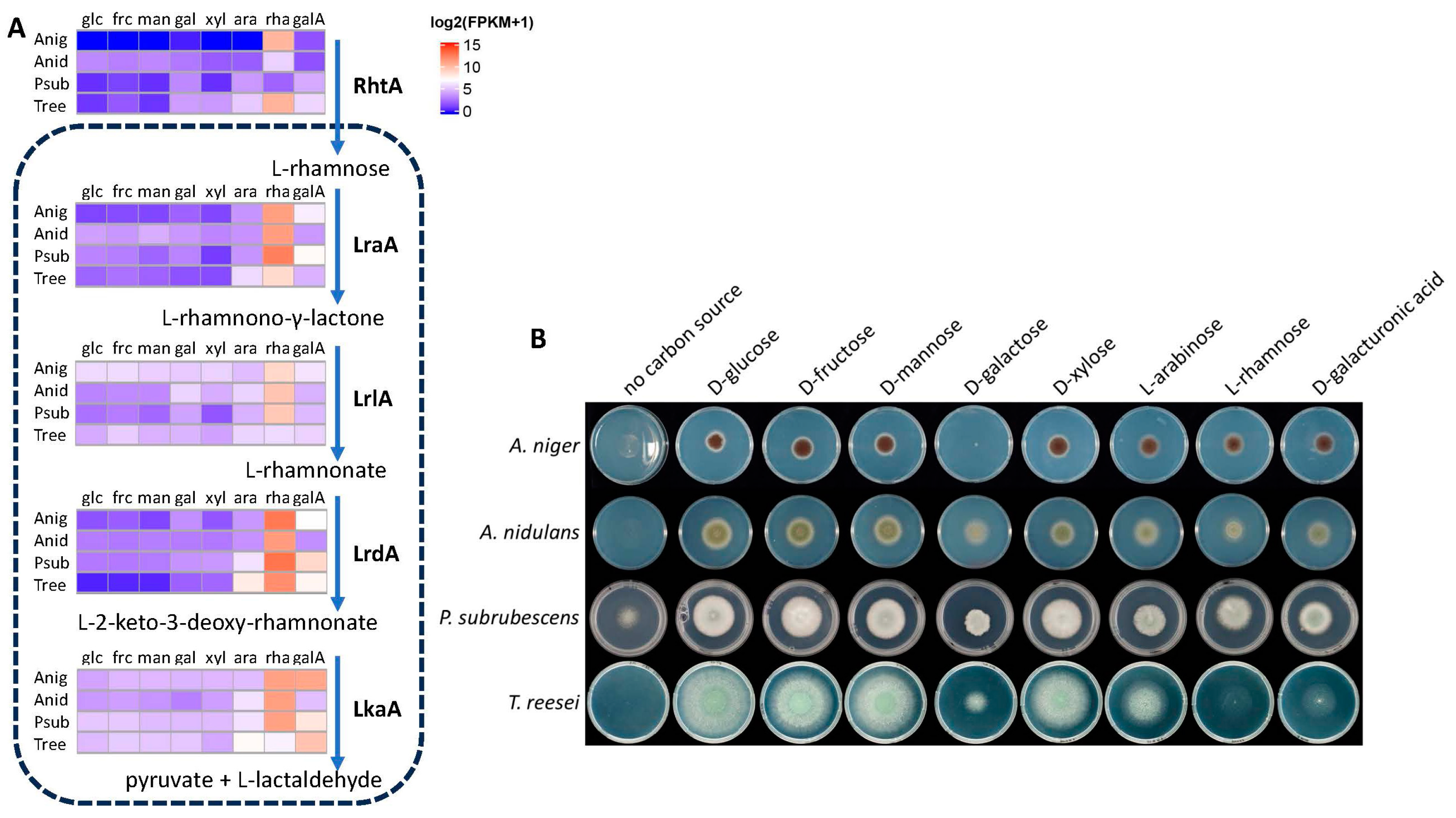
3.2. Expression of L-Rhamnose Catabolic Pathway Genes in Four Fungi Correlates with Their Growth on L-Rhamnose

To obtain a deeper understanding of L-rhamnose catabolism in A. niger and other fungi we performed an expression analysis of all genes involved in this pathway as well as their orthologs in three other fungi. These fungi were chosen as we have identical expression data available for them, generated under the same conditions and using the same methodologies, which minimizes the variation due to the experimental approach. These three fungi were previously chosen as A. nidulans, which is a relative of A. niger and a broadly used model species for fungal biology, while the other two are biotechnologically relevant fungi that are from close (P. subrubescens) or more distant (T. reesei) fungal genera. In addition, we included the expression of the previously identified L-rhamnose transporter-encoding gene RhtA from A. niger [7]) and its orthologs, and we correlated these results with growth profile analysis (Figure 3).
The Eurotiomycete species show similar growth on most of the studied monosaccharides. The only exception is the lack of growth of A. niger on D-galactose which has been shown to be due to non-functional D-galactose transport during germination [31]. The Sordariomycete T. reesei shows poor growth on D-galacturonic acid, while growth on L-rhamnose is almost completely abolished. A possible explanation for this could be the low expression of the lrlA and lkaA orthologs in T. reesei, which is significantly lower than those in most of the other species (Figure 3). As the T. reesei orthologs from lrlA and lkaA are not present in the cluster, other genes may encode these functions in T. reesei. However, the poor growth on L-rhamnose matches better with a poor expression of pathway genes.
Interestingly, the rhtA orthologs of A. nidulans and P. subrubescens show very low expression on L-rhamnose (Figure 3), putting their involvement in L-rhamnose uptake in question. In a recent evaluation of sugar transporters of these species [25], several other candidate L-rhamnose transporters were identified, which may explain the growth of these two species despite low rhtA expression.

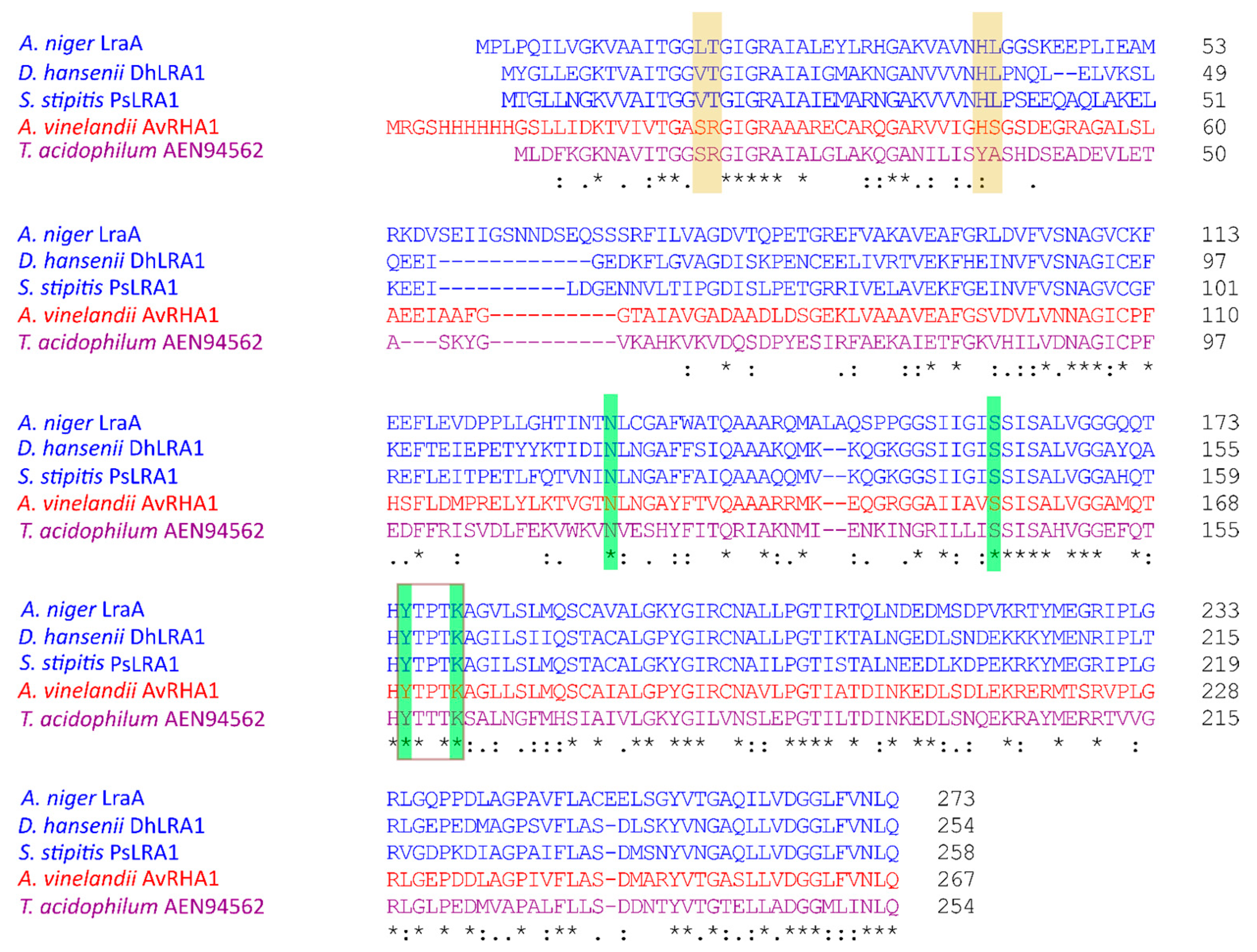
3.3. LraA Is Highly Specific for L-Rhamnose

To determine the substrate specificity of A. niger LraA, we recombinantly produced it in E. coli. The theoretical molecular mass of this enzyme is 28.7 kDa, which matches the value observed on SDS–PAGE after production of the enzyme (Figure S3). The substrate specificity of LraA was tested with 13 substrates (Figure 4), of which two were ketoses and 11 aldoses. LraA is only highly active on L-rhamnose (63.4 ± 5.7 U·mg−1), with a 55-fold lower activity on L-fucose (1.15 ± 0.43 U·mg−1). No activity was observed on any of the other substrates. These two sugars share the same configuration on two chiral centers: (R) on C-3 and (S) on C-5 (Figure 4), suggesting that this may be required for LraA to be active.
As the activity was highly specific for L-rhamnose, and the activity on L-fucose was very low, which would make kinetic values for L-fucose less convincing, we only calculated kinetic constants for the conversion of L-rhamnose. The results of this analysis were compared to the values of previously published microbial L-rhamnose dehydrogenases and are shown in Table 1. LraA has a high affinity for L-rhamnose which is comparable to the bacterial enzyme AvLRA1 [13], which also has a similar turnover as LraA. LraA has a strict dependency on NAD+ as a cofactor, which confirms studies with L-rhamnose dehydrogenases from the yeasts S. stipitis and D. hansenii [13,19,32]. Interestingly, while all characterized fungal L-rhamnose dehydrogenases are only active with NAD+ [13,19,32], bacterial L-rhamnose dehydrogenases are able to use both NAD+ and NADP+, and archaeal enzymes display NADP+ cofactor specificity [29,30]. To identify possible reasons for this, we performed a sequence alignment of five characterized microbial L-rhamnose dehydrogenases using Clustal Omega version 1.2.2 [33], which showed the differences in their N-terminal cofactor-binding motifs (Figure 5). Threonine (T) residue Thr-19 in A. niger LraA is conserved in other fungal L-rhamnose dehydrogenases and indicates NAD+ dependency, while arginine (R) at the corresponding position designates NADP+ preference. Arginine favors the binding of NADP+ due to the presence of a negatively charged phosphate in this cofactor [34,35]. The residues for cofactor-binding were studied in more detail in a bacterial LRA [30]. An R15T mutant of this enzyme modified specificity to NAD+, which is consistent with the presence of a T at this position in A. niger LraA (Figure 5). Of the other three residues implicated in coenzyme specificity in this study [30], A. niger LraA contains an L at the position of S14 and S37 of the bacterial enzyme, while H36 is conserved (Figure 5). The S to L mutations do not seem to alter the cofactor preference to NADP+, as A. niger LraA is strictly NAD+-dependent. The substrate-binding sites identified in the bacterial enzyme [30] are all conserved in A. niger LraA, but the bacterial enzyme has a broader substrate specificity than A. niger LraA, suggesting the involvement of additional residues that limit its substrate range.
L-rhamnose dehydrogenases belong to the large and functionally heterogeneous short-chain dehydrogenases/reductases superfamily (SDR) [36]. A. niger LraA contains conserved motifs for SDR, such as the previously mentioned N-terminal glycine-rich motif TGGLTGIGR (residues 15–23 in LraA), which is a nucleotide-binding Rossmann fold, and a catalytic tetrad (N131-S161-Y175-K179) with Y and K located in the catalytic center and responsible for substrate-binding [37]. All aligned enzymes (Figure 5) belong to the SDR superfamily, and they share the identical catalytic tetrad, which suggests the same mechanism of catalysis. Interestingly, A. niger LraA is more specific than the other L-rhamnose dehydrogenases in that it was only active on L-rhamnose and L-fucose. PsLRA1 from S. stipitis, DhLRA1 from D. hansenii, and AvLRA1 from the bacterium A. vinelandii showed a broader substrate specificity with activity on L-rhamnose, L-lyxose, L-fucose, and L-mannose [17,30], while Rha1 from S. stipitis and the archaeal enzyme were active on L-rhamnose, L-lyxose, and L-mannose [19,29]. These four sugars share the same configuration on C-3, and L-rhamnose, L-fucose, and L-mannose also on C-5 (Figure 4). According to the low number of tested L-rhamnose dehydrogenases, it is difficult to build a strong conclusion on their substrate preferences. It seems, though, that for the substrate recognition by the fungal L-rhamnose dehydrogenases, the C-3 configuration is essential. An expansion in the number of characterized LRA-enzymes from a taxonomically diverse set of organisms in future studies would benefit the assignment of functional residues related to substrate preference.
The oxidation of a sugar to a sugar acid is not very common in Eukaryotes. Enzymes catalyzing those reactions act similarly, although they belong to different protein families. One example is the NAD-dependent dehydrogenase-catalyzed oxidation of D-galactose to D-galactono-lactone in the non-phosphorylated De Ley–Doudoroff pathway in A. niger [38]. This pathway is one of three described for D-galactose metabolism next to the Leloir and oxidoreductive pathways [39,40]. Another example is L-fucose metabolism, which is very similar to L-rhamnose catabolism. L-fucose is converted to L-fuconate, and finally, in the last step of the pathway, to pyruvate and L-lactaldehyde [15]. This may in part explain why these enzymes have higher substrate specificity than, e.g., the PF00248 sugar reductases, as mentioned before.
In conclusion, our results clearly indicate the lack of additional enzymes in A. niger that would be able to compensate for the loss of LraA in L-rhamnose catabolism. The lraA gene shows a highly specific expression pattern for L-rhamnose, and LraA has high biochemical specificity towards L-rhamnose, suggesting that this enzyme is specifically used in the L-rhamnose pathway. L-rhamnose is the least preferred sugar for A. niger as it is only consumed when no other sugars are present [41]. The low preference for this sugar may have prevented the need for the evolutionary development of redundancy of the enzymes of the L-rhamnose pathway. Although this low redundancy would cause a risk in a single mutation preventing use of L-rhamnose as a carbon source, this would not significantly impact growth of A. niger in natural environments. It can be a benefit for metabolic engineering in industrial applications as it provides a simpler approach to produce L-rhamnose-based metabolic intermediates as biochemicals. Future studies should address this and also the impact of introducing alternatives from other organisms that are less specific.

Supplementary Materials

The following supporting information can be downloaded at https://www.mdpi.com/article/10.3390/jof11040301/s1: Figure S1: Alignment of A. niger LraA and 8837; Figure S2: Expression profiles of NRRL3_8837 and lraA; Figure S3: Result of SDS–PAGE (sodium dodecyl sulfate–polyacrylamide gel electrophoresis) of purified LraA; Table S1: List of genomic data sources used for BLASTP search; Table S2: Statistical analysis of the expression of the genes in this study.

Author Contributions

Conceptualization, R.P.d.V.; methodology, A.T., L.X. and M.P.; formal analysis, A.T., L.X. and M.P.; investigation, A.T., L.X. and M.P.; resources, R.P.d.V.; data curation, L.X. and M.P.; writing—original draft preparation, A.T.; writing—review and editing, L.X., M.P., M.R.M. and R.P.d.V.; supervision, R.P.d.V.; funding acquisition, A.T. and R.P.d.V. All authors have read and agreed to the published version of the manuscript.

Funding

A.T. was supported by the PROM Program financed by the Polish National Agency for Academic Exchange. Funding from the Academy of Finland (grant no.: 348443) to M.R.M. is acknowledged. L.X. gratefully acknowledges the China Scholarship Council Scholarship (grant no: 202107720100). M.P. was supported by a Horizon Europe 101094287 (Bioindustries 4.0) to R.P.d.V.

Institutional Review Board Statement

Not applicable.

Informed Consent Statement

Not applicable.

Data Availability Statement

The original contributions presented in this study are included in the article. Further inquiries can be directed to the corresponding author.

Conflicts of Interest

The authors declare no conflicts of interest.

References

  1. Albersheim, P.; Darvill, A.G.; O’Neill, M.A.; Schols, H.A.; Voragen, A.G.J. An hypothesis: The same six polysaccharides are components of the primary cell walls of all higher plants. In Pectins and Pectinases; Visser, J., Voragen, A.G.J., Eds.; Elsevier Science: Amsterdam, The Netherlands, 1996; Volume 14, pp. 47–55. [Google Scholar]
  2. Kaczmarska, A.; Pieczywek, P.M.; Cybulska, J.; Zdunek, A. Structure and functionality of Rhamnogalacturonan I in the cell wall and in solution: A review. Carbohydr. Polym. 2022, 278, 118909. [Google Scholar] [CrossRef] [PubMed]
  3. Mikshina, P.V.; Petrova, A.A.; Faizullin, D.A.; Zuev, Y.F.; Gorshkova, T.A. Tissue-specific rhamnogalacturonan I forms the gel with hyperelastic properties. Biochemistry 2015, 80, 915–924. [Google Scholar] [CrossRef] [PubMed]
  4. Mohnen, D. Pectin structure and biosynthesis. Curr. Opin. Plant Biol. 2008, 11, 266–277. [Google Scholar] [CrossRef] [PubMed]
  5. Khosravi, C.; Kun, R.S.; Visser, J.; Aguilar-Pontes, M.V.; de Vries, R.P.; Battaglia, E. In vivo functional analysis of L-rhamnose metabolic pathway in Aspergillus niger: A tool to identify the potential inducer of RhaR. BMC Microbiol. 2017, 17, 214. [Google Scholar] [CrossRef]
  6. Rodionova, I.A.; Li, X.; Thiel, V.; Stolyar, S.; Stanton, K.; Fredrickson, J.K.; Bryant, D.A.; Osterman, A.L.; Best, A.A.; Rodionov, D.A. Comparative genomics and functional analysis of rhamnose catabolic pathways and regulons in bacteria. Front. Microbiol. 2013, 4, 407. [Google Scholar] [CrossRef]
  7. Sloothaak, J.; Odoni, D.I.; Martins dos Santos, V.A.P.; Schaap, P.J.; Tamayo-Ramos, J.A. Identification of a novel L-rhamnose uptake transporter in the filamentous fungus Aspergillus niger. PLoS Genet. 2016, 12, e1006468. [Google Scholar] [CrossRef]
  8. Wilson, D.M.; Ajl, S. Metabolism of L-rhamnose by Escherichia coli. I. L-rhamnose isomerase. J. Bacteriol. 1957, 73, 410–414. [Google Scholar] [CrossRef]
  9. Akhy, M.T.; Brown, C.M.; Old, D.C. L-Rhamnose utilisation in Salmonella typhimurium. J. Appl. Bacteriol. 1984, 56, 269–274. [Google Scholar] [CrossRef]
  10. Badia, J.; Baldoma, L.; Aguilar, J.; Boronat, A. Identification of the rhaA, rhaB and rhaD gene products from Escherichia coli K-12. FEMS Microbiol. Lett. 1989, 53, 253–257. [Google Scholar] [CrossRef]
  11. Schwartz, N.B.; Abram, D.; Feingold, D.S. L-Rhamnulose 1-phosphate aldolase of Escherichia coli. The role of metal in enzyme structure. Biochemistry 1974, 13, 1726–1730. [Google Scholar] [CrossRef]
  12. MacCabe, A.P.; Ninou, E.I.; Pardo, E.; Orejas, M. Catabolism of L-rhamnose in A. nidulans proceeds via the non-phosphorylated pathway and is glucose repressed by a CreA-independent mechanism. Microb. Cell Factories 2020, 19, 188. [Google Scholar] [CrossRef]
  13. Watanabe, S.; Saimura, M.; Makino, K. Eukaryotic and bacterial gene clusters related to an alternative pathway of nonphosphorylated L-rhamnose metabolism. J. Biol. Chem. 2008, 283, 20372–20382. [Google Scholar] [CrossRef] [PubMed]
  14. Watanabe, S.; Makino, K. Novel modified version of nonphosphorylated sugar metabolism-an alternative L-rhamnose pathway of Sphingomonas sp. FEBS J. 2009, 276, 1554–1567. [Google Scholar] [CrossRef]
  15. Rigo, L.U.; Marechal, L.R.; Vieira, M.M.; Veiga, L.A. Oxidative pathway for L-rhamnose degradation in Pullularia pullulans. Can. J. Microbiol. 1985, 31, 817–822. [Google Scholar] [CrossRef]
  16. Koivistoinen, O.M.; Arvas, M.; Headman, J.R.; Andberg, M.; Penttilä, M.; Jeffries, T.W.; Richard, P. Characterisation of the gene cluster for L-rhamnose catabolism in the yeast Scheffersomyces (Pichia) stipitis. Gene 2012, 492, 177–185. [Google Scholar] [CrossRef]
  17. Riley, R.; Haridas, S.; Wolfe, K.H.; Lopes, M.R.; Hittinger, C.T.; Goker, M.; Salamov, A.A.; Wisecaver, J.H.; Long, T.M.; Calvey, C.H.; et al. Comparative genomics of biotechnologically important yeasts. Proc. Natl. Acad. Sci. USA 2016, 113, 9882–9887. [Google Scholar] [CrossRef] [PubMed]
  18. Chroumpi, T.; Aguilar-Pontes, M.V.; Peng, M.; Wang, M.; Lipzen, A.; Ng, V.; Grigoriev, I.V.; Mäkelä, M.R.; de Vries, R.P. Identification of a gene encoding the last step of the L-rhamnose catabolic pathway in Aspergillus niger revealed the inducer of the pathway regulator. Microbiol. Res. 2020, 234, 126426. [Google Scholar] [CrossRef]
  19. Koivistoinen, O.M.; Hilditch, S.; Voutilainen, S.P.; Boer, H.; Penttilä, M.; Richard, P. Identification in the yeast Pichia stipitis of the first l-rhamnose-1-dehydrogenase gene. FEBS J. 2008, 275, 2482–2488. [Google Scholar] [CrossRef]
  20. Li, J.; Chroumpi, T.; Garrigues, S.; Kun, R.S.; Meng, J.; Salazar-Cerezo, S.; Aguilar-Pontes, M.V.; Zhang, Y.; Tejomurthula, S.; Lipzen, A.; et al. The sugar metabolic model of Aspergillus niger can only be reliably transferred to fungi of its phylum. J. Fungi 2022, 8, 1315. [Google Scholar] [CrossRef]
  21. Chroumpi, T.; Peng, M.; Aguilar-Pontes, M.V.; Muller, A.; Wang, M.; Yan, J.; Lipzen, A.; Ng, V.; Grigoriev, I.V.; Mäkelä, M.R.; et al. Revisiting a ‘simple’ fungal metabolic pathway reveals redundancy, complexity and diversity. Microb. Biotechnol. 2021, 14, 2525–2537. [Google Scholar] [CrossRef]
  22. Mojzita, D.; Vuoristo, K.; Koivistoinen, O.M.; Penttilä, M.; Richard, P. The ‘true’ L-xylulose reductase of filamentous fungi identified in Aspergillus niger. FEBS Lett. 2010, 584, 3540–3544. [Google Scholar] [CrossRef] [PubMed]
  23. Katoh, K.; Asimenos, G.; Toh, H. Multiple alignment of DNA sequences with MAFFT. Methods Mol. Biol. 2009, 537, 39–64. [Google Scholar] [CrossRef] [PubMed]
  24. Tamura, K.; Stecher, G.; Peterson, D.; Filipski, A.; Kumar, S. MEGA6: Molecular Evolutionary Genetics Analysis version 6.0. Mol. Biol. Evol. 2013, 30, 2725–2729. [Google Scholar] [CrossRef]
  25. Xu, L.; Li, J.; Gonzalez Ramos, V.M.; Lyra, C.; Wiebenga, A.; Grigoriev, I.V.; de Vries, R.P.; Mäkelä, M.R.; Peng, M. Genome-wide prediction and transcriptome analysis of sugar transporters in four ascomycete fungi. Bioresour. Technol. 2024, 391, 130006. [Google Scholar] [CrossRef] [PubMed]
  26. Gu, Z.; Eils, R.; Schlesner, M. Complex heatmaps reveal patterns and correlations in multidimensional genomic data. Bioinformatics 2016, 32, 2847–2849. [Google Scholar] [CrossRef]
  27. Love, M.I.; Huber, W.; Anders, S. Moderated estimation of fold change and dispersion for RNA-seq data with DESeq2. Genome Biol. 2014, 15, 550. [Google Scholar] [CrossRef]
  28. Bjellqvist, B.; Hughes, G.J.; Pasquali, C.; Paquet, N.; Ravier, F.; Sanchez, J.C.; Frutiger, S.; Hochstrasser, D. The focusing positions of polypeptides in immobilized pH gradients can be predicted from their amino acid sequences. Electrophoresis 1993, 14, 1023–1031. [Google Scholar] [CrossRef]
  29. Kim, S.M.; Paek, K.H.; Lee, S.B. Characterization of NADP+-specific L-rhamnose dehydrogenase from the thermoacidophilic Archaeon Thermoplasma acidophilum. Extremophiles 2012, 16, 447–454. [Google Scholar] [CrossRef]
  30. Yoshiwara, K.; Watanabe, S.; Watanabe, Y. Crystal structure of l-rhamnose 1-dehydrogenase involved in the nonphosphorylative pathway of l-rhamnose metabolism in bacteria. FEBS Lett. 2021, 595, 637–646. [Google Scholar] [CrossRef]
  31. Fekete, E.; de Vries, R.P.; Seiboth, B.; vanKuyk, P.A.; Sandor, E.; Metz, B.; Kubicek, C.P.; Karaffa, L. D-Galactose uptake is nonfunctional in the conidiospores of Aspergillus niger. FEMS Microbiol. Lett. 2012, 329, 198–203. [Google Scholar] [CrossRef]
  32. Twerdochlib, A.L.; Pedrosa, F.O.; Funayama, S.; Rigo, L.U. L-rhamnose metabolism in Pichia stipitis and Debaryomyces polymorphus. Can. J. Microbiol. 1994, 40, 896–902. [Google Scholar] [CrossRef]
  33. Madeira, F.; Pearce, M.; Tivey, A.R.N.; Basutkar, P.; Lee, J.; Edbali, O.; Madhusoodanan, N.; Kolesnikov, A.; Lopez, R. Search and sequence analysis tools services from EMBL-EBI in 2022. Nucleic Acids Res. 2022, 50, W276–W279. [Google Scholar] [CrossRef] [PubMed]
  34. Pletnev, V.Z.; Weeks, C.M.; Duax, W.L. Rational proteomics II: Electrostatic nature of cofactor preference in the short-chain oxidoreductase (SCOR) enzyme family. Proteins 2004, 57, 294–301. [Google Scholar] [CrossRef]
  35. Tanaka, N.; Nonaka, T.; Nakanishi, M.; Deyashiki, Y.; Hara, A.; Mitsui, Y. Crystal structure of the ternary complex of mouse lung carbonyl reductase at 1.8 A resolution: The structural origin of coenzyme specificity in the short-chain dehydrogenase/reductase family. Structure 1996, 4, 33–45. [Google Scholar] [CrossRef] [PubMed]
  36. Kallberg, Y.; Oppermann, U.; Persson, B. Classification of the short-chain dehydrogenase/reductase superfamily using hidden Markov models. FEBS J. 2010, 277, 2375–2386. [Google Scholar] [CrossRef]
  37. Shah, B.S.; Tetu, S.G.; Harrop, S.J.; Paulsen, I.T.; Mabbutt, B.C. Structure of a short-chain dehydrogenase/reductase (SDR) within a genomic island from a clinical strain of Acinetobacter baumannii. Acta Crystallogr. Sect. F Struct. Biol. Commun. 2014, 70, 1318–1323. [Google Scholar] [CrossRef] [PubMed]
  38. Elshafei, A.M.; Abdel-Fatah, O.M. Evidence for a non-phosphorylated route of galactose breakdown in cell-free extracts of Aspergillus niger. Enzym. Microb. Technol. 2001, 29, 76–83. [Google Scholar] [CrossRef]
  39. Frey, P.A. The Leloir pathway: A mechanistic imperative for three enzymes to change the stereochemical configuration of a single carbon in galactose. FASEB J. 1996, 10, 461–470. [Google Scholar] [CrossRef]
  40. Mojzita, D.; Herold, S.; Metz, B.; Seiboth, B.; Richard, P. L-xylo-3-hexulose reductase is the missing link in the oxidoreductive pathway for D-galactose catabolism in filamentous fungi. J. Biol. Chem. 2012, 287, 26010–26018. [Google Scholar] [CrossRef]
  41. Mäkelä, M.R.; Aguilar-Pontes, M.V.; van Rossen-Uffink, D.; Peng, M.; de Vries, R.P. The fungus Aspergillus niger consumes sugars in a sequential manner that is not mediated by the carbon catabolite repressor CreA. Sci. Rep. 2018, 8, 6655. [Google Scholar] [CrossRef]
Figure 1. The fungal non-phosphorylated L-rhamnose metabolic pathway (A) and the LRA gene cluster in fungi (B). Red = lraA and its orthologs; purple = lrlA and its orthologs; blue = lrdA and its orthologs; green = rhaR transcriptional regulator and its orthologs; grey = genes not belonging to the cluster. Gene numbers below the graphs are obtained from JGI MycoCosm (https://mycocosm.jgi.doe.gov/mycocosm/home, accessed on 15 January 2025) (Table S1).
Jof 11 00301 g001
Figure 2. Phylogenetic analysis of LraA and its homologs in selected fungi and bacteria. Maximum Likelihood tree (500 bootstraps) is a representative of an MAFFT amino acid alignment. Sequences in bold represent characterized enzymes. The numbers behind the species are protein IDs from JGI MycoCosm (https://mycocosm.jgi.doe.gov/mycocosm/home, accessed on 17 February 2025). The numbers in brackets are NCBI accession numbers. Blue, red, orange, and green fonts indicate Eurotiomycete enzymes, Sordariomycete enzymes, characterized yeast enzymes [13,19], and characterized bacterial enzymes [14,29,30], respectively. A. niger LxrA [25] and its ortholog from A. nidulans were used as an outgroup.
Jof 11 00301 g002
Figure 3. Expression (A) and growth (B) profiles of A. niger, A. nidulans, P. subrubescens, and T. reesei. (A) Expression of genes involved in L-rhamnose transport and catabolism. Transcriptome data were obtained from [20,25]. Anig = A. niger, Anid = A. nidulans, Psub = P. subrubescens, Tree = T. reesei, glc = D-glucose, frc = D-fructose, man = D-mannose, xyl = D-xylose, ara = L-arabinose, rha = L-rhamnose, gala = D-galacturonic acid. (B) Growth profiles of A. niger, A. nidulans, P. subrubescens, and T. reesei on several monosaccharides and D-galacturonic acid.
Jof 11 00301 g003
Figure 4. Fisher projections of substrates used in LraA substrate specificity analysis. The structures present the chiral configurations of the different sugars and demonstrate the high specificity of LraA. The red boxes indicate identical chiral centers for L-fucose and L-rhamnose.
Jof 11 00301 g004
Figure 5. Clustal Omega alignment of characterized L-rhamnose dehydrogenases. Blue font = fungal, red font = bacterial, and purple font = archaeal L-rhamnose dehydrogenases. Yellow highlight = conserved Thr or Arg in cofactor-binding motif; green highlight = catalytic tetrad; brown frame = catalytic site. Symbols underneath the alignment mean: * = fully conserved amino acid; : and . similar type of amino acid.
Jof 11 00301 g005
Table 1. Kinetic constants of A. niger LraA and published LRA enzymes for L-rhamnose.
OrganismEnzymeKm
[mM]
kcat
[min−1]
kcat/Km
[mM−1 min−1]
Reference
Aspergillus nigerLraA2.4 ± 0.92149.2 ± 183.4904.8 ± 30.7This study
Scheffersomyces stipitisPsLRA11.7 ± 0.01510.0 ± 20.0885.0 ± 8.0[13]
S. stipitisRha11.5 ± 0.0NMNM[19]
Debaryomyces hanseniiDhLRA19.4 ± 1.12860.0 ± 230.0307.0 ± 11.0[13]
Azotobacter vinelandiiAvLRA12.2 ± 0.15010.0 ± 178.02250.0 ± 51.0[13,30]
2.6 ± 0.1 *2230.0 ± 43.0 *856.0 ± 28.0 *
Thermoplasma acidophilum-0.5 *1341.3 *2915.9 *[29]
* With NADP as a cofactor; NM: not mentioned; values are means ± SD, n = 3; SD for T. acidophilum was not mentioned in the original paper.
Disclaimer/Publisher’s Note: The statements, opinions and data contained in all publications are solely those of the individual author(s) and contributor(s) and not of MDPI and/or the editor(s). MDPI and/or the editor(s) disclaim responsibility for any injury to people or property resulting from any ideas, methods, instructions or products referred to in the content.

