Article

Global Diversity and Taxonomy of Sidera (Hymenochaetales, Basidiomycota): Four New Species and Keys to Species of the Genus

Institute of Microbiology, School of Ecology and Nature Conservation, PO Box 61, Beijing Forestry University, Beijing 100083, China
*
Authors to whom correspondence should be addressed.
J. Fungi 2021, 7(4), 251; https://doi.org/10.3390/jof7040251
Submission received: 19 January 2021 / Revised: 23 March 2021 / Accepted: 23 March 2021 / Published: 26 March 2021
(This article belongs to the Section Fungal Evolution, Biodiversity and Systematics)

Abstract

:
The genus Sidera is a polypore genus with resupinate, white to cream or buff fresh basidioma, poroid or hydnoid hymenophore, a monomitic or dimitic hyphal system with generative hyphae bearing clamp connections, the presence of rosette-like crystals and allantoid to lunate basidiospores. We study the phylogeny and diversity of Sidera herein by using both morphological and molecular methods. Phylogenetic analyses are based on the ITS dataset, the combined 2-locus dataset (5.8S + nLSU) and 7-locus dataset (ITS + nLSU + RPB1 + RPB2 + TEF1 + mtSSU + nSSU) of 15 taxa of Sidera all over the world. Among them, four species are new to science and described and illustrated in this paper, viz. S. inflata, S. malaysiana, S. punctata and S. roseo-bubalina. In addition, three taxa were treated as Sidera vulgaris sensu lato. An identification key of the 14 accepted species of Sidera worldwide is provided.

1. Introduction

The genus Sidera Miettinen & K.H. Larss. (Rickenellaceae and Hymenochaetales) was established by Miettinen and Larsson [1] based on molecular and morphological analyses to accommodate Sidera lunata (Romell ex Bourdot & Galzin) K.H. Larss., S. lowei (Rajchenb.) Miettinen, S. lenis (P. Karst.) Miettinen and S. vulgaris (Fr.) Miettinen, with S. lenis selected as its type [1]. Sidera has a worldwide distribution and is characterized by white-rot, resupinate, white to cream or buff, mostly waxy fresh basidioma, poroid or hydnoid hymenophore, a monomitic or dimitic hyphal system with generative hyphae bearing clamp connections, the presence of rosette-like crystals and allantoid to lunate basidiospores [1,2].
To date, ten species are accepted in the genus, i.e., Sidera lenis (= Physisporus lenis P. Karst., Rabenhorst 1886), S. vulgaris (= Polyporus vulgaris Fr., [3]), S. lowei (Rajchenb.) Miettinen (= Ceriporiopsis lowei Rajchenb., [4]), S. lunata (= Grandinia lunata Romell ex Bourdot & Galzin, [5]), S. minutipora (Rodway & Cleland) Y.C. Dai et al. (= Poria minutipora Rodway & Cleland [6]), S. vesiculosa Rui Du & M. Zhou [7], S. minutissima Y.C. Dai et al. [2], S. parallela Y.C. Dai et al. [2], S. srilankensis Y.C. Dai et al. [2] and S. tenuis Y.C. Dai et al. [2].
During investigations on the diversity of polypores in tropical Asia, four resupinate polypore specimens were collected from China and Malaysia. They were characterized by a monomitic or dimitic hyphal system with generative hyphae bearing clamp connections, the presence of rosette-like crystals and allantoid to lunate basidiospores. These morphological characteristics demonstrated that these specimens may represent species of Sidera. To confirm their affinities, phylogenetic analyses were carried out based on the internal transcribed spacer (ITS) regions, the large subunit nuclear ribosomal RNA gene (nLSU), the largest subunit of RNA polymerase II (RPB1), the second largest subunit of RNA polymerase II (RPB2), the translation elongation factor 1-α gene (TEF1), the small subunit mitochondrial rRNA gene sequences (mtSSU) and the small subunit (nSSU) of nuclear ribosomal RNA gene. As a result, these specimens were found to represent four new terminal lineages in the Sidera clade. In addition, the specimens or literature and sequences of all 14 currently accepted taxa of Sidera were studied, with their morphological characteristics summarized in Table 1. Furthermore, an identification key of accepted species is provided.

2. Materials and Methods

2.1. Morphological Studies

Morphological descriptions were based on field notes and dry herbarium specimens. Microscopic measurements and drawings were made from slide preparations of dry tissues stained with Cotton Blue and Melzer’s reagent following Dai (2010) [10]. Pores were measured by subjectively choosing the straightest line of pores possible and measuring how many fit per mm. The following abbreviations were used: KOH = 5% potassium hydroxide; CB = Cotton Blue; CB– = acyanophilous in Cotton Blue; IKI = Melzer’s reagent; IKI– = neither amyloid nor dextrinoid in Melzer’s reagent; L = mean spore length (arithmetic average of all spores); W = mean spore width (arithmetic average of all spores); Q = variation in the L/W ratios between specimens studied; n (a/b) = number of spores (a) measured from given number of specimens (b). In presenting spore size variation, 5% of measurements were excluded from each end of the range, and this value is given in parentheses. Special color terms follow Anonymous (1969) and Petersen (1996) [11,12]. Herbarium abbreviations follow Thiers (2018) [13]. The studied specimens were deposited at the herbarium of the Institute of Microbiology, Beijing Forestry University (BJFC).

2.2. DNA Extraction, PCR and Sequencing

Total genomic DNA was extracted from dried specimens by a CTAB rapid plant genome extraction kit (Aidlab Biotechnologies Company, Limited, Beijing, China) according to the manufacturer’s instructions with some modifications [14,15]. The ITS regions were amplified with primer pairs ITS5 and ITS4 [16]. The nLSU regions were amplified with primer pairs LR0R and LR7 (https://sites.duke.edu/vilgalyslab/rdna_primers_for_fungi/ (accessed on 24 March 2021)). The mtSSU regions were amplified with primer pairs MS1 and MS2 [16]. The nSSU regions were amplified with primer pairs NS1 and NS4 [16]. The TEF1 regions were amplified with primer pairs EF1-983F and EF1-1567R [17]. The RPB1 regions were amplified with primer pairs RPB1-Af and RPB1-Cf [18]. The RPB2 regions were amplified with primer pairs fRPB2-f5F and bRPB2-7.1R [19].
The PCR procedure for ITS and mtSSU was as follows: initial denaturation at 95 °C for 3 min, followed by 34 cycles at 94 °C for 40 s, 54 °C for ITS and 55 °C for mtSSU for 45 s and 72 °C for 1 min, and a final extension of 72 °C for 10 min. The PCR procedure for nLSU, nSSU and TEF1 was as follows: initial denaturation at 94 °C for 1 min, followed by 34 cycles at 94 °C for 30 s, 50 °C for nLSU and 59 °C for TEF1 for 1 min and 72 °C for 1.5 min, and a final extension of 72 °C for 10 min. The PCR procedure for RPB1 and RPB2 was as follows: initial denaturation at 94 °C for 2 min, followed by 10 cycles at 94 °C for 40 s, 60 °C for 40 s and 72 °C for 2 min, then followed by 37 cycles at 94 °C for 45 s, 55 °C for 1.5 min and 72 °C for 2 min, and a final extension of 72 °C for 10 min. The PCR products were purified and sequenced in the Beijing Genomics Institute, China, with the same primers used in the PCR reactions.

2.3. Phylogenetic Analyses
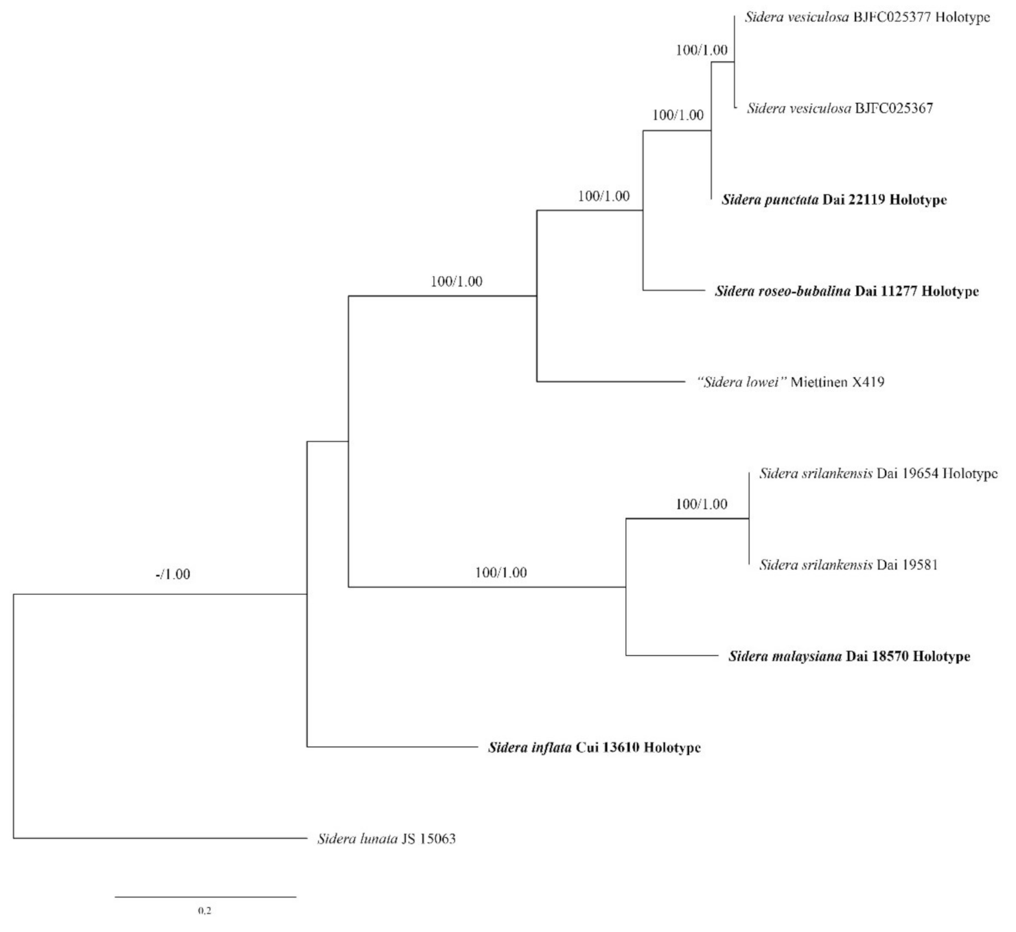
Three combined matrices were reconstructed for phylogenetic analyses as an ITS dataset, a two-gene dataset (5.8S + nLSU) and a 7-gene dataset (ITS + nLSU + RPB1 + RPB2 + TEF1 + mtSSU + nSSU). Phylogenetic analyses were performed with maximum likelihood (ML) and Bayesian Inference (BI) methods. Sequences generated in this study were aligned with additional sequences downloaded from GenBank (Table 2) in MAFFT 6 ([20]; http://mafft.cbrc.jp/alignment/server/ (accessed on 24 March 2021)) using the “G–INS–i” strategy and manually adjusted in BioEdit [21]. Prior to phylogenetic analysis, ambiguous sequences at the start and the end were deleted, and gaps were manually adjusted to optimize the alignment. The data matrix was edited in Mesquite v3.04 software [22]. Sequence alignment was deposited at TreeBase (submission ID 27909, 27910 and 27911). Sequences of Exidia candia Lloyd and Exidiopsis calcea (Pers.) K. Wells outside Hymenochaetales were used as the outgroup [1,12,23] in the combined 2-gene dataset (5.8S + nLSU) and 7-gene dataset (ITS + nLSU + RPB1 + RPB2 + TEF1 + mtSSU + nSSU). The sequence of Sidera lunata (Romell ex Bourdot & Galzin) K.H. Larss. was used as the outgroup in the ITS dataset.
Sequences were also analyzed using maximum likelihood (ML) with RAxML-HPC through the CIPRES Science Gateway ([24]; http://www.phylo.org (accessed on 24 March 2021)). Statistical support values (BS) were obtained using nonparametric bootstrapping with 1000 replicates. The optimal substitution models for the combined dataset were determined using the Akaike Information Criterion (AIC) implemented in MrModeltest 2.2 [25] after scoring 24 models of evolution by PAUP* version 4.0 beta 10 [26]. The selected model applied in the Bayesian phylogenetic inference (BI) analyses and maximum likelihood (ML) analyses was the model GTR + I + G.
The BI analysis was performed with MrBayes 3.2.5 [27]. Four Markov chains were run for two runs from random starting trees for 3 million generations (ITS, 5.8S + nLSU and ITS + nLSU + RPB1 + RPB2 + TEF1 + mtSSU + nSSU) until the split deviation frequency value reached <0.01, and trees were sampled every 1000 generation. The first 25% of the sampled trees were discarded as burn-in, and the remaining ones were used to reconstruct a majority rule consensus tree and calculate Bayesian posterior probabilities (BPP) of the clades.
Branches that received bootstrap support for maximum likelihood (BS), and Bayesian posterior probabilities (BPP) greater than 70% (BS) and 0.95 (BPP) were considered as significantly supported, respectively. FigTree v1.4.2 [28] was used to visualize the resulting tree.

3. Results

3.1. Phylogenetic Analyses

ITS is the most important locus for barcoding fungi, especially at the generic and species levels. However, the genetic variation inside Sidera is significant and, for example, the alignment of the whole ITS region is difficult among Sidera polypores [1]. Therefore, we used the most stable and conservative portion of ITS (5.8S) and LSU to analyse the phylogenetic relationship of Sidera species (Figure 1). The concatenated 5.8S + nLSU dataset contained sequences from 46 fungal specimens representing 15 Sidera taxa (three are treated as S. vulgaris sensu lato; (Table 2). Sequences of Exidia candia and Exidiopsis calcea were used as the outgroup [1,12,23]. The best model for the 5.8S + nLSU dataset estimated and applied in the Bayesian analysis was GTR + I+G. The ML analysis resulted in the best tree (Figure 1). BI analyses resulted in almost identical tree topologies compared to the ML analysis, with an average standard deviation of split frequencies of 0.009930 (BI). Additionally, only the ML tree is presented along with the support values from the BI analyses. Our new species and their related species are nested in Group A and Group B. Thus, we used the whole ITS region to analyse the phylogenetic relationship of new Sidera species with their closely related species (Figure 2).
The ITS dataset contained sequences from ten fungal specimens representing eight Sidera taxa (four new species and another four Sidera taxa). The sequence of Sidera lunata was used as the outgroup. The best model for the ITS dataset estimated and applied in the Bayesian analysis was GTR + I + G. The ML analysis resulted in the best tree (Figure 2). BI analyses resulted in almost identical tree topologies compared to the ML analysis, with an average standard deviation of split frequencies of 0.001361 (BI). Thus, only the ML tree is presented along with the support values from the BI analyses.
The concatenated ITS + nLSU + RPB1 + RPB2 + TEF1 + mtSSU + nSSU dataset contained sequences from 46 fungal specimens representing 15 Sidera taxa (three were treated as S. vulgaris sensu lato; Table 2). Sequences of Exidia candia and Exidiopsis calcea were used as the outgroup [1,12,23]. The best model for the ITS + nLSU + RPB1 + RPB2 + TEF1 + mtSSU + nSSU dataset estimated and applied in the Bayesian analysis was GTR + I+G. The ML analysis resulted in the best tree (Figure 3). BI analyses resulted in almost identical tree topologies compared to the ML analysis, with an average standard deviation of split frequencies of 0.009821 (BI). Additionally, only the ML tree is presented along with the support values from the BI analyses.
The phylogenetic trees (Figure 2 and Figure 3) revealed four new and independent lineages represented by our specimens, indicating that they are phylogenetically distinct from the species currently known in the genus. In addition, another three taxa were treated as S. vulgaris sensu lato, although they formed four independent lineages too.

3.2. Taxonomy

1.
Sidera inflata Z.B. Liu & Y.C. Dai, sp. nov. Figure 4 and Figure 5.
MycoBank number: MB 838380.
Etymology—Inflata (Lat.): referring to the species having inflated skeletal hyphae in KOH.
Type—China, Hainan Province, Baisha County, Yinggeling Nature Reserve, on rotten angiosperm wood, 17 November 2015, B.K. Cui 13610 (holotype BJFC 028475).
Basidioma—Annual, resupinate, soft corky when fresh and dry, up to 4 cm long, 1.5 cm wide and less than 1 mm thick at center; pore surface white to buff and shiny when fresh, becoming cream to buff yellow and shiny when dry; sterile margin distinct, white, cottony, thinning out; pores angular, 9–10 per mm; dissepiments thin, lacerate; subiculum very thin to almost absent; tubes concolorous with the poroid surface, less than 1 mm long.
Hyphal structure—Hyphal system dimitic; generative hyphae bearing clamp connections; skeletal hyphae dominant; all hyphae IKI–, CB–, skeletal hyphae obviously becoming swollen in KOH.
Subiculum—Generative hyphae hyaline, thin-walled, occasionally branched, 1–2 μm in diameter; skeletal hyphae thick-walled with a wide lumen, rarely branched, interwoven, 2–4 μm diameter; rosette-like crystals rarely present.
Tubes—Generative hyphae hyaline, thin-walled, occasionally branched, 1–2 μm in diameter; skeletal hyphae thick-walled with a wide lumen, occasionally branched, interwoven, usually covered by abundant fine thorn-like crystals at dissepiment edge, 2–4 μm diameter; rosette-like crystals abundant, 2–8 μm in diameter; cystidia absent; cystidioles present, fusoid, hyaline, thin-walled, basally swollen, with a sharp or often hyphoid neck, 13–15 × 2.5–3 μm; basidia clavate, hyaline, bearing four sterigmata and a basal clamp connection, 7–8 × 3.5–4.5 μm; basidioles similar in shape to basidia, but slightly shorter.
Basidiospores—Allantoid, hyaline, thin-walled, smooth, occasionally with one or two guttules, IKI–, CB–, (2.9–)3–3.3(–3.5) × (0.8–)0.9–1.1 μm, L = 3.03 μm, W = 1 μm, Q = 3.03 (n = 60/1).
Notes—Sidera inflata was found in China, and the species is characterized by annual, resupinate basidioma with a white to buff fresh pore surface, which becomes cream to buff-yellow upon drying; angular pores (9–10 per mm); a dimitic hyphal system; skeletal hyphae at dissepiment edge bearing abundant fine thorn-like crystals; skeletal hyphae in all structures obviously swelling in KOH; and allantoid basidiospores measuring 3–3.3 × 0.9–1.1 μm. Morphologically, S. inflata can be distinguished from other species in Sidera by its skeletal hyphae at the dissepiment edge, which are usually covered by abundant fine thorn-like crystals. Although S. inflata clustered together with the S. vulgaris sensu lato (Dai 21057 and Dai 22151) with a moderate support (87% BS, 0.94 BPP; Figure 3), the latter taxon had perennial basidioma, and its skeletal hyphae were unchanged in KOH.
2.
Sidera malaysiana Z.B. Liu & Y.C. Dai, sp. Nov. Figure 6 and Figure 7
MycoBank number: MB 838381.
Etymology—Malaysiana (Lat.): referring to the species occurring in Malaysia.
Type—Malaysia, Selangor, Forest Research Institute of Malaysia, on rotten angiosperm wood, 15 April 2018, Y.C. Dai 18570 (holotype BJFC 026859).
Basidioma—Annual, resupinate, very difficult to separate from substrate, soft corky when fresh and dry, up to 4 cm long, 2 cm wide and less than 1 mm thick at center; pore surface white to cream when fresh and dry; sterile margin indistinct; pores round to angular, 9–11 per mm; dissepiments thin, entire; subiculum very thin to almost absent; tubes white, less than 1 mm long.
Hyphal structure—Hyphal system dimitic; generative hyphae bearing clamp connections; skeletal hyphae dominant; all hyphae IKI–, CB–, skeletal hyphae slightly swollen in KOH.
Subiculum—Generative hyphae hyaline, thin-walled, rarely branched, 1–2 μm in diameter; skeletal hyphae dominant, occasionally branched, interwoven, usually covered by abundant irregular crystals and fine thorn-like crystals, 1.5–3.5 μm diameter; rosette-like crystals occasionally present, 3–5 μm in diameter.
Tubes—Generative hyphae hyaline, thin-walled, rarely branched, 1–2.5 μm in diameter, dominating at dissepiment edges; skeletal hyphae thick-walled, occasionally branched, interwoven, 1.5–3.5 μm diameter; rosette-like and irregular rhomboidal crystals abundant at dissepiment edges; cystidia absent; cystidioles present, fusoid, hyaline, thin-walled, basally swollen, with a sharp or often hyphoid neck, 9–13 × 2.2–3.5 μm; basidia clavate, hyaline, with a basal clamp connection and four sterigmata, 7.8–15 × 3–4.3 μm; basidioles similar in shape to basidia, but slightly shorter.
Basidiospores—Lunate, hyaline, thin-walled, smooth, usually with two or three guttules, IKI–, CB–, (2.8–)2.9–3.2(–3.3) × 1–1.2(–1.4) μm, L = 3.16 μm, W = 1.12 μm, Q = 2.82 (n = 60/1).
Notes—Sidera malaysiana was found in Malaysia, and the species is characterized by annual, resupinate basidioma with a white to cream pore surface, round to angular pores (9–11 per mm), a dimitic hyphal system, skeletal hyphae in all structures become slightly swollen in KOH, subicular skeletal hyphae bearing rosette-like crystals, irregular crystals and fine thorn-like crystals and lunate basidiospores measuring 2.9–3.2 × 1–1.2 μm. Morphologically, S. malaysiana can be distinguished from other species in Sidera by its subicular skeletal hyphae, which are usually covered by abundant irregular crystals and fine thorn-like crystals. S. malaysiana is closely related to S. srilankensis in our phylogeny (100% BS, 1.00 BPP; Figure 2), but S. malaysiana is different from S. srilankensis due to its smaller pores (9–11 per mm vs. 6–8 per mm, [2]) and skeletal hyphae in all structures becoming slightly swollen in KOH, while skeletal hyphae are unchanged in KOH in S. srilankensis. S. malaysiana resembles S. parallela due to its white fresh pore surface. However, S. malaysiana differs from S. parallela due to its smaller pores (9–11 per mm vs. 6–8 per mm, [2]). In addition, tramal hyphae are parallel in S. parallela, while they are interwoven in S. malaysiana. Additionally, they are phylogenetically distant (Figure 2).
3.
Sidera punctata Z.B. Liu & Y.C. Dai, sp. nov. Figure 8 and Figure 9.
MycoBank number: MB 838384.
Etymology—Punctata (Lat.): referring to the species having cushion-shaped basidioma.
Type—China, Hainan Province, Haikou, Guanlanhu, on rotten angiosperm wood, 18 November 2020, Y.C. Dai 22119 (holotype BJFC 036011).
Basidioma—Annual, resupinate, soft corky when fresh and dry, up to 13 cm long, 4 cm wide and less than 1 mm thick at center; pore surface white to cream when fresh, becoming cinnamon buff to white when dry; sterile margin distinct, white, cottony, thinning out; pores round, 8–9 per mm; dissepiments thin, entire; subiculum very thin to almost absent; tubes darker than the poroid surface, less than 1 mm long.
Hyphal structure—Hyphal system monomitic; generative hyphae bearing clamp connections; all hyphae IKI–, CB–, unchanged in KOH.
Subiculum—Generative hyphae hyaline, thin-walled, often branched, interwoven, 1.5–2.5 μm in diameter; rosette-like crystals frequently present, 2–7 μm in diameter.
Tubes—Generative hyphae hyaline, thin-walled, sometimes branched, loosely interwoven, 1.5–3 μm in diameter, some hyphae, especially hyphae at dissepiment edge with swollen tips which are globose, bottle-shaped or irregularly elongated; rosette-like crystals occasionally present; cystidia absent; cystidioles absent; basidia clavate, hyaline, bearing four sterigmata and a basal clamp connection, 12.5–13.5 × 3.5–4.5 μm; basidioles similar in shape to basidia, but slightly shorter.
Basidiospores—Allantoid to lunate, hyaline, thin-walled, smooth, occasionally with one to three guttules, IKI–, CB–, (3.5–)3.8–4.8(–5) × 1–1.3(–1.4) μm, L = 4.27 μm, W = 1.21 μm, Q = 3.53 (n = 60/1).
Notes—Sidera punctata was discovered in China, and the species is characterized by annual, resupinate basidioma with a white to cream fresh pore surface which becomes cinnamon-buff to white upon drying, round pores (8–9 per mm), a monomitic hyphal system, and allantoid to lunate basidiospores measuring 3.8–4.8 × 1–1.3 μm. Phylogenetically, S. punctata is close to S. vesiculosa (100% BS, 1.00 BPP; Figure 2) and it is similar to S. vesiculosa by annual, resupinate basidioma and some generative hyphae with swollen tips, but S. punctata has a cinnamon-buff to white dry pore surface, while the pore surface is cream upon drying in S. vesiculosa. Above all, S. punctata can be distinguished from other species in Sidera by its rosette-like crystals are more abundant in subiculum than in tubes, while rosette-like crystals are more abundant in tubes than in subiculum in other members of the genus.
4.
Sidera roseo-bubalina Z.B. Liu & Y.C. Dai, sp. nov. Figure 10 and Figure 11.
MycoBank number: MB 838382.
Etymology—Roseo-bubalina (Lat.): referring to the species having pinkish buff hymenophore.
Type—China, Henan Province, Neixiang County, Baotianman Nature Reserve, on rotten wood of Quercus, 22 September 2009, Y.C. Dai 11277 (holotype BJFC 007251).
Basidioma—Annual, resupinate, soft corky when dry, up to 7 cm long, 3 cm wide, and less than 1 mm thick at center; pore surface pinkish buff to yellowish brown when dry; sterile margin distinct, white, cottony, thinning out; pores round, 6–7 per mm; dissepiments thin, entire to lacerate; subiculum very thin to almost absent; tubes concolorous with the poroid surface, less than 1 mm long.
Hyphal structure—Hyphal system monomitic; generative hyphae bearing clamp connections; all hyphae IKI–, CB–, unchanged in KOH.
Subiculum—Generative hyphae hyaline, thin-walled, often branched, interwoven, 2–3 μm in diameter; rosette-like crystals occasionally present.
Tubes—Generative hyphae hyaline, thin-walled, often branched, interwoven, 2–3.5 μm in diameter, some hyphae, especially hyphae at dissepiment edge with swollen tips which are globose, bottle-shaped or irregularly elongated; rosette-like crystals abundant, 4–6 μm in diameter; cystidia absent; cystidioles present, fusoid, hyaline, thin-walled, basally swollen, with a sharp or often hyphoid neck, 15–22 × 4–4.5 μm; basidia clavate, hyaline, bearing four sterigmata and a basal clamp connection, 8.5–11 × 4.5–5 μm; basidioles similar in shape to basidia, but slightly shorter.
Basidiospores—Lunate, hyaline, thin-walled, smooth, occasionally with one to four guttules, IKI–, CB–, (3.5–)3.9–4.5(–4.8) × (0.7–) 0.8–1 μm, L = 4.22 μm, W = 0.93 μm, Q = 4.53 (n = 60/1).
Notes—Sidera roseo-bubalina was discovered in China, and the species is characterized by annual, resupinate basidioma with a pinkish buff to yellowish brown dry pore surface, round pores (6–7 per mm), a monomitic hyphal system and lunate basidiospores measuring 3.9–4.5 × 0.8–1 μm. Morphologically, S. roseo-bubalina and S. lowei share annual, resupinate basidioma and similar pores (6–7 per mm in S. roseo-bubalina vs. 6–8 per mm in S. lowei [1]), but S. roseo-bubalina has lunate basidiospores, which are usually <1 μm wide, while S. lowei has allantoid basidiospores, which are usually >1 μm wide. Phylogenetically, S. roseo-bubalina is close to S. punctata and S. vesiculosa (100% BS, 1.00 BPP; Figure 2), and these three species share annual and resupinate basidioma, and some generative hyphae with swollen tips, but S. roseo-bubalina has a pinkish buff to yellowish brown dry pore surface, while the pore surface is cream upon dryied in S. vesiculosa and cinnamon-buff to white when dried in S. punctata. In addition, S. roseo-bubalina differs from the other two species due to its larger pores (6–7 per mm in S. roseo-bubalina, 8–9 per mm in S. punctata and 7–9 per mm in S. vesiculosa, [7]).
5.
Sidera vulgaris sensu lato
Specimens examined—Belarus, Brestskaya Voblasts, Belavezhskaya Pushcha National Park, on rotten wood of Picea, 19 October 2019, Y.C. Dai 21057 (BJFC 032716 and MSK). China, Guangxi Province, Guiping County, Xishan Scenic Spot, on rotten wood of Pinus, 25 December 2020, Y.C. Dai 22151 (BJFC 036043); Shannxi Province, Zhashui County, Niubeiliang Forest Park, on fallen angiosperm trunk, 16 September 2013, B.K. Cui 11216 (BJFC 015331). USA, Connecticut, New Haven, West Rock Park, on rotten stump of Pinus, 15 July 2012, Y.C. Dai 12730 (BJFC 013037).
Previously, the ten species of Sidera, viz. S. lenis, S. lowei, S. lunata, S. minutipora, S. minutissima, S. parallela, S. srilankensis, S. tenuis, S. vesiculosa and S. vulgaris were described or transferred to the genus. In this paper, S. inflata, S. malaysiana, S. punctata and S. roseo-bubalina were described as new to science, and they have resupinate, white to cream or buff fresh basidiocarps; a monomitic or dimitic hyphal system with generative hyphae bearing clamp connections; the presence of rosette-like crystals; and allantoid to lunate basidiospores. These characteristics fit well with the generic concept of Sidera. Thus far, 14 species are accepted in Sidera, and a key of accepted species is provided below.
A key to species of Sidera worldwide
1. Hymenium grandinioid to odontioidS. lunata
1. Hymenium poroid2
2. Hyphal system monomitic3
2. Hyphal system dimitic6
3. Basidiospores mostly <1 μm in width4
3. Basidiospores mostly >1 μm in width5
4. Pores 7–9 per mm; basidiospores 2.9–3.7 μm longS. vesiculosa
4. Pores 6–7 per mm; basidiospores 3.9–4.5 μm longS. roseo-bubalina
5. Pores 6–8 per mm; cystidioles present, some branched S. lowei
5. Pores 8–9 per mm; cystidioles absentS. punctata
6. Basidiospores >1.5 μm in widthS. lenis
6. Basidiospores <1.5 μm in width7
7. Skeletal hyphae becoming swollen in KOH8
7. Skeletal hyphae almost unchanged in KOH10
8. Pores 5–7 per mm; basidiospores 3.7–4.3 μm longS. minutipora
8. Pores 9–11 per mm; basidiospores 2.9–3.3 μm long9
9. Basidiospores allantoid, skeletal hyphae distinctly swollen in KOHS. inflata
9. Basidiospores lunate, skeletal hyphae slightly swollen in KOHS. malaysiana
10. Tramal hyphae parallel along the tubesS. parallela
10. Tramal hyphae interwoven11
11. Generative hyphae at dissepiments even S. srilankensis
11. Generative hyphae at dissepiments with swollen tips12
12. Basidiospores <3.6 μm longS. vulgaris
12. Basidiospores >3.8 μm long13
13. Sterile margin distinct, fimbriate; basidiospore Length/width <4S. minutissima
13. Sterile margin indistinct to almost absent; basidiospore Length/width >4S. tenuis

4. Discussion

As is well known, the genus Sidera is challenging for distinguishing species morphologically, and two species, S. lenis and S. vulgaris, were recognized before phylogenetical analyses.
Poria krawtzewii Pilát was treated as a synonym of P. lenis (P. Karst.) Sacc. [29], but it is different from the S. lenis complex because of ellipsoid spores [30]. We summarize another five synonyms of Sidera lenis (Index Fungorum and MycoBank): Poria lunulispora Pilát (type from Siberia), P. chakasskensis Pilát (type from Siberia), P. earlei Murrill (type from Jamaica), P. tenuipora Murrill (type from Jamaica) and P. montana Murrill (type from Jamaica).
Poria earlei, P. montana and P. tenuipora were described from the Caribbean (Jamaica) (Murrill [31,32]). The type of specimens of three species were studied by Niemelä and Dai [8]. They found that P. earlei and P. montana are conspecific, and the species is perennial, resupinate and has small pores (7–9 per mm). Sidera punctata and S. vesiculosa have similar pores (8–9 per mm in S. punctata vs. 7–9 per mm in S. vesiculosa [7]) to P. tenuipora as well, but the former two species have a monomitic hyphal system, while P. tenuipora has a dimitic hyphal system. S. vesiculosa differs from S. punctata due to the presence of vesicular cells of swollen hyphae in the subiculum.
Poria chakasskensis and P. lunulispora were described from Siberia [33,34]. Kotlaba and Pouzar [30,35] found that P. chakasskensis has basidiospores measuring 5.5–8.5 × 2–2.4 μm and represents Ceriporia purpurea (Fr.) Donk; P. lunulispora was collected on the wood of Pinus, and is true Diplomitoporus lenis (= Sidera lenis). Hence, P. chakasskensis and P. lunulispora are different from our newly described species.
Du et al. [2] summarized three synonyms of Sidera vulgaris (Index Fungorum and MycoBank): Boletus papyraceus Schrank, B. proteus Bolton and B. cellulosus O.F. Müll, and all of them were originally described from Europe. In addition, the specimen Ryvarden 37198 from New Zealand was named S. vulgaris by Miettinen and Larsson too [1]. In the present paper, we found three taxa with similar morphologies to S. vulgaris, but we did not study the types of the above-mentioned taxa, and no sequence data are available for them. Although our three taxa formed three distinct lineages in our phylogenies (Figure 3), we refrained from describing these as new, and treated them as S. vulgaris sensu lato in this paper (Table 2). The description of these species is the subject of a forthcoming paper.
Ceriporiopsis lowei (= Sidera lowei) was described from Northern Brazil [4]. The specimen Miettinen X426 (Ryvarden 38817) from New Zealand clustered together with a Venezuelan specimen Miettinen X419, and both specimens were considered as Sidera lowei by Miettinen and Larsson [1]. We did not examine the type of Sidera lowei; thus, we regard Miettinen X419 and Miettinen X426 as “S. lowei”.
The sequence of OTU1581 is from GenBank. We failed to obtain specimens of the taxa, but it formed a distinct lineage within the Sidera clade, so we treated OTU1581 as Sidera sp. temporarily here.
Polypores are an extensively studied group of Basidiomycota, and more than 1500 species have been recorded in the world [36,37,38,39,40,41,42]. Molecular phylogenies have demonstrated that more new taxa exist in the world [43,44,45,46,47], and more crypto species will be confirmed after molecular analyses of some traditional species in sensu lato. Thus, in order to understand the diversity, phylogeny and evolution of the fungi, future taxonomic and phylogenetic work should be based on both molecular and morphological characteristics.

Author Contributions

Conceptualization, Y.-C.D. and Z.-B.L.; methodology, Z.-B.L.; performing the experiment, Z.-B.L.; formal analysis, Z.-B.L.; validation, Z.-B.L., Y.-C.D., M.Z. and Y.Y.; resources, Y.-C.D.; writing—original draft preparation, Z.-B.L.; writing—review and editing, Y.-C.D.; visualization, Z.-B.L.; supervision, Y.-C.D. and Y.Y.; project administration, Y.Y.; funding acquisition, Y.-C.D. All authors have read and agreed to the published version of the manuscript.

Funding

The research is supported by the National Natural Science Foundation of China (Project No. 31870007; 31530002).

Institutional Review Board Statement

Not applicable.

Informed Consent Statement

Not applicable.

Data Availability Statement

Publicly available datasets were analyzed in this study. This data can be found here: https://www.ncbi.nlm.nih.gov/; https://www.mycobank.org/page/Simple%20names%20search; http://purl.org/phylo/treebase, submission ID 27909, 27910 and 27911.

Acknowledgments

Not applicable.

Conflicts of Interest

The authors declare that there are no conflict of interest.

References

  1. Miettinen, O.; Larsson, K.H. Sidera. A new genus in Hymenochaetales with poroid and hydnoid species. Mycol. Prog. 2011, 10, 131–141. [Google Scholar] [CrossRef]
  2. Du, R.; Wu, F.; Gate, G.M.; Dai, Y.C.; Tian, X.M. Taxonomy and phylogeny of Sidera (Hymenochaetales, Basidiomycota): Four new species and keys to species of the genus. MycoKeys 2020, 68, 115–135. [Google Scholar] [CrossRef]
  3. Fries, E.M. Systema Mycologicum. Berlingius Lindae. 1821, 1, 1–520. [Google Scholar]
  4. Rajchenberg, M. Type studies of Polyporaceae (Aphyllophorales) described by J. Rick. Nord. J. Bot. 1987, 7, 553–568. [Google Scholar] [CrossRef]
  5. Bourdot, H.; Galzin, A. Hyménomycètes de France. I. Hétèrobasidiés. Bull. Soc. Mycol. Fr. 1909, 25, 15–36. [Google Scholar]
  6. Rodway, L.; Cleland, J.B. Notes on the genus Poria No. 3. Pap. Proc. R. S. Tasman. 1929, 17, 7–24. [Google Scholar]
  7. Du, R.; Wang, L.; Zhou, M.; Chen, J.J. A new species of Sidera (Hymenochaetales, Basidiomycota) from tropical Asia. Phytotaxa. 2019, 387, 165–171. [Google Scholar] [CrossRef]
  8. Niemelä, T.; Dai, Y.C. Polypore Skeletocutis lenis and its sib S. vulgaris. Ann. Bot. Fenn. 1997, 34, 133–140. [Google Scholar]
  9. Niemelä, T. Polypores, lignicolous fungi. Norrlinia 2005, 13, 1–320, (In Finnish, with English summary). [Google Scholar]
  10. Dai, Y.C. Hymenochaetaceae (Basidiomycota) in China. Fungal Divers. 2010, 45, 131–343. [Google Scholar] [CrossRef]
  11. Anonymous. Flora of British Fungi. Colour Identification Chart; Her Majesty’s Stationery Office: London, UK, 1969. [Google Scholar]
  12. Petersen, J.H. Farvekort. The Danish Mycological Society’ s Colour-Chart; Foreningen til Svampekundskabens Fremme: Greve, Italy, 1996; pp. 1–6. [Google Scholar]
  13. Thiers, B. Index Herbariorum: A Global Directory of Public Herbaria and Associated Staff; New York Botanical Garden’s Virtual Herbarium: New York, NY, USA, 2018; Available online: http://sweetgum.nybg.org/science/ih/ (accessed on 15 January 2021).
  14. Cui, B.K.; Li, H.J.; Ji, X.; Zhou, J.L.; Song, J.; Si, J.; Yang, Z.L.; Dai, Y.C. Species diversity, taxonomy and phylogeny of Polyporaceae (Basidiomycota) in China. Fungal Divers. 2019, 97, 137–392. [Google Scholar] [CrossRef]
  15. Shen, L.L.; Wang, M.; Zhou, J.L.; Xing, J.H.; Cui, B.K.; Dai, Y.C. Taxonomy and phylogeny of Postia. Multi–gene phylogeny and taxonomy of the brown–rot fungi: Postia and its related genera. Pers. Mol. Phylogeny Evol. Fungi 2019, 42, 101–126. [Google Scholar] [CrossRef] [Green Version]
  16. White, T.J.; Bruns, T.; Lee, S.; Taylor, J. Amplification and direct sequencing of fungal ribosomal RNA genes for phylogenetics. In PCR Protocols: A Guide to Methods and Applications; Innis, M.A., Gefand, D.H., Sninsky, J.J., White, J.T., Eds.; Academic Press: San Diego, CA, USA, 1990; pp. 315–322. [Google Scholar] [CrossRef]
  17. Rehner, S.A.; Buckley, E. A Beauveria phylogeny inferred from nuclear ITS and EF1-alpha sequences: Evidence for cryptic diversification and links to Cordyceps teleomorphs. Mycologia 2005, 97, 84–98. [Google Scholar] [CrossRef]
  18. Matheny, P.B.; Liu, Y.J.; Ammirati, J.F.; Hall, B.D. Using RPB1 sequences to improve phylogenetic inference among mushrooms (Inocybe, Agaricales). Am. J. Bot. 2002, 89, 688–698. [Google Scholar] [CrossRef] [PubMed]
  19. Matheny, P.B. Improving phylogenetic inference of mushrooms with RPB1 and RPB2 nucleotide sequences (Inocybe, Agaricales). Molec. Phylogenet. Evolut. 2005, 35, 1–20. [Google Scholar] [CrossRef] [PubMed]
  20. Katoh, K.; Toh, H. Recent developments in the MAFFT multiple sequence alignment program. Brief. Bioinform. 2008, 9, 286–298. [Google Scholar] [CrossRef] [Green Version]
  21. Hall, T.A. Bioedit: A user–friendly biological sequence alignment editor and analysis program for Windows 95/98/NT. Nucleic Acids Symp. Ser. 1999, 41, 95–98. [Google Scholar]
  22. Maddison, W.P.; Maddison, D.R. Mesquite: A Modular System for Evolutionary Analysis. Available online: https://www.mesquiteproject.org/ (accessed on 24 March 2021).
  23. Yuan, Y.; Ji, X.H.; Wu, F.; He, S.H.; Chen, J.J. Two new Gloeoporus (Polyporales, Basidiomycota) from tropical China. Nova Hedwig. 2016, 103, 169–183. [Google Scholar] [CrossRef]
  24. Miller, M.A.; Holder, M.T.; Vos, R.; Midford, P.E.; Liebowitz, T.; Chan, L.; Hoover, P.; Warnow, T. The CIPRES Portals. CIPRES. Available online: http://www.phylo.org/sub_sections/portal (accessed on 4 August 2009).
  25. Nylander, J.A.A. MrModeltest v2. Program Distributed by the Author; Evolutionary Biology Centre: Uppsala, Sweden, 2004. [Google Scholar]
  26. Swofford, D.L. PAUP: Phylogenetic Analysis using Parsimony Version 4.0b10; Sinauer Associates: Sunderland, MA, USA, 2002. [Google Scholar]
  27. Ronquist, F.; Teslenko, M.; Mark, P.; Avres, D.L.; Darling, A.; Höhna, S.; Larget, B.; Liu, L.; Suchard, M.A.; Huelsenbeck, J.P. MrBayes3.2: Efficient Bayesian phylogenetic inference and model choice, across a large model space. Syst. Biol. 2012, 61, 539–542. [Google Scholar] [CrossRef] [Green Version]
  28. Rambaut, A. Molecular Evolution, Phylogenetics and Epidemiology. FigTree ver. 1.4 Software. Available online: http://tree.bio.ed.ac.uk/software/figtree/ (accessed on 24 March 2021).
  29. Pilát, A. Polyporaceae. In Atlas des Champognons de l’s Europe; Kavina, C., Pilát, A., Eds.; Privately Published: Prague, Czech Republic, 1936–1942; pp. 1–624,374.
  30. Kotlaba, F.; Pouzar, Z. Type studies of polypores described by A. Pilát. Ceska Mykol. 1991, 45, 91–97. [Google Scholar]
  31. Murrill, W.A. Light-colored resupinate polypores 1. Mycologia 1920, 12, 77–92. [Google Scholar] [CrossRef]
  32. Murrill, W.A. Light-colored resupinate polypores 2. Mycologia 1920, 12, 299–308. [Google Scholar] [CrossRef]
  33. Pilát, A. Additamenta ad floram Sibiriae Asiaeque orientalis mycologicam. Bull. Soc. Mycol. Fr. 1933, 49, 256–339. [Google Scholar]
  34. Pilát, A. Additamenta ad floram Sibiriae Asiaeque orientalis mycologicam. Pars Tertia. Bull. Soc. Mycol. Fr. 1935, 51, 351–426. [Google Scholar]
  35. Kotlaba, F.; Pouzar, Z. Type studies of polypores described by A. Pilát-III. Ceská Mycol. 1989, 43, 36–44. [Google Scholar]
  36. Ryvarden, L.; Johansen, I. A Preliminary Polypore Flora of East Africa; Fungiflora: Oslo, Norway, 1980; pp. 1–636. [Google Scholar]
  37. Buchanan, P.K.; Ryvarden, L. An annotated checklist of polypore and polypore-like fungi recorded from New Zealand. N. Z. J. Bot. 2000, 38, 265–323. [Google Scholar] [CrossRef] [Green Version]
  38. Núñez, M.; Ryvarden, L. East Asian polypores 2. Synop. Fungorum. 2001, 14, 165–522. [Google Scholar]
  39. Ryvarden, L. Neotropical polypores 1. Synop. Fungorum. 2004, 19, 1–229. [Google Scholar]
  40. Dai, Y.C. Polypore diversity in China with an annotated checklist of Chinese polypores. Mycoscience 2012, 53, 49–80. [Google Scholar] [CrossRef]
  41. Zhou, L.W.; Nakasone, K.K.; Burdsall, H.H.; Ginns, J.; Vlasák, J.; Miettinen, O.; Spirin, V.; Niemelä, T.; Yuan, H.S.; He, S.H.; et al. Polypore diversity in North America with an annotated checklist. Mycol. Prog. 2016, 15, 771–790. [Google Scholar] [CrossRef]
  42. Ryvarden, L.; Melo, I. Poroid fungi of Europe, 2nd edition. In Synop. Fungorum; Ryvarden, L., Melo, I., Eds.; Fungiflora AS: Oslo, Norway, 2017; pp. 1–430. [Google Scholar]
  43. Decock, C.; Yombiyeni, P.; Memiaghe, H. Hymenochaetaceae from the Guineo-Congolian rainforest: Phylloporia flabelliforma sp. nov. and Phylloporia gabonensis sp. nov., two undescribed species from Gabon. Cryptogam. Mycol. 2016, 36, 449–467. [Google Scholar] [CrossRef]
  44. Miettinen, O.; Ryvarden, L. Polypore genera Antella, Austeria, Butyrea, Citripora, Metuloidea and Trulla (Steccherinaceae, Polyporales). Ann. Bot. Fenn. 2016, 53, 157–172. [Google Scholar] [CrossRef]
  45. Ji, X.H.; He, S.H.; Chen, J.J.; Si, J.; Wu, F.; Zhou, L.W.; Vlasák, J.; Tian, X.M.; Dai, Y.C. Global diversity and phylogeny of Onnia (Hymenochaetaceae) species on gymnosperms. Mycologia 2017, 109, 27–34. [Google Scholar] [CrossRef] [PubMed]
  46. Spirin, V.; Vlasák, J.; Miettinen, O. Studies in the Antrodia serialis group (Polyporales, Basidiomycota). Mycologia 2017, 109, 217–230. [Google Scholar] [CrossRef] [PubMed]
  47. Wu, F.; Dai, S.J.; Vlasák, J.; Spirin, V.; Dai, Y.C. Phylogeny and global diversity of Porodaedalea, a genus of gymnosperm pathogens in the Hymenochaetales. Mycologia 2019, 111, 40–53. [Google Scholar] [CrossRef]
Figure 1. Phylogeny of Sidera and related species generated by ML analyses based on combined 5.8S + nLSU sequences. Branches are labeled with maximum likelihood bootstrap >50% and Bayesian posterior probabilities >0.90, respectively. New species are indicated in bold.
Jof 07 00251 g001
Figure 2. Phylogeny of Sidera new species and related species generated by ML analyses based on ITS sequences. Branches are labeled with maximum likelihood bootstrap >50% and Bayesian posterior probabilities >0.90, respectively. New species are indicated in bold.
Jof 07 00251 g002
Figure 3. Phylogeny of Sidera and related species generated by ML analyses based on combined ITS + nLSU + RPB1 + RPB2 + TEF1 + mtSSU + nSSU sequences. Branches are labeled with maximum likelihood bootstrap >50% and Bayesian posterior probabilities >0.90, respectively. New species are indicated in bold.
Jof 07 00251 g003
Figure 4. A basidioma of Sidera inflata (from the holotype Cui 13610). Scale bar = 10 mm.
Jof 07 00251 g004
Figure 5. Microscopic structures of Sidera inflata (holotype, Cui 13610). (a) Basidiospores. (b) Basidia and basidioles. (c) Cystidioles. (d) Hyphae from subiculum. (e) Hyphae from trama. (f) Hyphae at dissepiment edge. Drawing by Meng Zhou.
Jof 07 00251 g005
Figure 6. A basidioma of Sidera malaysiana (from the holotype Dai 18570). Scale bar = 10 mm.
Jof 07 00251 g006
Figure 7. Microscopic structures of Sidera malaysiana (holotype, Dai 18570). (a) Basidiospores. (b) Basidia and basidioles. (c) Cystidioles. (d) Hyphae from subiculum. (e) Hyphae from trama. (f) Hyphae at dissepiment edge. Drawing by Meng Zhou.
Jof 07 00251 g007
Figure 8. A basidioma of Sidera punctata (from the holotype Dai 22119). Scale bar = 10 mm.
Jof 07 00251 g008
Figure 9. Microscopic structures of Sidera punctata (holotype, Dai 22119). (a) Basidiospores. (b) Basidia and basidioles. (c) Hyphae from subiculum. (d) Hyphae from trama. (e) Hyphae at dissepiment edge. Drawing by Meng Zhou.
Jof 07 00251 g009
Figure 10. A dry basidioma of Sidera roseo-bubalina (from the holotype Dai 11277). Scale bar = 10 mm.
Jof 07 00251 g010
Figure 11. Microscopic structures of Sidera roseo-bubalina (holotype, Dai 11277). (a) Basidiospores. (b) Basidia and basidioles. (c) Cystidioles. (d) Hyphae from subiculum. (e) Hyphae from trama. (f) Hyphae at dissepiment edge. Drawing by Meng Zhou.
Jof 07 00251 g011
Table 1. The main characteristics of Sidera species. Pore and basidiospore sizes partly from Du et al. (2020) [2], Rajchenberg (1987) [4], Du et al. (2019) [7], Niemelä and Dai (1997) [8] and Niemelä (2005) [9].
SpeciesGrowing HabitHymenophoreHyphal SystemCystidiolesSkeletal Hyphae in KOHSpores ShapeSpore Size (µm)
S. inflataAnnualPoroid, 9–10/mmDimiticPresentSwollenAllantoid3–3.3 × 0.9–1.1
S. lenisPerennialPoroid, 4–6/mmDimiticPresentSwollenAllantoid to lunate3.9–4.9 × 1.5–2
S. loweiAnnualPoroid, 6–8/mmMonomiticPresent, some branched-Allantoid3.5–5 × 1–1.2
S. lunataAnnualHydnoid, 8–9/mmMonomiticPresent-Allantoid2.5–3.8 × 1.6–1.9
S. malaysianaAnnualPoroid, 9–11/mmDimiticPresentSwollenLunate2.9–3.2 × 1–1.2
S. minutiporaAnnualPoroid, 5–7/mmDimiticPresentSwollenAllantoid3.7–4.3 × 1–1.3
S. minutissimaAnnualPoroid, 7–9/mmDimiticPresentAlmost unchangedAllantoid3.8–4.4 × 0.9–1.3
S. parallelaAnnualPoroid, 6–8/mmDimiticPresentAlmost unchangedLunate2.8–3.3 × 0.9–1.2
S. punctataAnnualPoroid, 8–9/mmMonomiticAbsent-Allantoid to lunate3.8–4.8 × 1–1.3
S. roseo-bubalinaAnnualPoroid, 6–7/mmMonomiticPresent-Lunate3.9–4.5 × 0.8–1
S. srilankensisAnnualPoroid, 6–8/mmDimiticPresentAlmost unchangedLunate3.5–4 × 1–1.3
S. tenuisAnnualPoroid, 8–10/mmDimiticPresentAlmost unchangedAllantoid4.2–5 × 0.8–1
S. vesiculosaAnnualPoroid, 7–9/mmMonomiticPresent-Allantoid to lunate2.9–3.7 × 0.6–1
S. vulgarisPerennialPoroid, 6–8/mmDimiticPresent, some branchedAlmost unchangedAllantoid to lunate2.9–3.6 × 0.9–1.4
New species are shown in bold.
Table 2. Information for the sequences used in this study.
SpeciesSpecimen No.LocalityGenBank Accession No.
ITSnLSURPB1RPB2TEF1mtSSUnSSU
Ceriporiopsis aneirinaMUAF 888Czech RepublicEU340895EU368503 EU368504
Contumyces rosellaRedhead 7501 U66452U66452
Exidia candidaVS 8588RussiaKY801871KY801896 KY801920
Exidiopsis calceaMW 331CanadaAF291280AF291326
Gloeoporus dichrousKHL 11173NorwayEU118627EU118627
G. hainanensisDai 15253ChinaKU360402KU360408
Globulicium hiemaleHjm 19007SwedenDQ873595DQ873595 DQ873594
Hyphodermella poroidesDai 12045ChinaKX008367KX011852
Odonticium romelliiMurdoch 38FinlandMF319073MF318929
Oxyporus corticolaKHL 13217EstoniaDQ873641DQ873641 DQ873640
Repetobasidium conicumKHL 12338USADQ873647DQ873647 DQ873646
Resinicium furfuraceumKHL 11738FinlandDQ873648DQ873648
Rickenella melleaLamoure 74 U66438U66438
Skvortzovia pinicolaKHL 12224USADQ873637DQ873637 DQ873636
Sidera inflataCui 13610ChinaMW198480 * MW418088 *
S. lenisMiettinen 11036FinlandFN907914FN907914
S. loweiMiettinen X419VenezuelaFN907917FN907917
S. loweiMiettinen X426New ZealandFN907919FN907919
S. lunataJS 15063NorwayDQ873593DQ873593
S. malaysianaDai 18570MalaysiaMW198481 *MW192007 * MW424992 *
S. minutiporaGates FF257AustraliaFN907922FN907922
S. minutiporaCui 16720AustraliaMN621349MN621348MW526261 *MW505865 *MW446248 *MW424986 *MW418078 *
S. minutissimaDai 19529Sri LankaMN621352MN621350MW526262 * MW424994 *MW418079 *
S. minutissimaDai 18471AChinaMW198482 *MW192008 *
S. parallelaCui 10346ChinaMK346145 MW424991 *MW418082 *
S. parallelaCui 10361ChinaMK346144
S. punctataDai 22119ChinaMW418438 *MW418437 * MW418085 *
S. roseo-bubalinaDai 11277ChinaMW198483 * MW418081 *
S. sp.OTU1581JapanMT594995
S. srilankensisDai 19581Sri LankaMN621345MN621347MW526265 * MW427604 *MW424993 *MW418089 *
S. srilankensisDai 19654Sri LankaMN621344MN621346 MW505868 *MW427602 *MW424989 *MW418087 *
S. tenuisDai 18697AustraliaMK331865MK331867MW526264 *MW505866 *MW427600 *MW424988 *MW418083 *
S. tenuisDai 18698AustraliaMK331866MK331868 MW505867 *MW427601 *MW424985 *MW418084 *
S. vesiculosaBJFC025367SingaporeMH636565MH636567
S. vesiculosaBJFC025377SingaporeMH636564MH636566
S. vulgaris sensu latoRyvarden 37198New ZealandFN907918FN907918
S. vulgaris sensu latoDai 12730USAMW198478 *
S. vulgaris sensu latoDai 21057BelarusMW198484 *MW192009 * MW505869 *MW427603 *MW424987 *MW418090 *
S. vulgaris sensu latoDai 22151ChinaMW477794 *MW474965 * MW477795 *
S. vulgaris sensu latoCui 11216ChinaMW198485 *
Skeletocutis amorphaMiettinen 11038FinlandFN907913FN907913
S. chrysellaMiettinen 9472FinlandFN907916FN907916
S. lilacinaHHB 10522spUSAKY948834KY948894
S. yuchengiiFBCC 1132ChinaKY953045KY953045KY953143 KY953109KY953142
S. yunnanensisDai 15709ChinaKU950434KU950436MW526263 * MW427605 *MW424990 *MW418080 *
S. odoraL 13763spCanadaKY948830KY948893KY949046
* Newly generated sequences for this study. New species are shown in bold.
Publisher’s Note: MDPI stays neutral with regard to jurisdictional claims in published maps and institutional affiliations.

