Article

The Change in Fatty Acids and Sugars Reveals the Association between Trifoliate Orange and Endophytic Fungi

1
College of Horticulture and Gardening, Yangtze University, Jingzhou 434025, China
2
ICAR-Central Citrus Research Institute, Nagpur 440033, Maharashtra, India
3
Department of Chemistry, Faculty of Science, University of Hradec Králové, 50003 Hradec Králové, Czech Republic
4
Botany and Microbiology Department, College of Science, King Saud University, P.O. Box 2460, Riyadh 11451, Saudi Arabia
5
Plant Production Department, College of Food and Agricultural Sciences, King Saud University, P.O. Box 2460, Riyadh 11451, Saudi Arabia
6
Department of Botany, Swami Shraddhanand College, University of Delhi, Delhi 110036, India
*
Author to whom correspondence should be addressed.
J. Fungi 2021, 7(9), 716; https://doi.org/10.3390/jof7090716
Submission received: 28 July 2021 / Revised: 19 August 2021 / Accepted: 27 August 2021 / Published: 31 August 2021
(This article belongs to the Special Issue Plant and Fungal Interactions)

Abstract

:
Endophytes have the ability to improve plant nutrition alongside their agronomic performance, among which arbuscular mycorrhizal fungi provide the most benefits to their host. Previously, we reported for the first time that an arbuscular mycorrhizal-like fungus Piriformospora indica had the ability to colonize roots of trifoliate orange (Poncirus trifoliata) and conferred positive effects on nutrient acquisition. Present study showed the changes in fatty acids and sugars to unravel the physiological and symbiotic association of trifoliate orange with P. indica and an arbuscular mycorrhizal fungus, Funneliformis mosseae singly or in combination. All the endophytic fungi collectively increased fructose, glucose, and sucrose content in leaves and roots, along with a relatively higher increase with P. indica inoculation than with F. mosseae alone or dual inoculation. Treatment with P. indica increased the concentration of part unsaturated fatty acids such as C18:3N6, C20:2, C20:3N6, C20:4N6, C20:3N3, C20:5N3, C22:1N9, and C24:1. Additionally, P. indica induced the increase in the concentration of part saturated fatty acids such as C6:0, C8:0, C13:0, C14:0, and C24:0. F. mosseae hardly changed the content of fatty acids, except for increase in C14:0 and C20:5N3. Double inoculation only reduced the C21:0, C10:0, C12:0, C18:3N3, and C18:1 content and increased the C20:5N3 content. These endophytic fungi up-regulated the root PtFAD2, PtFAD6, PtΔ9, and PtΔ15 gene expression level, coupled with a higher expression of PtFAD2 and PtΔ9 by P. indica than by F. mosseae. It was concluded that P. indica exhibited a stronger response, for sugars and fatty acids, than F. mosseae on trifoliate orange. Such results also reveal the Pi (an in vitro culturable fungus) as a bio-stimulator applying to citriculture.

1. Introduction

Terrestrial plants are reported to form mutual symbioses with endophytic fungi in roots, without causing any damage to host plants [1,2]. Amongst endophytic fungi, arbuscular mycorrhizal (AM) fungi predominantly occurring in the soil have the ability to colonize roots of more than 72% of terrestrial plants and, thus, form arbuscular mycorrhizas in the roots [3]. In such a mutualistic relationship, host plants provide the required substances to AM fungi for their growth and, in reciprocation, AM fungi facilitate water and nutrient acquisition of the host [4,5]. As a result, host plants offer mycorrhizal-C pool in the roots, which inevitably affects the extent of carbohydrate accumulation in the plant roots [6]. However, plant roots also utilize a substantial amount of carbohydrates for their growth and functions [7,8]. Within endophytes, the AM-like fungus, Piriformospora indica (Pi) of the Sebacinales, Basidiomycota, is extensively reported to colonize many plants for their growth promotion and imparting tolerance against varied biotic and abiotic stress [9,10,11,12].
The role of fatty acids (FAs) in response to mycorrhization for imparting drought tolerance to host plants is well documented [5]. FAs, as a main component of cell membrane, are divided into saturated fatty acids (SFAs) and unsaturated fatty acids (UFAs). Recent studies showed that AM fungi obtained FAs from host plants [4,13], further supporting the operation of the AM-regulated lipid pathway into the arbuscule-containing cells of host plants [13]. In Rhizophagus irregularis-mediated responses, myristic acid (C14:0) was used as a source of FAs for the hyphal growth of germinated spores, and C14:0 also induced the formation of secondary spores with features of a branched absorbing structure and dense coils [14]. On the other hand, (S)-12-methyltetradecanoic acid (a methyl-branched chain FA) released from bacterial cultures induced the branching of hyphae originated from mother spores leading to the formation of secondary spores, and palmitoleic acid (a SFA type) aided in the production of a higher number of secondary spores than the bacterial (S)-12-methyltetradecanoic acid [3]. One of our earlier studies showed that AM fungal colonization distinctly increased contents of UFAs in trifoliate orange by up-regulating the root FA desaturase (FAD) gene (e.g., FAD2 and FAD6) under either optimum water or soil water deficit conditions [5], suggesting further that the composition and saturation of FAs are governed by AM fungi under soil drought conditions. Hua et al. [15] demonstrated that the host metabolic compounds and metabolite pathways were reprogrammed following symbiosis upon inoculation with endophytic fungi. However, little is known about the effects of endophytic fungi on the composition and saturation of FAs synthesized by host plants.
Citrus is globally one of the most important fruit crops and displays lesser root hair growth in the field, but is considered highly dependent on AM fungi [16]. In vitro propagation of AM fungi is the major drawback, while Pi isolated in 1996 displayed an in vitro propagation ability without roots being able to colonize citrus [10,17].
The present study aims to study the response of trifoliate orange (Poncirus trifoliata L. Raf., a widely used citrus rootstock) to being colonized by an AM fungus (Funneliformospora mosseae, Fm) versus an AM-like fungus Pi: (i) changes in composition and saturation levels of FAs; (ii) changes in root sugar; (iii) expression levels of FA desaturase genes. Such work would reveal the symbiotic association between trifoliate orange and endophytic fungi.

2. Materials and Methods

2.1. Endophytic Fungi

Fm was provided by the Bank of Glomales in China. The AM fungus strain was propagated using Trifolium repens L. as a host plant for 16 weeks under potted conditions. The 80 g inoculum of Fm containing spores (20 spores/g), mycelia, and AM fungi-colonized root fragments was added into the pot as AM fungal treatment at the time of transplanting of trifoliate orange.
Pi was propagated following the protocol of Yang et al. [17]. A 5 × 5 mm fungal mass was activated twice at 30 °C for 7 days on solid culture medium of potato dextrose agar. The activated mycelium was proliferated on the liquid culture medium of potato dextrose broth under dark for 7 days. Spore suspensions were collected, washed with 0.05% Tween 20 in ddH2O, and centrifuged at 4000× g/min for 7 min. The collected spores were mixed with distilled water at a ratio of 1:20, and the number of spores was determined colorimetrically at 600 nm, reaching the concentration of 3.27 × 10⁸ CFU/mL.

2.2. Experimental Setup

The three 4-leaf-old trifoliate orange seedlings grown in autoclaved sands were transplanted into a 1.4 L pot, in which autoclaved (0.11 MPa, 2 h) growth substrates (1.5 kg) consisting of coarse-textured loam soil and sand (5:3, v/v) were supplied. A single Fm inoculum of 80 g per pot was applied at transplanting, and a single inoculation of Pi with 20 mL spore suspension was performed. The dual inoculation of Fm and Pi consisted of an 80 g inoculum of Fm and 20 mL spore suspension of Pi together during plant transplanting. Non-fungi-treated plants (control) were inoculated without any of the two fungi materials as the control. These treated plants were grown for 140 days under controlled environmental conditions, as previously described by Yang et al. [17].
The experiment was, therefore, arranged with four inoculations with single Fm, single Pi, dual Fm + Pi, and non-fungi control. Each treatment had ten replicates in a completely randomized design.

2.3. Measurement of Root Fungal Colonization and Plant Biomass

The roots were removed from the growing medium. A segment taken from the middle part of the root was carefully washed, cut into 1 cm long root segments, cleared with 10% KOH solutions at 95 °C for 1.5 h, followed by bleaching with 10% H2O2 solution for 15 min, acidified with 0.2 mol/L HCl for 1 h, and, finally, stained with 0.05% (w/v) trypan blue in lactophenol for 3 min [18]. The root fungal colonization degree was computed as the percentage of fungal colonized root number versus total observed root number as suggested by Yang et al. [17]. The shoots and roots were dried in oven (75 °C, 48 h) and weighed to record their biomass.

2.4. Measurement of Sugars and FA Contents

The fructose, glucose, and sucrose concentrations of leaves and roots were determined using the colorimetric method suggested by Wu et al. [6]. Root FAs were extracted and quantified according to the protocol outlined by Wu et al. [5], with an external standard with the NU-CHEK-PREP 37 fatty acid methyl ester-mixed solution (1000 μg/mL).

2.5. Relative Expression of Root FA Desaturase Genes

Root samples were ground with liquid nitrogen, and total RNA was extracted with the help of Plant RNA Extraction Kit (Takara Bio. Inc.). The RNA was reverse-transcribed into cDNA using a PrimeScriptTM RT reagent kit with cDNA eraser. The sequences of four FA desaturase genes, including FA desaturase 2 (PtFAD2), FA desaturase 6 (PtFAD6), Δ9 FA desaturase (PtΔ9), and Δ15 FA desaturase (PtΔ15) were obtained based on the sweet orange database (http://citrus.hzau.edu.cn/, 10 October 2020), and specific primer sequences (Table 1) were designed using the Primer Premier 5.0. The reaction system and condition of qRT-PCR were carried out according to the method described by Wu et al. [5]. Relative gene expression was worked out according to the method suggested by Livak and Schmittgen [19], based on β-actin as a house-keeping gene [20]. The measured transcripts were normalized to the relative expression value in non-fungi-inoculated plants.

2.6. Data Analysis

The data generated by the experiment were analyzed with one-factor analysis of variance through the SAS software. Duncan’s multiple range tests were used at the 0.05% level to compare the significant difference amongst the treatments.

3. Results

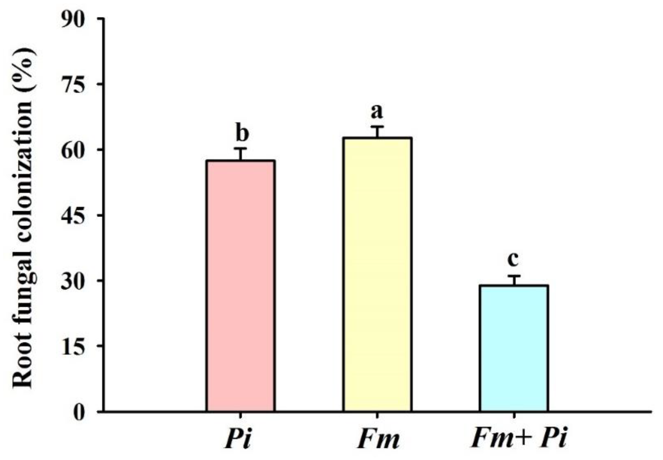
3.1. Root Colonization after Inoculation of Endophytic Fungi

No fungal colonization was found in the non-inoculated trifoliate orange seedlings, while fungal colonization (28.82–62.68%) was present in the roots of Fi, Pi, and Fi + Pi-inoculated seedlings, respectively (Figure 1b–d and Figure 2). The root fungal colonization was observed in a decreasing order of Fm > Pi > Fm + Pi (Figure 2).

3.2. Effects of Endophytic Fungi on Biomass Production

Single as well dual fungal inoculation collectively improved shoot and root biomass (Figure 1a and Figure 3a,b). Compared with the control (non-fungi-inoculated treatment), endophytic fungi inoculation significantly improved the shoot biomass by 77%, 83%, and 74%, respectively, with Pi, Fm, and the combination of Fm + Pi (Figure 3a). The root biomass was increased by 87%, 73%, and 65% with Pi, Fm, and Fm + Pi (Figure 3b), respectively, compared with the control. In brief, the improvement of shoot biomass did not show significant differences between endophytic fungal treatments, but the differences of the root biomass improvement were significant, showing a decreasing trend with Pi > Fm > Fm + Pi.

3.3. Effects of Endophytic Fungi on Sugar Concentrations of Leaves and Roots

Pi increased the concentration of sucrose, glucose, and fructose by 41%, 159%, and 16%, respectively, in leaves with the corresponding increase of 124%, 65%, and 23% in roots over the control (Table 2), while Fm increased sucrose, glucose, and fructose concentrations by 25%, 135%, and 11%, respectively, in leaves with the corresponding increase of 37%, 46%, and 21% in roots, compared with the control. The dual inoculation increased sucrose, glucose, and fructose concentrations in leaves by 13%, 29%, and 10%, respectively, with the corresponding increase of 104%, 50%, and 15% in roots, compared with the control. In addition, the increase in sugar was distinctly higher with Pi than with Fm and Fm + Pi, independent of leaves and roots.

3.4. Effects of Endophytic Fungi on FAs Contents in Roots

Fifteen SFAs and fourteen UFAs were identified in roots of trifoliate orange, and C11:0, C14:1, C15:1, C22:2, and C22:6N3 were not identified (Table 3). The major SFAs in trifoliate orange comprised of C16:0 and C18:0, accounting for 57–58% and 36–37%, respectively, out of total SFAs, while the important UFAs in roots were C18:1, C18:2, and C18:3N3, amounting to 8–18%, 55–56%, and 15%, respectively, out of total UFAs in roots.
Single inoculation with Pi significantly increased part UFAs concentrations such as C18:3N6, C20:2, C20:3N6, C20:4N6, C20:3N3, C20:5N3, C22:1N9, and C24:1 by 64%, 51%, 103%, 35%, 27%, 190%, 41%, and 69%, while it did not affect C16:1, C17:1, C18:1, C18:2, C18:3N3, and C20:1 concentrations. Single inoculation with Fm had no significant effect on the concentration of UFAs except that the C20:5N3 of UFAs, which was significantly increased by 158%. In addition, the double inoculation of Fm and Pi triggered the increase in C20:5N3 by 331% and the decrease in C18:1 by 28% and C18:3N3 by 22%, coupled with no change in other UFAs.
As for SFAs, single inoculation with Pi distinctly increased the C6:0, C8:0, C13:0, C14:0, and C24:0 concentration by 45%, 39%, 12%, 31%, and 36%, respectively, whereas it had no significant effect on other SFAs. Fm inoculation did not have any effects on all SFA concentrations, except the significant increase in C14:0 by 17%. Dual inoculation of Fm and Pi only reduced the C10:0, C12:0, and C21:0 concentration by 25%, 17%, and 18%, respectively. The content of all detected SFAs (except C17:0) almost showed a decreasing trend of Pi > Fm > Fm + Pi among endophytic fungal inoculation treatments.

3.5. Effects of Endophytic Fungi on Relative Expression of FA Desaturase Genes

The expression level of PtΔ9, PtFAD2, PtFAD6, and PtΔ15 was increased by 413%, 298%, 508%, and 298%, respectively, with Pi inoculation compared with the control (Figure 4). Fm inoculation significantly induced the expression of PtΔ9, PtFAD2, PtFAD6, and PtΔ15 by 304%, 309%, 202%, and 308%, respectively, and dual inoculation of Fm + Pi registered a 103%, 198%, 108%, and 250% higher expression level of PtΔ9, PtFAD2, PtFAD6, and PtΔ15, compared with the control. In brief, the relative expression levels of PtΔ9, PtΔ15, PtFAD2, and PtFAD6 genes showed a decreasing trend of Pi > Fm > Fm + Pi > control among the four treatments.

4. Discussion

In this study, we observed that three endophytic fungi inoculations displayed a differential magnitude of root fungal colonization in the decreasing order of Fm > Pi > Fm + Pi, indicating that trifoliate orange is preferentially colonized by Fm. Two endophytic fungi (Fm and Pi) colonized the roots of trifoliate orange when inoculated separately, but their dual inoculation of Fm + Pi failed to register the same magnitude of root fungal colonization. It has been documented that the proliferation of Pi in roots needs dead cells of host plants [21]. Thus, when Fm and Pi were co-present in the roots of trifoliate orange, the cell death induced by Pi may have reduced the colonization of living cells by Fm. More work needs to be started around the change of cells colonized by both AM fungi and Pi.
AM fungi are reported to acquire most of the carbohydrates, especially hexose, from the host root, transforming them into typical fungal carbohydrates, since AM fungi operate through a typical process of mutualistic symbiosis, depending upon the host plant to be able to transport the photosynthetic products for their multiplication [22]. In this process, AM fungi only absorb and utilize small molecules as glucose, while sucrose and other macromolecular substances need to be cleaved into glucose and fructose before being absorbed and utilized by mycorrhizal plants [23]. Our study showed that a higher sucrose content in the leaves of mycorrhizal plants was beneficial for the downward movement of sucrose in phloem tissue. Thus, sucrose in roots of mycorrhizal plants is accountable to the cleavage through catabolic enzymes into hexose for onward mycorrhizal development, or the development of a more effective root morphology of mycorrhizal plants, thereby, consuming more sucrose for respiration as a pre-requisite of AM fungi to be more efficacious [24]. Fm inoculation increased the glucose content, due to the existence of a better carbon pool in mycorrhizal roots requiring a comparatively higher amount of sucrose to move from leaves to roots; thus, reducing the sucrose cleavage in leaves and increasing the glucose content as a cause-and-effect relationship in the process of AM symbiosis. Amongst different carbohydrates, sucrose has the ability to stimulate a lateral root formation [25]. Glucose, as an important regulator of plant growth and development, participates in the gibberellin and cytokinin signaling pathway [26]. The higher the carbohydrate content in the roots of endophytic fungi-inoculated trifoliate orange, the more beneficial it is to the better growth of roots. Among Fm and Pi, we found a higher increase in fructose, glucose, and sucrose in leaves and roots by Pi inoculation than by Fm inoculation or dual inoculation, indicating that Pi triggered a stronger demand for C and C pools in roots than the two fungi inoculations. In order to understand the relationship between endophytic fungi and carbohydrate metabolism, we need to further bring the role of endogenous hormones, especially abscisic acid, gibberellins, and cytokinin, to unravel the underlying mechanisms operating in the host-endophytic fungi interaction.
Our results showed an increase in many UFA concentrations such as C18:3N6, C20:2, C20:3N6, C20:4N6, C20:3N3, C20:5N3, C22:1N9, and C24:1 after inoculation with single Pi, thereby leading to a higher degree of unsaturation in the Pi-inoculated plants, which would maintain a higher cell membrane fluidity and lower innate immunity against exogenous fungal colonization [27]. In contrast, Fm hardly changed the content of any UFAs (except the increase in C20:5N3). Thus, this appears to imply that roots of trifoliate orange were more susceptible to Pi colonization than Fm colonization. AM fungi (Fm) did not change any SFA contents (except the increase in C14:0), and Pi inoculation also increased five SFA (including C14:0) contents. A common feature of both fungi was the increased content of the SFA C14:0 as well as the UFA C20:5N3. Sugiura et al. [14] also reported that C14:0 could favor the mycelial growth of budding mycorrhizal fungal spores as well as the formation of secondary spores. Therefore, in FAs, C14:0 could be used as a characterizer to evaluate the developmental status of endophytic fungi. However, the double inoculation of Fm and Pi did not induce any significant change in the C14:0 concentration, indicating that the growth of both fungi consumed excess C14:0 and, therefore, triggered no change in C14:0 in roots of the host. An increase in C20:5N3 under fungal colonization conditions was found in Medicago truncatula plants inoculated with Glomus intraradices [28]. Some Mortierella fungi also produced C20:5N3 [29], while it is not known whether the increase in C20:5N3 in the fungi-colonized trifoliate orange resulted from the production of these fungi in colonized roots.
In our study, the expression level of PtΔ9, PtΔ15, PtFAD2, and PtFAD6 in the root was collectively up-regulated by three inoculations with single Fm, single Pi, and an Fm + Pi combination, compared with non-fungi inoculation, with the upregulation of single Pi being higher in PtΔ9 and PtFAD2 than in single Fm, and dual inoculation showing a weaker expression pattern. Similar results were observed in our previous study in trifoliate orange after inoculation with Fm under ample water conditions [5]. It is a known fact that Δ9 introduced the first double bond into C16:0 and C18:0, and transformed them into C16:1(Δ9) and C18:1 (Δ9) [5]. Although PtΔ9 was induced by these fungal inoculation, root C16:1 and C18:1 were not increased, and it is possible that root C16:1 and C18:1 were utilized by the endophytic fungi. Δ15 could desaturate C18:2 into C18:3 [30] and, thus, the C18:3 content of roots was increased by Pi inoculation, but not by Fm and Fm + Pi, implying a fungi-independent event. FAD2 is located in the endoplasmic reticulum and is responsible for the synthesis of all unsaturated glycerides [31], and FAD6 is distributed on the plastid membrane responsible for the further desaturation of membrane lipid of the plastid membrane [32]. However, an increase in part UFAs was found only in Pi-inoculated roots. In fact, spores of AM fungi had the presence of FAs, such as palmitoleic, palmitic, and oleic acids [33,34]. Thus, AM fungi are able to synthesize the lipids from the sugars that they receive from the host plant to sustain their growth [35]. On the other hand, arbuscular mycorrhizas receive FAs from their host plant [13]. The change in FA components and the FA desaturase enzyme gene expression in AM fungi-colonized roots was very complicated, due to the inconsistency of variation and the large number of FAs in plants. In addition, the root material in this study was collected 140 days after endophytic fungal colonization, and many of the fungi-induced lipid changes had already occurred [36]. Therefore, a future analysis of changes within 1–2 weeks of endophytic fungal inoculation may better elucidate the relationship between endophytic fungi and root lipids.

5. Conclusions

Fm displayed a stronger ability than Pi to colonize the roots of trifoliate orange seedlings, but Pi caused more accumulation of carbohydrate in leaves and roots and a higher expression level of PtFAD2 and PtΔ9 than Fm. Pi also triggered stronger changes in the unsaturated and saturated fatty acids content than Fm, coupled with the collective increase in C14:0 and C20:5N3. These observations also put forth a fact that the root endophytic fungus Pi had stronger responses of sugars and fatty acids than Fm, implying differently symbiotic mechanisms between the two fungi. Such results provide a better pathway for the future application of Pi (an in vitro culturable fungus) to citriculture than Fm (a fungus that is difficult to grow in vitro).

Author Contributions

Conceptualization, R.-C.L., Y.-N.Z., L.-L.M., and L.Y.; methodology, R.-C.L., Q.-S.W., L.-L.M., A.H., E.F.A., and K.K.; data curation and statistical analysis, R.-C.L., L.-L.M., and Q.-S.W.; writing—original draft preparation, R.-C.L., Y.-N.Z., L.-L.M., and L.Y.; writing—review and editing, A.H., E.F.A., A.K.S., K.K., B.G., and Q.-S.W.; supervision, Q.-S.W. All authors have read and agreed to the published version of the manuscript.

Funding

This work was supported by the Plan in Scientific and Technological Innovation Team of Outstanding Young Scientists, Hubei Provincial Department of Education (T201604). The authors would like to extend their sincere appreciation to the Researchers Supporting Project Number (RSP-2021/356), King Saud University, Riyadh, Saudi Arabia.

Institutional Review Board Statement

Not applicable.

Informed Consent Statement

Not applicable.

Data Availability Statement

All the data supporting the findings of this study are included in this article.

Conflicts of Interest

The authors declare no conflict of interest.

References

  1. Knapp, D.G.; Imrefi, I.; Boldpurev, E.; Csikos, S.; Akhmetova, G.; Berek-Nagy, P.J.; Otgonsuren, B.; Kovacs, G.M. Root-colonizing endophytic fungi of the dominant grass Stipa krylovii from a mongolian steppe grassland. Front. Microbiol. 2018, 10, 2565. [Google Scholar] [CrossRef] [PubMed]
  2. Hadacek, F.; Kraus, G.F. Plant root carbohydrates affect growth behaviour of endophytic microfungi. FEMS Microbiol. Ecol. 2002, 41, 161–170. [Google Scholar] [CrossRef] [PubMed]
  3. Bonfante, P. The future has roots in the past: The ideas and scientists that shaped mycorrhizal research. New Phytol. 2018, 220, 982–995. [Google Scholar] [CrossRef] [PubMed] [Green Version]
  4. Jiang, Y.N.; Wang, W.X.; Xie, Q.J.; Liu, N.; Liu, L.X.; Wang, D.P.; Wang, E. Plants transfer lipids to sustain colonization by mutualistic mycorrhizal and parasitic fungi. Science 2017, 356, 1172–1175. [Google Scholar] [CrossRef] [Green Version]
  5. Wu, Q.S.; He, J.D.; Srivastava, A.K.; Zou, Y.N.; Kuca, K. Mycorrhizas enhance drought tolerance of citrus by altering root fatty acid compositions and their saturation levels. Tree Physiol. 2019, 39, 1149–1158. [Google Scholar] [CrossRef] [PubMed]
  6. Wu, Q.S.; Srivastava, A.K.; Li, Y. Effect of mycorrhizal symbiosis on growth behavior and carbohdyrate metabolism of trifoliate orange under different substrate P levels. J. Plant Growth Regul. 2015, 34, 495–508. [Google Scholar] [CrossRef]
  7. Wu, Q.S.; Li, G.H.; Zou, Y.N. Improvement of root system architecture in peach (Prunus persica) seedlings by arbuscular mycorrhizal fungi, related to allocation of glucose/sucrose to root. Not. Bot. Horti Agrobot. 2011, 39, 232–236. [Google Scholar] [CrossRef] [Green Version]
  8. Meng, L.L.; He, J.D.; Zou, Y.N.; Wu, Q.S.; Kuča, K. Mycorrhiza-released glomalin-related soil protein fractions contribute to soil total nitrogen in trifoliate orange. Plant Soil Environ. 2020, 66, 183–189. [Google Scholar] [CrossRef]
  9. Lee, Y.; Johnson, J.M.; Chien, C.; Sun, C.; Cai, D.; Lou, B.G.; Oelmuller, R.; Yeh, K. Growth promotion of Chinese cabbage and Arabidopsis by Piriformospora indica is not stimulated by mycelium-synthesized auxin. Mol. Plant-Microbe Interact. 2011, 24, 421–431. [Google Scholar] [CrossRef] [Green Version]
  10. Franken, P. The plant strengthening root endophyte Piriformospora indica: Potential application and the biology behind. Appl. Microbiol. Biot. 2012, 96, 1455–1464. [Google Scholar] [CrossRef] [Green Version]
  11. Unnikumar, K.R.; Sree, K.S.; Varma, A. Piriformospora indica: A versatile root endophytic symbiont. Symbiosis 2013, 60, 107–113. [Google Scholar] [CrossRef]
  12. Yang, L.; Cao, J.L.; Zou, Y.N.; Wu, Q.S.; Kuča, K. Piriformospora indica: A root endophytic fungus and its roles in plants. Not. Bot. Horti Agrobot. 2020, 48, 1–13. [Google Scholar] [CrossRef] [Green Version]
  13. Luginbuehl, L.H.; Menard, G.N.; Kurup, S.; van Erp, H.; Radhakrishnan, G.V.; Breakspear, A.; Oldroyd, G.E.D.; Eastmond, P.J. Fatty acids in arbuscular mycorrhizal fungi are synthesized by the host plant. Science 2017, 356, 1175–1178. [Google Scholar] [CrossRef] [PubMed] [Green Version]
  14. Sugiura, Y.; Akiyama, R.; Tanaka, S.; Yano, K.; Kameoka, H.; Marui, S.; Saito, M.; Kawaguchi, M.; Akiyama, K.; Saito, K. Myristate can be used as a carbon and energy source for the asymbiotic growth of arbuscular mycorrhizal fungi. Proc. Natl. Acad. Sci. USA 2020, 117, 25779–25788. [Google Scholar] [CrossRef] [PubMed]
  15. Hua, M.D.; Kumar, R.S.; Shyur, L.; Cheng, B.Y.; Tian, Z.H.; Oelmuller, R.; Yeh, K. Metabolomic compounds identified in Piriformospora indica-colonized Chinese cabbage roots delineate symbiotic functions of the interaction. Sci. Rep. 2017, 7, 9291. [Google Scholar] [CrossRef] [PubMed] [Green Version]
  16. Wu, Q.S.; Srivastava, A.K.; Zou, Y.N. AMF-induced tolerance to drought stress in citrus: A review. Sci. Hortic. 2013, 164, 77–87. [Google Scholar] [CrossRef]
  17. Yang, L.; Zou, Y.N.; Tian, Z.H.; Wu, Q.S.; Kuča, K. Effects of beneficial endophytic fungal inoculants on plant growth and nutrient absorption of trifoliate orange seedlings. Sci. Hortic. 2021, 277, 109815. [Google Scholar] [CrossRef]
  18. Phillips, J.M.; Hayman, D.S. Improved procedures for clearing roots and staining parasitic and vesicular-arbuscular mycorrhizal fungi for rapid assessment of infection. Trans. Br. Mycol. Soc. 1970, 55, 158–161. [Google Scholar] [CrossRef]
  19. Livak, K.J.; Schmittgen, T.D. Analysis of relative gene expression data using real-time quantitative PCR and the 2−ΔΔCt method. Methods 2001, 25, 402–408. [Google Scholar] [CrossRef]
  20. Liu, Q.; Xu, J.; Liu, Y.Z.; Zhao, X.L.; Deng, X.X.; Guo, L.L.; Gu, J.Q. A novel bud mutation that confers abnormal patterns of lycopene accumulation in sweet orange fruit (Citrus sinensis L. Osbeck). J. Exp. Bot. 2007, 58, 4161–4171. [Google Scholar] [CrossRef]
  21. Deshmukh, S.; Hueckelhoven, R.; Schaefer, P.; Imani, J.; Sharma, M.; Weiss, M.; Waller, F.; Kogel, K.H. The root endophytic fungus Piriformospora indica requires host cell death for proliferation during mutualistic symbiosis with barley. Proc. Natl. Acad. Sci. USA 2006, 103, 18450–18457. [Google Scholar] [CrossRef] [PubMed] [Green Version]
  22. Bago, B.; Pfeffer, P.E.; Abubaker, J.; Jun, J.; Allen, J.; Brouillette, J.N.; Douds, D.; Lammers, P.; Shacharhill, Y. Carbon export from arbuscular mycorrhizal roots involves the translocation of carbohydrate as well as lipid. Plant. Physiol. 2003, 131, 1496–1507. [Google Scholar] [CrossRef] [Green Version]
  23. Schubert, A.; Allara, P.; Morte, A. Cleavage of sucrose in roots of soybean (Glycine max) colonized by an arbuscular mycorrhizal fungus. New Phytol. 2004, 161, 495–501. [Google Scholar] [CrossRef] [PubMed]
  24. Baier, M.C.; Keck, M.; Godde, V.; Niehaus, K.; Kuster, H.; Hohnjec, N. Knockdown of the symbiotic sucrose synthase MtSucS1 affects arbuscule maturation and maintenance in mycorrhizal roots of Medicago truncatula. Plant. Physiol. 2010, 152, 1000–1014. [Google Scholar] [CrossRef] [PubMed] [Green Version]
  25. Roycewicz, P.; Malamy, J.E. Dissecting the effects of nitrate, sucrose and osmotic potential on Arabidopsis root and shoot system growth in laboratory assays. Philos. Trans. R. Soc. B 2012, 367, 1489–1500. [Google Scholar] [CrossRef] [Green Version]
  26. Hu, M.Y.; Li, H.; Zhang, Y. Photosynthesis and related physiological characteristics affected by exogenous glucose in wheat seedlings under water stress. Acta Agron. Sin. 2009, 35, 724–732. [Google Scholar] [CrossRef]
  27. Howe, G.; Schilmiller, A.L. Oxylipin metabolism in response to stress. Curr. Opin. Plant. Biol. 2002, 5, 230–236. [Google Scholar] [CrossRef]
  28. Stumpe, M.; Carsjens, J.G.; Stenzel, I.; Gobel, C.; Lang, I.; Pawlowski, K.; Hause, B.; Feussner, I. Lipid metabolism in arbuscular mycorrhizal roots of Medicago truncatula. Phytochemistry 2005, 66, 781–791. [Google Scholar] [CrossRef] [Green Version]
  29. Shimizu, S.; Kawashima, H.; Shinmen, Y.; Akimoto, K.; Yamada, H. Production of eicosapentaenoic acid by Mortierella fungi. J. Am. Oil Chem. Soc. 1988, 65, 1455–1459. [Google Scholar] [CrossRef]
  30. Cao, F.L.; Wang, H.L.; Yu, W.W.; Cheng, H. Progress of research on fatty acid desaturase and their coding genes in higher plant. J. Nanjing For. Univ. 2012, 36, 125–132. [Google Scholar]
  31. Dyer, J.M.; Mullen, R.T. Immunocytological localization of two plant fatty acid desaturases in the endoplasmic reticulum. FEBS Lett. 2001, 494, 44–47. [Google Scholar] [CrossRef]
  32. Hildebrand, D. Lipid Biosynthesis. In Plant Metabolism and Biotechnolog; Ashihara, H., Crozier, A., Komanine, A., Eds.; John Wiley & Sons, Ltd.: Chichester, UK, 2011; pp. 27–66. [Google Scholar]
  33. Beilby, J.P. Fatty acid and sterol composition of ungerminated spores of the vesicular-arbuscular mycorrhizal fungus, Acaulospora laevis. Lipids 1980, 15, 949–952. [Google Scholar] [CrossRef]
  34. Cheeld, H.; Bhutada, G.; Beaudoin, F.; Eastmond, P.J. DES2 is a fatty acid Δ11 desaturase capable of synthesizing palmitvaccenic acid in the arbuscular mycorrhizal fungus Rhizophagus irregularis. FEBS Lett. 2020, 594, 1770–1777. [Google Scholar] [CrossRef] [PubMed] [Green Version]
  35. Pfeffer, P.; Douds, D.; Bécard, G.; Shachar-Hill, Y. Carbon uptake and the metabolism and transport of lipids in an arbuscular mycorrhiza. Plant. Physiol. 1999, 120, 587–598. [Google Scholar] [CrossRef] [PubMed] [Green Version]
  36. Kameoka, H.; Tsutsui, I.; Saito, K.; Kikuchi, Y.; Handa, Y.; Ezawa, T.; Hayashi, H.; Kawaguchi, M.; Akiyama, K. Stimulation of asymbiotic sporulation in arbuscular mycorrhizal fungi by fatty acids. Nat. Microbiol. 2019, 4, 1654–1660. [Google Scholar] [CrossRef]
Figure 1. Plant growth response (a) and root fungal colonization (bd) of trifoliate orange seedlings by Funneliformis mosseae (Fm) and Piriformospora indica (Pi) singly and in combination. (a) Plant growth change after inoculation; (b) intraradical hyphae, vesicles, and arbuscules in Fm-inoculated roots; (c) pear-shaped spores and vesicles in dual inoculated roots by Fm + Pi; (d) groups of pear-shaped spores in Pi-inoculated roots.
Jof 07 00716 g001
Figure 2. Effects of Piriformospora indica (Pi) and Funneliformis mosseae (Fm) singly and in combination on the root fungal colonization in trifoliate orange seedlings. Data (means ± SD, n = 10) followed by different letters above the bars indicate significant differences (p < 0.05) between treatments.
Jof 07 00716 g002
Figure 3. Effects of Piriformospora indica (Pi) and Funneliformis mosseae (Fm) singly and in combination on shoot (a) and root (b) biomass of trifoliate orange seedlings. Data (means ± SD, n = 10) followed by different letters above the bars indicate significant differences (p < 0.05) between treatments.
Jof 07 00716 g003
Figure 4. Effects of Piriformospora indica (Pi) and Funneliformis mosseae (Fm) singly and in combination on the relative expression levels of four FA desaturase genes in roots of trifoliate orange seedlings. Data (means ± SD, n = 3) followed by different letters above the bars indicate significant differences (p < 0.05) between treatments.
Jof 07 00716 g004
Table 1. Primer sequences of selective genes.
GenesGene IDsSequence (5’→3’)-ForwardSequence (5’→3’)-Reverse
PtFAD2Orange1.1t02241AGGAGGCAAGAGTGGAGGATAAGGGGAGCAGGTGGACGAATGTCTG
PtFAD6Cs8g17450CTGCACGGAGATACAGCTTGGCGGAATGTGAGGAGCCGTATGATGC
PtΔ9orange1.1t03533TGCCTGCTCACTTGATGTACGATGCTCCTCCAGCCTTCTGATTCTTGC
PtΔ15Cs6g08600CAAGAACTGGTCTAGCAGCCTCAGATGTGGCTGGACCTTGTGACTTAC
β-ActinCs1g05000CCGACCGTATGAGCAAGGAAATTCCTGTGGACAATGGATGGA
Table 2. Effects of Piriformospora indica (Pi) and Funneliformis mosseae (Fm) singly and in combination on tissue concentration (mg/g DW) of sucrose, glucose, and fructose in trifoliate orange seedlings.
TreatmentsSucrose Glucose Fructose
LeavesRootsLeavesRootsLeavesRoots
Control58.86 ± 6.57 c25.54 ± 4.61 b66.30 ± 6.41 c86.16 ± 27.40 b112.81 ± 4.96 c114.80 ± 8.03 b
Fm73.47 ± 4.77 b34.95 ± 7.82 b155.45 ± 3.48 ab125.42 ± 7.68 a125.76 ± 2.61 ab138.09 ± 1.96 a
Pi83.11 ± 3.63 a57.25 ± 9.56 a171.42 ± 34.44 a141.84 ± 9.15 a130.50 ± 0.40 a141.34 ± 5.56 a
Fm + Pi 66.55 ± 2.43 b52.01 ± 11.68 a85.43 ± 7.88 c129.45 ± 12.77 a123.56 ± 3.13 b132.53 ± 7.15 a
Data (means ± SD, n = 4) followed by different letters in the column indicate significant differences (p < 0.05).
Table 3. Effects of Piriformospora indica (Pi) and Funneliformis mosseae (Fm) singly and in combination on the root fatty acid composition of trifoliate orange seedlings.
FA TypesFA SpeciesTreatments (μg/g FW)
ControlPiFmFm + Pi
Saturated
FAs
Methyl hexanoate (C6:0) 1.47 ± 0.48 b2.13 ± 0.30 a1.96 ± 0.17 ab1.63 ± 0.09 ab
Methyl octanoate (C8:0)0.53 ± 0.05 b0.92 ± 0.21 a0.50 ± 0.02 b0.45 ± 0.01 b
Methyl decanoate (C10:0)1.18 ± 0.17 ab1.44 ± 0.20 a1.04 ± 0.08 bc0.89 ± 0.06 c
Methyl undecanoate (C11:0)NDNDNDND
Methyl laurate (C12:0)8.80 ± 0.57 a9.56 ± 0.69 a8.06 ± 1.27 ab7.31 ± 0.43 b
Methyl tridecanoate (C13:0)1.32 ± 0.14 b1.48 ± 0.19 a1.39 ± 0.07 ab1.30 ± 0.14 ab
Methyl myristate (C14:0)18.14 ± 0.90 c23.80 ± 1.70 a21.15 ± 1.10 b17.76 ± 1.18 c
Methyl pentadecanoate (C15:0)11.38 ± 0.90 a13.86 ± 0.63 a12.84 ± 1.51 a11.30 ± 1.85 a
Methyl palmitate (C16:0)1210.76 ± 114.10 a1379.62 ± 172.93 a1274.72 ± 14.76 a1227.51 ± 22.79 a
Methyl heptadecanoate (C17:0)16.98 ± 2.19 a16.91 ± 1.59 a17.07 ± 3.18 a15.93 ± 2.23 a
Methyl stearate (C18:0)767.37 ± 124.56 a823.26 ± 28.44 a791.65 ± 22.28 a757.79 ± 30.67 a
Methyl arachidate (C20:0)12.89 ± 2.34 ab13.69 ± 0.82 a12.01 ± 0.13 ab10.80 ± 0.64 b
Methyl heneicosadienoate (C21:0)2.11 ± 0.19 a2.14 ± 0.14 a1.92 ± 0.20 ab1.72 ± 0.22 b
Methyl behenate (C22:0)13.62 ± 1.36 ab15.82 ± 1.68 a13.46 ± 1.90 ab12.02 ± 1.16 b
Methyl tricosanoate (C23:0)13.22 ± 1.43 ab15.54 ± 1.74 a12.16 ± 1.84 b10.70 ± 1.01 b
Methyl lignocerate (C24:0)26.45 ± 3.96 b36.00 ± 5.54 a28.76 ± 5.06 ab26.88 ± 3.24 b
Unsaturated FAsMethyl myristoleate (C14:1)NDNDNDND
Methyl pentadecenoate (C15:1)NDNDNDND
Methyl palmitoleate (C16:1)9.97 ± 2.21 a9.15 ± 1.83 a7.61 ± 2.99 a8.90 ± 1.47 a
Methyl heptadecenoate (C17:1)8.45 ± 1.77 a9.09 ± 0.32 a9.59 ± 0.34 a8.47 ± 0.15 a
Methyl oleate (C18:1)175.21 ± 20.81 a162.46 ± 25.39 ab158.08 ± 2.69 ab125.60 ± 20.85 b
Methyl linoleate (C18:2)548.39 ± 40.90 a542.91 ± 42.95 a490.88 ± 90.61 a459.15 ± 29.9 a
Methyl gamma-Linolenate (C18:3N6)6.51 ± 0.56 b10.66 ± 1.35 a7.49 ± 2.45 b8.24 ± 0.39 ab
Methyl linolenate (C18:3N3)149.23 ± 21.49 a159.62 ± 16.98 a133.37 ± 13.39 ab115.82 ± 6.63 b
cis-11-Eicosenoic acid methyl ester (C20:1)8.68 ± 0.82 ab9.90 ± 1.01 a8.96 ± 0.13 ab7.84 ± 0.25 b
cis-11,14-Eicosadienoic acid methyl ester (C20:2)2.31 ± 0.27 b3.48 ± 0.77 a2.79 ± 0.46 ab2.75 ± 0.34 ab
Cis-11,14,-Eicosatrienotic acid methyl ester (C20:3N6)15.61 ± 0.78 b31.62 ± 6.99 a18.98 ± 2.30 b19.13 ± 1.19 b
Arachidonate (C20:4N6)34.44 ± 2.96 b46.50 ± 6.40 a27.85 ± 6.81 b28.58 ± 0.01 b
Cis-11,14,17-Eicosatrienoate acid methyl ester (C20:3N3)0.81 ± 0.08 b1.03 ± 0.07 a0.92 ± 0.05 ab0.82 ± 0.01 b
Cis-5,8,11,14,17-eicosapentaenoate tic acid methyl ester (C20:5N3)1.56 ± 0.43 c4.53 ± 0.33 b4.02 ± 1.58 b6.721 ± 1.58 a
Methyl erucate (C22:1N9)8.68 ± 1.90 b12.23 ± 0.86 a11.22 ± 1.72 ab10.99 ± 2.15 ab
Cis-13,16-Docosadienotic acid methyl ester (C22:2)NDNDNDND
Cis-4,7,10,13,16,19-Docosahexaenotic acid methyl ester (C22:6N3)NDNDNDND
Methyl cis-15-tetracosenoate (C24:1)15.84 ± 2.56 b26.73 ± 6.12 a17.76 ± 2.96 b18.05 ± 1.99 b
Data (means ± SD, n = 4) followed by different letters in the row indicate significant differences (p < 0.05). ND, no detected corresponding FA.
Publisher’s Note: MDPI stays neutral with regard to jurisdictional claims in published maps and institutional affiliations.

