Article

Development of PCR-Based Race-Specific Markers for Differentiation of Indian Fusarium oxysporum f. sp. cubense, the Causal Agent of Fusarium Wilt in Banana

by
Raman Thangavelu
1,*,
Esack Edwinraj
1,2,
Muthukathan Gopi
1,
Periyasamy Pushpakanth
1,
Kotteswaran Sharmila
1,3,
Manivasakan Prabaharan
1,
Murugan Loganathan
1 and
Subbaraya Uma
1
1
ICAR—National Research Center for Banana, Plant Pathology Division, Tiruchirappalli 620102, Tamil Nadu, India
2
Research and Development Division, MIRO Forestry SL Ltd., Mile 91, Tonkolili District, Sierra Leone
3
Department of Bioinformatics, Tamil Nadu Agriculture University, Lawley Road, Coimbatore 641003, Tamil Nadu, India
*
Author to whom correspondence should be addressed.
J. Fungi 2022, 8(1), 53; https://doi.org/10.3390/jof8010053
Submission received: 30 November 2021 / Revised: 31 December 2021 / Accepted: 3 January 2022 / Published: 5 January 2022
(This article belongs to the Special Issue Towards the Integrated Management of Fusarium Wilt of Banana)

Abstract

:
Fusarium wilt caused by Fusarium oxysporum f. sp. cubense (Foc), is the most lethal soil-borne fungal pathogen infecting bananas. Foc race 1 (R1) and 4 (R4) are the two most predominant races affecting the economically important Cavendish group of bananas in India. A total of seven vegetative compatibility groups (VCGs) from three pathogenic races were isolated during our field survey and were found to be highly virulent towards cv. Grande Naine. According to comparative genome analyses, these Indian Foc VCGs were diverse in genomic organization and effector gene profiles. As a result, false-positive results were obtained with currently available molecular markers. In this context, the study has been initiated to develop PCR-based molecular markers for the unambiguous identification of Indian Foc R1 and R4 isolates. Whole-genome sequences of Foc R1 (GCA_011316005.3), Foc TR4 (GCA_014282265.3), and Foc STR4 (GCA_016802205.1), as well as the reference genomes of Foc (ASM799451v1) and F. oxysporum f. sp. lycopersici (Fol; ASM14995v2), were aligned to identify unique variable regions among the Foc races. Using putative chromosome and predicted gene comparison, race-specific unique Foc virulence genes were identified. The putative lineage-specific identified genes encoding products secreted in xylem (SIX) that may be necessary for disease development in the banana. An in silico analysis was performed and primers were designed from a region where sequences were dissimilar with other races to develop a specific marker for Foc R1, R4, TR4, and STR4. These race-specific markers allowed target amplification in the characterized highly virulent Foc isolates, and did not show any cross-amplification to any other Foc races, VCGs or banana pathogens, Fusarium species, and non-pathogenic Fusarium oxysporum isolates. The study demonstrated that the molecular markers developed for all the three Foc races of India could detect the pathogen in planta and up to 0.025 pg µL−1 DNA levels. Thus, the markers developed in this study are novel and could potentially be useful for the accurate diagnosis and detection of the Indian Foc races which are important for the effective management of the disease.

1. Introduction

Banana and plantain (Musa spp.) are the most important fruit crop in the world and approximately 148 million tones are produced annually from 135 countries [1]. It serves as a staple food for more than 400 million people worldwide and a source of income to many developing countries. Although several hundreds of varieties are grown in different parts of the world, the Cavendish group of bananas contributes to 47% of global production, with an export of 22.3 million tones which worth US$ 13.8 billion per annum. However, Fusarium wilt is caused by a soil-borne fungal disease Fusarium oxysporum f. sp. cubense (Foc) and is becoming a major limiting factor of banana production worldwide. The chlamydospores produced by the Foc allow the fungus to thrive in the soil in the absence of a host, and susceptible varieties were unable to be cultivated economically for decades [2,3]. The symptoms produced by this Fusarium wilt pathogen include chlorosis in leaves, pseudostem splitting with internal vascular discoloration, and eventually death of the entire plants.
Based on the pathogenicity towards susceptible banana cultivars, Foc is classified into three races: race 1 (R1), race 2 (R2), and race 4 (R4). The Foc R1 attack mainly the variety Gros Michel (AAA), Silk (AAB), Pome (AAB), and Pisang Awak (ABB), Foc R2 attacks cooking type (ABB) of bananas and R4 (Tropical Race 4 -TR4; and sub-Tropical Race 4-STR4) attacks Cavendish group and also the R1 and R2 susceptible varieties of bananas. Among the two types of Foc R4, the Foc STR4 attacks Cavendish in the subtropics, and the Foc TR4 attacks bananas grown in both tropics and subtropics. Among all these races, the Foc TR4 (vegetative compatibility group (VCG) 01213/16) is considered as the most destructive because of its much broader host range and high virulence compared to other strains of Foc races. At present this lethal strain has spread to 21 different countries, namely Taiwan, Malaysia, Indonesia, China, Australia, Philippines, Oman, Jordan, Mozambique, Lebanon, Laos, Vietnam, Myanmar, Israel, India, Colombia, Thailand, Turkey, Mayotte, Peru, and the United Kingdom [4]. A report from FAO says that by 2028, globally, the Foc TR4 damage to bananas would lead to a loss of 160,000 ha of banana cultivation area, 2.8 million tonnes of banana production, a loss of direct employment for approximately 240,000 banana workers, and a 9.2% rise in the global reference price of bananas [5].
In India, 30.86 million tonnes of bananas are produced annually, which contributes 21% to global production [6]. About 20 different varieties of bananas are commercially grown in an area of approximately 860,000 ha [4]. These include Cavendish (AAA), Rasthali (Silk-AAB), Ney Poovan (AB), Karpuravalli (Pisang Awak-ABB), Monthan (Cooking bananas-ABB), Hill banana (Pome-AAB), Poovan (Mysore-AAB), Red banana (AAA), etc. Among these, the Cavendish group of bananas alone occupies 57% of the cultivated area and most of the export (90%) is from Cavendish banana. Unfortunately, Fusarium wilt is becoming a major production constraint in almost all the banana-growing regions of India, as in the present world situation. A recent survey revealed the incidence in Cavendish banana range from 1% to 45% and sometimes >70%, resulting in the abandonment of the banana fields, switching over to non-remunerative crops like maize (only ₹25,000 as against ₹2–3 lakhs from the banana), and migration of banana workers to distant states of India for the employment [7]. The Foc samples collected from Cavendish banana grown in different states of India indicated the presence VCGs such as 0124, 0125, 01220 of Foc R1, 0120 of Foc STR4, and 01213/16 of Foc TR4 which all are found to attack most of the commercial varieties of banana including Cavendish bananas. Interestingly the VCGs, 0125 and 01220 attacking Cavendish banana were identified in the state of Bihar and Uttar Pradesh, where the Fusarium wilt TR4 was also reported [8]. At present, these Cavendish infecting strains of Foc are widely distributed across India viz., Tamil Nadu, Kerala, Gujarat, Maharashtra, Madhya Pradesh, West Bengal, Uttar Pradesh, and Bihar [9], while VCG0120 (STR4) was reported from Gujarat and Madhya Pradesh, where the Cavendish is grown as a monoculture (Figure 1).
To date, only a few effective control strategies have been devised to protect the bananas from Fusarium wilt; nevertheless, their effectiveness remains minimal [10]. In this context, the best strategy to manage this disease is to use resistant genotypes [11], however, these are not always preferred by the farmers/consumers. Under these circumstances, the only way to prevent the spread of this disease is to implement adequate phytosanitary measures and early detection of the pathogen in the non-symptomatic infected plants, contaminated water, and soil [12]. The traditional method of identifying Foc pathogen and its races is based on symptomatology, culture morphology, and microscopic examination of the mycelia and spore characters which is time-consuming, destructive, and requires expertise [13,14,15]. The widely used VCG method for identifying Foc strains also requires 15–20 days, but it gives no indication of genetic relatedness between different VCGs and is ineffective for some Foc isolates which in this case are classified as unknown VCGs. Moreover, there are no differentials available to distinguish Foc races based on pathogenicity on different varieties of banana as some Foc strains isolated from Gros Michel (a differential cultivar of Foc R1) may cause disease in cooking type bananas (differential cultivar to Foc R2) and Foc strains isolated from cooking type bananas may cause disease in Foc R1 susceptible cultivars (Rasthali AAB) [8,16,17]. A recent study found that the Foc VCGs 0125 and 01220 belonging to Foc R1 could cause disease in Cavendish banana which is considered as Foc R4 differential (8). Therefore, to address the aforesaid challenges in pathogen identification, molecular markers that are more precise and reproducible are desperately needed.
Several molecular techniques have been developed so far which are paramount for the initial detection, identification and race determination of Foc isolates from various geographical regions. Numerous primers targeting various genes (e.g., ef1-α, IGS, SIX, etc.) and Polymerase Chain Reaction (PCR) techniques, such as conventional PCR, quantitative PCR (qPCR), real-time fluorescent loop-mediated isothermal amplification (LAMP), and diversity arrays technology sequencing (DArTseq) have been established [18,19,20,21]. Some techniques and primers are more effective than others in recognizing Foc races or VCGs. PCR-based techniques such as RAPD, SCAR, AFLPs, DAF, and SNP markers have been employed by García-Bastidas et al. [22]; Ploetz, [23]; Ordóñez et al. [24]; Chittarath et al. [25]; Hung et al. [26]; Zheng et al. [27]; Thangavelu et al. [28] to recognize different races of Foc. Similarly, the qPCR method has been developed for race typing Foc isolates which enables absolute quantification of pathogen DNA at low concentrations, and often samples from the plant, water, and soil are used to diagnose Foc R4 [29] and TR4 [20]. LAMP is a portable and easy-to-use quantitative detection method developed for the detection of Foc R4 [18] and TR4 [30]. DArTseq, a genotyping technology, was used to obtain DNA markers unique to Foc TR4 isolates [30]. The Foc4 0422F2/R2/P2 probe-based test, described as a Foc R4 detection tool by Yang et al. [31], was found to be specific and sensitive in targeting both VCGs 01213/16 and 0121. Li et al. [32] identified a potential virulence gene for diagnosis Foc TR4 through T-DNA insertional mutagenesis and developed a marker based on thermal asymmetric interlaced-PCR (TAIL-PCR) analysis.
These aforesaid molecular diagnostic methods were generally based on genes and core genomic regions and are unlikely to be closely associated with pathogenic characteristics of each race. The Foc TR4 diagnostic method developed based on the intergenic spacer region (IGS) of the nuclear ribosomal gene cluster of Foc is being widely used across the world [19]. However, this detection assay was not designed to detect VCGs which are closely related to TR4 and can also be able to infect Cavendish cultivars [24,33,34]. Besides, we found in a laboratory experiment that this marker also gives a false-positive result with Foc R1 isolated from cv. Ney Poovan (AB), Rasthali (Silk AAB) and Pisang Awak (ABB). Earlier, Lin et al. [21] developed molecular markers to detect Foc R4 strains by using random primers. Moreover, the homologs of secreted in the xylem (SIX) genes which contribute to the virulence of various formae specialis of Fusarium oxysporum (Fo) are also being exploited for diagnostic purposes and the VCGs of Foc vary in SIX protein profile and their respective gene sequence [34,35,36,37,38,39]. As there are strong links between virulence genes and pathogenicity, they have been regarded as excellent markers for host-specific Foc strains [32]. In this direction, Carvalhais et al. [40] developed diagnostic assays based on conventional PCR targeting SIX genes which accurately detected Foc R1, STR4, and TR4 as well as two VCGs (0121 and 0122) causing Fusarium wilt in bananas. But unfortunately, the marker developed by Carvalhais et al. [40] for Foc R1 had given a positive result for the Foc STR4 (Personnel observation). Therefore, developing unambiguous Foc race-specific molecular markers for Indian Foc isolates infecting Cavendish bananas is a pressing requirement. For this, we exploited the whole-genome sequences of all the Foc races of India and successfully developed molecular markers from the sequences of SIX genes (for Foc TR4, STR4, and R4) and a hypothetical effector protein associated with virulence (for Foc R1). Although just a few Foc specific primers are currently available, the simultaneous use of these primers will ensure not only the accuracy, but also false-positive or negative results do not occur [41] in diagnosing and detecting Indian Foc isolates which have variations in whole-genome sequences. Therefore, the study aimed to: (1) identify virulence-associated genes with putative roles in Fusarium wilt from whole-genome sequences published by Thangavelu et al. [42]; (2) find a unique variable region that could be used to design markers for different races (Foc R1, R4, TR4, and STR4) for specific detection; and (3) determine the specificity and sensitivity of the newly developed primers in identifying highly virulent Indian Foc isolates amongst characterized and uncharacterized Fo populations collected from various locations of India.

2. Materials and Methods

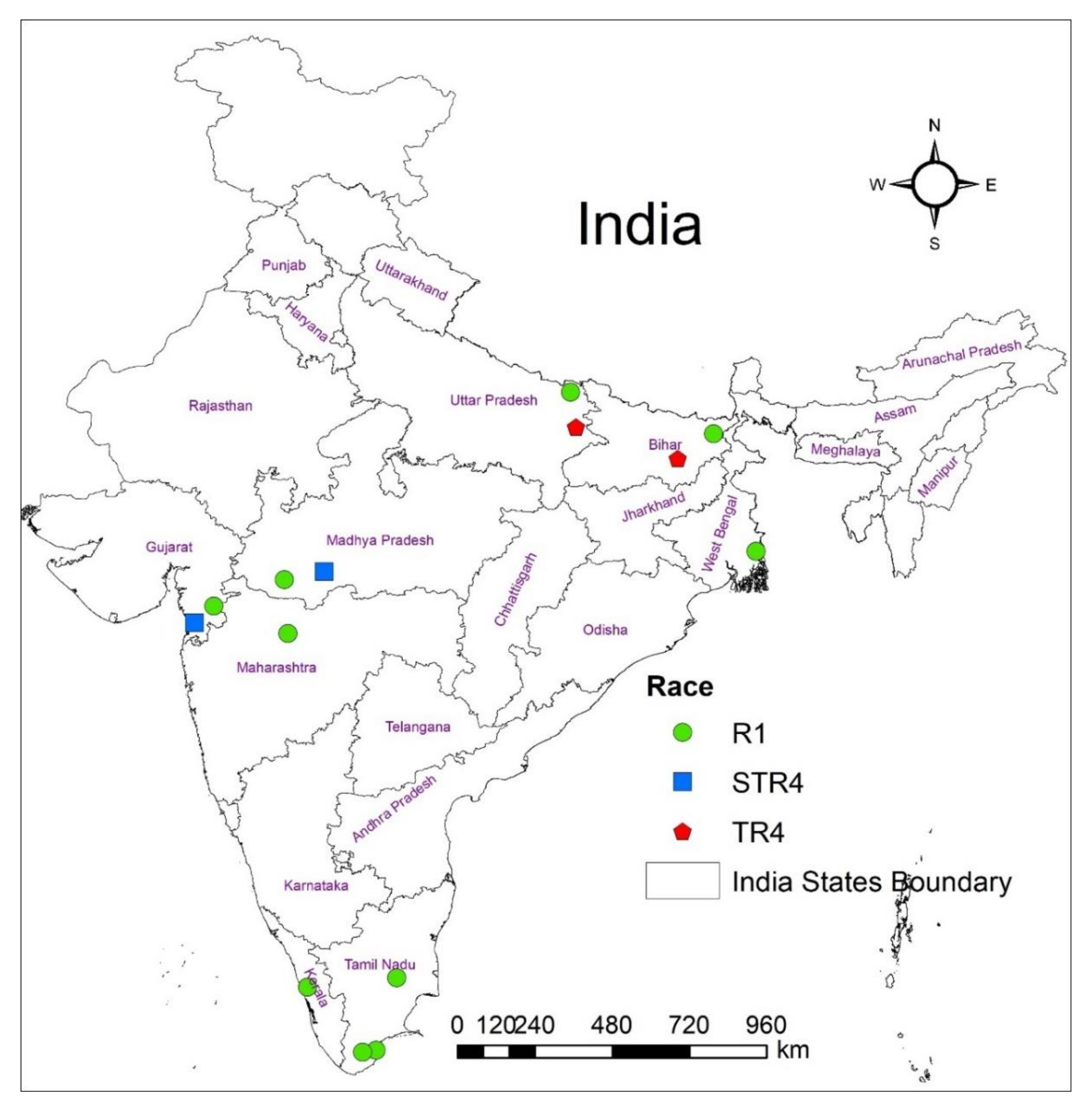
2.1. Collection of Foc Races

A total of 61 Foc isolates were isolated from different banana-growing states of India (11 isolates from Uttar Pradesh, 5 from Gujarat, 13 from Bihar, 23 from Tamil Nadu, 5 from Kerala, and 4 from Madhya Pradesh; Figure 1 and Table 1). The race typing of Foc was confirmed by pathogenicity tests on the respective banana cultivars and also on the cv. Grande Naine. Further, the genomic DNA of 23 reference Foc isolates (Supplementary Table S1) of different VCGs (obtained from QDPI and F, Brisbane, Australia), five isolates of nonpathogenic Fo, one each of Pseudocercospora eumusae, Colletotrichum musae, Fusarium solani, and Klebsiella variicola obtained from Department of Plant Pathology, ICAR-National Research Center for Banana, Tiruchirappalli was included in the study (Table 1).
All Indian Foc races of different VCGs employed in the present study were isolated from the vascular strand of pseudostem of a symptomatic Grande Naine cv. (Cavendish AAA subgroup). VCG analysis with nit-M (Nitrate non-utilizing mutants) testers [43], in planta pathogenicity testing using micropropagated Grande Naine plantlets, and molecular confirmation with race-specific PCR markers (e.g., [19,40,44]) were performed to reconfirm the virulence and races of the isolate before the whole-genome sequencing. Thangavelu et al. [28] furnished a detailed description of the procedures elsewhere.

2.2. DNA Extraction and Quantification

Genomic DNA was isolated according to the method described by Dellaporta et al. [45] with slight modifications. Freeze-dried fungal mycelium (1 g) and overnight grown bacterial culture (2.0 mL) were frozen in liquid nitrogen and ground to fine powders using a precooled mortar and pestle. DNA was extracted using 5 mL modified TNE buffer (100 mM Tris-HCl, pH 8.0; 50 mM Na2EDTA, pH 8.0; 50 mM NaCl; 8 μM β-mercaptoethanol; 1% SDS and 10 μg mL−1 RNase) and incubated at 65 °C for 1 h. A 0.33× volume of 5 M CH3CO2K was added, mixed, and centrifuged at 10,000 rpm for 5 min. The supernatant was transferred into a new tube and mixed with an equal volume of isopropanol to precipitate crude DNA. The samples were incubated at −20 °C overnight and centrifuged at 4 °C at 10,000 rpm for 10 min. After resuspending the DNA pellet in 300 μL dH2O, an equal volume of phenol-chloroform/isoamyl alcohol (25:24:1; v/v) was added and mixed thoroughly. The upper aqueous layer was transferred to a fresh tube after centrifugation at 10,000 rpm for 5 min at 4 °C, and 0.1 × volume of 5 M potassium acetate and 2.5 × volume of absolute ethanol (70%) were added. The supernatant was decanted following the centrifugation at 10,000 rpm for 10 min at 4 °C, and the DNA pellet was washed with 300 μL of 75% ethanol, left to air dry, and then finally dissolved in 1 × TE buffer (10 mM Tris-HCl, pH 8; 0.1 mM EDTA) for further analysis. Total DNA yield and purity were quantified by measuring OD at 260 nm using the NanoDrop LITE (Thermo Scientific, Waltham, MA, USA).

2.3. Sequence Alignment and Primer Design

Genomic sequences of Foc isolates (PRJNA552452, PRJNA687504 and accession numbers SAMN12206536, SAMN12206882, and SAMN17147622) along with the reference genomes of Fol4287 (ASM14995v2; txid426428) and Foc TR4 (ASM799451v1; txid61366) were obtained from the National Center for Biotechnology Information (NCBI). The Interactive Genomics Viewer was used to visualize and align the whole-genome sequences. Genome-wide distribution of DNA polymorphisms was initially analyzed by calculating their frequency of every 100 kb interval of reference genomes at chromosome levels. To identify the potential pathogenicity-related proteins, a Basic Local Alignment Search Tool (BLASTp) search was performed against the Pathogen–Host Interaction database (PHI-base) with a threshold E-value of ≤1 × 10⁻⁵ [46]. SIX genes in the genome of Indian Foc races are defined using BLAST analysis with the assembled genome to SIX genes in the NCBI database [47,48]. Targets for primer design were searched on differential regions of the genome, as these contained previously targeted gene candidates for race differentiation. Regions specific to each race were targeted specifically. Using this approach, different primer sequences were isolated as candidates for differentiation based on their genome. NCBI BLAST was used to determine the likelihood of genetic conservation within the genome based on shared sequences with related species to narrow the possible pool of target loci. In addition, sequences containing coding regions and hypothetical proteins were explicitly targeted specifically for primer design. Following BLAST analysis, primer design properties such as GC content and amplicon size were used to narrow further. Consequently, primer pairs for Foc R1, R4, TR4, and STR4 were designed manually and checked for quality and content with the Integrated DNA Technologies PrimerQuest™ Tool. All primers were synthesized by Sigma-Aldrich® (St. Louis, MO, USA) and stored at −20 °C. All primers used in this study are listed in Supplementary Table S2.

2.4. Primer Design and Polymerase Chain Reaction (PCR) Conditions

To achieve this goal, the whole-genome sequences of three races of Foc (R1, TR4, and STR4), as well as two other F. oxysporum (Fol, Foc) from NCBI were used for in silico analyses (www.ncbi.nlm.nih.gov accessed on 4 January 2020). The genome comparison finds the unique effector genes and variable regions between race/VCGs through SIX sequences from different Foc races were found to be highly conserved in general. When the homology of different races was compared by using a comprehensive suite of molecular biology and next-generation sequencing analysis tools, our target races, (Foc R1 and R4) revealed variations in a certain portion of sequences at different locations on chromosome 14 compared to all other races. The variable sequences facilitated the development of Foc R1 and R4 markers.
The primers were designed to target SIX gene sequences extracted from the whole-genome of Indian Foc races using Primer3 software [47] and were tested for specificity in silico using ‘In silico simulation of molecular biology experiments software’ (www.insilico.ehu.es accessed on 4 January 2020). Only variable regions were chosen over highly conserved regions as they may possess race-specific signature sequences required for identifying Foc R1, R4, TR4, and STR4. A total of 46 potential race-specific primer sets were designed to discriminate the Foc races in which 32 were for TR4, 1 for R4, 9 for R1, and 4 for STR4. Table 2 shows the specific region and expected amplification for these selected Foc primers that were cull out from variable regions of the genome. EmeraldAmp® PCR Master Mix (Takara, Shiga, Japan) was used in Polymerase Chain Reaction (PCR) to amplify the target regions with respective markers. A 20 µL of the PCR reaction mixture containing forward and reverse primers (0.5 µM µL−1 each), EmeraldAmp® PCR master mix (9.0 µL), ultra-pure water (8 µL), and 50 ng µL−1 DNA was used for PCR amplification. PCR was performed using the following conditions in a thermocycler (Takara, Shiga, Japan): denaturation at 95 °C for 5 min followed by 30 cycles of amplification at 95 °C for 30 s, annealing at 65 °C for 40 s and 72 °C for 45 s, and final elongation at 72 °C for 5 min, but in case of Foc TR4, R4 and STR4 specific amplification annealing was carried out at 66 °C for 40 s. Electrophoresis was undertaken using 2% agarose gel at 100 V for 30 min and visualized on the gel documentation system under ultraviolet light (302 nm).

2.5. Optimization of the Developed Marker

All the new primer sets were evaluated to determine the optimal annealing temperature, the level of specificity, and the detection limit of genomic DNA. To optimize the annealing temperature, a gradient PCR from 58 to 68 °C was performed and the highest temperature that did not diminish the brightness of the amplicon was selected. To assess specificity, each primer set was tested with a diverse selection of nonpathogenic Fo isolates, F. solani, Foc VCGs of all three races, and other banana pathogens such as P. eumusae, C. musae, and K. variicola. PCR samples were prepared to the same concentration levels as described above, with approximately 25 ng of DNA per reaction. Along with the non-target samples, a positive Foc control and negative water control were also included. To evaluate the sensitivity of the newly developed PCR marker, genomic DNA was isolated from various standard Foc isolates and standardized to 25 ng µL−1, then serially diluted down to 0. 02 pg µL−1 of DNA. DNA concentrations were quantified in a Qubit fluorometer using the Quant-iT High-Sensitivity dsDNA Assay Kit (Thermo Fisher Scientific Inc., Waltham, MA, USA) before dilution. To determine the minimum sensitivity, these dilutions were amplified using the specified race-specific primers. PCR results (4 µL per sample) were imaged on a 2% agarose gel and the lowest successful amplification was determined to be the detection limit of the new primer set. Reproducibility of the PCR assay was further confirmed by two different operators, on three different occasions, with the same set of samples. All tests included two technical replicates, and the results demonstrated that the primers met all repeatability and robustness validation criteria.

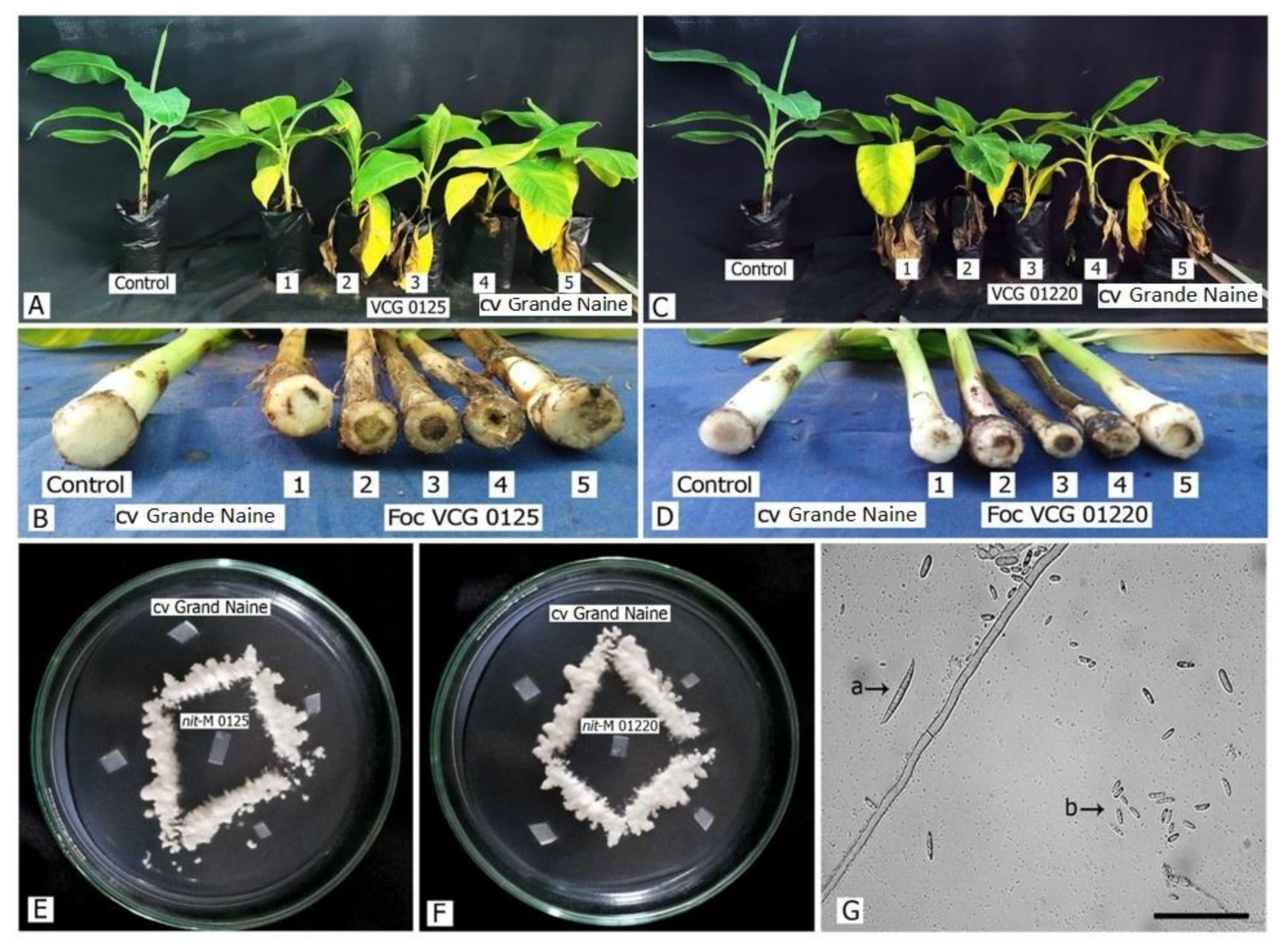
2.6. In Planta Detection of Foc Races

The experiment used three-month-old tissue-cultured plants of cv. Grande Naine and planted in poly grow bags (30 × 10 × 15 cm) filled with a sterilized potting mixture containing red earth, sand, and farmyard cattle manure in equal proportions and were kept under glasshouse conditions for 10-days for initial acclimatization. Fifteen-day-old pure Foc R1 (VCG 0125, 0124 and 01220) and Foc R4 (TR4 and STR4) cultures multiplied individually in the sand: maize (19:1) medium containing approximately 106 CFU g−1 was applied once in the soil around the plants in each plant at a 1 inch depth. The experiment was conducted as a randomized complete block design with three replications and each replication containing five plants. These inoculated plants were maintained in a glasshouse at 28 °C, 80% relative humidity, and 16 h light. After 25 days of inoculation, the rhizome and pseudostem were subjected to systematic examination by split into two halves and disease evaluation was carried out based on the percentage of the discolored area in the corm on a 0–5 scale [49]. Samples from the first half of the rhizome and vascular strands were plated on Komada’s medium for selective isolation of Foc and confirmation and the other half was used for genomic DNA extraction for in planta detection of Foc races. Total genomic DNA from 1 g of vascular strands and corm was extracted using the DNeasy® plant mini kit (Qiagen Cat no. 69104) according to the manufacturer’s instructions. In planta detection was performed using the respective primers designed as mentioned in Table 2 to amplify Foc strains of R1, R4, TR4, and STR4.

3. Results

The genome of Indian Foc races was assembled at chromosome-scale and the genome size was ranged from 61 to 63 Mb with ~48.5% GC. The genome of Indian Foc TR4 (VCG01213/16) is 16.3–26.4% higher in size in terms of nt bp, whereas, Foc R1 is 16.8–22.5% and Foc STR4 is 23.9% higher than the reference genome. A total of 21,842 intact protein-coding genes were predicted from the consensus gene sets of the Foc R1 genome assembly, while, the Foc STR4 and TR4 genome assemblies contain 17,118 and 17,745 protein-coding regions, respectively. The results revealed that the Foc R1 contained a maximum of 524 tRNAs and 223 rRNAs, TR4 has a maximum of 358 tRNAs and 134 rRNAs, and STR4 has a total of 304 tRNAs and 121 rRNAs in the genome. A total of 8756 (39.5%) out of 22,151 proteins coding genes in Foc R1, 5384 (28.4%) out of 18,946 protein-coding genes in Foc STR4, and 11,220 (57.1%) out of 19,651 protein-coding genes from Foc TR4 were annotated from the Gene Ontology database with a cutoff E-value of 10−5. A total of 4866 high-quality SNPs were found within the Indian Foc isolates, in which 1502 in Foc R1, 1844 in Foc STR4 and 1520 in Foc TR4, while a total of 139,023 InDels (88,532 insertions and 50,491 deletions) in which 53,602 in Foc R1, 33,298 in Foc STR4 and 52,123 in Foc TR4. The study identified 1738 (Foc R1) to 2809 (Foc STR4) putative virulence-associated genes in the genome of Indian Foc isolates in which a total of 94, 283, and 258 virulence genes are unique to the Foc R1, TR4, and STR4 genomes, respectively. The number of effector genes encoded by the genome varies in which the lowest 17 genes were present in Foc TR4, a moderate 20 genes in Foc R1, and a maximum of 28 genes in Foc STR4. Similarly, the number of genes involved in increased virulence was 13, 23, and 27 in Foc TR4, STR4, and R1, respectively. These virulence genes were the target for the primer synthesis for the race-specific identification of the Indian Foc isolates.

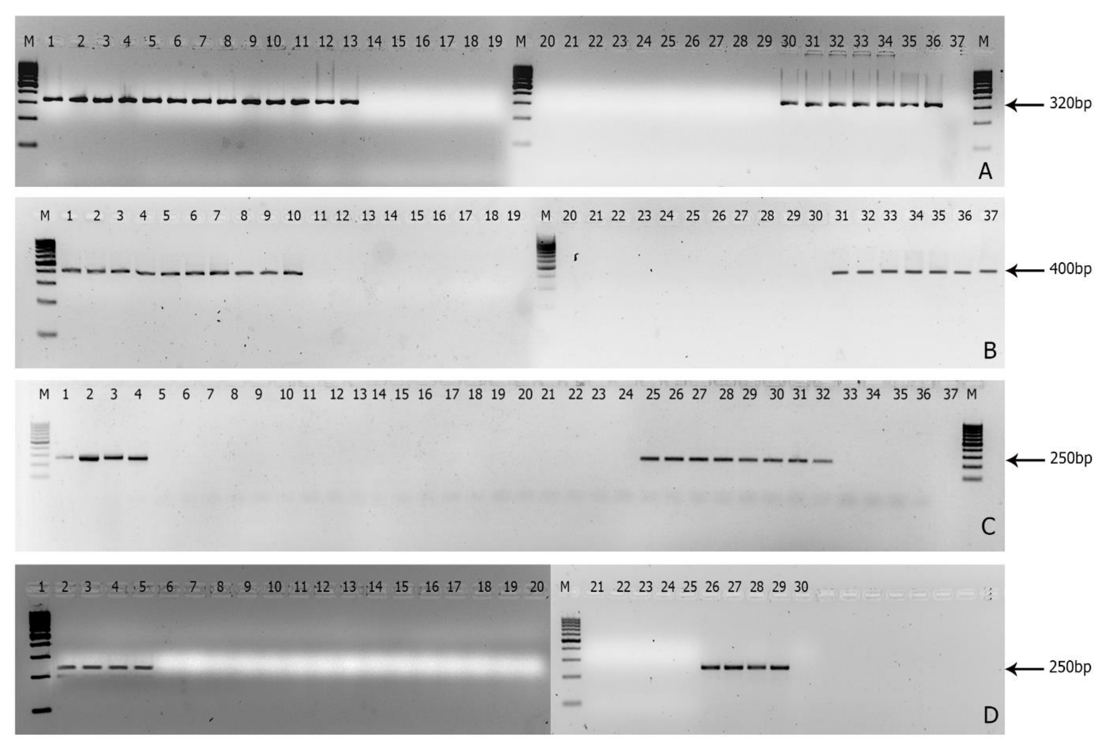
3.1. Screening of Race-Specific Markers and Its Specificity to Indian Foc

Out of 61 Foc isolates, 27 representative isolates of different races were subjected to the specificity analysis. Out of nine primers tested; the Foc R1F/Foc R1R primer set targeted to gene XM_018394505.1 encoding a hypothetical protein (Table 2) resulted in 320 bp amplicon only with Foc R1 VCGs 0124, 0125, and 01220 (Figure 2A). In the case of Foc R4, only one primer set FocR4F/FocR4R targeting gene KF548063.1 (SIX8a) resulted in 400 bp amplicon only with Foc R4 VCGs of 0120, 01213, 01216, and 01213/16 (Figure 2B). Of the 31 primer sets tested for Foc TR4, FocTR4F/FocTR4R primer targeted to gene KX434998.1 (SIX1) generated 250 bp amplicon with Foc TR4 VCGs of 01216, 01213, and 01213/16 (Figure 2C). Out of a four primer set designed for STR4, FocSTR4F/FocSTR4R targeting KX434398.1 (SIX7) gene resulted in the generation of 250 bp amplicon with Foc STR4 VCG 0120 (Figure 2D).

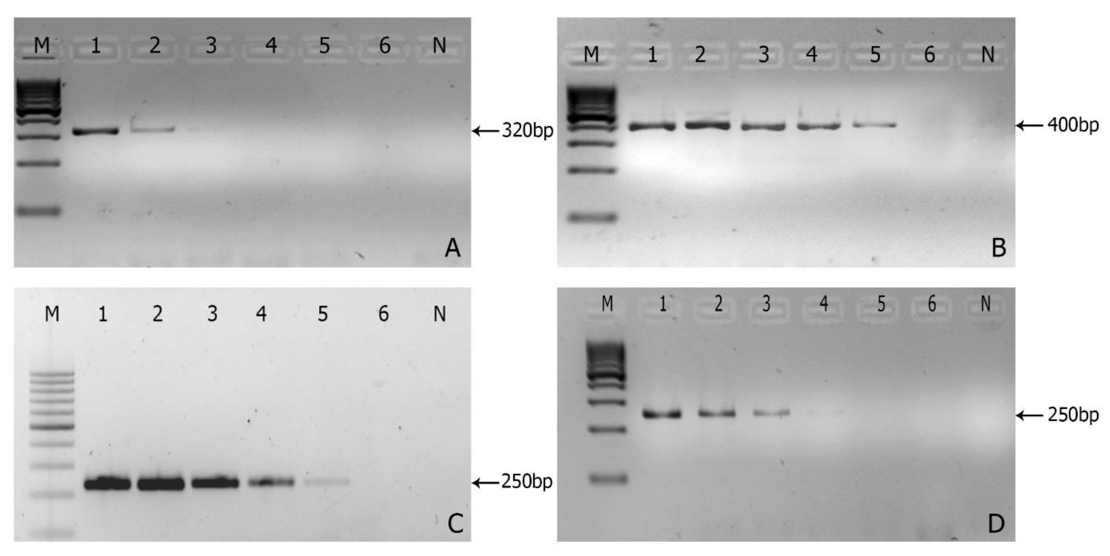
3.2. Sensitivity of the Markers

The gel showing PCR amplification products demonstrated that the lowest successful amplification was observed at 1.0 pg µL−1 and 0.1 pg µL−1 of genomic DNA on Foc R1 and R4, respectively (Figure 3A,B), and the optimal annealing temperature of the race-specific primer ranged between 58 to 66 °C depending on the race (Table 2).

3.3. In Planta Detection

The inoculation of Foc isolates of R1, STR4, and TR4 on the cv. Grande Naine resulted in the expression of typical external symptoms of leaf yellowing after 18–20 days of inoculation (Figure 4). All the Foc races inoculated tissue-cultured banana plants exhibited severe wilting and internal vascular discoloration in both the corm and pseudostem at 25–30 days after inoculation (Figure 4A,C). The study observed no differences in symptoms between the Foc races inoculated plants in terms of the number of days required for symptom expression or severity (Figure 4B,D). The tested Foc races were successfully recovered from rhizomes using Komada’s medium (Figure 4E,F) and were further confirmed by the spore morphology (Figure 4G) and molecular method using race-specific primers. Genomic DNA (50 ng) isolated from corm samples from infected plants was used for PCR amplification and successfully generated the respective amplicon using the respective primer set designed for the Foc races (Foc R1 and Foc R4, TR4 and STR4) diagnostics.

4. Discussion

The purpose of this study was to develop PCR-based molecular markers that can be used to accurately identify Indian Foc R1 and R4 which differ in genome size and SIX organization [42] with other reference Foc of the same races. Molecular methods based on Foc race identification have been extensively investigated by several researchers such as Lin et al. [21], Fourie et al. [50], Dita et al. [19], Carvalhais et al. [40], and Magdama [51]. However, the present study used molecular markers that were already available as a positive check [19,40,44], which failed to amplify the Indian Foc isolates due to genome variations [42]. There are no race-specific markers specifically available for detecting any of the Indian Foc races. Therefore, the study aimed to develop molecular markers that target the effector gene for the accurate detection of Indian Foc pathogens infecting banana crops [52,53]. This study presumes that PCR-based molecular markers are relatively affordable, robust, repeatable, trustworthy, and effective for the early detection of Foc in bananas.
A range of molecular diagnostic methods has previously been proposed to distinguish races of Foc [40]. For instance, Lin et al. [21], Fourie et al. [50], Dita et al. [19], and Magdama [51] demonstrated the use of molecular markers. However, the findings were found to have a major drawback in validating the Foc races which have been described elsewhere by Carvalhais et al. [40]. To address these issues, this study utilized effector genes as targets for a molecular diagnostic tool for Foc races, as each race of Indian VCGs has unique effector genes [42].
Hypothetical protein XM_018394505.1 of Fol4287 is present in Chr-1, 3, 6 and 15 of Foc R1 VCG0124 has 3036 bp length which ranged from 1525,130 to 1528165 nt in the genome, where the race-specific primer was targeted in Chr-15 between 875,832 to 875,813 and 875,510 to 875,529 of ~320 bp, respectively, for forward and reverse primers (Supplementary Figure S1A). A comparison of different VCGs revealed that VCG0124 exhibits polymorphism at 320 nt, as shown by multiple sequence analysis (Supplementary Figure S1B). The unique SNP allows race-specific identification of Indian Foc R1 which includes VCG 0124, 0215, and 01220. Carvalhais et al. [40] previously reported SIX6 to diagnose Foc R1 VCGs; however, the primer developed did not show the expected amplification product for five Foc R1 isolates, including VCGs 0124 and 0123. Therefore, this study believes that the newly developed primer has paramount importance in diagnosing the Indian Foc R1 as it is widely distributed in all the sampled states, especially in Cavendish bananas (Figure 1).
For the detection of Foc R4, Lin et al. [21] proposed an RAPD primer to diagnose Foc R4 VCGs. However, Dita et al. [19] and Magdama, [51] reported that the primers are cross-reactive with Foc R1 and nonpathogenic Foc. Therefore, in this study, a set of primers FocR4F/FocR4R that has an amplicon size of ~400 bp was designed to target both TR4 and STR4 isolates based on SIX8a which is present in Chr-14. Commonly, SIX8a is present in the Foc STR4 and TR4 which is similar to KF548063.1 of Fol4287 and has 713 bp in length from where the primer was designed in 483–502 nt and 661–680 nt, respectively for forward and reverse primer (Supplementary Figure S2A). From Figure 2B, the primer was found to be specific to Indian Foc R4 isolates that do not have any cross-reactions. This result was further supported by the results of Carvalhais et al. [40], Fraser-Smith et al. [54], and Czislowski et al. [34], who reported that SIX8a is present in both TR4 and STR4, and SIX8b is specific to STR4 and can be utilized for molecular diagnosis of Foc R4 isolates of divergent origin. In contrast to the above, our previous whole-genome study revealed that SIX8b is present in both the Indian races of Foc R4 [42]. Therefore, the newly developed FocR4F/FocR4R primer containing both conserved and differential regions in 483–502 nt and 661–680 nt, respectively (Supplementary Figure S2B) were utilized for effective diagnosis of Foc R4 isolates of the study area.
For the specific identification of Foc TR4, SIX1a_266 primer has been effectively utilized by Carvalhais et al. [40], but with a restriction digestion method. In this method, primers flanked a recognition site of the enzyme HpyAV, which is only present in the SIX1 gene homolog “a”, which is unique to TR4 of VCG 01213/16 was used for diagnosis [34]. To simplify the method, FocTR4F/FocTR4R primer was designed to identify the Indian Foc TR4 which has 17% polymorphism with gaps (Supplementary Figure S3A). The SIX1a gene was encoded in the CM028841.1 of Chr-14 which ranged between 1,474,171 and 1,475,025 with a total of 851 bp nt. However, the actual sequence length of SIX1a used by Carvalhais et al. [40] was 815 bp nt which is 83% similar to Indian Foc TR4 (Supplementary Figure S3A). Moreover, the SIX1a primer of Carvalhais et al. [40] included the homologous of ‘a’ to ‘g’, yet the Foc TR4 of Indian VCG01213/16 has a single homologous copy. From Figure 2C, it was observed that the primer was proved to be repeatable, robust, and specific to the amplicon size ~250 bp, where the forward primer binding was at 162–181 nt and reverse primer binding at 366–383 nt (Supplementary Figure S3B).
Based on the report of Czislowski et al. [40], the present study makes use of SIX7a for the identification of STR4 as it is found to be specific to this race with a wide range of VCGs such as 0120, 0120/15, 0126, 0129, 01211, and 01215. We previously reported that the genome of Foc STR4 VCG0120 has a total of 14 homologous copies of SIX8 across the different chromosomes. Therefore, using SIX8 based primer for the specific detection of Indian Foc STR4 remains a matter of risk as it has a polymorphic site in the primers based on homologous variants as reported by Carvalhais et al. [40]. Gene KM503196.1 encoding SIX7 in Foc STR4 having 663 bp nt was selected as a template to design the primer, where the forward primer binding at 310–329 nt and reverse primer binding at 535–554 nt with the amplicon size of ~248 bp (Supplementary Figure S4A). The gene was present in the Chr-14 (CM028826.1) of Indian Foc STR4 between 1,288,728 and 1,289,395 nt (Supplementary Figure S4B). From Figure 2D, the study found that the newly developed primer FocSTR4F/FocSTR4R was robust and specific.
Based on the aforementioned findings, the study corroborates the results of Fraser-Smith et al. [54], Rocha et al. [53], Ayukawa et al. [55], who developed molecular markers for the effective detection of Foc races. As the developed molecular markers allowed the identification of a strain of Foc R1 and R4, these race-specific markers can identify Foc races through conventional PCR within a few hours whereas pathogenicity testing using differential cultivars requires several months for race determination [20]. Thus, time-bound remedies to standing banana can be implemented immediately rather than taking several months for the identification to manage this lethal disease. Maymon et al. [56] recently used primers developed by O’Neill et al. [57] for recognizing Foc races in Israel, which were originally developed for Foc strains of northern Queensland. Similarly, the newly developed markers of this study can be used to identify these emerging Foc races with larger genome sizes and diverse SIX gene profiles, besides sensitizing the Indian Foc isolates in other parts of the nation or world.
After race typing, isolates were rearranged and assessed based on their geographic state of origin. For this, the races of respective isolates were identified and all the states were combined. Out of 61 samples subjected, a maximum of 68.9% are R1 (44) followed by 31.1% are R4 (19), in which 6.6% are STR4 (4) and 24.6% are TR4 (15). Of the eight states surveyed, Foc R1 has been the dominant race present in all the states of India (Figure 1). However, the Foc TR4 and STR4 are identified in two states where Foc TR4 was distributed in Uttar Pradesh and Bihar while Foc STR4 was identified from Gujarat and Madhya Pradesh. The locations and samples were randomly selected during our survey. Foc isolates differed in percentages from previously reported studies in which race 1 had been the most common race, instead of other races used in this study [33]. The markers used in this present study should enhance the speed and accuracy of the existing diagnostic ability for Foc and provide a jumping-off point to investigate similar regions involved in pathogenicity and race development for various researchers. This study exploited the variation within the whole-genome sequences of Foc races for developing Foc races (R1, R4, TR4, and ST4) specific markers.

5. Conclusions

To conclude, the present study has resulted in the development of Foc races’ specific molecular markers targeting the effector gene for the accurate diagnosis and detection of Indian Foc R1, R4, TR4, and STR4 strains affecting a wide range of banana cultivars including the Cavendish group of bananas which forms the major exporting variety in the world including India. These markers were extensively tested with a large number of isolates of each Foc race collected from different geographical locations of different states of India to prove their specificity and robustness in diagnosing all the Foc races infecting bananas. Moreover, these markers were also proved to be useful in in planta detection and also with up to 0.025 pg µL−1 levels of Foc DNA present in the sample. Therefore, the markers developed would be useful not only for quarantine purposes worldwide but also in designing and developing effective and sustainable management practices for this destructive disease.

Supplementary Materials

The following are available online at https://www.mdpi.com/article/10.3390/jof8010053/s1. Table S1: Details of reference Foc isolates (and their VCGs status) used in this study. Table S2: List of primers designed specifically for Foc races and their Gene ID. Figure S1: Forward and reverse primers binding site (A) and DNA sequence alignment of hypothetical protein (B) for FocR1 of FocR1F/Foc R1R. Figure S2: Forward and reverse primers binding site (A) and DNA sequence alignment of SIX8a (B) for Foc R4 of Foc R4F/Foc R4R. Figure S3: Sequence comparison of SIX1a (A) and forward and reverse primers binding site (B). Figure S4: Forward and reverse primers binding site (A) and multiple sequence alignment of SIX7a (B) for Foc STR4 of FocSTR4F/FocSTR4R.

Author Contributions

R.T., E.E. contributed equally: R.T. conceived the project, design the experiments, compiled data, and wrote the manuscript; E.E. performed data analyses, primer designing, performed the experiments, and wrote the manuscript; M.G., K.S., P.P. and M.P. reagents/materials/analysis tools, performed the experiments; and M.L. and S.U. wrote the manuscript and made the corrections. All authors have read and agreed to the published version of the manuscript.

Funding

This paper was made possible by a grant from the Indian Council of Agricultural Research (ICAR), New Delhi, for an extramural project named “Integrated Management of Fusarium wilt Tropical Race-4: A Devastating Strain on Banana” F.No. 7-2/2019-Hort.Sci-1.

Institutional Review Board Statement

Not applicable.

Informed Consent Statement

Not applicable.

Data Availability Statement

The assembled short-read genome sequences of Foc Race1 (VCG0124), Foc STR4 (VCG0120), and Foc TR4 (VCG 01213/16) infecting Cavendish group banana have been deposited at DDBJ/EMBL/GenBank under accession number GCA_011316005.3, GCA_016802205.1, and GCA_014282265.3, respectively. All the data generated or analyzed during this study are included in this published article.

Acknowledgments

We acknowledge gratefully the financial support provided for this study through extramural projects rendered by the ICAR, New Delhi, and the facilities provided by the Director. The authors are thankful to C, Anuradha, Biotechnology Division, ICAR-NRCB for technical assistance. We are grateful for and appreciate the contribution from the CGIAR Research Program on Roots, Tubers, and Bananas (RTB) and the CGIAR Fund Donors for covering the open access fee.

Conflicts of Interest

The authors declare that the research was conducted in the absence of any commercial or financial relationships that could be construed as a potential conflict of interest.

References

  1. FAO. FAOSTAT Database Collections. Available online: https://www.fao.org/faostat/en/#data/ (accessed on 27 September 2021).
  2. Stover, R.H. Fusarial Wilt (Panama Disease) of Bananas and Other Musa Species; Commonwealth Mycological Institute: Kew, UK, 1962. [Google Scholar]
  3. Stover, R.H. Fusarium wilt of banana: Some history and current status of the disease. In Fusarium Wilt of Banana; Ploetz, R.C., Ed.; American Phytopathology Society Press: St. Paul, MN, USA, 1990; Volume 1, p. 70. [Google Scholar]
  4. Thangavelu, R.; Loganathan, M.; Arthee, R.; Prabakaran, M.; Uma, S. Fusarium Wilt: A Threat to Banana Cultivation and Its Management; CABI: Wallingford, UK, 2020; Volume 15, pp. 1–24. [Google Scholar] [CrossRef]
  5. Altendorf, S. Banana Fusarium Wilt Tropical Race 4: A Mounting Threat to Global Banana Markets? FAO: Rome, Italy, 2019. [Google Scholar]
  6. Musapedia Banana-Producing Countries. Available online: https://www.promusa.org/Banana-producing+countries+portal#footnotefao (accessed on 27 September 2021).
  7. Mustaffa, M.M.; Thangavelu, R. Status of Fusarium wilt in India. In V International Symposium on Banana: ISHS-ProMusa Symposium on Global Perspectives on Asian Challenges 897; ISHS: Leuven, Belgium, 2011; Volume 897, pp. 323–330. [Google Scholar]
  8. Thangavelu, R.; Gopi, M.; Pushpakanth, P.; Loganathan, M.; Edwin Raj, E.; Marimuthu, N.; Prabakaran, M.; Uma, S. First Report of Fusarium oxysporum f. sp. cubense VCG 0125 and VCG 01220 of Race 1 Infecting Cavendish Bananas (Musa sp. AAA) in India. Plant Dis. 2021, 105, 1215. [Google Scholar] [CrossRef] [PubMed]
  9. Thangavelu, R.; Edwin Raj, E.; Muthukathan, G.; Murugan, L.; Uma, S. Draft Genome Resource of a Novel Virulent Fusarium oxysporum f. sp. cubense Race 1 Strain (VCG 0124) Infecting Cavendish (AAA) Group of Banana in India. Plant Dis. 2021, 105, 2708–2710. [Google Scholar] [CrossRef] [PubMed]
  10. Forsyth, L.M.; Smith, L.J.; Aitken, E.A.B. Identification and characterization of nonpathogenic Fusarium oxysporum capable of increasing and decreasing Fusarium wilt severity. Mycol. Res. 2006, 110, 929–935. [Google Scholar] [CrossRef] [PubMed]
  11. Diener, A.C.; Ausubel, F.M. Resistance to Fusarium oxysporum 1, a Dominant Arabidopsis Disease-Resistance Gene, Is Not Race Specific. Genetics 2005, 171, 305–321. [Google Scholar] [CrossRef] [Green Version]
  12. Carter, C.; Toms, S. The contours of critical accounting. Crit. Perspect. Account. 2010, 21, 171–182. [Google Scholar] [CrossRef]
  13. Daniells, J.; Davis, D.; Peterson, R.; Pegg, K. Goldfinger: Not as resistant to Sigatoka/yellow Sigatoka as first thought. Infomusa 1995, 4, 6. [Google Scholar]
  14. Yergeau, E.; Filion, M.; Vujanovic, V.; St-Arnaud, M. A PCR-denaturing gradient gel electrophoresis approach to assess Fusarium diversity in asparagus. J. Microbiol. Methods 2005, 60, 143–154. [Google Scholar] [CrossRef]
  15. Jurado, M.; Vázquez, C.; Marín, S.; Sanchis, V.; Teresa González-Jaén, M. PCR-based strategy to detect contamination with mycotoxigenic Fusarium species in maize. Syst. Appl. Microbiol. 2006, 29, 681–689. [Google Scholar] [CrossRef]
  16. Kung’u, J.; Jefries, P. Races and virulence of Fusarium oxysporum f. sp. cubense on local banana cultivars in Kenya. Ann. Appl. Biol. 2001, 139, 343–349. [Google Scholar] [CrossRef]
  17. Thangavelu, R.; Mustaffa, M.M. First Report on the Occurrence of a Virulent Strain of Fusarium Wilt Pathogen (Race-1) Infecting Cavendish (AAA) Group of Bananas in India. Plant Dis. 2010, 94, 1379. [Google Scholar] [CrossRef]
  18. Li, B.; Du, J.; Lan, C.; Liu, P.; Weng, Q.; Chen, Q. Development of a loop-mediated isothermal amplification assay for rapid and sensitive detection of Fusarium oxysporum f. sp. cubense race 4. Eur. J. Plant Pathol. 2013, 135, 903–911. [Google Scholar] [CrossRef]
  19. Dita, M.A.; Waalwijk, C.; Buddenhagen, I.W.; Souza, J.T.; Kema, G.H.J. A molecular diagnostic for tropical race 4 of the banana fusarium wilt pathogen. Plant Pathol. 2010, 59, 348–357. [Google Scholar] [CrossRef]
  20. Aguayo, J.; Mostert, D.; Fourrier-Jeandel, C.; Cerf-Wendling, I.; Hostachy, B.; Viljoen, A.; Ioos, R. Development of a hydrolysis probe-based real-time assay for the detection of tropical strains of Fusarium oxysporum f. sp. cubense race 4. PLoS ONE 2017, 12, e0171767. [Google Scholar] [CrossRef] [PubMed]
  21. Lin, Y.H.; Chang, J.Y.; Liu, E.T.; Chao, C.P.; Huang, J.W.; Chang, P.F.L. Development of a molecular marker for specific detection of Fusarium oxysporum f. sp. cubense race 4. Eur. J. Plant Pathol. 2009, 123, 353–365. [Google Scholar] [CrossRef]
  22. García-Bastidas, F.; Ordóñez, N.; Konkol, J.; Al-Qasim, M.; Naser, Z.; Abdelwali, M.; Salem, N.; Waalwijk, C.; Ploetz, R.C.; Kema, G.H.J. First Report of Fusarium oxysporum f. sp. cubense Tropical Race 4 Associated with Panama Disease of Banana outside Southeast Asia. Plant Dis. 2014, 98, 694. [Google Scholar] [CrossRef] [PubMed]
  23. Ploetz, R.C. Fusarium wilt of banana. Phytopathology 2015, 105, 1512–1521. [Google Scholar] [CrossRef] [PubMed] [Green Version]
  24. Ordoñez, N.; García-Bastidas, F.; Laghari, H.B.; Akkary, M.Y.; Harfouche, E.N.; al Awar, B.N.; Kema, G.H.J. First Report of Fusarium oxysporum f. sp. cubense Tropical Race 4 Causing Panama Disease in Cavendish Bananas in Pakistan and Lebanon. Plant Dis. 2016, 100, 209. [Google Scholar] [CrossRef]
  25. Chittarath, K.; Mostert, D.; Crew, K.S.; Viljoen, A.; Kong, G.; Molina, A.B.; Thomas, J.E. First Report of Fusarium oxysporum f. sp. cubense Tropical Race 4 (VCG 01213/16) Associated with Cavendish Bananas in Laos. Plant Dis. 2018, 102, 449. [Google Scholar] [CrossRef]
  26. Hung, T.N.; Hung, N.Q.; Mostert, D.; Viljoen, A.; Chao, C.P.; Molina, A.B. First report of fusarium wilt on Cavendish bananas, caused by Fusarium oxysporum f. sp. cubense tropical race 4 (VCG 01213/16), in Vietnam. Plant Dis. 2018, 102, 448. [Google Scholar] [CrossRef]
  27. Zheng, S.-J.; García-Bastidas, F.A.; Li, X.; Zeng, L.; Bai, T.; Xu, S.; Yin, K.; Li, H.; Fu, G.; Yu, Y.; et al. New Geographical Insights of the Latest Expansion of Fusarium oxysporum f. sp. cubense Tropical Race 4 Into the Greater Mekong Subregion. Front. Plant Sci. 2018, 9, 457. [Google Scholar] [CrossRef] [Green Version]
  28. Thangavelu, R.; Mostert, D.; Gopi, M.; Devi, P.G.; Padmanaban, B.; Molina, A.B.; Viljoen, A. First detection of Fusarium oxysporum f. sp. cubense tropical race 4 (TR4) on Cavendish banana in India. Eur. J. Plant Pathol. 2019, 154, 777–786. [Google Scholar] [CrossRef]
  29. Lin, Y.H.; Su, C.C.; Chao, C.P.; Chen, C.Y.; Chang, C.J.; Huang, J.W.; Chang, P.F.L. A molecular diagnosis method using real-time PCR for quantification and detection of Fusarium oxysporum f. sp. cubense race 4. Eur. J. Plant Pathol. 2013, 135, 395–405. [Google Scholar] [CrossRef]
  30. Ordóñez, N.; Salacinas, M.; Mendes, O.; Seidl, M.F.; Meijer, H.J.G.; Schoen, C.D.; Kema, G.H.J. A loop-mediated isothermal amplification (LAMP) assay based on unique markers derived from genotyping by sequencing data for rapid in planta diagnosis of Panama disease caused by Tropical Race 4 in banana. Plant Pathol. 2019, 68, 1682–1693. [Google Scholar] [CrossRef] [Green Version]
  31. Yang, L.L.; Sun, L.X.; Ruan, X.L.; Qiu, D.Y.; Chen, D.H.; Cai, X.Q.; Li, H.P. Development of a single-tube duplex real-time fluorescence method for the rapid quantitative detection of Fusarium oxysporum f. sp. cubense race 1 (FOC1) and race 4 (FOC4) using TaqMan probes. Crop Prot. 2015, 68, 27–35. [Google Scholar] [CrossRef]
  32. Li, M.; Shi, J.; Xie, X.; Leng, Y.; Wang, H.; Xi, P.; Zhou, J.; Zhong, S.; Jiang, Z. Identification and application of a unique genetic locus in diagnosis of Fusarium oxysporum f. sp. cubense tropical race 4. Can. J. Plant Pathol. 2013, 35, 482–493. [Google Scholar] [CrossRef]
  33. Mostert, D.; Molina, A.B.; Daniells, J.; Fourie, G.; Hermanto, C.; Chao, C.-P.; Fabregar, E.; Sinohin, V.G.; Masdek, N.; Thangavelu, R.; et al. The distribution and host range of the banana Fusarium wilt fungus, Fusarium oxysporum f. sp. cubense, in Asia. PLoS ONE 2017, 12, e0181630. [Google Scholar] [CrossRef] [PubMed] [Green Version]
  34. Czislowski, E.; Fraser-Smith, S.; Zander, M.; O’Neill, W.T.; Meldrum, R.A.; Tran-Nguyen, L.T.T.; Batley, J.; Aitken, E.A.B. Investigation of the diversity of effector genes in the banana pathogen, Fusarium oxysporum f. sp. cubense, reveals evidence of horizontal gene transfer. Mol. Plant Pathol. 2018, 19, 1155–1171. [Google Scholar] [CrossRef] [Green Version]
  35. Lievens, B.; van Baarlen, P.; Verreth, C.; van Kerckhove, S.; Rep, M.; Thomma, B.P.H.J. Evolutionary relationships between Fusarium oxysporum f. sp. lycopersici and F. oxysporum f. sp. radicis-lycopersici isolates inferred from mating type, elongation factor-1α and exopolygalacturonase sequences. Mycol. Res. 2009, 113, 1181–1191. [Google Scholar] [CrossRef] [PubMed]
  36. De Sain, M.; Rep, M. The role of pathogen-secreted proteins in fungal vascular wilt diseases. Int. J. Mol. Sci. 2015, 16, 23970–23993. [Google Scholar] [CrossRef]
  37. Van Dam, P.; Fokkens, L.; Schmidt, S.M.; Linmans, J.H.J.; Kistler, H.C.; Ma, L.-J.; Rep, M. Effector profiles distinguish formae speciales of Fusarium oxysporum. Environ. Microbiol. 2016, 18, 4087–4102. [Google Scholar] [CrossRef]
  38. Jangir, P.; Mehra, N.; Sharma, K.; Singh, N.; Rani, M.; Kapoor, R. Secreted in Xylem Genes: Drivers of Host Adaptation in Fusarium oxysporum. Front. Plant Sci. 2021, 12, 628611. [Google Scholar] [CrossRef] [PubMed]
  39. Chakrabarti, A.; Ganapathi, T.R.; Mukherjee, P.K.; Bapat, V.A. MSI-99, a magainin analogue, imparts enhanced disease resistance in transgenic tobacco and banana. Planta 2003, 216, 587–596. [Google Scholar] [CrossRef] [PubMed]
  40. Carvalhais, L.C.; Henderson, J.; Rincon-Florez, V.A.; O’Dwyer, C.; Czislowski, E.; Aitken, E.A.B.; Drenth, A. Molecular diagnostics of banana fusarium wilt targeting Secreted-in-xylem genes. Front. Plant Sci. 2019, 10, 547. [Google Scholar] [CrossRef]
  41. Li, C.; Mostert, G.; Zuo, C.; Beukes, I.; Yang, Q.; Sheng, O.; Kuang, R.; Wei, Y.; Hu, C.; Rose, L.; et al. Diversity and Distribution of the Banana Wilt Pathogen Fusarium oxysporum f. sp. cubense in China. Fungal Genom. Biol. 2013, 3, 6. [Google Scholar] [CrossRef]
  42. Raman, T.; Edwin Raj, E.; Muthukathan, G.; Loganathan, M.; Periyasamy, P.; Natesh, M.; Manivasakan, P.; Kotteeswaran, S.; Rajendran, S.; Subbaraya, U. Comparative Whole-Genome Sequence Analyses of Fusarium Wilt Pathogen (Foc R1, STR4 and TR4) Infecting Cavendish (AAA) Bananas in India, with a Special Emphasis on Pathogenicity Mechanisms. J. Fungi 2021, 7, 717. [Google Scholar] [CrossRef]
  43. Correll, J.C. Nitrate Nonutilizing Mutants of Fusarium oxysporum and Their Use in Vegetative Compatibility Tests. Phytopathology 1987, 77, 1640. [Google Scholar] [CrossRef]
  44. Li, C.; Shao, J.; Wang, Y.; Li, W.; Guo, D.; Yan, B.; Xia, Y.; Peng, M. Analysis of banana transcriptome and global gene expression profiles in banana roots in response to infection by race 1 and tropical race 4 of Fusarium oxysporum f. sp. cubense. BMC Genom. 2013, 14, 851. [Google Scholar] [CrossRef] [Green Version]
  45. Dellaporta, S.L.; Wood, J.; Hicks, J.B. A plant DNA minipreparation: Version II. Plant Mol. Biol. Report. 1983, 1, 19–21. [Google Scholar] [CrossRef]
  46. Winnenburg, R.; Baldwin, T.K.; Urban, M.; Rawlings, C.; Köhler, J.; Hammond-Kosack, K.E. PHI-base: A new database for pathogen-host interactions. Nucleic Acids Res. 2006, 34, D459–D464. [Google Scholar] [CrossRef]
  47. Winnenburg, R.; Urban, M.; Beacham, A.; Baldwin, T.K.; Holland, S.; Lindeberg, M.; Hansen, H.; Rawlings, C.; Hammond-Kosack, K.E.; Köhler, J. PHI-base update: Additions to the pathogen-host interaction database. Nucleic Acids Res. 2008, 36, D572–D576. [Google Scholar] [CrossRef]
  48. Schmidt, S.M.; Houterman, P.M.; Schreiver, I.; Ma, L.; Amyotte, S.; Chellappan, B.; Boeren, S.; Takken, F.L.W.; Rep, M. MITEs in the promoters of effector genes allow prediction of novel virulence genes in Fusarium oxysporum. BMC Genom. 2013, 14, 119. [Google Scholar] [CrossRef] [Green Version]
  49. Zuo, C.; Deng, G.; Li, B.; Huo, H.; Li, C.; Hu, C.; Kuang, R.; Yang, Q.; Dong, T.; Sheng, O.; et al. Germplasm screening of Musa spp. For resistance to Fusarium oxysporum f. sp. cubense tropical race 4 (Foc TR4). Eur. J. Plant Pathol. 2018, 151, 723–734. [Google Scholar] [CrossRef]
  50. Fourie, G.; Steenkamp, E.T.; Gordon, T.R.; Viljoen, A. Evolutionary relationships among the Fusarium oxysporum f. sp. cubense vegetative compatibility groups. Appl. Environ. Microbiol. 2009, 75, 4770–4781. [Google Scholar] [CrossRef] [Green Version]
  51. Magdama, F.; Monserrate-Maggi, L.; Serrano, L.; Sosa, D.; Geiser, D.M.; Jiménez-Gasco, M.D. Comparative analysis uncovers the limitations of current molecular detection methods for Fusarium oxysporum f. sp. cubense Race 4 strains. PLoS ONE 2019, 14, e0222727. [Google Scholar] [CrossRef]
  52. Jelinski, N.A.; Broz, K.; Jonkers, W.; Ma, L.-J.; Kistler, H.C. Effector Gene Suites in Some Soil Isolates of Fusarium oxysporum Are Not Sufficient Predictors of Vascular Wilt in Tomato. Phytopathology 2017, 107, 842–851. [Google Scholar] [CrossRef]
  53. Rocha, L.O.; Laurence, M.H.; Ludowici, V.A.; Puno, V.I.; Lim, C.C.; Tesoriero, L.A.; Summerell, B.A.; Liew, E.C.Y. Putative effector genes detected in Fusarium oxysporum from natural ecosystems of Australia. Plant Pathol. 2016, 65, 914–929. [Google Scholar] [CrossRef] [Green Version]
  54. Fraser-Smith, S.; Czislowski, E.; Meldrum, R.A.; Zander, M.; O’Neill, W.; Balali, G.R.; Aitken, E.A.B. Sequence variation in the putative effector gene SIX8 facilitates molecular differentiation of Fusarium oxysporum f. sp. cubense. Plant Pathol. 2014, 63, 1044–1052. [Google Scholar] [CrossRef]
  55. Ayukawa, Y.; Hanyuda, S.; Fujita, N.; Komatsu, K.; Arie, T. Novel loop-mediated isothermal amplification (LAMP) assay with a universal QProbe can detect SNPs determining races in plant pathogenic fungi. Sci. Rep. 2017, 7, 4253. [Google Scholar] [CrossRef]
  56. Maymon, M.; Sela, N.; Shpatz, U.; Galpaz, N.; Freeman, S. The origin and current situation of Fusarium oxysporum f. sp. cubense tropical race 4 in Israel and the Middle East. Sci. Rep. 2020, 10, 1590. [Google Scholar] [CrossRef] [PubMed]
  57. O’Neill, W.T.; Henderson, J.; Pattemore, J.A.; O’Dwyer, C.; Perry, S.; Beasley, D.R.; Tan, Y.P.; Smyth, A.L.; Goosem, C.H.; Thomson, K.M.; et al. Detection of Fusarium oxysporum f. sp. cubense tropical race 4 strain in northern Queensland. Australas. Plant Dis. Notes 2016, 11, 33. [Google Scholar] [CrossRef] [Green Version]
Figure 1. Distribution of Foc races across the different states of India. Color and shape denote Foc races infecting Cavendish banana.
Jof 08 00053 g001
Figure 2. Development of molecular markers for the specific detection of Foc races; (A): amplicons of FocR1F/FocR1R primer set for Foc R1; (B): amplicons of FocR4F/FocR4R primer set for Foc R4; (C): amplicons of FocTR4F/FocTR4R primer set for Foc TR4; and (D): amplicons of FocSTR4F/FocSTR4R primer set for Foc STR4. For A: M: Marker; Lane 1–13: Foc R1 VCGs; Lane 14–20: Foc VCGs of TR4 and STR4 (VCG 01213, 01216, 01213/16, 0120, 0121, 0129, and 01211); Lane 21: Pseudocercospora eumusae; Lane 22: Colletotrichum musae; Lane 23: Klebsiella variicola; Lane 24–28: Nonpathogenic Fusarium oxysporum (npFo) isolates; Lane 29: Fusarium solani; and Lane 30–36; Foc VCGs of Race 1 (VCGs 0124, 01220, 0125, 0124/5, 0123, 0126 and 01210; and Lane 37; no template control. For B: M: Marker; Lane 1–10: Foc race 4 isolates (both TR4 and STR4); Lane 11–17: Foc VCGs of R1 (VCG0124, 01220, 0125, 0124/5, 0123, 0126 and 01210); Lane 18–22: npFo isolates; Lane 23: Pseudocercospora eumusae; Lane 24: Colletotrichum musae; Lane 25: Klebsiella varicola; Lane 26: Fusarium solani; Lane 27–30: no template control; Lane 31–37: Foc R4 VCGs (01213, 01216, 01213/16, 0120, 0121, 0129 and 01211); For C: M: Marker; Lane 1–4: Foc VCG 01213, 01216, 01213 and 01213/16; Lane 5–12: Foc R1 and STR4 VCGs (VCG 0124, 01220, 0125, 0124/5, 0123, 0126 and 01210 and 0120); Lane 13–17: npFo isolates; Lane 18: F. solani; Lane 19: Pseudocercospora eumusae; Lane 20: Colletotrichum musae; Lane 21: Klebsiella variicola; Lane 22 and 23 npFo isolates Lane 24–32; Foc TR4 isolates; Lane 33–36: Foc isolates of R1 and STR4; and Lane 37: no template control. For D: Lane 1: Marker; Lane 2–5: Foc STR4 isolates; Lane 6–16: Foc VCGs of TR4 and R1 (VCG01213, 01216, 01213/16, 0124, 01220, 0125, 0124/5, 0123, 0126, 01210 and 0128); Lane 17–20; npFo isolates; Lane 21: Fusarium solani; Lane 22: Pseudocercospora eumusae; Lane 23: Colletotrichum musae; Lane 24: Klebsiella varicola; Lane 25–28: Foc VCGs of STR4 (0120, 0121, 0129 and 01211); and Lane 29–30: no template control.
Jof 08 00053 g002
Figure 3. Sensitivity of the primer sets FocR1F/FocR1R (A), FocR4F/FocR4R (B), FocTR4F/FocTR4R (C), and FocSTR4F/FocSTR4R (D). A serial dilution of genomic DNA ranging from 200 ng to 0.02 pg µL−1 was used; where 1: 200 ng; 2: 100 ng; 3: 10 ng; 4: 1 ng; 5: 10 pg; and 6: 1 pg. Lanes M—100 bp Marker; N—Negative control.
Jof 08 00053 g003
Figure 4. Banana cv. Grande Naine 25–30 days after inoculation with different races of Foc isolates. (A,C): Pathogenicity test of VCG0125 and VCG01220; (B,D): Cross-section of rhizome for vascular discoloration; (E,F): isolation and confirmation of VCGs by nit-M testers; and (G): Spore morphology of 7 days old culture of Foc VCG 0125 where a: macroconidia and b: microconidia, the scale bar is 100 µm and the image taken under 40× magnification on the Nikon® light microscope Eclipse TE2000.
Jof 08 00053 g004
Table 1. Details of Foc isolates used in the study, code, host genome, geographical origin, and their race/vegetative compatibility group (VCG) characterization and pathogenicity on the cv. Grande Naine.
S. No.IsolateCultivar
(Genome Status)
Geographical
Locations
RaceVCG Pathogenicity on cv. Grande Naine (AAA)PCR Diagnosis for
TR4STR4R4R1
1SU1Grande Naine (AAA)Karjan, Gujarat101220+---+
2SU2Grande Naine (AAA)Karjan, Gujarat101220+---+
3SU3Grande Naine (AAA)Surat, Gujarat40120+-++-
4SU4Grande Naine (AAA)Karjan, Gujarat101220+---+
5SU5Grande Naine (AAA)Karjan, Gujarat101220+---+
6UP1Grande Naine (AAA)Gurlirumkadava, UP10125+---+
7UP2Grande Naine (AAA)Pilavagam, UP101220+---+
8UP3Grande Naine (AAA)Kaleelnagar, UP10125+---+
9UP4Grande Naine (AAA)Puphyhee, UP401216++-+-
10UP5Grande Naine (AAA)Sabeyadala, UP101220+---+
11UP6Grande Naine (AAA)Buchiyang, UP10125+---+
12UP7Grande Naine (AAA)Siswabazar,UP401216++-+-
13UP8Grande Naine (AAA)Siswabazar,UP401216++-+-
14UP9Grande Naine (AAA)Siswabazar,UP401216++-+-
15UP10Grande Naine (AAA)Siswabazar,UP401216++-+-
16UP11Grande Naine (AAA)Siswabazar,UP401216++-+-
17B1Robusta (AAA)Kattihar, Bihar401213/16++-+-
18B2Robusta (AAA)Kattihar, Bihar401213/16++-+-
19B3Robusta (AAA)Kattihar, Bihar401213/16++-+-
20B4Grande Naine (AAA)Kattihar, Bihar401213/16++-+-
21B5Grande Naine (AAA)Katihar, Bihar401213/16++-+-
22B6Monthan (AAA)Katihar, Bihar101220+---+
23B7Monthan (AAA)Katihar, Bihar101220+---+
24B8Monthan (AAA)Katihar, Bihar101220+---+
25B9Malbhog (AAA)Katihar Bihar10125+---+
26B10Grande Naine (AAA)Katihar Bihar401216++-+-
27B11Grande Naine (AAA)Katihar, Bihar401216++-+-
28B12Grande Naine (AAA)Purnia, Bihar401216++-+-
29B13Grande Naine (AAA)Purnia, Bihar401216++-+-
30TN1Grande Naine (AAA)Theni, TN10124+---+
31TN2Grande Naine (AAA)Theni, TN10125+---+
32TN3Robuta (AAA)Theni, TN101220+---+
33TN4Grande Naine (AAA)Theni, TN10125+---+
34TN5Papoulou (AAB)Theni, TN101220+---+
35TN6Grande Naine (AAA)Theni, TN10125+---+
36TN7Grande Naine (AAA)Theni, TN10125+---+
37TN8Grande Naine (AAA)Theni, TN10125+---+
38TN9Grande Naine (AAA)Theni, TN10124+---+
39TN10Grande Naine (AAA)Theni, TN10125+---+
40TN11Grande Naine (AAA)Theni, TN10125+---+
41TN12Grande Naine (AAA)Theni, TN10124+---+
42TN13Grande Naine (AAA)Theni. TN101220+---+
43TN14Grande Naine (AAA)Theni, TN10125+---+
44TN15Karpuravalli (ABB)Kattuputhur, TN10125+---+
45TN16Rasthali (AAB)Namakkal, TN10125+---+
46TN17Rasthali (AAB)Tirunelveli, TN10125+---+
47TN18Monthan (ABB)Tuticorin, TN101220+---+
48TN19Ney Poovan (AB)Tuticorin, TN101220+---+
49TN20Grande Naine (AAA)Coimbatore, TN1*+---+
50TN21Karpuravalli (ABB)Coimbatore, TN101220+---+
51TN22Rasthali (AAB)Coimbatore, TN1*+---+
52TN23Ney Poovan (AB)Coimbatore, TN1*+---+
53Ke1Rasthali (AAB)Thrissur, Kerala1*+---+
54Ke2Rasthali (AAB)Thrissur, Kerala1*+---+
55Ke3Rasthali (AAB)Thrissur, Kerala1*+---+
56Ke4Big Ebanga (AAB)Thrissur, Kerala10125+---+
57Ke5Rasthali (AAB)Thrissur, Kerala1*+---+
58MP1Grande Naine (AAA)Burhanpur, MP40120+-++-
59MP2Grande Naine (AAA)Burhanpur, MP40120+-++-
60MP3Grande Naine (AAA)Burhanpur, MP40120+-++-
61MP4Grande Naine (AAA)Burhanpur, MP1*+---+
Race designation according to newly developed molecular markers of this study and VCGs by nit-M testers. * Isolates are not complemented with any of the respective races, it is considered as a race of unknown VCG.
Table 2. List of Foc race-specific primers used in this study.
Primer NameGene IDPrimer sequence (5′ to 3′)Length
(bp)
Annealing
Conditions
FocR1F
FocR1R
XM_018394505.1
(Hypothetical protein)
TACCTCCTTGGTCGACAGGT
CAGACTTCCAACGTCTCGGT
320 bp62 °C for 30 s
30 cycles
FocR4F
FocR4R
KF548063.1
(SIX8a)
CGCACTCTTACGTTGAGGAT
TCCACGCAACACTAGCTACT
400 bp66 °C for 30 s
30 cycles
FocTR4F
FocTR4R
KX434998.1
(SIX1a)
TGATTTGCCGTGGAATGACA
TGGTCTTGACACGACCCA
250 bp65 °C for 30 s
30 cycles
FocSTR4F
FocSTR4R
KM503196.1
(SIX7a)
GCGCAAGTAGTCTTGCTTCC
ATTAAGCGGTTGGCGTATTG
250 bp58 °C for 30 s
30 cycles
Publisher’s Note: MDPI stays neutral with regard to jurisdictional claims in published maps and institutional affiliations.

