Article

Identification of Key Regulatory Pathways of Basidiocarp Formation in Pleurotus spp. Using Modeling, Simulation and System Biology Studies

1
ICAR-Directorate of Mushroom Research, Solan 173 213, India
2
Department of Agricultural & Biological Engineering, Purdue University, West Lafayette, IN 47906, USA
3
ICAR-Indian Institute of Soil and Water Conservation, Research Center, Udhagamandalam 643 006, India
4
National Institute of Agricultural Extension Management (MANAGE), Hyderabad 500 030, India
5
Division of Plant Pathology, Faculty of Agriculture, Sher-e-Kashmir University of Agricultural Sciences & Technology of Jammu, Jammu 180 009, India
6
Department of Plant Pathology, College of Horticulture, Dr. Yashwant Singh Parmar University of Horticulture and Forestry, Nauni, Solan 173 230, India
7
Division of Biochemistry, Faculty of Agriculture, Sher-e-Kashmir University of Agricultural Sciences & Tech-nology of Jammu, Jammu 180 009, India
8
Division of Microbiology, Faculty of Agriculture, Sher-e-Kashmir University of Agricultural Sciences & Tech-nology of Jammu, Jammu 180 009, India
*
Authors to whom correspondence should be addressed.
Present address: ICAR-Indian Institute of Soil and Water Conservation, Dehradun 248 195, India.
J. Fungi 2022, 8(10), 1073; https://doi.org/10.3390/jof8101073
Submission received: 14 September 2022 / Revised: 3 October 2022 / Accepted: 7 October 2022 / Published: 13 October 2022
(This article belongs to the Special Issue Edible and Medicinal Macrofungi)

Abstract

:
Pleurotus (Oyster mushroom) is an important cultivated edible mushroom across the world. It has several therapeutic effects as it contains various useful bio-molecules. The cultivation and crop management of these basidiomycete fungi depends on many extrinsic and intrinsic factors such as substrate composition, growing environment, enzymatic properties, and the genetic makeup, etc. Moreover, for efficient crop production, a comprehensive understanding of the fundamental properties viz. intrinsic–extrinsic factors and genotype-environment interaction analysis is required. The present study explores the basidiocarp formation biology in Pleurotus mushroom using an in silico response to the environmental factors and involvement of the major regulatory genes. The predictive model developed in this study indicates involvement of the key regulatory pathways in the pinhead to fruit body development process. Notably, the major regulatory pathways involved in the conversion of mycelium aggregation to pinhead formation and White Collar protein (PoWC1) binding flavin-chromophore (FAD) to activate respiratory enzymes. Overall, cell differentiation and higher expression of respiratory enzymes are the two important steps for basidiocarp formation. PoWC1 and pofst genes were participate in the structural changes process. Besides this, the PoWC1 gene is also involved in the respiratory requirement, while the OLYA6 gene is the triggering point of fruiting. The findings of the present study could be utilized to understand the detailed mechanism associated with the basidiocarp formation and to cultivate mushrooms at a sustainable level.

1. Introduction

Pleurotus is a widely grown edible mushroom and its rank moved from second to first position. Pleurotus contributes to 26% of global mushroom production [1,2]. The huge species diversity of this genus, as well as its ability to utilize the different types of agricultural wastes or substrates, makes its adaptation favorable in different climates. This mushroom is nutritionally rich with varied medicinal benefits. For instance, many bioactive compounds are reported in oyster mushrooms such as polysaccharides, proteins, lectins, phenolic compounds, terpenoids and other useful myco-compounds [3]. Moreover, its other usages, such as bioremediation [4], dye making [5], cosmetics [6], etc., have already been reported in the last decade. For the successful production and effective crop handling of Pleurotus, knowledge of fruiting is essential, but the complete fruiting mechanism has not yet been fully understood. The biological phenomenon of transition from vegetative phase to reproductive phase in mushrooms is always a topic of curiosity amongst researchers. Since mushrooms contribute to a healthy diet vis à vis being storehouses of bioactive compounds, it is important to understand the mechanisms of fruiting to cultivate mushrooms at a sustainable level. Pleurotus production is largely concentrated in Asian countries such as China, Japan, South Korea, Taiwan, Thailand, Vietnam and India. Besides the Asian countries, it is significantly grown in more than 25 countries. The Pleurotus species is the largest among cultivated edible mushrooms at global level. There has been a rapid increase in Pleurotus cultivation in the last three decades. The total production registered a many-fold increase from 1987 to 2019. In 2019, the total global production of Pleurotus was recorded at around 11.18 million tonnes [2]. The life cycle of Pleurotus begins as spores, which germinate and form mycelial threads that colonize on lignocellulose wastes. The mycelia digest the substrate via its active enzyme system and convert it into colonized mycelial mass. The colonized substrate produces the fruit bodies under favorable conditions. The fruit body produces a white-colored spore mass from the gills. Compatible spores with different mating types fuse to produce a dikaryotic mycelium which continues the life cycle in nature. This life cycle in Pleurotus is known as the heterothallic life cycle [7]. Pleurotus as a genus is well known for its species diversity and easy cultivation practices. However, its full productivity is yet to be realized due to the limited availability of potential germplasm with variable response to the growing environment and the agronomic practices. The genotype × environment interaction is a critical factor to enhance the Pleurotus productivity, as environmental factors are directly linked to its fruiting initiation, development and maturity. The fruiting of oyster mushrooms is affected by light, humidity, pH of the substrate, oxygen concentration, and temperature, and thus plays a crucial role in the fruiting mechanism [8]. For indoor cultivation, temperature and other parameters are variable for different Pleurotus species [9]. The variation in these parameters may cause drastic changes in the yield. Linking the external stimulus of fruiting to the molecular mechanism is essential for a uniform production cycle. The genotype×environment interactions can be better explained when all the intrinsic and extrinsic factors are correlated in a systematic approach. There are several metabolic and molecular pathways that regulate the fruiting development in Pleurotus [10]. In response to the interaction of their intrinsic (genetic) and extrinsic (physical, chemicals) factors, the cells will undergo a differentiation process within the fruit body’s developing tissues [11]. All these phenomena lead to the creation of the pattern that creates the specific distribution of differentiated tissues in the multicellular structure. The initiation and development of the fruit body in Pleurotus requires different stages viz. hyphal knot formation, primordial initiation, primordial formation and fruit body formation [10,12]. Various studies have been conducted in other mushrooms to understand the molecular mechanism of fruiting. In model fungal species, the studies on fruiting mechanism were conducted in Schizophyllum commune [13], Coprinopsis cinerea [14] and Agrocybe aegerita [15]. In most of the basidiomycetes mushrooms, the whole process of fruit body development fungus deploys mechanisms for communication, cell differentiation and development [16]. Theoretically, after hyphal knot converts into primordia, these primordial structures convert into mature fruiting body under suitable environmental conditions. The lower part of primordia forms the stem/stipe while upper parts forms cap or pileus [17]. In many studies, certain compounds/proteins are found to be responsible for fruiting in mushrooms, such as ostreolysin/hemolysins, [18] hydrophobin [19], lectins [14], oxidative enzymes/cytochrome 450 [20], and expansins [21]. The complexity involved in the total fruiting process make it difficult to understand whole process in one frame. Bioinformatics can provide an opportunity to solve the complex puzzle of the mechanism even in paucity of information. Using an in silico study, we can perform more accurate analysis without expending the chemicals in a wet laboratory [22]. Therefore, based on the available literature, we have linked the system biology approaches to construct the predictive model for the fruiting mechanism in the Pleurotus mushroom. The studies were also undertaken on simulation, rate kinetics of constructed model, network analysis and topological studies of network of fruiting in Pleurotus spp.

2. Materials and Methods

2.1. Development of Model

To find out the molecular mechanism of fruit body development with response to external stimulus in the Pleurotus mushroom, the literature was accessed by various sources, such as Pubmed, Google Scholar, and Research Gate, etc. The role of the genes responsible for fruiting and their probable mechanisms were noted and the system biology was studied. Using systems biology graphical notation (SBGN), the system biology studies were conducted in Cell Designer4.1. The Cell Designer software enables the biologist to depict biological process (catalysis, inhibition, activation, phosphorylation, etc.) and molecules (DNA, RNA, proteins, etc.) in cells as a diagram/model to show the biological network pathways using graphical notation symbols. In the present study, a model was constructed consisting of a biological network (Figure 1) and pathways to understand the biology of fruit body development in Pleurotus spp. Complex Pathway Simulator (COPASI), Systems Biology Markup Language (SBML) and ordinary differential equation (ODE) Solver were used for channels simulations in Cell Designer software for identification of principal parameters, variable quantity and simulation. Cell Designer required the Java programming language for operations on operating system (OS) platforms of Windows [23]. The simulation work was processed on a hardware configuration comprised of HP Pavilion Intel core-i5-7400T CPU with 2.40 GHz Processor and 8 GB RAM on 64 bit Windows OS with Java compatible for both 32 and 64 bit environment.

2.2. Kinetic Rate Reaction Calculation and Model Simulation

In the constructed model, each reaction was calculated for kinetic rate equations using SBML (Systems Biology Markup Language) squeezer version plugin 1.3 of Cell Designer. SBML is a machine-readable format that helps in describing biological processes such as metabolic pathways, cell signaling pathways, etc. [24]. The rate equations generated using Cell Designer help in terms of being error-free and quick compared to manual calculation. Moreover, a large set of Vmax (maximal reaction rate) calculations for each reaction can be performed for a complicated model. The SBML squeezer plugin has a unique feature which harvests data from the model of all biological components in SBGN representation and then chooses to withdraw the necessary data from the Systems Biology Ontology (SBO) annotations [25,26]. The simulation and dynamics of constructed model were studied using SBML ODE Solver Library (SOSlib) in Cell Designer. SOSlib is a programming library used for study of chemical reaction network models utilizing symbolic and numerical analysis encoded in the SBML. The SOSlib in applied computing is used for computational biology in genetics studies, system biology, etc. [27]. Quantitative investigation of biological networks was performed by normal differential equations and ODE-based simulations through graphical user interface.

2.3. Network Analysis

Network Analyzer is an effective and efficient tool present in Cytoscape that enables us to compute topological network parameters in both undirected and directed networks [28]. This open-source software helps in integrating, examining, and visualizing any biological and complex networks. Cytoscape has the ability to add on third-party-developed tools to improve visualization and analysis [29]. The Biological Network Manager (BiNoM) plugin (version 2.5) of Cytoscape was used. The plugin is useful to import Cell Designer SBML formats files [30]. Interactive networks representation with different styles can be brilliantly achieved by Cytoscape [31]. Using Cytoscape ver. 2.8.3, Pleurotus spp. fruiting mechanism model was visualized for molecular interaction. The data of the model developed by Cell Designer was exported and used in Cytoscape. BiNoM is also used for analyzing complex biological networks using SBML, SBGN, and BioPAX formats for complicated network structures. In the present study, we used Cytoscape for investigating and visualizing the crucial genes, proteins and paths responsible for Pleurotus fruiting.

3. Results

For Pleurotus spp., previous studies were conducted on various extrinsic and intrinsic factors that affect fruiting but no clear mechanisms or pathways have been suggested to understand the biology of fruit body development. Using system biology graphical notation (SBGN), we tried to establish a fruit body development model accommodating some of the most important environmental parameters responsible for fruiting. Here, the models were created to attempt to find out the responsible key genes, enzymes and pathways to obtain insight into the fruit body development as well as the effect of the environment on the development and biology. The models showed similarities to earlier studies found in the existing literature and identify some key components responsible for fruiting in Pleurotus [17]. The model comprises four compartments (environment, fungal cell, mitochondria and substrate), 53 species, 11 genes, 11 mRNA, 16 proteins and 39 reactions (Figure 2).

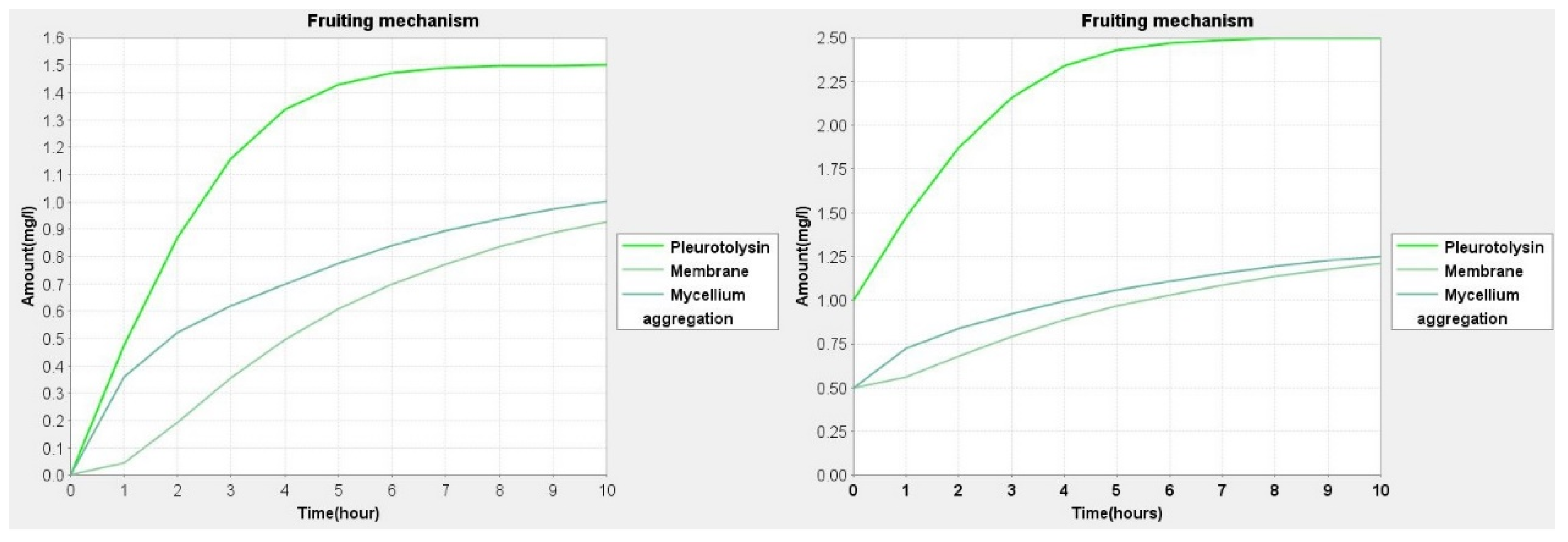
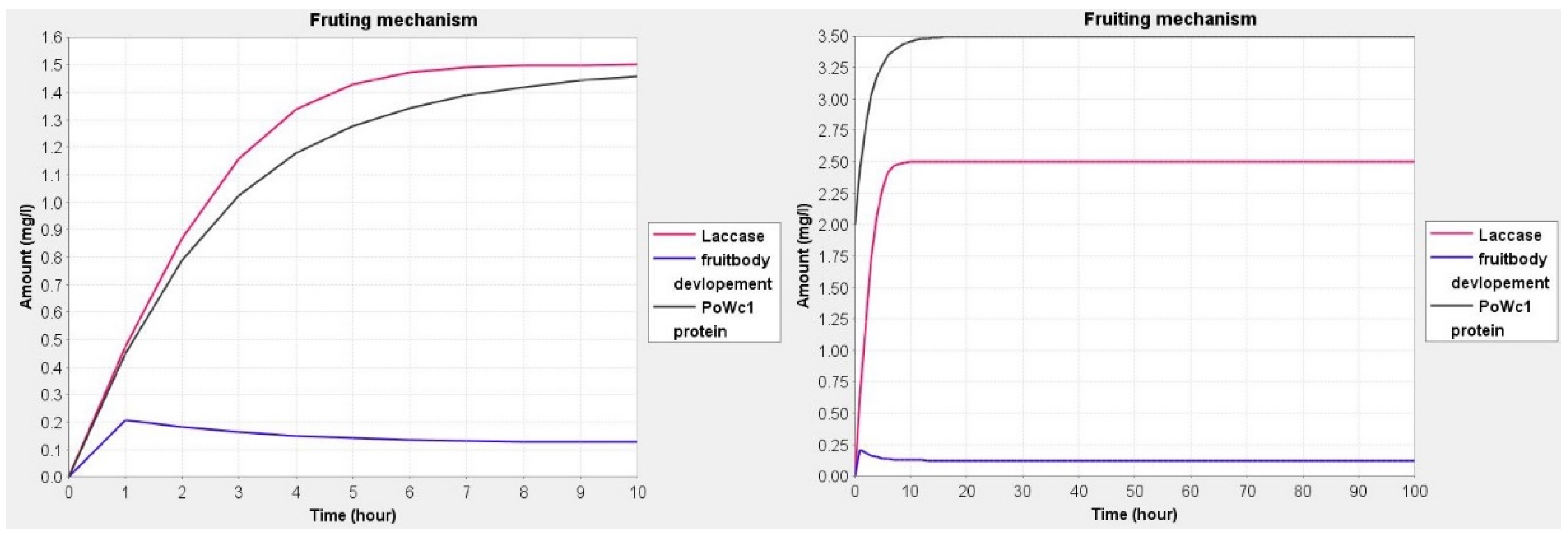
3.1. Dynamic Behavior Studies and Model Simulations

The prediction and behavior by simulation are used to understand the series of events taking place during the fruit development process in mushrooms. The predicted dynamic behavior ascertains the effect of the expression of genes in different environments. Simulation studies are helpful in the interpretation of the fate of various molecular species and metabolites with the degree of importance in the model. In simulation, although it is difficult to estimate the amount or concentration of molecular species/metabolites, an approximation was set up using previous wet laboratory-based studies. Knowing the amount of molecular species in quantitative terms will help us to find links and association amongst species [25,32]. In our model, we have used the range of 0.5 to 2.5 for each molecular species. The values of genes were set at 0.5, proteins were set at 2.0 and mRNA was set at 1.0. The value 2.5 was set for phenotypes and phenotypic responses. In each species, the variation of 0.5 to 1.0 was given and checks the changes in expression in the fungal cell (Figure 3, Figure 4 and Figure 5). Simple molecules were set at 0.5 due to its basal amount. The values for Pleurotolysin and membrane receptors were between 0.0 and 1.0 to study the effect on mycelium aggregation or knot formation. Similarly, the values for hydrophobin and Pleurotus White Collar complex protein were between 0.0 and 2.0 to study the effect on pinhead formation and fruit body devolvement. All the dynamics are provided in Supplementary files.

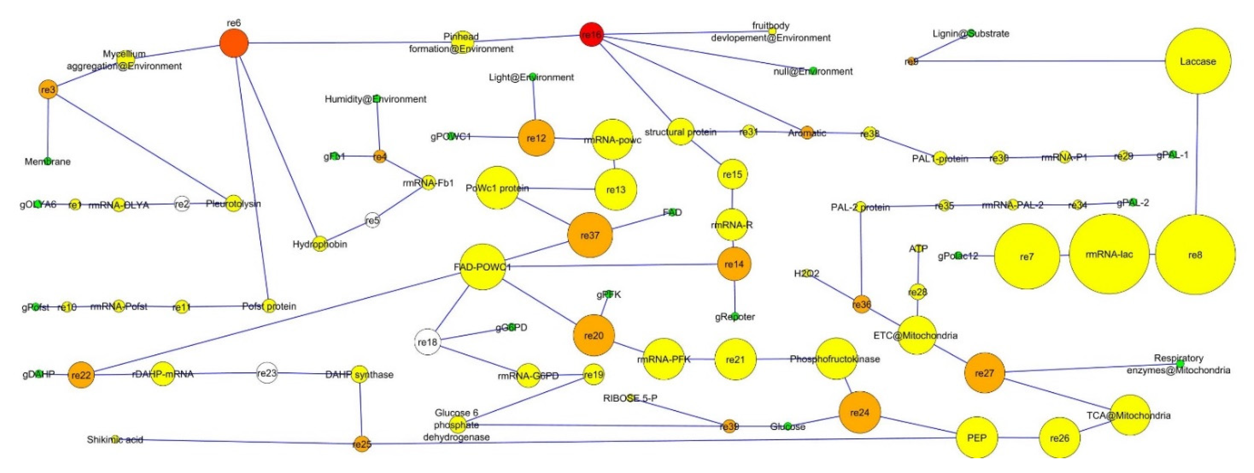
3.2. Topological Analysis of Fruit Body Development

The model analyzed for the network analysis showed the series of events predicted to occur during fruiting, and a total of 89 nodes and 90 edges were developed. The simple topology parameters are number of nodes, number of edges, connected components, clustering coefficient, network diameter, network radius, shortest paths, average neighbors numbers, density, radius, heterogeneity, centralization, self-loops and characteristic path length [28]. The network was developed using a directed network to estimate the average path length. The simple parameters analyzed in the network analysis are presented in Table 1.
The clustering coefficient of a node is the degree to which the nodes cluster together. The value range is between 0 to 1. The clustering coefficient is the ratio of the number of edges between the neighbors to the maximum number of edges that could possibly exist between the neighbors [33]. The number of connected components indicates the connectivity of a network and is inversely related. The neighborhood connectivity distribution shows that there is a decreasing function in k, which means that in the network, the edges are between low-connected and highly connected nodes [34] (Figure 6). The network diagram shows the node size “in-degree” and node color “betweeness centrality” and can be visualized in Figure 6. The map helped in the determination of the hub nodes. The nodes represented in red color are hub nodes and have a significant regulatory character in Pleurotus fruiting. The other nodes in yellow color had the least regulatory effect on the fruiting mechanism pathways. The green-color nodes show the moderate effect on the model pathway (Figure 6).
In the model, the fruit body development process involves most of the regulatory pathway re-16. The other important regulatory pathways were the conversion of mycelium aggregation to pinhead formation re-6 and White Collar protein (PoWC1) PoWC1-binding flavin-chromophore FAD to activate respiratory enzymes re-37. The other important pathways according to importance were re-20, re-24, re-27, re-12, re-14, re-22, re-3, re-36, re-25, re-39, re-4 and re-9 (Figure 6).
In network analysis, the edges and nodes are connected in terms of degree. In the directed node approach, in-degree distribution estimates the edges approaching a node, whereas the out degree distribution takes in account those edges which are leaving the node [35]. The nodes can be treated differently for different networks, for example in metabolic network, reactions are taken as edges and the metabolic intermediates are observed as nodes, while in gene regulatory networks, the nodes are genes and its regulatory molecules are edges. Moreover, in the protein–protein interactions network, nodes are proteins and edges are bonds between them [36]. The biological properties of the hubs are important in the fruiting mechanism to judge the role of protein or genes. Hubs are usually an intermediate, gene or protein with key regulatory roles (Figure 7). In the model, some highly connected nodes, such as Pleurotus White Collar protein (PoWC1) protein, are important in fruiting and mostly in the fruit body development. The Pleurotus mushroom is considered as one of the most important mushrooms not only for its edible use but also for its myco-remediation potential [37]. It is also essential that its fruiting be propagated in the environment to liberate the spores. The fruiting mechanism still needs to be studied in detail. A study showed that 596 unigenes were expressed during fruit body formation [38]. In, Pleurotus genetic improvement this research may be helpful in the development of early varieties using the important fruiting genes (Table 2).

4. Discussion

To find more detailed fruiting mechanism pathways and their response to different environmental factors, an in silico experiment was designed. Under the specific environmental conditions, development of the fruiting body starts with mycelium/aerial hyphal aggregation. Nutrient, light, humidity and temperature, etc., are some of the factors responsible for fruit body development [43,50,51]. These aerial dikaryotic hyphal aggregates continuously develop into primordia/pinhead which further differentiate into the mature mushroom fruiting body [51]. In our study, we broadened our knowledge on how different environmental factors, developmental genes (receptors genes, expression genes) and enzymes are responsible for the molecular mechanisms of fruiting in Pleurotus [52]. The predictive model helps to gain an insight into various genes and enzymes and their temporal expression which affects the fruiting mechanisms. Through studying Pleurotus, it is known that Pleurotolysin has 138 amino acid residues (16 k Da) and a pore-forming protein has been found to have a role in fruiting [39]. The OLYA6 gene, also known as Ostreolysin A6 (Aa-pri1 gene) transcript, is found in primordial stages and in immature fruit bodies. These proteins are associated with the membrane-attack complex/perforin (MACPF) domains and have various biological roles, such as defense and attack, organism development, cell adhesion and signaling [53]. Protein Pleurotolysin consists of non-associated components A and B. Pleurotolysin A is an egerolysin-like protein, while Pleurotolysin B has a MACPF domain [53]. It is observed that Pleurotolysin A and B together can cause hemolytic activity. In some of the studies, it is seen that Pleurotolysin A interacts with the membrane by recognizing the polar head group of sphingomyelin and eventually interacts with the hydrophobic ends of cholesterol and other phospholipids [54]. Moreover, the presence of both cholesterol and sphingomyelin affects Pleurotolysin binding to mono- and bilayer lipids, and lipid vesicle permeabilization. There are various explanations for the interaction, including that the Lysophospholipids act as a signaling molecule and thus regulate the pore forming ability of Pleurotolysin which transforms the cell differentiation from a vegetative to reproductive phase [55]. It is also noted in various studies that Pleurotolysin cannot increase the permeability of the lipid vesicle [56], but they may bind to it.
During the simulation of the network, we have found that reaction-3 (re3) (Figure 6) had some role in cell differentiation but we could not find a major role in the whole basidiocarp development. Although, it can be assumed that OLYA6 may be the triggering point of fruiting. Phenotypically hyphae aggregation may be governed by this protein. In our simulation study, it was found that the Pleurotolysin is an important protein for mycelium aggregation (Figure 3).
Another important step after mycelium aggregation is pinhead formation. In a humid environment, the hydrophobin gene is the most highly expressed gene of all during fruit body development. Hydrophobin is cysteine-rich and abundantly expresses fungal proteins [57,58]. Hydrophobins lower surface tension, letting the hyphae escape and initiating pinhead growth (aerial structure, aerial hyphae and fruit body) studied in Schizophyllum commune [40,41]. In the model, the hydrophobin Fbh1 gene is transcribed and translated in high humidity and produces hydrophobin proteins that interfere in aerial hyphae, spores and fruiting body development. Structurally, the Fbh1 and POH1 hydrophobin proteins contain two cysteine and 113 amino acids clusters [59]. During basiodiocarp formation, the hydrophobin protein is involved in hyphal adhesion and protects against desiccation and helps in gas exchange regulation. In Pleurotus ostreatus, the POH1 (P. ostreatus hydrophobin 1) gene showed a regulating effect for limiting pinhead development while POH2 and POH3 expressed specific levels of hydrophobins at the vegetative phase [60]. Penase et al., in 2002 and 2004, investigated two genes, i.e., vmh3 and Fbh1 from P. florida that were expressed in vegetative mycelium and fruiting body development, respectively [42,43]. Various other hydrophobins are reported in other mushrooms such as in the bracket mushroom (SC1, SC3 and SC4) [61], while in the button mushroom, ABH1 and ABH2 were detected in the mature fruit body [41,62].
In simulation network, reaction no. 6 (re6) in Figure 6 is also a significant reaction catalyzed by hydrophobin and the Pofst protein. This reaction is more important than reaction 3 in terms of significance as represented by the network analysis diagram. The Pofst3 gene is responsible for controlling the number and shape of sporocarps. Pofst3 is involved in the regulation by inhibiting the cluster formation in P. ostreatus [44]. Hyphae aggregation may be governed by this protein. In our simulation study, it was seen that the Pleurotolysin is an important protein for mycelium aggregation. It is seen that hydrophobin and the Pofst protein play an important role in pinhead formation. In reaction dynamics, as the concentration of hydrophobin increases, the pinhead number also increases, but a high concentration of the Pofst protein decreases the number of pinheads, but in the presence of both proteins a regulated amount of pinhead is formed (Figure 4). Pinhead to fruit body formation is a more complex process governed by various genes which include structural genes, metabolic genes, respiratory genes and secondary metabolites genes, etc. In Pleurotus, as reported in studies, blue light significantly contributes to the growth of pileus [49]. Transcriptomics study showed that blue light enzymes, mainly glyceraldehyde-3-phosphate dehydrogenase (GAPDH), 6-phosphogluconate dehydrogenase (6PGD) and phosphoenol pyruvate carboxykinase (PEPCK), significantly upregulated pileus formation by using specific pathways such as glycolysis/gluconeogenesis and pentose phosphate pathway (PPP) where 6PGD and PEPCK are rate-controlling genes in the pentose phosphate pathway and gluconeogenesis, respectively, and GAPDH is crucial in the glycolysis pathway [45]. A photoreceptor phototropic-like protein called Pleurotus White Collar 1 combines with FAD (flavin adenine dinucleotide) to form the PoWC1–FAD complex that induces the transcription and expression of respiratory genes by binding to their promoter site and inducing gene expression that leads to fruiting body development. In the model, it can be seen that both Glucose-6 phosphate dehydrogenase (G6PD) and phosphofructokinase (PFK) enzymes are involved in phosphoenol pyruvate (PEP) formation (Figure 2). The G6PD enzyme is mainly representative of the PEP pathway, but it showed lower activity as compared to hexokinase and glucose-6 phosphate isomerase (glycolytic pathway enzymes) for mycelial and non-sporulating stages. At the time of sporulation, the glycolytic pathway enzymes increase significantly as compared to other mycelial stages in the P. ostreatus mushroom [63]. A similar study, by Schwalb in 1974, reported that, in Schizophyllum commune, the sugar metabolism, glycolytic pathway was a major route during fruit body development [64]. After PEP product, the 3-deoxy-D-arabinoheptulosonate 7-phosphate synthase (DAHP) enzyme participated, and the end result is the formation of shikimic acid, collectively it is known as a shikimic acid pathway [65]. PEP entered into the TCA (tricarboxylic acid cycle) cycle in mitochondria followed by the electron transport chain (ETC) reaction to generate ATP via oxidative phosphorylation. The mitochondrial ETC consists of different complexes and these complexes form reactive oxygen as a byproduct during ATP formation and any imbalance or stress, especially heat stress, or recovery in their antioxidant systems [66,67]. During this phase, blue light also helps in the expression of genes encoding carbohydrate-active enzymes (CAZymes) during primordial differentiation to the fruiting body [46]. Moreover, Laccase is also one factor that is responsible for fruit body structuring and secondary metabolite formation in the Pleurotus mushroom. Laccases are multi-copper polyphenol oxidase enzymes which help in delignification, pathogenesis and pigmentation of the fruiting body [68,69]. PoLac1–12 putative laccase genes clusters have been noted in the genome of the P. ostreatus mushroom. As a matter of fact, during a transcription regulation study of Laccase genes, they were found to be involved in various stages, including mycelium, primodium and fruiting body formation [47]. The PoLac 12 gene is supposed to be responsible for lignin degradation after the formation of the mRNA-Polac12 complex. This complex finally degrades lignin and helps to develop primordia and the fruiting body of P. ostreatus [70]. Other Laccase genes such as PoLac2 and PoLac (POXC) have been overexpressed in the formation of the prophase of mycelium vegetative growth in P. ostreatus and are so closely related to lignin degradation [71]. In network analysis, it was found that reaction 13 and reaction 37 are the most significant. Both reactions are linked with fruit body development. In reaction 13, mostly PAL genes, Laccase and the ATP development process are important, while a significant role can be observed for the PoWC1 protein (Figure 6). In simulation studies, it is seen that, as the PoWC1 protein increases, the fruit body development increases. Moreover, PAL1, PAL2, G6PD, Laccase, structural proteins, and electron transport chain enzymes have positive and direct effects on the body development of Pleurotus.
Some other genes shown in the model, such as Phenylalanine ammonium-lysae (PAL), are protein genes, which were highly expressed in the phenylpropanoid pathway [72], that develop the phenolic compound in fruiting bodies. PAL genes expression are found under abiotic stresses, mainly UV light, temperature, salts, etc., that lead to the collection of phenolic compounds [47]. Two PAL genes, i.e., PAL1 and PAL2 are identified in P. ostreatus genome during the developmental stage, the expression of both genes was shown in all developmental stages but the highest expression was observed in fruit gills [73]. The expression of the PAL2 gene significantly up-regulated the process during primordia, fruiting body and spore development in P. ostreatus [48]. PALs, along with tyrosinases, are involved in pileus pigment formation [49].

5. Conclusions

This study widens our knowledge of the advancements in the omics analysis technology by using different bioinformatics tools, demonstrating how the developmental and regulated genes/enzymes in the model are responsible for the fruiting body development of the edible Pleurotus mushroom. We studied a series of genes in our study and three genes showed importance based on the role of regulatory pathways, i.e., PoWC1 (blue light), the Fbh1 gene (hydrophobin) and the Pofst gene. PoWC1 is involved in fulfilling both structural changes and helps to fulfill respiratory requirements, while the Fbh1 gene (hydrophobin) and Pofst gene are mainly responsible for structural change. Moreover, OLYA6 may be the triggering point of fruiting but does not act as the governor of key regulatory pathways. Several other biosynthesis genes (PAL, PoLAC gene) and respiratory enzymes (G6PD, PEP, DAHP synthase, PFK) are regulating the entire fruiting body development process during the primordial, fruiting body and spore development process in the Pleurotus mushroom. To understand the specific role of differentially expressed genes might help determine good candidates for in-depth experimental studies. Additional comprehension of different environmental factors and genes involved enhancing or supporting fruiting body induction and development is censorious from the trade point of view and will be helpful in the future for commercial Pleurotus mushroom cultivation.

Supplementary Materials

The following supporting information can be downloaded at: https://data.mendeley.com/datasets/7rhvryrkzg/1.

Author Contributions

Conceptualization, A.B. and P.B.; methodology, A.B., P.B. and K.S.; software, K.S. and A.B.; validation, S.K.A., M.N., B.K. and K.K.; formal analysis, A.B. and S.K.; investigation, S.K.; writing—original draft preparation, A.B., K.S., M.N., S.K.A. and M.S.; writing—review and editing, V.P.S., S.G., A.S., M.G. and U.D. All authors have read and agreed to the published version of the manuscript.

Funding

This research received no external funding.

Institutional Review Board Statement

Not applicable for studies not involving humans or animals.

Informed Consent Statement

Not applicable.

Data Availability Statement

Not applicable.

Acknowledgments

Authors acknowledge Director, ICAR-DMR for his support for conducting work.

Conflicts of Interest

The authors declare no conflict of interest.

References

  1. Royse, D.J.; Baars, J.; Tan, Q. Current overview of mushroom production in the world. In Edible and Medicinal Mushrooms: Technology and Applications; Zied, D.C., Pardo-Giménez, A., Eds.; John Wiley & Sons Ltd.: New York, NY, USA, 2017; pp. 5–13. [Google Scholar]
  2. Singh, M.; Kamal, S.; Sharma, V.P. Species and Region-wise Mushroom Production in Leading Mushroom Producing Countries—China, Japan, USA, Canada and India. Mushroom Res. 2021, 30, 99–108. [Google Scholar] [CrossRef]
  3. Reis, F.S.; Barros, L.; Martins, A.; Ferreira, I.C.F.R. Chemical composition and nutritional value of the most widely appreciated cultivated mushrooms: An inter-species comparative study. Food Chem. Toxicol. 2012, 50, 191–197. [Google Scholar] [CrossRef] [PubMed] [Green Version]
  4. Lopes, R.D.O.; Pereira, P.M.; Pereira, A.R.B.; Fernandes, K.V.; Carvalho, J.F.; da Silva de França, A.; Valente, R.H.; da Silva, M.; Ferreira-Leitão, V.S. Atrazine, desethylatrazine (DEA) and desisopropylatrazine (DIA) degradation by Pleurotus ostreatus INCQS 40310. Biocatal. Biotransform. 2020, 38, 415–430. [Google Scholar] [CrossRef]
  5. Pozdnyakova, N.N.; Jarosz-Wilkolazka, A.; Polak, J.; Grąz, M.; Turkovskaya, O.V. Decolourisation of anthraquinone-and anthracene-type dyes by versatile peroxidases from Bjerkandera fumosa and Pleurotus ostreatus D1. Biocatal. Biotransform. 2015, 33, 69–80. [Google Scholar] [CrossRef]
  6. Wu, Y.; Choi, M.-H.; Li, J.; Yang, H.; Shin, H.-J. Mushroom Cosmetics: The Present and Future. Cosmetics 2016, 3, 22. [Google Scholar] [CrossRef]
  7. Singh, M.; Kamal, S. Genetic Aspects and Strategies for Obtaining Hybrids. In Edible and Medicinal Mushrooms; John Wiley & Sons, Ltd.: Chichester, UK, 2017; pp. 35–87. ISBN 9781119149446. [Google Scholar]
  8. Wösten, H.A.B.; Wessels, J.G.H. The Emergence of Fruiting Bodies in Basidiomycetes. In Growth, Differentiation and Sexuality; Springer: Berlin/Heidelberg, Germany, 2006; pp. 393–414. [Google Scholar]
  9. Stamets, P. Growing Gourmet and Medicinal Mushrooms, 3rd ed.; Ten speed press: Toronto, ON, Canada, 2000. [Google Scholar]
  10. Zhang, J.; Ren, A.; Chen, H.; Zhao, M.; Shi, L.; Chen, M.; Wang, H.; Feng, Z. Transcriptome Analysis and Its Application in Identifying Genes Associated with Fruiting Body Development in Basidiomycete Hypsizygus marmoreus. PLoS ONE 2015, 10, e0123025. [Google Scholar] [CrossRef] [Green Version]
  11. Wessels, J.G.H. Tansley Review No. 45 Wall growth, protein excretion and morphogenesis in fungi. New Phytol. 1993, 123, 397–413. [Google Scholar] [CrossRef]
  12. Barh, A.; Sharma, V.P.; Annepu, S.K.; Kamal, S.; Sharma, S.; Bhatt, P. Genetic improvement in Pleurotus (oyster mushroom): A review. 3 Biotech 2019, 9, 322. [Google Scholar] [CrossRef]
  13. Han, C.-H.; Zhang, G.-Q.; Wang, H.-X.; Ng, T.B. Schizolysin, a hemolysin from the split gill mushroom Schizophyllum commune. FEMS Microbiol. Lett. 2010, 309, 115–121. [Google Scholar] [CrossRef]
  14. Kamada, T.; Sano, H.; Nakazawa, T.; Nakahori, K. Regulation of fruiting body photomorphogenesis in Coprinopsis cinerea. Fungal Genet. Biol. 2010, 47, 917–921. [Google Scholar] [CrossRef]
  15. Herzog, R.; Solovyeva, I.; Bölker, M.; Lugones, L.G.; Hennicke, F. Exploring molecular tools for transformation and gene expression in the cultivated edible mushroom Agrocybe aegerita. Mol. Genet. Genom. 2019, 294, 663–677. [Google Scholar] [CrossRef]
  16. Krizsán, K.; Almási, É.; Merényi, Z.; Sahu, N.; Virágh, M.; Kószó, T.; Mondo, S.; Kiss, B.; Bálint, B.; Kües, U.; et al. Transcriptomic atlas of mushroom development reveals conserved genes behind complex multicellularity in fungi. Proc. Natl. Acad. Sci. USA 2019, 116, 7409–7418. [Google Scholar] [CrossRef] [Green Version]
  17. Pelkmans, J.F.; Lugones, L.G.; Wösten, H.A.B. Fruiting Body Formation in Basidiomycetes. In Growth, Differentiation and Sexuality; Springer International Publishing: Berlin/Heidelberg, Germany, 2016; pp. 387–405. [Google Scholar]
  18. Berne, S.; Križaj, I.; Pohleven, F.; Turk, T.; Maček, P.; Sepčić, K. Pleurotus and Agrocybe hemolysins, new proteins hypothetically involved in fungal fruiting. Biochim. Biophys. Acta-Gen. Subj. 2002, 1570, 153–159. [Google Scholar] [CrossRef]
  19. Peñas, M.M.; Ásgeirsdóttir, S.A.; Lasa, I.; Culiañez-Macià, F.A.; Pisabarro, A.G.; Wessels, J.G.H.; Ramírez, L. Identification, characterization, and in situ detection of a fruit-body-specific hydrophobin of Pleurotus ostreatus. Appl. Environ. Microbiol. 1998, 64, 4028–4034. [Google Scholar] [CrossRef] [Green Version]
  20. Muraguchi, H.; Kamada, T. A mutation in the eln2 gene encoding a cytochrome P450 of Coprinus cinereus affects mushroom morphogenesis. Fungal Genet. Biol. 2000, 29, 49–59. [Google Scholar] [CrossRef]
  21. Suzuki, H.; Vuong, T.V.; Gong, Y.; Chan, K.; Ho, C.Y.; Master, E.R.; Kondo, A. Sequence diversity and gene expression analyses of expansin-related proteins in the white-rot basidiomycete, Phanerochaete carnosa. Fungal Genet. Biol. 2014, 72, 115–123. [Google Scholar] [CrossRef]
  22. Bhatt, P.; Barh, A. Bioinformatic Tools to Study the Soil Microorganisms: An In Silico Approach for Sustainable Agriculture. In In Silico Approach for Sustainable Agriculture; Springer: Singapore, 2018; pp. 169–182. [Google Scholar]
  23. Funahashi, A.; Morohashi, M.; Matsuoka, Y.; Jouraku, A.; Kitano, H. CellDesigner: A graphical biological network editor and workbench interfacing simulator. In Introduction to Systems Biology; Humana Press: Totowa, NJ, USA, 2007; pp. 422–434. ISBN 9781588297068. [Google Scholar]
  24. Hucka, M.; Bergmann, F.T.; Dräger, A.; Hoops, S.; Keating, S.M.; Le Novère, N.; Myers, C.J.; Olivier, B.G.; Sahle, S.; Schaff, J.C.; et al. The Systems Biology Markup Language (SBML): Language Specification for Level 3 Version 2 Core. J. Integr. Bioinform. 2018, 15, 20170081. [Google Scholar] [CrossRef]
  25. Pathak, R.K.; Baunthiyal, M.; Pandey, N.; Pandey, D.; Kumar, A. Modeling of the jasmonate signaling pathway in Arabidopsis thaliana with respect to pathophysiology of Alternaria blight in Brassica. Sci. Rep. 2017, 7, 16790. [Google Scholar] [CrossRef] [Green Version]
  26. Bhatt, P.; Sethi, K.; Gangola, S.; Bhandari, G.; Verma, A.; Adnan, M.; Singh, Y.; Chaube, S. Modeling and simulation of atrazine biodegradation in bacteria and its effect in other living systems. J. Biomol. Struct. Dyn. 2020, 40, 3285–3295. [Google Scholar] [CrossRef]
  27. Machné, R.; Finney, A.; Müller, S.; Lu, J.; Widder, S.; Flamm, C. The SBML ODE Solver Library: A native API for symbolic and fast numerical analysis of reaction networks. Bioinformatics 2006, 22, 1406–1407. [Google Scholar] [CrossRef] [Green Version]
  28. Assenov, Y.; Ramírez, F.; Schelhorn, S.-E.; Lengauer, T.; Albrecht, M. Computing topological parameters of biological networks. Bioinform. Appl. 2008, 24, 282–284. [Google Scholar] [CrossRef] [Green Version]
  29. Su, G.; Morris, J.H.; Demchak, B.; Bader, G.D. Biological Network Exploration with Cytoscape 3. Curr. Protoc. Bioinform. 2014, 47, 8–13. [Google Scholar] [CrossRef] [Green Version]
  30. Zinovyev, A.; Viara, E.; Calzone, L.; Barillot, E. BiNoM: A Cytoscape plugin for manipulating and analyzing biological networks. Bioinformatics 2008, 24, 876–877. [Google Scholar] [CrossRef] [Green Version]
  31. Suderman, M.; Hallett, M. Tools for visually exploring biological networks. Bioinformatics 2007, 23, 2651–2659. [Google Scholar] [CrossRef]
  32. Bhatt, P.; Pal, K.; Bhandari, G.; Barh, A. Modelling of the methyl halide biodegradation in bacteria and its effect on environmental systems. Pestic. Biochem. Physiol. 2019, 158, 88–100. [Google Scholar] [CrossRef]
  33. Max Planck Institute for Informatics NetworkAnalyzer. Available online: https://med.bioinf.mpi-inf.mpg.de/netanalyzer/help/2.5/#refBarabasi04 (accessed on 18 January 2021).
  34. Maslov, S.; Sneppen, K. Specificity and stability in topology of protein networks. Science 2002, 296, 910–913. [Google Scholar] [CrossRef] [Green Version]
  35. Kohl, M.; Wiese, S.; Warscheid, B. Cytoscape: Software for visualization and analysis of biological networks. Methods Mol. Biol. 2011, 696, 291–303. [Google Scholar] [CrossRef]
  36. Barabási, A.L.; Oltvai, Z.N. Network biology: Understanding the cell’s functional organization. Nat. Rev. Genet. 2004, 5, 101–113. [Google Scholar] [CrossRef]
  37. Chun, S.C.; Muthu, M.; Hasan, N.; Tasneem, S.; Gopal, J. Mycoremediation of PCBs by Pleurotus ostreatus: Possibilities and Prospects. Appl. Sci. 2019, 9, 4185. [Google Scholar] [CrossRef] [Green Version]
  38. Lee, S.H.; Kim, B.G.; Kim, K.J.; Lee, J.S.; Yun, D.W.; Hahn, J.H.; Kim, G.H.; Lee, K.H.; Suh, D.S.; Kwon, S.T.; et al. Comparative analysis of sequences expressed during the liquid-cultured mycelia and fruit body stages of Pleurotus ostreatus. Fungal Genet. Biol. 2002, 35, 115–134. [Google Scholar] [CrossRef]
  39. Berne, S.; Pohleven, J.; Vidic, I.; Rebolj, K.; Pohleven, F.; Turk, T.; Ek, P.M.; Sonnenberg, A.; Sepčić, K.; Eastwood, D.C. Ostreolysin enhances fruiting initiation in the oyster mushroom (Pleurotus ostreatus). Mycol. Res. 2007, 111, 1431–1436. [Google Scholar] [CrossRef] [PubMed]
  40. Wösten, H.A.B.; Wessels, J.G.H. Hydrophobins, from molecular structure to multiple functions in fungal development. Mycoscience 1997, 38, 363–374. [Google Scholar] [CrossRef]
  41. Lugones, L.; Wosten, H.B.; Wessels, J.H. A hydrophobin (ABH3) specifically secreted by vegetatively growing hyphae of Agaricus bisporus (common white button mushroom). Microbiology 1998, 144, 2345–2353. [Google Scholar] [CrossRef] [PubMed] [Green Version]
  42. Peñas, M.M.; Aranguren, J.; Ramírez, L.; Pisabarro, A.G. Structure of gene coding for the fruit body-specific hydrophobia Fbh1 of the edible basidiomycete Pleurotus ostreatus. Mycologia 2004, 96, 75–82. [Google Scholar] [CrossRef]
  43. Peñas, M.M.; Rust, B.; Larraya, L.M.; Ramírez, L.; Pisabarro, A.G. Differentially regulated, vegetative-mycelium-specific hydrophobins of the edible basidiomycete Pleurotus ostreatus. Appl. Environ. Microbiol. 2002, 68, 3891–3898. [Google Scholar] [CrossRef] [Green Version]
  44. Qi, Y.; Chen, H.; Zhang, M.; Wen, Q.; Qiu, L.; Shen, J. Identification and expression analysis of Pofst3 suggests a role during Pleurotus ostreatus primordia formation. Fungal Biol. 2019, 123, 200–208. [Google Scholar] [CrossRef]
  45. Wang, H.; Tong, X.; Tian, F.; Jia, C.; Li, C.; Li, Y. Transcriptomic profiling sheds light on the blue-light and red-light response of oyster mushroom (Pleurotus ostreatus). AMB Express 2020, 10, 10. [Google Scholar] [CrossRef] [Green Version]
  46. Xie, C.; Gong, W.; Zhu, Z.; Yan, L.; Hu, Z.; Peng, Y. Comparative transcriptomics of Pleurotus eryngii reveals blue-light regulation of carbohydrate-active enzymes (CAZymes) expression at primordium differentiated into fruiting body stage. Genomics 2018, 110, 201–209. [Google Scholar] [CrossRef]
  47. Lettera, V.; Piscitelli, A.; Leo, G.; Birolo, L.; Pezzella, C.; Sannia, G. Identification of a new member of Pleurotus ostreatus laccase family from mature fruiting body. Fungal Biol. 2010, 114, 724–730. [Google Scholar] [CrossRef]
  48. Liu, X.B.; Xia, E.H.; Li, M.; Cui, Y.Y.; Wang, P.M.; Zhang, J.X.; Xie, B.G.; Xu, J.P.; Yan, J.J.; Li, J.; et al. Transcriptome data reveal conserved patterns of fruiting body development and response to heat stress in the mushroomforming fungus Flammulina filiformis. PLoS ONE 2020, 15, e0239890. [Google Scholar] [CrossRef]
  49. Du, F.; Zou, Y.; Hu, Q.; Zhang, H.; Ye, D. Comparative transcriptomic analysis reveals molecular processes involved in pileus morphogenesis in Pleurotus eryngii under different light conditions. Genomics 2020, 112, 1707–1715. [Google Scholar] [CrossRef]
  50. Ohm, R.A.; De Jong, J.F.; Lugones, L.G.; Aerts, A.; Kothe, E.; Stajich, J.E.; De Vries, R.P.; Record, E.; Levasseur, A.; Baker, S.E.; et al. Genome sequence of the model mushroom Schizophyllum commune. Nat. Biotechnol. 2010, 28, 957–963. [Google Scholar] [CrossRef] [PubMed] [Green Version]
  51. Muraguchi, H.; Umezawa, K.; Niikura, M.; Yoshida, M.; Kozaki, T.; Ishii, K.; Sakai, K.; Shimizu, M.; Nakahori, K.; Sakamoto, Y.; et al. Strand-Specific RNA-Seq Analyses of Fruiting Body Development in Coprinopsis cinerea. PLoS ONE 2015, 10, e0141586. [Google Scholar] [CrossRef] [Green Version]
  52. Varga, T.; Krizsán, K.; Földi, C.; Dima, B.; Sánchez-García, M.; Sánchez-Ramírez, S.; Szöllősi, G.J.; Szarkándi, J.G.; Papp, V.; Albert, L.; et al. Megaphylogeny resolves global patterns of mushroom evolution. Nat. Ecol. Evol. 2019, 3, 668–678. [Google Scholar] [CrossRef] [PubMed] [Green Version]
  53. Ota, K.; Butala, M.; Viero, G.; Serra, M.D.; Sepčić, K.; Maček, P. Fungal MACPF-like proteins and aegerolysins: Bi-component pore-forming proteins? In MACPF/CDC Proteins—Agents of Defence, Attack and Invasion; Anderluh, G., Gilbert, R., Eds.; Springer Dordrecht: Berlin/Heidelberg, Germany, 2014; Volume 80, pp. 271–291. [Google Scholar]
  54. Tomita, T.; Noguchi, K.; Mimuro, H.; Ukaji, F.; Ito, K.; Sugawara-Tomita, N.; Hashimoto, Y. Pleurotolysin, a novel sphingomyelin-specific two-component cytolysin from the edible mushroom Pleurotus ostreatus, assembles into a transmembrane pore complex. J. Biol. Chem. 2004, 279, 26975–26982. [Google Scholar] [CrossRef] [Green Version]
  55. Sepcic, K.; Berne, S.; Potrich, C.; Turk, T.; Macek, P.; Menestrina, G. Interaction of ostreolysin, a cytolytic protein from the edible mushroom Pleurotus ostreatus, with lipid membranes and modulation by lysophospholipids. Eur. J. Biochem. 2003, 270, 1199–1210. [Google Scholar] [CrossRef] [PubMed] [Green Version]
  56. Kurahashi, A.; Sato, M.; Kobayashi, T.; Nishibori, K.; Fujimori, F. Homologous genes, Pe. pleurotolysin A and Pe. ostreolysin, are both specifically and highly expressed in primordia and young fruiting bodies of Pleurotus eryngii. Mycoscience 2014, 55, 113–117. [Google Scholar] [CrossRef]
  57. Kulkarni, S.S.; Nene, S.N.; Joshi, K.S. A comparative study of production of hydrophobin like proteins (HYD-LPs) in submerged liquid and solid state fermentation from white rot fungus Pleurotus ostreatus. Biocatal. Agric. Biotechnol. 2020, 23, 101440. [Google Scholar] [CrossRef]
  58. Schuren, F.H.J.; Wessels, J.G.H. Two genes specifically expressed in fruiting dikaryons of Schizophyllum commune: Homologies with a gene not regulated by mating-type genes. Gene 1990, 90, 199–205. [Google Scholar] [CrossRef]
  59. Kershaw, M.J.; Talbot, N.J. Hydrophobins and Repellents: Proteins with Fundamental Roles in Fungal Morphogenesis. Fungal Genet. Biol. 1998, 23, 18–33. [Google Scholar] [CrossRef]
  60. Ásgeirsdóttir, S.A.; De Vries, O.M.H.; Wessels, J.G.H. Identification of three differentially expressed hydrophobins in Pleurotus ostreatus (oyster mushroom). Microbiology 1998, 144, 2961–2969. [Google Scholar] [CrossRef] [Green Version]
  61. Van Wetter, M.A.; Wösten, H.A.B.; Wessels, J.G.H. SC3 and SC4 hydrophobins have distinct roles in formation of aerial structures in dikaryons of Schizophyllum commune. Mol. Microbiol. 2000, 36, 201–210. [Google Scholar] [CrossRef]
  62. De Groot, P.W.J.; Schaap, P.J.; Sonnenberg, A.S.M.; Visser, J.; Van Griensven, L.J.L.D. The Agaricus bisporus hypA gene encodes a hydrophobin and specifically accumulates in peel tissue of mushroom caps during fruit body development. J. Mol. Biol. 1996, 257, 1008–1018. [Google Scholar] [CrossRef]
  63. Chakraborty, T.K.; Das, N.; Mukherjee, M. Evidences of high carbon catabolic enzyme activities during sporulation of Pleurotus ostreatus (Florida). J. Basic Microbiol. 2003, 43, 462–467. [Google Scholar] [CrossRef]
  64. Schwalb, M.N.; Shanler, A. Phototropic and geotropic responses during the development of normal and mutant fruit bodies of the basidiomycete Schizophyllum commune. J. Gen. Microbiol. 1974, 82, 209–212. [Google Scholar] [CrossRef] [Green Version]
  65. Kojima, M.; Kimura, N.; Miura, R. Regulation of primary metabolic pathways in oyster mushroom mycelia induced by blue light stimulation: Accumulation of shikimic acid. Sci. Rep. 2015, 5, 8630. [Google Scholar] [CrossRef] [Green Version]
  66. Zhou, S.; Ma, F.; Zhang, X.; Zhang, J. Carbohydrate changes during growth and fruiting in Pleurotus ostreatus. Fungal Biol. 2016, 120, 852–861. [Google Scholar] [CrossRef]
  67. Lee, S.; Tak, E.; Lee, J.; Rashid, M.; Murphy, M.P.; Ha, J.; Kim, S.S. Mitochondrial H2O2 generated from electron transport chain complex i stimulates muscle differentiation. Cell Res. 2011, 21, 817–834. [Google Scholar] [CrossRef] [Green Version]
  68. Jiao, X.; Li, G.; Wang, Y.; Nie, F.; Cheng, X.; Abdullah, M.; Lin, Y.; Cai, Y. Systematic analysis of the Pleurotus ostreatus laccase gene (PoLac) Family and functional characterization of PoLac2 involved in the degradation of cotton-straw lignin. Molecules 2018, 23, 880. [Google Scholar] [CrossRef] [Green Version]
  69. Barh, A.; Kumari, B.; Sharma, S.; Annepu, S.K.; Kumar, A.; Kamal, S.; Sharma, V.P. Mushroom mycoremediation: Kinetics and mechanism. In Smart Bioremediation Technologies: Microbial Enzymes; Elsevier: Amsterdam, The Netherlands, 2019; pp. 1–22. ISBN 9780128183076. [Google Scholar]
  70. Park, M.; Kim, M.; Kim, S.; Ha, B.; Ro, H.-S. Differential Expression of Laccase Genes in Pleurotus ostreatus and Biochemical Characterization of Laccase Isozymes Produced in Pichia pastoris. Mycobiology 2015, 43, 280–287. [Google Scholar] [CrossRef]
  71. Pezzella, C.; Autore, F.; Giardina, P.; Piscitelli, A.; Sannia, G.; Faraco, V. The Pleurotus ostreatus laccase multi-gene family: Isolation and heterologous expression of new family members. Curr. Genet. 2009, 55, 45–57. [Google Scholar] [CrossRef] [PubMed]
  72. Zou, Y.; Zhang, M.; Qu, J.; Zhang, J. iTRAQ-Based Quantitative Proteomic Analysis Reveals Proteomic Changes in Mycelium of Pleurotus ostreatus in Response to Heat Stress and Subsequent Recovery. Front. Microbiol. 2018, 9, 2368. [Google Scholar] [CrossRef] [PubMed]
  73. Hou, L.; Wang, L.; Wu, X.; Gao, W.; Zhang, J.; Huang, C. Expression patterns of two pal genes of Pleurotus ostreatus across developmental stages and under heat stress. BMC Microbiol. 2019, 19, 231. [Google Scholar] [CrossRef] [PubMed]
Figure 1. System biology graphical notation (SBGN) symbols used in Cell Designer for modeling of fruiting mechanism in Pleurotus.
Jof 08 01073 g001
Figure 2. Fruiting mechanism in Pleurotus constructed using Cell Designer in fungal cells, in connection with the substrate and environment. The diagram shows the simple molecules, proteins, mRNA, DNA, enzymes, catalysis, and inhibition, etc.
Jof 08 01073 g002
Figure 3. Simulation curve for Pleurotolysin protein and membrane receptor with respect to mycelium aggregation, the amount of Pleurotolysin was set at 0.0 and 1.0, respectively, while and membrane receptor and mycelium aggregation were set to 0 and 0.5.
Jof 08 01073 g003
Figure 4. Dynamics of hydrophobin protein and Pofst protein with respect to pinhead formation, the amount of hydrophobin and pinhead formation were set at 0.0 and 1.0, respectively, while Pofst protein was set at 0.0 and 0.5.
Jof 08 01073 g004
Figure 5. Dynamics of PoWC1 protein and Laccase with respect to fruit body development, the amount of PoWC1 protein was set at 0.0 and 2.0, respectively, while Laccase and fruit body development were set at 0.0.
Jof 08 01073 g005
Figure 6. Mapping of the fruiting mechanism pathway: visualization of complete network pathway to map hub nodes.
Jof 08 01073 g006
Figure 7. Module style view of fruiting mechanism pathway network node with physical simulation and catalysis.
Jof 08 01073 g007
Table 1. Simple topology parameters of fruiting network.
Sl. No.Simple Topology ParametersValues
1.Clustering coefficient0.0
2.Connected components2.0
3.Network diameter (largest distance between two nodes)18.0
4.Network radius1
5.Shortest paths747(%)
6.Characteristic path length/average shortest path length5.63
7.Average number of neighbors/average connectivity2.02
8.Network density0.0
Table 2. Role of genes and protein in Pleurotus fruit body formation and development.
Sl. No.ProteinRoleReference
1PleurotolysinPore-forming protein having role in fruiting[39]
2HydrophobinsInitiate pinhead growth makes aerial structure-aerial hyphae and fruit body[40,41,42,43]
3Pofst proteinRegulation of fruiting by inhibiting the excessive cluster formation[44]
4Glyceraldehyde-3-phosphate dehydrogenase (GAPDH)Role in glycolysis/gluconeogenesis and pentose phosphate pathway to promote fruitbody growth[45]
56-phosphogluconate dehydrogenase (6PGD)Role in glycolysis/gluconeogenesis and pentose phosphate pathway to promote fruitbody growth[45]
6Phosphoenol pyruvate carboxykinase (PEPCK)Role in glycolysis/gluconeogenesis and pentose phosphate pathway to promote fruitbody growth[45]
7Pleurotus White Collar proteinIt induces transcription and expression of respiratory genes[46]
8Carbohydrate-active enzymesPrimordial differentiation to fruiting body with help of blue light[46]
9LaccasesRole in fruit body formation[47]
10PAL genesExpression upregulated during primodia fruiting body and spore development. These genes along with tyrosinases involved in pileus pigment formation[48,49]
Publisher’s Note: MDPI stays neutral with regard to jurisdictional claims in published maps and institutional affiliations.

