Abstract
The orphan but highly virulent pathogen Pythium insidiosum causes pythiosis in humans and animals. Surgery is a primary treatment aiming to cure but trading off losing affected organs. Antimicrobial drugs show limited efficacy in treating pythiosis. Alternative drugs effective against the pathogen are needed. In-house drug susceptibility tests (i.e., broth dilution, disc diffusion, and radial growth assays) have been established, some of which adapted the standard protocols (i.e., CLSI M38-A2 and CLSI M51) designed for fungi. Hyphal plug, hyphal suspension, and zoospore are inocula commonly used in the drug susceptibility assessment for P. insidiosum. A side-by-side comparison demonstrated that each method had advantages and limitations. Minimum inhibitory and cidal concentrations of a drug varied depending on the selected method. Material availability, user experience, and organism and drug quantities determined which susceptibility assay should be used. We employed the hyphal plug and a combination of broth dilution and radial growth methods to screen and validate the anti-P. insidiosum activities of several previously reported chemicals, including potassium iodide, triamcinolone acetonide, dimethyl sulfoxide, and ethanol, in which data on their anti-P. insidiosum efficacy are limited. We tested each chemical against 29 genetically diverse isolates of P. insidiosum. These chemicals possessed direct antimicrobial effects on the growth of the pathogen in a dose- and time-dependent manner, suggesting their potential application in pythiosis treatment. Future attempts should focus on standardizing these drug susceptibility methods, such as determining susceptibility/resistant breakpoints, so healthcare workers can confidently interpret a result and select an effective drug against P. insidiosum.
1. Introduction
The filamentous organism Pythium insidiosum is a member of the oomycetes that belong to the Kingdom Stramenopiles and causes a fatal infectious condition called pythiosis in humans and animals [1]. The number of pythiosis cases has been increasingly documented in tropical and subtropical countries [2]. P. insidiosum inhabits freshwater, where it colonizes a water plant and produces zoospores to complete its life cycle [3,4,5,6]. Clinical manifestations of pythiosis include cutaneous granulomatous ulcers [7,8], gastrointestinal lesion [9,10], corneal ulcers [11,12], arteritis [13,14], and disseminated infection [15,16]. A definitive diagnosis of pythiosis relies on culture-based identification [17,18,19,20], histopathological examination [21,22], serological assays [23,24,25,26,27], molecular methods [28,29,30,31,32,33], and proteomic assessment [34,35].
The overall mortality rate in humans and animals with pythiosis is ~30% [2]. Antimicrobial medication (i.e., itraconazole, terbinafine, amphotericin B, linezolid, azithromycin, and doxycycline) is usually ineffective in pythiosis treatment [36,37,38,39,40,41,42,43,44,45,46,47,48,49,50]. However, some ocular pythiosis patients showed a favorable response after such medical treatment. Surgical removal of an infected organ is the primary treatment for a pythiosis patient [51,52,53,54,55]. Administration of the immunotherapeutic antigen extracted from P. insidiosum could reduce disease morbidity and mortality [56,57,58,59,60,61,62]. In some pythiosis patients, surgical intervention is impossible or fails to remove all infected tissue [63,64,65,66]. The management of such cases relies on antimicrobial agents and other treatment modalities to control the disease and prevent a recurrence. Some unconventional medications (i.e., potassium iodide (KI) [15,39,46,67,68,69,70,71,72,73,74,75,76,77,78], triamcinolone acetonide (TA) [68,79,80,81,82], dimethyl sulfoxide (DMSO) [83,84,85,86,87], and ethanol (EtOH) [88,89,90]) have been implemented for the management of several pythiosis patients and show satisfactory treatment outcomes.
KI, TA, DMSO, and EtOH were not originally designed for use as antimicrobial agents. Their information regarding the anti-P. insidiosum effect is limited and has not been comprehensively explored. KI is an inorganic salt used for many purposes. In the industry, it has been applied for inhibiting corrosion, facilitating chemical transformation, and catalyzing biodiesel [91,92,93]. In medicine, this chemical has been utilized in various clinical conditions, such as protecting the thyroid gland against the iodine-131 radioisotope, controlling inflammation in dermatoses, and treating several mycoses (i.e., sporotrichosis, cryptococcosis, entomophthoramycosis, and pythiosis) [94,95,96]. Administration of KI, as a part of the treatment, led to a clinical improvement in some humans, horses, and sheep with cutaneous pythiosis [15,39,46,67,69,72,75,77,78,97]. Additionally, KI has been used in treating other forms of pythiosis (i.e., vascular, ocular, disseminated) [54,69,70,71,74,77,98,99,100].
Steroids (i.e., TA, prednisone, and dexamethasone) modulate the immune response, an important property used for controlling many medical conditions, such as allergies, autoimmune diseases, and inflammatory disorders [101,102,103]. Regarding pythiosis treatment, TA has been used as a monotherapy in some affected horses that fully recovered from the disease [68,79,80,81]. When combined with surgery, immunotherapy, and other medications (i.e., terbinafine, itraconazole, and mefenoxam), prednisone successfully cured dogs with pythiosis [63,104]. However, prednisone and dexamethasone in treating human pythiosis led to disease progression [38,53,66,76]. The direct and indirect effects of steroids against P. insidiosum need further exploration to understand its underlying antimicrobial mechanism.
DMSO is a solvent used for preparing chemical solutions [105]. It possesses anti-inflammatory, antioxidant, and antimicrobial properties [106,107,108,109]. DMSO was combined with amphotericin B in treating horses with pythiosis [87]. It was also post-surgically administered as an adjunctive treatment in several affected horses [86]. Such clinical applications of DMSO were associated with improved treatment outcomes in all 32 affected horses [86,87]. DMSO is commonly used to solubilize a test drug for in vitro susceptibility studies of P. insidiosum [110,111,112,113]. A direct effect of DMSO against P. insidiosum should be assessed for its possible interference in the interpretation of drug susceptibility results and its potential clinical use in pythiosis treatment.
EtOH is an antiseptic agent with a broad antimicrobial activity [114,115,116]. It has been used as a part of the treatment during ocular surgery, such as laser in situ keratomileusis (LASIK) [117], management of iris cyst [118,119], and periorbital arteriovenous malformation [120]. In addition to surgery, medication, and cryotherapy, absolute EtOH was topically applied to treat and prevent recurrent infection in some patients with Pythium keratitis [88,89,90]. An initial assessment showed that as low as 20% EtOH could markedly inhibit the in vitro growth of a clinical isolate of P. insidiosum [88]. Due to its reported safety and anti-P. insidiosum activity, using EtOH as an adjunctive treatment to improve the clinical treatment outcome of ocular pythiosis patients, is promising [89,90].
As mentioned above, KI, TA, DMSO, and EtOH could be alternative medications for treating pythiosis. However, information on their antimicrobial effects against biologically diverse isolates of P. insidiosum is lacking. Regarding in vitro drug susceptibility evaluation, there is no standardized method for P. insidiosum. Several in-house methods have been established, which can be divided into agar-based (i.e., radial growth and disc diffusion) and broth-based (i.e., broth dilution) techniques, employing various inoculum types (i.e., hyphal plug, hyphal suspension, and zoospores) [121,122,123,124,125,126,127,128,129]. Other than procedure duration and complexity, a different susceptibility method or inoculum type could provide a different result, particularly minimal inhibitory concentration (MIC) [111,130]. The current study aims to (i) compare various in vitro susceptibility assays for their advantages and disadvantages and (ii) comprehensively assess the anti-P. insidiosum activity of KI, TA, DMSO, and EtOH using an appropriate in vitro susceptibility assay. This study suggests how drug susceptibility assessment for P. insidiosum can be selected and performed in a clinical laboratory and describes the potential use of KI, TA, DMSO, and EtOH in pythiosis treatment.
2. Materials and Methods
2.1. Microorganisms
Twenty-nine P. insidiosum isolates were tested for their drug susceptibility against KI, TA, DMSO, and EtOH. Associated information regarding the affected host, infected tissue, country of origin, and phylogenetic group (clade) of the pathogens are provided in Table 1. The identity of each isolate was confirmed using PCR and sequence homology analysis [28,29,30,33,131,132,133,134,135,136]. All organisms were maintained on Sabouraud dextrose (SD) agar (1% peptone (Gibco Thermofisher, Detroit, MI, USA), 2% glucose (Himedia, Maharashtra, India), 1.2% agar (Difco BD, Le Pont de Claix, France), and distilled water) and subcultured monthly until use. Each agar plate was prepared by pouring 20 mL of the sterile warm SD medium (pH 7.2) into a 9 cm diameter petri dish and letting it set at room temperature.

Table 1.
Drug susceptibility analyses (i.e., determining minimal inhibitory (MIC) and cidal (MCC) concentrations and percent growth reduction) of potassium iodide (KI), triamcinolone acetonide (TA), dimethyl sulfoxide (DMSO), and ethanol (EtOH) tested against 29 P. insidiosum isolates. The 10 P. insidiosum isolates (indicated by an asterisk) were selected to test each chemical with radial growth assay. Broth dilution assay and hyphal plug inoculum are used to evaluate P. insidiosum’s drug susceptibility. MIC50 and MCC50 represent drug concentrations inhibiting and killing 50% of the isolates tested. MIC90 and MCC90 are the drug concentrations that inhibit and kill 90% of the studied population.
2.2. Preparation of an Inoculum
Three inoculum types (i.e., hyphal plug, hyphal suspension, and zoospore) were prepared for in vitro drug susceptibility analysis of P. insidiosum. Hyphal plugs were excised using a Cork borer (5 mm in diameter) from the edge of a P. insidiosum colony (7 days of age) actively growing on SD agar and used as an inoculum as described elsewhere [122,137]. For the agar-based susceptibility assays (i.e., radial growth and disc diffusion), the organism side of each hyphal plug was faced down onto an SD agar plate containing a drug of choice [121,137]. The hyphal suspension was prepared by scraping the surface of a P. insidiosum colony on an SD agar plate in the presence of 10 mL of sterile distilled water. The resulting hyphal suspension was adjusted to 80–85% transmittance using a spectrophotometer (at 530-nm wavelength) and diluted to 1:10 in SD broth [1% peptone (Gibco Thermofisher, Detroit, MI, USA), 2% glucose (Himedia, Maharashtra, India), and distilled water; pH 7.2] [121,128,130].
Zoospores, the asexual stage of P. insidiosum, were generated following the previously described methods with some modifications [121,138,139]. Briefly, a hyphal plug was placed on SD agar and co-incubated with sterile grass leaves at 37 °C for 24 h. The grass leaves were transferred to a 50-mL beaker and submerged in 20 mL of the induction medium, which is a mixture of 0.5 mL solution A (1 M K2HPO4, 1 M KH2PO4, and 1 M (NH4)2HPO4), 0.1 mL solution B (0.5 M MgCl2·6H2O and 0.5 M CaCl2·2H2O), and 1000 mL sterile distilled water [121,138,139]. The released zoospores (usually observed within 8–12 h) were quantitated using a hemocytometer, and cell density was adjusted to 2–3 × 10³ cells/mL for broth dilution assay and 3–5 × 10⁴ cells/mL for disc diffusion analysis (see below) [112,121,129,140,141].
2.3. In Vitro Susceptibility Assays
Three in vitro susceptibility methods were used in this study: broth dilution, radial growth assay, and disc diffusion. The selection of inoculum types for each assay relied on the previously reported susceptibility assays [111,112,121,123,130]. The broth dilution assay was performed using either multiple tubes (i.e., 5 mL test tubes) or a 24-well plate containing P. insidiosum inoculum (i.e., hyphae plug, hyphae suspension or zoospore) in SD broth at various drug concentrations [111,126,130,142]. A susceptibility readout relied on the presence (growth/resistant) or absence (no growth/susceptible) of a growing colony by the naked eye.
The radial growth assay was conducted using a set of SD agar plates containing various drug concentrations. A test drug was added to the desired concentration in warm SD agar (~56 °C), mixed well, poured (20 mL) into a 9 cm diameter petri dish, and let the plate settle at room temperature [111,122,125,137]. A hyphal plug containing an actively growing P. insidiosum colony was placed face down on a drug-containing SD agar [122,125], incubated at 37 °C, and checked for radial growth daily for 2 days. The result was reported as a relative percent radial growth of a P. insidiosum colony in a drug-containing agar compared with that in a drug-free medium (control) [111,122,125].
Disc diffusion assay employed a 6 mm sterile paper disc (grade AA discs, WhatmanTM, GE Healthcare Life Sciences, Buckinghamshire, UK) containing 20 μL of a test drug at the desired concentration [123,143,144,145]. The drug-soaked disc was placed 2 cm away from a hyphae plug which was inoculated in a way that the organism directly contacted a plain SD agar [121]. A clear zone (P. insidiosum inhibition zone) around the disc was measured as described elsewhere [146,147,148].
Result interpretation and report for each method were performed as follows. For broth dilution and radial growth assays, anti-P. insidiosum activity of a test drug was reported as minimum inhibitory concentration (MIC), which indicates the lowest drug concentration that completely inhibits the P. insidiosum growth [121,130,149]. For disc diffusion assay, an inhibition zone indicated a positive drug susceptibility result [144,146]. Minimum cidal concentration (MCC) was the lowest drug concentration that showed no growth after subculturing a drug-treated organism on a drug-free SD agar [121,126,142,150]. MIC50 and MCC50 represented the drug concentration inhibiting and killing 50% of the P. insidiosum isolates tested. MIC90 and MCC90 depicted the same but quantified the cut-off at 90% of the test isolates [151,152,153]. The experiments were conducted in duplicate (when all 29 isolates were tested) or triplicates (when up to 10 isolates were studied). MIC and MCC were recorded after incubating the organism at 37 °C for 2 days.
2.4. Comparison of the In Vitro Susceptibility Assays
Disulfiram (Unidrug Innovative Pharma Technologies, India; ≥98% purity) was used as a standard substance to compare the advantages and disadvantages of broth dilution, radial growth, and disc diffusion assays for in vitro drug susceptibility testing against 3 isolates of P. insidiosum (i.e., Pi009, Pi050, and Pi052). Disulfiram dissolved in DMSO (Farmitalia Carlo Erba, Milano, Italy) was 2-fold diluted to a concentration range of 2–128 μg/mL for broth dilution and radial growth assays and 1000–64,000 μg/mL for disc diffusion assay. The final DMSO concentration in each disulfiram solution, including no-drug control, was 2% (v/v) in the SD medium.
2.5. Evaluation of Anti-P. insidiosum Activity of Potassium Iodide, Triamcinolone Acetonide, DMSO, and Ethanol
KI (Suksapan Panit, Thailand), TA (Tokyo Chemical Industry, Japan; >98.0% purity), DMSO, and EtOH (Sigma Aldrich, Germany; ≥99.9% purity) were tested against all 29 P. insidiosum isolates (Table 1). Drug concentration ranges were 8–128 mg/mL (in SD broth) for KI, 32–512 μg/mL (in 1% DMSO) for TA, 1–16% (in SD broth) for DMSO, and 25–99.9% (in water) for EtOH. Negative (no drug) and positive (64 μg/mL disulfiram) controls were performed in every experiment.
Broth dilution assay and hyphal plug inoculum were employed to evaluate P. insidiosum’s susceptibility to KI, TA, and DMSO (Table 1). The radial growth assay was also selected to test a broader range of TA (32–1024 μg/mL), KI (8–128 mg/mL), and DMSO (0.25–8%) against 10 P. insidiosum isolates (i.e., Pi001, Pi008, Pi009, Pi032, Pi037, Pi052, Pi054, Pi057, Pi094, and Pi105). Regarding EtOH susceptibility testing, a P. insidiosum hyphal plug from 10 isolates was immersed in 500 µL of 25–100% EtOH for 1, 2.5, 5, and 10 min. Each EtOH-exposed hyphal plug was washed with sterile water and subcultured on a plain SD agar plate at 37 °C for 2 days [88] before calculating a percent radial growth in reference to the EtOH-unexposed organism (control). The percent growths of all P. insidiosum isolates tested with different drug concentrations were compared using STATA 17 (StataCorp, TX, USA). The Kruskal Wallis test and quantile regression were performed with 95% confidence.
3. Results and Discussion
3.1. Comparison of Inoculum Types for In Vitro Drug Susceptibility
Due to its marked antimicrobial property and availability in our laboratory [111], disulfiram was an anti-P. insidiosum drug used for comparing the performances of three in vitro susceptibility methods (i.e., broth dilution, radial growth, and disc diffusion) and three inoculum types (i.e., hyphal plug, hyphal suspension, and zoospores) prepared from three different isolates of the organism (i.e., Pi009, Pi050, and Pi052) (Table 2). Preparation of the hyphal plug was relatively feasible, robust, and reproducible, especially when multiple isolates were simultaneously tested. On the other hand, the hyphal and zoospore suspensions were time-consuming and complicated to prepare, even from a single isolate, limiting their use in a high throughput drug susceptibility screening. The inoculum size of hyphal and zoospore suspensions can be accurately estimated using a spectrophotometer (transmittance measurement) or a light microscope (cell counting) [110,128,130,139,154]. It should be cautioned that sediment and non-homogenous suspension of the hyphae or zoospores could occur and interfere with the result reading and interpretation. Some investigators use P. insidiosum zoospores in drug susceptibility assays, mainly broth dilution [112,124,152]. On our hands, the preparation of zoospores usually provided a low, inadequate, and unreproducible yield (generally less than 1000 zoospores/mL). Inconsistent zoospore quantities, even prepared from the same isolate, were also observed. Due to these limitations, the current study excluded zoospore for use as an inoculum.
Table 2.
Side-by-side performance comparison of 3 in vitro drug susceptibility methods and 3 inoculum types for assessing anti-P. insidiosum activity of disulfiram. Abbreviations: MIC, minimal inhibitory concentration; ND, not conducted due to the zoospore production providing inadequate yield for the in vitro susceptibility assays.
The hyphal plug was more versatile as it can be used in various susceptibility methods (i.e., broth dilution, radial growth, and disc diffusion). Concerning the radial growth and disc diffusion assays, it is difficult to spot and keep the hyphal suspension at the inoculated location on an agar plate because the liquid nature made it scatter or splash during the assay manipulation, transportation, and incubation. Thus, the hyphal suspension was most suitable for broth dilution performed using a multi-well plate or a set of test tubes [128,130,154]. Because zoospore is challenging to produce, some investigators replaced this inoculum type with hyphal suspension for in vitro drug susceptibility testing [130]. Preparing the hyphal suspension is more complicated than the hyphal plug but simpler than zoospore production. However, the uniform inoculum size of hyphal suspension (prepared and adjusted using a spectrophotometer) made it reliable for testing against a drug.
3.2. Advantages and Disadvantages of each In Vitro Susceptibility Assay
P. insidiosum is an oomycete whose microscopic morphology resembles filamentous fungi. A standard guideline for in vitro drug susceptibility against P. insidiosum has not been established. In vitro drug susceptibility analysis of P. insidiosum has been adapted from the standard methods of the fungi, such as CLSI M38-A2 (a procedure and interpretation guideline for broth dilution assay) [129,155,156] and CLSI M51 (a procedure and interpretation guideline for disc diffusion) [129,143]. Additionally, radial growth assay is another useful method for assessing anti-P. insidiosum drug activity [122,125,137,146]. Each method has advantages and limitations compared with the others (Table 2). All of these in-house assays (i.e., broth dilution, radial growth, and disc diffusion) have been used for testing the anti-P. insidiosum activity of a drug of interest [112,123,125,137,140,145,157]. We performed a side-by-side performance comparison of these methods and described their advantages and disadvantages below.
Broth dilution assay can be performed in test tubes or multi-well plates using any inoculum types (i.e., hyphal suspension, hyphal plug, and zoospore) and a small volume of drug solution and liquid medium (i.e., RPMI-1640 and SD broth) [111,112,130,150]. A result of the broth dilution assay can be qualitatively reported as “Growth” or “No growth” and quantitatively reported as a percent growth reduction roughly estimated by the naked eye [111,112,158]. The P. insidiosum growth and viability could be precisely assessed using mycelium dried weight measurement and MTT-based colorimetric analysis, respectively, as suggested by the other investigators [159,160]. Regarding radial growth assay (synonym: agar dilution assay [161,162]), preparing an agar plate for this method required a much higher drug amount (46 mg of disulfiram were needed for testing, in triplicate, against three isolates) and media volume (i.e., SD agar, vegetable extract agar, and nutrient agar) than broth dilution and disc diffusion tests (7 and 23 mg of disulfiram were respectively required) [122,125,146]. However, the radial growth assay offered a rapid and precise assessment of growth inhibition, as also described by other investigators [111,161,163]. Both broth dilution and radial growth assays can provide MICs of a drug tested against P. insidiosum. For disc diffusion, it has been commonly used for screening the anti-P. insidiosum activity of a new compound (i.e., plant extract [142,148]) and for quickly assessing the susceptibility of a clinical isolate of P. insidiosum against a drug of choice [45]. The significant advantage of this method over radial growth is the ability to screen and compare multiple drugs against P. insidiosum simply by observing a growth inhibition zone (the larger the inhibition zone, the higher the drug potency) [45,123,148]. As a downside, the disc diffusion did not provide a drug MIC unless performed an antimicrobial gradient method, such as E-test (bioMérieux, France) and MIC test strip (Liofilchem, Italy), as described by Loreto et al. [112,129,162]. Such MIC measurements are only available for certain drugs, limiting their use in the anti-P. insidiosum drug screening.
Different in vitro susceptibility methods or inoculum types could affect the MIC of the same drug. Some other factors, especially the medium type, could also influence the drug MIC [155,162]. Controlling such factors (i.e., using the same medium, method, and inoculum type) is essential to ensure the reliability of the in vitro drug susceptibility results. As observed in this study, broth dilution using the hyphal plug showed a higher disulfiram MIC (32–64 μg/mL) than the hyphal suspension (8 μg/mL) (Table 2). This noticeable difference could result from the hyphal plug having a piece of drug-free agar attached to one side of the P. insidiosum colony. This could partially prevent the organism from direct exposure to disulfiram. For the hyphal suspension, the organism was wholly immersed and exposed to the drug. Another possible explanation is that fragmented hyphae presented in the hyphal suspension (as a result of scraping the organism out of a colony) could be more vulnerable to a drug than intact organisms in the hyphal plug. When using the hyphal plug as an inoculum, broth dilution and radial growth methods demonstrated similar MICs: 32–64 μg/mL for broth dilution and 64 μg/mL for radial growth (Table 2). In the disc diffusion assay, the drug concentration in the disc is not MIC. The disc was soaked with a high disulfiram concentration (i.e., 2000 and 4000 μg/mL, depending on a P. insidiosum isolate tested) because the drug in the disc needed to diffuse into the plain agar and generate a drug concentration gradient. Interpretation of the disc diffusion assay relies on an organism’s inhibition zone. Suppose a markedly lower disulfiram concentration was used (i.e., 2–128 μg/mL) as in the other methods; the disc diffusion assay could fail to generate an optimal drug concentration gradient for inhibiting the P. insidiosum growth. We found that the inhibition zone did not appear when testing a disulfiram concentration lower than 2000 μg/mL. Nevertheless, a different MIC generated by each susceptibility method is not a concern if clinical outcome data are available for determining the method-specific drug susceptible/resistant breakpoint.
3.3. Evaluation of Alternative Chemicals for Anti-P. insidiosum Activity
P. insidiosum resists the conventional drugs designed to inhibit the fungi (i.e., ergosterol synthesis and chitin synthase inhibitors). This may be due to P. insidiosum possessing different cell wall components and sterol biosynthesis enzymes that are not proper targets of those antifungal drugs [1,122,164,165,166]. Searching for a new and effective anti-P. insidiosum agent is one of the priorities. This study evaluated the anti-P. insidiosum activity of several alternative chemicals (i.e., KI, TA, DMSO, and EtOH) against 29 clinical and environmental isolates of P. insidiosum. Because of the feasibility, robustness, and reproducibility described above, broth dilution and hyphal plug were selected as this study’s primary drug susceptibility method and inoculum type.
(1) Potassium iodide: MIC and MCC of KI ranged from ≤8 to 32 mg/mL and ≤8 to 64 mg/mL, respectively. MICs that inhibited at least 50% (MIC50) and 90% (MIC90) of 29 P. insidiosum isolates tested were 32 mg/mL. The same concentration was also defined as MCC50 and MCC90, as KI killed at least 50% and 90% of the organism population, respectively (Table 1). The susceptibility evaluation of KI against 10 P. insidiosum isolates using the radial growth assay showed that the organism growths were 6%, 31%, and 61% reduced after exposure to 8, 16, and 32 mg/mL of KI, respectively (Figure 1A). Moreover, the growths were inhibited entirely by 64 and 128 mg/mL of KI (Figure 1A). The statistical analysis showed that the growths were significantly reduced at concentrations of at least 16 mg/mL (p-value < 0.05). KI has been used to treat several fungal infections, such as sporotrichosis, basidiobolomycosis, and cryptococcosis [94,96,167]. Additionally, it has been used as a part of the pythiosis treatment [54,68,69,70,71,74,77,78,98,99]. KI, in the form of a saturated solution, is administered orally for weeks or months until the infection dissolves [15,39,67,72,74,75,97,99]. A recommended KI dose for treating an infectious disease in human adults is up to 7.5 g/day [96]. After ingestion, KI is readily absorbed, rapidly distributed in the body, and mainly excreted in the urine [94]. Long-term use of KI in human patients could lead to some adverse effects, such as iodism, potassium toxicity, and abnormal thyroid metabolism [94,96]. No hepatic and renal toxicity is noted in horses with pythiosis treated with KI for 2 months [72]. Favorable clinical outcomes following KI administration (in conjunction with other treatment modalities) have been documented in sheep, horses, and humans with pythiosis [15,39,67,72,75,78,97]. On the contrary, unresponsiveness is observed in some pythiosis cases after KI treatment [39,67,69,100].
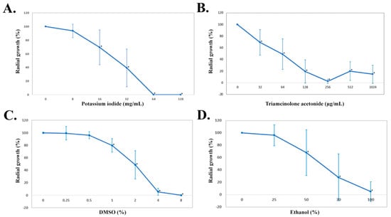
Figure 1.
Growth reduction of P. insidiosum after treatment with various concentrations of potassium iodide (A), triamcinolone acetonide (B), DMSO (C), and ethanol (D). The radial growths are averaged based on 10 representative isolates of P. insidiosum after 2−day exposure to potassium iodide, triamcinolone acetonide, and DMSO, and 1−min exposure to absolute EtOH. An asterisk indicates a statistically significant growth reduction compared to no-drug control.
Some investigators proposed that KI modulates the immune response (i.e., inhibiting the white blood cell chemotaxis, suppressing the oxygen intermediates production by immune cells, and exerting anti-inflammatory activity) to promote the elimination of pathogenic fungi [96,168,169,170]. The KI-dependent immune modulation is also a possible mechanism of action for eliminating P. insidiosum. In our in vitro study, KI appeared to directly affect P. insidiosum growth and viability. The chemical at 32 mg/mL (MCC90) killed most P. insidiosum isolates tested. Another piece of evidence supporting the direct effect of KI on a microorganism comes from Hiruma and Kagawa [171]. They microscopically demonstrate that KI could inhibit germination and physically destroy the fungus Sporothrix schenckii. However, the exact mechanism of KI’s antifungal action still needs further investigation. Apart from KI, sodium iodide is another iodine salt infrequently used in treating pythiosis, providing uncertain clinical outcomes [84,172,173].
(2) Triamcinolone acetonide: TA is a synthetic glucocorticoid widely used for treating autoimmune diseases and cancers [102,103,174,175]. TA is an immunomodulator that could activate macrophages, increase an interleukin-10 level, and reduce eosinophils and immunoglobulin E antibodies [176,177]. Intramuscular injection of TA, as a monotherapy, cured several horses with cutaneous pythiosis [68,79,80,81,82]. This study evaluated TA for its antimicrobial effect against P. insidiosum. The broth dilution method demonstrated that the range of TA MICs and MCCs spanned from ≤32 to >512 µg/mL. MIC50, MIC90, MCC50, and MCC90 of TA were greater than 512 µg/mL (the maximal TA concentration used in broth dilution; Table 1). Only one isolate (Pi050) from a cutaneous pythiosis patient in the United States exhibited MCC less than 32 µg/mL. Five of 6 TA-sensitive isolates (i.e., Pi060, Pi057, Pi075, Pi077, and Pi094) showed MCCs of 128–256 µg/mL, but their growths resumed when incubating in an agar plate with a higher TA concentration (i.e., 512 µg/mL). Colonies of most TA-insensitive organisms (MIC and MCC > 512 µg/mL) that exposed to TA at 512 µg/mL were larger than at 256 µg/mL. To confirm this observation, the radial growth assay was used to evaluate TA (concentration range: 32–1024 µg/mL) for its antimicrobial activity against 10 representative isolates of P. insidiosum. Similar results were observed, in which the growths were significantly reduced in a dose-dependent manner until the organisms resumed growing at a TA concentration higher than 256 µg/mL (Figure 1B). This paradoxical phenomenon has been described as the Eagle effect, in which an organism regrows at the drug concentration above MCC [178,179]. Although the underlying mechanism of the Eagle effect is unclear, it could result from drug impurity, reduction of autolytic activity, increase in drug-inactivated enzyme, and reduction of reactive oxygen species [178,180]. In our case, drug purity should not be the cause since a high-purity TA (98%) was used. However, we observed limited solubility of TA in a solvent at a concentration greater than 256 µg/mL, which led to drug precipitation and, thus, lower-than-expected antimicrobial drug activity. Although the mechanism of anti-P. insidiosum effect of TA is unknown, we proposed that the direct antimicrobial effect (especially for TA-sensitive isolates) and the immunomodulatory properties of TA could contribute to the elimination of P. insidiosum.
(3) Dimethyl sulfoxide: DMSO has been used as a solvent, antioxidant, anti-inflammatory, and antimicrobial agent [105,106,109,181]. Regarding its antimicrobial activity, DMSO can inhibit some bacteria (i.e., Escherichia coli and Pseudomonas aeruginosa) and fungi (i.e., Botrytis cinerea, dermatophytes and Candida albicans) [181,182,183,184]. DMSO was intravenously administered, in conjunction with amphotericin B, in 15 horses with cutaneous pythiosis [87]. It was also topically applied to the post-surgical skin lesion of 17 affected horses [86]. DMSO can promote recovery in such infected horses [86,87]. In this study, we elaborated on the clinical finding by investigating the in vitro antimicrobial activity of DMSO against 29 P. insidiosum isolates. Based on the broth dilution method, DMSO MICs and MCCs ranged from 2 to 8% (v/v). MIC50, MIC90, MCC50, and MCC90 were all at 8% DMSO (Table 1). The radial growth assay was also used to test 10 representative P. insidiosum isolates against various DMSO concentrations (i.e., 0.25%, 0.5%, 1%, 2%, 4%, and 8%) and demonstrated dose-dependent growth reductions (i.e., 0.8%, 4.0%, 20.3%, 51.4%, 94.6% and 100.0%, respectively; Figure 1C). Compared with the no-drug control, the organism growths were significantly reduced following the exposure to at least 1% of DMSO (p-value < 0.05; Figure 1C). As shown here, DMSO concentrations, particularly down to 2%, can kill P. insidiosum (Table 1). The anti-P. insidiosum mechanism of DMSO action might be the same as described in other pathogens, such as increasing the membrane permeability, altering the expression of cell wall protein, and changing the enzymatic activity [108,185]. Various DMSO concentrations have been applied for many medical and scientific purposes. For example, 90% DMSO is commonly used in skin diseases [186], and 50% DMSO shows a treatment benefit in eye diseases and interstitial cystitis [187,188]. For a laboratory experiment involving cell culture, 10% DMSO is used as a cryo-preservative agent [189]. Depending on an administered dose and route, several adverse effects of DMSO could be noticed, for example, retinal apoptosis, hemolysis, fibrinogen precipitation, cardiac arrhythmia, and genetic changes [189,190,191].
(4) Ethanol: EtOH is an antiseptic agent [116]. Previous reports show that, when used locally as an adjunctive treatment with surgery and other medications, absolute EtOH can lead to favorable clinical outcomes in treating a small group of ocular pythiosis patients [88,89,90]. In the current study, various EtOH concentrations (i.e., 25%, 50%, 70%, and 100%) were tested for their antimicrobial effect against 10 P. insidiosum isolates at several time points (i.e., 1, 2.5, 5, and 10 min). Compared with the no-drug control, the P. insidiosum growths were significantly reduced after 1 min exposure to 50% (p-value = 0.03), 70% (p-value < 0.001), and 100% (p-value < 0.001), but not 25% (p-value = 0.53) EtOH (Figure 1D). Like the other EtOH concentrations, 25% EtOH inhibited the organism’s growth more significantly when the exposure time was longer, such as 5 min (p-value = 0.01) and 10 min (p-value < 0.001) (Supplementary Figure S1). Taken together, EtOH can inhibit the organism in a dose- and time-dependent manner: higher concentration and longer exposure time enhance growth suppression. In a clinical setting, Agarwal et al. have topically applied absolute (100%) EtOH at the infection site of a few ocular pythiosis cases for 1 min, resulting in favorable treatment response [88]. We augmented their finding by challenging 29 isolates of P. insidiosum with absolute EtOH for 1 min. The result showed complete growth inhibition in 83% of all isolates tested (n = 24; Table 1). Regarding the mechanism of action, EtOH affects, for example, fungal organisms in various ways that lead to abnormal mitotic spindle, abnormal morphology, and reduced cell membrane permeability [192]. EtOH toxicities (i.e., cell lysis, inducing apoptosis, and suppressing cell proliferation) are a concern when using this chemical [193,194]. Nevertheless, EtOH could be a potential alternative agent for managing a local P. insidiosum infection.
4. Conclusions and Perspectives
Novel, alternative, or repurposed drugs effective against P. insidiosum (an orphan but highly virulent pathogen) are urgently needed. Assessing the antimicrobial activity of a drug of interest requires a standardized in vitro susceptibility test. However, no such test is available for this organism. Several in-house drug susceptibility tests (i.e., broth dilution, disc diffusion, and radial growth assays) have been established, some of which adapted the standard methods (i.e., CLSI M38-A2 and CLSI M51 protocols) designed for fungi. Hyphal plug, hyphal suspension, and zoospores are inoculum types commonly used in the drug susceptibility assessment for P. insidiosum. We demonstrated that each method has advantages and limitations compared to the others (Table 2). Selecting an assay and inoculum type depends on material availability; the experience of a laboratory worker; and the number of isolates and drugs to be tested
In this study, we employed the hyphal plug (served as an inoculum) and a combination of broth dilution and radial growth methods to screen and validate the anti-P. insidiosum activities of four chemicals (i.e., KI, TA, DMSO, and EtOH). Other investigators have preliminarily reported these chemicals as effective agents in treating pythiosis. We augmented their findings by extensively testing these chemicals against 29 genetically diverse isolates of P. insidiosum (Table 1). The results show that KI, TA, DMSO, and EtOH possessed an antimicrobial effect against P. insidiosum in a dose- or time-dependent manner. This information suggests that these chemicals could be potentially applied systematically (i.e., KI and TA) or locally (i.e., DMSO and EtOH) to treat pythiosis. The mechanism of action of these chemicals needs to be elucidated to understand how they work.
There is no standardized method for in vitro drug susceptibility analysis of P. insidiosum. Future attempts should emphasize standardizing the drug susceptibility methods, including determination of susceptibility and resistant breakpoints, for P. insidiosum, so healthcare workers can confidently read and interpret a result for selecting the most effective drug against the pathogen.
Supplementary Materials
The following supporting information can be downloaded at: https://www.mdpi.com/article/10.3390/jof8111116/s1, Figure S1: Growth reduction of P. insidiosum following the treatment with various ethanol concentrations at several time points.
Author Contributions
H.Y. and T.K.: Conceptualization; H.Y. and T.K.: Funding acquisition; H.Y., T.L., T.R., W.Y., Y.K., P.S.-C. and P.P.: Methodology and Resources; H.Y.: Visualization; T.K.: Supervision; H.Y.: Writing—original draft; T.K.: Writing—review and editing. All authors have read and agreed to the published version of the manuscript.
Funding
This work was supported by the Faculty of Graduate Studies, Mahidol University, Thailand (H.Y.); Program in Translational Medicine, Faculty of Medicine, Ramathibodi Hospital, Mahidol University, Thailand (H.Y.); School of Medicine and Health Sciences, Atma Jaya Catholic University of Indonesia, Indonesia (H.Y.); National Research Council of Thailand and Mahidol University, Thailand (Grant numbers: N42A650339; T.K.); and Faculty of Medicine, Ramathibodi Hospital, Mahidol University, Thailand (Grant number: CF_65003; T.K.).
Institutional Review Board Statement
This work was approved by the Institutional Review Board of the Faculty of Medicine, Ramathibodi Hospital, Mahidol University (approval number: MURA2022/324).
Informed Consent Statement
Not applicable.
Data Availability Statement
Not applicable.
Conflicts of Interest
The authors declare no conflict of interest.
References
- Gaastra, W.; Lipman, L.J.; De Cock, A.W.; Exel, T.K.; Pegge, R.B.; Scheurwater, J.; Vilela, R.; Mendoza, L. Pythium Insidiosum: An Overview. Vet. Microbiol. 2010, 146, 1–16. [Google Scholar] [CrossRef] [PubMed]
- Yolanda, H.; Krajaejun, T. Global Distribution and Clinical Features of Pythiosis in Humans and Animals. J. Fungi 2022, 8, 182. [Google Scholar] [CrossRef] [PubMed]
- Htun, Z.M.; Laikul, A.; Pathomsakulwong, W.; Yurayart, C.; Lohnoo, T.; Yingyong, W.; Kumsang, Y.; Payattikul, P.; Sae-Chew, P.; Rujirawat, T.; et al. Identification and Biotyping of Pythium Insidiosum Isolated from Urban and Rural Areas of Thailand by Multiplex PCR, DNA Barcode, and Proteomic Analyses. J. Fungi 2021, 7, 242. [Google Scholar] [CrossRef] [PubMed]
- Jara, M.; Holcomb, K.; Wang, X.; Goss, E.M.; Machado, G. The Potential Distribution of Pythium Insidiosum in the Chincoteague National Wildlife Refuge, Virginia. Front. Vet. Sci. 2021, 8, 640339. [Google Scholar] [CrossRef] [PubMed]
- Vanittanakom, N.; Szekely, J.; Khanthawong, S.; Sawutdeechaikul, P.; Vanittanakom, P.; Fisher, M.C. Molecular Detection of Pythium Insidiosum from Soil in Thai Agricultural Areas. Int. J. Med. Microbiol. 2014, 304, 321–326. [Google Scholar] [CrossRef]
- Mendoza, L.; Hernandez, F.; Ajello, L. Life Cycle of the Human and Animal Oomycete Pathogen Pythium Insidiosum. J. Clin. Microbiol. 1993, 31, 2967–2973. [Google Scholar] [CrossRef]
- Mosbah, E.; Karrouf, G.I.; Younis, E.A.; Saad, H.S.; Ahdy, A.; Zaghloul, A.E. Diagnosis and Surgical Management of Pythiosis in Draft Horses: Report of 33 Cases in Egypt. J. Equine Vet. Sci. 2012, 32, 164–169. [Google Scholar] [CrossRef]
- Cardona-Álvarez, J.A.; Montes-Vergara, D.; Reyes-Bossa, B.J. Pythiosis Mamaria En Una Yegua Criolla Colombiana. Rev. Colomb. Cienc. Anim. 2021, 13, e867. [Google Scholar] [CrossRef]
- Manço, M.H.; Ferreira, S.K.; Perossi, I.F.S.; Klein, M.; Pelógia, M.E.S.; Carra, G.J.U.; de Souza, C.D.D.; Costa, M.T.; Bosco, S.; Moraes, P.C.; et al. Metastatic Calcification and Granulomatous Grastroenteritis Associated to Pythium Insidiosum in a Dog. Braz. J. Vet. Pathol. 2021, 14, 50–55. [Google Scholar]
- Parambeth, J.C.; Lawhon, S.D.; Mansell, J.; Wu, J.; Clark, S.D.; Sutton, D.; Gibas, C.; Wiederhold, N.P.; Myers, A.N.; Johnson, M.C.; et al. Gastrointestinal Pythiosis with Concurrent Presumptive Gastrointestinal Basidiobolomycosis in a Boxer Dog. Vet. Clin. Pathol. 2019, 48, 83–88. [Google Scholar] [CrossRef]
- Nonpassopon, M.; Jongkhajornpong, P.; Aroonroch, R.; Koovisitsopit, A.; Lekhanont, K. Predisposing Factors, Clinical Presentations, and Outcomes of Contact Lens-Related Pythium Keratitis. Cornea 2021, 40, 1413–1419. [Google Scholar] [CrossRef] [PubMed]
- Hou, H.; Wang, Y.; Tian, L.; Wang, F.; Sun, Z.; Chen, Z. Pythium Insidiosum Keratitis Reported in China, Raising the Alertness to This Fungus-like Infection: A Case Series. J. Med. Case Rep. 2021, 15, 619. [Google Scholar] [CrossRef] [PubMed]
- Sermsathanasawadi, N.; Praditsuktavorn, B.; Hongku, K.; Wongwanit, C.; Chinsakchai, K.; Ruangsetakit, C.; Hahtapornsawan, S.; Mutirangura, P. Outcomes and Factors Influencing Prognosis in Patients with Vascular Pythiosis. J. Vasc. Surg. 2016, 64, 411–417. [Google Scholar] [CrossRef] [PubMed]
- Chitasombat, M.N.; Petchkum, P.; Horsirimanont, S.; Sornmayura, P.; Chindamporn, A.; Krajaejun, T. Vascular Pythiosis of Carotid Artery with Meningitis and Cerebral Septic Emboli: A Case Report and Literature Review. Med. Mycol. Case Rep. 2018, 21, 57–62. [Google Scholar] [CrossRef] [PubMed]
- Krajaejun, T.; Sathapatayavongs, B.; Pracharktam, R.; Nitiyanant, P.; Leelachaikul, P.; Wanachiwanawin, W.; Chaiprasert, A.; Assanasen, P.; Saipetch, M.; Mootsikapun, P.; et al. Clinical and Epidemiological Analyses of Human Pythiosis in Thailand. Clin. Infect. Dis. 2006, 43, 569–576. [Google Scholar] [CrossRef] [PubMed]
- Franco, D.M.; Aronson, J.F.; Hawkins, H.K.; Gallagher, J.J.; Mendoza, L.; McGinnis, M.R.; Williams-Bouyer, N. Systemic Pythium Insidiosum in a Pediatric Burn Patient. Burns 2010, 36, e68–e71. [Google Scholar] [CrossRef] [PubMed]
- Krajaejun, T.; Chongtrakool, P.; Angkananukul, K.; Brandhorst, T.T. Effect of Temperature on Growth of the Pathogenic Oomycete Pythium Insidiosum. Southeast Asian J. Trop. Med. Public Health 2010, 41, 1462–1466. [Google Scholar]
- Grooters, A.M.; Whittington, A.; Lopez, M.K.; Boroughs, M.N.; Roy, A.F. Evaluation of Microbial Culture Techniques for the Isolation of Pythium Insidiosum from Equine Tissues. J. Vet. Diagn. Investig. 2002, 14, 288–294. [Google Scholar] [CrossRef]
- Lohnoo, T.; Yingyong, W.; Jongruja, N.; Krajaejun, T. Comparison of Storage Methods to Preserve the Pathogenic Oomycete Pythium Insidiosum. Southeast Asian J. Trop. Med. Public Health 2018, 49, 421–427. [Google Scholar]
- Chaiprasert, A.; Samerpitak, K.; Wanachiwanawin, W.; Thasnakorn, P. Induction of Zoospore Formation in Thai Isolates of Pythium Insidiosum. Mycoses 1990, 33, 317–323. [Google Scholar] [CrossRef]
- Rathi, V.M.; Murthy, S.I.; Mitra, S.; Yamjala, B.; Mohamed, A.; Sharma, S. Masked Comparison of Trypan Blue Stain and Potassium Hydroxide with Calcofluor White Stain in the Microscopic Examination of Corneal Scrapings for the Diagnosis of Microbial Keratitis. Indian J. Ophthalmol. 2021, 69, 2457. [Google Scholar] [PubMed]
- Keeratijarut, A.; Karnsombut, P.; Aroonroch, R.; Srimuang, S.; Sangruchi, T.; Sansopha, L.; Mootsikapun, P.; Larbcharoensub, N.; Krajaejun, T. Evaluation of an In-House Immunoperoxidase Staining Assay for Histodiagnosis of Human Pythiosis. Southeast Asian J. Trop. Med. Public Health 2009, 40, 1298–1305. [Google Scholar] [PubMed]
- Chechi, J.L.; Rotchanapreeda, T.; da Paz, G.S.; Prado, A.C.; Oliveira, A.L.; Vieira, J.C.S.; Buzalaf, M.A.R.; Rodrigues, A.M.; dos Santos, L.D.; Krajaejun, T.; et al. Prospecting Biomarkers for Diagnostic and Therapeutic Approaches in Pythiosis. J. Fungi 2021, 7, 423. [Google Scholar] [CrossRef]
- Jaturapaktrarak, C.; Payattikul, P.; Lohnoo, T.; Kumsang, Y.; Laikul, A.; Pathomsakulwong, W.; Yurayart, C.; Tonpitak, W.; Krajaejun, T. Protein A/G-Based Enzyme-Linked Immunosorbent Assay for Detection of Anti-Pythium Insidiosum Antibodies in Human and Animal Subjects. BMC Res. Notes 2020, 13, 135. [Google Scholar] [CrossRef] [PubMed]
- Chitasombat, M.N.; Jongkhajornpong, P.; Lekhanont, K.; Krajaejun, T. Recent Update in Diagnosis and Treatment of Human Pythiosis. PeerJ 2020, 8, e8555. [Google Scholar] [CrossRef] [PubMed]
- Krajaejun, T.; Imkhieo, S.; Intaramat, A.; Ratanabanangkoon, K. Development of an Immunochromatographic Test for Rapid Serodiagnosis of Human Pythiosis. Clin. Vaccine Immunol. 2009, 16, 506–509. [Google Scholar] [CrossRef] [PubMed]
- Jindayok, T.; Piromsontikorn, S.; Srimuang, S.; Khupulsup, K.; Krajaejun, T. Hemagglutination Test for Rapid Serodiagnosis of Human Pythiosis. Clin. Vaccine Immunol. 2009, 16, 1047–1051. [Google Scholar] [CrossRef]
- Kammarnjesadakul, P.; Palaga, T.; Sritunyalucksana, K.; Mendoza, L.; Krajaejun, T.; Vanittanakom, N.; Tongchusak, S.; Denduangboripant, J.; Chindamporn, A. Phylogenetic Analysis of Pythium Insidiosum Thai Strains Using Cytochrome Oxidase II (COX II) DNA Coding Sequences and Internal Transcribed Spacer Regions (ITS). Med. Mycol. 2011, 49, 289–295. [Google Scholar] [CrossRef]
- Rujirawat, T.; Patumcharoenpol, P.; Kittichotirat, W.; Krajaejun, T. Oomycete Gene Table: An Online Database for Comparative Genomic Analyses of the Oomycete Microorganisms. Database Oxf. 2019, 2019, baz082. [Google Scholar] [CrossRef]
- Krajaejun, T.; Kittichotirat, W.; Patumcharoenpol, P.; Rujirawat, T.; Lohnoo, T.; Yingyong, W. Genome Data of Four Pythium Insidiosum Strains from the Phylogenetically-Distinct Clades I, II, and III. BMC Res. Notes 2021, 14, 197. [Google Scholar] [CrossRef]
- Behera, H.S.; Barik, M.R.; Das, S.; Sharma, S. Simple Polymerase Chain Reaction Assay to Differentiate between Fungal and Pythium Insidiosum Keratitis. Clin. Exp. Ophthalmol. 2021, 49, 630–632. [Google Scholar] [CrossRef] [PubMed]
- Lohnoo, T.; Jongruja, N.; Rujirawat, T.; Yingyon, W.; Lerksuthirat, T.; Nampoon, U.; Kumsang, Y.; Onpaew, P.; Chongtrakool, P.; Keeratijarut, A.; et al. Efficiency Comparison of Three Methods for Extracting Genomic DNA of the Pathogenic Oomycete Pythium Insidiosum. J. Med. Assoc. Thai. 2014, 97, 342–348. [Google Scholar] [PubMed]
- Rujirawat, T.; Sridapan, T.; Lohnoo, T.; Yingyong, W.; Kumsang, Y.; Sae-Chew, P.; Tonpitak, W.; Krajaejun, T. Single Nucleotide Polymorphism-Based Multiplex PCR for Identification and Genotyping of the Oomycete Pythium Insidiosum from Humans, Animals and the Environment. Infect. Genet. Evol. 2017, 54, 429–436. [Google Scholar] [CrossRef] [PubMed]
- Krajaejun, T.; Lohnoo, T.; Jittorntam, P.; Srimongkol, A.; Kumsang, Y.; Yingyong, W.; Rujirawat, T.; Reamtong, O.; Mangmee, S. Assessment of Matrix-Assisted Laser Desorption Ionization-Time of Flight Mass Spectrometry for Identification and Biotyping of the Pathogenic Oomycete Pythium Insidiosum. Int. J. Infect. Dis. 2018, 77, 61–67. [Google Scholar] [CrossRef]
- Mani, R.; Vilela, R.; Kettler, N.; Chilvers, M.I.; Mendoza, L. Identification of Pythium Insidiosum Complex by Matrix-Assisted Laser Desorption Ionization-Time of Flight Mass Spectrometry. J. Med. Microbiol. 2019, 68, 574–584. [Google Scholar] [CrossRef]
- Thanathanee, O.; Enkvetchakul, O.; Rangsin, R.; Waraasawapati, S.; Samerpitak, K.; Suwan-apichon, O. Outbreak of Pythium Keratitis during Rainy Season: A Case Series. Cornea 2013, 32, 199–204. [Google Scholar] [CrossRef]
- Hilton, R.E.; Tepedino, K.; Glenn, C.J.; Merkel, K.L. Swamp Cancer: A Case of Human Pythiosis and Review of the Literature. Br. J. Dermatol. 2016, 175, 394–397. [Google Scholar] [CrossRef]
- Kirzhner, M.; Arnold, S.R.; Lyle, C.; Mendoza, L.L.; Fleming, J.C. Pythium Insidiosum: A Rare Necrotizing Orbital and Facial Infection. J. Pediatr. Infect. Dis. Soc. 2014, 4, e10–e13. [Google Scholar] [CrossRef]
- Watanabe, M.J.; de Moura Alonso, J.; Alves, A.L.G.; Yamada, A.L.M.; de Moraes Gimenes Bosco, S.; Rodrigues, C.A.; Hussni, C.A. Equine Pythiosis: Report of 28 Cases from São Paulo State, Brazil. Semina Ciênc. Agrár. 2015, 36, 909–915. [Google Scholar] [CrossRef][Green Version]
- Doria, R.G.; Freitas, S.H.; Linardi, R.L.; de Souza Mendonça, F.; Arruda, L.P.; Boabaid, F.M.; Valadão, C.A. Treatment of Pythiosis in Equine Limbs Using Intravenous Regional Perfusion of Amphotericin B. Vet. Surg. 2012, 41, 759–765. [Google Scholar] [CrossRef]
- Laohapensang, K.; Rutherford, R.B.; Supabandhu, J.; Vanittanakom, N. Vascular Pythiosis in a Thalassemic Patient. Vascular 2009, 17, 234–238. [Google Scholar] [CrossRef] [PubMed]
- Torvorapanit, P.; Chuleerarux, N.; Plongla, R.; Worasilchai, N.; Manothummetha, K.; Thongkam, A.; Langsiri, N.; Diewsurin, J.; Kongsakpaisan, P.; Bansong, R.; et al. Clinical Outcomes of Radical Surgery and Antimicrobial Agents in Vascular Pythiosis: A Multicenter Prospective Study. J. Fungi 2021, 7, 114. [Google Scholar] [CrossRef] [PubMed]
- Gurnani, B.; Narayana, S.; Christy, J.; Rajkumar, P.; Kaur, K.; Gubert, J. Successful Management of Pediatric Pythium Insidiosum Keratitis with Cyanoacrylate Glue, Linezolid, and Azithromycin: Rare Case Report. Eur. J. Ophthalmol. 2022, 32, NP87–NP91. [Google Scholar] [CrossRef] [PubMed]
- Chatterjee, S.; Agrawal, D. Azithromycin in the Management of Pythium Insidiosum Keratitis. Cornea 2018, 37, e8–e9. [Google Scholar] [CrossRef] [PubMed]
- Maeno, S.; Oie, Y.; Sunada, A.; Tanibuchi, H.; Hagiwara, S.; Makimura, K.; Nishida, K. Successful Medical Management of Pythium Insidiosum Keratitis Using a Combination of Minocycline, Linezolid, and Chloramphenicol. Am. J. Ophthalmol. Case Rep. 2019, 15, 100498. [Google Scholar] [CrossRef]
- Ros Castellar, F.; Jiménez, C.S.; del Hierro Zarzuelo, A.; Ambrosio, A.H.; de Ios Bueis, A.B. Intraocular Minocycline for the Treatment of Ocular Pythiosis. Am. J. Health Syst. Pharm. 2017, 74, 821–825. [Google Scholar] [CrossRef]
- Medhasi, S.; Chindamporn, A.; Worasilchai, N. A Review: Antimicrobial Therapy for Human Pythiosis. Antibiotics 2022, 11, 450. [Google Scholar] [CrossRef]
- Ahirwar, L.K.; Kalra, P.; Sharma, S.; Mohamed, A.; Mittal, R.; Das, S.; Bagga, B. Linezolid Shows High Safety and Efficacy in the Treatment of Pythium Insidiosum Keratitis in a Rabbit Model. Exp. Eye Res. 2021, 202, 108345. [Google Scholar] [CrossRef]
- Bagga, B.; Kate, A.; Mohamed, A.; Sharma, S.; Das, S.; Mitra, S. Successful Strategic Management of Pythium Insidiosum Keratitis with Antibiotics. Ophthalmology 2020, 128, 169–172. [Google Scholar] [CrossRef]
- Sane, S.S.; Madduri, B.; Mohan, N.; Mittal, R.; Raghava, J.V.; Fernandes, M. Improved Outcome of Pythium Keratitis with a Combined Triple Drug Regimen of Linezolid and Azithromycin. Cornea 2021, 40, 888–893. [Google Scholar] [CrossRef]
- Permpalung, N.; Worasilchai, N.; Plongla, R.; Upala, S.; Sanguankeo, A.; Paitoonpong, L.; Mendoza, L.; Chindamporn, A. Treatment Outcomes of Surgery, Antifungal Therapy and Immunotherapy in Ocular and Vascular Human Pythiosis: A Retrospective Study of 18 Patients. J. Antimicrob. Chemother. 2015, 70, 1885–1892. [Google Scholar] [CrossRef] [PubMed]
- Hoffman, M.A.; Cornish, N.E.; Simonsen, K.A. A Painful Thigh Lesion in an Immunocompromised 11-Year-Old Boy. Pediatr. Infect. Dis. J. 2011, 30, 1011–1017. [Google Scholar] [CrossRef] [PubMed]
- Kate, A.; Bagga, B.; Ahirwar, L.K.; Mishra, D.K.; Sharma, S. Unusual Presentation of Pythium Keratitis as Peripheral Ulcerative Keratitis: Clinical Dilemma. Ocul. Immunol. Inflamm. 2021, 1–4. [Google Scholar] [CrossRef] [PubMed]
- Chitasombat, M.N.; Larbcharoensub, N.; Chindamporn, A.; Krajaejun, T. Clinicopathological Features and Outcomes of Pythiosis. Int. J. Infect. Dis. 2018, 71, 33–41. [Google Scholar] [CrossRef]
- Perkins, M.J.; Rosario, D.J.; Wickes, B.L.; Krajaejun, T.; Sherwood, J.E.; Mody, R.M. Severe Skin and Soft Tissue Pythiosis Acquired in a Hot Spring in the Southwestern United States, a Case Report and Review of North American Cases. Travel Med. Infect. Dis. 2022, 48, 102349. [Google Scholar] [CrossRef]
- dos Santos, C.E.; Ubiali, D.G.; Pescador, C.A.; Zanette, R.A.; Santurio, J.M.; Marques, L.C. Epidemiological Survey of Equine Pythiosis in the Brazilian Pantanal and Nearby Areas: Results of 76 Cases. J. Equine Vet. Sci. 2014, 34, 270–274. [Google Scholar] [CrossRef]
- Mendoza, L.; Mandy, W.; Glass, R. An Improved Pythium Insidiosum-Vaccine Formulation with Enhanced Immunotherapeutic Properties in Horses and Dogs with Pythiosis. Vaccine 2003, 21, 2797–2804. [Google Scholar] [CrossRef]
- Santurio, J.M.; Catto, J.B.; Leal, A.B.M.; Leal, A.T. Tratamento Imunoterápico Da Pitiose Equina. Comun. Tec. 2001, 67, 1–3. [Google Scholar]
- Frey, F.; Velho, J.R.; Lins, L.A.; Nogueira, C.E.W.; Santurio, J.M. Pitiose Equina Na Região Sul Do Brasil. Rev. Port. Cienc. Vet. 2007, 102, 107–111. [Google Scholar]
- Reanpang, T.; Orrapin, S.; Orrapin, S.; Arworn, S.; Kattipatanapong, T.; Srisuwan, T.; Vanittanakom, N.; Lekawanvijit, S.P.; Rerkasem, K. Vascular Pythiosis of the Lower Extremity in Northern Thailand: Ten Years’ Experience. Int. J. Low. Extrem. Wounds 2015, 14, 245–250. [Google Scholar] [CrossRef]
- Puangsricharern, V.; Chotikkakamthorn, P.; Tulvatana, W.; Kittipibul, T.; Chantaren, P.; Reinprayoon, U.; Kasetsuwan, N.; Satitpitakul, V.; Worasilchai, N.; Chindamporn, A. Clinical Characteristics, Histopathology, and Treatment Outcomes of Pythium Keratitis: A Retrospective Cohort Study. Clin. Ophthalmol. 2021, 15, 1691–1701. [Google Scholar] [CrossRef] [PubMed]
- Yolanda, H.; Krajaejun, T. History and Perspective of Immunotherapy for Pythiosis. Vaccines 2021, 9, 1080. [Google Scholar] [CrossRef]
- Reagan, K.L.; Marks, S.L.; Pesavento, P.A.; Maggiore, A.D.; Zhu, B.Y.; Grooters, A.M. Successful Management of 3 Dogs with Colonic Pythiosis Using Itraconzaole, Terbinafine, and Prednisone. J. Vet. Intern. Med. 2019, 33, 1434–1439. [Google Scholar] [CrossRef]
- Tavares, T.R.; Frias, N.C.; Lopes, N.C.; Maldos, P.C.W.; Galvão-Dias, M.A.; Reis-Menezes, A.A. Equine Pitiosis in São Paulo: Case Report; Icophai: Pernambuco, Brazil, 2013; p. 1. [Google Scholar]
- Susaengrat, N.; Torvorapanit, P.; Plongla, R.; Chuleerarux, N.; Manothummetha, K.; Tuangsirisup, J.; Worasilchai, N.; Chindamporn, A.; Permpalung, N. Adjunctive Antibacterial Agents as a Salvage Therapy in Relapsed Vascular Pythiosis Patients. Int. J. Infect. Dis. 2019, 88, 27–30. [Google Scholar] [CrossRef] [PubMed]
- Thitithanyanont, A.; Mendoza, L.; Chuansumrit, A.; Pracharktam, R.; Laothamatas, J.; Sathapatayavongs, B.; Lolekha, S.; Ajello, L. Use of an Immunotherapeutic Vaccine to Treat a Life-Threatening Human Arteritic Infection Caused by Pythium Insidiosum. Clin. Infect. Dis. 1998, 27, 1394–1400. [Google Scholar] [CrossRef] [PubMed]
- Ubiali, D.G.; Pereira, A.H.; Boabaid, F.M.; Dutra, V.; Nakazato, L.; Campos, C.G.; Colodel, E.M.; Pescador, C.A.; Riet-Correa, F. Successful Potassium Iodide Treatment for Rhinofacial Pythiosis in Sheep. J. Med. Mycol. 2022, 32, 101233. [Google Scholar] [CrossRef]
- Lemos, G.; Petrucci, L.; Vieira, V.; di Filippo, P. Treatment of Equine Cutaneous Pythiosis with Triamcinolone Acetate and Potassium Iodide. Rev. Acadêmica Ciênc. Anim. 2018, 16, e162507. [Google Scholar]
- Wanachiwanawin, W.; Thianprasit, M.; Fucharoen, S.; Chaiprasert, A.; Ayudhya, N.S.N.; Sirithanaratkul, N.; Piankijagum, A. Fatal Arteritis Due to Pythium Insidiosum Infection in Patients with Thalassaemia. Trans. R. Soc. Trop. Med. Hyg. 1993, 87, 296–298. [Google Scholar] [CrossRef]
- Chetchotisakd, P.; Pairojkul, C.; Porntaveevudhi, O.; Sathapatayavongs, B.; Mairiang, P.; Nuntirooj, K.; Patjanasoontorn, B.; Saew, O.; Chaiprasert, A.; Haswell-Elkins, M. Human Pythiosis in Srinagarind Hospital: One Year’s Experience. J. Med. Assoc. Thai. 1992, 75, 248–254. [Google Scholar]
- Sathapatayavongs, B.; Leelachaikul, P.; Prachaktam, R.; Atichartakarn, V.; Sriphojanart, S.; Trairatvorakul, P.; Jirasiritham, S.; Nontasut, S.; Eurvilaichit, C.; Flegel, T. Human Pythiosis Associated with Thalassemia Hemoglobinopathy Syndrome. J. Infect. Dis. 1989, 159, 274–280. [Google Scholar] [CrossRef]
- Salomão-Nascimento, R.B.; Frazão-Teixeira, E.; de Oliveira, F. Hepatic and Renal Analysis in Horses with Pythiosis Treated with Potassium Iodate, through the Detection of Serum Proteins, Nitrogenated Substances and Enzymes. Rev. Bras. Med. Veterinária 2010, 32, 105–110. [Google Scholar]
- Pupaibool, J.; Chindamporn, A.; Patarakul, K.; Suankratay, C.; Sindhuphak, W.; Kulwichit, W. Human Pythiosis. Emerg. Infect. Dis. 2006, 12, 517. [Google Scholar] [CrossRef] [PubMed]
- Thianprasit, M.; Chaiprasert, A.; Imwidthaya, P. Human Pythiosis. Curr. Top. Med. Mycol. 1996, 7, 43–54. [Google Scholar] [PubMed]
- Imwidthaya, P. Human Pythiosis in Thailand. Postgrad. Med. J. 1994, 70, 558–560. [Google Scholar] [CrossRef]
- Mendoza, L.; Prasla, S.H.; Ajello, L. Orbital Pythiosis: A Non-Fungal Disease Mimicking Orbital Mycotic Infections, with a Retrospective Review of the Literature. Mycoses 2004, 47, 14–23. [Google Scholar] [CrossRef]
- Tanphaichitra, D. Tropical Disease in the Immunocompromised Host: Melioidosis and Pythiosis. Rev. Infect. Dis. 1989, 11, S1629–S1643. [Google Scholar] [CrossRef]
- Prasertwitayakij, N.; Louthrenoo, W.; Kasitanon, N.; Thamprasert, K.; Vanittanakom, N. Human Pythiosis, a Rare Cause of Arteritis: Case Report and Literature Review. Semin. Arthritis Rheum. 2003, 33, 204–214. [Google Scholar] [CrossRef]
- Souto, E.P.F.; Maia, L.A.; Neto, E.G.M.; Kommers, G.D.; Junior, F.G.; Riet-Correa, F.; Galiza, G.J.; Dantas, A.F. Pythiosis in Equidae in Northeastern Brazil: 1985–2020. J. Equine Vet. Sci. 2021, 105, 103726. [Google Scholar] [CrossRef]
- Cardona-Álvarez, J.A.; Vargas-Vilória, M.; Patarroyo-Salcedo, J. Cutaneous Pythiosis in Horses Treated with Triamcinolone Acetonide. Part 1. Clinical Characterization. Rev. MVZ Córdoba 2016, 21, 5511–5524. [Google Scholar] [CrossRef]
- Romero, A.; García, J.; Balestié, S.; Malfatto, F.; Vicentino, A.; Sallis, E.S.V.; Schild, A.L.; Dutra, F. Equine Pythiosis in the Eastern Wetlands of Uruguay. Pesqui. Veterinária Bras. 2019, 39, 469–475. [Google Scholar] [CrossRef]
- Baldrich Romero, N.E.; Patiño Quiroz, B.; Peña, H.; Juan Carlos, C.R. Primer Reporte de Pythiosis En Área Rual de Florencia-Caquetá. Rev. Electron. Vet. 2016, 17, 1–10. [Google Scholar]
- Dias, D.; Doria, R.; Pereira, R.; Canola, P.; Di Filippo, P. Topical Treatment Using Amphotericin B and DMSO for an Atypically Located Equine Cutaneous Pythiosis. Acta Sci. Vet. 2012, 40, 1088. [Google Scholar]
- Videla, R.; van Amstel, S.; O’Neill, S.H.; Frank, L.A.; Newman, S.J.; Vilela, R.; Mendoza, L. Vulvar Pythiosis in Two Captive Camels (Camelus Dromedarius). Sabouraudia 2012, 50, 212–224. [Google Scholar] [CrossRef] [PubMed]
- Dowling, B.; Dart, A.; Kessell, A.; Pascoe, R.; Hodgson, D. Cutaneous Phycomycosis in Two Horses. Aust. Vet. J. 1999, 77, 780–783. [Google Scholar] [CrossRef] [PubMed]
- Atiba, A.; Ghazy, A.; Hamad, M. Evaluating the Efficacy of Surgical Excision and Topical Dimethyl Sulphoxide (DMSO) in the Treatment of Equine Cutaneous Pythiosis. Iran. J. Vet. Res. 2020, 21, 301–307. [Google Scholar] [PubMed]
- Dória, R.G.; Carvalho, M.B.; Freitas, S.H.; Laskoski, L.M.; Colodel, E.M.; Mendonça, F.S.; Silva, M.A.; Grigoletto, R.; Neto, P.F. Evaluation of Intravenous Regional Perfusion with Amphotericin B and Dimethylsulfoxide to Treat Horses for Pythiosis of a Limb. BMC Vet. Res. 2015, 11, 152. [Google Scholar] [CrossRef]
- Agarwal, S.; Srinivasan, B.; Janakiraman, N.; Therese, L.K.; KrishnaKumar, S.; Patel, N.; Thenmozhi, V.; Iyer, G. Role of Topical Ethanol in the Treatment of Pythium Insidiosum Keratitis—A Proof of Concept. Cornea 2020, 39, 1102–1107. [Google Scholar] [CrossRef]
- Agarwal, S.; Iyer, G.; Srinivasan, B.; Agarwal, M.; Kumar, S.P.S.; Therese, L.K. Clinical Profile of Pythium Keratitis: Perioperative Measures to Reduce Risk of Recurrence. Br. J. Ophthalmol. 2018, 102, 153–157. [Google Scholar] [CrossRef]
- Agarwal, S.; Iyer, G.; Srinivasan, B.; Benurwar, S.; Agarwal, M.; Narayanan, N.; Lakshmipathy, M.; Radhika, N.; Rajagopal, R.; Krishnakumar, S.; et al. Clinical Profile, Risk Factors and Outcome of Medical, Surgical and Adjunct Interventions in Patients with Pythium Insidiosum Keratitis. Br. J. Ophthalmol. 2019, 103, 296–300. [Google Scholar] [CrossRef]
- Khadom, A.A.; Yaro, A.S. Protection of Low Carbon Steel in Phosphoric Acid by Potassium Iodide. Prot. Met. Phys. Chem. Surf. 2011, 47, 662–669. [Google Scholar] [CrossRef]
- Zhao, X.; Lu, X.; Wei, A.; Jia, X.; Chen, J.; Lu, K. Potassium Iodide Promoted Thiolation of Pyrazolones and Benzofurans Using Aryl Sulfonyl Chlorides as Sulfenylation Reagents. Tetrahedron Lett. 2016, 57, 5330–5333. [Google Scholar] [CrossRef]
- Xie, W.; Li, H. Alumina-Supported Potassium Iodide as a Heterogeneous Catalyst for Biodiesel Production from Soybean Oil. J. Mol. Catal. Chem. 2006, 255, 1–9. [Google Scholar] [CrossRef]
- Sterling, J.B.; Heymann, W.R. Potassium Iodide in Dermatology: A 19th Century Drug for the 21st Century—Uses, Pharmacology, Adverse Effects, and Contraindications. J. Am. Acad. Dermatol. 2000, 43, 691–697. [Google Scholar] [CrossRef]
- Reiners, C.; Schneider, R. Potassium Iodide (KI) to Block the Thyroid from Exposure to I-131: Current Questions and Answers to Be Discussed. Radiat. Environ. Biophys. 2013, 52, 189–193. [Google Scholar] [CrossRef] [PubMed]
- Costa, R.O.; de Macedo, P.M.; Carvalhal, A.; Bernardes-Engemann, A.R. Use of Potassium Iodide in Dermatology: Updates on an Old Drug. An. Bras. Dermatol. 2013, 88, 396–402. [Google Scholar] [CrossRef] [PubMed]
- Tabosa, I.; Riet-Correa, F.; Nobre, V.; Azevedo, E.; Reis-Junior, J.; Medeiros, R. Outbreaks of Pythiosis in Two Flocks of Sheep in Northeastern Brazil. Vet. Pathol. 2004, 41, 412–415. [Google Scholar] [CrossRef]
- Wanachiwanawin, W.; Mendoza, L.; Visuthisakchai, S.; Mutsikapan, P.; Sathapatayavongs, B.; Chaiprasert, A.; Suwanagool, P.; Manuskiatti, W.; Ruangsetakit, C.; Ajello, L. Efficacy of Immunotherapy Using Antigens of Pythium Insidiosum in the Treatment of Vascular Pythiosis in Humans. Vaccine 2004, 22, 3613–3621. [Google Scholar] [CrossRef]
- Khunkhet, S.; Rattanakaemakorn, P.; Rajatanavin, N. Pythiosis Presenting with Digital Gangrene and Subcutaneous Nodules Mimicking Medium Vessel Vasculitis. JAAD Case Rep. 2015, 1, 399–402. [Google Scholar] [CrossRef]
- de Moraes Gimenes Bosco, S.; Bagagli, E.; Araújo Jr, J.P.; Candeias, J.M.G.; De Franco, M.F.; Marques, M.E.A.; Mendoza, L.; de Camargo, R.P.; Marques, S.A. Human Pythiosis, Brazil. Emerg. Infect. Dis. 2005, 11, 715. [Google Scholar] [CrossRef]
- Nassar, A.; Atef, H.; Eldeeb, F.; Alakad, R. Comparison of Fractional Laser-Assisted Drug Delivery and Intralesional Injection of Triamcinolone Acetonide in Nail Psoriasis. J. Dtsch. Dermatol. Ges. 2022, 20, 788–796. [Google Scholar] [CrossRef]
- Jeal, W.; Faulds, D. Triamcinolone Acetonide. Drugs 1997, 53, 257–280. [Google Scholar] [CrossRef] [PubMed]
- Timmermans, S.; Souffriau, J.; Libert, C. A General Introduction to Glucocorticoid Biology. Front. Immunol. 2019, 10, 1545. [Google Scholar] [CrossRef] [PubMed]
- Cridge, H.; Hughes, S.M.; Langston, V.C.; Mackin, A.J. Mefenoxam, Itraconazole, and Terbinafine Combination Therapy for Management of Pythiosis in Dogs (Six Cases). J. Am. Anim. Hosp. Assoc. 2020, 56, 307. [Google Scholar] [CrossRef] [PubMed]
- Tashrifi, Z.; Khanaposhtani, M.M.; Larijani, B.; Mahdavi, M. Dimethyl Sulfoxide: Yesterday’s Solvent, Today’s Reagent. Adv. Synth. Catal. 2020, 362, 65–86. [Google Scholar] [CrossRef]
- Elisia, I.; Nakamura, H.; Lam, V.; Hofs, E.; Cederberg, R.; Cait, J.; Hughes, M.R.; Lee, L.; Jia, W.; Adomat, H.H.; et al. DMSO Represses Inflammatory Cytokine Production from Human Blood Cells and Reduces Autoimmune Arthritis. PLoS ONE 2016, 11, e0152538. [Google Scholar] [CrossRef]
- Kirkwood, Z.I.; Millar, B.C.; Downey, D.G.; Moore, J.E. Antimicrobial Effect of Dimethyl Sulfoxide and N, N-Dimethylformamide on Mycobacterium Abscessus: Implications for Antimicrobial Susceptibility Testing. Int. J. Mycobacteriol. 2018, 7, 134–136. [Google Scholar]
- Hazen, K.C. Influence of DMSO on Antifungal Activity during Susceptibility Testing in Vitro. Diagn. Microbiol. Infect. Dis. 2013, 75, 60–63. [Google Scholar] [CrossRef]
- Sanmartín-Suárez, C.; Soto-Otero, R.; Sánchez-Sellero, I.; Méndez-Álvarez, E. Antioxidant Properties of Dimethyl Sulfoxide and Its Viability as a Solvent in the Evaluation of Neuroprotective Antioxidants. J. Pharmacol. Toxicol. Methods 2011, 63, 209–215. [Google Scholar] [CrossRef]
- Ianiski, L.B.; Stibbe, P.C.; Denardi, L.B.; Weiblen, C.; Soares, M.P.; de Souza Silveira Valente, J.; Sangioni, L.A.; Pereira, D.I.B.; Santurio, J.M.; de Avila Botton, S. In Vitro Anti-Pythium Insidiosum Activity of Amorolfine Hydrochloride and Azithromycin, Alone and in Combination: Antimicrobial Effect Anti-P. Insidiosum. Med. Mycol. 2021, 59, 67–73. [Google Scholar] [CrossRef]
- Krajaejun, T.; Lohnoo, T.; Yingyong, W.; Rujirawat, T.; Kumsang, Y.; Jongkhajornpong, P.; Theerawatanasirikul, S.; Kittichotirat, W.; Reamtong, O.; Yolanda, H. The Repurposed Drug, Disulfiram, Inhibits Urease and Aldehyde Dehydrogenase and Prevents in Vitro Growth of the Oomycete Pythium Insidiosum. Antimicrob. Agents Chemother. 2019, 63, e00609-19. [Google Scholar] [CrossRef]
- Loreto, E.S.; Tondolo, J.S.; Oliveira, D.C.; Santurio, J.M.; Alves, S.H. In Vitro Activities of Miltefosine and Antibacterial Agents from Macrolide, Oxazolidinone, and Pleuromutilin Classes against Pythium Insidiosum and Pythium Aphanidermatum. Antimicrob. Agents Chemother. 2018, 62, e01678-17. [Google Scholar] [CrossRef] [PubMed]
- Jesus, F.; Ferreiro, L.; Bizzi, K.; Loreto, E.; Pilotto, M.; Ludwig, A.; Alves, S.; Zanette, R.; Santurio, J. In Vitro Activity of Carvacrol and Thymol Combined with Antifungals or Antibacterials against Pythium Insidiosum. J. Mycol. Med. 2015, 25, e89–e93. [Google Scholar] [CrossRef]
- Le Dare, B.; Gicquel, T. Therapeutic Applications of Ethanol: A Review. J. Pharm. Pharm. Sci. 2019, 22, 525–535. [Google Scholar] [CrossRef] [PubMed]
- Mukherjee, P.K.; Mohamed, S.; Chandra, J.; Kuhn, D.; Liu, S.; Antar, O.S.; Munyon, R.; Mitchell, A.P.; Andes, D.; Chance, M.R.; et al. Alcohol Dehydrogenase Restricts the Ability of the Pathogen Candida Albicans to Form a Biofilm on Catheter Surfaces through an Ethanol-Based Mechanism. Infect. Immun. 2006, 74, 3804–3816. [Google Scholar] [CrossRef] [PubMed]
- Kampf, G.; Pitten, F.; Heeg, P.; Christiansen, B. Efficacy of Two Ethanol-Based Skin Antiseptics on the Forehead at Shorter Application Times. BMC Microbiol. 2007, 7, 85. [Google Scholar] [CrossRef]
- Haw, W.W.; Manche, E.E. Treatment of Progressive or Recurrent Epithelial Ingrowth with Ethanol Following Laser in Situ Keratomileusis. J. Refract. Surg. 2001, 17, 63–68. [Google Scholar] [CrossRef]
- Shields, C.L.; Arepalli, S.; Lally, E.B.; Lally, S.E.; Shields, J.A. Iris Stromal Cyst Management with Absolute Alcohol–Induced Sclerosis in 16 Patients. JAMA Ophthalmol. 2014, 132, 703–708. [Google Scholar] [CrossRef]
- Zohreh, B.; Aliasghar, K. Epithelial Iris Cyst Treatment with Intracystic Ethanol Irrigation. Ophthalmology 2003, 110, 1601–1605. [Google Scholar] [CrossRef]
- Su, L.X.; Jia, R.B.; Wang, D.M.; Lv, M.M.; Fan, X.D. Absolute Ethanol Embolization of Arteriovenous Malformations in the Periorbital Region. Cardiovasc. Intervent. Radiol. 2015, 38, 632–641. [Google Scholar] [CrossRef]
- Yolanda, H.; Krajaejun, T. Review of Methods and Antimicrobial Agents for Susceptibility Testing against Pythium Insidiosum. Heliyon 2020, 6, e03737. [Google Scholar] [CrossRef]
- Lerksuthirat, T.; Sangcakul, A.; Lohnoo, T.; Yingyong, W.; Rujirawat, T.; Krajaejun, T. Evolution of the Sterol Biosynthetic Pathway of Pythium Insidiosum and Related Oomycetes Contributes to Antifungal Drug Resistance. Antimicrob. Agents Chemother. 2017, 61, e02352-16. [Google Scholar] [CrossRef] [PubMed]
- Posri, P.; Suthiwong, J.; Thongsri, Y.; Yenjai, C. Antifungal Activity of Compounds from the Stems of Dalbergia Stipulacea against Pythium Insidiosum. Nat. Prod. Res. 2021, 35, 2823–2830. [Google Scholar] [CrossRef] [PubMed]
- Worasilchai, N.; Chindamporn, A.; Plongla, R.; Torvorapanit, P.; Manothummetha, K.; Chuleerarux, N.; Permpalung, N. In Vitro Susceptibility of Antibacterial Agents against Thai Pythium Insidiosum Isolates. Antimicrob. Agents Chemother. 2020, 64, e02099-19. [Google Scholar] [CrossRef] [PubMed]
- Brown, T.A.; Grooters, A.M.; Hosgood, G.L. In Vitro Susceptibility of Pythium Insidiosum and a Lagenidium sp to Itraconazole, Posaconazole, Voriconazole, Terbinafine, Caspofungin, and Mefenoxam. Am. J. Vet. Res. 2008, 69, 1463–1468. [Google Scholar] [CrossRef] [PubMed]
- Trolezi, R.; Azanha, J.M.; Paschoal, N.R.; Chechi, J.L.; Silva, M.J.D.; Fabris, V.E.; Vilegas, W.; Kaneno, R.; Junior, A.F.; de Moraes G Bosco, S. Stryphnodendron Adstringens and Purified Tannin on Pythium Insidiosum: In Vitro and in Vivo Studies. Ann. Clin. Microbiol. Antimicrob. 2017, 16, 7. [Google Scholar] [CrossRef]
- Araújo, M.J.A.M.; de Moraes Gimenes Bosco, S.; Sforcin, J.M. Pythium Insidiosum: Inhibitory Effects of Propolis and Geopropolis on Hyphal Growth. Braz. J. Microbiol. 2016, 47, 863–869. [Google Scholar] [CrossRef]
- de Souza Silveira Valente, J.; da Silva Fonseca, A.d.O.; Denardi, L.B.; Dal Ben, V.S.; de Souza Maia Filho, F.; Baptista, C.T.; Braga, C.Q.; Zambrano, C.G.; Alves, S.H.; de Avila Botton, S.; et al. In Vitro Susceptibility of Pythium Insidiosum to Melaleuca Alternifolia, Mentha Piperita and Origanum Vulgare Essential Oils Combinations. Mycopathologia 2016, 181, 617–622. [Google Scholar] [CrossRef]
- Loreto, É.S.; Tondolo, J.S.; Pilotto, M.B.; Alves, S.H.; Santurio, J.M. New Insights into the in Vitro Susceptibility of Pythium Insidiosum. Antimicrob. Agents Chemother. 2014, 58, 7534–7537. [Google Scholar] [CrossRef]
- Fonseca, A.; Pereira, D.; Maia Filho, F.; Osório, L.; Maroneze, B.; Valente, J.; Pötter, L.; Meireles, M. In Vitro Susceptibility of Zoospores and Hyphae of Pythium Insidiosum to Antifungals. J. Antimicrob. Chemother. 2014, 69, 1564–1567. [Google Scholar] [CrossRef]
- Krajaejun, T.; Kittichotirat, W.; Patumcharoenpol, P.; Rujirawat, T.; Lohnoo, T.; Yingyong, W. Data on Whole Genome Sequencing of the Oomycete Pythium Insidiosum Strain CBS 101555 from a Horse with Pythiosis in Brazil. BMC Res. Notes 2018, 11, 880. [Google Scholar] [CrossRef]
- Patumcharoenpol, P.; Rujirawat, T.; Lohnoo, T.; Yingyong, W.; Vanittanakom, N.; Kittichotirat, W.; Krajaejun, T. Draft Genome Sequences of the Oomycete Pythium Insidiosum Strain CBS 573.85 from a Horse with Pythiosis and Strain CR02 from the Environment. Data Brief 2018, 16, 47–50. [Google Scholar] [CrossRef] [PubMed]
- Rujirawat, T.; Patumcharoenpol, P.; Lohnoo, T.; Yingyong, W.; Lerksuthirat, T.; Tangphatsornruang, S.; Suriyaphol, P.; Grenville-Briggs, L.J.; Garg, G.; Kittichotirat, W.; et al. Draft Genome Sequence of the Pathogenic Oomycete Pythium Insidiosum Strain Pi-S, Isolated from a Patient with Pythiosis. Genome Announc. 2015, 3, e00574-15. [Google Scholar] [CrossRef] [PubMed]
- Tangphatsornruang, S.; Ruang-Areerate, P.; Sangsrakru, D.; Rujirawat, T.; Lohnoo, T.; Kittichotirat, W.; Patumcharoenpol, P.; Grenville-Briggs, L.J.; Krajaejun, T. Comparative Mitochondrial Genome Analysis of Pythium Insidiosum and Related Oomycete Species Provides New Insights into Genetic Variation and Phylogenetic Relationships. Gene 2016, 575, 34–41. [Google Scholar] [CrossRef]
- Keeratijarut, A.; Lohnoo, T.; Yingyong, W.; Rujirawat, T.; Srichunrusami, C.; Onpeaw, P.; Chongtrakool, P.; Brandhorst, T.T.; Krajaejun, T. Detection of the Oomycete Pythium Insidiosum by Real-Time PCR Targeting the Gene Coding for Exo-1, 3-β-Glucanase. J. Med. Microbiol. 2015, 64, 971–977. [Google Scholar] [CrossRef] [PubMed]
- Kittichotirat, W.; Krajaejun, T. Application of Genome Sequencing to Study Infectious Diseases. J. Infect. Antimicrob. Agents 2019, 36, 47–58. [Google Scholar]
- Krajaejun, T.; Lowhnoo, T.; Yingyong, W.; Rujirawat, T.; Fucharoen, S.; Strobel, G.A. In Vitro Antimicrobial Activity of Volatile Organic Compounds from Muscodor Crispans against the Pathogenic Oomycete Pythium Insidiosum. Southeast Asian J. Trop. Med. Public Health 2012, 43, 1474. [Google Scholar]
- Zimmermann, C.E.; Jesus, F.P.; Schlemmer, K.B.; Loreto, É.S.; Tondolo, J.S.; Driemeier, D.; Alves, S.H.; Ferreiro, L.; Santurio, J.M. In Vivo Effect of Minocycline Alone and in Combination with Immunotherapy against Pythium Insidiosum. Vet. Microbiol. 2020, 243, 108616. [Google Scholar] [CrossRef]
- Santurio, J.; Leal, A.; Leal, A.; Festugatto, R.; Lubeck, I.; Sallis, E.; Copetti, M.; Alves, S.; Ferreiro, L. Three Types of Immunotherapics against Pythiosis Insidiosi Developed and Evaluated. Vaccine 2003, 21, 2535–2540. [Google Scholar] [CrossRef]
- Argenta, J.S.; Alves, S.H.; Silveira, F.; Maboni, G.; Zanette, R.A.; Cavalheiro, A.S.; Pereira, P.L.; Pereira, D.I.; Sallis, E.S.; Pötter, L.; et al. In Vitro and in Vivo Susceptibility of Two-Drug and Three-Drug Combinations of Terbinafine, Itraconazole, Caspofungin, Ibuprofen and Fluvastatin against Pythium Insidiosum. Vet. Microbiol. 2012, 157, 137–142. [Google Scholar] [CrossRef]
- Pereira, D.I.B.; Santurio, J.M.; Alves, S.H.; Argenta, J.S.; Pötter, L.; Spanamberg, A.; Ferreiro, L. Caspofungin in Vitro and in Vivo Activity against Brazilian Pythium Insidiosum Strains Isolated from Animals. J. Antimicrob. Chemother. 2007, 60, 1168–1171. [Google Scholar] [CrossRef]
- Wittayapipath, K.; Yenjai, C.; Prariyachatigul, C.; Hamal, P. Evaluation of Antifungal Effect and Toxicity of Xanthyletin and Two Bacterial Metabolites against Thai Isolates of Pythium Insidiosum. Sci. Rep. 2020, 10, 4495. [Google Scholar] [CrossRef] [PubMed]
- Suthiwong, J.; Thongsri, Y.; Yenjai, C. A New Furanocoumarin from the Fruits of Scaevola Taccada and Antifungal Activity against Pythium Insidiosum. Nat. Prod. Res. 2017, 31, 453–459. [Google Scholar] [CrossRef] [PubMed]
- Sriphana, U.; Thongsri, Y.; Ardwichai, P.; Poopasit, K.; Prariyachatigul, C.; Simasathiansophon, S.; Yenjai, C. New Lignan Esters from Alyxia Schlechteri and Antifungal Activity against Pythium Insidiosum. Fitoterapia 2013, 91, 39–43. [Google Scholar] [CrossRef]
- Sribuhom, T.; Thongsri, Y.; Yenjai, C. Potential Antifungal Activity against Pythium Insidiosum of Isoflavonoids from the Stems of Dalbergia Cultrata. Asian J. Chem. 2020, 32, 1788–1792. [Google Scholar] [CrossRef]
- Thongsri, Y.; Aromdee, C.; Yenjai, C.; Kanokmedhakul, S.; Chaiprasert, A.; Hamal, P.; Prariyachatigul, C. Detection of Diketopiperazine and Pyrrolnitrin, Compounds with Anti-Pythium Insidiosum Activity, in a Pseudomonas Stutzeri Environmental Strain. Biomed. Pap. Med. Fac. Univ. Palacky Olomouc. Czech Repub. 2014, 158, 378–383. [Google Scholar] [CrossRef]
- Sriphana, U.; Thongsri, Y.; Prariyachatigul, C.; Pakawatchai, C.; Yenjai, C. Clauraila E from the Roots of Clausena Harmandiana and Antifungal Activity against Pythium Insidiosum. Arch. Pharm. Res. 2013, 36, 1078–1083. [Google Scholar] [CrossRef] [PubMed]
- Suthiwong, J.; Sriphana, U.; Thongsri, Y.; Promsuwan, P.; Prariyachatigul, C.; Yenjai, C. Coumarinoids from the Fruits of Micromelum Falcatum. Fitoterapia 2014, 94, 134–141. [Google Scholar] [CrossRef] [PubMed]
- de Souza Silveira Valente, J.; da Silva Fonseca, A.d.O.; Brasil, C.L.; Sagave, L.; Flores, F.C.; da Silva, C.d.B.; Sangioni, L.A.; Pötter, L.; Santurio, J.M.; de Avila Botton, S.; et al. In Vitro Activity of Melaleuca Alternifolia (Tea Tree) in Its Free Oil and Nanoemulsion Formulations against Pythium Insidiosum. Mycopathologia 2016, 181, 865–869. [Google Scholar] [CrossRef]
- Wittayapipath, K.; Laolit, S.; Yenjai, C.; Chio-Srichan, S.; Pakarasang, M.; Tavichakorntrakool, R.; Prariyachatigul, C. Analysis of Xanthyletin and Secondary Metabolites from Pseudomonas Stutzeri ST1302 and Klebsiella Pneumoniae ST2501 against Pythium Insidiosum. BMC Microbiol. 2019, 19, 78. [Google Scholar] [CrossRef]
- Loreto, E.S.; Tondolo, J.S.; Santurio, J.M.; Alves, S.H. Screening of Antibacterial Drugs for Antimicrobial Activity against Pythium Insidiosum. Med. Mycol. 2018, 57, 523–525. [Google Scholar]
- Cavalheiro, A.S.; Zanette, R.A.; Spader, T.B.; Lovato, L.; Azevedo, M.I.; Botton, S.; Alves, S.H.; Santurio, J.M. In Vitro Activity of Terbinafine Associated to Amphotericin B, Fluvastatin, Rifampicin, Metronidazole and Ibuprofen against Pythium Insidiosum. Vet. Microbiol. 2009, 137, 408–411. [Google Scholar] [CrossRef] [PubMed]
- Cavalheiro, A.S.; Maboni, G.; de Azevedo, M.I.; Argenta, J.S.; Pereira, D.I.; Spader, T.B.; Alves, S.H.; Santurio, J.M. In Vitro Activity of Terbinafine Combined with Caspofungin and Azoles against Pythium Insidiosum. Antimicrob. Agents Chemother. 2009, 53, 2136–2138. [Google Scholar] [CrossRef] [PubMed]
- de Souza Silveira Valente, J.; Braga, C.Q.; Brasil, C.L.; Baptista, C.T.; Reis, G.F.; Panagio, L.A.; Nakazato, G.; de Oliveira Hübner, S.; Soares, M.P.; de Avila Botton, S.; et al. In Vitro Anti-Pythium Insidiosum Activity of Biogenic Silver Nanoparticles. Med. Mycol. 2019, 57, 858–863. [Google Scholar] [CrossRef] [PubMed]
- Cantón, E.; Espinel-Ingroff, A.; Pemán, J. Trends in Antifungal Susceptibility Testing Using CLSI Reference and Commercial Methods. Expert Rev. Anti Infect. Ther. 2009, 7, 107–119. [Google Scholar] [CrossRef] [PubMed]
- Loreto, E.; Tondolo, J.; de Jesus, F.; Engelmann, A.; de Andrade, C.; Santurio, J.; Zanette, R.; Kommers, G.; Silva, T.; Alves, S. Efficacy of Miltefosine Therapy against Subcutaneous Experimental Pythiosis in Rabbits. J. Mycol. Médicale 2019, 30, 100919. [Google Scholar] [CrossRef]
- Itaqui, S.R.; Verdi, C.M.; Tondolo, J.S.; da Luz, T.S.; Alves, S.H.; Santurio, J.M.; Loreto, É.S. In Vitro Synergism between Azithromycin or Terbinafine and Topical Antimicrobial Agents against Pythium Insidiosum. Antimicrob. Agents Chemother. 2016, 60, 5023–5025. [Google Scholar] [CrossRef]
- Argenta, J.S.; Santurio, J.M.; Alves, S.H.; Pereira, D.I.; Cavalheiro, A.S.; Spanamberg, A.; Ferreiro, L. In Vitro Activities of Voriconazole, Itraconazole, and Terbinafine Alone or in Combination against Pythium Insidiosum Isolates from Brazil. Antimicrob. Agents Chemother. 2008, 52, 767–769. [Google Scholar] [CrossRef]
- Loreto, É.S.; Mario, D.A.N.; Denardi, L.B.; Alves, S.H.; Santurio, J.M. In Vitro Susceptibility of Pythium Insidiosum to Macrolides and Tetracycline Antibiotics. Antimicrob. Agents Chemother. 2011, 55, 3588–3590. [Google Scholar] [CrossRef]
- McMeekin, D.; Mendoza, L. In Vitro Effect of Streptomycin on Clinical Isolates of Pythium Insidiosum. Mycologia 2000, 92, 371–373. [Google Scholar] [CrossRef]
- Belisle, R.J.; Hao, W.; McKee, B.; Arpaia, M.L.; Manosalva, P.; Adaskaveg, J.E. New Oomycota Fungicides with Activity against Phytophthora Cinnamomi and Their Potential Use for Managing Avocado Root Rot in California. Plant Dis. 2019, 103, 2024–2032. [Google Scholar] [CrossRef]
- Balouiri, M.; Sadiki, M.; Ibnsouda, S.K. Methods for in Vitro Evaluating Antimicrobial Activity: A Review. J. Pharm. Anal. 2016, 6, 71–79. [Google Scholar] [CrossRef] [PubMed]
- Miao, J.; Dong, X.; Lin, D.; Wang, Q.; Liu, P.; Chen, F.; Du, Y.; Liu, X. Activity of the Novel Fungicide Oxathiapiprolin against Plant-Pathogenic Oomycetes. Pest Manag. Sci. 2016, 72, 1572–1577. [Google Scholar] [CrossRef] [PubMed]
- Latijnhouwers, M.; de Wit, P.J.; Govers, F. Oomycetes and Fungi: Similar Weaponry to Attack Plants. Trends Microbiol. 2003, 11, 462–469. [Google Scholar] [CrossRef] [PubMed]
- Larwood, D.J. Nikkomycin Z—Ready to Meet the Promise? J. Fungi 2020, 6, 261. [Google Scholar] [CrossRef]
- Rzeszutek, E.; Díaz-Moreno, S.M.; Bulone, V. Identification and Characterization of the Chitin Synthase Genes from the Fish Pathogen Saprolegnia Parasitica. Front. Microbiol. 2019, 10, 2873. [Google Scholar] [CrossRef]
- Reis, É.G.; Gremião, I.D.; Kitada, A.A.; Rocha, R.F.; Castro, V.S.; Barros, M.B.; Menezes, R.C.; Pereira, S.A.; Schubach, T.M. Potassium Iodide Capsule Treatment of Feline Sporotrichosis. J. Feline Med. Surg. 2012, 14, 399–404. [Google Scholar] [CrossRef]
- Honma, K.; Saga, K.; Onodera, H.; Takahashi, M. Potassium Iodide Inhibits Neutrophil Chemotaxis. Acta Derm. Venereol. 1990, 70, 247–249. [Google Scholar]
- Maiti, P.; Das, S.; Ghosh, T.; Dey, R. Effects of Potassium Iodide on Low Avid Immunological Reactions: Probable Mechanism of Action on Selective Fungal Infections. Ann. Med. Health Sci. Res. 2013, 3, 397–401. [Google Scholar] [CrossRef]
- Miyachi, Y.; Niwa, Y. Effects of Potassium Iodide, Colchicine and Dapsone on the Generation of Polymorphonuclear Leukocyte-Derived Oxygen Intermediates. Br. J. Dermatol. 1982, 107, 209–214. [Google Scholar] [CrossRef]
- Hiruma, M.; Kagawa, S. Ultrastructure of Sporothrix Schenckii Treated with Iodine-Potassium Iodide Solution. Mycopathologia 1987, 97, 121–127. [Google Scholar] [CrossRef]
- Chaffin, M.; Schumacher, J.; Hooper, N. Multicentric Cutaneous Pythiosis in a Foal. J. Am. Vet. Med. Assoc. 1992, 201, 310–312. [Google Scholar] [PubMed]
- Wellehan, J.F.; Farina, L.L.; Keoughan, C.G.; Lafortune, M.; Grooters, A.M.; Mendoza, L.; Brown, M.; Terrell, S.P.; Jacobson, E.R.; Heard, D.J. Pythiosis in a Dromedary Camel (Camelus Dromedarius). J. Zoo Wildl. Med. 2004, 35, 564–568. [Google Scholar] [CrossRef] [PubMed]
- Pitarokoili, K.; Sgodzai, M.; Grüter, T.; Bachir, H.; Motte, J.; Ambrosius, B.; Pedreiturria, X.; Yoon, M.S.; Gold, R. Intrathecal Triamcinolone Acetonide Exerts Anti-Inflammatory Effects on Lewis Rat Experimental Autoimmune Neuritis and Direct Anti-Oxidative Effects on Schwann Cells. J. Neuroinflamm. 2019, 16, 58. [Google Scholar] [CrossRef] [PubMed]
- Gumus, S.; Yariktas, M.; Naziroglu, M.; Uguz, A.C.; Aynali, G.; Baspinar, S. Effect of Corticosteroid (Triamcinolone Acetonide) and Chlorhexidin on Chemotherapy-Induced Oxidative Stress in Buccal Mucosa of Rats. Ear. Nose. Throat J. 2016, 95, E36–E43. [Google Scholar] [CrossRef] [PubMed]
- Gawchik, S.M.; Saccar, C.L. A Risk-Benefit Assessment of Intranasal Triamcinolone Acetonide in Allergic Rhinitis. Drug Saf. 2000, 23, 309–322. [Google Scholar] [CrossRef]
- Siebelt, M.; Korthagen, N.; Wei, W.; Groen, H.; Bastiaansen-Jenniskens, Y.; Müller, C.; Waarsing, J.H.; De Jong, M.; Weinans, H. Triamcinolone Acetonide Activates an Anti-Inflammatory and Folate Receptor–Positive Macrophage That Prevents Osteophytosis in Vivo. Arthritis Res. Ther. 2015, 17, 352. [Google Scholar] [CrossRef]
- Prasetyoputri, A.; Jarrad, A.M.; Cooper, M.A.; Blaskovich, M.A. The Eagle Effect and Antibiotic-Induced Persistence: Two Sides of the Same Coin? Trends Microbiol. 2019, 27, 339–354. [Google Scholar] [CrossRef]
- Shah, P.M. Paradoxical Effect of Antibiotics: I. The ‘Eagle Effect’. J. Antimicrob. Chemother. 1982, 10, 259–260. [Google Scholar] [CrossRef]
- Eagle, H.; Musselman, A.D. The Rate of Bactericidal Action of Penicillin in Vitro as a Function of Its Concentration, and Its Paradoxically Reduced Activity at High Concentrations against Certain Organisms. J. Exp. Med. 1948, 88, 99–131. [Google Scholar] [CrossRef]
- Ansel, H.C.; Norred, W.P.; Roth, I.L. Antimicrobial Activity of Dimethyl Sulfoxide against Escherichia Coli, Pseudomonas Aeruginosa, and Bacillus Megaterium. J. Pharm. Sci. 1969, 58, 836–839. [Google Scholar] [CrossRef]
- Petruccelli, V.; Brasili, E.; Varone, L.; Valletta, A.; Pasqua, G. Antifungal Activity of Dimethyl Sulfoxide against Botrytis Cinerea and Phytotoxicity on Tomato and Lettuce Plants. Plant Biosyst. 2020, 154, 455–462. [Google Scholar] [CrossRef]
- Randhawa, M.A. Dimethyl Sulfoxide (DMSO) Inhibits the Germination of Candida Albicans and the Arthrospores of Trichophyton Mentagrophytes. Nippon. Ishinkin Gakkai Zasshi 2008, 49, 125–128. [Google Scholar] [CrossRef]
- Randhawa, M.A. The Effect of Dimethyl Sulfoxide (DMSO) on the Growth of Dermatophytes. Nippon. Ishinkin Gakkai Zasshi 2006, 47, 313–318. [Google Scholar] [CrossRef] [PubMed]
- León-García, M.C.; Ríos-Castro, E.; López-Romero, E.; Cuéllar-Cruz, M. Evaluation of Cell Wall Damage by Dimethyl Sulfoxide in Candida Species. Res. Microbiol. 2017, 168, 732–739. [Google Scholar] [CrossRef] [PubMed]
- Young, V.L.; Boswell, C.; Centeno, R.F.; Watson, M.E. DMSO: Applications in Plastic Surgery. Aesthet. Surg. J. 2005, 25, 201–209. [Google Scholar] [CrossRef] [PubMed][Green Version]
- Tomoe, H. In What Type of Interstitial Cystitis/Bladder Pain Syndrome Is DMSO Intravesical Instillation Therapy Effective? Transl. Androl. Urol. 2015, 4, 600. [Google Scholar] [PubMed]
- Hoang, C.; Nguyen, A.K.; Nguyen, T.Q.; Fang, W.; Han, B.; Hoang, B.X.; Tran, H.D. Application of Dimethyl Sulfoxide as a Therapeutic Agent and Drug Vehicle for Eye Diseases. J. Ocul. Pharmacol. Ther. 2021, 37, 441–451. [Google Scholar] [CrossRef] [PubMed]
- Verheijen, M.; Lienhard, M.; Schrooders, Y.; Clayton, O.; Nudischer, R.; Boerno, S.; Timmermann, B.; Selevsek, N.; Schlapbach, R.; Gmuender, H.; et al. DMSO Induces Drastic Changes in Human Cellular Processes and Epigenetic Landscape in Vitro. Sci. Rep. 2019, 9, 4641. [Google Scholar] [CrossRef]
- Galvao, J.; Davis, B.; Tilley, M.; Normando, E.; Duchen, M.R.; Cordeiro, M.F. Unexpected Low-Dose Toxicity of the Universal Solvent DMSO. FASEB J. 2014, 28, 1317–1330. [Google Scholar] [CrossRef]
- Awan, M.; Buriak, I.; Fleck, R.; Fuller, B.; Goltsev, A.; Kerby, J.; Lowdell, M.; Mericka, P.; Petrenko, A.; Petrenko, Y.; et al. Dimethyl Sulfoxide: A Central Player since the Dawn of Cryobiology, Is Efficacy Balanced by Toxicity? Regen. Med. 2020, 15, 1463–1491. [Google Scholar] [CrossRef]
- Jones, R.P. Biological Principles for the Effects of Ethanol. Enzyme Microb. Technol. 1989, 11, 130–153. [Google Scholar] [CrossRef]
- Perkins, T.W.; Kumar, A.; Kiland, J.A. Corneal Decompensation Following Bleb Revision with Absolute Alcohol: Clinical Pathological Correlation. Arch. Ophthalmol. 2006, 124, 738–741. [Google Scholar] [CrossRef] [PubMed]
- Oh, J.Y.; Yu, J.M.; Ko, J.H. Analysis of Ethanol Effects on Corneal Epithelium. Invest. Ophthalmol. Vis. Sci. 2013, 54, 3852–3856. [Google Scholar] [CrossRef] [PubMed]

Publisher’s Note: MDPI stays neutral with regard to jurisdictional claims in published maps and institutional affiliations. |
© 2022 by the authors. Licensee MDPI, Basel, Switzerland. This article is an open access article distributed under the terms and conditions of the Creative Commons Attribution (CC BY) license (https://creativecommons.org/licenses/by/4.0/).