Article

Applying EDTA in Chelating Excess Metal Ions to Improve Downstream DNA Recovery from Mine Tailings for Long-Read Amplicon Sequencing of Acidophilic Fungi Communities

Institute for the Development of Energy for African Sustainability, University of South Africa, Christiaan De Wet/Pioneer Dr. P.O. Box X6, Florida 1710, South Africa
*
Author to whom correspondence should be addressed.
J. Fungi 2022, 8(5), 419; https://doi.org/10.3390/jof8050419
Submission received: 1 March 2022 / Revised: 7 April 2022 / Accepted: 8 April 2022 / Published: 20 April 2022
(This article belongs to the Topic Fungal Diversity)

Abstract

:
The hostile environment of mine tailings contains unique microbial life capable of bioleaching. The metagenomic analysis of such an environment provides an in-depth understanding of the microbial life and its potential, especially in biomining operations. However, DNA recovery from samples collected in those environments is challenging due to the presence of metal ions that interfere with the DNA analysis. A varied concentration of EDTA (4–13 µg/µL) to chelate the metal ions of enriched tailing samples prior to DNA extraction was performed. The results show that 9 µg/µL of EDTA was effective in most samples. However, the increasing concentration of EDTA negatively affected the DNA recovery. The sequencing of the successfully extracted DNA revealed a diverse range of fungal genera, some of which have not been previously reported in tailing or bioleaching applications. The dominant genera include Fodinomyces, Penicillium, Recurvomuces, Trichoderma, and Xenoacremonium; their traits were determined using the FungalTraits database. This study demonstrates the need to include a preliminary metal-chelating step using EDTA before DNA extractions for samples collected from metal-rich environments. It further showed the need for optimization but provided a benchmark range, particularly for tailings. However, we caution that a further EDTA removal step from the extracted DNA should be included to avoid its interferences in downstream applications.

Graphical Abstract

1. Introduction

Mine tailings sites frequently contain distinct and phylogenetically diverse microbial communities that can be exploited for their specific metal-extraction capabilities (bioleaching) [1]. Despite the presence of both chemolithotrophic prokaryotes and heterotroph eukaryotes in these environments, the research is focused more on the chemolithotrophic members of the community in comparison to the eukaryotic members [2,3]. However, in recent years, the characteristics of the eukaryotic members, particularly their acid-tolerance, have piqued the interest of scholars and scientists in studying and applying them in bioleaching processes [4]. Depending on the research question, bioleaching research employs either a culture-dependent method or a culture-independent method or a combination of the two [5]. However, profiling such microbial communities relies on metagenomics approaches as they have been shown to be very useful in rapidly elucidating large sample areas. Metagenomic DNA isolated from tailing environments is a potential genetic resource from which the uncultured microbial species’ phylogenetic affiliation can be determined, and their genetic potential can be explored by identifying novel genes with potential applications in bioleaching processes [6].
The foundation of various biological applications in microbial profiling depends on accurate and reliable DNA analysis. Separating pure DNA from cell matrix or biological samples has proven to be extremely complicated and challenging in a complex environment [7]. Tailings are one of the most challenging environmental samples to recover pure DNA from [6]. They have high concentrations of heavy metals, which are impurities in the form of metal ions that have an impact on downstream DNA applications [8,9]. According to published research, these metal ions can interfere with the DNA analysis at various stages, from extraction to polymerase chain reaction (PCR) [6,10,11].
Metal ions cause interferences during DNA analysis; they act as cofactors required to increase the activity of various enzymes/nucleases such as Deoxyribonuclease (DNase) [12]. DNase is an endonuclease that hydrolyses double-stranded DNA, and divalent metal ions such as magnesium (Mg2+) and calcium (Ca2+) increase its activity [12,13,14,15]. Although DNA is packaged in a nucleus and protected from nucleases, it is exposed to nucleases during DNA extraction [16]. Metal ions in extracted DNA can also prevent successful PCR [10]; Ca2+, for instance, is an inorganic substance known to have inhibitory effects on PCR [13].
Commercial extraction kits/protocols are designed to extract DNA in its purest form, removing most impurities derived from the biological sample content or cell matrix [17]. To address metal ion removal or stability during extraction, lysis buffer contains chelating agents that are capable of stabilizing metal ions such as iron, magnesium, manganese, cobalt, zinc, lead, copper, calcium, etc. [10,18,19]. Chelating agents include ethylene diamine tetraacetic acid (EDTA) and ethylene glycol-bis(2-aminoethylether)-N,N,N′,N′-tetraacetic acid (EGTA) [10,17]. EDTA and EGTA have similar properties and can chelate nearly all the same metals, such as Mg2+, Cu2+, Fe2+, Mn2+, Ni2+, and Zn2+ ions [20]. However, the affinity of EDTA and EGTA for magnesium varies, with EDTA having a high affinity and EGTA having a low affinity [21]. EDTA chelates metal ions at a 1:1 ratio regardless of cation charge and forms stable chelating complexes with cations [22].
The metal content of environmental samples and the success of DNA extraction protocols differ depending on the sample’s chemical composition. According to Kuffel et al. [10], the impact of metal ion interference on the recovery of DNA is determined by the concentration of the various metals involved. As a result, the challenges of successful DNA extraction or PCR are expected with tailing samples with their associated high concentration of metal ions.
Nearly all of the previous investigations relating to metal interference in DNA extraction can only be found in forensic science, with studies conducted on methods of optimizing DNA extraction and PCR processes in relation to metal ion interferences and on blood, bone, and other samples collected from crime scenes [10,11,23]. Their findings thus far suggest the pre-treatment of samples containing significantly higher concentrations of metal ions with chelating agents such as EDTA, EGTA, and CHELEX (a chelating agent manufactured by Bio-Rad laboratories) before extraction [24]. This pre-treatment is an accepted protocol addition despite the included presence of chelating agents in all DNA extraction buffers found in commercial kits. However, it appears that the studies carried out investigating this likely interference for environmental samples, especially tailings, are few to non-existent
EDTA was chosen as the chelating agent in this study because it has a higher affinity for chelating Mg2+ ions (cofactor for DNase) compared to EGTA and does not interfere with most chemicals used in standard buffers. Moreover, previous studies demonstrated that the chelating ability of EDTA is effective between 4–13 µg/µL [23]. Beyond this concentration, EDTA may affect DNA recovery negatively. In this study, EDTA (4–13 µg/µL) was used as a metal ion chelating agent in tailing samples to optimize DNA recovery and PCR, with the goal of profiling the potential bioleaching fungal communities present in the tailing samples.

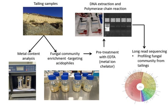
2. Materials and Methods

2.1. Site Description and Soil Sampling

Mine tailing samples were collected at three different tailing storage sites in Krugersdorp, Gauteng province, South Africa (mixed tailing and tailing A—26.132750, 27.711464, tailing B—26.133074, 27.767206, and tailing C—26.135979, 27.740172). A diagonal sampling method was used [25], and the samples were collected at various depths: depth 1: 0–15 cm, depth 2: 15–30 cm, and depth 3: 30–45 cm, while mixed tailings (mixed tailings before re-processing) were collected on the surface. Each depth’s samples were combined (for instance, all 0–15 cm depth samples were combined to form one sample per sampling site), and from each tailing storage site, a total of three samples were obtained. All samples were collected using sterile zipper bags and transported to the university laboratory on ice, and upon arrival, the samples were stored at 4 °C until further analysis.

2.2. Characterization of Mine Tailing Samples

The pH of the tailings was determined by dissolving 1 g of tailings in 10 mL of dH2O and allowing it to sit for an hour before taking pH measurements with an AD1030 pH meter. For heavy metal content analysis, 0.25 g samples were digested by mixing nitric acid (HNO3) and hydrochloric acid (HCL) at a ratio of 3:1 in a microwave digester (CEM Mars-6using Xpress plus vessels, MAD TECHNOLOGY (PTY) LTD, Johannesburg, South Africa), heated to 180 °C, and held there for 10 min. The samples were then diluted up to 50 mL in falcon tubes. The metals in the solution were initially scanned, followed by quantification using inductively coupled plasma atomic emission spectroscopy (ICP-AES) (Shimadzu ICPE-9820, Gauteng, South Africa).

2.3. Microbial Profiling and Isolation

2.3.1. Enrichment and DNA Extraction

Two different selective media (Table 1) were used to enrich the fungal communities found in the tailing samples. The purpose of enrichment was to target the acidophilic/acid-tolerant communities with bioleaching potential and to increase their population for isolation. The two media were prepared as follows:
  • Solution A contains all the compounds listed in Table 1 (for each media) except iron (II) sulfate or sulfur, which were sterilized by autoclaving at 112 °C for 30 min.
  • Solution B contains either iron (II) sulfate (44.2 g/L in liquid media and 6 g/L on solid media, maintained at pH 2 to avoid oxidation) or sulfur (30 g/L in liquid media and 10 g/L) [26,27]. The solutions were sterilized by filtration using 0.2 µm sterile filters and added to solution A aseptically to prepare iron- or sulfur-containing media [28].
For the initial enrichment, only solution A was prepared, in which 20 g of tailings was added to 250 mL of the two specific media in Duran Schott bottles. The contents of the tailings were used as an energy source. The samples were enriched for 12 days at 30 °C in a shaker incubator at 140 rpm (Labotec (PTY) LTD, Model 353, Gauteng, South Africa); this was referred to as S1. After 12 days, 20 mL was transferred to the fresh liquid media containing both solution A and solution B (iron (II) sulfate heptahydrate served as the energy source) and incubated for a further 12 days; this was referred to as S2.

2.3.2. Optimization of DNA Extraction

Several failed attempts at extracting DNA and running a successful PCR from the enriched tailing samples led to the assumption that metal ions may have been interfering with the DNA extraction process. To stabilize the metal ions in the samples, 10 mL of S1 and S2 samples was centrifuged at 14,000 rpm, and the supernatant was discarded. The pellet was resuspended with EDTA (4–13 µg/µL) and treated at room temperature for an hour on a rotating mixer at 60 rpm before extraction [24]. The samples were further centrifuged to remove the EDTA and resuspended in 250 µL dH2O. The samples with no EDTA treatment were centrifuged and only resuspended in dH2O. DNA extractions were conducted using Zymo soil/faecal and BIOMICS extraction kits (Inqaba Biotechnical Industries (Pty) Ltd., Pretoria, South Africa), according to the manufacturer’s instructions. The ratio of absorbance at 260 and 280 nm was used to check the purity of the extracted DNA using a nanodrop (Mfg year 2016) and quantified using a QUBIT fluorometer (ThermoFisher, Edenvale, South Africa). To further confirm the chelating ability of EDTA, a PCR amplification of the ITS gene was conducted using ITS1—5′ CTTGGTCATTTAGAGGAAGTAA-3′ and ITS4—5′ CCTCCGCTTATTGATATGC-3′ primers [31]. The ITS gene was amplified in a 25 µL reaction containing DNA template, 5 µM of each primer, and 12.5 µL of Biolabs 2X Master mix (Inqaba Biotechnical Industries (Pty) Ltd., Pretoria, South Africa). The PCR amplification was performed as follows: initial denaturation at 95 °C for 30 s, 35 cycles of denaturation at 95 °C for 30 s, annealing at 55 °C for 1 min, extension at 68 °C for 1 min, and a final extension at 68 °C for 5 min. One percent agarose gel electrophoresis was used to view the amplified gene. The DNA samples treated with EDTA that resulted in a successful PCR amplification were submitted to Inqaba Biotechnical Industries (Pty) Ltd., Pretoria, South Africa for long amplicons sequencing with PacBio sequel II.

2.3.3. Bioinformatics and Statistics

A total of 1,375,847 demultiplexed reads were obtained across 46 samples, with an average of 29,909 reads per sample. The DADA2 ITS pipeline workflow [32] with technology specific for PacBio [33] was used. The DADA2 R package v1.18.0 was installed on R studio. The sequences were quality filtered and denoised, and the chimeric sequences were removed. The DADA2 default naïve Bayesian classifier [34] was used to assign taxonomy to the amplicon sequence variants based on the UNITE database [35]. The resulting OTU table with assigned taxonomy was exported to Excel for relative abundance plots at different levels. The alpha and beta diversities plots were generated using MicrobiomeAnalyst (a comprehensive statistical, visual, and meta-analysis of microbiome data).

3. Results

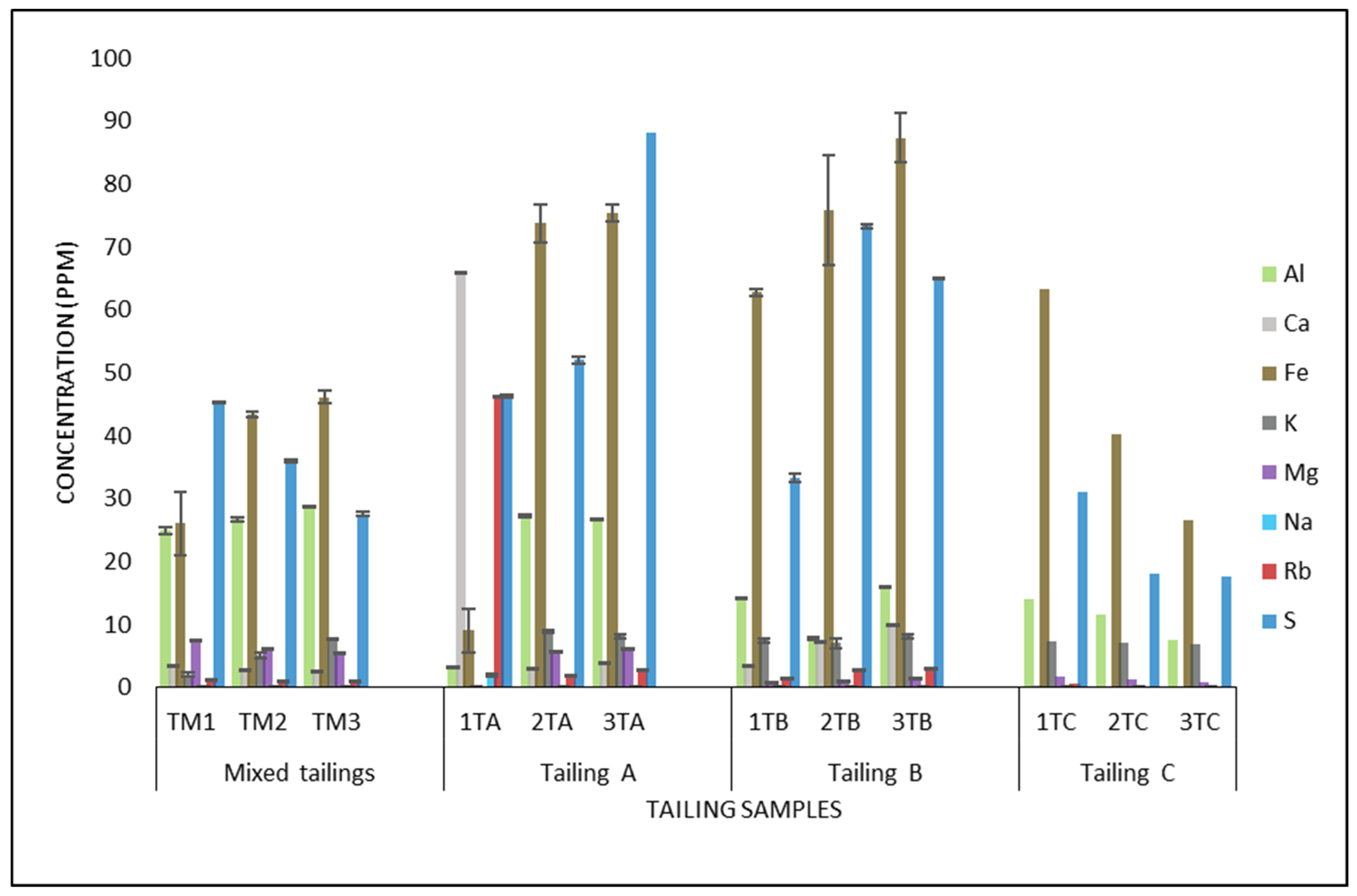
3.1. Tailing Characterization

Table 2 shows the pH measurements of the four different tailing samples, which ranged from 3.9 to 1.8 in all four tailing sites. The pH range in tailings A and B is the lowest. Figure 1 depicts the metal content analysis of the tailings at various depths using ICP-EOS. The highest concentrations of iron and sulfur were observed in all the tailings, followed by aluminum in varying concentrations. The differences in the tailings revealed that tailings A and B had the highest concentrations of the three metals, with iron at 75.7 ppm and 87.3 ppm and sulfur at 88.1 ppm and 73.2 ppm, respectively. Rubidium and calcium were observed to be highest in tailing A only at 46.3 and 65.8 ppm, respectively. Rubidium (except in tailing A depth 1), Calcium (except in tailing A depth 1), potassium, and sodium were found in lower concentrations.

3.2. DNA Optimization

An EDTA concentration of 9 µg/µL was found to be effective for metal ion chelation in S1 samples. This was confirmed by comparing the 260/280 ratio DNA concentration and the successful PCR amplification of the ITS1–ITS4 region in S1 samples from both enrichment media. The 260/280 ratio and the DNA concentration improved in the EDTA-treated samples (Table 3). The average ratio of the samples without EDTA treatment in GYEM and YSM was 1.68 and 1.18, respectively, and improved to 2.0 and 1.75, respectively, in the samples treated with EDTA. The DNA concentration increased from 1.31 ng/mL in GYEM and 0.35 ng/mL in YSM in the control samples to 74.4 ng/mL and 30.7 ng/mL, respectively, in the EDTA-treated samples. The PCR amplification of the ITS region was successful in all the EDTA-treated DNA samples (Figure S1).
EDTA concentrations ranging from 9–13 µg/µL were used in the S2 samples. The average ratio of the samples without EDTA treatment in GYEM and YSM was 1.72 and 1.07, respectively, whereas the average ratio improvement in the samples treated with EDTA was 2.38 and 1.70, respectively. In GYEM, the average DNA concentration increased from 1.52 ng/mL in the samples that were not treated with EDTA to 39.72 ng/mL in the samples that were treated with EDTA. In YSM S2, the samples without EDTA treatment yielded no DNA, whereas the samples with EDTA yielded 1.92 ng/mL on average. Amplification of the ITS region by PCR was mostly successful in the GYEM S2 samples treated with EDTA (Figure S1).

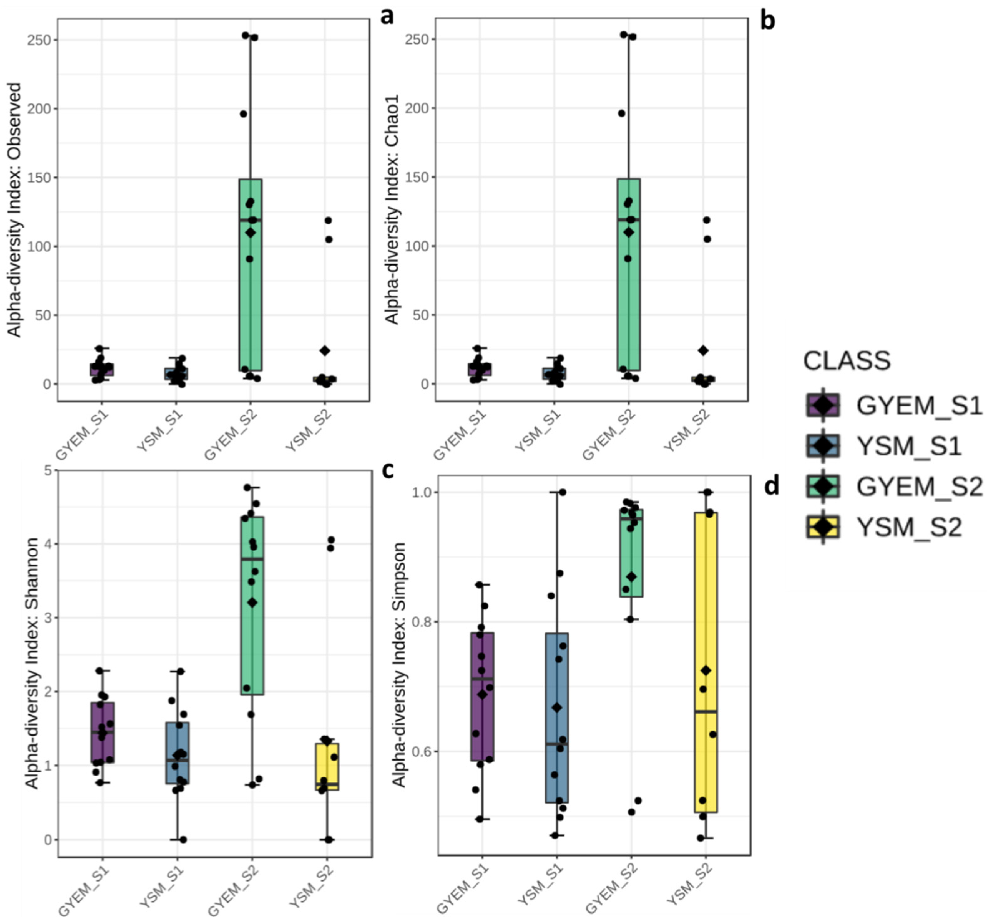
3.3. Alpha and Beta Diversities

A total of 766,039 sequences were clustered into 564 OTUs, after filtering, using the following criteria—a minimum count of 4 OTUs per sample and the OTU number was 167 using MicrobiomeAnalyst (a comprehensive statistical, visual, and meta-analysis of microbiome data). The alpha diversities were measured at the feature level using the Kruskal–Wallis statistical method (Figure 2); significant differences for alpha-diversity indices S1 and S2 were observed. The statistical p-values were as follows: observed and chao1—p < 0.002, Shannon—p < 0.003 and Simpson—p > 0.006. The highest diversity index was observed in GYEM S2, and the lowest diversity index was observed in YSM S2. The beta diversity was also measured using the Bray–Curtis index distance method, and the PERMANOVA statistical method was used (F-value: 2.5639; R-squared: 0.15479; p-value < 0.001) (Figure 3). Beta diversity compares the number of species shared between samples; from Figure 3, overlaps can be seen from all the four grouped samples (based on media (GYEM and YSM) and enrichment (S1 and S2).

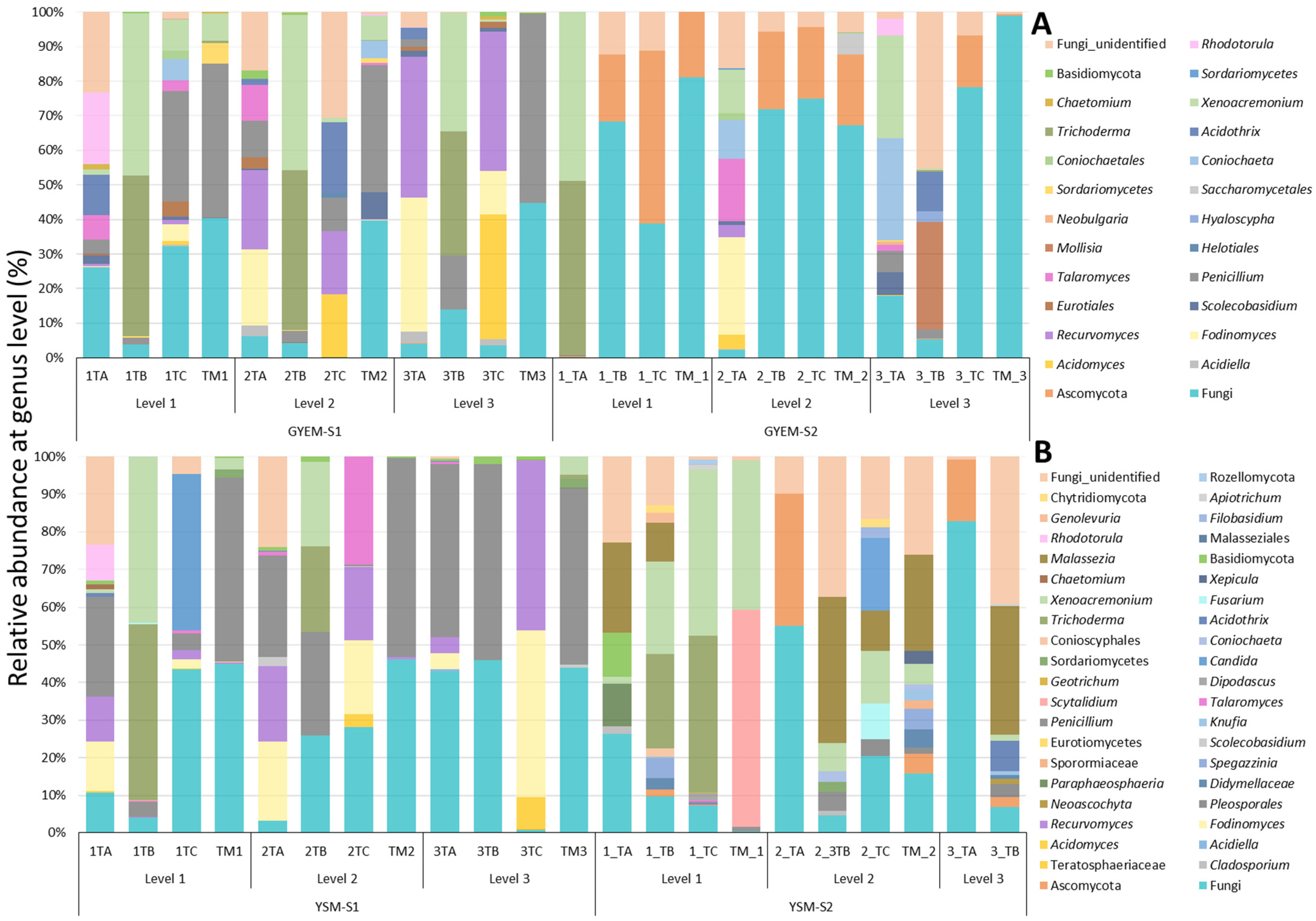
3.4. Taxonomic Diversity of Microbial Community on GYEM and YSM Enrichment

The relative abundance of the microbial communities enriched with glucose yeast extract and yeast sucrose media was determined at the phylum and genus depths comprising at least 1% in at least one sample (Figure 2 and Figure 3). There was an abundance of the Ascomycota, Basidiomycota, Chytridiomycota, and Rozellomycota phyla (Figure 4). Despite the apparent dominance of these phyla in the various tailing sites, each enrichment medium had its own distinct fungal community composition (Figure 5). The phylum Ascomycota was the most prevalent in both enrichment media, represented by the genera Penicillium, Xenoacremonium, Talaromyces, Fodinomyces, Recurvomyces, and Trichoderma. The phylum Basidiomycota was abundant in YSM-S2, with the genera Rhodotorula and Malassezia representing this phylum.
The enrichment media were selected to target the acid-tolerant fungi with bioleaching potential. The microorganisms in the tailings were first enriched in GYEM and YSM using the content of the tailing as a source of energy (S1) and then sub-cultured in the same media, but iron (II) sulfate heptahydrate was used as a source of energy (S2). The following paragraphs compare the diversity abundance of S1 and S2 on both enrichment media per tailing site at genus level (Figure 5). Furthermore, by using the PacBio platform to sequence the full-length ITS region of the extracted DNA, the taxonomic profile of some fungal populations in the tailing samples was resolved to the species level (see Figure S2).
Tailing A: The genera Penicillium, Fodinimyces, and Recurvomyces were observed in most depths of the tailing A samples for both media in the S1 and S2 enrichments, ranging from 4.2–45%. Talaromyces was observed only in GYES-S1 depth 1 and 2 (7.09% and 10.5%), while Trichoderma and Xenoacremonium were observed only in depth 1 and 2 of GYEM-S2 (12.6–50.6%). The genus Rhodotorula was only observed in depth 1 S1 of both media enrichment but was most dominant in GYEM-S1 at 20.6%. Unique genera to GYEM-S2 depth 1 and 3 were Trichoderma (50.6%), Scoleobasidium (6.6%), and Acidothrix (20.8%), whereas Paraphaesophaeria (11.3%) Basidiomycota (11.5%), and Malassezia (23.8%) were unique to YSM-S2 depth 1.
Tailing B: Similar to tailing A, Penicillium, Fodinomyces, and Recurvomyces were observed in most depths of the tailing B samples for both media in the S1 and S2 enrichments, ranging from 15.7–52%. Malassezia was only observed in YSM-S2 depth 1 and 2 at 10.11% and 38.4%, respectively, while Acidothrix was observed in S2 depth 3 in both media. Mollisia (3.1%) was unique to GYEM-S2 depth 3, and Spegazzinia (5.3%) was unique to YSM-S2.
Tailing C: The genera Penicillium, Fodinimyces, Acidomyces, and Recurvomyces were observed in most depths of the tailing C samples for both media in S1 and S2 enrichments, ranging from 4.9–44%. Penicillium was observed in S1 of both media (GYEM-S1 depth 1 at 31.9% and 2 at 9.7%, YSM-S1 depth 1 at 4.1%). Xenoacremonium (8.9%) was observed in the GYEM-S1 depth, while in YSM-S2 it was observed at depth 1 and 2 (43.6% and 13.8%). Talaromyces (28.7%), Coniochaeta (6.2%), and Acidothrix (20.3%) were unique to GYEM-S1 depth 1 and 2, while Candida (41.8%) was unique to YSM-S2 depth 2 and Fusarium (9.5%) and Pleosporale (4.4%) were unique to YSM-S2 depth 2.
Mixed tailings: The genera Penicillium and Xenoacremonium were abundant in most depths of mixed tailing samples for both media in the S1 and S2 enrichments, ranging from 7.9–52.7%. Sorduriomycetes was observed in both media S1 but was most abundant in GYEM at 5.87%. Scolecobasidium (7.5%) was unique to GYEM-S1 depth 2, whereas Scytalidium (57.6%), Malassezia (25.6%), and Spegazzinia (5.3%) were unique to YSM-S2 depth 1 and 2.
The FungalTraits database was used to determine the dominant fungal taxa’s ecological information and functional assignment [36,37]. This database categorizes the functional assignment into various “traits”, such as primary and secondary lifestyles, endophytic interaction capacity, aquatic habitat, and many others. The FungalTraits database revealed ecological and functional information for eight of the 17 dominant genera. According to the findings of the ecological functional assignment, for primary lifestyle, the genera are mostly soil and wood saprotrophs. Their secondary lifestyle and endophytic interaction capacity are primarily foliar-endophytic, with a few genera assigned to root-associated and rock-inhabiting. Table 4 contains more detailed information on the ecological data and the functional assignment found for the fungal isolates present in this study.

4. Discussion

4.1. DNA Extraction Optimization

Metal ion interference on DNA recovery is largely determined by metal concentration [10]. As shown in S1 and S2 of both enrichment media, the effectiveness of EDTA in chelating metal ions varies with the metal content and concentration. In S1, the main energy source was the tailing contents (Figure 1), whereas in S2 the main energy source was the inclusion of iron (II) sulfate heptahydrate. As a result of the higher Fe2+ ion concentrations, the S2 samples may have required a higher EDTA concentration to complete chelation. Fe2+ may also be responsible for the low DNA recovery and the encountered difficulty in running PCR in S2 samples. These findings are similar to an observation made by Kuffel et al. [10] when they investigated metal ion interferences in forensic DNA analysis from crime scenes, where DNA extractions were carried out from swabs of a variety of metal objects, including bullets, cartridge casings, gun surfaces, knives, metal wires, and surfaces, as well as calcium-rich bone samples. They caution that the metal ions’ impacts on DNA recovery, extraction, and amplification are not entirely understood. However, their investigations demonstrate the effects of some metals on DNA amplification, showing that inhibitory effects were observed with zinc, tin, iron (II), and copper, with 50% of the inhibitory concentration (IC50) values much below 1 mM. Thus, their study finding supports the observation made in this present study regarding the inhibitory effects observed with Fe2+ ions.
The low recovery of DNA and failed PCR may also be attributed to the high concentration of EDTA used in S2 as compared to the S1 samples. According to a study conducted by Khosravinia and Ramesha [23], EDTA concentrations > 11 µg/µL are known to reduce DNA recovery. Beyond chelating, Lopata et al. [20] also showed that EDTA affects enzyme activity by tightly binding to the active site of enzymes such as Taq DNA polymerase, dUTPase, and dNTPase, resulting in low or no PCR products. As an example, Taq polymerase requires Mg2+ for activation, while EDTA tends to compete with Mg2+ for binding, resulting in enzyme inhibition. This trend of the poor activity of enzymes in the presence of higher concentrations of EDTA and metal ions was also observed by Kuffel et al. [10] and they highlighted that KOD polymerase was more metal-resistant than Taq polymerase in such instances
A ratio of absorbance at 260 and 280 nm can be used to assess DNA purity, and a ratio of ≈1.8 and even up to 2.0 is regarded as pure. However, a ratio above 2.0 shows the likelihood of RNA contamination of the DNA being assessed for purity. Moreover, a ratio of >2 is related to DNA degradation and is showing, rather, a measurement of free nucleotides, as described in the protocol manual for the NanoDrop instrument [38]. Moreover, a ratio of 1.6 suggests the presence of proteins, phenol, or other contaminants that absorb strongly at or near 280 nm [39]. The S2 samples had an average ratio of 1.7 after treatment with EDTA, which is likely due to any one of the factors described.

4.2. Taxonomic Diversity of Microbial Community in Tailing Samples

Metamorphic environments such as tailings are characterized by high metal concentrations and extremely low pH ranges. These factors contribute to the diversity structure of the acidophilic or acid-tolerant communities found in that environment [40,41]. As a result, the taxonomic abundance of the fungal community observed in the four tailing samples was influenced by their habitat. Furthermore, the nutrients provided by the enrichment media also contributed to the taxonomic abundance. Tailing A and B had the lowest pH ranges of <2.1, which decreased with each depth, whereas mixed tailing and tailing C had pH ranges of >3 for all depths. However, the four tailings had similar metal content with high concentrations of iron, sulfur, and aluminum, though the concentration differed (Figure 1). This explains why genera such Penicillium, Fodinomyces, and Recurvomyces were dominant in most depths of the different tailings. Penicillium and Fodinomyces are both acidophilic (growing only at pH below 5) and acid-tolerant (growing at pH ranging below or above 5) [1]. Furthermore, their abundance in both enrichment media demonstrates their ability to thrive in different carbon sources [1,42]. Penicillium and Fodinomyces have been applied in bioleaching and have demonstrated metal tolerance and good bioleaching efficiencies [1,43]. In our research, no report of Recurcomyces in tailing samples or its application in bioleaching was found, making this report the first report of its identification in tailings. However, this genus has been described by Selbmann et al. [44] as a rock-inhabiting yeast found in cold, dry environments, and this ecological feature is supported by analysis conducted using the FungalTraits database (Table 4). Furthermore, Coleine et al. [45] suggested that nonsporulating fungi found in rock habitats may occasionally be dispersed between continents, for example via dust, and adapt to other ecologically similar habitats. As a result, the characteristics of Recurvomyces, such as growing in an extreme environment, as described by Coleine et al. [45], Selbmann et al. [5], Ametrano et al. [46], and Selbmann et al. [44] are evident enough to support the possibility of its existence in tailings. Further investigations of this genus’ bioleaching efficiency are therefore required.
Another dominant genus that was observed in most of the tailings (except in tailing B), especially in depths 1 and 2, is Xenoacremonium. This genus, previously referred to as Acremonium recifei (now renamed Xenoacremonium recifei L. by Lombard and Crous) [47], is a plant pathogen involved in wood decay and has also been isolated from petroleum-contaminated sites [48,49]. Ecological information from FungalTrails also shows that it is a wood saprotroph; thus, its characterization as a wood decaying pathogen is valid. This is likely the first report of this genus in tailing samples. No studies have yet linked this genus to bioleaching processes or applications. Acidothrix is also another genus observed to be dominant mostly in depth 3 of the tailing samples (except tailing C). Acidothrix exhibits both acid-tolerant and acidophilic characteristics and is mostly found in extreme habitats such as acidic soils [50]. This explains its detection only at depth 3, where the lowest pH ranges were observed.
Although similar genera were observed in most tailings, some genera were only found in specific tailing samples. For example, Trichoderma, Rhodotorula, and Paraphaesophaeria were only observed in tailing A, Mollisia in tailing B, Candida, Acidomyces, Coniochaeta, Fusarium, and Pleosporale in tailing C, and Scytalidium in mixed tailings. Though they are unique to each tailing, Trichoderma, Candida, Acidomyces, Rhodotorula, Fusarium, Coniochaeta, and Scytalidium have been observed to share similar characteristics such as acid and metal tolerance. Their occurrence in tailing samples has been reported as well as their application to the bioleaching process [43,51,52]. The fungus Paraphaesophaeria was only found in tailing A and is known to be a plant-associated (primarily a desert plant) fungi with Cu, Cr, and Cd tolerance [53]. It has also been reported in manganese oxidation [54]. Mollisia is another genus found only in tailing B. It is an endophyte that is associated with plants and is also involved in plant decay [55,56]. Its presence in tailings is likely due to the current efforts to revegetate tailings to prevent dust dispersion during windy days, as well as other environmental concerns linked to airborne pollution [57].
A comparison of the observed genera in the two media revealed genera that were unique to each. For example, Malassezia and Spegazzinia were observed only in YSM S2 and Scoleobasidium, which was unique to GYEM. The genus Malassezia has been reported in flotation tailings of the Kevitsa mine in Finland [58] and in zinc and copper tailings [59]. Because the genus is known to dominate parts of the human microbiota [60], it was suggested by Miettinen et al. [59] that its presence in tailing samples may have come as result of human contamination. However, ecological information from FungalTraits assigned Malassezia as a soil saprotroph that is associated with plant roots. This may be a demonstration of the genera’s versatility and mobility. Thus, its ability to survive in low pH and high concentrations of iron (II) sulfate heptahydrate in the S2 media enrichment, as observed in this study, implies that members of this genus are potentially acid-tolerant and further investigations may be required to determine their bioleaching potential. According to Amend [61], Malassezia are ecologically hyper-diverse and have been reported in diverse environmental sources, such as Antarctic soils [62], hydrothermal vents, and deep-sea sediments [61]. Spegazzenia is a wood saprotroph with no endophytic capability, as described in Table 4, obtained from FungalTraits. This genus of Spegazzenia was observed by Samarakoon et al. [63] to be associated with Musa sp. (banana). No reports of Spegazzenia in the past have associated any member of this genus with tailing or bioleaching. Scoleobasidium is an endophyte that was previously isolated in tomato fruits, as reported by Mahmoud and Narisawa, [64]. The genus showed the ability to increase plant biomass in the presence of an organic nitrogen source. The plant growth promoting ability was also reported by Hamayun et al. [65] in soybean plants. Therefore, just like Mollisia, its introduction to tailings is likely due to the revegetation process at these tailing sites. These four genera, however, have not been previously reported in association with bioleaching processes and will require extensive investigations. This validates the need for routine bioprospecting of extreme environments for microorganisms that may potentially be useful for industrial applications and biomining operations.

5. Conclusions

In this work, the application of EDTA to stabilize divalent metal ions was explored as a result of the difficulties encountered in extracting DNA from the metal-rich environment of tailings. Our investigation showed that the use of EDTA improved the DNA recovery for S1 samples with no interference with the downstream application. However, in the S2 samples, the use of a high EDTA concentration led to low DNA recovery, which impacts the downstream application. It is therefore recommended that a necessary preliminary application of optimal concentrations of EDTA is required to extract DNA from environmental samples obtained from metal-rich environments such as those present in tailings. However, we caution that a further EDTA removal step must be included above the conventional wash step advised by the DNA extraction kit manufacturers. The elimination of the additional EDTA is needed to ensure the integrity of the DNA extracts and to prevent enzyme inhibition during downstream analysis.
The fungal ITS1 long-read amplicon sequencing revealed the presence and varied compositions of the acidophilic and acid-tolerant fungal diversity at different depths of the four tailing samples. It further elucidated novel fungi genera that had not been previously identified in tailing samples and were likely unique to these mines in Krugersdorp, Gauteng, South Africa. The findings of this study clearly support the use of culture-independent techniques and long-read amplicon sequencing to gain a more in-depth understanding of biodiversity. However, culture-independent studies do not provide enough opportunities to visualize (morphological characteristics), to test functions (such as bioleaching ability), and to investigate the genetics and genomics of these acidophiles. This limitation is being solved by the creation of the FungalTraits database. Nonetheless, they provide great insights with their high throughput that prompt further investigation, particularly with regard to applications and further tests of microbial efficiencies and abilities. As such, they are excellent tools for bioprospecting vast sample areas.

Supplementary Materials

The following supporting information can be downloaded at: https://www.mdpi.com/article/10.3390/jof8050419/s1, Figure S1: gel image showing samples with successful amplification of ITS region, Figure S2: evolutionary relationships of taxa resolved to species level.

Author Contributions

Methodology, software, validation, formal analysis; investigation, R.N.; data curation and writing—original draft preparation, R.N.; writing—review and editing, supervision, project administration, T.S.M. and G.N.I., funding acquisition, T.S.M. All authors have read and agreed to the published version of the manuscript.

Funding

This research was funded by the Technology Innovation Agency (TIA), grant number 2018/FUN/0166, the South African Department of Science and Innovation (DSI), grant number DST/CON 0197/2017, and the National Research Foundation (NRF), grant number 138093.

Institutional Review Board Statement

Not applicable.

Informed Consent Statement

Not applicable.

Data Availability Statement

The original sequencing data obtained in this study were submitted to the NCBI Sequence Read Archive (SRA) database (fungi accession number: PRJNA803379).

Acknowledgments

The authors would like to thank David Heisi, Matshidiso Tsotetsi, Morena Mokoena, Asheal Mutungwazi, and Lebohang Koloti for their assistance with the tailing sample collection.

Conflicts of Interest

The authors declare no conflict of interest.

References

  1. Vázquez-Campos, X.; Kinsela, A.S.; Waite, T.D.; Collins, R.N.; Neilan, B.A. Fodinomyces uranophilus gen. nov. sp. nov. and Coniochaeta fodinicola sp. nov., two uranium mine-inhabiting Ascomycota fungi from northern Australia. Mycologia 2014, 106, 1073–1089. [Google Scholar] [CrossRef]
  2. Liang, J.; Liu, J.; Yang, T.; Wang, P.D.; Zhang, S.C.; Jia, P.; Liao, B.; Shu, W.S.; Li, J.T. Contrasting soil fungal communities at different habitats in a revegetated copper mine wasteland. Soil Ecol. Lett. 2020, 2, 8–19. [Google Scholar] [CrossRef] [Green Version]
  3. Jia, T.; Wang, R.; Fan, X.; Chai, B. A comparative study of fungal community structure, diversity and richness between the soil and the phyllosphere of native grass species in a copper tailings dam in Shanxi Province, China. Appl. Sci. 2018, 8, 1297. [Google Scholar] [CrossRef] [Green Version]
  4. Huerta-Rosas, B.; Cano-Rodríguez, I.; Gamiño-Arroyo, Z.; Gómez-Castro, F.I.; Carrillo-Pedroza, F.R.; Romo-Rodríguez, P.; Gutiérrez-Corona, J.F. Aerobic processes for bioleaching manganese and silver using microorganisms indigenous to mine tailings. World J. Microbiol. Biotechnol. 2020, 36, 124. [Google Scholar] [CrossRef]
  5. Selbmann, L.; Stoppiello, G.A.; Onofri, S.; Stajich, J.E. Culture-Dependent and Amplicon Sequencing Approaches Reveal Diversity and Distribution of Black Fungi in Antarctic Cryptoendolithic Communities. J. Fungi 2021, 7, 213. [Google Scholar] [CrossRef]
  6. Desai, C.; Madamwar, D. Extraction of inhibitor-free metagenomic DNA from polluted sediments, compatible with molecular diversity analysis using adsorption and ion-exchange treatments. Bioresour. Technol. 2007, 98, 761–768. [Google Scholar] [CrossRef]
  7. Liu, M.; Ding, X.; Wang, X.; Li, J.; Yang, H.; Yin, Y. Extraction of DNA from complex biological sample matrices using guanidinium ionic liquid modified magnetic nanocomposites. RSC Adv. 2019, 9, 23119–23128. [Google Scholar] [CrossRef] [Green Version]
  8. Colin, Y.; Goberna, M.; Verdú, M.; Navarro-Cano, J.A. Successional trajectories of soil bacterial communities in mine tailings: The role of plant functional traits. J. Environ. Manag. 2019, 241, 284–292. [Google Scholar] [CrossRef]
  9. Bernardino, A.F.; Pais, F.S.; Oliveira, L.S.; Gabriel, F.A.; Ferreira, T.O.; Queiroz, H.M.; Mazzuco, A.C.A. Chronic trace metals effects of mine tailings on estuarine assemblages revealed by environmental DNA. PeerJ 2019, 2019, e8042. [Google Scholar] [CrossRef]
  10. Kuffel, A.; Gray, A.; Daeid, N.N. Impact of metal ions on PCR inhibition and RT-PCR efficiency. Int. J. Legal Med. 2021, 135, 63–72. [Google Scholar] [CrossRef]
  11. Moreno, L.I.; McCord, B.R. Understanding metal inhibition: The effect of copper (Cu2+) on DNA containing samples. Forensic Chem. 2017, 4, 89–95. [Google Scholar] [CrossRef]
  12. Guéroult, M.; Picot, D.; Abi-Ghanem, J.; Hartmann, B.; Baaden, M. How cations can assist dnase i in DNA binding and hydrolysis. PLoS Comput. Biol. 2010, 6, e1001000. [Google Scholar] [CrossRef] [PubMed] [Green Version]
  13. Schrader, C.; Schielke, A.; Ellerbroek, L.; Johne, R. PCR inhibitors—Occurrence, properties and removal. J. Appl. Microbiol. 2012, 113, 1014–1026. [Google Scholar] [CrossRef] [PubMed]
  14. Dominguez, K.; Ward, W.S. A novel nuclease activity that is activated by Ca2+ chelated to EGTA. Syst. Biol. Reprod. Med. 2009, 55, 193–199. [Google Scholar] [CrossRef] [PubMed] [Green Version]
  15. Melgar, E.; Goldthwait, D.A. Deoxyribonucleic acid nucleases. II. The effects of metals on the mechanism of action of deoxyribonuclease I. J. Biol. Chem. 1968, 243, 4409–4416. [Google Scholar] [CrossRef]
  16. El-Ashram, S.; Al Nasr, I.; Suo, X. Nucleic acid protocols: Extraction and optimization. Biotechnol. Rep. 2016, 12, 33–39. [Google Scholar] [CrossRef] [Green Version]
  17. Heikrujam, J.; Kishor, R.; Behari Mazumder, P. The Chemistry Behind Plant DNA Isolation Protocols. Biochem. Anal. Tools–Methods Bio-Molecules Stud. 2020, 8, 131–141. [Google Scholar] [CrossRef]
  18. Bonsu, D.O.M.; Higgins, D.; Austin, J.J. Forensic touch DNA recovery from metal surfaces—A review. Sci. Justice 2020, 60, 206–215. [Google Scholar] [CrossRef]
  19. Feng, D.; van Deventer, J.S.J. Thiosulphate leaching of gold in the presence of ethylenediaminetetraacetic acid (EDTA). Miner. Eng. 2010, 23, 143–150. [Google Scholar] [CrossRef]
  20. Lopata, A.; Jójárt, B.; Surányi, É.V.; Takács, E.; Bezúr, L.; Leveles, I.; Bendes, Á.; Viskolcz, B.; Vértessy, B.G.; Tóth, J. Beyond chelation: EDTA tightly binds taq DNA polymerase, MutT and dUTPase and directly inhibits dNTPase activity. Biomolecules 2019, 9, 621. [Google Scholar] [CrossRef] [Green Version]
  21. Portzehl, H.; Caldwell, P.C.; Rüegg, J.C. The dependence of contraction and relaxation of muscle fibres from the crab Maia squinado on the internal concentration of free calcium ions. BBA-Spec. Sect. Biophys. Subj. 1964, 79, 581–591. [Google Scholar] [CrossRef]
  22. Zaitoun, M.A.; Lin, C.T. Chelating behavior between metal ions and EDTA in sol—gel matrix. J. Phys. Chem. B 1997, 101, 1857–1860. [Google Scholar] [CrossRef]
  23. Khosravinia, H.; Ramesha, K.P. Influence of EDTA and magnesium on DNA extraction from blood samples and specificity of polymerase chain reaction. Afr. J. Biotechnol. 2007, 6, 184–187. [Google Scholar] [CrossRef]
  24. Hall, N.E.L.; Axelrod, D.E. Interference of cellular ferric ions with DNA extraction and the application to methods of DNA determination. Anal. Biochem. 1977, 79, 425–430. [Google Scholar] [CrossRef]
  25. Tang, C.; Zhong, J.; Lv, Y.; Liu, X.; Li, Y.; Zhang, M.; Yan, X.; Sun, W. Response and dynamic change of microbial community during bioremediation of uranium tailings by bacillus sp. Minerals 2021, 11, 967. [Google Scholar] [CrossRef]
  26. Hassanshahian, M.; Ghoebani, S. Isolation and Characterization of Iron and Sulfur- Oxidizing Bacteria from Maiduk Copper Mine at Shahrbabk Province in Iran. Geomicrobiol. J. 2018, 35, 261–265. [Google Scholar] [CrossRef]
  27. Martínez, P.; Gálvez, S.; Ohtsuka, N.; Budinich, M.; Cortés, M.P.; Serpell, C.; Nakahigashi, K.; Hirayama, A.; Tomita, M.; Soga, T.; et al. Metabolomic study of Chilean biomining bacteria Acidithiobacillus ferrooxidans strain Wenelen and Acidithiobacillus thiooxidans strain Licanantay. Metabolomics 2012, 9, 247–257. [Google Scholar] [CrossRef] [Green Version]
  28. Dong, Y.; Lin, H.; Xu, X.; Zhou, S. Hydrometallurgy Bioleaching of different copper sul fi des by Acidithiobacillus ferrooxidans and its adsorption on minerals. Hydrometallurgy 2013, 140, 42–47. [Google Scholar] [CrossRef]
  29. Kishimoto, N.; Tano, T. Acidophilic heterotrophic bacteria isolated from acidic mine drainage, sewage, and soils. J. Gen. Appl. Microbiol. 1987, 33, 11–25. [Google Scholar] [CrossRef] [Green Version]
  30. Amiri, F.; Mousavi, S.M.; Yaghmaei, S. Enhancement of bioleaching of a spent Ni/Mo hydroprocessing catalyst by Penicillium simplicissimum. Sep. Purif. Technol. 2011, 80, 566–576. [Google Scholar] [CrossRef]
  31. Rasheed, A.; Cloete, E.; Khasa, D. Isolation and identification of iron ore—Solubilising fungus. S. Afr. J. Sci. 2010, 106, 1–6. [Google Scholar] [CrossRef]
  32. Callahan, B.J.; McMurdie, P.J.; Rosen, M.J.; Han, A.W.; Johnson, A.J.A.; Holmes, S.P. DADA2 ITS Pipeline Workflow (1.8). 2020. Available online: https://benjjneb.github.io/dada2/ITS_workflow.html (accessed on 22 November 2021).
  33. Callahan, B.J.; Wong, J.; Heiner, C.; Oh, S.; Theriot, C.M.; Gulati, A.S.; McGill, S.K.; Dougherty, M.K. High-throughput amplicon sequencing of the full-length 16S rRNA gene with single-nucleotide resolution. Nucleic Acids Res. 2019, 47, e103. [Google Scholar] [CrossRef] [PubMed] [Green Version]
  34. Wang, Q.; Garrity, G.M.; Tiedje, J.M.; Cole, J.R. Naïve Bayesian classifier for rapid assignment of rRNA sequences into the new bacterial taxonomy. Appl. Environ. Microbiol. 2007, 73, 5261–5267. [Google Scholar] [CrossRef] [PubMed] [Green Version]
  35. Kõljalg, U.; Nilsson, R.H.; Abarenkov, K.; Tedersoo, L.; Taylor, A.F.S.; Bahram, M.; Bates, S.T.; Bruns, T.D.; Bengtsson-Palme, J.; Callaghan, T.M.; et al. Towards a unified paradigm for sequence-based identification of fungi. Mol. Ecol. 2013, 22, 5271–5277. [Google Scholar] [CrossRef] [PubMed] [Green Version]
  36. Tanunchai, B.; Ji, L.; Schroeter, S.A.; Wahdan, S.F.M.; Hossen, S.; Delelegn, Y.; Buscot, F.; Lehnert, A.S.; Alves, E.G.; Hilke, I.; et al. FungalTraits vs. FUNGuild: Comparison of Ecological Functional Assignments of Leaf- and Needle-Associated Fungi across 12 Temperate Tree Species. Microb. Ecol. 2022, 1–18. [Google Scholar] [CrossRef]
  37. Põlme, S.; Abarenkov, K.; Henrik Nilsson, R.; Lindahl, B.D.; Clemmensen, K.E.; Kauserud, H.; Nguyen, N.; Kjøller, R.; Bates, S.T.; Baldrian, P.; et al. FungalTraits: A user-friendly traits database of fungi and fungus-like stramenopiles. Fungal Divers. 2020, 105, 1–16. [Google Scholar] [CrossRef]
  38. Kargar, S. Determination of DNA Concentration and Purity by Ultraviolet Spectrophotometry. Academia. Available online: https://www.academia.edu/29266669/Determination_of_DNA_Concentration_and_Purity_by_Ultraviolet_Spectrophotometry (accessed on 7 April 2022).
  39. Lucena-Aguilar, G.; Sánchez-López, A.M.; Barberán-Aceituno, C.; Carrillo-Ávila, J.A.; López-Guerrero, J.A.; Aguilar-Quesada, R. DNA Source Selection for Downstream Applications Based on DNA Quality Indicators Analysis. Biopreserv. Biobank. 2016, 14, 264–270. [Google Scholar] [CrossRef] [Green Version]
  40. Xue, P.; Carrillo, Y.; Pino, V.; Minasny, B.; Mcbratney, A.B. Soil Properties Drive Microbial Community Structure in a Large Scale Transect in South Eastern. Sci. Rep. 2018, 8, 11725. [Google Scholar] [CrossRef] [Green Version]
  41. Hu, X.; Wang, J.; Lv, Y.; Liu, X.; Zhong, J. Effects of Heavy Metals/Metalloids and Soil Properties on Microbial Communities in Farmland in the Vicinity of a Metals Smelter. Front. Microbiol. 2021, 12, 2347. [Google Scholar] [CrossRef]
  42. Xia, M.; Bao, P.; Liu, A.; Wang, M.; Shen, L.; Yu, R.; Liu, Y.; Chen, M.; Li, J.; Wu, X.; et al. Bioleaching of low-grade waste printed circuit boards by mixed fungal culture and its community structure analysis. Resour. Conserv. Recycl. 2018, 136, 267–275. [Google Scholar] [CrossRef]
  43. Begum, S.; Rath, S.K.; Rath, C.C. Applications of Microbial Communities for the Remediation of Industrial and Mining Toxic Metal Waste: A Review. Geomicrobiol. J. 2021, 1–12. [Google Scholar] [CrossRef]
  44. Selbmann, L.; de Hoog, G.S.; Zucconi, L.; Isola, D.; Ruisi, S.; Gerrits van den Ende, A.H.G.; Ruibal, C.; De Leo, F.; Urzì, C.; Onofri, S. Drought meets acid: Three new genera in a dothidealean clade of extremotolerant fungi. Stud. Mycol. 2008, 61, 1–20. [Google Scholar] [CrossRef] [PubMed]
  45. Coleine, C.; Stajich, J.E.; Ríos, A.D.L.; Selbmann, L.; Coleine, C.; Stajich, J.E.; Ríos, A.D.L.; Selbmann, L.; Coleine, C.; Stajich, J.E. Beyond the extremes: Rocks as ultimate refuge for fungi in drylands Beyond the extremes: Rocks as ultimate refuge for fungi in drylands. Mycologia 2021, 113, 108–133. [Google Scholar] [CrossRef] [PubMed]
  46. Ametrano, C.G.; Muggia, L.; Grube, M. Fungi in Extreme Environments: Ecological Role and Biotechnological Significance. Fungi Extrem. Environ. Ecol. Role Biotechnol. Significance 2019, 119–143. [Google Scholar] [CrossRef]
  47. Lombard, L.; van der Merwe, N.A.; Groenewald, J.Z.; Crous, P.W. Generic concepts in Nectriaceae. Stud. Mycol. 2015, 80, 189–245. [Google Scholar] [CrossRef] [Green Version]
  48. Patel, J.N.; Parmar, F.A.; Upasani, U.V. Isolation and Characterization of Pathogens Causing Disease in Pomegranate (Punica granatum L.), India. IJIRSET 2021, 10. [Google Scholar] [CrossRef]
  49. Taha, H.; Shivanand, P.; Khoo, D.H.; Mohammad, Y.H.; Matussin, N.B.A.; Metali, F. Identification of culturable petroleum-degrading bacteria and fungi from petroleum-contaminated sites in Brunei Darussalam. J. Environ. Sci. Health-Part A Toxic/Hazardous Subst. Environ. Eng. 2020, 55, 1542–1547. [Google Scholar] [CrossRef]
  50. Hujslová, M.; Kubátová, A.; Kostovčík, M.; Blanchette, R.A.; de Beer, Z.W.; Chudíčková, M.; Kolařík, M. Three new genera of fungi from extremely acidic soils. Mycol. Prog. 2014, 13, 819–831. [Google Scholar] [CrossRef] [Green Version]
  51. Tansengco, M.; Tejano, J.; Coronado, F.; Gacho, C.; Barcelo, J. Heavy metal tolerance and removal capacity of trichoderma species isolated from mine tailings in Itogon, Benguet. Environ. Nat. Resour. J. 2018, 16, 39–57. [Google Scholar] [CrossRef]
  52. Ou, S.N.; Liang, J.L.; Jiang, X.M.; Liao, B.; Jia, P.; Shu, W.S.; Li, J.T. Physiological, Genomic and Transcriptomic Analyses Reveal the Adaptation Mechanisms of Acidiella bohemica to Extreme Acid Mine Drainage Environments. Front. Microbiol. 2021, 12, 705839. [Google Scholar] [CrossRef]
  53. An, H.; Liu, Y.; Zhao, X.; Huang, Q.; Yuan, S.; Yang, X.; Dong, J. Characterization of cadmium-resistant endophytic fungi from Salix variegata Franch. in Three Gorges Reservoir Region, China. Microbiol. Res. 2015, 176, 29–37. [Google Scholar] [CrossRef] [PubMed]
  54. Takano, K.; Itoh, Y.; Ogino, T.; Kurosawa, K.; Sasaki, K. Phylogenetic analysis of manganese-oxidizing fungi isolated from manganese-rich aquatic environments in Hokkaido, Japan. Limnology 2006, 7, 219–223. [Google Scholar] [CrossRef]
  55. Tanney, J.B.; Seifert, K.A. Mollisiaceae: An overlooked lineage of diverse endophytes. Stud. Mycol. 2020, 95, 293–380. [Google Scholar] [CrossRef] [PubMed]
  56. Clark, T.N.; Ellsworth, K.T.; Decken, A.; Johnson, J.A.; Gray, C.A. The Application of nmr and lc-hrms Based Prioritization Strategies for the Discovery of Natural Products Produced by Endophytic Fungi from Medicinal Plants. Doctoral Dissertation, University of New Brunswick, Fredericton and Saint John, NB, Canada, 2019. [Google Scholar]
  57. Zappelini, C.; Karimi, B.; Foulon, J.; Lacercat-Didier, L.; Maillard, F.; Valot, B.; Blaudez, D.; Cazaux, D.; Gilbert, D.; Yergeau, E.; et al. Diversity and complexity of microbial communities from a chlor-alkali tailings dump. Soil Biol. Biochem. 2015, 90, 101–110. [Google Scholar] [CrossRef]
  58. Bomberg, M.; Miettinen, H.; Musuku, B.; Kinnunen, P. First insights to the microbial communities in the plant process water of the multi-metal Kevitsa mine. Res. Microbiol. 2020, 171, 230–242. [Google Scholar] [CrossRef]
  59. Miettinen, H.; Bomberg, M.; Le, T.M.K.; Kinnunen, P. Identification and metabolism of naturally prevailing microorganisms in zinc and copper mineral processing. Minerals 2021, 11, 156. [Google Scholar] [CrossRef]
  60. Sparber, F.; Ruchti, F.; LeibundGut-Landmann, S. Host Immunity to Malassezia in Health and Disease. Front. Cell. Infect. Microbiol. 2020, 10, 198. [Google Scholar] [CrossRef]
  61. Amend, A. From Dandruff to Deep-Sea Vents: Malassezia-like Fungi Are Ecologically Hyper-diverse. PLoS Pathog. 2014, 10, e1004277. [Google Scholar] [CrossRef] [Green Version]
  62. Garmendia, G.; Alvarez, A.; Villarreal, R.; Martínez-Silveira, A.; Wisniewski, M.; Vero, S. Fungal diversity in the coastal waters of King George Island (maritime Antarctica). World J. Microbiol. Biotechnol. 2021, 37, 142. [Google Scholar] [CrossRef]
  63. Samarakoon, B.C.; Phookamsak, R.; Wanasinghe, D.N.; Chomnunti, P.; Hyde, K.D.; McKenzie, E.H.C.; Promputtha, I.; Xu, J.C.; Li, Y.J. Taxonomy and phylogenetic appraisal of Spegazzinia musae sp. nov. And S. deightonii (Didymosphaeriaceae, Pleosporales) on Musaceae from Thailand. MycoKeys 2020, 70, 19–37. [Google Scholar] [CrossRef]
  64. Mahmoud, R.S.; Narisawa, K. A new fungal endophyte, Scolecobasidium humicola, promotes tomato growth under organic nitrogen. PLoS ONE 2013, 8, e78746. [Google Scholar] [CrossRef] [PubMed]
  65. Hamayun, M.; Khan, S.A.; Kim, H.Y.; Chaudhary, M.F.; Hwang, Y.H.; Shin, D.H.; Kim, I.K.; Lee, B.H.; Lee, I.J. Gibberellin Production and Plant Growth Enhancement by Newly Isolated Strain of Scolecobasidium tshawytschae. J. Microbiol. Biotechnol. 2009, 19, 560–565. [Google Scholar] [PubMed]
Figure 1. Heavy metal characterization of 4 different tailings at different depths. 1–3—sampling depth, T—tailing, A–C—different tailings, and M—mixed tailing.
Jof 08 00419 g001
Figure 2. Alpha diversity indices of fungi: (a)—observed OTUs (richness index), (b)—Chao1 (total richness), (c)—Shannon index (richness and evenness) and (d)—Simpson index (number of species present, as well as the relative abundance of each species).
Jof 08 00419 g002
Figure 3. Bray−Curtis measures of beta-diversity visualised using principal coordinate analysis (PCoA) for the comparison of fungal diversity for enrichment (S1 and S2) of tailing samples using GYEM and YSM.
Jof 08 00419 g003
Figure 4. Relative abundance of fungi at phyla depth: (A)—glucose yeast extract media and (B)—yeast sucrose media.
Jof 08 00419 g004
Figure 5. Relative abundance of fungi at genus depth: (A)—glucose yeast extract media and (B)—yeast sucrose media.
Jof 08 00419 g005
Table 1. Media composition for enrichment and isolation of bioleaching microorganisms.
Name Composition
General
Basic bioleaching media composition3.0 g/L (NH4)2SO4, 0.5 g/L MgSO4·7H2O, 0.5 g/L K2HPO4, 0.1 g/L KCl, 44.2 g/L FeSO4·7H2O or 30 g/L sulfur and 1.5% Agar bacteriological
Compounds Different in Each MediapHReferences
Glucose yeast extract medium (GYEM)5 g/L glucose, 0.05 g/L yeast extract3[29]
Yeast sucrose media (YSM)100 g/L sucrose, 1.5 g/L NaNO3, 1.6 g/L yeast extract3[30]
Table 2. pH measurements of the different tailings.
DepthsMixed TailingsTailing ATailing BTailing C
1: 0–15 cm 3.27 ± 0.752.1 ± 0.0173.0 ± 1.063.945 ± 0.02
2: 15–30 cm3.305 ± 0.461.8 ± 0.042.8 ± 1.123.7 ± 0.04
3: 30–45 cm3.25 ± 0.611.8 ± 0.0052.8 ± 1.123.7 ± 0.01
Table 3. Effectiveness of EDTA as chelating agent based on DNA purity, concentration, and successful amplification of ITS region.
Sample IDA260/A280Fluorometer (ng/mL)Reads Count
Glucose Yeast Extract Media
S1S2S1S2S1S2
No EDTAEDTANo EDTAEDTANo EDTAEDTANo EDTAEDTAEDTAEDTA
TM12.01.951.832.501.2578.51.5014.443,55725,392
TM21.552.002.02.331.181.092.002.0845,88243,225
TM31.402.001.502.66low0.592.5014.510,31976,036
1TA1.602.102.102.401.311400.5819.660,8033868
2TA1.52.001.932.700.981721.957.5919,9806725
3TA2.02.001.552.751.516.640.887.7260,6336365
1TB1.661.922.002.200.892252.8042.134,189125,058
2TB1.501.91.012.20low37.80.947.6151,55344,624
3TB2.01.951.352.200.841391.2113.543,36522,567
1TC1.712.141.882.251.298.88low2.0937,91545,172
2TC1.502.141.61.921.0726.03.0834031,218100,441
3TC1.802.002.002.505.48580.865.54704575,451
Yeast Sucrose Media
S1S2S1S2S1S2
No EDTAEDTANo EDTAEDTANo EDTAEDTANo EDTAEDTAEDTAEDTA
TM11.71.620.224.001.8514.8low0.7529,4269131
TM21.541.501.401.501.471.54low0.8827,541681
TM31.621.881.351.500.985.74low1.3238,058failed
1TA1.701.70.18--low9.31low0.5022,704568
2TA0.181.851.802.50low7.79low2.6023,80533,873
3TA1.271.871.442.25low183low3.3724,54220,572
1TB0.901.781.351.36low64.4low2.0634,0331193
2TB1.441.621.551.58low10.1low3.7868161433
3TB0.711.721.282.00low16.5low2.5711,6431261
1TC1.01.860.812.00low45.1low1.1913,8493809
2TC1.401.930.23--low9.34low2.8120,6931685
3TC0.751.751.271.75low1.30low1.2127,148failed
Note: 1–3 represents depths (1: 0–15 cm, 2: 15–30 cm, 3: 20–45 cm), respectively. TM—mixed tailing; TA—tailing A; TB—tailing B, and TC—tailing C.
Table 4. Ecological information and functional assignment of dominant fungal taxa using FungalTraits database.
GeneraPrimary LifestyleSecondary LifestyleComment on Lifestyle Endophytic Interaction Capability Plant Pathogenic CapacityDecay Substrate Decay Type Aquatic HabitatAnimal Biotrophic CapacityGrowth Form Fruitbody Type
Penicilliumunspecified saprotrophfoliar endophytetoxin-producing, animal parasite some species, mycoparasite some speciesfoliar endophyte0soilmoldpartly aquaticopportunistic human parasitefilamentous mycelium0
Recurvomycessoil saprotrophrock-inhabiting000soil0non-aquatic0filamentous myceliumperithecium (hymenium hidden, narrow opening)
Talaromycesunspecified saprotroph0hypervariable, thermophile, some species animal parasite some species various saprotrophs00leaf/fruit/seed, soil, animal materialmoldpartly aquaticopportunistic human parasitefilamentous mycelium0
Xenoacremoniumwood saprotroph0000wood0partly marine (partly non-aquatic)opportunistic human parasitefilamentous mycelium0
Rhodotorulaunspecified saprotrophfoliar endophyte0foliar endophyte000partly aquaticopportunistic human parasiteyeastnone
Acidothrixsoil saprotroph0000soil0non-aquatic0filamentous myceliumnone
Malasseziasoil saprotrophroot-associated0root-associatedroot-associatedroots, soil0partly aquaticanimal-associated/opportunistic human parasiteyeastnone
Spegazziniawood saprotroph00no endophytic capacity0wood0non-aquatic0filamentous myceliumperithecium (hymenium hidden, narrow opening)
Coniochaetaunspecified saprotrophfoliar endophytehypervariablefoliar endophyteleaf/fruit/seed pathogenleaf/fruit/seed, soil, dung, animal material0non-aquaticanimal parasitefilamentous myceliumcleistothecium (closed, spherical)
Fodinimyces, Trichoderma, Scoleobasidium, Paraphaesophaeria, Mollisia, Sardariomycetes, and Scytalidium were not detected on FungalTraits database.
Publisher’s Note: MDPI stays neutral with regard to jurisdictional claims in published maps and institutional affiliations.

