Article

Identification and Functional Characterization of Adenosine Deaminase in Mucor circinelloides: A Novel Potential Regulator of Nitrogen Utilization and Lipid Biosynthesis

1
Colin Ratledge Center for Microbial Lipids, School of Agricultural Engineering and Food Science, Shandong University of Technology, Zibo 255000, China
2
Department of Food Sciences, College of Food Science and Engineering, Lingnan Normal University, Zhanjiang 524048, China
3
Department of Botany and Microbiology, Faculty of Science, Al-Azhar University, Assiut 71524, Egypt
4
Department of Genetics and Microbiology (Associated Unit to IQFR-CSIC), Faculty of Biology, University of Murcia, 3100 Murcia, Spain
*
Author to whom correspondence should be addressed.
These authors contributed equally to this work.
J. Fungi 2022, 8(8), 774; https://doi.org/10.3390/jof8080774
Submission received: 29 June 2022 / Revised: 17 July 2022 / Accepted: 22 July 2022 / Published: 26 July 2022
(This article belongs to the Section Fungal Cell Biology, Metabolism and Physiology)

Abstract

:
Adenosine deaminase (ADA) is an enzyme distributed in a wide variety of organisms that cleaves adenosine into inosine. Since inosine plays an important role in nitrogen metabolism, ADA may have a critical function in the regulation of fatty acid synthesis. However, the role of ADA in oleaginous fungi has not been reported so far. Therefore, in this study, we identified one ada gene encoding ADA (with ID scaffold0027.9) in the high lipid-producing fungus, Mucor circinelloides WJ11, and investigated its role in cell growth, lipid production, and nitrogen metabolism by overexpressing and knockout of this gene. The results showed that knockout of the ada altered the efficiency of nitrogen consumption, which led to a 20% increment in the lipid content (25% of cell dry weight) of the engineered strain, while overexpression of the ada showed no significant differences compared with the control strain at the final growth stage; however, interestingly, it increased lipid accumulation at the early growth stage. Additionally, transcriptional analysis was conducted by RT-qPCR and our findings indicated that the deletion of ada activated the committed steps of lipid biosynthesis involved in acetyl-CoA carboxylase (acc1 gene), cytosolic malic acid enzyme (cme1 gene), and fatty acid synthases (fas1 gene), while it suppressed the expression of AMP-activated protein kinase (ampk α1 and ampk β genes), which plays a role in lipolysis, whereas the ada-overexpressed strain displayed reverse trends. Conclusively, this work unraveled a novel role of ADA in governing lipid biosynthesis and nitrogen metabolism in the oleaginous fungus, M. circinelloides.

1. Introduction

Microbial lipids, as environmentally sustainable and economically viable alternative sources of unusual lipids and fuels [1,2], have obtained increased attention since they have been used in the designing of high-value lipid products, particularly polyunsaturated fatty acids (PUFAs) such as alpha-linolenic acid (ALA), gamma-linolenic acid (GLA), and stearidonic acid (SDA), and others which have a variety of applications (i.e., in cosmetics, food, and healthcare industries) [3,4]. It is now possible to optimize the microbial lipid synthesis pathway to meet increasing market demands through metabolic engineering in microorganisms [5].
Nowadays, in order to produce a large number of oleochemicals, genetic modifications are performed in the varied microorganisms that are able to accumulate large quantities of lipids [6]. An oleaginous fungus, Mucor circinelloides, has been widely used for microbial lipid and lipid derivative production since it can accumulate lipids as nearly 36% of its cell dry weight (CDW); meanwhile, this fungus has efficient genetic engineering tools and a clear and known genetic background [7,8]. Moreover, its metabolism, specifically the production of the most diverse fatty acids, has been extensively studied and characterized [8,9]. Previous studies showed that a cascade of biochemical events has been triggered by nitrogen deficiency, which disturbed the TCA cycle and redirected carbon flux to lipid biosynthesis in M. circinelloides [10,11,12]. As postulated by Ratledge, lipid synthesis in M. circinelloides was regulated by a disparate number of enzymes, such as malic enzyme (ME), ATP-citrate lyase (ACL), acetyl-CoA carboxylase (ACC), and fatty acid synthase (FAS), which have been identified as conditional levers for fatty acid synthesis [11,13,14]. Many efforts, such as increasing the supply of precursors and diverting the carbon flux to TAGs, were made to increase the lipid production by changing the expression levels of the key enzymes involved in lipid synthesis in M. circinelloides [15,16,17]. Therefore, the genetic modification of the key enzymes involved in lipid synthesis was one of the celebrated strategies developed for achieving the profitable production of lipids in oleaginous fungi [18].
Adenosine deaminase (ADA; EC 3.5.4.4), as a crucial deaminating enzyme, catalyzes the irreversible conversions of adenosine to inosine and 2′-deoxyadenosine to 2′-deoxyinosine [19]. Meanwhile, it was ubiquitously expressed and involved in many cellular processes, including the biosynthesis and regulation of nucleic acids [20]. The ADA purified from Aspergillus oryzae has been found to catalyze the deamination of free adenine, adenine nucleotides, and oligonucleotides [21]. In addition, Lin et al. constructed adenosine deaminase-deficient recombinants to block the transformation of adenosine into hypoxanthine and enhance glutathione production in Escherichia coli and Saccharomyces cerevisiae [22]. Previous studies have also demonstrated that ADA is a possible producer of reactive oxygen species (ROS), which cause lipid peroxidation in cells and have a negative correlation with antioxidant enzymes [23,24]. In addition, increasing evidence has highlighted that ADA is associated with increased levels of hemoglobin (HbA1c), which plays a critical role in the derangement of lipogenesis in diabetes [25,26]. These studies showed that there is a connection between lipid metabolism and adenosine deaminase; however, research has rarely worked on the association of lipid metabolism with ADA, especially in oleaginous microorganisms.
Therefore, the present study aimed to investigate the role of ADA in the regulation of lipid biosynthesis in M. circinelloides. We constructed the ada gene knockout and overexpressing strains and studied their growth patterns, and the lipid accumulation in the cells. Additionally, we measured the mRNA expression levels of fatty acid metabolism-related genes in M. circinelloides to investigate the interconnection between ADA and lipid biosynthesis. To the best of our knowledge, this study provides the first report exploring the role of ADA in lipid accumulation and nitrogen metabolism in M. circinelloides WJ11.

2. Results

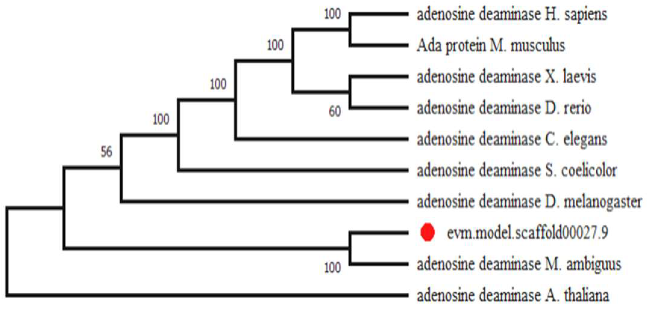
2.1. Identification of Gene Coding for the ADA in M. circinelloides

Based on the known completed genomic sequencing of M. circinelloides WJ11, a potential gene of adenosine deaminase ADA (namely ada encoded by scaffold0027.9) was identified by the genome-wide analysis. To further evaluate the general properties of the ada gene, the bioinformatical analysis of the genes was carried out by online website tools including ProtParam, CFSSP, and SOPMA. The ADA protein consists of 338 amino acids and exhibits good stability in water (the instability index is 38.79). Analysis of the grand average of hydropathicity (−0.250) indicated that ADA is hydrophilic. In addition, the ADA_AMPD domain (cd00443) was observed in ADA through CDD blast in the conserved domain database (NCBI-CDD), which suggested that this protein may have a catalytic role in adenosine deamination.
Phylogenetic evolution analysis was employed to explore the evolutionary relationship of the ADA in M. circinelloides WJ11 and its homologous proteins from other species. As shown in Figure 1, ADA protein occurs ubiquitously in animals, plants, and eukaryotic microorganisms. The ADA of M. circinelloides WJ11 shared 89% sequence similarities with GAN00815.1 of Mucor ambiguus, about 21% with AAH76532.1 of Danio rerio, and 23% with NP_000013.2 of Homo sapiens. Analysis of the amino acid sequences by NCBI-CDS exhibited that they all contain the conserved domain of the metallo-dependent hydrolase superfamily (cl00281), which has a conserved metal-binding site deprotonating a water molecule for a nucleophilic attack on the substrate in the common reaction mechanism. Similar to the tertiary structure of mouse ADA, the predicted three-dimensional structure of the ADA in M. circinelloides displayed an α/β-barrel structure with a zinc atom within the active site, which can catalyze the deamination of adenosine (Figure S1) [27]. Meanwhile, ADA has been observed in all human tissues in three isoforms: ADA1, ADA1+CP, and ADA2, involved in extracellular adenosine degradation [28]. Furthermore, an extensive search of the Danio rerio genome, followed by a phylogenetic analysis, confirmed the presence of three distinct members of ADA (ADA1, ADA2, and ADAL), which may take part in a complex regulation of adenosine deaminase activity [29]. In summary, these findings signified that the putative ADA could be responsible for adenosine degradation in M. circinelloides.

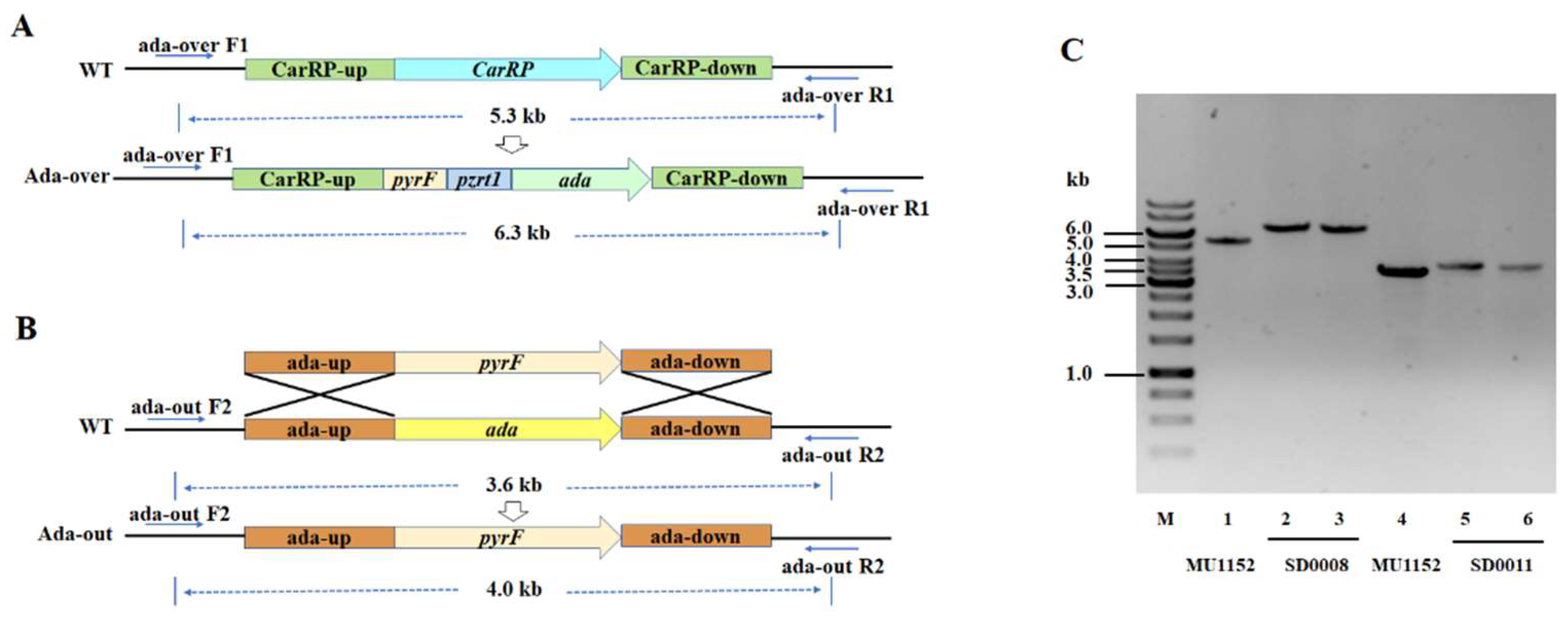
2.2. Generation of Ada Overexpressing and Knockout Transformants of M. circinelloides

The ada overexpressing and knockout mutants of M. circinelloides were generated to investigate the effect of ADA on lipid biosynthesis in M. circinelloides. The gene ada was overexpressed using the plasmid pMAT2075, which contained the pyrF gene as the selectable marker and a promoter zrt1, flanked by the sequences corresponding to regions surrounding the carotenogenic carRP gene (Figure 2A). Then, the gene overexpressing fragment was cut by Sma I and transformed into MU760, which is the leucine and uracil auxotroph of WJ11, by electroporation. The selection of homokaryotic transformants was conducted using the methods described by Rodríguez-Frómeta et al. [30]. The transformant was checked by PCR amplification with the relevant primers, ada-over-F1/R1, which obtained a 6.3-kb fragment from the ada-overexpressed strain, while a 5.3-kb fragment was obtained from the control strain (Figure 2C). The knockout plasmid was designed by a gene replacement strategy, using the pyrF gene as the selective marker and flanking the adjacent sequences of the ada gene to allow homologous recombination (Figure 2B). The fragments obtained from these designed vectors were cut by Not I and transformed into MU760. The construction of the ada-knockout mutant was confirmed by PCR amplification with the ada-out-F2/R2 primers (Figure 2C). The amplification results showed an expected 4.0-kb band in the ada-knockout strain and a 3.6-kb band in the control, respectively. Consequently, the above PCR analysis validated the target gene had been integrated into the genome of the overexpressed transformant and that was named as SD0008. Meanwhile, the absence of the 3.6-kb wild-type fragment indicated that the ada wide-type allele had been replaced and the transformant was named as SD0011, which was the homokaryon for the ada knockout.

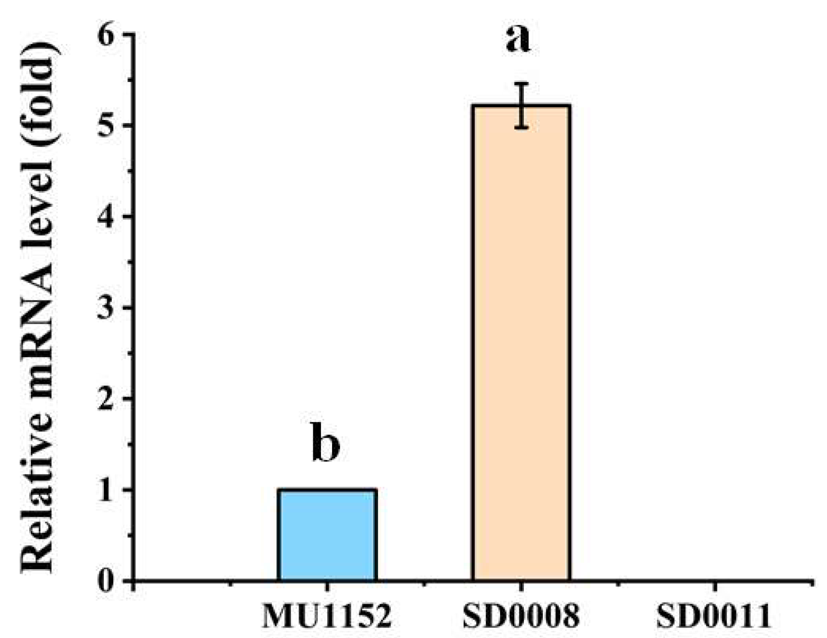
2.3. Analysis of Ada Expression in the Transformants

The relative mRNA levels of ada in the control MU1152, ada-overexpressing strain SD0008 and ada-knockout strain SD0011 were measured by qRT-PCR. Compared to the control strain, the transcription level of ada in the ada-overexpressing strain (SD0008) increased 5.2-fold at 24 h, which confirmed the ada gene was successfully overexpressed. However, the ada gene was deleted in SD0011, according to a marginal expression level of ada as shown in Figure 3.

2.4. Ada Regulated Nitrogen Metabolism and Affected Cell Growth and Lipid Accumulation

To investigate the role of ADA in the lipid biosynthesis of M. circinelloides, cell growth and lipid accumulation in SD0008 (ada-overexpressing strain) and SD0011(ada-knockout strain) were analyzed during 96 h of cultivation in K&R medium, supplemented with leucine.
As shown in Figure 4A, the cell growth patterns of three strains were affected by the ada gene expression level. Overexpression of ada increased nitrogen assimilation, cell growth, and the lipid accumulation of the fungus before 24 h of cultivation (from 7.20% in the control to 11.05% in SD0008) (Figure 4D). After that, cell proliferation of the ada overexpressing strain was inhibited and the cell dry weight was much less than the control, while the lipid accumulation rate (0.13 g/h) was lower than that in the control (0.31 g/h). Thus, the final cell lipid content (at 96 h) was nearly the same as that of the control strain. In contrast, ada disruption decreased nitrogen assimilation and cell growth before 24 h of cultivation, while at the later stage, lipid-free CDW increased by about 4.51% (Figure S2) and the lipid accumulation was increased by 20% compared to the control. Although it is not clear how the ada gene regulated the growth and lipid biosynthesis in the transformants, the results indicated that ada altered nitrogen depletion to face the cellular stress.

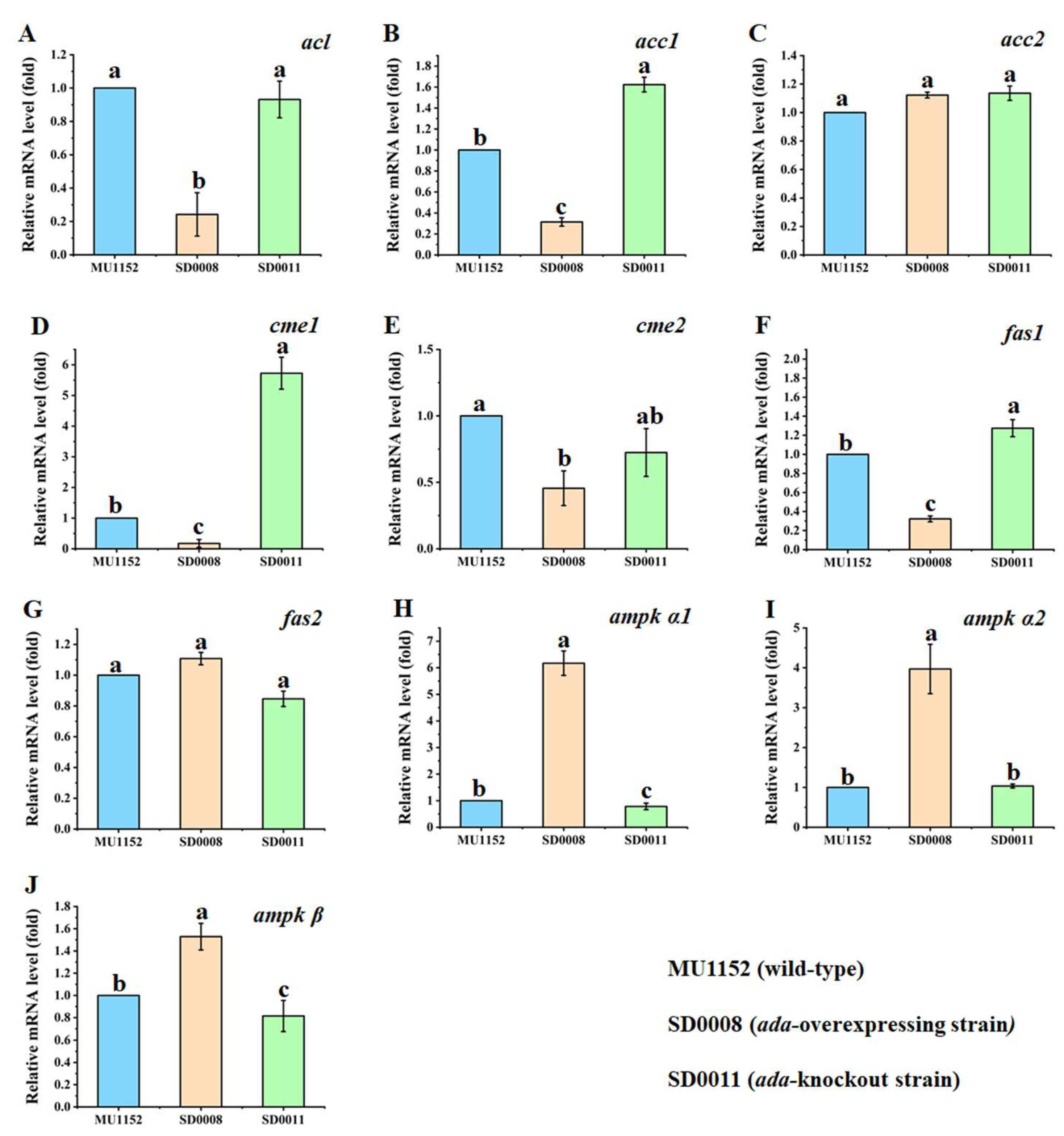
2.5. Impacts of Ada Gene Manipulation on the Expression Levels of the Key Genes for Fatty Acid Biosynthesis

Among the essential enzymes involved in lipid biosynthesis in M. circinelloides, ATP-citrate lyase (ACL) was encoded by acl (gene ID: scaffold00037.10), acetyl-CoA carboxylase (ACC) was encoded by acc1 (gene ID: scaffold00021.30) and acc2 (gene ID: scaffold00023.50), the cytosolic malic acid enzyme (ME) was encoded by cme1 (gene ID: scaffold00036.12) and cme2 (gene ID: scaffold00049.37), and fatty acid synthase was encoded by fas1 (gene ID: scaffold00002.57) and fas2 (gene ID: scaffold00111.12). In addition, as the negative regulator for lipid accumulation, AMP-activated protein kinase (AMPK) contains three subunits; its α subunit was encoded by ampk α1 (gene ID: scaffold00144.16) and ampk α2 (gene ID: scaffold00046.26), and its β subunit was encoded by ampk β (gene ID: scaffold00011.47), respectively. As the manipulation of the ADA expression level made a difference in the lipogenesis of M. circinelloides, the effects of ada overexpression and deletion on the expression of the pivotal genes in this fungus mentioned above were investigated by qRT-PCR at 24 h, when the nitrogen was exhausted in the fermentation medium.
Our results showed that the relative expression levels of acc1 and fas1 were slightly increased when ada was deleted (Figure 5B,F). In contrast, acl, acc1, and fas1 expression levels were significantly downregulated in the ada-overexpressed mutant (Figure 5A,B,F). Meanwhile, the transcriptional levels of acc2 and fas2 of the recombinant strains had no differences compared with the control (Figure 5C,G). Figure 5D,E show the lessened transcription levels of both cme1 and cme2 in SD0008 compared with those of the control strain, while the cme1 gene was considerably upregulated in the ada-knockout strain. As a negative regulator of lipid biosynthesis, the three genes encoding AMPK were extremely upregulated in the ada-overexpressed mutant and the relative mRNA levels of ampk α1 and ampk β were decreased slightly in the ada-knockout mutant (Figure 5H–J). The qRT-PCR results suggested that the manipulation of ada can regulate the expression of acc1, fas1, and ampk, and therefore contribute to the alteration of lipid metabolism in M. circinelloides.

3. Discussion

Lipid accumulation can be triggered when nitrogen is depleted in the growth medium, which is a unique process in the cytoplasm of oleaginous microorganisms. As nitrogen is one of the major constituents of many cellular compounds, the cells stagnated the biosynthesis of proteins and nucleic acids after nitrogen deficiency which caused the excessive carbon flux flowed to fatty acid biosynthesis [31,32]. Thus, ADA, as a crucial enzyme involved in the biosynthesis and regulation of nucleic acids and protein [20,33], should have an important role in the switching of protein/nucleic acid biosynthesis to the lipid accumulation process [19]. However, only a few studies have investigated the function of ADA in autoimmune diseases and type 2 diabetes mellitus [25,34,35], and no significant work has elucidated its effect on lipid accumulation. In this study, we identified an ADA encoding gene in oleaginous fungus, M. circinelloides WJ11, which contained an ADA_AMPD domain (cd00443). The phylogenetic tree for ADA in WJ11 and the homologous proteins from other species revealed that ADA was conserved in animals, plants, and fungi (Figure 1), which showed catalytic activity in adenosine deamination. In addition, the previous work illustrated that the depletion of adenosine by ADA enhanced insulin sensitivity, which inhibited lipolysis in the isolated adipose tissue of humans and rats [36,37]. Therefore, ADA connects nitrogen metabolism and lipid biosynthesis in an unknown regulating mechanism.
To unravel the novel role of ADA in governing fatty acid synthesis in oleaginous fungi, we constructed the ada gene overexpression and knockout strains by genetic strategies (Figure 2). Growth analysis revealed that the overexpression of ada resulted in the enhanced cell growth and efficiency of ammonium utilization before 24 h compared to the control (Figure 3A,C), which resulted in an early nitrogen exhaustion in SD0008 (Figures S3 and S4). Subsequently, the lipid accumulation was triggered earlier in SD0008, which led to a higher lipid production in the gene overexpressing transformant in the early stage (Figure 3A,D). However, because of the early nitrogen exhaustion, the cell growth was inhibited after 24 h during the fermentation process in SD0008. A biochemical hypothesis has been postulated that fungal mycelia continue to uptake glucose even though nitrogen deficiency limited cell growth, thus obliging the organism to accumulate surplus carbon as lipids [38]. Nevertheless, there was no evidence that the inhibited cell proliferation at the later stage caused by ada overexpression has an influence on the final lipid production. However, disruption of ada significantly retarded the absorption of nitrogen and showed a negative effect on the cell growth in the presence of nitrogen compared to the control, but at the later stage of growth (after 24 h), both growth (13.45 g/L of lipid-free CDW in SD0011 and 12.87 g/L of lipid-free CDW in the control) and lipid production (24.93% in SD0011 and 20.82% in the control) were enhanced (Figure 3D), suggesting that the absence of ADA improved carbon flux from cell growth to lipid accumulation, due to its role in the regulation of nitrogen metabolism. Similarly, the adenosine concentration in the extracellular space was regulated and adenosine’s action on its membrane receptors was modified by ADA in human [39,40]. Moreover, the delay in cell growth before 24 h may result from events associated with the accumulation of extracellular adenosine, which inhibited cell proliferation, and resulted in the further deceleration of the nitrogen uptake [41,42]. Additionally, Hoshinoa et al. revealed that the removal of endogenous adenosine by ADA resulted in an immediate rise in lipolytic activity [43]. Therefore, the stock of adenosine could be a possible cause for the increased lipid production in the ada-knockout strain, since adenosine has been considered to be a major endogenous antilipolytic factor [44], regulating the balance between lipolysis and lipogenesis [45,46].
To further investigate the specific role of ADA in fatty acid biosynthesis, we conducted the qRT-PCR experiment to test the expression levels of lipid metabolism-related genes in the transformants. In most oleaginous organisms, ACL and ME are proposed to provide, respectively, acetyl-CoA and NADPH for fatty acid (FA) synthesis [47,48]. The acl gene from M. musculus overexpressed in oleaginous yeast, Y. lipolytica, enhanced the lipid accumulation from 7.3% to 23.1% of CDW, whereas the inactivation of ACL1 of Y. lipolytica decreased FA synthesis by 60 to 80% [49,50]. Additionally, the overexpression of ME genes from M. circinelloides improved lipid accumulation by 2.5- and 2-fold in M. circinelloides and Rhodotorula glutinis, separately [13,51]. Therefore, compared with the control, the similar lipid production in the ada-overexpressed strain was related to the insufficient precursors and NADPH, which consistent with the declined mRNA levels of acl and cme, as displayed in Figure 5A,D,E. The transcriptional results of acc1 and fas1 (Figure 5B,F) showed significant increments in the knockout mutant compared to the other two strains. As the first and last step of palmitic acid biosynthesis, the substantial function of ACC and FAS in regulating lipid biosynthesis has been widely studied in various organisms. It has been documented that acc overexpression in the non-oleaginous yeast and bacteria, as well as in some plants, showed an overall enhancement in fatty acid production [52,53,54,55]. Similarly, the expression of FAS systems in Saccharomyces cerevisiae triggered short-chain fatty acid production, whereas the degradation of FASs in parasitic lifecycle-based insect species led to the loss of lipogenesis [56,57]. Thus, the evidence suggested that increased expression of acc1 and fas1 genes is likely to result in the promotion of the conversion of the precursors for fatty acid synthesis, which ultimately leads to more lipid production in the ada-knockout strain [58,59]. AMPK, acted as a negative regulator of lipid biosynthesis in oleaginous fungi [60,61], was also chosen to check the exchanged expression levels affected by ada manipulation. It is noteworthy that a significant upregulation of the AMPK subunit genes (α1, α2, and β) in the ada-overexpressed mutant was observed compared to that of the control (Figure 5E–G). The downregulation of AMPK genes observed in the ada disruption mutant could also be consistent with an impaired ability for fatty acid exploitation, that may be an explanation for the increased lipid production [62,63].
One of the unexpected findings from our study was that ADA was involved in the nitrogen metabolism of M. circinelloides. However, the lipid yield generated by the ada-knockout mutant was higher, indicating that the activation of ADA revealed the negative effects in motivating the lipid production. Considering the ability to modulate signaling metabolites in cells, the ADA in M. circinelloides may also regulate lipid metabolism through the signal molecules. Recently, research showed that ADA also increased the levels of the second messenger, cyclic AMP (cAMP), which leads to the activation of cAMP-dependent protein kinase A (PKA) and stimulates lipolysis together with other proteins [64,65]. Therefore, studies should be continued to perform a deeper analysis and to fully understand the function and regulation mechanisms of ADA in oleaginous organisms.

4. Materials and Methods

4.1. Strains, Transformation, and Fermentation Conditions

The Escherichia coli DH5α was used for cloning and plasmid construction and propagation, and was grown in Luria–Bertani (LB) medium at 37 °C, with shaking at 200 rpm, supplemented with ampicillin or kanamycin (100 mg/L) for plasmid maintenance [66]. Strain MU760, a leucine and uracil auxotroph of M. circinelloides WJ11, was used as the recipient strain in all the transformation experiments. Cultures were grown at 28 °C in YNB, YPG, or MMC medium, which were adjusted to pH 4.5 or 3.2 for mycelial or colonial growth, separately, and supplemented with uracil (200 μg/mL) or leucine (600 μg/mL) when required [18]. Transformation and selection procedures were carried out as previously described [67].
The fungal spores (~105 to 106) of each M. circinelloides strain were inoculated into 100 mL of Kendrick and Ratledge (K&R) medium in 500 mL flasks equipped with baffles and cultured in a rotating shaker at 130 rpm, at 28 °C for 24 h, and the resultant seed cultures were used for inoculation at 10% (v/v) into a 1.5-L bioreactor [68]. These fermenters were operated at 28 °C and stirred at 700 rpm, with aeration at 2.0 vvm, and pH controlled at 6.0 by 2 mol/L NaOH. The high nitrogen medium was the same as the modified K&R medium, except for ammonium tartrate at 10 g/L.

4.2. Identification and Bioinformatics Analysis of ADA Gene in M. circinelloides

Based on the genome annotation of M. circinelloides WJ11, a putative ADA gene was retrieved according to the gene information and the presence of the conserved domains of ADA proteins from other organisms. The phylogenetic tree was constructed by using the MEGA 6.0 program, based on the sequences of ADA obtained from the NCBI database using systematic BLAST searches (www.ncbi.nlm.nih.gov/BLAST, accessed on 10 September 2015). The molecular weight, protein isoelectric point, instability index, aliphatic index, and tertiary structure were analyzed by using ProtParam (https://web.expasy.org/protparam/, accessed on 10 September 2015), CFSSP (https://www.biogem.org/tool/choufasman/index.php, accessed on 10 September 2015), SOPMA (https://npsa-prabi.ibcp.fr/cgi-bin/npsa_automat.pl?page=npsa_sopma.html, accessed on 10 September 2015), and SWISS-MODEL (https://swissmodel.expasy.org, accessed on 10 September 2015).

4.3. Plasmid Construction

The plasmid pCRC8 was constructed for overexpressed ada gene in M. circinelloides by the modification of pMAT2075 [69], harboring a construction contained in the ada gene under the promoter of zrt1, as well as the pyrF gene of M. circinelloides as a selectable marker, which was surrounded by 1 kb up- and downstream sequences of the carRP gene to allow its chromosomal integration by homologous recombination. The ada fragment was obtained by PCR amplification from the genome of M. circinelloides WJ11 using the primers ada-F/ada-R, which contained 25 bp homologous sequences on both sides of linearized pMAT2075, cut by Nhe I restriction endonuclease. The PCR fragment was cloned into linearized pMAT2075 to generate plasmid pCRC8 by using the one-step cloning kit (Takara). The primer sequences can be found in Supplementary Table S1.
Plasmid pCRC43 was constructed to disrupt the ada gene that comprised the M. circinelloides pyrF gene, flanked by 1 kb of the up- and downstream regions of the ada gene, according to the previously described method [18]. The up- and downstream fragments of the ada gene were obtained by PCR amplification with the primer pairs, adaup-NotΙ-SmaΙ-F/adaup-R and adadown-F/adadown-NotΙ-SmaI-R, separately. Then, the pyrF was amplified using the primer pair pyrF-F/pyrF-R. These three fragments were joined using fusion PCR with the primer pair adaup-NotΙ-SmaΙ-F/adadown-NotΙ-SmaI-R, and the PCR product was cloned into pUC18 after digested with Not I and Sma I.

4.4. Biochemical Analysis of the Fermentation Process

The biomass of the fungal strains was filtered using a Buchner funnel and washed thrice with distilled water to remove the medium excess, and frozen overnight at −80 °C. Then, the frozen biomass was lyophilized and determined gravimetrically. The glucose oxidase Perid-test kit (Rongsheng) was used to determine the glucose concentration in the medium, and ammonium was calculated by the indophenol method [70].

4.5. Determination of Lipid Accumulation in Transformants

The lipids were extracted from the mycelia as previously described by Folch et al., with minor modifications [71]. Approximately 15 mg of dry mycelia was mixed with the chloroform/methanol (2:1, v/v), and the pentadecanoic acid (15:0) was used as the internal standard. Methylation was performed with 10% HCl/methanol (w/w) and the fatty acid methyl esters (FAMEs) were separated with n-hexane (HPLC grade). Finally, the FAMEs were analyzed by gas chromatography (GC) equipped with a column: 30 m × 0.32 mm, 0.25 μm (DB-Waxetr). The program was: 120 °C for 3 min, ramp to 200 °C at 5 °C/min, then ramp to 220 °C at 4 °C/min, and hold for 2 min. Finally, the lipid content was calculated from the data determined by GC.

4.6. RNA Isolation and Gene Expression Analysis by qRT-PCR

The qRT-PCR was subsequently carried out to quantify the levels of genes expression. The total RNA was extracted from the fungal mycelium after being grown in a 1 L fermenter for 24 h. The qRT-PCR was performed using specific primers (Supplementary Materials Table S1) and the SYBR Green Realtime PCR Master Mix kit (Roche). The actin gene of M. circinelloides was served as the housekeeping gene. All the data were analyzed through relative quantification for qRT-PCR (2−ΔΔCt).

4.7. Statistical Analysis

All the experiments were performed in triplicate and data were presented as means ± S.D. The Student’s t test of IBM SPSS Statistics 22 was used for statistical analysis of the results, and p < 0.05 was considered as a significant different.

5. Conclusions

In this study, the results provided evidence for the important roles of ADA in lipid accumulation and nitrogen metabolism in M. circinelloides WJ11. Analysis of overexpression and deletion strains for the ADA gene revealed that ADA, as a regulator for lipid biosynthesis, regulated the expression levels of genes encoding the key enzymes of lipolysis and lipogenesis and, therefore, affected the redistribution of carbon flux. Specifically, we showed that the genetic modification of ADA in M. circinelloides altered the capacity of nitrogen consumption and lipid biosynthesis. Nevertheless, further investigation is needed regarding how ADA influences nitrogen metabolism and achieves a high lipid yield in a shorter fermentation time.

Supplementary Materials

The following supporting information can be downloaded at: https://www.mdpi.com/article/10.3390/jof8080774/s1, Figure S1: The predicted three-dimensional (3D) structure of ADA in M. circinelloides; Figure S2: Lipid-free cell dry weight (CDW); Figure S3: Lipid-free CDW/Glucose; Figure S4: Lipid-free CDW/Ammonium; Table S1: List of primers used in this study; Table S2: Cell growth rate of mutants and the control strain.

Author Contributions

Conceptualization, Y.S. and S.L.; methodology, S.L. and J.Y.; software, H.M. and S.P.; validation, H.M. and X.W.; formal analysis, S.L.-G.; investigation, S.L. and J.Y.; resources, S.L.-G. and X.W.; data curation, H.M. and C.W.; writing—original draft preparation, S.L.; writing—review and editing, S.L. and H.M.; visualization, S.P. and C.W.; supervision, Y.S.; project administration, Y.S.; funding acquisition, Y.S. All authors have read and agreed to the published version of the manuscript.

Funding

This research was funded by the National Natural Science Foundation of China (grant no. 31972851).

Institutional Review Board Statement

Not applicable.

Informed Consent Statement

Not applicable.

Data Availability Statement

Not applicable.

Acknowledgments

We thank Qing Liu and Yao Zhang for technical support.

Conflicts of Interest

The authors declare no conflict of interest.

References

  1. Papanikolaou, S.; Rontou, M.; Belka, A.; Athenaki, M.; Gardeli, C.; Mallouchos, A.; Kalantzi, O.; Koutinas, A.A.; Kookos, I.K.; Zeng, A.P.; et al. Conversion of biodiesel-derived glycerol into biotechnological products of industrial significance by yeast and fungal strains. Eng. Life Sci. 2017, 17, 262–281. [Google Scholar] [CrossRef] [PubMed]
  2. Ledesma-Amaro, R.; Nicaud, J.-M. Yarrowia lipolytica as a biotechnological chassis to produce usual and unusual fatty acids. Prog. Lipid Res. 2016, 61, 40–50. [Google Scholar] [CrossRef] [PubMed]
  3. Carlsson, A.S.; Yilmaz, J.L.; Green, A.G.; Stymne, S.; Hofvander, P. Replacing fossil oil with fresh oil—With what and for what? Eur. J. Lipid Sci. Technol. 2011, 113, 812–831. [Google Scholar] [CrossRef] [Green Version]
  4. Gong, Y.; Wan, X.; Jiang, M.; Hu, C.; Hu, H.; Huang, F. Metabolic engineering of microorganisms to produce omega-3 very long-chain polyunsaturated fatty acids. Prog. Lipid Res. 2014, 56, 19–35. [Google Scholar] [CrossRef]
  5. Park, Y.K.; Nicaud, J.M. Metabolic Engineering for Unusual Lipid Production in Yarrowia lipolytica. Microorganisms 2020, 8, 1937. [Google Scholar] [CrossRef]
  6. Czerwiec, Q.; Idrissitaghki, A.; Imatoukene, N.; Nonus, M.; Thomasset, B.; Nicaud, J.M.; Rossignol, T. Optimization of cyclopropane fatty acids production in Yarrowia lipolytica. Yeast 2019, 36, 143–151. [Google Scholar] [CrossRef] [PubMed]
  7. Morin-Sardin, S.; Nodet, P.; Coton, E.; Jany, J.-L. Mucor: A Janus-faced fungal genus with human health impact and industrial applications. Fungal Biol. Rev. 2017, 31, 12–32. [Google Scholar] [CrossRef]
  8. Rodrigues Reis, C.E.; Bento, H.B.S.; Carvalho, A.K.F.; Rajendran, A.; Hu, B.; De Castro, H.F. Critical applications of Mucor circinelloides within a biorefinery context. Crit. Rev. Biotechnol. 2019, 39, 555–570. [Google Scholar] [CrossRef]
  9. Zhang, Y.; Wang, Y.; Yang, J.; Yang, W.; Wang, X.; Wu, C.; Song, Y. Improved gamma-Linolenic Acid Production from Cellulose in Mucor circinelloides via Coexpression of Cellobiohydrolase and Delta-6 Desaturase. J. Agric. Food Chem. 2022, 70, 4373–4381. [Google Scholar] [CrossRef]
  10. Nordin, N.; Yusof, N.; Maeda, T.; Mustapha, N.A.; Mohd Yusoff, M.Z.; Raja Khairuddin, R.F. Mechanism of carbon partitioning towards starch and triacylglycerol in Chlorella vulgaris under nitrogen stress through whole-transcriptome analysis. Biomass Bioenergy 2020, 138, 105600. [Google Scholar] [CrossRef]
  11. Ratledg, C. Regulation of lipid accumulation in oleaginous micro-organisms. Biochem. Soc. 2002, 30, 1047–1050. [Google Scholar] [CrossRef] [Green Version]
  12. Ratledge, C. Fatty acid biosynthesis in microorganisms being used for Single Cell Oil production. Biochimie 2004, 86, 807–815. [Google Scholar] [CrossRef] [PubMed]
  13. Zhang, Y.; Adams, I.P.; Ratledge, C. Malic enzyme: The controlling activity for lipid production? Overexpression of malic enzyme in Mucor circinelloides leads to a 2.5-fold increase in lipid accumulation. Microbiology (Reading) 2007, 153, 2013–2025. [Google Scholar] [CrossRef] [PubMed] [Green Version]
  14. Hussain, S.A.; Hameed, A.; Khan, M.A.K.; Zhang, Y.; Zhang, H.; Garre, V.; Song, Y. Engineering of Fatty Acid Synthases (FASs) to Boost the Production of Medium-Chain Fatty Acids (MCFAs) in Mucor circinelloides. Int. J. Mol. Sci. 2019, 20, 786. [Google Scholar] [CrossRef] [PubMed] [Green Version]
  15. Wang, X.; Mohamed, H.; Bao, Y.; Wu, C.; Shi, W.; Song, Y.; Yang, J. Heterologous Expression of Two Malate Transporters From an Oleaginous Fungus Mucor circinelloides Improved the Lipid Accumulation in Mucor lusitanicus. Front. Microbiol. 2021, 12, 774825. [Google Scholar] [CrossRef]
  16. Tang, X.; Chen, H.; Gu, Z.; Zhang, H.; Chen, Y.Q.; Song, Y.; Chen, W. Role of g6pdh and leuB on Lipid Accumulation in Mucor circinelloides. J. Agric. Food Chem. 2020, 68, 4245–4251. [Google Scholar] [CrossRef]
  17. Zhang, L.; Zhang, H.; Song, Y. Identification and Characterization of Diacylglycerol Acyltransferase from Oleaginous Fungus Mucor circinelloides. J. Agric. Food Chem. 2018, 66, 674–681. [Google Scholar] [CrossRef]
  18. Yang, J.; Cánovas-Márquez, J.T.; Li, P.; Li, S.; Niu, J.; Wang, X.; Nazir, Y.; López-García, S.; Garre, V.; Song, Y. Deletion of Plasma Membrane Malate Transporters Increased Lipid Accumulation in the Oleaginous Fungus Mucor circinelloides WJ11. J. Agric. Food Chem. 2021, 69, 9632–9641. [Google Scholar] [CrossRef]
  19. Gao, Z.W.; Wang, X.; Zhang, H.Z.; Lin, F.; Liu, C.; Dong, K. The roles of adenosine deaminase in autoimmune diseases. Autoimmun. Rev. 2021, 20, 102709. [Google Scholar] [CrossRef]
  20. Challa, A.; Johnson, S.; Robertson, K.; Gumasekaran, M. Properties of adenosine deaminase from Candida albicans. J. Basic Microbiol. 1998, 39, 97–101. [Google Scholar] [CrossRef]
  21. Wolfenden, R.; Sharpless, T.K.; Allan, R. Substrate Binding by Adenosine Deaminase. J. Biol. Chem. 1967, 242, 977–983. [Google Scholar] [CrossRef]
  22. Lin, J.; Liao, X.; Du, G.; Chen, J. Enhancement of glutathione production in a coupled system of adenosine deaminase-deficient recombinant Escherichia coli and Saccharomyces cerevisiae. Enzym. Microb. Technol. 2009, 44, 269–273. [Google Scholar] [CrossRef]
  23. Erkilic, K.; Evereklioglu, C.; Cekmen, M.; Ozkiris, A.; Duygulu, F.; Dogan, H. Adenosine deaminase enzyme activity is increased and negatively correlates with catalase, superoxide dismutase and glutathione peroxidase in patients with Behcet’s disease: Original contributions/clinical and laboratory investigations. Mediat. Inflamm 2003, 12, 107–116. [Google Scholar] [CrossRef] [PubMed] [Green Version]
  24. Gitanjali, G.; Sudeep, G.; Neerja, G.; Deepak, A.; Priyanka, S. The effect of Hyperglycemia on some biochemical parameters in diabetes mellitus. J. Clin. Diag Res. 2010, 4, 3181–3186. [Google Scholar]
  25. Havilah, P.; Pandit, V.B.; Durga, P.K. Adenosine Deaminase Activity in Type-2 Diabetes Mellitus—An Independent Marker of Glycemic Status and Stimulator of Lipid Peroxidation. Int. J. Chem. Life Sci. 2013, 2, 1175–1178. [Google Scholar]
  26. Niraula, A.; Thapa, S.; Kunwar, S.; Lamsal, M.; Baral, N.; Maskey, R. Adenosine deaminase activity in type 2 diabetes mellitus: Does it have any role? BMC Endocr. Disord. 2018, 18, 58. [Google Scholar] [CrossRef]
  27. Wilson, D.K.; Rudolph, F.B.; Quiocho, F.A.R. Atomic Structure of Adenosine Deaminase Complexed with a Transition-State Analog: Understanding Catalysis and Immunodeficiency Mutations. Science 1991, 252, 1278–1284. [Google Scholar] [CrossRef]
  28. Franco, R.; Mallol, J.; Casadó, V.; Lluis, C.; Canela, E.I.; Saura, C.; Blanco, J.; Ciruela, F. Ecto-Adenosine Deaminase: An Ecto-Enzyme and a Costimulatory Protein Acting On a Variety of Cell Surface Receptors. Drug Dev. Res. 1998, 45, 261–268. [Google Scholar] [CrossRef]
  29. Rosemberg, D.B.; Rico, E.P.; Guidoti, M.R.; Dias, R.D.; Souza, D.O.; Bonan, C.D.; Bogo, M.R. Adenosine deaminase-related genes: Molecular identification, tissue expression pattern and truncated alternative splice isoform in adult zebrafish (Danio rerio). Life Sci. 2007, 81, 1526–1534. [Google Scholar] [CrossRef]
  30. Rodriguez-Frometa, R.A.; Gutierrez, A.; Torres-Martinez, S.; Garre, V. Malic enzyme activity is not the only bottleneck for lipid accumulation in the oleaginous fungus Mucor circinelloides. Appl. Microbiol. Biotechnol. 2013, 97, 3063–3072. [Google Scholar] [CrossRef]
  31. Yang, Z.K.; Ma, Y.H.; Zheng, J.W.; Yang, W.D.; Liu, J.S.; Li, H.Y. Proteomics to reveal metabolic network shifts towards lipid accumulation following nitrogen deprivation in the diatom Phaeodactylum tricornutum. J. Appl. Phycol. 2014, 26, 73–82. [Google Scholar] [CrossRef] [PubMed] [Green Version]
  32. Tang, X.; Zan, X.; Zhao, L.; Chen, H.; Chen, Y.Q.; Chen, W.; Song, Y.; Ratledge, C. Proteomics analysis of high lipid-producing strain Mucor circinelloides WJ11: An explanation for the mechanism of lipid accumulation at the proteomic level. Microb. Cell Fact. 2016, 15, 35. [Google Scholar] [CrossRef] [PubMed] [Green Version]
  33. Fuller, J.S.; Sugden, P.H. Stimulation of protein synthesis, glucose uptake and lactate output by insulin and adenosine deaminase in the rat heart. Fed. Eur. Biochem. Soc. 1986, 201, 246–250. [Google Scholar] [CrossRef] [Green Version]
  34. Saghiri, R.; Ghashghai, N.; Movaseghi, S.; Poursharifi, P.; Jalilfar, S.; Bidhendi, M.A.; Ghazizadeh, L.; Ebrahimi-Rad, M. Serum adenosine deaminase activity in patients with systemic lupus erythematosus: A study based on ADA1 and ADA2 isoenzymes pattern. Rheumatol. Int. 2012, 32, 1633–1638. [Google Scholar] [CrossRef]
  35. Kose, K.; Yazici, C.; Ascioglu, O. The evaluation of lipid peroxidation and adenosine deaminase activity in patients with Behcet’s disease. Clin. Biochem. 2001, 34, 125–129. [Google Scholar] [CrossRef]
  36. Koupenova, M.; Ravid, K. Adenosine, adenosine receptors and their role in glucose homeostasis and lipid metabolism. J. Cell Physiol. 2013, 228, 1703–1712. [Google Scholar] [CrossRef]
  37. Johansson, S.M.; Lindgren, E.; Yang, J.N.; Herling, A.W.; Fredholm, B.B. Adenosine A1 receptors regulate lipolysis and lipogenesis in mouse adipose tissue-interactions with insulin. Eur. J. Pharmacol. 2008, 597, 92–101. [Google Scholar] [CrossRef]
  38. Botham, P.A.; Ratledge, C. A Biochemical Explanation for Lipid Accumulation in Candida 107 and Other Oleaginous Micro-organisms. J. Gen. Microbiol. 1979, 114, 361–375. [Google Scholar] [CrossRef] [Green Version]
  39. Larijani, B.; Heshmat, R.; Ebrahimi-Rad, M.; Khatami, S.; Valadbeigi, S.; Saghiri, R. Diagnostic Value of Adenosine Deaminase and Its Isoforms in Type II Diabetes Mellitus. Enzyme Res. 2016, 2016, 9526593. [Google Scholar] [CrossRef]
  40. Namiot, Z.; Barańczuk, E.; Marcinkiewicz, M.; Wójcik, B. Adenosine deaminase activity in various parts of the gastrointestinal tract of streptozotocin treated rats. Die Pharm. 1993, 48, 950. [Google Scholar]
  41. Zurovec, M.; Dolezal, T.; Gazi, M.; Pavlova, E.; Bryant, P.J. Adenosine deaminase-related growth factors stimulate cell proliferation in Drosophila by depleting extracellular adenosine. Proc. Natl. Acad. Sci. USA 2002, 99, 4403–4408. [Google Scholar] [CrossRef] [PubMed] [Green Version]
  42. Marcelino, H.; Nogueira, V.C.; Santos, C.R.A.; Quelhas, P.; Carvalho, T.M.A.; Fonseca-Gomes, J.; Tomas, J.; Diogenes, M.J.; Sebastiao, A.M.; Cascalheira, J.F. Adenosine inhibits human astrocyte proliferation independently of adenosine receptor activation. J. Neurochem. 2020, 153, 455–467. [Google Scholar] [CrossRef] [PubMed]
  43. Hoshino, T.; Yamada, K.; Masuoka, K.; Tsuboi, I.; Itoh, K.; Nonaka, K.; Oizumi, K. Elevated adenosine deaminase activity in the serum of patients with diabetes mellitus. Diabetes Res. Clin. Pract. 1994, 25, 97–102. [Google Scholar] [CrossRef]
  44. Johansson, S.M.; Yang, J.N.; Lindgren, E.; Fredholm, B.B. Eliminating the antilipolytic adenosine A1 receptor does not lead to compensatory changes in the antilipolytic actions of PGE2 and nicotinic acid. Acta Physiol. 2007, 190, 87–96. [Google Scholar] [CrossRef]
  45. Cheng, J.T.; Liu, I.M.; Chi, T.C.; Shinozuka, K.; Lu, F.H.; Wu, T.J.; Chang, C.J. Role of Adenosine in Insulin-Stimulated Release of Leptin From Isolated White Adipocytes of Wistar Rats. Diabetes 2000, 49, 20–24. [Google Scholar] [CrossRef] [Green Version]
  46. Ribeiro, M.J.; Sacramento, J.F.; Gallego-Martin, T.; Olea, E.; Melo, B.F.; Guarino, M.P.; Yubero, S.; Obeso, A.; Conde, S.V. High fat diet blunts the effects of leptin on ventilation and on carotid body activity. J. Physiol. 2018, 596, 3187–3199. [Google Scholar] [CrossRef]
  47. Ratledge, C.; Boulyon, C.A. ATP: Citrate Lyase—The Regulatory Enzyme for Lipid Biosynthesis in Lipornyces starkeyi? J. Gen. Microbiol. 1981, 127, 423–426. [Google Scholar] [CrossRef] [Green Version]
  48. Ratledge, C.; Wynn, J.P. Malic enzyme is a major source of NADPH for lipid accumulation by Aspergillus nidulans. Microbiology 1997, 143, 253–257. [Google Scholar] [CrossRef] [Green Version]
  49. Zhang, H.; Zhang, L.; Chen, H.; Chen, Y.Q.; Chen, W.; Song, Y.; Ratledge, C. Enhanced lipid accumulation in the yeast Yarrowia lipolytica by over-expression of ATP:citrate lyase from Mus musculus. J. Biotechnol. 2014, 192, 78–84. [Google Scholar] [CrossRef]
  50. Dulermo, T.; Lazar, Z.; Dulermo, R.; Rakicka, M.; Haddouche, R.; Nicaud, J.M. Analysis of ATP-citrate lyase and malic enzyme mutants of Yarrowia lipolytica points out the importance of mannitol metabolism in fatty acid synthesis. Biochim. Biophys. Acta 2015, 1851, 1107–1117. [Google Scholar] [CrossRef] [Green Version]
  51. Li, Z.; Sun, H.; Mo, X.; Li, X.; Xu, B.; Tian, P. Overexpression of malic enzyme (ME) of Mucor circinelloides improved lipid accumulation in engineered Rhodotorula glutinis. Appl. Microbiol. Biotechnol. 2013, 97, 4927–4936. [Google Scholar] [CrossRef] [PubMed]
  52. Ruenwai, R.; Cheevadhanarak, S.; Laoteng, K. Overexpression of acetyl-CoA carboxylase gene of Mucor rouxii enhanced fatty acid content in Hansenula polymorpha. Mol. Biotechnol. 2009, 42, 327–332. [Google Scholar] [CrossRef] [PubMed]
  53. Wang, J.; Xu, R.; Wang, R.; Haque, M.E.; Liu, A. Overexpression of ACC gene from oleaginous yeast Lipomyces starkeyi enhanced the lipid accumulation in Saccharomyces cerevisiae with increased levels of glycerol 3-phosphate substrates. Biosci. Biotechnol. Biochem. 2016, 80, 1214–1222. [Google Scholar] [CrossRef] [Green Version]
  54. Davis, M.S.; Solbiati, J.; Cronan, J.E., Jr. Overproduction of acetyl-CoA carboxylase activity increases the rate of fatty acid biosynthesis in Escherichia coli. J. Biol. Chem. 2000, 275, 28593–28598. [Google Scholar] [CrossRef] [Green Version]
  55. Klaus, D.; Ohlrogge, J.B.; Neuhaus, H.E.; Dormann, P. Increased fatty acid production in potato by engineering of acetyl-CoA carboxylase. Planta 2004, 219, 389–396. [Google Scholar] [CrossRef] [PubMed]
  56. Visser, B.; Roelofs, D.; Hahn, D.A.; Teal, P.E.; Marien, J.; Ellers, J. Transcriptional changes associated with lack of lipid synthesis in parasitoids. Genome Biol. Evol. 2012, 4, 752–762. [Google Scholar] [CrossRef] [PubMed] [Green Version]
  57. Kajiwara, S.; Oura, T.; Shishido, K. Cloning of a fatty acid synthase component FAS1 gene from Saccharomyces kluyveri and its functional complementation of S. cerevisiae fas1 mutant. Yeast Seq. Rep. 2001, 18, 1339–1345. [Google Scholar] [CrossRef] [PubMed] [Green Version]
  58. Mills, C.A.; Trub, A.G.; Hirschey, M.D. Sensing Mitochondrial Acetyl-CoA to Tune Respiration. Trends Endocrinol. Metab. 2019, 30, 1–3. [Google Scholar] [CrossRef]
  59. Boone, A.; Brownsey, R.; Elliott, J.; Kulpa, J.; Lee, W. Regulation of acetyl-CoA carboxylase. Biochem. Soc. Trans. 2006, 34, 223–227. [Google Scholar] [CrossRef]
  60. Seip, J.; Jackson, R.; He, H.; Zhu, Q.; Hong, S.P. Snf1 is a regulator of lipid accumulation in Yarrowia lipolytica. Appl. Environ. Microbiol. 2013, 79, 7360–7370. [Google Scholar] [CrossRef] [Green Version]
  61. Nosheen, S.; Naz, T.; Yang, J.; Hussain, S.A.; Fazili, A.B.A.; Nazir, Y.; Li, S.; Mohamed, H.; Yang, W.; Mustafa, K.; et al. Role of Snf-beta in lipid accumulation in the high lipid-producing fungus Mucor circinelloides WJ11. Microb. Cell Fact. 2021, 20, 52. [Google Scholar] [CrossRef] [PubMed]
  62. Tachibana, C.; Yoo, J.Y.; Tagne, J.B.; Kacherovsky, N.; Lee, T.I.; Young, E.T. Combined global localization analysis and transcriptome data identify genes that are directly coregulated by Adr1 and Cat8. Mol. Cell Biol. 2005, 25, 2138–2146. [Google Scholar] [CrossRef] [PubMed] [Green Version]
  63. Fang, K.; Wu, F.; Chen, G.; Dong, H.; Li, J.; Zhao, Y.; Xu, L.; Zou, X.; Lu, F. Diosgenin ameliorates palmitic acid-induced lipid accumulation via AMPK/ACC/CPT-1A and SREBP-1c/FAS signaling pathways in LO2 cells. BMC Complement. Altern. Med. 2019, 19, 255. [Google Scholar] [CrossRef] [PubMed] [Green Version]
  64. Heimann, E.; Nyman, M.; Degerman, E. Propionic acid and butyric acid inhibit lipolysis and de novo lipogenesis and increase insulin-stimulated glucose uptake in primary rat adipocytes. Adipocyte 2015, 4, 81–88. [Google Scholar] [CrossRef] [Green Version]
  65. Yang, A.; Mottillo, E.P. Adipocyte lipolysis: From molecular mechanisms of regulation to disease and therapeutics. Biochem. J. 2020, 477, 985–1008. [Google Scholar] [CrossRef]
  66. Hanahan, D. Studies on Transformation of Escherichia coli with Plasmids. J. Mol. Biol. 1983, 166, 557–580. [Google Scholar] [CrossRef]
  67. Nicolas, F.E.; Navarro-Mendoza, M.I.; Perez-Arques, C.; Lopez-Garcia, S.; Navarro, E.; Torres-Martinez, S.; Garre, V. Molecular Tools for Carotenogenesis Analysis in the Mucoral Mucor circinelloides. Methods Mol. Biol 2018, 1852, 221–237. [Google Scholar] [CrossRef]
  68. Kendrick, A.; Ratledge, C. Desaturation of polyunsaturated fatty acids in Mucor circinelloides and the involvement of a novel membrane-bound malic enzyme. Eur. J. Biochem. 1992, 209, 667–673. [Google Scholar] [CrossRef]
  69. Yang, J.; Khan, M.A.K.; Lopez-Garcia, S.; Nosheen, S.; Nazir, Y.; Zhang, H.; Garre, V.; Song, Y. Improved SDA Production in High Lipid Accumulating Strain of Mucor circinelloides WJ11 by Genetic Modification. Am. J. Biochem. Biotechnol. 2020, 16, 138–147. [Google Scholar] [CrossRef] [Green Version]
  70. Chaney, A.L.; Marbach, E.P. Modified reagents for determination of urea and ammonia. Clin. Chem. 1962, 8, 130–132. [Google Scholar] [CrossRef]
  71. Folch, J.; Lees, M.; Stanley, G.H.S. A Simple Method for the Isolation and Purification of Total Lipides from Animal Tissues. J. Biol. Chem. 1957, 226, 497–509. [Google Scholar] [CrossRef]
Figure 1. Phylogenetic analysis of ADA using MEGA6. The ADA from the model organism was identified by BLASTp and aligned using the neighbor-joining method. The sequences are Homo sapiens (NP_000013.2), Mus musculus (AAH02075.1), Xenopus laevis (NP_001085740.1), Danio rerio (AAH76532.1), Caenorhabditis elegans (NP_872091.1), Streptomyces coelicolor (CAC33066.1), Drosophila melanogaster (NP_649866.1), M. circinelloides, Mucor ambiguus (GAN00815.1), and Arabidopsis thaliana (NP_192397.2).
Jof 08 00774 g001
Figure 2. Overexpression and knockout of ada gene. (A) The structure of ada-overexpressing plasmid, pCRC8. (B) Genomic structure of ada wild-type locus (middle) and upon homologous recombination (lower) with the replacement fragment (upper). (C) PCR amplification of the plasmid region in MU1152 (WT), and ada-overexpressing and ada-knockout strains, SD0008 and SD0011.
Jof 08 00774 g002
Figure 3. Transcription levels of the ada gene in the overexpressing strain SD0008, knockout strain SD0011, and the wide-type strain MU1152. Error bars represent the standard deviations. Different letters indicate significant differences, p < 0.05. For each group, three biological repetitions were used.
Jof 08 00774 g003
Figure 4. Cell dry weight (A), residual glucose (B), ammonium (C), and lipid accumulation (D) of ada-overexpressing and ada-knockout strains cultured in 1 L K&R medium were measured. The values are means ± standard deviations (bars) of the three independent experiments.
Jof 08 00774 g004
Figure 5. Expression profiles of key genes in ada-overexpressing and ada-knockout strains at 24 h. (A) acl, ATP-citrate lyase; (B) acc1, (C) acc2, acetyl-CoA carboxylase; (D) cme1, (E) cme2, cytosolic malic acid enzyme; (F) fas1, (G) fas2, fatty acid synthase; and (H) ampk α1, (I) ampk α2, (J) ampk β, AMP-activated protein kinase. Data were collected from three biological replicates and showed significant differences (p < 0.05) when they do not share common superscripts.
Jof 08 00774 g005
Publisher’s Note: MDPI stays neutral with regard to jurisdictional claims in published maps and institutional affiliations.

