Article

Regulatory Mechanism of Peroxisome Number Reduction Caused by FgPex4 and FgPex22-like Deletion in Fusarium graminearum

Key Laboratory of Agricultural Microbiology, College of Plant Protection, Shandong Agricultural University, Tai’an 271018, China
*
Authors to whom correspondence should be addressed.
J. Fungi 2023, 9(11), 1083; https://doi.org/10.3390/jof9111083
Submission received: 17 October 2023 / Revised: 1 November 2023 / Accepted: 2 November 2023 / Published: 6 November 2023

Abstract

:
Peroxisomes are single-membrane-bound organelles that play critical roles in eukaryotic cellular functions. Peroxisome quantity is a key factor influencing the homeostasis and pathogenic processes of pathogenic fungi. The aim of the present study was to investigate the underlying mechanisms of the reduction in number of peroxisomes in Fusarium graminearum consequent to FgPex4 and FgPex22-like deletion. The number of peroxisomes decreased by 40.55% and 39.70% when FgPex4 and FgPex22-like, respectively, were absent. Peroxisome biogenesis-related proteins, as well as inheritance- and division-related dynamin-like proteins were reduced at the transcriptional level in the mutant strains. In addition, the degree of pexophagy was intensified and the accumulation of ubiquitinated FgPex5 was also increased in F. graminearum when FgPex4 or FgPex22-like was absent. The findings suggest that FgPex4 and FgPex22-like influence the number of peroxisomes by influencing peroxisome biogenesis and pexophagy.

1. Introduction

Peroxisomes are dynamic single-membrane organelles that are essential for the normal growth and development of eukaryotes. Peroxisomes contain at least 50 types of enzymes and participate in several biochemical pathways, such as fatty acid β-oxidation, glyoxylic acid cycle, reactive oxygen species regulation, methanol metabolism, and penicillin synthesis. Currently, more than 30 types of proteins involved in peroxisome assembly, differentiation, and genetic regulation have been reported in fungi. The proteins are called peroxins (encoding gene Pex and protein Pexp). Zhang et al. (2021) identified a homologous protein of Pex22p in Fusarium graminearum and named it FgPex22-like. The protein contains a predicted Pex4 binding site and interacts with FgPex4 as a membrane anchor protein of FgPex4 [1]. Pex4p is a ubiquitin-conjugating enzyme (UBC) and is required for efficient protein transport to peroxisomes [2,3]. FgPex4 and FgPex22-like can reportedly participate in the regulation of the development and pathogenicity of filamentous fungi [1,4].
Pex5 is a matrix protein receptor that can bind to the matrix protein containing the type 1 peroxisome target signal (PTS1) [5,6,7,8] and transport it to the lumen of peroxisomes. After unloading the matrix protein, Pex5p returns to the cytoplasm to continue its function as a receptor. The normal process of the cycle relies on the monoubiquitination of Pex5p [9,10,11,12], which is extracted from the peroxisome membrane into the cytoplasm using Pex1p and Pex6p (and also Pex15p) and initiates a new cycle following deubiquitination. Pex4p and Pex22p comprise the E2 unit required for monoubiquitination. Pex2p, Pex10p, and Pex12p play equally important roles because they are the E3 ligases of monoubiquitination of Pex5p. Once a certain peroxin related to Pex5p circulation is lost, Pex5p is unable to undergo monoubiquitination but undergoes polyubiquitination catalyzed by Ubc4p [13,14], after which it is degraded by the 26S proteasome [13].
Peroxisomes’ homeostasis is strictly regulated by their synthesis and degradation. Currently, there are two major mechanisms of peroxisome proliferation: one is de novo formation of peroxisomes, and the other is the fission of mature peroxisome. During de novo formation of peroxisomes, the endoplasmic reticulum contributes a part of the peroxisome membrane wherein the key proteins, such as Pex3p and Pex16p, are inserted to form a pre-peroxisome [15,16,17,18]. With the continuous insertion of membrane proteins and introduction of matrix proteins, peroxisomes grow and mature continuously. The continuous incorporation of the peroxisome membrane and matrix proteins during growth improves the peroxisome constantly and it matures eventually into a normally functioning organelle. The mature peroxisome can continue to extend, become tubular, and undergo cleavage to facilitate peroxisome proliferation, which is the second major pathway of peroxisome proliferation. Mature peroxisomes in Saccharomyces cerevisiae cells are stretched and contracted by the proteins Pex11p, Pex25p, and Pex27p and divide with the help of the dynamin-related proteins Vps1p, Dnm1p, and Fis1p [19,20,21,22,23]. After becoming an independent organelle, some peroxisomes migrate to daughter cells with the assistance of Inp1p, Myo2p, Inp2p, and Rho1p proteins [24,25,26,27,28,29,30]. Meanwhile, the remaining immature peroxisomes continue to improve by incorporating additional membrane and matrix proteins.
Autophagy is the process by which cells digest their own substances including cytoplasmic proteins and organelles. Therefore, autophagy is one of the main pathways of organelle degradation. Under nutrient-deficient, hypoxic, or damaged organelle conditions, the cell wraps some substances in the cytoplasm and transports them to lysosomes or vacuoles, wherein they are degraded to produce amino acids, monosaccharides, and other decomposition products for the cell to reuse [31,32], maintain its balance, and also help the cell to survive stress conditions [33]. Autophagy has significant implications for the growth and development of organisms, immune defense, tumor suppression, and neurodegenerative diseases. Currently, more than 40 autophagy-related (ATG) proteins are associated with autophagy in yeast [34,35,36,37], with ATG8 being the most notable. It is widely present in organisms, performs critical physiological functions, and is highly conserved across species. The ATG8 protein family participates in the formation of autophagosome membranes by binding to phosphatidylethanolamine (PE) and plays an important role in autophagosome formation [37]. Therefore, it can be used as a marker of autophagy. Pexophagy is the main pathway of peroxisome degradation [38] and is a type of selective autophagy [39,40,41]. Although the precise mechanism of pexophagy triggered by various stimuli may vary in mammals, a relatively reliable model posits that during diverse pexophagy processes, some proteins on the peroxisome membrane undergo ubiquitination in the initial stage. In addition, peroxisomal proteins play important roles in pexophagy. Macropexophagy depends on two peroxisomal proteins involved in peroxisome biogenesis. One is Pex3p, the core component of the peroxisome membrane protein (PMP)-targeting mechanism, which must be removed from the membrane and subjected to proteasome treatment [42]. The second is Pex14p, a docking protein in the peroxisomal protein import machinery. Pex14p is necessary for macropexophagy because it can act as a docking protein for the initial factor of pexophagy [43,44]. Pex1p, Pex6p, Pex15p, and Pex13p also play critical roles in pexophagy regulation [45,46].
The peroxisome number is strictly regulated by their biosynthesis and pexophagy. A study has shown that the during the F. graminearum infection process, the number of peroxisomes increases significantly [47]. In addition, a decrease in protein expression levels or coding gene deletion of Pex1p, Pex2p, Pex4p, Pex11p, and Pex14p can lead to a decrease in the number of peroxisomes in fungi [4,48,49,50,51,52]. Considering current knowledge on peroxisome synthesis and degradation in various organisms, the aim of the present study was to investigate the regulatory mechanisms underlying the reduction in the number of peroxisomes in FgPex4 and FgPex22-like gene knockout strains of F. graminearum. Peroxisome origin (biogenesis, inheritance, and division of peroxisomes) and degradation (autophagy and pexophagy) were analyzed. According to the results of the present study, the reduction in the number of peroxisomes caused by the deletion of FgPex4 and FgPex22-like genes is a consequence of both the blockage of the source pathway and the intensification of peroxisome autophagy.

2. Materials and Methods

2.1. Fungal Strains and Growth Assays

The transformants used in the present study were obtained by protoplast transformation of the wild-type strain PH-1 of F. graminearum. Double-knockout mutants were generated using the split-marker approach [53,54] based on single-knockout mutants. Single knockout mutants were obtained using hygromycin genes, whereas the double knockout mutants were obtained using G418 genes. The single knockout mutants, ΔFgPex4 and ΔFgPex22-like, have been described previously [1,4]. The strains were cultured using different media depending on the objective of the experiment. The well-known potato dextrose agar (PDA) (200 g potato, 15–20 g glucose, 15 g agar, and 1 L water) medium was used to transfer the cultures of the strains.

2.2. Strain Construction

FgPex4 and FgPex22-like genes were knocked out in the laboratory prior and maintained in sterile water at 4 °C. The primers used to amplify each target gene are listed in Table S1. The open reading frame of each targeted gene was replaced with a geneticin (G418) resistance cassette. ATG1 (FGSG_05547) was knocked out in ΔFgPex4 to establish a double-knockout mutant strain ΔΔATG1/FgPex4 using geneticin (G418) as a screening marker. A double-knockout mutant strain ΔΔATG1/FgPex22-like was constructed using the same method. All the mutants were identified via PCR using specific primers (Table S1).
The native promoter, GFP sequence, and coding sequence of ATG8 (FGSG_10740) of F. graminearum were amplified using primers ATG8-1-F/R, ATG8-2-F/R, and ATG8-3-F/R, respectively, and then linked by overlapping PCR (three pairs of primers, Table S1). The purified DNA product was linked to XhoI-digested pFL2 vector using a cloning enzyme and then introduced into Escherichia coli strain DH5α to propagate the recombined pFL2-GFP-ATG8 plasmid. Other recombinant plasmids pFL2-Pex14-GFP, pFL2-Pex5-GFP, pFL2-mCherry-PTS1, and pFL2-PTS2-GFP were constructed using the same strategy. The FGSG_10362 hypothetical protein, which is predicted to encode the long-chain-fatty-acid CoA ligase and contains the conserved PTS1 signal peptide (AKL) at the C-terminus, was selected as a peroxisomal matrix protein PTS1 marker in the present study. Similarly, the FGSG_04243 3-ketoacyl-CoA thiolase, which is found in the National Center for Biotechnology Information database and retains a typical PTS2 signal peptide, was used as a peroxisomal matrix protein PTS2 marker. According to the different experimental purposes, the specially constructed recombinant vector was transformed into wild-type strains and deletion mutants using previously described methods [55]. E. coli containing recombinant plasmid was stored in 15% glycerol at −20 °C. All the fungal transformants were conserved in sterile water at 4 °C.

2.3. Statistics of the Number of Peroxisomes

The strain was cultured in CM medium for 24 h, and then the mycelia were taken to the center of the slide to make a temporary slide specimen. The number of peroxisomes in the mycelia was observed using a laser confocal microscope, the LSM800 (Zeiss Laboratories, Gottingen, Germany). An area 150 µm away from the top of the mycelium was selected for observation, with each sample observed to be approximately 1000 µm in length.

2.4. Quantitative Real-Time PCR

Total RNA was isolated from hyphae of the PH-1, ΔFgPex4, and ΔFgPex22-like strains using RNA isolator (Total RNA Extraction Reagent, Vazyme Biotech Co., Ltd., Nanjing, China). cDNA was reverse-transcribed from an equivalent amount of RNA. qRT-PCR experiments were performed according to the manufacturer instructions for Hieff® qPCR SYBR® Green Master Mix, No Rox (Yeasen Biotechnology Co., Ltd., Shanghai, China) containing DNA polymerase, SYBR Green I, dNTPs, and Mg2+. qRT-PCR was performed by simply adding templates and primers to the amplification system, greatly simplifying the operation process and reducing the probability of contamination. A Lightcycler® 96 instrument (Roche Inc., Branchburg, NJ, USA) was used for DNA strand amplification, signal detection, and data display. The quantitative results were only analyzed when the amplification curve was S-shaped, the Ct value fell between 20 and 30, and the melting curve was a single peak. The relative expression level of each gene was calculated using a previously described method [56].

2.5. Autophagy and Pexophagy Detection

The recombinant plasmid pFL2-GFP-ATG8 was introduced into the wild-type strain PH-1 and mutant strains ΔFgPex4 and ΔFgPex22-like via PEG-mediated transformation. The correct transformant strains PH-1/GFP:FgATG8, ΔFgPex4/GFP:FgATG8, and ΔFgPex22-like/GFP:FgATG8 were cultured in liquid complete medium (CM) (10 g dextrose, 2 g tryptone, 1 g yeast extract, 1 g casamino acids, 6 g NaNO3, 0.5 g KCl, 1 g MgSO4·7H2O, 1.5 g KH2PO4, 15 g agar, 1 L water, pH 6.5) at 25 °C for 24 h, and the mycelia were transferred to liquid nitrogen-deficient medium MM-N (1 g KH2PO4, 0.5 g KCl, 0.5 g MgSO4·7H2O, 0.01 g FeSO4·7H2O, 30 g sucrose, 1 L water, pH 6.9, added another 200 μL fusarium trace elements: 5 g citric acid, 5 g ZnSO4·7H2O, 0.25 g CuSO4·5H2O, 100 mL water) for another 12 h. Mycelia cultured in the above different media were stained with 10 µM CMAC (7-amino-4-chloromethylcoumarin) solution, and their autophagy was observed under a Zeiss LSM800 laser confocal microscope (Zeiss Laboratories, Gottingen, Germany). Mycelial samples were obtained for Western blotting analysis using the same culture method. The total proteins were extracted with filamentous fungal protein extraction kit (BestBio, Shanghai, China) and their concentrations were determined using the bicinchoninic acid method, separated with 12.5% sodium dodecyl sulfate-polyacrylamide gel electrophoresis (SDS-PAGE), transferred to 0.45 µm polyvinylidenefluoride membrane (Millipore, Schwalbach, Germany), and hybridized using GFP monoclonal antibody and β-actin monoclonal antibodies. β-actin was used as a loading control. The assay method of pexophagy was similar to that of autophagy. The difference was that pFL2-Pex14-GFP was introduced into three different strains instead of pFL2-GFP-ATG8, correct transformant strains were transferred to oleate medium (1% peptone, 0.3% yeast extract, 0.1% oleate, and 1% Tween-20) for 20 h after being cultured in CM for 24 h, and were then transferred to MM-N medium for 12 h. Mycelial samples were analyzed using SDS-PAGE and immunoblotting. Autophagy and pexophagy under different conditions were assessed based on the proportion of free GFP in protein immunoblotting.

2.6. Polyubiquitination Detection of Pex5

Strains PH-1, ΔFgPex4, and ΔFgPex22-like were cultured in potato dextrose broth (PDB, liquid PDA) medium at 25 °C and 180 rpm for 1 d, filtered with sterile three-layer lens paper, rinsed with pre-cooled PBS buffer, quickly dried, and frozen in liquid nitrogen. The subsequent experiments (protein extraction, trypsin digestion, affinity enrichment, LC-MS/MS analysis) and database search were performed as described previously [57,58]. In addition, ubiquitination of Pex5p (FGSG_01174) detected via Co-IP. pFL2-Pex5-GFP carrier was introduced into the protoplasts of PH-1, ΔFgPex4, and ΔFgPex22-like, and the transformants were verified using PCR. The total proteins (Input) were also extracted using a filamentous fungal protein extraction kit (BestBio, Shanghai, China) and their concentrations were determined using the bicinchoninic acid method. An equivalent amount of protein was detected using Western blot analysis with an anti-GFP (1:5000 dilution, Abcam, Cambridge, UK) and anti-actin (1:2000 dilution, Huadingbio, Nanjing, China) antibody. Western blots of proteins eluted from the protein A/G-agarose-anti-GFP were detected with the anti-GFP and anti-ubiquitin (1:1000 dilution, Cell Signaling Technology, Boston, MA, USA) antibody. IgG served as a negative control to exclude the possibility of contamination in the experiment.

2.7. Detection of the Recovery of Peroxisome Function

The strains were inoculated onto a solid oleic acid medium (6 g sucrose was replaced by 160 μL oleic acid and 5 mL NP40 in 200 mL MM) with oleic acid as the sole carbon source. Inoculated Petri dishes were inverted and incubated at 25 °C for 7 days. Growth was observed and images were recorded. In addition, the strains were inoculated into a liquid oleic acid medium and shaken for 7 days at 25 °C and 180 rpm. The mycelia were collected and placed in an oven until the weight of the mycelia remained constant. Finally, the dry weight of the mycelium was confirmed. The experiment was repeated multiple times.

2.8. Determination of Matrix Protein Introduction

The constructed vectors pFL2-mCherry-PTS1 and pFL2-PTS2-GFP were transformed into strains PH-1, ΔFgPex4, and ΔFgPex22-like using protoplast transformation methods, and the correct transformants were obtained via PCR verification. The correct transformants were inoculated in 100 mL liquid CM, cultured at 25 °C and 180 rpm for 1 day. The mycelia were picked onto a glass slide, and their fluorescence was observed using a laser confocal microscope, the LSM800.

3. Results

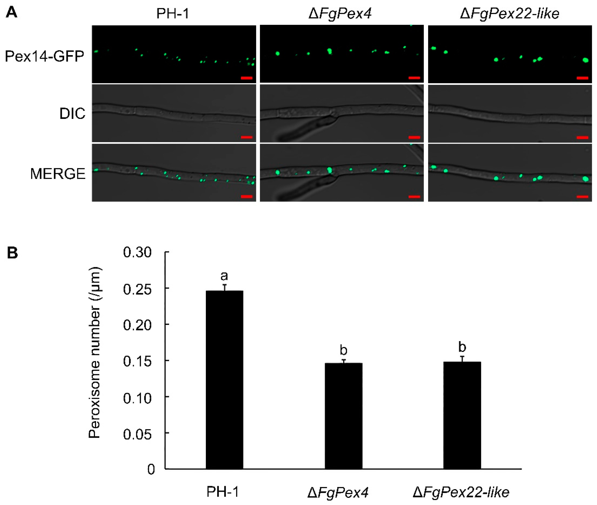
3.1. Deletion of FgPex4 and FgPex22-like Genes Reduced Number of Peroxisomes

To investigate the effect of FgPex4 and FgPex22-like gene knockout on the number of peroxisomes in F. graminearum, green fluorescent protein (GFP) was used to label the peroxisomal membrane protein FgPex14 in PH-1, ΔFgPex4, and ΔFgPex22-like strains, and the number of fluorescent dots in the mycelia cultured in liquid complete medium (CM) for 24 h was subsequently observed with an LSM800 laser confocal microscope to estimate the number of peroxisomes in each strain. For the main observation area, a region 150 μm away from the top of the mycelium was selected. Each strain was observed with a mycelium length of approximately 1000 μm. The results showed that the number of peroxisomes in F. graminearum decreased by 40.55% and 39.70% after the deletion of FgPex4 and FgPex22-like genes, respectively (Figure 1).

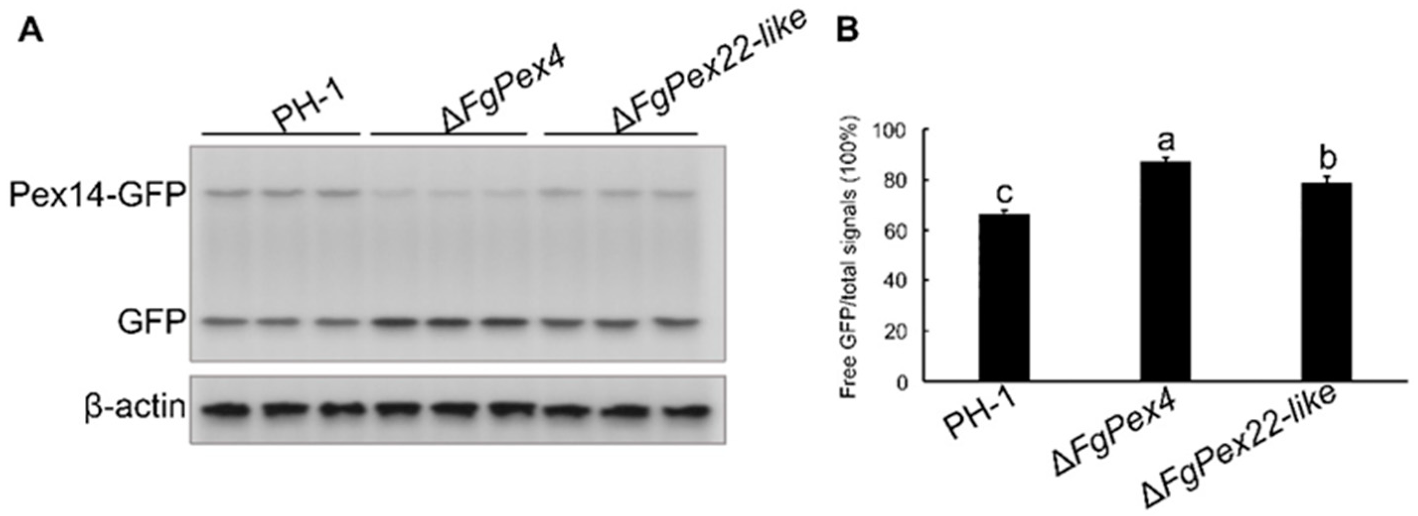
3.2. Peroxisome Synthesis Affected by the Knockout of FgPex4 and FgPex22-like Genes

To investigate the impact of FgPex4 and FgPex22-like deletions on peroxisome synthesis, the expression at the transcriptional level of proteins related to synthesis, division, and inheritance was examined. Fresh hyphae of the strains under study were transferred into 100 mL of CM liquid medium and cultured at 25 °C and 180 rpm. After 24 h of culture, the hyphae were collected to extract RNA and reverse-transcribed to obtain cDNA for use in subsequent experiments. Differences in the expression of peroxisome synthesis-related genes, FgPex3, FgPex16, FgPex19, FgPex11, FgPex11b, FgPex11c, FgPex11c2, FgPex11-2, and dynamic genes related to inheritance and division, FgMyo2, FgVps1, FgDnm1, and FgFis1, were detected at the transcription level using quantitative real-time PCR (qRT-PCR). Simultaneously, a difference in the expression of PMP70 at the transcriptional level, which accounts for a large proportion of PMPs, was detected. The internal reference used in this experiment was glyceraldehyde 3-phosphate dehydrogenase (GAPDH). The results showed that in both the FgPex4 and FgPex22-like gene knockout strains, the expression of peroxisome biogenesis-related genes, inheritance- and division-related genes, and the PMP70 gene at the transcriptional level was significantly lower than that of the wild-type strain PH-1 (Figure 2). β-actin, a gene with little relationship with peroxisome, was used as an additional reference gene. Therefore, after the deletion of FgPex4 and FgPex22-like genes, one of the factors that decreased the number of peroxisomes was that the source of peroxisomes was affected.

3.3. Knockout of FgPex4 and FgPex22-like Genes Affected Peroxisome Degradation

3.3.1. Effect of FgPex4 and FgPex22-like Deletions on Autophagy

Autophagy-related degradation pathways were investigated to verify if the reduction in the number of peroxisomes caused by knockout of FgPex4 and FgPex22-like genes was related to difference in degree of peroxisome degradation. Autophagy has become a popular research topic in recent decades. In-depth research has revealed proteins that can be used to label autophagosomes, with the autophagy-related protein ATG8 being the most recognized. Free GFP indicates that GFP-ATG8 is degraded during autophagy, so that the higher the proportion of free GFP in the total signal in each lane, the greater the degree of autophagy. The vector pFL2-GFP-ATG8 was introduced into the protoplast of PH-1, ΔFgPex4, and ΔFgPex22-like strains to obtain the correct transformants PH-1/GFP:FgATG8, ΔFgPex4/GFP:FgATG8, and ΔFgPex22-like/GFP:FgATG8. Mycelia cultured in CM medium for 24 h were transferred to nitrogen-deficient medium (MM-N) and collected after 0, 6, and 12 h. Total protein was extracted from the hyphal samples collected at different intervals and detected with an anti-GFP antibody. The proportion of free GFP in the total signal was calculated according to the gray value of the band. β-actin was used as an internal parameter. The results showed that the degree of autophagy of ΔFgPex4 and ΔFgPex22-like was not significantly different from that of the wild type strain PH-1 cultured in CM for 24 h and nitrogen-deficient medium (MM-N) for 0, 6, and 12 h (Figure 3A,B and Figure S1). The hyphae were stained with 10 µM 7-amino-4-chloromethylcoumarin (CMAC) to track the location of the vacuoles, and blue fluorescence in the vacuoles and green fluorescence of the GFP:FgATG8 protein were observed using laser confocal microscopy. Regardless of ΔFgPex4 or ΔFgPex22-like, the mycelia cultured in CM were similar to the wild-type strain PH-1. GFP:FgATG8 was either located at the phagophore assembly site next to the vacuole or diffused in the cytoplasm. After MM-N-induced autophagy, the three strains presented the same state; that is, GFP:FgATG8 was located in the vacuole, and autophagy occurred (Figure 3C). This indicates that autophagy was not associated with the decrease in the number of peroxisomes in the ΔFgPex4 and ΔFgPex22-like strains.

3.3.2. Deletion of FgPex4 and FgPex22-like Aggravated the Pexophagy of F. graminearum

To clearly determine whether pexophagy was related to the decrease of peroxisome number, the pFL2-FgPex14-GFP vector was introduced into PH-1, ΔFgPex4, and ΔFgPex22-like protoplasts to obtain the correct transformants PH-1/FgPex14:GFP, ΔFgPex4/FgPex14:GFP, and ΔFgPex22-like/FgPex14:GFP. The total hyphal protein was extracted, and anti-GFP antibody was used to detect FgPex14-GFP protein and free GFP protein in the total protein; pexophagy was preliminarily predicted according to the proportion of free GFP after inducing nitrogen deficiency for 0 and 24 h. Anti-β-actin antibody was used as internal reference antibody. The results indicated that the color of free GFP bands in strains ΔFgPex4 and ΔFgPex22-like was darker, and the value of the ratio of free GFP to total signals was also higher compared to the wildtype PH-1. This observation was consistent whether the mycelia were cultured in CM for 24 h or induced by nitrogen deficiency for 24 h (Figure 4 and Figure S2). The above results indicate that the loss of FgPex4 and FgPex22-like could aggravate pexophagy. Therefore, it can be inferred that the decrease in the number of peroxisomes in ΔFgPex4 and ΔFgPex22-like strains was also associated with pexophagy.

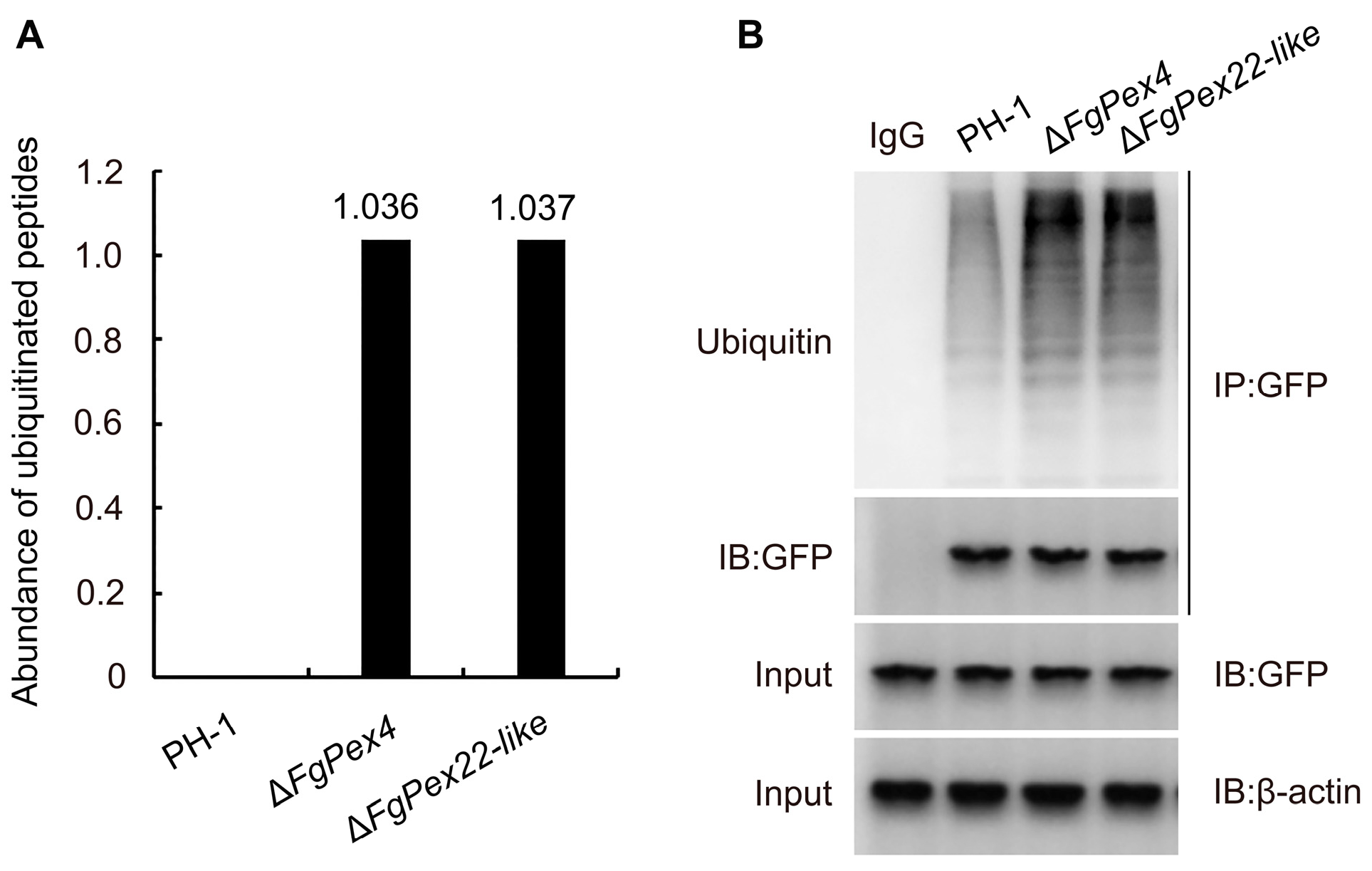
3.4. Effect of FgPex4 and FgPex22-like Genes Deletion on Polyubiquitination of Pex5p

The signal of peptide ubiquitination can be measured using label-free quantification and date-dependent acquisition methods. The results showed that when Pex5p could normally function as a peroxisome target signal receptor in wild-type PH-1, no specific ubiquitination modified signals with lysine as a ubiquitination site were detected. However, specific signals of ubiquitination modification sites were detected in ΔFgPex4 and ΔFgPex22-like strains (Figure 5A). The constructed pFL2-FgPex5-GFP vector was introduced into the protoplasts of wild-type strain PH-1 and mutant strains ΔFgPex4, ΔFgPex22-like via protoplast transformation, and the protein was extracted from the correct transformants. The protein mixture and FgPex5-GFP protein co-purified from the protein mixture were detected using a GFP antibody, and the ubiquitination of FgPex5 in the sample was detected using a ubiquitin antibody. The results showed that compared with the wild-type PH-1, ubiquitination of FgPex5 increased significantly in the mutants ΔFgPex4 and ΔFgPex22-like (Figure 5B). The result highlights that ubiquitinated Pex5p accumulation in the two mutant strains was significantly stronger than that in the wild-type strain.

3.5. Effect of Blocking the Pexophagy Pathway on the Number of Peroxisomes

A decrease in the number of peroxisomes in ΔFgPex4 and ΔFgPex22-like strains aggravated pexophagy. Therefore, whether the number of peroxisomes increase in the mutant strains if pexophagy-related pathways in organisms are blocked remained unclear. Split-marker PCR technology was used to construct an ATG1 knockout fragment, which was introduced into the PH-1/FgPex14:GFP, ΔFgPex4/FgPex14:GFP, and ΔFgPex22-like/FgPex14:GFP strains via protoplast transformation. After PCR validation, the correct mutants ΔATG1, ΔΔATG1/Pex4, and ΔΔATG1/Pex22-like were obtained. The number of peroxisomes in the six strains was determined using laser confocal microscopy. The results showed that the number of peroxisomes in ΔΔATG1/Pex4, and ΔΔATG1/Pex22-like mutants was higher than that in ΔFgPex4/FgPex14:GFP, and ΔFgPex22-like/FgPex14:GFP strains (Figure 6). This result demonstrated that the decrease in the number of peroxisomes in ΔFgPex4 and ΔFgPex22-like mutant strains was related to pexophagy.

3.6. Effects of FgPex4 and FgPex22-like Deletions on Peroxisome Integrity

As damage to peroxisomes can also cause pexophagy, we examined whether peroxisomal function was damaged. The colony diameters of wild-type PH-1, two gene-deficient strains ΔFgPex4 and ΔFgPex22-like, and three ATG1 deficient strains, ΔATG1, ΔΔATG1/FgPex4, and ΔΔATG1/FgPex22-like, growing on oleic acid medium, were examined. The results showed that ΔΔATG1/FgPex4 and ΔΔATG1/FgPex22-like did not return to normal growth after blocking pexophagy (Figure 7A,B). Furthermore, the dry weight of the mycelia was determined, which showed no significant difference in the mycelium quantity between ΔATG1 and wild-type strains, whereas the mycelial dry weights of ΔΔATG1/FgPex4 and ΔΔATG1/FgPex22-like did not revert to the wild-type level (Figure 7C). Labeling PTS1 and PTS2 with mCherry and GFP, respectively, showed that the loss of FgPex4 and FgPex22-like prevented matrix proteins from entering the peroxisomes normally (Figure 8). This resulted in damaged peroxisomes, which may also be a reason for the intensification of pexophagy.

4. Discussion

In eukaryotes, peroxisomes are dynamic organelles that respond rapidly to various stimuli. In the filamentous fungus Neurospora crassa, the deletion of the Pex33 not only affects the biogenesis of glyoxysomes and Woronin bodies but also reduces the density of peroxisomes [59]. In S. cerevisiae, Pex30p, Pex31p, and Pex32p, along with the upstream Pex28p and Pex29p, regulate the proliferation of peroxisomes, and deletion of Pex30 could increase the number of peroxisomes [60]. Pex11p is a key protein in peroxisomal division. With the cooperation of the Pex25p and Pex27p membrane proteins, Pex11p is believed to be involved in the elongation and constriction process that occurs during peroxisome fission. Our results indicated that FgPex4 and FgPex22-like are also closely related to the regulation of peroxisome quantity, and the deletion of FgPex4 and FgPex22-like resulted in a reduction in peroxisome quantity in ΔFgPex4 and ΔFgPex22-like strains (Figure 1). In summary, FgPex4 and FgPex22-like are two essential peroxins that maintain the dynamic balance of peroxisomes in F. graminearum.
During peroxisome inheritance and division, several dynamin-like proteins, such as Vps1p, Dnm1p, and Dnm1p-anchoring protein Fis1p [19,20,21,22], participate in the mitochondrial division process. The proteins related to peroxisome synthesis, heredity, and division were downregulated at the transcriptional level, and the main components of the peroxisome membrane, PMP70, were also downregulated in ΔFgPex4 and ΔFgPex22-like strains (Figure 2). Myo2p is a V-type myosin that translocates peroxisomes into dividing cells in S. cerevisiae [19,28]. Although there is no direct evidence in filamentous fungi that Myo2p is associated with peroxisome transport, our results do indicate that the absence of peroxins FgPex4 and FgPex22-like seriously affects the expression of Myo2p at the transcriptional level. Furthermore, in the NCBI database, comparisons of Myo2p between S. cerevisiae and F. graminearum showed higher sequence consistency, and their structural domains were also very similar. Therefore, we speculate that Myo2p in F. graminearum is also related to peroxisome transport. Based on the results, it is evident that the deletion of FgPex4 and FgPex22-like affects the normal regulation of these proteins at the transcriptional level, thus affecting the expression of proteins and normal proliferation of peroxisomes.
Pexophagy is the main pathway of peroxisome degradation, and Pex14p is a key protein in pexophagy that determines whether pexophagy can occur [40,44,61]. Therefore, the degree of degradation of Pex14-GFP protein can indicate the degree of pexophagy. In the present study, the deletion of FgPex4 and FgPex22-like accelerated the degradation of Pex14-GFP (Figure 4), thereby indicating the intensification of pexophagy. In Hansenula polymorpha, Pex4p are not required for this pexophagy [61]. Therefore, we speculate that the role of Pex4p orthologs is likely to not be conserved in fungal pexophagy.
In mammals, ubiquitination of peroxins, such as Pex5p, Pex3p, and PMP34, is believed to be involved in the activation of pexophagy [62,63,64]. Some studies have shown that in human cells, the accumulation of ubiquitinated Pex5p on the peroxisome membrane is considered a signal of pexophagy through the combination of the ubiquitin-binding protein NBR1 [65], suggesting that the accumulation of polyubiquitinated receptors may be a signal for pexophagy in mammalian cells [62]. To verify pexophagy in F. gramiearum is associated with the accumulation of ubiquitinated Pex5p, we detected ubiquitinated Pex5p accumulation and observed that the absence of FgPex4 and FgPex22-like did indeed cause ubiquitinated Pex5p accumulation. Similarly, previous studies have found that the absence of pex1p, Pex13p, and Pex26p can lead to the accumulation of ubiquitinated Pex5p and induces pexophagy [46,66]. Therefore, the accumulation of ubiquitinated Pex5p may be closely related to pexophagy in filamentous fungi.
After the main degradation pathway of peroxisomes was blocked, the number of peroxisomes increased significantly; however, their function did not recover (Figure 7). In addition, the deletion of FgPex4 and FgPex22-like not only caused the mislocalization of matrix protein with PTS1 signal but also caused the mislocalization of matrix protein with PTS2 signal (Figure 8), a result consistent with the defect trait caused by the deletion of Pex4 and Pex22 in the fungus Podospora anserina [3]. Therefore, we speculate that the reason for the lack of restoration of peroxisome function is due to peroxisome dysfunction.
In summary, the decrease in the number of peroxisomes caused by the absence of FgPex4 and FgPex22-like is due to the inhibition of peroxisome synthesis and the intensification of pexophagy. Moreover, the absence of FgPex4 and FgPex22-like can lead to the accumulation of ubiquitinated Pex5p and the mislocalization of matrix proteins with PTS1 or PTS2 signals. In future studies, we will try to explore the causes of pexophagy associated with FgPex4 and FgPex22-like deletions.

Supplementary Materials

The following supporting information can be downloaded at: https://www.mdpi.com/article/10.3390/jof9111083/s1, Table S1. Primers used in this study. Figure S1. Autophagy detection of ΔFgPex4 and ΔFgPex22-like strains cultured in CM for 24 h. (A) Western blotting was used to detect the autophagy of the strains cultured in CM for 24 h. The fresh mycelia at the edge of the fungal colony were transferred into CM medium and incubated for 24 h. The results of anti-GFP antibody detection were used to identify autophagy of the strain. β-actin was used as an internal control. (B) The calculated proportion of free GFP in the total band signal. Error bars on the histograms represent the standard error of three repeated tests. Different letters on the bars for each treatment indicate significant difference at p < 0.05 based on Duncan’s multiple range test. Figure S2. Pexophagy detection of ΔFgPex4 and ΔFgPex22-like strains cultured in CM for 24 h. (A) WB was used to detect the degree of pexophagy in strains PH-1/FgPex14:GFP, ΔFgPex4/FgPex14:GFP, and ΔFgPex22-like/FgPex14:GFP. Mycelia were cultured in CM medium for 24 h. Anti-GFP antibody was used to detect the degree of degradation of Pex14-GFP and β-actin was used as an internal control. (B) The calculated proportion of free GFP in the total band signal. The grayscale value of the strip was measured using ImageJ software. Error bars on the histograms represent the standard error of three repeated tests. Different letters on the bars for each treatment indicate significant difference at p < 0.05 based on Duncan’s multiple range test.

Author Contributions

L.Z. and J.Y. designed the experiments, supervised the work, and edited the manuscript; C.L. performed the majority of the experiments, analyzed the data, and wrote the draft manuscript; Z.B., H.X. and R.Z. participated in the article modification; J.W. performed data curation. Y.L. provided guidance and revised the manuscript. All authors have read and agreed to the published version of the manuscript.

Funding

This work was supported by the Wheat Innovation Team of Shandong Province Modern Agricultural Industry Technology System (SDAIT-01-09); Natural Science Foundation of Shandong Province (ZR2021QC141) and Funds of Shandong “Double Tops” Program (SYL2017XTTD11).

Institutional Review Board Statement

Not applicable.

Informed Consent Statement

Not applicable.

Data Availability Statement

Not applicable.

Conflicts of Interest

The authors declare no conflict of interest.

References

  1. Zhang, L.; Liu, C.; Wang, M.; Tao, Y.; Liang, Y.; Yu, J. Peroxin FgPEX22-Like Is Involved in FgPEX4 Tethering and Fusarium graminearum Pathogenicity. Front. Microbiol. 2021, 12, 756292. [Google Scholar] [CrossRef] [PubMed]
  2. Mano, S.; Hayashi, Y.; Hikino, K.; Otomo, M.; Kanai, M.; Nishimura, M. Ubiquitin-conjugating activity by PEX4 is required for efficient protein transport to peroxisomes in Arabidopsis thaliana. J. Biol. Chem. 2022, 298, 102038. [Google Scholar] [CrossRef]
  3. Suaste-Olmos, F.; Zirión-Martínez, C.; Takano-Rojas, H.; Peraza-Reyes, L. Meiotic development initiation in the fungus Podospora anserina requires the peroxisome receptor export machinery. Biochim. Biophys. Acta Mol. Cell Res. 2018, 1865, 572–586. [Google Scholar] [CrossRef] [PubMed]
  4. Zhang, L.; Wang, L.; Liang, Y.; Yu, J. FgPEX4 is involved in development, pathogenicity, and cell wall integrity in Fusarium graminearum. Curr. Genet. 2019, 65, 747–758. [Google Scholar] [CrossRef]
  5. Skowyra, M.L.; Rapoport, T.A. PEX5 translocation into and out of peroxisomes drives matrix protein import. Mol. Cell 2022, 82, 3209–3225.e7. [Google Scholar] [CrossRef]
  6. Gould, S.J.; Keller, G.A.; Hosken, N.; Wilkinson, J.; Subramani, S. A conserved tripeptide sorts proteins to peroxisomes. J. Cell Biol. 1989, 108, 1657–1664. [Google Scholar] [CrossRef]
  7. Miura, S.; Kasuya-Arai, I.; Mori, H.; Miyazawa, S.; Osumi, T.; Hashimoto, T.; Fujiki, Y. Carboxyl-terminal consensus Ser-Lys-Leu-related tripeptide of peroxisomal proteins functions in vitro as a minimal peroxisome-targeting signal. J. Biol. Chem. 1992, 267, 14405–14411. [Google Scholar] [CrossRef]
  8. Miyazawa, S.; Osumi, T.; Hashimoto, T.; Ohno, K.; Miura, S.; Fujiki, Y. Peroxisome targeting signal of rat liver acyl-coenzyme A oxidase resides at the carboxy terminus. Mol. Cell Biol. 1989, 9, 83–91. [Google Scholar]
  9. Williams, C.; van den Berg, M.; Sprenger, R.R.; Distel, B. A conserved cysteine is essential for Pex4p-dependent ubiquitination of the peroxisomal import receptor Pex5p. J. Biol. Chem. 2007, 282, 22534–22543. [Google Scholar] [CrossRef]
  10. Platta, H.W.; El Magraoui, F.; Schlee, D.; Grunau, S.; Girzalsky, W.; Erdmann, R. Ubiquitination of the peroxisomal import receptor Pex5p is required for its recycling. J. Cell Biol. 2007, 177, 197–204. [Google Scholar] [CrossRef]
  11. Carvalho, A.F.; Pinto, M.P.; Grou, C.P.; Alencastre, I.S.; Fransen, M.; Sa-Miranda, C.; Azevedo, J.E. Ubiquitination of mammalian Pex5p, the peroxisomal import receptor. J. Biol. Chem. 2007, 282, 31267–31272. [Google Scholar] [CrossRef] [PubMed]
  12. Okumoto, K.; Misono, S.; Miyata, N.; Matsumoto, Y.; Mukai, S.; Fujiki, Y. Cysteine ubiquitination of PTS1 receptor Pex5p regulates Pex5p recycling. Traffic 2011, 12, 1067–1083. [Google Scholar] [CrossRef] [PubMed]
  13. Kiel, J.A.; Emmrich, K.; Meyer, H.E.; Kunau, W.H. Ubiquitination of the peroxisomal targeting signal type 1 receptor, Pex5p, suggests the presence of a quality control mechanism during peroxisomal matrix protein import. J. Biol. Chem. 2005, 280, 1921–1930. [Google Scholar] [CrossRef] [PubMed]
  14. Kragt, A.; Voorn-Brouwer, T.; van den Berg, M.; Distel, B. The Saccharomyces cerevisiae peroxisomal import receptor Pex5p is monoubiquitinated in wild type cells. J. Biol. Chem. 2005, 280, 7867–7874. [Google Scholar] [CrossRef]
  15. Haan, G.J.; Baerends, R.J.; Krikken, A.M.; Otzen, M.; Veenhuis, M.; van der Klei, I.J. Reassembly of peroxisomes in Hansenula polymorpha pex3 cells on reintroduction of Pex3p involves the nuclear envelope. FEMS Yeast Res. 2006, 6, 186–194. [Google Scholar] [CrossRef]
  16. Hoepfner, D.; Schildknegt, D.; Braakman, I.; Philippsen, P.; Tabak, H.F. Contribution of the Endoplasmic Reticulum to Peroxisome Formation. Cell 2005, 122, 85–95. [Google Scholar] [CrossRef]
  17. Kragt, A.; Voorn-Brouwer, T.; van den Berg, M.; Distel, B. Endoplasmic Reticulum-directed Pex3p Routes to Peroxisomes and Restores Peroxisome Formation in a Saccharomyces cerevisiae pex3Δ Strain. J. Biol. Chem. 2005, 280, 34350–34357. [Google Scholar] [CrossRef]
  18. Tam, Y.Y.C.; Fagarasanu, A.; Fagarasanu, M.; Rachubinski, R.A. Pex3p Initiates the Formation of a Preperoxisomal Compartment from a Subdomain of the Endoplasmic Reticulum in Saccharomyces cerevisiae. J. Biol. Chem. 2005, 280, 34933–34939. [Google Scholar] [CrossRef]
  19. Hoepfner, D.; van den Berg, M.; Philippsen, P.; Tabak, H.F.; Hettema, E.H. A role for Vps1p, actin, and the Myo2p motor in peroxisome abundance and inheritance in Saccharomyces cerevisiae. J. Cell Biol. 2001, 155, 979–990. [Google Scholar] [CrossRef]
  20. Koch, A.; Yoon, Y.; Bonekamp, N.A.; McNiven, M.A.; Schrader, M. A role for Fis1 in both mitochondrial and peroxisomal fission in mammalian cells. Mol. Biol. Cell 2005, 16, 5077–5086. [Google Scholar] [CrossRef]
  21. Kuravi, K.; Nagotu, S.; Krikken, A.M.; Sjollema, K.; Deckers, M.; Erdmann, R.; Veenhuis, M.; van der Klei, I.J. Dynamin-related proteins Vps1p and Dnm1p control peroxisome abundance in Saccharomyces cerevisiae. J. Cell Sci. 2006, 119, 3994–4001. [Google Scholar] [CrossRef] [PubMed]
  22. Li, X.; Gould, S.J. The Dynamin-like GTPase DLP1 Is Essential for Peroxisome Division and Is Recruited to Peroxisomes in Part by PEX11. J. Biol. Chem. 2003, 278, 17012–17020. [Google Scholar] [CrossRef] [PubMed]
  23. Koch, J.; Brocard, C. PEX11 proteins attract Mff and human Fis1 to coordinate peroxisomal fission. J. Cell Sci. 2012, 125 Pt 16, 3813–3826. [Google Scholar]
  24. Fagarasanu, M.; Fagarasanu, A.; Tam, Y.Y.; Aitchison, J.D.; Rachubinski, R.A. Inp1p is a peroxisomal membrane protein required for peroxisome inheritance in Saccharomyces cerevisiae. J. Cell Biol. 2005, 169, 765–775. [Google Scholar] [CrossRef] [PubMed]
  25. Fagarasanu, M.; Fagarasanu, A.; Rachubinski, R.A. Sharing the wealth: Peroxisome inheritance in budding yeast. Biochim. Biophys. Acta 2006, 1763, 1669–1677. [Google Scholar] [CrossRef]
  26. Fagarasanu, A.; Fagarasanu, M.; Rachubinski, R.A. Maintaining peroxisome populations: A story of division and inheritance. Annu. Rev. Cell Dev. Biol. 2007, 23, 321–344. [Google Scholar] [CrossRef]
  27. Otzen, M.; Rucktaschel, R.; Thoms, S.; Emmrich, K.; Krikken, A.M.; Erdmann, R.; van der Klei, I.J. Pex19p contributes to peroxisome inheritance in the association of peroxisomes to Myo2p. Traffic 2012, 13, 947–959. [Google Scholar] [CrossRef]
  28. Fagarasanu, A.; Fagarasanu, M.; Eitzen, G.A.; Aitchison, J.D.; Rachubinski, R.A. The peroxisomal membrane protein Inp2p is the peroxisome-specific receptor for the myosin V motor Myo2p of Saccharomyces cerevisiae. Dev. Cell 2006, 10, 587–600. [Google Scholar] [CrossRef]
  29. Fagarasanu, A.; Mast, F.D.; Knoblach, B.; Jin, Y.; Brunner, M.J.; Logan, M.R.; Glover, J.N.; Eitzen, G.A.; Aitchison, J.D.; Weisman, L.S.; et al. Myosin-driven peroxisome partitioning in S. cerevisiae. J. Cell Biol. 2009, 186, 541–554. [Google Scholar] [CrossRef]
  30. Marelli, M.; Smith, J.J.; Jung, S.; Yi, E.; Nesvizhskii, A.I.; Christmas, R.H.; Saleem, R.A.; Tam, Y.Y.C.; Fagarasanu, A.; Goodlett, D.R.; et al. Quantitative mass spectrometry reveals a role for the GTPase Rho1p in actin organization on the peroxisome membrane. J. Cell Biol. 2004, 167, 1099–1112. [Google Scholar] [CrossRef]
  31. Mizushima, N.; Komatsu, M. Autophagy: Renovation of cells and tissues. Cell 2011, 147, 728–741. [Google Scholar] [CrossRef] [PubMed]
  32. Green, D.R.; Levine, B. To be or not to be? How selective autophagy and cell death govern cell fate. Cell 2014, 157, 65–75. [Google Scholar] [CrossRef] [PubMed]
  33. Lamb, C.A.; Yoshimori, T.; Tooze, S.A. The autophagosome: Origins unknown, biogenesis complex. Nat. Rev. Mol. Cell Biol. 2013, 14, 759–774. [Google Scholar] [CrossRef]
  34. Mochida, K.; Oikawa, Y.; Kimura, Y.; Kirisako, H.; Hirano, H.; Ohsumi, Y.; Nakatogawa, H. Receptor-mediated selective autophagy degrades the endoplasmic reticulum and the nucleus. Nature 2015, 522, 359–362. [Google Scholar] [CrossRef]
  35. Ohsumi, Y. Molecular dissection of autophagy: Two ubiquitin-like systems. Nat. Rev. Mol. Cell Biol. 2001, 2, 211–216. [Google Scholar] [CrossRef]
  36. Suzuki, K.; Kubota, Y.; Sekito, T.; Ohsumi, Y. Hierarchy of Atg proteins in pre-autophagosomal structure organization. Genes Cells 2007, 12, 209–218. [Google Scholar] [CrossRef]
  37. Nakatogawa, H.; Ichimura, Y.; Ohsumi, Y. Atg8, a ubiquitin-like protein required for autophagosome formation, mediates membrane tethering and hemifusion. Cell 2007, 130, 165–178. [Google Scholar] [CrossRef]
  38. Walker, C.L.; Pomatto, L.C.D.; Tripathi, D.N.; Davies, K.J.A. Redox Regulation of Homeostasis and Proteostasis in Peroxisomes. Physiol. Rev. 2018, 98, 89–115. [Google Scholar] [CrossRef]
  39. Oku, M.; Sakai, Y. Pexophagy in yeasts. Biochim. Biophys. Acta 2016, 1863, 992–998. [Google Scholar] [CrossRef]
  40. Germain, K.; Kim, P.K. Pexophagy: A Model for Selective Autophagy. Int. J. Mol. Sci. 2020, 21, 578. [Google Scholar] [CrossRef]
  41. Eberhart, T.; Kovacs, W.J. Pexophagy in yeast and mammals: An update on mysteries. Histochem. Cell Biol. 2018, 150, 473–488. [Google Scholar] [CrossRef] [PubMed]
  42. Bellu, A.R.; Salomons, F.A.; Kiel, J.A.; Veenhuis, M.; Van Der Klei, I.J. Removal of Pex3p is an important initial stage in selective peroxisome degradation in Hansenula polymorpha. J. Biol. Chem. 2002, 277, 42875–42880. [Google Scholar] [CrossRef] [PubMed]
  43. de Vries, B.; Todde, V.; Stevens, P.; Salomons, F.; van der Klei, I.J.; Veenhuis, M. Pex14p is not required for N-starvation induced microautophagy and in catalytic amounts for macropexophagy in Hansenula polymorpha. Autophagy 2006, 2, 183–188. [Google Scholar] [CrossRef] [PubMed]
  44. Bellu, A.R.; Komori, M.; van der Klei, I.J.; Kiel, J.A.; Veenhuis, M. Peroxisome biogenesis and selective degradation converge at Pex14p. J. Biol. Chem. 2001, 276, 44570–44574. [Google Scholar] [CrossRef]
  45. Nuttall, J.M.; Motley, A.M.; Hettema, E.H. Deficiency of the exportomer components Pex1, Pex6, and Pex15 causes enhanced pexophagy in Saccharomyces cerevisiae. Autophagy 2014, 10, 835–845. [Google Scholar] [CrossRef]
  46. Demers, N.D.; Riccio, V.; Jo, D.S.; Bhandari, S.; Law, K.B.; Liao, W.; Kim, C.; McQuibban, G.A.; Choe, S.K.; Cho, D.H.; et al. PEX13 prevents pexophagy by regulating ubiquitinated PEX5 and peroxisomal ROS. Autophagy 2023, 19, 1781–1802. [Google Scholar] [CrossRef]
  47. Chen, Y.; Zheng, S.; Ju, Z.; Zhang, C.; Tang, G.; Wang, J.; Wen, Z.; Chen, W.; Ma, Z. Contribution of peroxisomal docking machinery to mycotoxin biosynthesis, pathogenicity and pexophagy in the plant pathogenic fungus Fusarium graminearum. Environ. Microbiol. 2018, 20, 3224–3245. [Google Scholar] [CrossRef]
  48. Tam, Y.Y.; Torres-Guzman, J.C.; Vizeacoumar, F.J.; Smith, J.J.; Marelli, M.; Aitchison, J.D.; Rachubinski, R.A. Pex11-related proteins in peroxisome dynamics: A role for the novel peroxin Pex27p in controlling peroxisome size and number in Saccharomyces cerevisiae. Mol. Biol. Cell 2003, 14, 4089–4102. [Google Scholar] [CrossRef]
  49. Vizeacoumar, F.J.; Torres-Guzman, J.C.; Tam, Y.Y.C.; Aitchison, J.D.; Rachubinski, R.A. YHR150w and YDR479c encode peroxisomal integral membrane proteins involved in the regulation of peroxisome number, size, and distribution in Saccharomyces cerevisiae. J. Cell Biol. 2003, 161, 321–332. [Google Scholar] [CrossRef]
  50. Li, L.; Wang, J.; Chen, H.; Chai, R.; Zhang, Z.; Mao, X.; Qiu, H.; Jiang, H.; Wang, Y.; Sun, G. Pex14/17, a filamentous fungus-specific peroxin, is required for the import of peroxisomal matrix proteins and full virulence of Magnaporthe oryzae. Mol. Plant Pathol. 2017, 18, 1238–1252. [Google Scholar] [CrossRef]
  51. Wróblewska, J.P.; van der Klei, I.J. Peroxisome Maintenance Depends on De Novo Peroxisome Formation in Yeast Mutants Defective in Peroxisome Fission and Inheritance. Int. J. Mol. Sci. 2019, 20, 4023. [Google Scholar] [CrossRef] [PubMed]
  52. Zhang, L.; Liu, C.; Wang, L.; Sun, S.; Liu, A.; Liang, Y.; Yu, J.; Dong, H. FgPEX1 and FgPEX10 are required for the maintenance of Woronin bodies and full virulence of Fusarium graminearum. Curr. Genet. 2019, 65, 1383–1396. [Google Scholar] [CrossRef] [PubMed]
  53. Goswami, R.S. Targeted gene replacement in fungi using a split-marker approach. Methods Mol. Biol. 2012, 835, 255–269. [Google Scholar] [PubMed]
  54. Gravelat, F.N.; Askew, D.S.; Sheppard, D.C. Targeted gene deletion in Aspergillus fumigatus using the hygromycin-resistance split-marker approach. Methods Mol. Biol. 2012, 845, 119–130. [Google Scholar]
  55. Yun, Y.; Liu, Z.; Zhang, J.; Shim, W.B.; Chen, Y.; Ma, Z. The MAPKK FgMkk1 of Fusarium graminearum regulates vegetative differentiation, multiple stress response, and virulence via the cell wall integrity and high-osmolarity glycerol signaling pathways. Environ. Microbiol. 2014, 16, 2023–2037. [Google Scholar] [CrossRef]
  56. Livak, K.J.; Schmittgen, T.D. Analysis of relative gene expression data using real-time quantitative PCR and the 2(-Delta Delta C(T)) Method. Methods 2001, 25, 402–408. [Google Scholar] [CrossRef]
  57. He, D.; Li, M.; Damaris, R.N.; Bu, C.; Xue, J.; Yang, P. Quantitative ubiquitylomics approach for characterizing the dynamic change and extensive modulation of ubiquitylation in rice seed germination. Plant J. 2019, 101, 1430–1447. [Google Scholar] [CrossRef]
  58. Zhu, L.; Cheng, H.; Peng, G.; Wang, S.; Zhang, Z.; Ni, E.; Fu, X.; Zhuang, C.; Liu, Z.; Zhou, H. Ubiquitinome Profiling Reveals the Landscape of Ubiquitination Regulation in Rice Young Panicles. Genom. Proteom. Bioinform. 2020, 18, 305–320. [Google Scholar] [CrossRef]
  59. Managadze, D.; Wurtz, C.; Wiese, S.; Schneider, M.; Girzalsky, W.; Meyer, H.E.; Erdmann, R.; Warscheid, B.; Rottensteiner, H. Identification of PEX33, a novel component of the peroxisomal docking complex in the filamentous fungus Neurospora crassa. Eur. J. Cell Biol. 2010, 89, 955–964. [Google Scholar] [CrossRef]
  60. Vizeacoumar, F.J.; Torres-Guzman, J.C.; Bouard, D.; Aitchison, J.D.; Rachubinski, R.A. Pex30p, Pex31p, and Pex32p form a family of peroxisomal integral membrane proteins regulating peroxisome size and number in Saccharomyces cerevisiae. Mol. Biol. Cell 2004, 15, 665–677. [Google Scholar] [CrossRef]
  61. Zutphen, T.; Veenhuis, M.; van der Klei, I.J. Pex14 is the sole component of the peroxisomal translocon that is required for pexophagy. Autophagy 2008, 4, 63–66. [Google Scholar] [CrossRef] [PubMed]
  62. Kim, P.K.; Hailey, D.W.; Mullen, R.T.; Lippincott-Schwartz, J. Ubiquitin signals autophagic degradation of cytosolic proteins and peroxisomes. Proc. Natl. Acad. Sci. USA 2008, 105, 20567–20574. [Google Scholar] [CrossRef] [PubMed]
  63. Yamashita, S.; Abe, K.; Tatemichi, Y.; Fujiki, Y. The membrane peroxin PEX3 induces peroxisome-ubiquitination-linked pexophagy. Autophagy 2014, 10, 1549–1564. [Google Scholar] [CrossRef] [PubMed]
  64. Zhang, J.; Tripathi, D.N.; Jing, J.; Alexander, A.; Kim, J.; Powell, R.T.; Dere, R.; Tait-Mulder, J.; Lee, J.H.; Paull, T.T.; et al. ATM functions at the peroxisome to induce pexophagy in response to ROS. Nat. Cell Biol. 2015, 17, 1259–1269. [Google Scholar] [CrossRef]
  65. Deosaran, E.; Larsen, K.B.; Hua, R.; Sargent, G.; Wang, Y.; Kim, S.; Lamark, T.; Jauregui, M.; Law, K.; Lippincott-Schwartz, J.; et al. NBR1 acts as an autophagy receptor for peroxisomes. J. Cell Sci. 2013, 126 Pt 4, 939–952. [Google Scholar] [CrossRef]
  66. Law, K.B.; Bronte-Tinkew, D.; Di Pietro, E.; Snowden, A.; Jones, R.O.; Moser, A.; Brumell, J.H.; Braverman, N.; Kim, P.K. The peroxisomal AAA ATPase complex prevents pexophagy and development of peroxisome biogenesis disorders. Autophagy 2017, 13, 868–884. [Google Scholar] [CrossRef]
Figure 1. Effect of FgPex4 and FgPex22-like gene deletion on the number of peroxisomes. (A) Peroxisomes in mycelia. Several mycelia from the edge of the colony were transferred to 100 mL CM medium and cultured for 24 h in an incubator at 25 °C (180 rpm shaking speed). Fluorescence of young mycelia was observed using an LSM800 laser confocal microscope. Scale bar = 5 μm. (B) Number of peroxisomes per unit length of mycelia. We manually counted the number of peroxisomes in approximately 1000 μm of mycelium. Different letters on the bars for each treatment indicate significant difference at p < 0.05 by Duncan’s multiple range test. Measurements represent the average of three independent experiments. Error bars on the histograms represent the standard error of three repeated tests.
Jof 09 01083 g001
Figure 2. Effect of FgPex4 and FgPex22-like deletion on peroxisome synthesis pathway of Fusarium graminearum. (A) Relative quantitative analysis of peroxisome synthesis-related genes at the transcriptional level. The mycelia were collected and cultured in liquid CM medium for 24 h, and RNA was extracted and reverse-transcribed for repeated qRT-PCR experiments. GAPDH was used as an internal reference gene. (B) Relative quantitative analysis of peroxisome inheritance and division-related genes at transcriptional level. The total RNA of three strains (wild-type PH-1, ΔFgPex4, and ΔFgPex22-like) was obtained using the same method, and then a series of experiments and analyses were performed. GAPDH was used as an internal reference gene. (C) Relative quantitative analysis of PMP70 at the transcriptional level. Error bars on the histograms represent the standard error of three repeated tests. Different letters on the bars for each treatment indicate significant difference at p < 0.05 by Duncan’s multiple range test. All experiments were repeated three times with three replicates each time.
Jof 09 01083 g002
Figure 3. Effect of FgPex4 and FgPex22-like deletion on autophagy. (A) Western blotting was used to detect the autophagy of the strains induced in nitrogen-deficient medium MM-N for 0, 6, and 12 h. The fresh mycelia at the edge of the fungal colony were transferred into CM medium and incubated for 24 h and then transferred to MM-N medium for 0, 6, and 12 h. The results of anti-GFP antibody detection were used to identify autophagy of the strain. β-actin was used as an internal control. (B) The calculated proportion of free GFP in the total band signal. Error bars on the histograms represent the standard error of three repeated tests. Different letters on the bars for each treatment indicate significant difference at p < 0.05 based on Duncan’s multiple range test. (C) Detection of autophagy fluorescence signal. Strains PH-1/GFP:FgATG8, ΔFgPex4/GFP:FgATG8, and ΔFgPex22-like/GFP:FgATG8 were transferred from the CM culture medium to the MM-N culture medium for 12 h, stained with 10 µM CMAC, and the distribution of fluorescence in the mycelia observed under a laser confocal microscope. Scale bar = 10 µm. Green fluorescence represents GFP-FgATG8 protein, blue fluorescence represents vacuoles stained by CMAC, and turquoise represents their colocalization.
Jof 09 01083 g003
Figure 4. Pexophagy in ΔFgPex4 and ΔFgPex22-like. (A) After the mycelia were cultured in MM-N medium for 24 h, Pex14:GFP proteolysis was examined using Western blot in PH-1/FgPex14:GFP, ΔFgPex4/FgPex14:GFP, and ΔFgPex22-like/FgPex14:GFP strains. (B) Statistics of the degree of Pex14:GFP protein hydrolysis. Gray value of the strip was estimated and the proportion of GFP in the total signal was calculated using ImageJ software (1.53a). Error bars on the histograms represent the standard error of three repeated tests. Different letters on the bars for each treatment indicate significant difference at p < 0.05 based on Duncan’s multiple range test.
Jof 09 01083 g004
Figure 5. Effect of the deletion of FgPex4 and FgPex22-like on ubiquitinated FgPex5 accumulation. (A) Quantitative proteomics study of ubiquitination modification of FgPex5. The protein mixtures extracted from mycelial samples of three different strains were enzymatically hydrolyzed with trypsin and analyzed using liquid chromatography-mass spectrometry. (B) CO-IP results of FgPex5 ubiquitination accumulation. FgPex5 is labeled with GFP. The extracted protein mixtures and proteins co-purified with FgPex5-GFP from these protein mixtures (GFP IP) were detected with the anti-GFP antibody, and then the ubiquitination of FgPex5 protein was detected with an anti-ubiquitin antibody. Anti-actin antibody was used as an antibody for the control.
Jof 09 01083 g005
Figure 6. Effect of blocking the autophagy pathway on the number of peroxisomes. (A) Peroxisomes in mycelia. ATG1 was knocked out in the background of PH-1/FgPex14:GFP, ΔFgPex4/FgPex14:GFP, and ΔFgPex22-like/FgPex14:GFP strains to obtain the double knockout mutants ΔATG1/FgPex14:GFP, ΔΔATG1/FgPex4/FgPex14:GFP, and ΔΔATG1/FgPex22-like/FgPex14:GFP. The six strains were cultured in liquid PDB medium for 24 h, and the distribution of peroxisomes was observed under a laser confocal microscope. Scale bar = 5 μm. Green represents the peroxisome labeled with GFP, while turquoise represents the peroxisome with green fluorescence in the bright field. (B) Trends in the number of peroxisomes. The ratio of peroxisome number of ΔATG1/PH-1, ΔΔATG1/FgPex4FgPex4, and ΔΔATG1/FgPex22-likeFgPex22-like was analyzed. Error bars on the histograms represent the standard error of three repeated tests. Different letters on the bars for each treatment indicate significant difference at p < 0.05 based on the Duncan’s multiple range test.
Jof 09 01083 g006
Figure 7. Effect of blocking pexophagy on fatty acid utilization. (A) Growth of each strain on oleic acid medium at 25 °C for 7 days. Scale bar = 1000 μm (B) Diameter measurement of each strain. (C) Dry weight of mycelium of each strain in liquid oleic acid medium at 7 days post-incubation (25 °C, 180 rpm). Error bars on the histograms represent the standard error of three repeated tests. Different letters on the bars for each treatment indicate significant difference at p < 0.05 by Duncan’s multiple range test. Measurements represent the average of three independent experiments.
Jof 09 01083 g007
Figure 8. Effect of deletion of FgPex4 and FgPex22-like on the introduction of peroxisome matrix protein. The strains containing mCherry:PTS1 or PTS2:GFP were cultured in liquid CM for 1 day, and then the fluorescence distribution in the mycelia was observed using a laser confocal microscope LSM800. Scale bar = 10 µm. Red fluorescence and green fluorescence represent the distribution status of mCherry:PTS1 and PTS2:GFP in the mycelia, respectively.
Jof 09 01083 g008
Disclaimer/Publisher’s Note: The statements, opinions and data contained in all publications are solely those of the individual author(s) and contributor(s) and not of MDPI and/or the editor(s). MDPI and/or the editor(s) disclaim responsibility for any injury to people or property resulting from any ideas, methods, instructions or products referred to in the content.

