Article

Structure of the Mating-Type Genes and Mating Systems of Verpa bohemica and Verpa conica (Ascomycota, Pezizomycotina)

1
School of Food and Biological Engineering, Zhengzhou University of Light Industry, Zhengzhou 450002, China
2
Germplasm Bank of Wild Species, Yunnan Key Laboratory for Fungal Diversity and Green Development, Kunming Institute of Botany, Chinese Academy of Sciences, Kunming 650201, China
3
Institute of Agro-Products Processing, Yunnan Academy of Agricultural Sciences, Kunming 650221, China
4
College of Resource and Environment, Yunnan Agricultural University, Kunming 650100, China
5
School of Biodiversity Conservation, Southwest Forestry University, Kunming 650224, China
6
Agricultural Technology Promotion Station in Zhuoni County, Gannan 747600, China
*
Authors to whom correspondence should be addressed.
These authors contributed equally to this work.
J. Fungi 2023, 9(12), 1202; https://doi.org/10.3390/jof9121202
Submission received: 7 November 2023 / Revised: 6 December 2023 / Accepted: 12 December 2023 / Published: 15 December 2023
(This article belongs to the Special Issue Edible and Medicinal Macrofungi, 2nd Edition)

Abstract

:
Verpa spp. are potentially important economic fungi within Morchellaceae. However, fundamental research on their mating systems, the key aspects of their life cycle, remains scarce. Fungal sexual reproduction is chiefly governed by mating-type genes, where the configuration of these genes plays a pivotal role in facilitating the reproductive process. For this study, de novo assembly methodologies based on genomic data from Verpa spp. were employed to extract precise information on the mating-type genes, which were then precisely identified in silico and by amplifying their single-ascospore populations using MAT-specific primers. The results suggest that the MAT loci of the three tested strains of V. bohemica encompassed both the MAT1-1-1 and MAT1-2-1 genes, implying homothallism. On the other hand, amongst the three V. conica isolates, only the MAT1-1-1 or MAT1-2-1 genes were present in their MAT loci, suggesting that V. conica is heterothallic. Moreover, bioinformatic analysis reveals that the three tested V. bohemica strains and one V. conica No. 21110 strain include a MAT1-1-10 gene in their MAT loci, while the other two V. conica strains contained MAT1-1-11, exhibiting high amino acid identities with those from corresponding Morchella species. In addition, MEME analysis shows that a total of 17 conserved protein motifs are present among the MAT1-1-10 encoded protein, while the MAT1-1-11 protein contained 10. Finally, the mating type genes were successfully amplified in corresponding single-ascospore populations of V. bohemica and V. conica, further confirming their life-cycle type. This is the first report on the mating-type genes and mating systems of Verpa spp., and the presented results are expected to benefit further exploitation of these potentially important economic fungi.

1. Introduction

Verpa is an epigeous genus within Morchellaceae (Ascomycota, Pezizomycotina). The phylogenetic analysis of ribosomal DNA revealed a close relationship between it and the genus Morchella [1,2]. Hence, Verpa spp. are also called false or early morels, due to similar morphology of their ascocarp and earlier occurrence in spring in comparison with true morels (Morchella spp.) [3]. Verpa spp. are widespread in the northern temperate zones and were originally found in the northern parts of North America, Europe, and Asia. Globally, although they are not as common as true morels, the false morels are often found in a wide range of habitats and environmental conditions [4], even in the wild mushroom market. A total of 28 Verpa species are recorded in Index Fungorum (http://www.indexfungorum.org/Names/Names.asp, accessed 1 November 2023), among which, two species—V. bohemica and V. conica—have been extensively studied.
V. bohemica is locally used for dietary and antioxidant purposes in tribal areas of Kashmir Himalaya. However, when eaten in excess, they may cause gastrointestinal upset and loss of muscular coordination [5]. Thus, V. bohemica was eventually recognized as an inedible mushroom [6]. Meanwhile, many studies have indicated that V. bohemica presents quite impressive antibacterial and antifungal activities [5], as well as strong antioxidant activities [7,8]. Furthermore, high contents of lovastatin, γ-aminobutyric acid (GABA), and ergothioneine have been found in mycelial and single-cell biomasses of V. bohemica, implying that the fungus possesses effective anti-inflammatory, antioxidant, profibrinolytic, and antihypertensive effects [9]. On the other hand, apart from its strong antioxidant activity in different systems [10], V. conica has exhibited strong laccase-like multicopper oxidase activity enhancement in the presence of the naturally occurring phenolic compound guaiacol, showing great potential for plant litter decay [11]. However, there is scarce research on the genetic information, life cycle, and reproductive system of Verpa spp. in the existing literature.
The genetic locus responsible for the determination of the mating type in fungi is referred to as the mating-type locus. Ascomycetes regulate their sexual reproduction through a single mating-type locus [12] which contains idiomorphs [13], distinct and non-homologous mating-type “alleles” in heterothallic species which can encompass one or multiple genes, specifically known as MAT1-1 and MAT1-2, respectively [14]. The MAT1-1 idiomorph harbors the MAT1-1-1 gene, which encodes a protein containing an MATalpha_HMGbox domain. Similarly, the MAT1-2 idiomorph harbors the MAT1-2-1 gene, which encodes a protein containing a high-mobility group (HMG) domain [15]. In heterothallic ascomycete species, a single ascospore usually contains only one MAT idiomorph, MAT1-1 or MAT1-2, while in homothallic ascomycetes of the same lineage, there will be two mating types in the same genome, but the arrangement will often differ [15,16]. Apart from the core mating-type genes (MAT1-1-1 and MAT1-2-1), other mating-type genes on the MAT locus are lineage-specific and conserved at the family and class levels. To date, a total of 12 MAT1-1 genes (MAT1-1-1 to MAT1-1-12) and 15 MAT1-2 genes (MAT1-2-1 to MAT1-2-15) have been identified across various species [17,18].
While research on the mating type gene of Verpa has been limited, studies on this gene in closely related species such as Morchella have been more extensive. Liu et al. [19] have employed genome data to elucidate the heterothallic life cycle of Morchella importuna. Chai et al. [20] identified two new secondary MAT genes in Morchella spp.; namely MAT1-1-10 and MAT1-1-11. Expanding on their findings, Chai et al. [21] examined the structure of the mating-type loci across ten Morchella species, revealing that eight were heterothallic, while two were homothallic.
In this study, the whole genome of the two Verpa species (three strains per species) were sequenced, assembled, and annotated with the purpose of: (1) identifying the MAT loci of the two Verpa species, (2) analyzing the differences in MAT locus structure between Verpa and Morchella, and (3) determining the reproductive strategy of Verpa species (i.e., homothallic or heterothallic).

2. Materials and Methods

2.1. Fungal Cultivation and Ascosporic Isolates

Six strains of V. bohemica and V. conica were sourced from five provinces in China (Table 1). To obtain pure mycelium culture, tissue isolation was performed from fresh or naturally dried specimens. The specific procedure was as follows: A tissue block of approximately 0.5 cm2 in size was taken from the stipe part of the ascomata. After washing with sterile water 2–3 times, the block was disinfected with 75% alcohol for 1.5 min. Then, the tissue was sliced into 1–2 mm3 pieces and placed on suitable culture media (PDA, 200 g of potato extract, 20 g of glucose, and 20 g of agar per liter of medium) for constant temperature cultivation at 21 °C. The emerging tips of individual tissue blocks were then excised and transferred to a new PDA plate. Three successive cultivation cycles later, strains of a purified nature were successfully derived.
Among six Verpa specimens, the fruiting bodies of No. 20020 (V. bohemica) and No. 21120 (V. conica), which produced a large number of mature ascospores, were selected for the isolation of monosporic cultures. After the process of gradient dilution [22], three consecutive low-concentration gradient spore suspensions were independently applied onto three separate PDA plates and subjected to the same incubation conditions mentioned earlier. The emerging tips of individual ascospores were then excised and transferred to a new PDA plate under a stereomicroscope (Leica M205, Wilmington, DE, USA). Each monosporic isolate was assigned a corresponding Arabic numeral suffix. For example, 20020-1 refers to the first ascosporic isolate. All cultures were stored in the Germplasm Repository of the Kunming Institute of Botany, Chinese Academy of Sciences, Yunnan Province, China.

2.2. DNA Extraction and Sequencing Scheme

Fresh mycelium grown on PDA plates for 7 days were collected for DNA and RNA extraction. DNA was extracted from fungal mycelium using the modified cetyltrimethylammonium bromide (CTAB) method [23], and RNA contamination was eliminated using RNAse A digestion. Total RNA was extracted using TRIzol® Reagent (Invitrogen, Carlsbad, CA, USA) and purified with Plant RNA Purification Reagent (Invitrogen, Carlsbad, CA, USA). Both the extracted DNA and RNA were subjected to subsequent sequencing analysis.
A hybrid scheme of Illumina sequencing and Nanopore sequencing was utilized. For Illumina sequencing on the Illumina HiSeq4000 platform (San Diego, CA, USA), two paired-end (PE) libraries with insert fragment sizes of 270 bp and 500 bp, respectively, were constructed, and Illumina PE150 sequencing was performed, generating approximately 4 gigabases of raw data for each library. In the Nanopore sequencing approach, each sample was sequenced using the sensitive flow cells of the PromethION sequencer and underwent sequencing reactions to produce at least 5 gigabases of raw data. Additionally, RNA sequencing was conducted on the mycelia of each strain using the Illumina HiSeq4000 (San Diego, CA, USA) sequencing platform, with a PE library having an insert size of 270 bp and PE150 paired-end sequencing, generating 4 gigabases raw data for subsequent gene structure annotation. The original sequencing data were submitted to the Sequence Read Archive (SRA) database with access numbers SRR22519296–SRR22519313.

2.3. Genome Assembly and Annotation

The raw reads generated by Illumina sequencing were subjected to quality control using Trimmomatic v0.39 [24], filtering out low-quality reads, adapters, and reads containing ‘N’. Nanopore reads were trimmed using nanofilt 2.8.0 [25]. Entire genome assembly was completed using a hybrid approach that combined Illumina sequencing and Nanopore clean sequencing data. The de novo genome assembler suite, comprising NextDenovo v2.4.0 and NextPolish v1.3.1 (both developed by NextOmics; https://github.com/Nextomics, accessed on 1 December 2023), was utilized to produce highly accurate and continuous genome assembly. Gene prediction was performed using geta v2.6.1 (https://github.com/chenlianfu/geta, accessed on 1 December 2023), combined with ab initio, homologous protein alignment and RNA sequencing transcription alignment, followed by Nr annotation using a local Nr database, and custom scripts were utilized to organize and summarize the results into the final gene annotation file. The final de novo genomic assembly results were submitted to the National Center for Biotechnology Information (NCBI), and their respective public accession numbers can be found in Table 1.

2.4. Determination of the MAT Locus Structures

In the six available Verpa genome sequences, each MAT gene was selected based on the feature descriptions in the genome annotation files. Each MAT protein and its neighboring region were further confirmed through BLASTp with published homologous proteins. The functional domains of MAT genes were determined using a conservative domain search available on NCBI. Additionally, the domain alignment was depicted using the R package ggmsa (http://yulab-smu.top/ggmsa/, accessed on 1 December 2023).

2.5. Comparison of MAT Loci

Comparison was conducted on the MAT gene loci and their adjacent regions mined from the genome sequences of Verpa spp., and visualization was performed using R version 4.0.2 and the gggenes package version 0.4.1 (https://github.com/wilkox/gggenes, accessed on 1 December 2023). To create linear gene maps, protein gene coordinates were incorporated into a .csv file and imported into the gggenes package in RStudio [26]. The resulting genome maps of MAT gene loci were exported as .pdf files. The sequences of the two Morchella species [19] used for comparison were obtained from public genomic databases at NCBI with accession numbers QORM00000000 and QOKS00000000, respectively.

2.6. Motif Identification of Secondary MAT Genes

Conserved amino acid sequences of secondary MAT proteins were predicted using the online MEME tool (Multiple Em for Motif Elicitation) [27]. The XML file obtained from the online MEME tool was obtained by visualizing the Batch MEME Motifs Viz plugin in TBtools (v1.123) [28].

2.7. Phylogenetic Analysis

To confirm the accuracy of the MAT proteins in Verpa, published MAT protein sequences were selected to construct a phylogenetic tree [17,18]. All sequences underwent alignment using MAFFT version 7.508 [29] with options --maxiterate 1000 --retree 1 --localpair. Maximum likelihood (ML) phylogenies were generated from the alignment in IQ-TREE v2.2.0 [30]. Branch supports were assessed using the ultra-fast bootstrapping method with 1000 replicates [31].

2.8. Primer Design for MAT Genes

Primers of MAT1-1-1 and MAT1-2-1 were designed to determine the sexual reproduction mode of Verpa spp., additionally, primers were also designed to amplify MAT1-1-10 and MAT1-1-11. All primers were designed using Primer Premier v.5 with the optimal sequence length of 20 bp, optimal annealing temperature of 60 °C, and the fragment length prioritizing the crossing of a certain intron. Primers were synthesized by Beijing Tsingke Biotechnology. PCR amplification was performed in a 15 μL reaction mixture, including 7.5 μL of PCR mix (Beijing Tsingke Biotechnology, Beijing, China), 0.375 μL of each primer (10 μM), and 1 μL of genomic DNA (25 ng/μL). The PCR products were analyzed using 1.5% agarose gel electrophoresis. The primer sequences and key PCR cycling parameters are listed in Table 2.

3. Results

3.1. Structure of the MAT1–1-1 and MAT1–2-1 Genes of V. bohemica and V. conica

The gene structure and length vary inter-species and intra-species of Verpa. The predicted MAT1-1-1 genes in V. bohemica strains No. 20020, No. 20124, and No. 21108, as well as in V. conica strain No. 21110, each exhibited the presence of two introns, with the intron length as 57–66 and 56–57 bp, respectively. The lengths of these genes were different, measuring 1112 bp, 1112 bp, 1163 bp, and 1182 bp, respectively. The predicted MAT1-1-1 genes in Verpa spp. encoded proteins with the following amino acid sequences: 329 aa (No. 20020), 329 aa (No. 20124), 346 aa (No. 21108), and 355 aa (No. 21110) (Supplementary Table S1). The inferred amino acid sequences of these four Verpa strains all possessed a conserved MATalpha_HMGbox domain (GenBank: cl07856) with E-values ranging from 9.15 × 10⁻⁵ to 2.7 × 10⁻⁵ (Figure 1a). In the case of V. bohemica, all three predicted MATalpha_HMGbox domain spanned the first intron with the length of 66 bp, whereas it was interrupted by a 57 bp intron in V. conica No. 21110. The MATalpha_HMGbox domain in Verpa spp. contained an intron that spanned a conserved arginine codon, as indicated by the red arrow in Figure 1a.
Similarly, the predicted MAT1-2-1 genes in V. bohemica strains No. 20020, No. 20124, and No. 21108, as well as in V. conica strains No. 21117 and No. 21120, each exhibited the presence of three introns. These genes exhibited varying lengths—specifically, 1073 bp, 1073 bp, 1073 bp, 1081 bp, and 1081 bp—and their coding regions translated into protein sequences of 281 aa (No. 20020), 281 aa (No. 20124), 281 aa (No. 21108), 305 aa (No. 21117), and 305 aa (No. 21120), respectively (Supplementary Table S1). Additionally, alignment analysis of the aforementioned sequences revealed that a conserved HMG-box domain (GenBank: cd01389) with E-value from 2.8 × 10⁻²¹ to 1.6 × 10⁻²¹ in the MAT1-2-1 protein of Verpa spp. was identified (Figure 1b). All of the predicted MAT1-2-1 genes of V. bohemica had an HMG-box domain interrupted by two introns, whereas V. conica featured an HMG-box domain spanning three introns. The HMG-box domain of in Verpa spp. contained an intron that spanned a conserved serine codon, as indicated by the red arrow in Figure 1b.
The amino acid sequence also varies inter-species and intra-species of Verpa. The amino acid sequences in the C-termini regions exhibited high variability (Supplementary Figure S1). In stark contrast, the MAT1-2-1 protein was conserved among different species, even in the C-termini regions, with variations occurring only in specific regions, such as 119 aa to 142 aa in V. conica No. 21117 and 128 aa to 151 aa in No. 21120 (Supplementary Figure S2).

3.2. Structure of the MAT1–1-10 and MAT1-1-11 Genes of V. bohemica and V. conica

Within the MAT loci of the six investigated strains, aside from MAT1-1-1 and MAT1-2-1, additional open reading frames (ORFs) were detected. For V. bohemica, the top BLASTp hits for Gene05695 of No. 20020, Gene05694 of No. 20124, and Gene05692 of No. 21108 were all the MAT1-1-10 protein of M. sextelata (QQL94615) with e-values of 1 × 10−130, 7 × 10−131, and 9 × 10−109, respectively (Supplementary Table S2). The MAT loci of V. conica strains No. 21117 and No. 21120 exclusively contained the MAT1-2 idiomorph, lacking the MAT1-1 idiomorph, and both harbored another ORF. In the top BLASTp results for Gene05376 of No. 21117 and Gene05383 of No. 21120, apart from the two highest-ranking “hypothetical proteins” in the NCBI database, the best BLASTp hit corresponded to the MAT1-1-11 protein of M. sextelata (QQL94614) (Supplementary Table S2). Following the nomenclature guidelines outlined by Turgeon et al. [14] and Wilken et al. [17], we designated Gene05695 of No. 20020, Gene05694 of No. 20124, Gene05692 of No. 21108, and Gene05312 of No. 21110 as MAT1-1-10, and Gene05376 of No. 21117 and Gene05383 of No. 21120 as MAT1-1-11.
All three strains of V. bohemica possessed MAT1-1-10 genes with five introns, each with a length of 1782 bp. However, the MAT1-1-10 gene of V. conica strain No. 21110 contained three introns with a combined length of 1686 bp. The MAT1-1-10 genes in Verpa spp. encoded proteins with the amino acid lengths of 498 aa (No. 20020), 498 aa (No. 20124), 441 aa (No. 21108), and 507 aa (No. 21110). These hypothetical MAT1-1-10 proteins from three V. bohemica strains showcased amino acid identities surpassing 35% with the Morchella genus, while those of V. conica strain No. 21110 had amino acid identities of 38.60% (e-value = 1 × 10−114) and 37.94% (e-value = 2 × 10−104) with Morchella sp. Mes-20 (AVI61143) and M. rufobrunnea (QQL94651), respectively (Supplementary Figure S3). In contrast, the MAT1-1-11 genes found in V. conica possessed three introns measuring 1619 bp and 1995 bp in length, with the coding region encoding proteins of 445 and 424 amino acids, respectively (Supplementary Table S1). MAT loci of V. conica strains No. 21117 and No. 21120 solely encompassed the MAT1-2 idiomorph, lacking the MAT1-1 idiomorph, and both harbored a hypothetical MAT1-1-11. BLASTp analysis revealed that Gene05376 of No. 21117 and Gene05383 of No. 21120 exhibited amino acid identities of 45.59% (e-value = 4 × 10−114) and 45.59% (e-value = 1 × 10−113) with MAT1-1-11 from the Morchella genus (Supplementary Figure S4).

3.3. Phylogenetic Analysis of MAT Genes

As shown in Figure 2, identical MAT proteins were clustered together. The analysis revealed that the MAT1-1-1 genes from the genus Verpa formed a cluster with 100% support, subsequently clustering with those of the genus Morchella, both belonging to Morchellaceae, and collectively with the genus Tuber (Tuberaceae) and Phymatotrichum (Rhizinaceae) in the order Pezizales. The corresponding MAT1-1-10 and MAT1-1-11 genes of the genus Verpa also clustered with those of the genus Morchella, with both genes clustered under independent branches under 98% and 100% support, respectively (Figure 2a). Moreover, the MAT1-2-1 gene from Verpa exhibited a high-support cluster with its counterpart in Morchella, Tuber and Phymatotrichum (Figure 2b). This evidence substantiates the accuracy of the designated nomenclature for MAT genes within Verpa spp.

3.4. Analysis of Conserved Motifs of MAT1-1-10 and MAT1-1-11 Proteins

Incorporating the revelations derived from Morchella genus by Chai et al. [21], motif analysis was employed to characterize the domains within the MAT1-1-10 and MAT1-1-11 proteins. Through MEME analysis, a total of 17 recognized protein motifs were unveiled within the MAT1-1-10 protein, while the MAT1-1-11 protein contained 10 conserved protein motifs. Subsequently, the motifs were arranged in ascending order according to their e-values, as exemplified in Figure 3.
Within the 17 conserved protein motifs discovered in the MAT1-1-10 protein, motifs 14, 15 and 16 exhibited specificity towards Verpa spp., while motifs 13 and 17 demonstrated specificity towards Morchella spp.; furthermore, among Verpa spp., motifs 3 and 4 were exclusively present in V. bohemica and absent in V. conica. Similarly, variations were noted among diverse Morchella species in relation to the MAT1-1-10 protein. Specifically, motifs 13 and 17 were present in Esculenta clade species (Morchella sp. Mes-20, Morchella sp. Mes-6, Morchella sp. Mes-19, and M. crassipes), whereas they were absent in Elata clade species, including M. sextelata, M. importuna, and M. purpurascens. To establish the prevalence of this phenomenon among Morchella species, further substantiating data are required. Overall, the MAT1-1-10 proteins exhibited remarkably similar structures within the confines of the same species, although certain species lacked specific regions (as in M. crassipes). Conversely, significant conservation was observed at the 5′ and 3′ ends of the sequences among different species, while the region proximal to the 3′ end exhibited variability. Hence, if we define a domain for the MAT1-1-10 protein, we consider it reasonable to include motif 6, motif 1, motif 11, motif 2, motif 9, and motif 8 within this region. In fact, this is a continuous sequence that exists at the 5′ end of MAT1-1-10, where motif 1 and motif 2 have the highest e-values (Figure 3a). With regard to the MAT1-1-11 protein, out of the 10 conserved protein motifs, motif 7 and motif 10 were exclusively identified in Morchella species. Variations were detected at the 3′ end of the sequence, while other regions remained relatively conserved. Consequently, it is justifiable to designate the middle region as the domains of the MAT1-1-11 protein, such as the region composed of motif 2, motif 9, and motif 1, based on their coexistence and conservatism (Figure 3b). However, we maintain the perspective that, in the delineation of a domain for the MAT1-1-10 and MAT1-1-11 proteins, motif 1 should both be deemed as their central regions.

3.5. MAT Locus Gene Content of Verpa

With regard to the genus Morchella, which exhibited the closest phylogenetic relationship to the genus Verpa, we constructed a gene structure map of the MAT locus of Verpa spp., as compared with the published genome data of morels [19] and found that its gene structure differed. Here, we define the direction of the universal APN2 to be upstream of the MAT locus, while the SLA2 gene is downstream. In these cases, MAT gene order in Verpa and Morchella maintained consistency, but their quantity and location clearly differed. MAT1-1-1 and MAT1-2-1 had the same gene transcription direction, deviating from MAT1-1-10 and MAT1-1-11 in these fungi. The MAT loci of the three V. bohemica simultaneously contained both MAT1-1-1 and MAT1-2-1, and each possessed a MAT1-1-10 gene (Figure 4), indicating that it is a homothallic fungus at the molecular level. However, V. conica strain No. 21110 showed similarity to V. bohemica in MAT loci genes, but lacked the MAT1-2-1 gene, only containing MAT1-1-10 and MAT1-1-1 genes. On the other hand, strains No. 21117 and No. 21120 only contained MAT1-2-1 and also included an additional gene, MAT1-1-11 (Figure 4), suggesting that it has a heterothallic life cycle. In contrast, within the Verpa genus, APN2 and SLA2 genes are closely adjacent to the MAT locus. Between APN2 and SLA2, no additional coding genes were inserted except for the identified mating-type genes in Verpa spp. MAT locus. The actual situation could be inferred as compared to Morchella: the sequences at both ends (SDH2 to APN2, SLA2 to MBA1) of the Verpa MAT loci undergo complete in situ flipping (Figure 4). Furthermore, we adhered to the naming recommendations for the upstream and downstream genes of the MAT locus in M. importuna proposed by Du and Yang [32] and made uniform modifications, such as changing atp-3/ATPs4 to ATP4 and Mba1/Mab1 to MBA1. We also discovered proteins homologous to ATP4 and MBA1 in the genus Verpa. Nonetheless, the molecular functions of the genes flanking the MAT loci remain unknown.

3.6. PCR Amplifications of MAT Genes

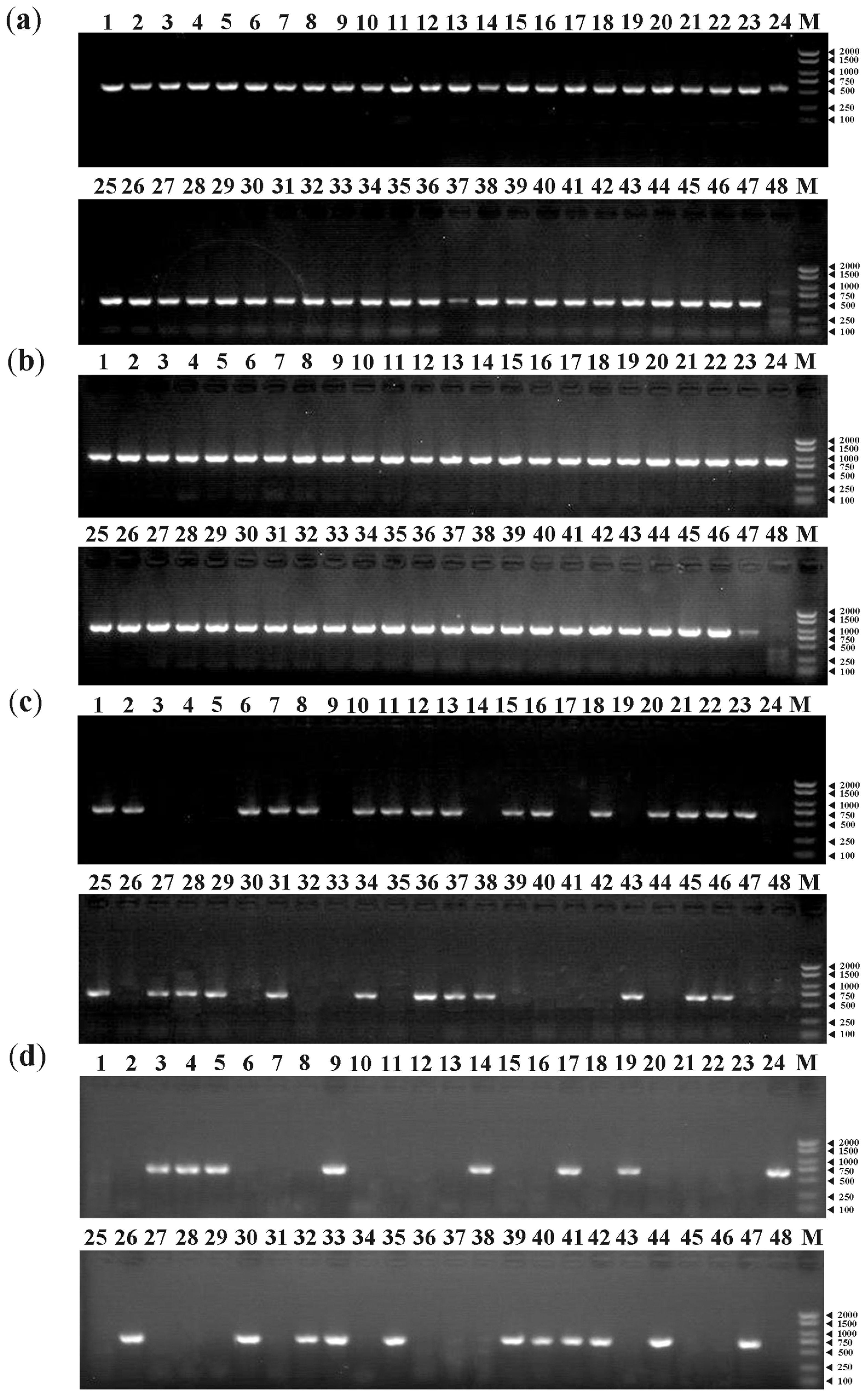
Primer pairs of the MAT1-1-1 (b1) and MAT1-2-1 (b2) genes were used to detect the mating type of single-ascospore populations in V. bohemica. Similarly, primer pairs of MAT1-1-1 (c1) and MAT1-2-1 (c2) genes were used for those in V. conica. Additionally, special primers were designed to amplify the MAT1-1-10 and MAT1-1-11 genes in V. bohemica and V. conica (Table 2). Among the 47 single-ascospore populations of V. bohemica, both the MAT1-1-1 and MAT1-2-1 gene were successfully amplified in each ascospore strain (Figure 5a,b). In the 47 single-ascospore populations of V. conica, strains that contained the MAT1-1-1 gene but lacked the MAT1-2-1 gene were amplified and identified as MAT1-1-1 gene, and vice versa. Two genes exhibited perfect complementarity within the single-ascospore population (Figure 5c,d). Additionally, primers for b10, c10, and c11 effectively amplified the MAT1-1-10 gene of V. bohemica, the MAT1-1-10 gene of V. conica, and the MAT1-1-11 gene of V. conica, respectively (Supplementary Figure S7a–c). Furthermore, we attempted to verify the presence of the MAT1-1-11 gene in V. bohemica using the primer pair c11, designed for the MAT1-1-11 gene of V. conica. The results revealed that the MAT1-1-11 gene is not present in V. bohemica, consistent with the genomic data. In this study of V. bohemica and V. conica, among the five mating-type genes, no sequence differences were detected between the single-ascospore strains and the parental strains.

4. Discussion

4.1. MAT Loci and Mating-Type Genes

Through our investigation, it was determined that all three strains of V. bohemica consistently exhibited a configuration of the MAT locus which includes three ORFs: the MAT1-1-1, MAT1-1-10, and the MAT1-2-1 genes. In contrast, V. conica exhibited an entirely distinct pattern, with each strain exclusively harboring one type of MAT idiomorph—either MAT1-1 or MAT1-2. Strain No. 21110 exclusively possessed the MAT1-1 idiomorph, encompassing the MAT1-1-1 and MAT1-1-10 genes. Conversely, strains No. 21117 and No. 21120 exclusively contained the MAT1-2 idiomorph, housing the MAT1-2-1 and MAT1-1-11 genes. This is the first report of the MAT1-1-10 and MAT1-1-11 genes in species other than those in the genus Morchella since Chai et al. [20] initially identified them in Morchella.
Compared to other protein-coding genes, sex-determining genes in ascomycetes have been found to evolve at a faster rate, making them useful for studying the phylogenetic relationships of fungi [33]. Previous studies on the phylogenetic analysis of mating-type genes have been conducted in ascomycetes [34,35]. In this study, phylogenetic trees were constructed using different MAT1-1 and MAT1-2 protein sequences, in order to corroborate the accuracy of the naming of MAT genes in the genus Verpa.
Glass et al. [36] first described the MAT1-1-1 gene in the matA (=MAT1-1) idiomorph of N. crassa, which encodes a DNA-binding protein with a conserved MATalpha_HMGbox domain. While previous research has suggested that the MAT1-1-1 gene typically contains an intron within its conserved domain [37,38,39], subsequent studies have revealed exceptions to this pattern (as in Sporothrix schenckii [40]). Nevertheless, in this study, the MAT1-1-1 gene in Verpa spp. clearly supported the previously established viewpoint (Figure 1a). The MAT1-2-1 gene harbored a conserved HMG-box domain, which has been demonstrated to play a role in DNA binding across plants, animals, and fungi [41,42,43]. In accordance with prior research, the ORF of the MAT1-2-1 gene in Verpa spp. included an intron spanning a serine codon (Figure 1b). The definition of a dependable domain for the MAT gene has been a persistent challenge. The MAT1-1-2 gene—the second MAT1-1 gene described—was exclusively present in Sordariomycetes [17]. Previous studies have aimed to establish a reliable structural domain for the MAT1-1-2 protein [15,37], which now has been included in the Protein Family (PFAM) database. Earlier studies suggested a role for the MAT1-1-2 gene in the development of sexual structures [44,45,46]; however, its precise function in sexual reproduction has remained enigmatic. This study attempted to define a motif for the recently discovered secondary MAT genes, MAT1-1-10 and MAT1-1-11, with the aim of contributing a novel domain definition for future research. However, it is crucial to note that this motif relies solely on data available in Morchella and Verpa. Subsequent discoveries are imperative to validate the applicability of this domain and confirm the functionality of this motif, necessitating further investigation.

4.2. Structure of Mating-Type Loci between Verpa bohemica and Verpa conica

The gene structure of the MAT idiomorph in Verpa spp. exhibited a resemblance to the corresponding structure in Morchella. For instance, the MAT idiomorph of V. bohemica No. 21110 and M04M24 of M. importuna, as reported by Liu et al. [19], shared a similar configuration. Similarly, the MAT idiomorph of V. bohemica aligned with the structure of M. rufobrunnea, as published by Chai et al. [21], where the MAT1-1-10, MAT1-1-1, and MAT1-2-1 genes were arranged in a tandem array. However, discernible structural disparities existed between the MAT loci of these two species. For example, M. importuna also possesses an alternative structure, consisting solely of one MAT1-2-1 gene in the MAT1-2 idiomorph [19]. Recent research has suggested that the MAT1-2 idiomorph of M. purpurascena and M. pulchella encompasses the MAT1-2-1 and MAT1-1-11 genes [21], corresponding to the structure observed in strains No. 21117 and No. 21120 of V. conica, as delineated in this investigation. However, the presence of a MAT1-1 gene in the MAT1-2 idiomorph remains a perplexing phenomenon. Similarly, the MAT1-2 idiomorph of Chaetomium globosum (Sordariomycetes) included a MAT1-1-2 gene located 1540 bp downstream of the MAT1-2-1 gene [15].
Butler et al. [47] were the first to propose the concept of MAT context in ascomycetes through their investigation of various yeasts and N. crassa. They observed that the MAT locus in Yarrowia lipolytica was located between the homologous genes APN2 and SLA2 in S. cerevisiae, and these two genes were also identified in the left and right adjacent sequences in N. crassa. Consequently, this shared structural arrangement led the authors to suggest that this configuration may be ancestral to all ascomycetes. Debuchy et al. [15] supported this conclusion and additionally proposed that APN2, SLA2, and APC5 are ancestral partners of the MAT locus in ascomycetes. However, this study raised questions about whether APC5 truly fits this description. In summary, a more comprehensive examination of the regions surrounding the MAT locus is expected to contribute to better understanding of the ancestral MAT locus structure in ascomycetes.

4.3. Homothallism in V. bohemica and Heterothallism in V. conica

In contrast to animals with sex chromosomes, the mechanism of sexual reproduction in fungi is governed by a solitary genetic locus known as the mating-type locus [48,49]. Compared to basidiomycetes, the mating-type loci of ascomycetes only contain a few relatively conserved genes, usually MAT1-1 and/or MAT1-2 [50]. Within the genomic data of the three strains of V. bohemica, it was ascertained that this species harbors both the MAT1-1-1 and MAT1-2-1 gene in the MAT locus, signifying the homothallic characteristic of V. bohemica. Concerning V. conica, genomic data of the three strains revealed that No. 21110 exclusively contained the MAT1-1 idiomorph, while No. 21117 and No. 21120 solely contained the MAT1-2 idiomorph, manifesting characteristics akin to heterothallic ascomycetes. In fact, the missing genotypes in the V. conica MAT locus could not be detected throughout the entire genome, either in silico or by PCR amplification. The conserved flanking sequences of these two distinct types validated their occupancy of the same chromosomal locus. PCR analysis further substantiated these findings, as we effectively amplified the MAT1-1-1, MAT1-2-1, and MAT1-1-10 genes within the single-spore strain of V. bohemica No. 20020. In contrast, within the single-spore population of V. conica No. 21120, MAT1-1-1 and MAT1-2-1 demonstrated complementary target products, while MAT1-1-10 and MAT1-1-11, which are intrinsically associated with these two genes, exhibited an identical banding pattern corresponding exclusively to their counterparts. Therefore, these results strongly indicate the homothallic life cycle of V. bohemica, while V. conica exhibits heterothallism. Discovery of the life cycle of Verpa spp. through genomic data analysis holds promise for providing further insights into their future utilization.

5. Conclusions

The results of this study revealed heterothallism in V. conica and homothallism in V. bohemica, indicative of the existence of both reproductive strategies within the Verpa genus. Leveraging genomic data enabled a comprehensive analysis of mating-type loci across six Verpa species. It was found that V. bohemica, a homothallic species, includes both MAT1-1-1 and MAT1-2-1 genes alongside an independent MAT1-1-10 within its mating-type loci, while V. conica, a heterothallic species, possesses either MAT1-1-1 or MAT1-2-1, with MAT1-1-10 and MAT1-1-11 genes specific to the MAT1-1 and MAT1-2 idiomorphs, respectively. Consistent with the pattern observed in other ascomycetes, the MAT1-1-1 gene in Verpa spp. demonstrated greater variability than the MAT1-2-1 gene, particularly within their conserved domains. Contrary to Morchella, where the SDH2 and MBA1 genes are adjacent to the MAT locus, the Verpa spp. displayed a significantly different flanking gene composition, with APN2 and SLA2 in proximity and SDH2 and MBA1 positioned at a greater distance. Yet, this arrangement is similar to the structure observed in Sordariomycetes. This remarkable finding challenges the traditional view of conserved flanking sequences within mating-type loci and their evolutionary role in MAT genes, diverging from the structure observed in the model organism N. crassa. This phenomenon calls for reevaluation of the significance of conserved flanking sequences in the evolution of MAT genes within the broader framework of mating-type loci.

Supplementary Materials

The following supporting information can be downloaded at: https://www.mdpi.com/article/10.3390/jof9121202/s1, Table S1: Sequence details of MAT1-1-1, MAT1-2-1, MAT1-1-10 and MAT1-1-11 gene in Verpa spp.; Table S2: BLASTp result of MAT1-1-10 and MAT1-1-11 protein in Verpa spp.; Figure S1: Alignment of MAT1-1-1 protein; Figure S2: Alignment of MAT1-2-1 protein; Figure S3: Alignment of MAT1-1-10 protein; Figure S4: Alignment of MAT1-1-11 protein; Figure S5: The detailed sequence information of MAT1-1-10 protein motif; Figure S6: The detailed sequence information of MAT1-1-11 protein motif; Figure S7:Agarose gel electrophoresis of the PCR products of MAT1-1-10 and MAT1-1-11.

Author Contributions

W.S., P.H. and W.L. article design, manuscript writing, review, and editing; P.H., review, investigation and editing; Y.C., X.S., L.W., J.Z., L.E., Q.H., Q.Y. and Z.Z. data processing and review; P.H. and F.Y., review, editing, discussions and project management. All authors have read and agreed to the published version of the manuscript.

Funding

This research was funded by the Basic Research Project of Yunnan Provincial Department of Science and Technology (grant number 202101AT070541). Yunnan Technology Innovation Talents Projects (202205AD160036) to Fuqiang Yu.

Institutional Review Board Statement

Not applicable.

Informed Consent Statement

Not applicable.

Data Availability Statement

Data are contained within the article and Supplementary Materials.

Acknowledgments

The authors sincerely thank those who helped collect the specimens, including Yongbing Guo, Xili Jia, Jianyong Han, Xiangsong Pu, Yongchang Liu and Tianqi Yao.

Conflicts of Interest

The authors declare no conflict of interest.

References

  1. Bunyard, B.A.; Nicholson, M.S.; Royse, D.J. Phylogenetic resolution of Morchella, Verpa, and Disciotis [Pezizales: Morchellaceae] based on restriction enzyme analysis of the 28S ribosomal RNA gene. Exp. Mycol. 1995, 19, 223–233. [Google Scholar] [CrossRef] [PubMed]
  2. O’Donnell, K.; Cigelnik, E.; Weber, N.S.; Trappe, J.M. Phylogenetic relationships among ascomycetous truffles and the true and false morels inferred from 18S and 28S ribosomal DNA sequence analysis. Mycologia 1997, 89, 48–65. [Google Scholar] [CrossRef]
  3. Aktas, S.; Karaselek, M.A. The taxonomicalch characterization of some helvella and its relatives by morphological and molecular data from turkey. Appl. Ecol. Environ. Res. 2019, 17, 7395–7405. [Google Scholar] [CrossRef]
  4. Anand, N.; Chowdhry, P.N. Taxonomic and molecular identification of Verpa bohemica: A newly explored fungi from Rajouri (J&K), India. Recent Res. Sci. Technol. 2013, 5, 9–12. [Google Scholar]
  5. Shameem, N.; Kamili, A.N.; Ahmad, M.; Masoodi, F.A.; Parray, J.A. Antimicrobial activity of crude fractions and morel compounds from wild edible mushrooms of North Western Himalaya. Microb. Pathog. 2017, 105, 356–360. [Google Scholar] [CrossRef] [PubMed]
  6. Lagrange, E.; Vernoux, J.P. Warning on false or true morels and button mushrooms with potential toxicity linked to hydrazinic toxins: An update. Toxins 2020, 12, 482. [Google Scholar] [CrossRef]
  7. Misirikl, D.; Elmastas, M.; Turkekul, İ. Determination of antioxidant activities of some wild mushrooms species in Tokat region. Bütünleyici Ve Anadolu Tıbbı Derg. 2019, 1, 24–32. [Google Scholar]
  8. Sheikh, P.; Dar, G.H.H.; Dar, A.; Shah, S.; Bhat, K. Chemical composition and anti-oxidant activities of some edible mushrooms of western Himalayas of India. Int. J. Plant Res. 2015, 28, 124–133. [Google Scholar] [CrossRef]
  9. Lin, S.Y.; Chen, Y.K.; Yu, H.T.; Barseghyan, G.S.; Asatiani, M.D.; Wasser, S.P.; Mau, J.L. Comparative study of contents of several bioactive components in fruiting bodies and mycelia of culinary-medicinal mushrooms. Int. J. Med. Mushrooms 2013, 15, 315–323. [Google Scholar] [CrossRef]
  10. Elmastas, M.; Isildak, O.; Turkekul, I.; Temur, N. Determination of antioxidant activity and antioxidant compounds in wild edible mushrooms. J. Food Compos. Anal. 2007, 20, 337–345. [Google Scholar] [CrossRef]
  11. Kellner, H.; Luis, P.; Buscot, F. Diversity of laccase-like multicopper oxidase genes in morchellaceae: Identification of genes potentially involved in extracellular activities related to plant litter decay. FEMS Microbiol. Ecol. 2007, 61, 153–163. [Google Scholar] [CrossRef] [PubMed]
  12. Coppin, E.; Debuchy, R.; Arnaise, S.; Picard, M. Mating types and sexual development in filamentous ascomycetes. Microbiol. Mol. Biol. Rev. 1997, 61, 411–428. [Google Scholar] [CrossRef] [PubMed]
  13. Metzenberg, R.L.; Glass, N.L. Mating type and mating strategies in Neurospora. BioEssays 1990, 12, 53–59. [Google Scholar] [CrossRef] [PubMed]
  14. Turgeon, B.G.; Yoder, O.C. Proposed nomenclature for mating type genes of filamentous ascomycetes. Fungal Genet. Biol. 2000, 31, 1–5. [Google Scholar] [CrossRef] [PubMed]
  15. Debuchy, R.; Turgeon, B.G. Mating-type structure, evolution, and function in euascomycetes. In Growth, Differentiation and Sexuality; Springer: Berlin/Heidelberg, Germany, 2006; pp. 293–323. [Google Scholar]
  16. Nelson, M.A. Mating systems in ascomycetes: A romp in the Sac. Trends Genet. 1996, 12, 69–74. [Google Scholar] [CrossRef] [PubMed]
  17. Wilken, P.M.; Steenkamp, E.T.; Wingfield, M.J.; de Beer, Z.W.; Wingfield, B.D. Which MAT gene? Pezizomycotina (ascomycota) mating-type gene nomenclature reconsidered. Fungal Biol. Rev. 2017, 31, 199–211. [Google Scholar] [CrossRef]
  18. Wilson, A.M.; Wilken, P.M.; Wingfield, M.J.; Wingfield, B.D. Genetic networks that govern sexual reproduction in the pezizomycotina. Microbiol. Mol. Biol. Rev. 2021, 85, e00020-21. [Google Scholar] [CrossRef]
  19. Liu, W.; Chen, L.; Cai, Y.; Zhang, Q.; Bian, Y. Opposite polarity monospore genome de novo sequencing and comparative analysis reveal the possible heterothallic life cycle of Morchella importuna. Int. J. Mol. Sci. 2018, 19, 2525. [Google Scholar] [CrossRef]
  20. Chai, H.; Chen, W.; Zhang, X.; Su, K.; Zhao, Y. Structural variation and phylogenetic analysis of the mating-type locus in the genus Morchella. Mycologia 2019, 111, 551–562. [Google Scholar] [CrossRef]
  21. Chai, H.; Liu, P.; Ma, Y.; Chen, W.; Tao, N.; Zhao, Y. Organization and unconventional integration of the mating-type loci in Morchella species. J. Fungi 2022, 8, 746. [Google Scholar] [CrossRef]
  22. Brock, T.D.; Madigan, M.T.; Martinko, J.M. Brock Biology of Microorganisms, 13th ed.; Benjamin Cummings: San Francisco, CA, USA, 2012. [Google Scholar]
  23. Inglis, P.W.; Pappas, M.D.C.R.; Resende, L.V.; Grattapaglia, D. Fast and inexpensive protocols for consistent extraction of high quality DNA and RNA from challenging plant and fungal samples for high-throughput SNP genotyping and sequencing applications. PLoS ONE 2018, 13, e0206085. [Google Scholar] [CrossRef]
  24. Bolger, A.M.; Lohse, M.; Usadel, B. Trimmomatic: A flexible trimmer for illumina sequence data. Bioinformatics 2014, 30, 2114–2120. [Google Scholar] [CrossRef] [PubMed]
  25. De Coster, W.; D’Hert, S.; Schultz, D.T.; Cruts, M.; Van Broeckhoven, C. NanoPack: Visualizing and processing long-read sequencing data. Bioinformatics 2018, 34, 2666–2669. [Google Scholar] [CrossRef] [PubMed]
  26. Allaire, J. RStudio: Integrated Development Environment for R; Boston, MA, USA, 2012; Volume 770, pp. 165–171. Available online: http://www.rstudio.com/ (accessed on 1 December 2023).
  27. Bailey, T.L.; Johnson, J.; Grant, C.E.; Noble, W.S. The MEME suite. Nucleic Acids Res. 2015, 43, W39–W49. [Google Scholar] [CrossRef]
  28. Chen, C.; Chen, H.; Zhang, Y.; Thomas, H.R.; Frank, M.H.; He, Y.; Xia, R. TBtools: An integrative toolkit developed for interactive analyses of big biological data. Mol. Plant 2020, 13, 1194–1202. [Google Scholar] [CrossRef] [PubMed]
  29. Katoh, K.; Misawa, K.; Kuma, K.; Miyata, T. MAFFT: A novel method for rapid multiple sequence alignment based on fast fourier transform. Nucleic Acids Res. 2002, 30, 3059–3066. [Google Scholar] [CrossRef]
  30. Nguyen, L.T.; Schmidt, H.A.; von Haeseler, A.; Minh, B.Q. IQ-TREE: A fast and effective stochastic algorithm for estimating maximum-likelihood phylogenies. Mol. Biol. Evol. 2015, 32, 268–274. [Google Scholar] [CrossRef] [PubMed]
  31. Minh, B.Q.; Nguyen, M.A.T.; von Haeseler, A. Ultrafast approximation for phylogenetic bootstrap. Mol. Biol. Evol. 2013, 30, 1188–1195. [Google Scholar] [CrossRef]
  32. Du, X.H.; Yang, Z.L. Mating systems in true morels (Morchella). Microbiol. Mol. Biol. Rev. 2021, 85, e00220-20. [Google Scholar] [CrossRef]
  33. Ferris, P.J.; Pavlovic, C.; Fabry, S.; Goodenough, U.W. Rapid evolution of sex-related genes in Chlamydomonas. Proc. Natl. Acad. Sci. USA 1997, 94, 8634–8639. [Google Scholar] [CrossRef]
  34. Martin, F.; Kohler, A.; Murat, C.; Balestrini, R.; Coutinho, P.M.; Jaillon, O.; Montanini, B.; Morin, E.; Noel, B.; Percudani, R.; et al. Périgord black truffle genome uncovers evolutionary origins and mechanisms of symbiosis. Nature 2010, 464, 1033–1038. [Google Scholar] [CrossRef] [PubMed]
  35. O’Donnell, K.; Ward, T.J.; Geiser, D.M.; Corby Kistler, H.; Aoki, T. Genealogical concordance between the mating type locus and seven other nuclear genes supports formal recognition of nine phylogenetically distinct species within the Fusarium graminearum clade. Fungal Genet. Biol. 2004, 41, 600–623. [Google Scholar] [CrossRef] [PubMed]
  36. Glass, N.L.; Grotelueschen, J.; Metzenberg, R.L. Neurospora crassa a mating-type region. Proc. Natl. Acad. Sci. USA 1990, 87, 4912–4916. [Google Scholar] [CrossRef] [PubMed]
  37. Kanematsu, S.; Adachi, Y.; Ito, T. Mating-type loci of heterothallic Diaporthe spp.: Homologous genes are present in opposite mating-types. Curr. Genet. 2007, 52, 11–22. [Google Scholar] [CrossRef] [PubMed]
  38. Waalwijk, C.; Mendes, O.; Verstappen, E.C.P.; De Waard, M.A.; Kema, G.H.J. Isolation and characterization of the mating-type idiomorphs from the wheat septoria leaf blotch fungus Mycosphaerella graminicola. Fungal Genet. Biol. 2002, 35, 277–286. [Google Scholar] [CrossRef] [PubMed]
  39. Wilken, P.M.; Steenkamp, E.T.; Wingfield, M.J.; de Beer, Z.W.; Wingfield, B.D. DNA loss at the Ceratocystis fimbriata mating locus results in self-sterility. PLoS ONE 2014, 9, e92180. [Google Scholar] [CrossRef]
  40. Teixeira, M.D.M.; Rodrigues, A.M.; Tsui, C.K.M.; De Almeida, L.G.P.; Van Diepeningen, A.D.; Van Den Ende, B.G.; Fernandes, G.F.; Kano, R.; Hamelin, R.C.; Lopes Bezerra, L.M.; et al. Asexual propagation of a virulent clone complex in a human and feline outbreak of Sporotrichosis. Eukaryot. Cell 2015, 14, 158–169. [Google Scholar] [CrossRef] [PubMed]
  41. Ait Benkhali, J.; Coppin, E.; Brun, S.; Peraza Reyes, L.; Martin, T.; Dixelius, C.; Lazar, N.; Van Tilbeurgh, H.; Debuchy, R. A network of HMG-box transcription factors regulates sexual cycle in the fungus Podospora anserina. PLoS Genet. 2013, 9, e1003642. [Google Scholar] [CrossRef]
  42. Debuchy, R.; Berteaux Lecellier, V.; Silar, P. Mating systems and sexual morphogenesis in ascomycetes. In Cellular and Molecular Biology of Filamentous Fungi; John Wiley & Sons, Ltd.: Hoboken, NJ, USA, 2010; pp. 499–535. ISBN 978-1-68367-129-9. [Google Scholar]
  43. Jacobi, V.; Dufour, J.; Bouvet, G.F.; Aoun, M.; Bernier, L. Identification of transcripts up-regulated in asexual and sexual fruiting bodies of the dutch elm disease pathogen Ophiostoma novo-ulmi. Can. J. Microbiol. 2010, 56, 697–705. [Google Scholar] [CrossRef]
  44. Arnaise, S.; Zickler, D.; Le Bilcot, S.; Poisier, C.; Debuchy, R. Mutations in mating-type genes of the heterothallic fungus Podospora anserina lead to self-fertility. Genetics 2001, 159, 545–556. [Google Scholar] [CrossRef]
  45. Klix, V.; Nowrousian, M.; Ringelberg, C.; Loros, J.J.; Dunlap, J.C.; Pöggeler, S. Functional characterization of MAT1-1-specific mating-type genes in the homothallic ascomycete Sordaria macrospora provides new insights into essential and nonessential sexual regulators. Eukaryot. Cell 2010, 9, 894–905. [Google Scholar] [CrossRef] [PubMed]
  46. Zheng, Q.; Hou, R.; Zhang, J.; Ma, J.; Wu, Z.; Wang, G.; Wang, C.; Xu, J.R. The MAT locus genes play different roles in sexual reproduction and pathogenesis in Fusarium graminearum. PLoS ONE 2013, 8, e66980. [Google Scholar] [CrossRef] [PubMed]
  47. Butler, G.; Kenny, C.; Fagan, A.; Kurischko, C.; Gaillardin, C.; Wolfe, K.H. Evolution of the MAT locus and its Ho endonuclease in yeast species. Proc. Natl. Acad. Sci. USA 2004, 101, 1632–1637. [Google Scholar] [CrossRef] [PubMed]
  48. Heitman, J. Evolution of sexual reproduction: A view from the fungal kingdom supports an evolutionary epoch with sex before sexes. Fungal Biol. Rev. 2015, 29, 108–117. [Google Scholar] [CrossRef]
  49. Heitman, J.; Sun, S.; James, T.Y. Evolution of fungal sexual reproduction. Mycologia 2013, 105, 1–27. [Google Scholar] [CrossRef]
  50. Casselton, L.; Challen, M. The mating type genes of the basidiomycetes. In Growth, Differentiation and Sexuality; Springer: Berlin/Heidelberg, Germany, 2006; pp. 357–374. [Google Scholar]
Figure 1. Alignment of part of the (a) MAT1-1-1 MATalpha_HMGbox domain and (b) MAT1-2-1 HMG box domain. The consensus sequences were based on a 60% similarity cutoff, while the sequence logo illustrates the relative representation of each amino acid per position. Red arrows indicate the positions of conserved introns. The figure was produced using the R package ggmsa; details of the sequences are available in the Supplementary Information (Supplementary Figures S2 and S3).
Jof 09 01202 g001
Figure 2. Phylogenetic trees of (a) MAT1-1 and (b) MAT1-2 genes constructed using IQ-TREE [30].
Jof 09 01202 g002
Figure 3. Distribution of conserved motifs of (a) MAT1-1-10 and (b) MAT1-1-11 proteins from Verpa spp. and Morchella spp. The scale bar at the bottom indicates the protein lengths, and sequence logos for each conserved motif are displayed below. Supplementary Figures S5 and S6 provide detailed sequences for each motif.
Jof 09 01202 g003
Figure 4. Genome maps of MAT loci for six Verpa species and two Morchella species. The names of the genes are labeled on the map and key genes are labeled with special colors. Genome maps of MAT loci were created using R package gggenes using fixed-length parameters.
Jof 09 01202 g004
Figure 5. MAT gene PCR products of single-ascospore populations No. 20020 and No. 21120: (a) MAT1-1-1 gene PCR products of No. 20020-1 to No. 20020-47; (b) MAT1-2-1 gene PCR products of No. 20020-1 to No. 20020-47; (c) MAT1-1-1 gene PCR products of No. 21120-1 to No. 21120-47; and (d) MAT1-2-1 gene PCR products of No. 21120-1 to No. 21120-47. The first lane represents the parental strain and the 48th lane contains ddH2O, serving as a negative control in all gel images. ‘M’ denotes DNA marker 2000 (2000, 1500, 1000, 750, 500, 250, and 100 bp).
Jof 09 01202 g005
Table 1. Ascocarps used in this study.
AscocarpSpeciesOriginGenbank
No. 20020Verpa bohemicaShennongjia HubeiJAUFSG000000000
No. 20124Verpa bohemicaLinxia GansuJAUFSF000000000
No. 21108Verpa bohemicaArba SichuanJAUFSE000000000
No. 21110Verpa conicaPengshui ChongqingJAUFSD000000000
No. 21117Verpa conicaTiaiyuan ShanxiJAUFSC000000000
No. 21120Verpa conicaXinyuan XinjiangJAUFSB000000000
Table 2. Primer sequences and PCR cycling parameters used in this study.
GeneCodeSequence (5′–3′)Annealing Temperature (°C)Product Size (bp)
MAT1-1-1 gene of V. bohemicab1fTCTCGCAGAAAGCCACGATT61481
b1rATCCTCATTGCGAGCGTAGG
MAT1-2-1 gene of V. bohemicab2fCTCCATCAAACGTGCGGTTC61654
b2rGGGGGTTTCTTGTCCGAAGT
MAT1-1-10 gene of V. bohemicab10fGGGCCTATCAATCAACGCTT60396
b10rAGGTAGCGTCTCAATGGGGT
MAT1-1-1 gene of V. conicac1fATCTCGCAGAAGTCCACGATT61580
c1rTTGATACCACAGCCGAGAAGG
MAT1-2-1 gene of V. conicac2fGTCGTGACTAAAACCGGCCT61500
c2rTCATTGCTGCGGTGGATCTT
MAT1-1-10 gene of V. conicac10fCGCGTCGTTAGTAGTGCGAT60819
c10rGAAGCTCAGTCCGGTGACAT
MAT1-1-11 gene of V. conicac11fTGGACGAAACGCTGGACTTT61648
c11rCCTTCTGGAACCCTGCAAGT
Disclaimer/Publisher’s Note: The statements, opinions and data contained in all publications are solely those of the individual author(s) and contributor(s) and not of MDPI and/or the editor(s). MDPI and/or the editor(s) disclaim responsibility for any injury to people or property resulting from any ideas, methods, instructions or products referred to in the content.