Article

Candida auris in Austria—What Is New and What Is Different

1
Division of Clinical Microbiology, Department of Laboratory Medicine, Medical University of Vienna, 1090 Vienna, Austria
2
Division of Infectious Diseases and Tropical Medicine, Department of Medicine I, Medical University of Vienna, 1090 Vienna, Austria
*
Author to whom correspondence should be addressed.
Current address: LABCON—Medizinische Laboratorien GmbH, Laborambulatorium am Kurpark Oberlaa, 1100 Vienna, Austria.
Current address: Institut für Klinische Pathologie und Molekularpathologie, Landesklinikum Horn-Allentsteig, 3580 Horn, Austria.
§
Current address: Division of Data, Statistics and Risk Assessment, Austrian Agency for Health and Food Safety AGES, 1220 Vienna, Austria.
J. Fungi 2023, 9(2), 129; https://doi.org/10.3390/jof9020129
Submission received: 17 November 2022 / Revised: 5 January 2023 / Accepted: 13 January 2023 / Published: 17 January 2023
(This article belongs to the Special Issue Infection Prevention and Control of Fungal Infections)

Abstract

:
Candida auris is a novel and emerging pathogenic yeast which represents a serious global health threat. Since its first description in Japan 2009, it has been associated with large hospital outbreaks all over the world and is often resistant to more than one antifungal drug class. To date, five C. auris isolates have been detected in Austria. Morphological characterization and antifungal susceptibility profiles against echinocandins, azoles, polyenes and pyrimidines, as well as the new antifungals ibrexafungerp and manogepix, were determined. In order to assess pathogenicity of these isolates, an infection model in Galleria mellonella was performed and whole genome sequencing (WGS) analysis was conducted to determine the phylogeographic origin. We could characterize four isolates as South Asian clade I and one isolate as African clade III. All of them had elevated minimal inhibitory concentrations to at least two different antifungal classes. The new antifungal manogepix showed high in vitro efficacy against all five C. auris isolates. One isolate, belonging to the African clade III, showed an aggregating phenotype, while the other isolates belonging to South Asian clade I were non-aggregating. In the Galleria mellonella infection model, the isolate belonging to African clade III exhibited the lowest in vivo pathogenicity. As the occurrence of C. auris increases globally, it is important to raise awareness to prevent transmission and hospital outbreaks.

Graphical Abstract

1. Introduction

Infections with Candida species are a severe public health problem across the world, especially since the emergence of Candida auris, a multidrug-resistant (MDR) nosocomial pathogen that can cause invasive candidiasis and is often associated with a high case fatality rate and therapeutic failure [1]. Candida auris was first isolated and described from ear discharge of a 70-year-old female patient in Tokyo, Japan, in 2009 [2]. Within a decade, this yeast had spread around the world at an alarming rate, causing numerous outbreaks in health care facilities [1,2,3,4,5,6,7,8,9].
C. auris has an extraordinary capacity to colonize the human body and environment for prolonged periods [10] and is very difficult to eradicate [11]. During the current COVID-19 pandemic, many health care systems around the world were overwhelmed, leading to over-occupied intensive care units (ICUs) and compromised infection prevention control. These circumstances might favor the nosocomial spread of C. auris infections [12,13]. Furthermore, C. auris is often misidentified by commercial identification systems in routine microbiology laboratories; therefore, the real prevalence of infections caused by C. auris may be underestimated.
Here, we describe phenotypic and genotypic features of the first five Austrian isolates. Given the rapid global emergence of C. auris and the associated hospital outbreaks, it is important to raise awareness about the appearance and possibility of outbreaks of C. auris in Austrian healthcare facilities.
The aim of this study was to describe the morphological characteristics and to investigate the isolates’ susceptibilities to commonly used antifungals, including the new antifungals ibrexafungerp and manogepix. In addition, mutations associated with antifungal resistance were investigated. A Galleria mellonella infection model was chosen to determine the in vivo pathogenicity of the five isolates. Finally, the study aims to assign the five strains to one of the four previously described phylogenetic clades of C. auris.

2. Materials and Methods

2.1. Sampling

Five isolates were detected in patients from Austria and sampled. In addition to the five clinical isolates, we included two control strains, CBS 12777 (South Asian clade I) and CBS 10913 (alias: B11220, first East Asian clade II from Japan [2]), in the phenotypic characterization (susceptibility testing, aggregating phenotype, in vivo pathogenicity). However, as there are no reference values for most of the data, these were included as type strains for comparison with the data obtained from the clinical isolates.

2.2. Phylogeographic Clade Assignment Using Whole Genome Sequencing (WGS)

DNA extraction was carried out using 48 h colonies from malt extract agar (MEA) plates, following a solution-based protocol with implemented bead-beating steps as described elsewhere [14]. Before preparing libraries, the total quantity of DNA was measured using the Qubit dsDNA High Sensitivity Assay system (Life Technologies, Carlsbad, CA, USA). The purity of DNA was determined by measuring the ratios A260/A280 and A260/A230 using a NanoDrop 2000c spectrophotometer (Thermo Fisher Scientific, Waltham, MA, USA).
In this study, WGS was carried out to classify the clinical isolates according to the four major phylogeographic clades. DNA-sequencing libraries were prepared from extracted DNA according to the Illumina DNA Prep workflow. DNA was denatured according to the protocol and diluted to a final loading concentration of 8 pM combined with a 5% PhiX spike-in (PhiX Control v3, Illumina) for sequencing on a v3-flowcell 2 × 300 bp on Illumina MiSeq (Illumina, San Diego, CA, USA).
The quality of the NGS run was evaluated with FastQC 0.11.4 [15]. The Mycosnp pipeline was used for phylogenetic analysis of the WGS data [16]. The sequence of the C. auris isolates were compared to B8441 (South Asian clade I) and B11221 (African clade III), which were obtained from www.candidagenome.org (accessed on 17 September 2022), and B11220 (East Asian clade II, GCA_003013715.2) and B11243 (South American clade IV, GCA_003014415.1), obtained from www.ncbi.nlm.nih.gov (accessed on 17 September 2022). For phylogenetic analysis, we compared SNPs called against the reference strains B8841, B11220, B11221 and B11243. A multiple sequence alignment of mating type loci and flanking genes of C. auris isolates Cau1-5 with B8441 (MTLa) and B11221 (MTLα) was generated to determine mating type [17].

2.3. Microscopic Morphology—Aggregating or Non-Aggregating Phenotype

Cellular appearance of all isolates was examined after 48 h of incubation at 37 °C in a liquid medium to determine whether aggregate formation is inducible in any of the C. auris strains through exposure to antifungal drugs. All isolates were microscopically screened for aggregating phenotype in a RPMI medium, Sabouraud medium and after exposure to different antifungals (caspofungin 0.016 mg/L, fluconazole 0.25 mg/L, 5-flucytosine 0.064 mg/L and amphotericin B 0.064 mg/L). All microscopic examinations, including aggregate-forming capacity after antifungal exposure, were performed in triplicates for all five clinical isolates, plus control strains CBS 10913 and CBS 12777, on three different days.

2.4. Infection Model in Galleria mellonella

For the infection model, G. mellonella larvae were used in a weight range of 250 mg ± 30 mg and after a day of fasting. Before injecting with the different C. auris strains, the larvae were washed with 96% ethanol to minimize the surface pathogens. The larvae were split into groups of 10 and kept in petri dishes. C. auris strains were cultured overnight on Sabouraud agar and resuspended in sterile PBS. Using the cell counter Luna-FL (Logos Biosystem), a cell suspension with a concentration of 5.0 × 10⁸ CFU/mL was obtained for each strain. For the trial, each group of larvae was inoculated with 10 µL of the C. auris suspension via injection in the last left pro-leg using a sterile Hamilton syringe (Hamilton Medical, Swiss). Thus, each larva was inoculated with 5.0 × 10⁶ yeast cells. A control group of 10 larvae was injected with 10 µL of PBS. Afterwards, the larvae were incubated at 37 °C and at different time points (12, 24, 48, 72, 96 and 120 h), the melanization and movement of the larvae were visually checked to evaluate survival. For comparison of pathogenicity, the median effective dose(ED50) values, which represent the time in hours after which 50% of the inoculated larvae population died, were calculated.

2.5. Antifungal Susceptibility Testing (AFST) including New Antifungals Ibrexafungerp and Manogepix

Antifungal susceptibility of all clinical isolates and the two control strains was determined according to the European Committee on Antimicrobial Susceptibility Testing (EUCAST) E.Def 7.3.2 microdilution method [18]. Each isolate was tested against the following eight antifungals (values represent the final concentrations after inoculation): anidulafungin (ANI; 0.008–16 mg/L), micafungin (MCA; 0.008–16 mg/L), fluconazole (FLC; 0.125–256 mg/L), posaconazole (POS; 0.016–32 mg/L), voriconazole (VOR; 0.008–16 mg/L), 5-flucytosine (5-FC; 0.032–64 mg/L), amphotericin B (AMB; 0.032–16 mg/L), ibrexafungerp (IBX; 0.016–8 mg/L) provided by Scynexis (Jersey City, NJ, USA), and manogepix (MGP; 0.002–16 mg/L) provided by Pfizer (New York City, NY, USA). There are no established species-specific clinical breakpoints for C. auris in EUCAST guidelines [19].

3. Results

3.1. Origin of Candida Auris Isolates

To date, five sporadic cases of C. auris have been reported in Austria—one infection and four cases of asymptomatic colonization. The first C. auris strain (Cau1) was isolated in January 2018 from the external auditory canal of a 22-year-old man who suffered from a therapy-refractory otitis externa that had persisted for almost four years despite antibiotic treatment. The patient was otherwise healthy. He was of Turkish ancestry and used to travel to Turkey frequently [20]. The second isolate (Cau2) was isolated in February 2020 from the external auditory canal of a 61-year-old male with a hematologic disease, followed by a strain (Cau3) isolated in May 2020 from the urinary tract of a trauma patient of Indian origin with regular travel activity. The isolate Cau4 was detected in October 2021 from a 60-year-old female patient in Valencia, Spain, after hospitalization because of a subarachnoid hemorrhage. The most recent isolate (Cau5) was detected in April 2022 in the urine of a 66-year-old female after hospitalization on Patmos and Rhodos, Greece, also due to subarachnoid hemorrhage. The four latter patients were colonized and did not show any signs of infection caused by C. auris. All patients came from different regions in Austria and were treated in different healthcare institutions. No epidemiologic links could be found between these five cases. In Table 1, all five clinical C. auris isolates are listed, including patient description, underlying medical condition, isolation site and origin of the isolate.

3.2. Phylogeographic Clade Assignment

Phylogenetic analysis using WGS revealed that the isolates Cau1, Cau2, Cau3 and Cau5 were genetically closest to South Asian clade I (see Table 2). Isolates Cau1 and Cau2, as well as Cau3 and Cau5, showed a higher degree of relatedness to each other with 44 SNPs and 76 SNPs, respectively. Overall, it is likely that the strains Cau1 and Cau2 belong to different subclades compared to Cau3 and Cau5, and reference strain B8441, as they are separated by more than 200 and 900 SNPs, respectively. In addition, the isolates could be separated from clades II, III and IV by 40,000 to 170,000 SNPs, whereas the isolate Cau4 was most closely related to African clade III, with 49 SNPs to clade III reference isolate B11221. Furthermore, analysis of the mating-type locus revealed that the clinical isolates Cau1, Cau2, Cau3 and Cau5 were MTLa homozygous (MTLa1 and MTLa2), thus confirming their assignment to South Asian clade I. The isolate Cau4 showed a homozygous MTLα, as is expected from African clade III.

3.3. Aggregating Phenotype of C. auris

When examining the cellular morphology of C. auris grown for 48 h at 37 °C in RPMI media, the isolates Cau1, Cau2, Cau3 and Cau5, as well as the two control strains CBS 12777 and CBS 10913, appeared as individual budding yeast cells. Isolate Cau4—assigned to African clade III—was forming aggregates in RPMI media and after exposure to different antifungal agents. Isolate Cau1 showed a non-aggregating phenotype in RPMI media, but formed small to large aggregates reproducibly, which could not be physically disrupted when exposed to different antifungals—see Figure 1.

3.4. Infection Model with Galleria mellonella

The G. mellonella infection model showed that for all tested C. auris isolates, a concentration of 5.0 × 10⁸ CFU/mL (5.0 × 10⁶ CFU/larva) is sufficient to kill G. mellonella within 120 h.
All of the PBS-injected larvae which were used as a negative control survived the experiment. Figure 2 and Table 3 demonstrate the different pathogenicity of the clinical isolates in comparison to the control strains. As shown in Table 3, the strains CBS 10913 and Cau4 are less pathogenic than the others.

3.5. Antifungal Susceptibility

In vitro susceptibilities of the five clinical C. auris isolates and two control strains to nine different commonly used antifungal agents and the new antifungals ibrexafungerp and manogepix are displayed in Table 4. All five isolates exhibited high MIC values to at least two different classes of antifungals. Both isolates Cau1 and Cau2 showed moderately elevated MIC values to the tested echinocandins anidulafungin (0.5 mg/L) and micafungin (0.125 mg/L)—as well as to amphotericin B (2 mg/L). Fluconazole showed low in vitro efficacy against isolates Cau3 and Cau5 with MIC values of 64 and >256 mg/L, respectively. The isolate Cau4 displayed a high fluconazole MIC of 64 mg/L as well as slightly elevated MIC values against echinocandins. The new antifungal substances ibrexafungerp and manogepix showed good in vitro activity against all tested isolates. The MIC values of manogepix were particularly low, with 0.008–0.032 mg/L.

3.6. Detection of Antifungal Resistance Mutations

No mutations in the genes ERG1, ERG2, ERG3 (associated with azole resistance), and MEC3 (associated with polyene resistance) could be detected in any of the clinical isolates [21]. In the isolates Cau3 and Cau5 showing high fluconazole MICs, the missense mutation p.Y132F in ERG11, which encodes 14-alpha-demethylase, was detected. The isolate Cau4 (African clade III) had two missense mutations (p.V125A, p.F126L) in ERG11 compared to the reference sequence of B8441 (South Asian clade I) and B11220 (East Asian clade II). Additionally, the isolates Cau3 and Cau5 had two missense mutations p.A583S (Cau3, Cau5) and p.S857L (Cau5) in TAC1b, which is a transcription factor for the drug efflux pumps Cdr1p, Cdr2p and Snq2p. Furthermore, the missense mutation p.F1367C was found in FKS1 in Cau1 and p.K74E (Cau1, Cau2, Cau3, Cau5) in CIS2. No mutations compared to the used reference genome of B11221 (African clade III) were found in the isolate Cau4.

4. Discussion

C. auris is a very heterogeneous, drug-resistant yeast that has been recognized as a serious health threat. Though C. auris has attracted much scientific attention, there are still many open questions. Therefore, the isolates detected in Austria have been investigated thoroughly in order to obtain more knowledge regarding the characteristics of this fungus and to check the strains for differences compared to previously described isolates. Thus, different methods, such as whole genome sequencing for classification of the phylogenetic clade, conventional phenotyping, assessing the pathogenicity an in vivo model, and the susceptibility pattern, as well as detecting resistance mutations, were applied and will be discussed in this order.

4.1. Phylogeographic Clades

In the present study, WGS of the five clinical isolates detected in Austria was conducted to determine their phylogeographic origin. The isolates Cau1, Cau2, Cau3 and Cau5 were separated by 46 to 225 SNPs, confirming the assumption that there are no epidemiological links between them. They were genetically closest to South Asian clade I and could be separated from clade II, III and IV by 45,000 to 165,000 SNPs. The patient colonized with Cau4 belonging to the African clade III was hospitalized in Valencia due to a subarachnoid hemorrhage. Since a large nosocomial outbreak with C. auris belonging to the African clade III has been described in a hospital in Valencia, it can be speculated that the patient acquired colonization during her hospital stay [22]. The patient colonized with Cau5 belonging to South Asian clade I was hospitalized on Patmos and Rhodos in Greece due to subarachnoid hemorrhage. From 2019–2021 74 cases and outbreaks in Greece have been reported including 58 cases in 2021, thus indicating a significant increase of cases in Greece. However, no further information which can be linked to our case has been made available [23].
Analysis of the mating-type locus—for further validation of the phylogenetic classification—revealed that our South Asian clade I isolates were MTLa homozygous (MTLa1 and MTLa2), supporting their assignment to South Asian clade I. In contrast to the other isolates, isolate Cau4 showed a homozygous MTLα as is described for African clade III strains [17].
The South Asian clade I is known for high rates of resistance to different antifungal agents and has been linked to invasive candidiasis and large-scale hospital outbreaks. However, isolates Cau1 and Cau2, belonging to South Asian clade I, may not explicitly display all of the properties associated with this clade. Both isolates were detected from the external auditory canal of two different patients—one with infection and one being only colonized. C. auris isolates associated with ear infections usually belong to East Asian Clade II. Welsh et al. indicate that this observation may be biased due to different testing methods. For example, in many laboratories, species-level identification is rarely performed from non-sterile sites such as the ear canal. Nonetheless, East Asian clade II is the only clade not associated with hospital outbreaks of invasive candidiasis [24].
The four C. auris clades can differ in their susceptibility profiles significantly. East Asian clade II is usually considered more susceptible than South Asian clade I. Although the isolates Cau1, Cau2, Cau3 and Cau5 belong to South Asian clade I, only Cau3 and Cau5 are fluconazole-resistant, while in previous studies, South Asian clade I has been shown to have a very high resistance rate of 97% to fluconazole [25]. However, levels of antifungal drug resistance can vary among strains significantly [24,26]. Nevertheless, four of our isolates showed elevated MIC values against amphotericin B (2–8 mg/L), a characteristic which, to date, has been found only in clades I and IV. Furthermore, all five isolates showed slightly elevated MICs for echinocandins, which is also in agreement with previous studies that reported cases of echinocandin resistance in clades I, III and IV [25,27].

4.2. Aggregating Phenotype

To date, only a few studies have investigated aggregate formation in C. auris. Borman et al. found that C. auris isolates can be roughly divided into aggregative or non-aggregative groups based on their growth characteristic phenotypes in culture. Isolates with an aggregative phenotype form large cellular agglomerates which cannot be physically disrupted. This is caused by a malfunctioning budding process, after which the daughter cells are not released. Aggregate-forming isolates were shown to be less pathogenic than isolates with non-aggregative phenotypes in a G. mellonella infection model [28]. Szekely et al. observed that differences in phenotypic behavior of C. auris isolates were clade-specific—including their growth characteristics and antifungal susceptibility profiles. In this study, aggregate-forming isolates belonged exclusively to the African clade III (n = 50) and not a single isolate from the South Asian clade I (n = 90) showed this phenotype. The authors also noted that an exposure to echinocandins and azoles caused isolates from the South Asian clade I to grow as aggregates. Cell morphology was unaffected by exposure to flucytosine or amphotericin B [29]. In isolate Cau4, belonging to the African clade III, the aggregating phenotype was detected. The other four clinical C. auris isolates belonging to South Asian clade I were non-aggregative: they appeared as single cells when cultured in standard mycological media. When the clinical isolates were exposed to antifungal drugs, the isolate Cau1 switched to the aggregative phenotype. However, in contrast to the findings of the previously mentioned study by Szekely et al., aggregation was also observed after exposure to flucytosine or amphotericin B. Aggregation might be an escape, stress response or a physical defense against antiseptic or antifungals in naturally non-aggregative C. auris isolates [29]. However, the reason for these observed differences in phenotypic behavior remains unclear, since the antifungal susceptibility profile of Cau1 did not differ from the other three South Asian clade I isolates in which aggregates were not detected. Further investigations are required to evaluate whether such in vitro differences in phenotypic behavior have implications for clinical practice.

4.3. Pathogenicity in the Galleria mellonella Infection Model

For the evaluation of pathogenicity, a G. mellonella infection model was performed. It became apparent that the four clinical isolates belonging to the South Asian clade I and the control strain CBS 12777 showed a much lower ED50. The isolate Cau4, which could be assigned to the African clade III, as well as the control strain CBS 10913 (East Asian clade II), showed a higher ED50 of 57.31 and 44.46, respectively. As already mentioned, aggregation was observed in isolate Cau4, which is in accordance with previous reports suggesting that aggregating isolates are less pathogenic in in vivo models in contrast to South Asian clade I isolates [28,30].

4.4. Antifungal Susceptibility

As EUCAST has not established species-specific clinical breakpoints for C. auris yet, an exact interpretation is difficult. All five clinical isolates of C. auris exhibited high MIC values to at least two different classes of antifungals. Since only four drug classes are available for the treatment of invasive Candida infections, this could limit therapeutic options.
In accordance with previous studies that reported the efficacy of the pyrimidine analogue 5-FC against C. auris [31,32,33], all five isolates showed low 5-FC MIC values of 0.064–0.25 mg/L. However, 5-FC is not used as monotherapy because in vivo resistance develops rapidly during treatment [34]. C. auris shows an extraordinarily high rate of fluconazole-resistant strains compared to other Candida species [1,35,36]. Therefore, echinocandins are usually the drug of choice for treatment of C. auris infections [37]. Nevertheless, the results of this study demonstrate that echinocandin resistance can develop in C. auris, thus confirming previous reports [4,26,38]. Cau1 and Cau2 were susceptible to azoles and showed moderately increased MIC values to both echinocandins and amphotericin B. In Cau3, the MIC of fluconazole was 64 mg/L, in addition to moderately elevated MIC values against echinocandins and amphotericin B. Moreover, the isolate Cau4 showed a high fluconazole MIC (64 mg/L). In contrast to the other strains, Cau4 was susceptible to anidulafungin and amphotericin B. The most recent isolate, Cau5, showed a similar resistance profile to that of Cau3, although fluconazole and amphotericin B MICs showed higher MIC values. These findings demonstrate the difficulty in choosing the appropriate antifungal treatment and the need for antifungal susceptibility testing. Furthermore, this highlights the urgent need for new antifungal drugs that may be effective against C. auris. The MIC values regarding the new antifungals ibrexafungerp and manogepix showed excellent in vitro activity, suggesting a promising alternative for an effective therapy, as has already been described [39,40,41,42,43]. In particular, manogepix showed potent in vitro activity with MIC values of 0.008–0.032 mg/L. This supports the assumption that manogepix could be a valuable therapeutic alternative in the treatment of C. auris [44,45,46].

4.5. Screening for Potential Resistance Mutations

Molecular mechanisms of antifungal resistance are not completely understood—this is especially true in the case of C. auris. However, NGS has been proven to be a powerful tool to detect mutations that are associated with antifungal resistance in Candida spp. [47,48]. When examining the five clinical isolates, a mutation in ERG11 (p.Y132F; hot spot 1 region), one of the most popular mutations associated with azole resistance in C. auris [49], was detected in Cau3 and Cau5. This was correlated with the elevated MIC values of 64 mg/L and >256 mg/L for fluconazole, respectively. The isolate Cau4 belonging to the African clade III, with a MIC of 64 mg/L for fluconazole, showed two missense mutations in ERG11 (p.V125A, p.F126L) compared to the reference genomes of clades I and II. Since this location is only six amino acids upstream of the well-known mutation p.Y132F, these target mutations may be associated with azole resistance. However, the absence of any mutations in ERG11 does not rule out the presence of azole resistance in C. auris. Kwon et al. reported that only in 5 of 38 fluconazole-resistant East Asian clade II C. auris isolates amino acid substitutions in ERG11 could be detected [50].
Regarding echinocandin resistance, Cau1 showed a mutation (p.F1367C) ten amino acids downstream of the FKS1 gene hot spot 2, which could explain the moderate resistance to echinocandins. However, Cau2, Cau3, Cau4 and Cau5 did not show any mutations in FKS1 despite elevated echinocandin MICs, thus suggesting other underlying molecular resistance mechanisms. WGS analysis revealed a missense mutation in CIS2 (p.K74E), which could be found in all South Asian clade I isolates (Cau1, Cau2, Cau3, Cau5). The CIS2 gene encodes gamma-glutamyl transpeptidase (GGT) and has been recently suspected to play a role in echinocandin resistance in C. auris [21]. Carolus et al. found that decreased amphotericin B susceptibility accompanied by fluconazole cross-resistance was caused by simultaneous premature stop codons in ERG3 and ERG11 [21]. Previously, amphotericin B resistance was only explained by an increased expression of genes involved in the biosynthesis of ergosterol, such as ERG1 and ERG2 [17]. The Austrian isolates did not show any mutations in ERG1, ERG2 and ERG3.
C. auris is still understudied compared to C. albicans and C. glabrata, even though it poses an urgent global threat and has been recently included in the Critical Priority Group of the WHO fungal priority pathogens list [51]. Further research is needed in order to understand the diverse underlying molecular resistance mechanisms associated with antifungal resistance, as this might aid in the development of new drugs [21,37].

5. Conclusions

More than thirteen years after the first description of C. auris in Japan [2], there are still numerous open questions. For example, little is known about its mechanisms and the development of resistance to antifungals.
The present study underlines the importance of raising awareness among healthcare providers of a potential spread of C. auris in Austria as the incidence of C. auris infections increases worldwide. All five clinical isolates being detected in Austrian patients show in vitro multidrug resistances. Four isolates belong to the South Asian clade I and one isolate could be assigned to African clade III. Both genotypical lineages have been associated with invasive infections and high case fatality rates [1]. It is conceivable that there are more cases of C. auris in Austria that have neither been detected nor reported. It is important to ensure a rapid identification of C. auris in patients and develop awareness of the threat this yeast may cause in order to prevent possible transmissions and hospital outbreaks.

Author Contributions

Conceptualization, K.S. and B.W.; methodology, K.S., C.W., S.S., S.G. and R.K.; software, K.S. and R.K.; validation, K.S. and R.K.; formal analysis, K.S. and R.K.; investigation, K.S., C.W., S.S., L.A., S.G. and R.K.; resources, B.W.; data curation, R.K.; writing—original draft preparation, K.S. and C.W.; writing—review and editing, B.W., C.W., R.K., B.S., H.L., I.C., A.M., S.S., L.A. and S.G.; visualization, K.S. and R.K.; supervision, B.W.; project administration, B.W.; funding acquisition, B.W. All authors have read and agreed to the published version of the manuscript.

Funding

This research received no external funding.

Institutional Review Board Statement

Not applicable.

Informed Consent Statement

Not applicable.

Data Availability Statement

The sequencing data are available in the BioProject database under accession number PRJNA923734.

Acknowledgments

We thank Shiva Pekard-Amenitsch—AGES Graz; Walter Buzina—MUG; Stefan Doppler—Kepler Linz; Dagmar Achleitner—SALK; Claudia Honsig—AGES Wien; Dorit Stahl—Klinik Landstraße for providing the clinical isolates. For assessment of MIC value to new antifungals, plates were provided by Thermo Fisher Scientific, Scynexis and Pfizer.

Conflicts of Interest

The authors declare no conflict of interest. This work was partially supported by Thermo Fisher Scientific. The sponsor had no role in the design of the study or the collection, analysis and interpretation of data.

References

  1. Lockhart, S.R.; Etienne, K.A.; Vallabhaneni, S.; Farooqi, J.; Chowdhary, A.; Govender, N.P.; Colombo, A.L.; Calvo, B.; Cuomo, C.A.; Desjardins, C.A.; et al. Simultaneous Emergence of Multidrug-Resistant Candida auris on 3 Continents Confirmed by Whole-Genome Sequencing and Epidemiological Analyses. Clin. Infect. Dis. 2017, 64, 134–140. [Google Scholar] [CrossRef] [PubMed] [Green Version]
  2. Satoh, K.; Makimura, K.; Hasumi, Y.; Nishiyama, Y.; Uchida, K.; Yamaguchi, H. Candida auris Sp. Nov., a Novel Ascomycetous Yeast Isolated from the External Ear Canal of an Inpatient in a Japanese Hospital. Microbiol. Immunol. 2009, 53, 41–44. [Google Scholar] [CrossRef] [PubMed]
  3. Clancy, C.J.; Nguyen, M.H. Emergence of Candida auris: An International Call to Arms. Clin. Infect. Dis. 2017, 64, 141–143. [Google Scholar] [CrossRef] [PubMed] [Green Version]
  4. Chowdhary, A.; Sharma, C.; Meis, J.F. Candida auris: A Rapidly Emerging Cause of Hospital-Acquired Multidrug-Resistant Fungal Infections Globally. PLoS Pathog. 2017, 13, e1006290. [Google Scholar] [CrossRef] [PubMed]
  5. Borman, A.M.; Szekely, A.; Johnson, E.M. Isolates of the Emerging Pathogen Candida auris Present in the UK Have Several Geographic Origins. Med. Mycol. 2017, 55, 563–567. [Google Scholar] [CrossRef]
  6. Wang, X.; Bing, J.; Zheng, Q.; Zhang, F.; Liu, J.; Yue, H.; Tao, L.; Du, H.; Wang, Y.; Wang, H.; et al. The First Isolate of Candida auris in China: Clinical and Biological Aspects Article. Emerg. Microbes Infect. 2018, 7, 1–9. [Google Scholar] [CrossRef] [Green Version]
  7. Sears, D.; Schwartz, B.S. Candida auris: An Emerging Multidrug-Resistant Pathogen. Int. J. Infect. Dis. 2017, 63, 95–98. [Google Scholar] [CrossRef] [Green Version]
  8. Schelenz, S.; Hagen, F.; Rhodes, J.L.; Abdolrasouli, A.; Chowdhary, A.; Hall, A.; Ryan, L.; Shackleton, J.; Trimlett, R.; Meis, J.F.; et al. First Hospital Outbreak of the Globally Emerging Candida auris in a European Hospital. Antimicrob. Resist. Infect. Control 2016, 5, 1–7. [Google Scholar] [CrossRef] [Green Version]
  9. Forsberg, K.; Woodworth, K.; Walters, M.; Berkow, E.L.; Jackson, B.; Chiller, T.; Vallabhaneni, S. Candida auris: The Recent Emergence of a Multidrug-Resistant Fungal Pathogen. Med. Mycol. 2019, 57, 1–12. [Google Scholar] [CrossRef] [Green Version]
  10. Iguchi, S.; Itakura, Y.; Yoshida, A.; Kamada, K.; Mizushima, R.; Arai, Y.; Uzawa, Y.; Kikuchi, K. Candida auris: A Pathogen Difficult to Identify, Treat, and Eradicate and Its Characteristics in Japanese Strains. J. Infect. Chemother. 2019, 25, 743–749. [Google Scholar] [CrossRef]
  11. Mulet Bayona, J.V.; Palop, N.T.; García, C.S.; Rodríguez, P.H.; de Medrano, V.A.L.; Gómez, C.F.; Cardona, C.G. Characteristics and Management of Candidaemia Episodes in an Established Candida auris Outbreak. Antibiotics 2020, 9, 558. [Google Scholar] [CrossRef] [PubMed]
  12. Chowdhary, A.; Sharma, A. The Lurking Scourge of Multidrug Resistant Candida auris in Times of COVID-19 Pandemic. J. Glob. Antimicrob. Resist. 2020, 22, 175–176. [Google Scholar] [CrossRef] [PubMed]
  13. Chowdhary, A.; Tarai, B.; Singh, A.; Sharma, A. Multidrug-Resistant Candida auris Infections in Critically Ill Coronavirus Disease Patients, India, April–July 2020. Emerg. Infect. Dis. 2020, 26, 2694–2696. [Google Scholar] [CrossRef]
  14. Spettel, K.; Galazka, S.; Kriz, R.; Camp, I.; Willinger, B. Do Candida Albicans Isolates with Borderline Resistant Micafungin Mics Always Harbor Fks1 Hot Spot Mutations? J. Fungi 2021, 7, 93. [Google Scholar] [CrossRef] [PubMed]
  15. Andrews, S. FastQC. Babraham Bioinforma 2010, 11583827. Available online: https://www.bioinformatics.babraham.ac.uk/projects/fastqc (accessed on 17 September 2022).
  16. Bagal, U.R.; Phan, J.; Welsh, R.M.; Misas, E.; Wagner, D.; Gade, L.; Litvintseva, A.P.; Cuomo, C.A.; Chow, N.A. MycoSNP: A Portable Workflow for Performing Whole-Genome Sequencing Analysis of Candida auris. Methods Mol. Biol. 2022, 2517, 215–228. [Google Scholar] [CrossRef]
  17. Muñoz, J.F.; Gade, L.; Chow, N.A.; Loparev, V.N.; Juieng, P.; Berkow, E.L.; Farrer, R.A.; Litvintseva, A.P.; Cuomo, C.A. Genomic Insights into Multidrug-Resistance, Mating and Virulence in Candida auris and Related Emerging Species. Nat. Commun. 2018, 9, 5346. [Google Scholar] [CrossRef] [Green Version]
  18. Arendrup, M.C.; Meletiadis, J.; Mouton, J.W.; Lagrou, K.; Hamal, P.; Guinea, J.; the Subcommittee on Antifungal Susceptibility Testing (AFST) of the ESCMID European Committee for Antimicrobial Susceptibility Testing (EUCAST). Antifungal MIC Method for Yeasts: Method for the Determination of Broth Dilution Minimum Inhibitory Concentrations of Antifungal Agents for Yeasts. Eucast 2020, 1–21. Available online: https://www.eucast.org (accessed on 15 March 2021).
  19. The European Committee on Antimicrobial Susceptibility Testing. Overview of Antifungal ECOFFs and Clinical Breakpoints for Yeasts, Moulds and Dermatophytes Using the EUCAST E.Def 7.3, E.Def 9.3 and E.Def 11.0 Procedures; Version 2. Eucast 2020. Available online: https://www.eucast.org (accessed on 15 March 2021).
  20. Pekard-Amenitsch, S.; Schriebl, A.; Posawetz, W.; Willinger, B.; Kölli, B.; Buzina, W. Isolation of Candida auris from Ear of Otherwise Healthy Patient, Austria, 2018. Emerg. Infect. Dis. 2018, 24, 1596–1597. [Google Scholar] [CrossRef] [Green Version]
  21. Carolus, H.; Pierson, S.; Muñoz, J.F.; Subotić, A.; Cruz, R.B.; Cuomo, C.A.; Van Dijck, P. Genome-Wide Analysis of Experimentally Evolved Candida auris Reveals Multiple Novel Mechanisms of Multidrug Resistance. MBio 2021, 12, e03333-20. [Google Scholar] [CrossRef] [PubMed]
  22. Ruiz-Gaitán, A.; Moret, A.M.; Tasias-Pitarch, M.; Aleixandre-López, A.I.; Martínez-Morel, H.; Calabuig, E.; Salavert-Lletí, M.; Ramírez, P.; López-Hontangas, J.L.; Hagen, F.; et al. An Outbreak Due to Candida auris with Prolonged Colonisation and Candidaemia in a Tertiary Care European Hospital. Mycoses 2018, 61, 498–505. [Google Scholar] [CrossRef] [PubMed] [Green Version]
  23. Kohlenberg, A.; Monnet, D.L.; Plachouras, D. Increasing Number of Cases and Outbreaks Caused by Candida auris in the EU/EEA, 2020 to 2021. Eurosurveillance 2022, 27, 2200846. [Google Scholar] [CrossRef]
  24. Welsh, R.M.; Sexton, D.J.; Forsberg, K.; Vallabhaneni, S.; Litvintseva, A. Insights into the Unique Nature of the East Asian Clade of the Emerging Pathogenic Yeast Candida auris. J. Clin. Microbiol. 2018, 57, e00007-19. [Google Scholar] [CrossRef] [Green Version]
  25. Chow, N.A.; Muñoz, J.F.; Gade, L.; Berkow, E.L.; Li, X.; Welsh, R.M.; Forsberg, K.; Lockhart, S.R.; Adam, R.; Alanio, A.; et al. Tracing the Evolutionary History and Global Expansion of Candida auris Using Population Genomic Analyses. MBio 2020, 11, e03364-19. [Google Scholar] [CrossRef] [PubMed]
  26. Sharma, C.; Kumar, N.; Pandey, R.; Meis, J.F.; Chowdhary, A. Whole Genome Sequencing of Emerging Multidrug Resistant Candida auris Isolates in India Demonstrates Low Genetic Variation. New Microbes New Infect. 2016, 13, 77–82. [Google Scholar] [CrossRef] [Green Version]
  27. Chakrabarti, A.; Sood, P. On the Emergence, Spread and Resistance of Candida auris: Host, Pathogen and Environmental Tipping Points. J. Med. Microbiol. 2021, 70, 001318. [Google Scholar] [CrossRef]
  28. Borman, A.M.; Szekely, A.; Johnson, E.M. Comparative Pathogenicity of United Kingdom Isolates of the Emerging Pathogen Candida auris and Other Key Pathogenic Candida Species. MSphere 2016, 1, e00189-16. [Google Scholar] [CrossRef] [Green Version]
  29. Szekely, A.; Borman, A.M.; Johnson, E.M. Candida auris Isolates of the Southern Asian and South African Lineages Exhibit Different Phenotypic and Antifungal Susceptibility Profiles In Vitro. J. Clin. Microbiol. 2019, 57, e02055-18. [Google Scholar] [CrossRef] [Green Version]
  30. Hernando-Ortiz, A.; Mateo, E.; Perez-Rodriguez, A.; de Groot, P.W.J.; Quindós, G.; Eraso, E. Virulence of Candida auris from Different Clinical Origins in Caenorhabditis Elegans and Galleria Mellonella Host Models. Virulence 2021, 12, 1063–1075. [Google Scholar] [CrossRef]
  31. Ben-Ami, R.; Berman, J.; Novikov, A.; Bash, E.; Shachor-Meyouhas, Y.; Zakin, S.; Maor, Y.; Tarabia, J.; Schechner, V.; Adler, A.; et al. Multidrug-Resistant Candida Haemulonii and C. Auris, Tel Aviv, Israel. Emerg. Infect. Dis. 2017, 23, 195–203. [Google Scholar] [CrossRef] [PubMed]
  32. Osei Sekyere, J. Candida auris: A Systematic Review and Meta-Analysis of Current Updates on an Emerging Multidrug-Resistant Pathogen. Microbiologyopen 2018, 7, e00578. [Google Scholar] [CrossRef] [PubMed] [Green Version]
  33. O’Brien, B.; Chaturvedi, S.; Chaturvedi, V. In Vitro Evaluation of Antifungal Drug Combinations against Multidrug-Resistant Candida auris Isolates from New York Outbreak. Antimicrob. Agents Chemother. 2020, 64, e02195-19. [Google Scholar] [CrossRef]
  34. Bidaud, A.L.; Botterel, F.; Chowdhary, A.; Dannaoui, E. In Vitro Antifungal Combination of Flucytosine with Amphotericin B, Voriconazole, or Micafungin against Candida auris Shows No Antagonism. Antimicrob. Agents Chemother. 2019, 63, e01393-19. [Google Scholar] [CrossRef] [PubMed]
  35. Berkow, E.L.; Lockhart, S.R. Fluconazole Resistance in Candida Species: A Current Perspective. Infect. Drug Resist. 2017, 10, 237–245. [Google Scholar] [CrossRef] [Green Version]
  36. Chowdhary, A.; Sharma, C.; Duggal, S.; Agarwal, K.; Prakash, A.; Singh, P.K.; Jain, S.; Kathuria, S.; Randhawa, H.S.; Hagen, F.; et al. New Clonal Strain of Candida auris, Delhi, India. Emerg. Infect. Dis. 2013, 19, 1670–1673. [Google Scholar] [CrossRef] [PubMed] [Green Version]
  37. Chaabane, F.; Graf, A.; Jequier, L.; Coste, A.T. Review on Antifungal Resistance Mechanisms in the Emerging Pathogen Candida auris. Front. Microbiol. 2019, 10, 2788. [Google Scholar] [CrossRef]
  38. Kordalewska, M.; Lee, A.; Park, S.; Berrio, I.; Chowdhary, A.; Zhao, Y.; Perlin, D.S. Understanding Echinocandin Resistance in the Emerging Pathogen Candida auris. Antimicrob. Agents Chemother. 2018, 62, e00238-18. [Google Scholar] [CrossRef] [Green Version]
  39. Ghannoum, M.; Isham, N.; Angulo, D.; Borroto-Esoda, K.; Barat, S.; Long, L. Efficacy of Ibrexafungerp (SCY-078) against Candida auris in an in Vivo Guinea Pig Cutaneous Infection Model. Antimicrob. Agents Chemother. 2020, 64, e00854-20. [Google Scholar] [CrossRef]
  40. Wiederhold, N.P.; Najvar, L.K.; Olivo, M.; Morris, K.N.; Patterson, H.P.; Catano, G.; Patterson, T.F. Ibrexafungerp Demonstrates In Vitro Activity against Fluconazole-Resistant Candida auris and In Vivo Efficacy with Delayed Initiation of Therapy in an Experimental Model of Invasive Candidiasis. Antimicrob. Agents Chemother. 2021, 65, e02694-20. [Google Scholar] [CrossRef]
  41. Larkin, E.; Hager, C.; Chandra, J.; Mukherjee, P.K. The Emerging Pathogen Candida auris: Growth Phenotype, Virulence Factors, Activity of Antifungals, and Effect of SCY-078, a Novel Glucan Synthesis Inhibitor, on Growth Morphology and Biofilm Formation. Antimicrob. Agents Chemother. 2017, 61, e02396-16. [Google Scholar] [CrossRef] [PubMed] [Green Version]
  42. Helleberg, M.; Jørgensen, K.M.; Hare, R.K.; Datcu, R.; Chowdhary, A.; Cavling Arendrup, M. Rezafungin In Vitro Activity against Contemporary Nordic Clinical Candida Isolates and Candida auris Determined by the EUCAST Reference Method. Antimicrob. Agents Chemother. 2020, 64, e02438-19. [Google Scholar] [CrossRef]
  43. Arendrup, M.C.; Jørgensen, M. In Vitro Activity of Ibrexafungerp (SCY-078) against Candida auris Isolates as Determined by EUCAST Methodology and Comparison with Activity against C. Albicans and C. Glabrata and with the Activities of Six Comparator Agents. Antimicrob. Agents Chemother. 2020, 64, e02136-19. [Google Scholar] [CrossRef]
  44. Arendrup, M.C.; Chowdhary, A.; Jørgensen, K.M.; Meletiadis, J. Manogepix (APX001A) in Vitro Activity against Candida auris: Head-to-Head Comparison of EUCAST and CLSI MICs. Antimicrob. Agents Chemother. 2020, 64, e00656-20. [Google Scholar] [CrossRef] [PubMed]
  45. Zhu, Y.C.; Kilburn, S.; Kapoor, M.; Chaturvedi, S.; Shaw, K.J.; Chaturvedi, V. In Vitro Activity of Manogepix against Multidrug-Resistant and Panresistant Candida auris from the New York Outbreak. Antimicrob. Agents Chemother. 2020, 64, e01124-20. [Google Scholar] [CrossRef] [PubMed]
  46. Maphanga, T.G.; Mpembe, R.S.; Naicker, S.D.; Govender, N.P. In Vitro Antifungal Activity of Manogepix and Other Antifungal Agents against South African Candida auris Isolates from Bloodstream Infections. Microbiol. Spectr. 2022, 10, e01717-21. [Google Scholar] [CrossRef]
  47. Garnaud, C.; Botterel, F.; Sertour, N.; Bougnoux, M.-E.; Dannaoui, E.; Larrat, S.; Hennequin, C.; Guinea, J.; Cornet, M.; Maubon, D. Next-Generation Sequencing Offers New Insights into the Resistance of Candida Spp. to Echinocandins and Azoles. J. Antimicrob. Chemother. 2015, 70, 2556–2565. [Google Scholar] [CrossRef] [Green Version]
  48. Biswas, C.; Chen, S.C.A.; Halliday, C.; Martinez, E.; Rockett, R.J.; Wang, Q.; Timms, V.J.; Dhakal, R.; Sadsad, R.; Kennedy, K.J.; et al. Whole Genome Sequencing of Candida Glabrata for Detection of Markers of Antifungal Drug Resistance. J. Vis. Exp. 2017, 2017, e56714. [Google Scholar] [CrossRef]
  49. Healey, K.R.; Kordalewska, M.; Ortigosa, C.J.; Singh, A.; Berrío, I.; Chowdhary, A.; Perlin, D.S. Limited ERG11 Mutations Identified in Isolates of Candida auris Directly Contribute to Reduced Azole Susceptibility. Antimicrob. Agents Chemother. 2018, 62, e01427-18. [Google Scholar] [CrossRef] [Green Version]
  50. Kwon, Y.J.; Shin, J.H.; Byun, S.A.; Choi, M.J.; Won, E.J.; Lee, D.; Lee, S.Y.; Chun, S.; Lee, J.H.; Choi, H.J.; et al. Candida auris Clinical Isolates from South Korea: Identification, Antifungal Susceptibility, and Genotyping. J. Clin. Microbiol. 2019, 57, e01624-18. [Google Scholar] [CrossRef]
  51. World Health Organization. WHO Fungal Priority Pathogens List to Guide Research, Development and Public Health Action; World Health Organization: Geneva, Switzerland, 2022; ISBN 9789240060241. [Google Scholar]
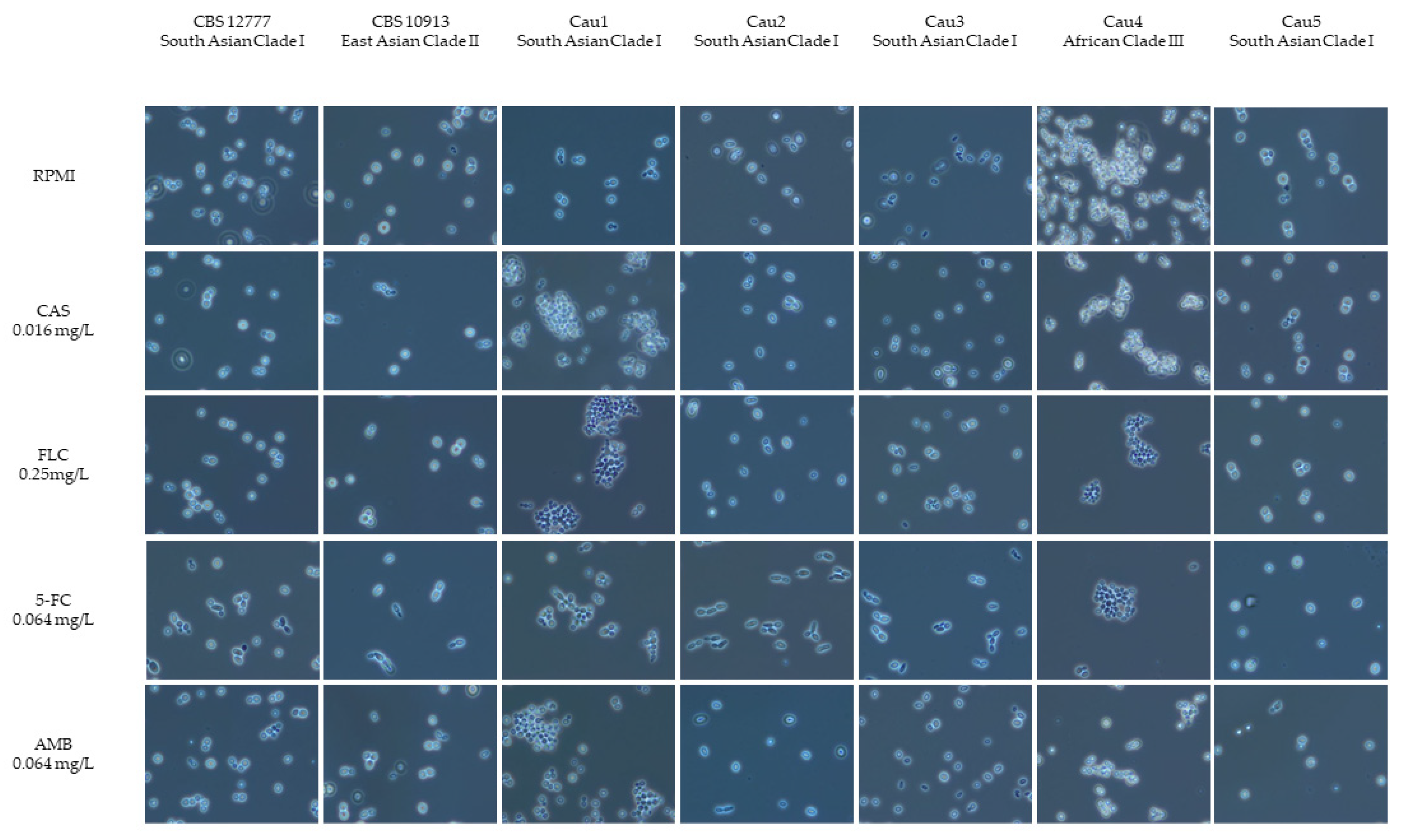
Figure 1. Cellular morphology of C. auris isolates. Microscopic appearance of C. auris cells grown in RPMI and by exposure to antifungal agents (CAS—caspofungin 0.016 mg/L), FLC—fluconazole (0.25 mg/L), 5-FC—5-flucytosine (0.064 mg/L), AMB—amphotericin B (0.064 mg/L). Suspensions were examined at 400× magnification by phase contrast microscopy.
Jof 09 00129 g001
Figure 2. Time–kill curves of the different C. auris strains in the G. mellonella infection model with a concentration of 5.0 × 10⁶ CFU/larva and PBS as a control group.
Jof 09 00129 g002
Table 1. Description of the clinical isolates detected in Austria.
IDIsolation DatePatient DescriptionUnderlying Medical ConditionSite of Isolation Travel History
Cau101/201822-year-old male patient
with Turkish ancestry
therapy-refractory otitis externaexternal auditory canalTurkey
Cau202/202061-year-old male patienthematologic malignancy, colonizationexternal auditory canalnone
Cau305/2020male patient with Indian ancestrytrauma, colonizationurinary tractIndia
Cau410/202160-year-old female patienthospitalization in Spain due to subarachnoid hemorrhage, colonizationthroatSpain
Cau504/202266-year-old female patienthospitalization in Greece due to subarachnoid hemorrhage, colonizationurinary tractGreece
Table 2. Number of SNPs detected in the Austrian C. auris isolates.
IsolateCau1Cau2Cau3Cau4Cau5B8441
(clade I)
South Asia
B11220
(clade II)
East Asia
B11221
(clade III)
Africa
B11243
(clade IV)
South America
Cau10442204224022595963,69144,333163,671
Cau24402044229420995963,73644,419163,671
Cau32202040423147697863,65844,359163,983
Cau442,24042,29442,314042,3004233259,80949157,871
Cau52252097642300093260,18442,259155,590
Table 3. ED50 values of different C. auris strains in the G. mellonella infection model, representing the time in hours after which 50% of the larvae population died.
Sample IDED50SECI (Lower)CI (Upper)Events
PBS controlNANANANA0
CBS1277728.150.1927.6228.6710
CBS1091357.311.2853.7660.8610
Cau126.560.1126.2526.8710
Cau214.250.1213.914.610
Cau328.150.1927.6228.6710
Cau444.460.1843.9644.9710
Cau522.350.0522.2222.4710
SE = standard error, CI = confidence interval, Events = total number of died larvae, NA = not applicable.
Table 4. In vitro susceptibility pattern of C. auris isolates tested by EUCAST microdilution method. MIC—minimum inhibitory concentration.
Antifungal AgentClinical IsolatesControl Strains
Cau1Cau2Cau3Cau4Cau5CBS 10913CBS 12777
MIC (mg/L)
EchinocandinsAnidulafungin0.50.50.0640.0320.250.0322
Micafungin0.1250.1250.1250.0640.250.0640.5
TriazolesFluconazole0.526464>2568>256
Posaconazole0.0320.0320.032≤0.0160.1250.0320.25
Voriconazole0.0080.0320.1250.510.0641
Pyrimidine analogues5-Flucytosine0.0640.1250.1250.250.1250.1250.125
PolyenesAmphotericin B2241812
New AntifungalsIbrexafungerp0.0640.1250.1250.1250.250.0320.25
Manogepix0.0080.0080.0080.0080.0320.0080.032
Disclaimer/Publisher’s Note: The statements, opinions and data contained in all publications are solely those of the individual author(s) and contributor(s) and not of MDPI and/or the editor(s). MDPI and/or the editor(s) disclaim responsibility for any injury to people or property resulting from any ideas, methods, instructions or products referred to in the content.