Article

Molecular Diagnosis and Vegetative Compatibility Group Analysis of Fusarium Wilt of Banana in Nepal

1
Nepal Agricultural Research Council, National Plant Pathology Research Centre, Lalitpur 44700, Bagmati Province, Nepal
2
Yunnan Key Laboratory of Green Prevention and Control of Agricultural Transboundary Pests, Agricultural Environment and Resources Institute, Yunnan Academy of Agricultural Sciences, Beijing Road 2238, Kunming 650205, China
3
Guangxi Key Laboratory of Biology for Crop Diseases and Insect Pests, Plant Protection Research Institute, Guangxi Academy of Agricultural Sciences, Daxue Road 174, Nanning 530007, China
4
Department of Entomology and Plant Pathology, North Carolina State University, Raleigh, NC 27695, USA
5
Nepal Agricultural Research Council, National Plant Breeding and Genetics Research Centre, Lalitpur 44700, Bagmati Province, Nepal
6
Bioversity International, Cali-Palmira CP, Palmira 763537, Colombia
7
Bioversity International, Parc Scientifique Agropolis II, CEDEX 5, 34397 Montpellier, France
8
Bioversity International, Beijing Road 2238, Kunming 650205, China
*
Authors to whom correspondence should be addressed.
These authors contributed equally to this work.
J. Fungi 2023, 9(2), 208; https://doi.org/10.3390/jof9020208
Submission received: 15 January 2023 / Revised: 1 February 2023 / Accepted: 2 February 2023 / Published: 5 February 2023
(This article belongs to the Special Issue Fusarium Species in Agriculture Industry)

Abstract

:
Fusarium wilt of banana (FWB), caused by Fusarium oxysporum f. sp. cubense (Foc), is the most important constraint of the banana industry globally. In Nepal, epidemics resembling FWB have been increasingly observed on the Malbhog cultivar in the past several years. However, the disease has not been officially reported yet, and consequently, little is known about the pathogen present across the country. In this study, we characterized 13 fungal strains isolated from banana plants of the Malbhog cultivar (Silk, AAB) showing symptoms similar to FWB in banana plantations in Nepal. All of the strains were typed as belonging to the F. oxysporum and caused FWB symptoms when inoculated in the Malbhog and Cachaco (Bluggoe, ABB) cultivars. No symptoms were observed in the Williams cultivar (Cavendish, AAA). Vegetative compatibility group (VCG) analysis classified the strains as VCG 0124 or VCG 0125. PCR analyses conducted with primers specific for Foc race 1 (Foc R1) or Foc tropical race 4 (TR4) revealed that all the strains reacted positively for Foc R1 and none for TR4. Altogether, our results demonstrated that the pathogen populations causing FWB of the Malbhog cultivar in Nepal were Foc R1. This work reported, for the first time, the occurrence of FWB in Nepal. Further studies with larger Foc populations are needed to better understand disease epidemiology to design sustainable disease management strategies.

1. Introduction

Banana is a major fruit crop and a high-value agricultural product in Nepal. It is cultivated on 18,329 ha areas across the country, with a total production of 278,890 tons and a productivity of 16.79 t/ha spanning 68 out of the 77 districts of Nepal [1,2]. Among them, Chitwan district is considered the major hub for banana production, followed by the Saptari, Jhapa, Morang, and Rupandehi districts. In Chitwan, banana farming is mainly concentrated in Bharatpur, Ratnanagar, Kalika, Khaireni, Jagatpur, and Thimura [3]. Nearly 15% of the area of banana-growing regions in Nepal is covered by a variety called Malbhog (Silk, AAB), which accounts for a 2493 ha area with 41,833 tons of production [4].
Diseases are major constraints to the banana production in Nepal. Fusarium wilt of banana (FWB), also known as Panama disease, is caused by the soil-borne fungus Fusarium oxysporum f. sp. cubense (Foc), which is a growing concern in Nepal. Foc was first described in Australia in 1874 [5]. Due to the exchange of planting material and the movement of spore-bearing soil, the pathogen has spread and has been reported in many countries of the world [6,7]. So far, three races of Foc (R1, R2, and R4) have been reported, based on the pathogenic characterization of different banana cultivars [8]. Foc R1, which caused FWB epidemic in the early 20th century, affects a range of cultivars, such as the Gros Michel (AAA), Silk (AAB), and Pisang Awak (ABB) subgroups, among others. Foc R2 affects cooking bananas belonging to the Bluggoe (ABB) subgroup. Foc R4, which is further divided into subtropical race 4 (SR4) and tropical race 4 (TR4), affects Cavendish cultivars, as well as bananas susceptible to Foc R1 and Foc R2.
Apart from races, Foc has been also grouped based on vegetative compatibility (VCGs). So far, 24 VCGs have been identified, but there is solid evidence that other VCGs also exists [9]. By far, TR4, which belongs to VCG 01213/16, is the most aggressive strain causing FWB. The strain, originating from Indonesia, has spread globally on different continents [10,11,12,13,14]. The strain is currently under strict quarantine regulations in most of the banana-producing countries worldwide. Therefore, the early detection of Foc TR4 is overarching, as it represents a serious threat for the export banana industry, banana diversity, and food security. Foc R1 strains belonging to VCG 0124 and VCG 0125 were also reported to infect the Cavendish banana variety, Grand Naine banana, under specific circumstances in India [15,16]. SR4 comprises strains belonging to VCG 0120, 0121, 0122, 0129, and VCG 01211 [17]. All the other VCGs, including VCG 0124 and VCG 0125, belong to Foc R1 or Foc R2 [17,18,19]. The VCG classification is imperfect; it is noted that VCG 0124 belongs to Foc R1, as well as Foc R2, while the VCG 0120 strain can be classified as both Foc R1 and SR4 [10,18,20]. This is considered to be the result of convergent evolution or the horizontal gene transfer of the F. oxysporum [10,21,22].
A syndrome similar to FWB appeared in banana plantations of Nepal in 2017 in the Chitwan district. It became widespread and destructive in almost all Malbhog banana-growing regions of Nepal, with a 30–90% disease incidence. In Chitwan, some farmers had to abandon the Malbhog variety, due to the high susceptibility to this disease. While field observations indicated that the disease corresponded to FWB, it was not clear which strain was associated with it. Both farmers and phytosanitary authorities were concerned about an eventual outbreak caused by Foc TR4 in Nepal because TR4 was already reported both in Bihar and Uttar Pradesh, India, which borders Nepal [23,24]. In the present study, we isolated the causal agent responsible for the disease outbreak in Malbhog bananas in Nepal, investigated the vegetative compatibility, and tested the pathogenicity to facilitate the development of effective disease management strategies in Nepal.

2. Materials and Methods

2.1. Sample Collection

In December 2017, an unusual phenomenon of banana plants (Malbhog, AAB) showing severe yellowing wilt symptoms before collapse was first reported in the Chitwan district of Nepal. Plant sampling from Malbhog bananas was conducted during two field surveys in December 2019 in the Chitwan and Nawalparasi districts. Visited locations were at an altitude of 170 m to 210 m. Diseased plants with typical external symptoms were found, and some xylem vessels appeared reddish-brown when the pseudostems were cut longitudinally. Altogether, thirteen samples from symptomatic tissue were collected from seven farmers’ fields of the Chitwan district and four farmers’ fields of the Nawalparasi district (Table 1).

2.2. Isolation and Identification

All collected samples were dried at room temperature before the isolation of the pathogen. Every sample was cut into small squares with a width of 0.5 cm using scissors. The scissors and tweezers were repeatedly heat-sterilized for every sample isolation. To prevent cross-contamination, gloves were changed when handling each sample. The cut pieces were immersed in 70% alcohol for 30 s for disinfection, followed by immersion in 0.1% mercury chloride for 30 s in the same way, and then rinsed three times with sterile water and placed on the plates with PDA medium with 50 µg/mL ampicillin. Then, the plates were kept in the dark at 30 °C for 5–7 days. The morphologies of the developed colonies were observed under a stereomicroscope, and the typical structures of Fusarium were observed using a compound microscope.

2.3. Pathogenicity Test

Tissue-cultured plantlets of Malbhog were planted in a steam-sterilized potting medium consisting of soil, sand, and farmyard manure in the ratio of 3:1:1 at the National Plant Pathology Research Centre (NPPRC), Khumaltar, Nepal. The pathogenicity test was performed when the plants reached 10–15 cm in height at the 3–4 leaf stage to ensure that the vascular system was fully developed. Six plants were used per treatment. These plants were inoculated with NP11 or NP12 single-spore isolates from the Chitwan and Nawalparasi districts, respectively. The conidial suspension adjusted was counted using a hemocytometer and was 6.3 × 10⁶ conidia/mL. The roots of plants were slightly wounded by a sterile knife, and 100 mL of spore suspension of the F. oxysporum isolate was then added to the plantlets by drenching. Plants drenched with water served as the control. All the plants were placed in the greenhouse. Inoculated plants were monitored regularly for the appearance of disease symptoms. When external symptoms appeared, the internal symptoms were also investigated by dissecting the pseudostem and corm. The pathogen was re-isolated from the inoculated plants and further characterized by PCR.
Pathogenicity tests were also carried out at the Institute of Plant Protection Research, Guangxi Key Laboratory of Biology for Crop Diseases and Insect Pests, Guangxi Academy of Agricultural Sciences, China. Banana plantlets from the Cachaco (Bluggoe, ABB) and Williams (Cavendish, AAA) cultivars were grown and inoculated as afore mentioned. Nine plants were used per treatment, and each treatment was repeated three times. The wounded roots of the inoculated plantlets were soaked in a 106 conidia/mL spore suspension for 30 min and then planted in the pots. Those plantlets that soaked in sterile water for 30 min served as the healthy control. Twenty-four days after inoculation, the disease index was investigated by dissecting the corm. The classification criteria were as follows: scale 0, no symptoms in the corm; scale 1, discoloration area in the central longitudinal section of the corm was less than 25%; scale 3, discoloration area in the central longitudinal section of the corm was 26–50%; scale 5, discoloration area in the central longitudinal section of the corm was 51–75% area; scale 7, discoloration area in the central longitudinal section of the corm was more than 75%. The disease index was calculated according to the following equation [25]:
Disease index equation = [Σ (number of infected plants at a given disease scale × the disease scale)/(total tested plants × 7)] × 100
Statistical analysis was performed using R Studio version 4.0.3 (Agricolae package) for the disease index data. The data was transformed into log10 (1 + data) for normalization, and 1 in each data set was added to remove potential infinity values from the log-transformed data. The analysis was measured using two-way ANOVA with the LSD test.

2.4. Vegetative Compatibility Group Analyses

Nitrate-nonutilizing (nit) mutants of the wild-type Foc strains were generated in minimal medium (MM) [26] amended with KClO3 and incubated for 7–14 days at 25 °C. Spontaneous KClO3-resistant sectors were transferred to MM. Those that grew as thin colonies with no aerial mycelium were classified as nit mutants and were further characterized on media containing one of four different sources of nitrogen [27]. Finally, VCGs of all mutants were determined by pairing in MM with tester nit mutants from strains with known VCGs (VCG 0120, VCG 0121, VCG 0122, VCG 0123, VCG 0124, VCG 0125, VCG 0126, VCG 0127, VCG 0128, VCG 0129, VCG 01210, VCG 01211, VCG 01212, VCG 01213, VCG 01214, VCG 01215, VCG 01216, VCG 01217, VCG 01218, VCG 01219, VCG 01220, VCG 01221, VCG 01222, and VCG 01223) obtained from Professor Elizabeth Aitken, the University of Queensland, Australia. Complementation between different nit mutants resulted in dense aerial growth at the contact zone between the two colonies [27]. Eight isolates (NP1-1, NP2-1, NP4-1, NP5-1, NP6-1, NP7-1, NP8-1, and NP10-1) were used for vegetative compatibility group analyses.

2.5. PCR Analyses

Five- to seven-day-old cultures were used for polymerase chain reaction (PCR) amplification. Approximately 100 mg of mycelium was taken with a sterile toothpick and placed into a sterilized PCR tube with 50 µL of lysis buffer for microorganisms (Takara Bio, https://www.takarabio.com, accessed on 13 January 2020). After 15 min of thermal denaturation at 80 °C in the PCR machine, it was centrifuged briefly, and 1.5 µL of the supernatant was used as a template for the PCR reaction. The translation elongation factor 1α gene (TEF-1α) was amplified with primers EF-1 and EF-2 as internal positive controls using the following program: 95 °C for 2 min and 35 cycles of 95 °C for 30 s; 50 °C for 30 s; and 72 °C for 1 min, followed by an additional extension time for 10 min at 72 °C [21]. The primer sets W1805F/W1805R and W2987F/W2987R from Li et al. (2012) [20] were used to identify Foc R1 (354 bp) and Foc R4 (593 bp), respectively. Another pair of W2987F/W2987R from Li et al. (2013) [28] was used to identify TR4 (452 bp). [Note: the name of primer set W2987F/W2987R from Li et al. (2013) [28] is the same as that from Li et al. (2012) [20], so in this research, we labeled it as TR4-W2987F/TR4-W2987R.] The sequence information of all the primer sets is shown in Table 2. The PCR was performed using the Gold Mix (Code: TSE 101, tsingke, Beijing, China) according to the manufacturer’s protocol.
PCR products were electrophoresed in 1% agarose gel in an electrophoretic tank (Takara Bio) containing 1X TAE buffer for 20 min at 135 V. BIO-RAD ChemiDocTMXRS+ was used to take an image after electrophoresis.

2.6. Phylogenetic Analysis

The TEF-1α sequences of isolates were amplified by PCR. These PCR products were confirmed with aimed bands by agarose gel electrophoresis and purified using the DNA Gel Extraction kit TSP601 (Tsingke, Beijing, China). PCR products were then cloned and sequenced in the pMD18-T vector. The TEF-1α sequences were showed in Supplementary Table S1. Additionally, TEF-1α sequences of other F. oxysporum isolates were obtained from the publicly available genomes (https://www.ncbi.nlm.nih.gov/, accessed on 30 July 2021). The analysis involved 31 isolate sequences that were aligned with MUSCLE [29], and the phylogenetic analyses were performed using maximum likelihood (ML). The phylogenetic tree was constructed using the MEGA 6.0 software with the Kimura 2-parameter model [30]. The TEF-1α sequence from Ramlispora sorghi was used as the outgroup taxon (KM087651.1). Branches were tested for the inferred tree by bootstrap analysis on 1000 random trees.

3. Results

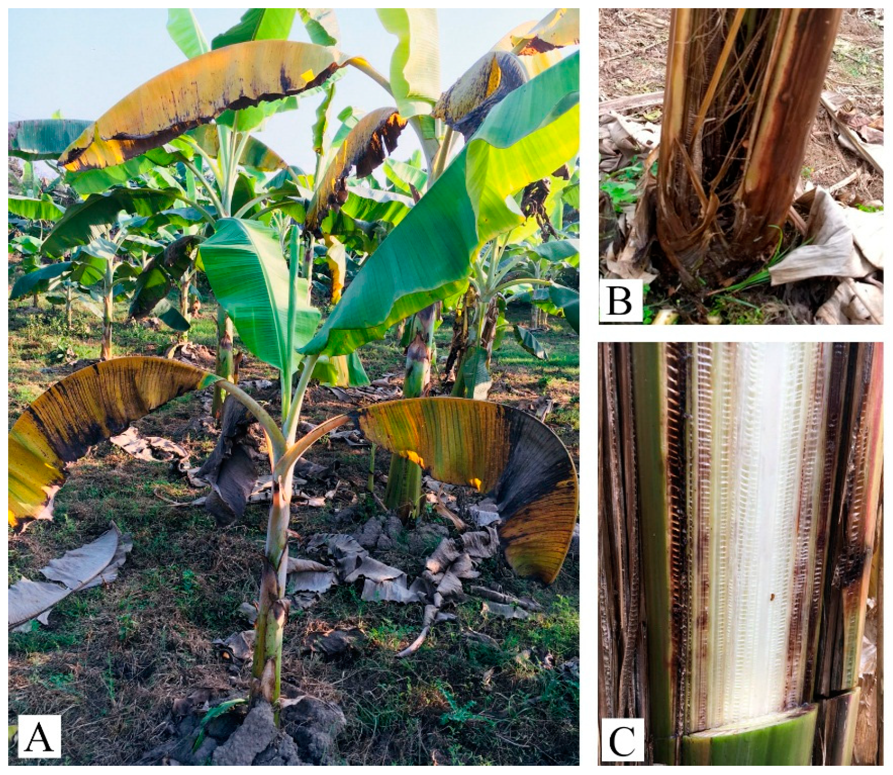
3.1. Disease Diagnosis with Field Symptoms

The problem of wilt disease has existed in banana orchards in Nepal since 2017. The disease was observed in some new and old orchards growing the Malbhog variety. Symptoms appeared as yellowing of the oldest leaves and splitting at the base of different types of infected plants. Some diseased, old yellow leaves split at the base. Vascular discoloration varied from pale yellow in the early stage to dark red or almost black in the later stage when viewed by cutting a cross-section or splitting the pseudostem base. Brownish streaks of the vascular tissue were also observed during the field survey (Figure 1). However, wilt disease was not observed in the Grand Naine banana variety.

3.2. Morphology of the Pathogen Isolate

Thirteen suspected pathogen isolates were extracted from thirteen corresponding samples collected from banana fields in the Chitwan and Nawalparasi districts (Table 1), and their morphological characteristics were studied. The isolates produced dense white and purple colonies, and mycelia were evenly spread all over the PDA medium (Figure 2A,B).
The microscopic examination of fungal isolates showed the presence of three types of asexual spores: macroconidia, microconidia, and chlamydospores. The macroconidia were thin-walled with a definite foot cell and a pointed apical cell (Figure 2C). Microconidia were oval- or kidney-shaped and were produced on micro-conidiophores in aerial mycelia (Figure 2D). Chlamydospores were thick-walled (Figure 2E), and these were usually produced singly in macroconidia, were intercalary, or were in terminal hyphae [31].

3.3. Pathogenicity Test

The pathogenicity test was performed on three banana cultivars: Malbhog (AAB), Cachaco (ABB), and Williams (AAA). Yellowing and wilting symptoms were observed in inoculated Malbhog plantlets after 21 days of pathogen inoculation (Figure 3A: left two plants). The non-inoculated plants (controls) showed no symptoms, and the leaves remained green (Figure 3A: right two plants). Dark spots and brownish discoloration of the vascular tissue in the pseudostem were also observed (Figure 3B). In severe cases, inoculated plantlets showed complete wilting or even death.
Similar symptoms were observed in inoculated Cachaco plantlets (Figure 4A,C). Wilting symptoms in plantlets inoculated with the isolates NP1-1, NP2-1, NP4-1, NP5-1, NP6-1, NP7-1, NP8-1, and NP10-1 were noticed after 24 days of pathogen inoculation. There was a highly significant response of the disease index on the varieties and interactions of isolates with varieties, while the response was non-significant in the case of isolates without varieties (Table 3). The pathogenicity of the F. oxysporum isolates to Cachaco and Williams was extremely significantly different (p < 2 × 10⁻¹⁶). All of the inoculated Cachaco plantlets showed different degrees of symptoms, and the disease index was greater than 26.19, while the inoculated Williams plantlets nearly had no disease symptoms. The disease index of Cachaco inoculated with the isolate NP4-1 was 66.67, which was significantly higher than that caused by NP2-1, NP5-1, NP6-1, and NP8-1 (p < 0.0465). The pathogen was re-isolated from the inoculated, symptomatic plants and confirmed with the original one using microscopic observation and PCR.

3.4. PCR Analysis

PCR analyses were performed using four primer sets (Figure 5). The W1805F/W1805R primer set previously associated with Foc R1 populations [20] produced the predicted 354 bp amplicon in all of the 13 tested isolates and from the Foc R1 strain used as the reference control. None of the tested isolates reacted to the Foc R4 or TR4 primer sets. The TR4 strain used as the positive control produced the predicted 593 and 452 bp amplicons, respectively. Altogether, our PCR results suggested that all Foc strains isolated from Malbhog bananas in Nepal were associated with Foc R1 populations.

3.5. Vegetative Compatibility Analyses

Nit mutants were successfully generated for eight Foc isolates (NP1-1, NP2-1, NP4-1, NP5-1, NP6-1, NP7-1, NP8-1, and NP10-1). Five (NP2-1, NP4-1, NP7-1, NP8-1, and NP10-1) out of the eight isolates were compatible with VCG 0124, since the development of aerial mycelia and the formation of heterokaryons at the line of intersection between colonies of the isolates and tester mutants were observed. The other isolate (NP5-1) was typed as VCG 0125, suggesting that VCG 0124 was the predominant isolate. NP1-1 and NP6-1 were of unknown VCGs (Figure 6 and Figure 7).

3.6. Phylogenetic Analysis

Nepal isolates and some other F. oxysporum isolates from the publicly available genomes were phylogenetically analyzed based on TEF-1α. The phylogenetic tree divided thirteen Nepal isolates into two clusters. The first cluster, including most of the isolates, viz., NP1-1, NP2-1, NP5-1, NP6-1, NP7-1, NP8-1, NP11, NP12, NP13, NP14, and NP15 were grouped with Foc NRRL 25607 and CAV 2612, which were also typed as VCG 0124 in previous studies [18,32]. The second cluster grouped NP4-1 and NP10-1 together with a set of F. oxysporum formae speciales, including cubense (NRRL 25367 NRRL 25609) and non-cubense, such as glycines (NRRL 25598) and melonis (NRRL 26178) (Figure 7).

4. Discussion

FWB causes severe yield losses globally, especially the TR4 strain, which is considered the biggest threat to global banana production [33]. Since 2017, typical symptoms of FWB have been increasingly observed in banana plantations in Nepal. However, identification and characterization of the causal agent is still lacking. In this research, we isolated and characterized the pathogen responsible for causing wilt disease on Malbhog banana plantations in the Chitwan and Nawalparasi districts, Nepal. Isolates, which exhibited morphological characteristics similar to the Fusarium oxysporum [34], were able to cause typical FWB symptoms in the Malbhog and Cachaco cultivars but not in the Cavendish cultivar, Williams. Field observations also revealed that the Cavendish cultivar, Grand Naine, which is resistant to Foc R1, had successfully replaced the susceptible Malbhog in the affected areas. Our PCR and VCG analyses grouped all the strains isolated as Foc R1 and revealed that six of these strains belonged to VCGs 0124 and 0125.
The phylogenetic tree based on TEF-1α divided the thirteen Nepal isolates into two clusters. The first cluster included most isolates grouped with Foc NRRL 25607 and CAV 2612, both of which belonged to VCG 0124 [18,32]. NP5-1, which belonged to VCG 0125, was also grouped with them. NP4-1 and NP10-1, both of which belonged to VCG 0124, were grouped into a separate branch. Results of genetic diversity derived from VCG and the phylogenetic tree were not fully consistent. It was probably due to the complex genetic diversity of Foc for convergent evolution or the horizontal gene transfer of F. oxysporum [10,21,22]. Moreover, TEF-1α was shown in some cases to be insufficient for distinguishing different formae speciales of Fusarium [35]. Nonetheless, results of the VCG revealed the diversity of Foc R1 in Nepal, although a larger number of isolates collected from other banana-producing regions may be suitable to further understand the Foc population biology in Nepal.
Banana is a high-value agricultural product and a major fruit in Nepal, in terms of production and domestic consumption (https://www.icimod.org/expanding-commercial-banana-production-in-nepal/, accessed on 8 June 2021). The current epidemic and spread of Foc R1 have severely affected the production of the traditional banana cultivar, Malbhog, which is one of the preferred local cultivars in Nepal. Our surveys revealed that FWB was prevalent in almost 90% of the Malbhog-growing area of the Chitwan and Nawalparasi districts. Surveys and farmer interviews during field visits indicated that such a rapid spread of FWB could be associated with the informal exchange of planting material from infested to healthy areas. In the absence of effective management practices, many growers chose to plant the Foc R1-resistant cultivar, Grand Naine. However, farmers and extension officers need to consider that Grand Naine is highly susceptible to Foc TR4, a more aggressive Foc strain already present in Bihar and Uttar Pradesh, India [23,24]. Therefore, prevention and exclusion measures against Foc TR4 using anticipatory approaches, as well as continuous Foc population monitoring, are strongly encouraged.

5. Conclusions

In conclusion, 13 fungal strains were isolated from banana plants of the Malbhog cultivar (Silk, AAB) showing symptoms similar to FWB in banana orchards in Nepal. Molecular diagnosis and vegetative compatibility group analysis were conducted for the evaluation of the FWB. All the strains caused FWB symptoms when inoculated in the Malbhog and Cachaco (Bluggoe, ABB) cultivars. No symptoms were observed in the Williams cultivar (Cavendish, AAA). PCR analyses with primers specific for Foc race 1 (R1) or Foc tropical race 4 (TR4) revealed that all the strains reacted positively for Foc R1 and none for TR4 in this study. Vegetative compatibility group (VCG) analysis further confirmed most strains as VCG 0124 or VCG 0125. The results demonstrated that the pathogen in this study causing FWB in Nepal are associated with Foc R1 in these limited samples. Larger Foc populations are necessary to better understand disease epidemiology for making sustainable disease management strategies in the next step.

Supplementary Materials

The following is available online at https://www.mdpi.com/article/10.3390/jof9020208/s1. Supplemental Table S1. TEF-1α sequences.

Author Contributions

Conceptualization, B.P., T.B., G.F. and S.-J.Z.; methodology, T.B., C.D., S.B., P.B.M. and S.M.; software, T.B. and J.S.; validation, S.B., P.B.M. and M.D.; investigation, B.P., S.B., P.B.M. and S.M.; data curation, T.B. and G.F.; writing—original draft preparation, B.P., T.B. and G.F.; writing—review and editing, M.D., M.R. and S.-J.Z.; supervision, M.D., M.R., G.F. and S.-J.Z.; project administration, S.-J.Z. and T.B.; funding acquisition, S.-J.Z. All authors have read and agreed to the published version of the manuscript.

Funding

This research was funded by National Natural Science Foundation of China (NSFC32161143001); Yunling Scholar Programme of Yunnan Provincial Government (YNWR-YLXZ-2018-018); Science and Technology Department of Yunnan Provincial Government (202102AE090003); the National Banana Industry and Technology System Project funded by the Ministry of Finance & Ministry of Agriculture and Rural Affairs of China (CARS-31-08); the Nepal Agricultural Research Council (NARC), Government of Nepal and the CGIAR Research Program on Roots, Tubers, and Bananas (RTB).

Institutional Review Board Statement

Not applicable.

Informed Consent Statement

Not applicable.

Data Availability Statement

Acknowledgments

We are thankful to banana growers in the Chitwan and Nawalparasi districts for their active participation during field survey and interaction. We thank Elizabeth Aitken, the University of Queensland, Australia for providing nit M mutants of the tester. We are also grateful to Thirtha Devi Ghimire, Bindu Laxmi Shrestha, and Pushpa Shrestha for their contributions during laboratory works and to Bikram Datt Pant from the Noble Research Institute, USA for reading the manuscript.

Conflicts of Interest

The authors declare no conflict of interest.

References

  1. MoALD. Statistical Information on Nepalese Agriculture 2077/78 (2020/21). Planning and Development Cooperation Coordination Division; Ministry of Agriculture and Livestock Development, Government of Nepal, Singha Durbar Kathmandu: Kathmandu, Nepal, 2020; p. 435.
  2. MoALD. Statistical Information on Nepalese Agriculture 2076/77 (2019/20). Planning and Development Cooperation Coordination Division; Ministry of Agriculture and Livestock Development, Government of Nepal, Singha Durbar Kathmandu: Kathmandu, Nepal, 2019; pp. 135–138.
  3. Puri, S. Banana Farmers Strike Gold as Demand Spikes. The Kathmandu Post. Ecology and Taxonomy of Fusarium Species. Ph.D. Thesis, Sydney University, Sydney, Australia, 2017. Available online: https://kathmandupost.com/money/2017/06/15/banana-farmers-strike-gold-as-demandspikes (accessed on 2 April 2020).
  4. Ghimire, T.B.; Manandhar, R.; Siwakoti, S.; Gautam, I.P.; Adhikari, R.N.; Bhattarai, M.D.; Khatiwada, S. Karya Bibaran Samadhanka Bikalpa Sahit Adhyan Pratobedan (Nepali Version) Submitted to Program Planning and Monitoring Sub-Committee of MoALD; Ministry of Agriculture and Livestock Development, Government of Nepal, Singha Durbar Kathmandu: Kathmandu, Nepal, 2018.
  5. Bancroft, J. Report of the board appointed to enquire into the cause of disease affecting livestock and plants. Votes Proc. 1876, 3, 1011–1038. [Google Scholar]
  6. Ploetz, R.C. Management of Fusarium wilt of banana: A review with special reference to tropical race 4. Crop Prot. 2015, 73, 7–15. [Google Scholar] [CrossRef]
  7. Pegg, K.G.; Coates, L.M.; Neill, W.T.O.; Turner, D.W. The epidemiology of Fusarium wilt of banana. Front. Plant Sci. 2019, 10, 1–19. [Google Scholar] [CrossRef] [PubMed]
  8. Pérez Vicente, L.; Dita, M.A. Technical Manual Fusarium wilt of Banana or Panama disease by Fusarium oxysporum f. sp. cubense: A review on history, symptoms, biology, epidemiology and management. In Proceedings of the Technical Manual: Prevention and Diagnostic of Fusarium Wilt (Panama Disease) of Banana Caused by Fusarium oxysporum f. sp. cubense Tropical Race 4 (TR4), Regional Workshop on the Diagnosis of Fusarium Wilt (Panama Disease) Caused by Fusarium oxysporum f. sp. cubense Tropical Race 4: Mitigating the Threat and Preventing Its Spread in the Caribbean, St. Augustine, Trinidad and Tobago, 5–9 May 2014; Pérez Vicente, L., Dita, M.A., Martinez-de la Parte, E., Eds.; FAO: Rome, Italy, 2014; pp. 5–30. [Google Scholar]
  9. Mostert, D.; Wicker, E.; de Jager, M.M.; Al Kaabi, S.M.; O’Neill, W.T.; Perry, S.; Li, C.; Yi, G.; Pegg, K.G.; Mostert, L.; et al. A polyphasic approach reveals novel genotypes and updates the genetic structure of the banana Fusarium wilt pathogen. Microorganisms 2022, 10, 269. [Google Scholar] [CrossRef]
  10. Ordoñez, N.; Seidl, M.F.; Waalwijk, C.; Drenth, A.; Kilian, A.; Thomma, B.P.H.J.; Ploetz, R.C.; Kema, G.H.J. Worse comes to worst: Bananas and Panama disease—When plant and pathogen clones meet. PLOS Pathog. 2015, 11, e1005197. [Google Scholar] [CrossRef] [PubMed]
  11. Acuña, R.; Rouard, M.; Leiva, A.M.; Marques, C.; Olortegui, J.A.; Ureta, C.; Cabrera-Pintado, R.M.; Rojas, J.C.; Lopez-Alvarez, D.; Cenci, A.; et al. First report of Fusarium oxysporum f. sp. cubense tropical race 4 causing Fusarium wilt in cavendish bananas in Peru. Plant Dis. 2022, 8, 106. [Google Scholar] [CrossRef]
  12. Aguayo, J.; Cerf-Wendling, I.; Folscher, A.B.; Fourrier-Jeandel, C.; Ioos, R.; Mathews, M.C.; Mostert, D.; Renault, C.; Wilson, V.; Viljoen, A. First report of Fusarium oxysporum f. sp. cubense tropical race 4 (TR4) causing banana wilt in the Island of Mayotte. Plant Dis. 2021, 105, 219. [Google Scholar] [CrossRef]
  13. García-Bastidas, F.A.; Quintero-Vargas, J.C.; Ayala-Vasquez, M.; Schermer, T.; Seidl, M.F.; Santos-Paiva, M.; Noguera, A.M.; Aguilera-Galvez, C.; Wittenberg, A.; Hofstede, R.; et al. First report of Fusarium wilt tropical race 4 in Cavendish bananas caused by Fusarium odoratissimum in Colombia. Plant Dis. 2020, 104, 994. [Google Scholar] [CrossRef]
  14. Zheng, S.-J.; García-Bastidas, F.A.; Li, X.; Bai, T.; Xu, S.; Yin, K.; Li, H.; Fu, G.; Yu, Y.; Yang, L.; et al. New geographical insights of the latest expansion of Fusarium oxysporum f. sp. cubense tropical race 4 into the great Mekong subregion. Front Plant Sci. 2018, 9, 457. [Google Scholar] [CrossRef]
  15. Thangavelu, R.; Mustaffa, M.M. First report on the occurrence of a virulent strain of Fusarium wilt pathogen (Race-1) infecting Cavendish (AAA) group of bananas in India. Plant Dis. 2010, 94, 1379. [Google Scholar] [CrossRef]
  16. Thangavelu, R.; Gopi, M.; Pushpakanth, P.; Loganathan, M.; Edxin Raj, E.; Marimuthu, N.; Prabakaran, M.; Uma, S. First report of Fusarium oxysporum f. sp. cubense VCG 0125 and VCG 01220 of race 1 infecting Cavendish bananas (Musa sp. AAA) in India. Plant Dis. 2021, 105, 1215. [Google Scholar] [CrossRef]
  17. Ghag, S.B.; Shekhawat, U.K.S.; Ganapathi, T.R. Fusarium wilt of banana: Biology, epidemiology and management. Int. J. Pest Manag. 2015, 61, 250–263. [Google Scholar] [CrossRef]
  18. Dita, M.A.; Waalwijk, C.; Buddenhagen, I.W.; Souza, M.T., Jr.; Kema, G.H.J. A molecular diagnostic for tropical race 4 of the banana fusarium wilt pathogen. Plant Pathol. 2010, 59, 348–357. [Google Scholar] [CrossRef]
  19. Fraser-Smith, S.; Czislowski, E.; Meldrum, R.A.; Zander, M.; O’Neil, W.; Balali, G.R.; Aitken, E.A.B. Sequence variation in the putative effector gene SIX8 facilitates molecular differentiation of Fusarium oxysporum f. sp. cubense. Plant Pathol. 2014, 63, 1044–1052. [Google Scholar] [CrossRef]
  20. Li, M.H.; Yu, X.Q.; Wang, H.F.; Zhou, J.; Xi, P.G.; Jiang, Z.D. Rapid detection and identification of Fusarium oxysporum f. sp. cubense Race 1 and Race 4. Sci. Agric. Sin. 2012, 45, 3971–3979. [Google Scholar]
  21. O’Donnell, K.; Kistler, H.C.; Cigelnik, E.; Ploetz, R.C. Multiple evolutionary origins of the fungus causing Panama disease of banana: Concordant evidence from nuclear and mitochondrial gene genealogies. Proc. Natl. Acad. Sci. USA 1998, 95, 2044–2049. [Google Scholar] [CrossRef] [PubMed]
  22. Fourie, G.; Steenkamp, E.T.; Gordon, T.R.; Viljoen, A. Evolutionary relationships among the Fusarium oxysporum f. sp. cubense vegetative compatibility groups. Appl. Env. Microbiol. 2009, 75, 4770–4781. [Google Scholar] [CrossRef]
  23. Damodaran, T.; Rajan, S.; Mishra, V.K.; Jha, S.K.; Gopal, R. First report of Fusarium wilt in banana caused by Fusarium oxysporum f. sp. cubense tropical race 4 in India. Plant Dis. 2019, 103, 1022. [Google Scholar] [CrossRef]
  24. Thangavelu, R.; Mostert, D.; Gopi, M.; Ganga Devi, P.; Padmanaban, B.; Molina, A.B.; Viljoen, A. First detection of Fusarium oxysporum f. sp. cubense tropical race 4 (TR4) on Cavendish banana in India. Eur. J. Plant Pathol. 2019, 154, 777–786. [Google Scholar] [CrossRef]
  25. McKinney, H.H. Influence of soil temperature and moisture on infection of wheat seedlings by Helminthosporium sativum. J. Agric. Res. 1923, 26, 195–217. [Google Scholar]
  26. Puhalla, J.E. Classification of strains of Fusarium oxysporum on the basis of vegetative compatibility. Can J. Bot. 1985, 63, 179–183. [Google Scholar] [CrossRef]
  27. Correll, J.C.; Klittich, C.J.R.; Leslie, J.F. Nitrate nonutilizing mutants of Fusarium oxysporum and their use in vegetative compatibility tests. Phytopathology 1987, 77, 1640–1646. [Google Scholar] [CrossRef]
  28. Li, M.; Shi, J.; Xie, X.; Leng, Y.; Wang, H.; Xi, P.; Zhou, P.; Zhong, S.; Jiang, Z. Identification and application of a unique genetic locus in diagnosis of Fusarium oxysporum f. sp. cubense tropical race 4. Can J. Plant Pathol. 2013, 35, 482–493. [Google Scholar] [CrossRef]
  29. Edgar, R.C. MUSCLE: A multiple sequence alignment method with reduced time and space complexity. BMC Bioinform. 2004, 5, 113. [Google Scholar] [CrossRef] [PubMed]
  30. Tamura, K.; Stecher, G.; Peterson, D.; Filipski, A.; Kumar, S. MEGA6: Molecular evolutionary genetics analysis version 6.0. Mol. Biol. Evol. 2013, 30, 2725–2729. [Google Scholar] [CrossRef] [PubMed]
  31. Nelson, P.E.; Toussoun, T.A.; Marasas, W.F.O. Fusarium Species: An Illustrated Manual for Identification; Pennsylvania State University Press: University Park, PA, USA, 1983. [Google Scholar]
  32. Karangwa, P.; Mostert, D.; Ndayihanzamaso, P.; Dubois, T.; Viljoen, A. Genetic diversity of Fusarium oxysporum f. sp. cubense in East and Central Africa. Plant Dis. 2018, 10, 552–560. [Google Scholar] [CrossRef] [PubMed]
  33. Kema, G.H.J.; Drenth, A.; Dita, M.; Jansen, K.; Vellema, S.; Stoorvogel, J.J. Editorial: Fusarium wilt of banana, a recurring threat to global banana production. Front. Plant Sci. 2021. [Google Scholar] [CrossRef]
  34. Beckman, C.H. The Nature of Wilt Diseases of Plants; American Phytopathological Society: St. Paul, MA, USA, 1987; p. 175. [Google Scholar]
  35. Duan, Y.; Qu, W.; Chang, S.; Li, C.; Xu, F.; Ju, M.; Zhao, R.; Wang, H.; Zhang, H.; Miao, H. Identification of pathogenic groups and pathogenic molecular characterization of Fusarium oxysporum f. sp. sesami in China. Phytopathology 2020, 110, 1093–1104. [Google Scholar] [CrossRef] [PubMed]
Figure 1. Different diseased plants (Malbhog variety) with various symptoms on different parts in the Chitwan district. (A) Whole plant, (B) splitting pseudostem, and (C) reddish to dark-brownish discoloration of the vascular system.
Jof 09 00208 g001
Figure 2. Morphological characteristics of the fungus isolated from banana orchards of the Chitwan and Nawalparasi districts. (A) Purple shades of Fusarium oxysporum f. sp. cubense (Foc) on PDA medium; (B) white shades of Foc on PDA medium; (C) macroconidia of Foc; (D) microconidia of Foc; and (E) chlamydospores of Foc. All scale bars 10 μm.
Jof 09 00208 g002
Figure 3. Pathogenicity test on the Malbhog (AAB) variety at the National Plant Pathology Research Center, NARC, Khumaltar, Nepal. (A) Inoculated (left two plants) and non-inoculated as control (right two plants); (B) rhizome discoloration caused by Foc isolates after inoculation.
Jof 09 00208 g003
Figure 4. Pathogenicity test results on the Cachaco (ABB) and Williams (AAA) cultivars at the Institute of Plant Protection Research, Guangxi Key Laboratory of Biology for Crop Diseases and Insect Pests, Guangxi Academy of Agricultural Sciences, China. (AC) Tests on Cachaco (ABB) cultivars; (A) inoculated; (B) non-inoculated; and (C) rhizome discoloration caused by Foc isolates after inoculation. (DF) Tests on Williams (AAA) cultivars; (D) inoculated; (E) non-inoculated; and (F) no symptoms in the rhizome after inoculation.
Jof 09 00208 g004
Figure 5. PCR identification of F. oxysporum f. sp. cubense (Foc) using primer sets (Table 2). (A) Primer set W1805F/W1805R; (B) Primer set W2987F/W2987R; (C) Primer set TR4-W2987F/TR4-W2987R; (D) Primer set EF-1/EF-2 was used to amplify the fragment of the translation elongation factor gene (TEF-1α) as the internal positive control. Lane M: molecular weight maker; NP1-1, NP2-1, NP4-1, NP5-1, NP6-1, NP7-1, NP8-1, NP10-1, NP11, NP12, NP13, NP14, and NP15 were the isolates from Nepal. The Foc R1 and TR4 isolates were from the Yunnan province of China; N-CK: water as negative control.
Jof 09 00208 g005
Figure 6. Compatibility assays between nit mutants on minimal medium (MM). (A,B) Heterokaryon formation when pairing the tester and some isolates; (C) no heterokaryon formation when pairing testers and isolate NP6-1.
Jof 09 00208 g006
Figure 7. Maximum-likelihood (ML) tree inferred from the translation elongation factor 1α gene (TEF-1α) sequences in selected Fusarium isolates. Isolates obtained in the present study are indicated in bold. Bootstrap values (≥50%) are indicated at the nodes. The phylogram is rooted to F. concentricum M8164R and F. mangiferae SJG3-4. NRRL 25607 belongs to VCG 0124, associated with Foc R2 [18]. CAV 2612 also belongs to VCG 0124, but it is associated with Foc R1 [32]. VCG / means unidentified VCG.
Jof 09 00208 g007
Table 1. Isolates of Fusarium oxysporum used in this study from the Chitwan and Nawalparasi districts of Nepal.
Isolate Sampling DateSiteVarietyLocationAltitude (m)
NP1-118 December 2019ChitwanMalbhog27°40′55″ N, 84°30′21″ E210
NP2-118 December 2019ChitwanMalbhog27°40′55″ N, 84°30′24″ E210
NP4-118 December 2019ChitwanMalbhog27°40′55″ N, 84°30′24″ E210
NP5-118 December 2019ChitwanMalbhog27°40′55″ N, 84°30′24″ E210
NP6-118 December 2019ChitwanMalbhog27°40′55″ N, 84°30′24″ E210
NP7-118 December 2019ChitwanMalbhog27°37′50″ N, 84°31′1″ E200
NP8-118 December 2019NawalparasiMalbhog27°37′3″ N, 84°6′1″ E170
NP10-118 December 2019NawalparasiMalbhog27°37′3″ N, 84°6′1″ E170
NP1110 December 2019ChitwanMalbhog27°40′52″ N, 84°30′26″ E220
NP1210 December 2019NawalparasiMalbhog27°37′3″ N, 84°6′1″ E170
NP1310 December 2019ChitwanMalbhog27°39′12″ N, 84°30′41″ E210
NP1410 December 2019ChitwanMalbhog27°39′10″ N, 84°30′41″ E210
NP1510 December 2019ChitwanMalbhog27°37′52″ N, 84°31′4″ E200
Table 2. The primer sets used to identify the Foc pathogen.
Primer SetsFungi to IdentifyPrimer SequencesReferences
W1805F/W1805RFoc R15′-GTTGAGTCTCGATAAACAGCAAT-3′[20]
5′-GACGAGGGGAGATATGGTC-3′
W2987F/W2987RFoc R45′-GCCGATGTCTTCGTCAGGTA-3′[20]
5′-CTGAGACTCGTGCTGCATGA-3′
W2987F/W2987R
(TR4-W2987F/TR4-W2987R)
TR45′-TGCCGAGAACCACTGACAA-3′[28]
5′-GCCGATGTCTTCGTCAGGTA-3′
EF-1/EF-2Fusarium spp.5′-ATGGGTAAGGA(A/G)GACAAGAC-3′[21]
5′-GGA(G/A)GTACCAGT(G/C)ATCATGTT-3′
Table 3. The disease index of isolates on the host.
IsolateVarietyDisease Index (%)
Cachaco (ABB)38.9 (1.59a)
Williams (AAA)0.53 (0.11b)
p-value<2 × 10⁻¹⁶
NP1-1 23.81 (0.84)
NP2-1 17.06 (0.85)
NP4-1 33.33 (0.91)
NP5-1 14.29 (0.73)
NP6-1 13.10 (0.70)
NP7-1 26.19 (0.98)
NP8-1 17.06 (0.97)
NP10-1 20.24 (0.81)
p-value0.11
NP1-1Cachaco (ABB)47.62 (1.68abc)
NP2-1Cachaco (ABB)33.33 (1.52bc)
NP4-1Cachaco (ABB)66.66 (1.83a)
NP5-1Cachaco (ABB)28.57 (1.47bc)
NP6-1Cachaco (ABB)26.19 (1.41c)
NP7-1Cachaco (ABB)50.79 (1.71ab)
NP8-1Cachaco (ABB)31.75 (1.51bc)
NP10-1Cachaco (ABB)40.48 (1.61abc)
NP1-1Williams (AAA)0.00 (0.00e)
NP2-1Williams (AAA)0.79 (0.18de)
NP4-1Williams (AAA)0.00 (0.00e)
NP5-1Williams (AAA)0.00 (0.00e)
NP6-1Williams (AAA)0.00 (0.00e)
NP7-1Williams (AAA)1.59 (0.25de)
NP8-1Williams (AAA)2.38 (0.43e)
NP10-1Williams (AAA)0.00 (0.00e)
p-value0.0465
Values are the means of three replications. Data were transformed using log10 (1 + data), the presented means are original, and the values inside the parentheses indicate transformed data; ANOVA was conducted on the transformed data. The mean values in the same column with the same letter are not statistically significantly different (p < 0.05).
Disclaimer/Publisher’s Note: The statements, opinions and data contained in all publications are solely those of the individual author(s) and contributor(s) and not of MDPI and/or the editor(s). MDPI and/or the editor(s) disclaim responsibility for any injury to people or property resulting from any ideas, methods, instructions or products referred to in the content.

