Article

A Saprophytic Fungus Tubeufia rubra Produces Novel Rubracin D and E Reversing Multidrug Resistance in Cancer Cells

1
Engineering and Research Center for Southwest Bio-Pharmaceutical Resources of National Education Ministry of China, Guizhou University, Guiyang 550025, China
2
School of Life Science, Guizhou University, Guiyang 550025, China
3
Guizhou Key Laboratory of Characteristic Microbial Research & Drug Development, Zunyi Medical University, Zunyi 563000, China
4
School of Food and Pharmaceutical Engineering, Guizhou Institute of Technology, Guiyang 550003, China
*
Authors to whom correspondence should be addressed.
J. Fungi 2023, 9(3), 309; https://doi.org/10.3390/jof9030309
Submission received: 29 December 2022 / Revised: 19 February 2023 / Accepted: 22 February 2023 / Published: 28 February 2023

Abstract

:
Two new (1, 2 viz Rubracin D and E) and sixteen known Glyceroglycolipids (318) in the saprophytic fungus Tubeufia rubra (PF02-2) from decaying wood in freshwater habitat were isolated and identified. Their chemical structures were elucidated via means of the extensive spectroscopic analyses of NMR, HR-ESI-MS and UV spectra, as well as comparison with literature data. The new compounds were assayed for the reversal activity of multidrug resistance (MDR) on MCF-7/ADM, K562/ADM and A549/ADM cell lines, and both compounds 1 and 2 reversed MDR in the three resistant cancer cell lines with concentration dependence. In the assay on K562/ADM, both new compounds had been proved to have remarkable MDR reversal effects, which were higher than those of the positive control viz Verapamil (Vrp). Meanwhile, in the assay on A549/ADM, compound 1 displayed significant MDR reversal effects, which were also higher than those of Vrp at certain concentrations. Furthermore, the Western blot assay proved that both new compounds reversed the MDR in the resistant cancer cell line viz MCF-7/ADM by inhibiting the overexpression of P-glycoprotein. This is the first report that the Glyceroglycolipids isolated firstly from the fungal genus Tubeufia reversed MDR in resistant cancer cells.

1. Introduction

Cancer is still a leading cause of death worldwide [1,2]. Multidrug resistance (MDR) in cancer cells is a key factor in malignancies treatment with chemotherapeutic drugs [3]. P-glycoprotein (P-gp), a 170-kDa transmembrane protein, plays an important role in the process of MDR with its overexpression. Searching for P-gp inhibitors that can be combined with anticancer drug therapies is one of the most efficient strategies to reverse cancer MDR [4]. Natural products are important bioactive agents for developing a new clinic drug because of their diverse structures and potential biological activities [5,6,7,8,9]. A large number of natural products have reported the potential to inhibit the overexpression of P-gp resulting in reversing MDR activity in tumors [10,11,12] with structures involved in alkaloids [13], flavonoids [14], lignans [15,16] and terpenoids [17,18].
To date, many bioactive secondary metabolites were reported from the microorganism, in 1993, Stierle et al. [19] found that the strain Taxomyces andreanae isolated from the phloem (inner bark) of Taxus brevifolia had the ability to produce taxol, and numerous natural products with complicated structure and novel activity [20,21,22]. For example, the esters of orsellinic acid derivatives globosumones A-C isolated from Chaetomium globosum exhibited significant cytotoxic activity against four cancer cell lines NCI-H460, MCF-7, SF-268, and WI-38 [23]. Two novel xanthone dimers phomoxanthones A and B obtained the endophytic fungus Phomopsis sp. BCC 1323 showed antimalarial and antitubercular activities [24]. A new chromone aspergilluone A produced by a marine sponge-associated fungus Aspergillus sp. LS57 displayed antimicrobial activity against Staphylococcus aureus and Bacillus subtilis [25]. The secondary metabolites from Helicomyces sp. No.19353 demonstrated significant antidiabetic activity [26]. Therefore, it is important to find active substances in microbial fermentation broth.
Our previous studies indicated that the crude ethyl acetate extract of the fermentation broth from the strain Tubeufia rubra (PF02-2) showed significant reversal activity of MDR in MCF-7/ADM cell line via inhibiting the overexpression of P-gp [27]. A novel nitrogen-containing compound Rubracin A with reversed MDR activity was isolated from the strain Tubeufia rubra (PF02-2) [28], while the other active compounds awaited clarification. In this study, the saprophytic fungus PF02-2 of the decaying wood in a freshwater habitat and the secondary metabolites from its fermented culture were further investigated. Herein, the isolation, structure elucidation and bio-activity assay of another new class of compounds from the crude ethyl acetate extract of PF02-2 were described in detail.

2. Materials and Methods

2.1. General Experimental Procedures

HR-ESI-MS was performed on a Waters Xevo G2 QTOF mass spectrometer. IR spectra were recorded with NICOLET iS10. UV spectra were obtained using a Shimadzu UV-2401A spectrophotometer. Optical rotations were measured on a JASCO-20C digital polarimeter. Then, 1D NMR and 2D NMR were performed on Bruker DRX-500, or AVANCE III-600 spectrometers with TMS as an internal standard. Silica gel and Sephadex LH-20 were used for column chromatography (CC). Medium-pressure liquid chromatography (MPLC) was performed on a Lisui EZ Purify III System including pump manager P03, detector modules P02, and fraction collector P01 and columns packed with RP-18 gel. Fractions were monitored by TLC and spots were visualized by heating silica gel plates sprayed with 10% H2SO4 in EtOH.

2.2. Fungus Materials

The fungal strain (PF02-2) was isolated from decaying wood in a freshwater habitat, which was collected in Fangchenggang City, Guangxi Province, China, in May 2016. This strain was reported as a new species viz Tubeufia rubra by Lu et al. [29]. The strain is preserved at the Engineering and Research Center for Southwest Bio-Pharmaceutical Resources of National Education Ministry of China, Guizhou University (GZCC 16038) and China Center for Type Culture Collection (CCTCC NO. M2019957), Wuhan University.

2.3. Fermentation, Extraction and Isolation

A stock culture of the strain was maintained at 4 °C on PDA medium (Solarbio). The stock culture was inoculated into potato dextrose agar medium (PDA) at 28 °C for 17 days. A flask of 250 mL containing 100 mL of seed medium (PDB) was inoculated with six agar chunks with an area of 1 × 1 cm2 from a well-grown culture of PF02-2 and shaken on a rotary shaker at 28 °C and 180 rpm for 3 days according to the preliminary experimental results. The seed cultures were then used to inoculate the production-scale fermentation. Each seed culture (2 mL/piece) was transferred into a flask of 1 L containing 200 g oat and 150 mL distilled water and fermented at 28 °C for 105 days. The whole fermented cultures (30 kg) were extracted three times with EtOAc at room temperature for 48 h on a rotary shaker (50 rpm), and the combined EtOAc extracts (100 L) were concentrated under reduced pressure to give a crude extract (2027.17 g) for further separation.
The crude extract (2027.17 g) was performed with a silica gel column (100–200 mesh, 12 × 150 cm) and eluted with petroleum ether, dichloromethane, ethyl acetate and methanol to obtain fractions Fr. 1→4 according to TLC monitor. Fr. 3 (61.4 g) was subjected to C18 reversed-phase silica gel (40–75 μm, 10 × 150 cm) and eluted with a gradient of MeOH/H2O (20:80–100:0, v/v) to give subfractions Fr. 3-1→Fr. 3-18. Fr. 3-5 (767 mg) was performed with a silica gel column (200–300 mesh, 1.7 × 30.5 cm) and eluted with dichloromethane -methanol-methanoic acid system (200:20:1→200:40:1, v/v) to give subfractions Fr. 3-5-1→Fr. 3-5-2. Fr. 3-5-2 (28 mg) was purified by Sephadex LH-20 (20–150 μm, 1.0 × 50 cm) using methanol to obtain 18 (17 mg). Fr. 3-6 (3.6 g) was subjected to a silica gel column (200–300 mesh, 3.2 × 45.7 cm) eluted with dichloromethane -methanol-methanoic acid system (220:20:1→200:40:1, v/v) to give five subfractions Fr. 3-6-1→Fr. 3-6-5. Fr. 3-6-2 (22 mg) was purified by Sephadex LH-20 (20–150 μm, 1.0 × 50 cm) using dichloromethane -methanol (1:1, v/v) to obtain 15 (9.0 mg). Fr. 3-6-3 (2.6 g) was purified on C18 reversed-phase silica gel (40–75 μm, 4.9 × 23 cm) and eluted with a gradient of MeOH/H2O (40:60→100:0, v/v) to give subfractions Fr. 3-6-3-1→Fr. 3-6-3-4. Fr. 3-6-3-3 (1.8 g) was subjected to a silica gel column (200–300 mesh, 3.2 × 45.7 cm) eluted with dichloromethane -methanol system (15:1→10:1, v/v) to yield 9 (12.3 mg). Fr. 3-7 (4.7 g) was performed with C18 reversed-phase silica gel (40–75 μm, 4.9 × 23 cm) and eluted with a gradient of MeOH/H2O (30:70→100:0, v/v) to give subfractions Fr. 3-7-1→ Fr. 3-7-7. Fr. 3-7-5 (213 mg) was purified via a silica gel (200–300 mesh, 1.7 × 23 cm) column eluted with dichloromethane -methanol system (20:1→10:1, v/v) to yield 11 (58 mg). Fr. 3-7-6 (298 mg) was subjected to a silica gel column (200–300 mesh, 1.7 × 30.5 cm) eluted with dichloromethane -methanol system (15:1→10:1, v/v) to yield 6 (72 mg). Fr. 3-8 (2.6 g) was performed with silica gel column chromatography (CC) (200–300 mesh, 3.2 × 45.7 cm) using CHCl2 -CH3OH (100:1→20:1, v/v) to obtain subfractions Fr. 3-8-1→Fr. 3-8-4. Fr. 3-8-3 (357 mg) was subjected to a silica gel column (200–300 mesh, 1.7 × 30.5 cm) eluted with dichloromethane -methanol system (20:1→10:1, v/v) to yield 13 (5.7 mg). Fr. 3-15 (9.23 g) was performed with silica gel CC (200–300 mesh, 8.0 × 61 cm) using CHCl2 -CH3OH (15:1→5:1, v/v) to obtain subfractions Fr. 3-15-1→Fr. 3-15-10. Fr. 3-15-4 (760 mg) was subjected to a silica gel column (200–300 mesh, 2.6 × 30.5 cm) eluted with dichloromethane -methanol system (15:1→10:1, v/v) to yield 1 (2.8 mg). Fr. 3-15-5 (358 mg) was subjected to a silica gel column (200–300 mesh, 1.7 × 30.5 cm) eluted with ethyl acetate-methanol system (50:1→30:1, v/v) to yield subfractions Fr. 3-15-5-1→3-15-5-4. Fr. 3-15-5-1 (52 mg) was purified by C18 reversed-phase silica gel (40–75 μm, 1.5 × 23 cm) and eluted with MeOH/H2O (80:20→100:0, v/v) to give 5 (27.3 mg). Fr. 3-15-6 (152 mg) was subjected to a silica gel column (200–300 mesh, 1.7 × 23 cm) and eluted with dichloromethane-methanol system (80:1→20:1, v/v) to yield 16 (3.1 mg) and fraction Fr. 3-15-6-1. Fr. 3-15-6-1 (109 mg) was purified by a silica gel column (200–300 mesh, 1.7 × 23 cm) and eluted with dichloromethane-methanol system (8:1, v/v) to yield 8 (13.6 mg). Fr. 3-15-7 (33 mg) was purified by a silica gel column (200–300 mesh, 1.7 × 23 cm) and eluted with dichloromethane-methanol system (20:1, v/v) to yield 14 (16.6 mg). Fr. 3-15-8 (104 mg) was performed with C18 reversed-phase silica gel (40–75 μm, 1.7 × 23 cm) and eluted with MeOH/H2O (95:25→100:0, v/v) to obtain 10 (53 mg). Fr. 3-15-9 (186 mg) was purified by a silica gel column (200–300 mesh, 1.7 × 23 cm) eluted with dichloromethane-methanol system (10:1, v/v) to yield 2 (7.3 mg). Fr. 3-16 (367 mg) was purified by a silica gel column (200–300 mesh, 1.7 × 30.5 cm) eluted with dichloromethane-methanol system (30:1→5:1, v/v) to obtain subfractions Fr. 3-16-1→Fr. 3-16-3. Fr. 3-16-3 (290 mg) was performed with a silica gel CC (200–300 mesh, 1.7 × 30.5 cm) using CHCl2 -CH3OH (15:1→5:1, v/v) to obtain compound 12 (5.8 mg) and 3 (159 mg).
Fr. 4 (35.77 g) was subjected to C18 reversed-phase silica gel (40–75 μm, 10 × 150 cm) and eluted with a gradient of MeOH/H2O (20:90→100:0, v/v) to give subfractions Fr. 4-1→Fr. 4-17. Fr. 4-11 (8.7 g) was purified on C18 reversed-phase silica gel (40–75 μm, 4.9 × 46 cm) and eluted with a gradient of MeOH/H2O (30:70→100:0, v/v) to give subfractions Fr. 4-11-1→Fr. 4-11-17. Fr. 4-11-6 (123 mg) was performed with a silica gel CC (200–300 mesh, 1.7 × 23 cm) using ethyl acetate-methanol (10:1→5:1, v/v) to obtain compound 4 (3.1 mg). Fr. 4-11-12 (2.35 g) was performed with a silica gel CC (200–300 mesh, 2.6 × 30.5 cm) using ethyl acetate-methanol (10:1-5:1, v/v) to obtain compound 7 (120 mg) and 17 (324 mg).

2.4. Spectral Data

Rubracin D (1) Colorless oil; [α ] D 28.8 = +62.06 (c = 0.13, CH3OH); UV (MeOH) λmax (log ε) 196.5 (4.17) and 193 (4.08) nm; IR (KBr) vmax 3400, 1739, 1644, 1243, 1072 cm−1; 1H (500 MHz) and 13C NMR (125 MHz), see Table 1 (MeOH-d4); HR-ESI-MS m/z 1005.60938 [M + Na]+ (calcd for C53H90O16Na, 1005.61211).
Rubracin E (2) Colorless oil; [α ] D 28.8 = +73.58 (c = 0.11, CH3OH); UV (MeOH) λmax (log ε) 196 (4.35); IR (KBr) vmax 3421, 1740, 1630, 1243, 1073 cm−1; 1H (500 MHz) and 13C NMR (125 MHz), see Table 1 (MeOH-d4); HR-ESI-MS m/z 981.61029 [M + Na]+ (calcd for C51H90O16Na, 981.61211).

2.5. Cytotoxicity Assays

MCF-7/ADM, K562/ADM and A549/ADM cells were purchased from Shanghai MEIXUAN Biological Science and Technology Co., Ltd. (Shanghai, China). Cytotoxicity assays were performed via the CCK-8 method in 96-well microplates [30]. A total of 100 µL cells were cultured at 37 °C under a humidified atmosphere of 5% CO2 in RPMI 1640 medium supplemented with 10% fetal bovine serum and dispersed in replicate 96-well plates with 3 × 10⁴cells/well for 24 h. After incubation, MCF-7/ADM cells were treated with 6.25, 12.5, 25, 50, 100, and 200 μg/mL tested compounds (12) or ADM (used as positive control) for 48 h at 37 °C in a humidified atmosphere of 5% CO2, while A549/ADM and K562/ADM cells were treated with six concentration gradients tested compounds (12) or ADM (diluted twice at an initial concentration of 25 μg/mL) for 48 h at 37 °C in a humidified atmosphere of 5% CO2. At the end of incubation, each well was added to 10 CCK-8 solution and continued to incubate for 3 h at 37 °C in a humidified atmosphere of 5% CO2. Finally, OD was measured at wavelength 490 nm. A total of 10 µL DMSO and 90 µL medium suspension were used as negative control and each assay was performed in triplicate.

2.6. Reversed MDR Assays

Reversed MDR assays were also performed using the CCK-8 method and its experimental procedure was the same as that of cytotoxicity assays. The difference mainly lay in the treatment of cells with tested compounds (12). In the assay on MCF-7/ADM, cells were treated with ADM at different concentrations (6.25, 12.5, 25, 50, 100, and 200 μg/mL) combined with 5, 10, 20 μg/mL compounds (12) and Verapamil (used as positive control), respectively. In the assays on A549/ADM and K562/ADM, cells were treated with ADM at different concentrations (0.78125, 1.5625, 3.125, 6.25, 12.5, and 25 μg/mL) combined with 5, 10, 20 μg/mL compounds (12) and Verapamil, respectively. Six concentration gradients ADM (6.25, 12.5, 25, 50, 100, or 200 μg/mL) were used as a negative control.

2.7. Western Blot (WB) Assay

MCF-7/ADM cells were seeded at a density of 2 × 10⁵ cells/well in 6-well plates and incubated for 24 h. Each tumor cell was exposed to each test compound at 0, 5, 10, and 20 μg/mL combined with ADM at the concentration of 65 μg/mL in triplicate for 48 h. Cells were washed three times with PBS and lysed directly in the RIPA (Radio Immunoprecipitation Assay) buffer. The lysates were centrifuged at 12,000 rpm for 5 min, and the supernatants were collected. The protein concentrations of the supernatants were determined using a BCA protein assay kit. SDS-PAGE was used to separate whole cell lysates having equivalent amounts of protein to be transferred to polyvinylidene difluoride (PVDF) membranes. PVDF membranes were incubated with primary antibodies and then with HRP-conjugated secondary antibodies. The proteins were then detected using the ECL detection kit.

2.8. Data Analysis

Each experiment was performed at least in triplicate. The IC50 values were calculated by Modified Kou’s method. The grayscale values were calculated by IPP 6.0. One-way analysis of variance (one-way ANOVA) was used to evaluate the data with the following significance levels: * p < 0.05, ** p < 0.01, *** p < 0.001, and p < 0.0001. The results are expressed as the mean standard deviation (SD) and activity-related figures were calculated by GraphPad Prism 8.0.

3. Results

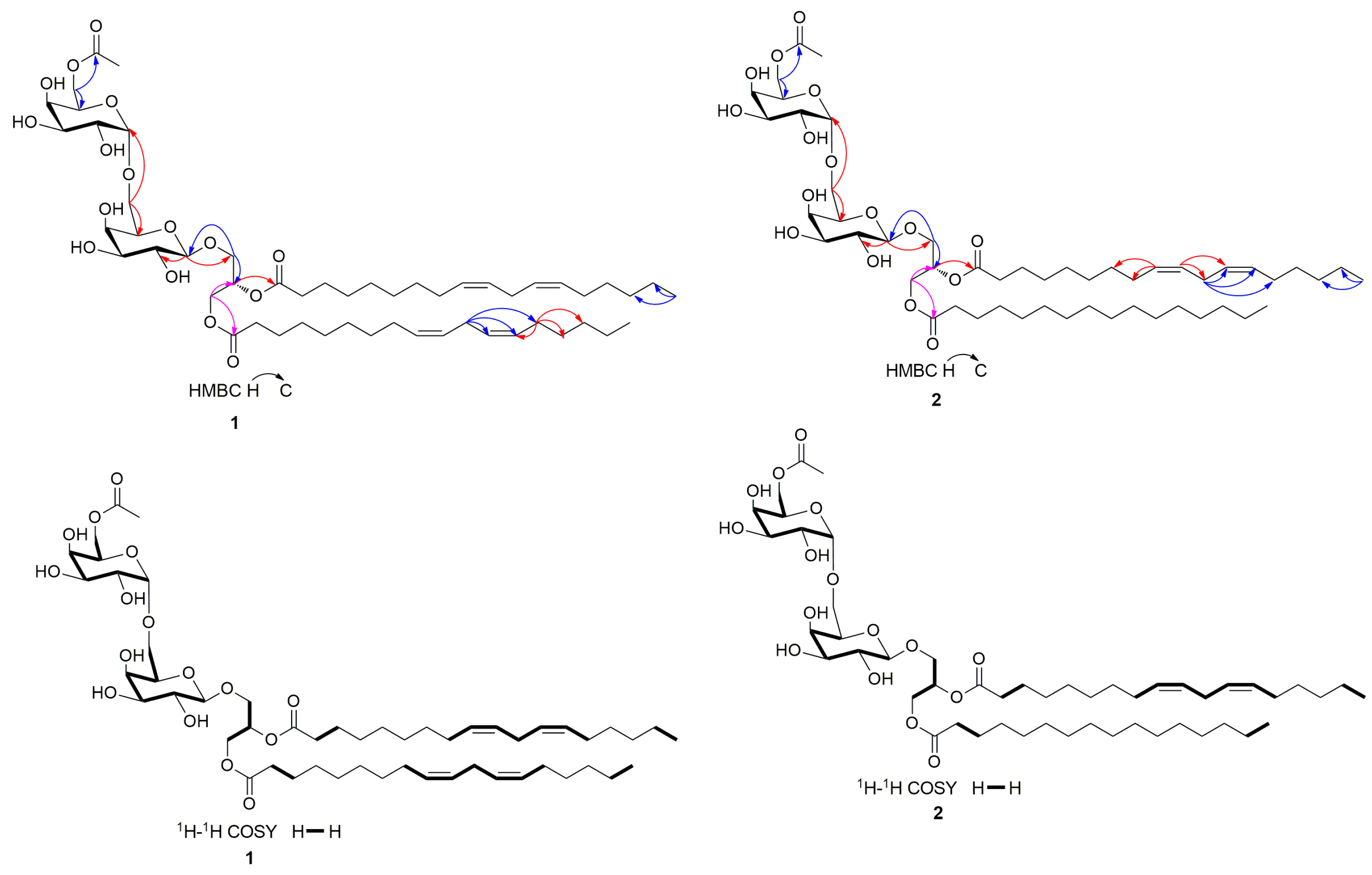
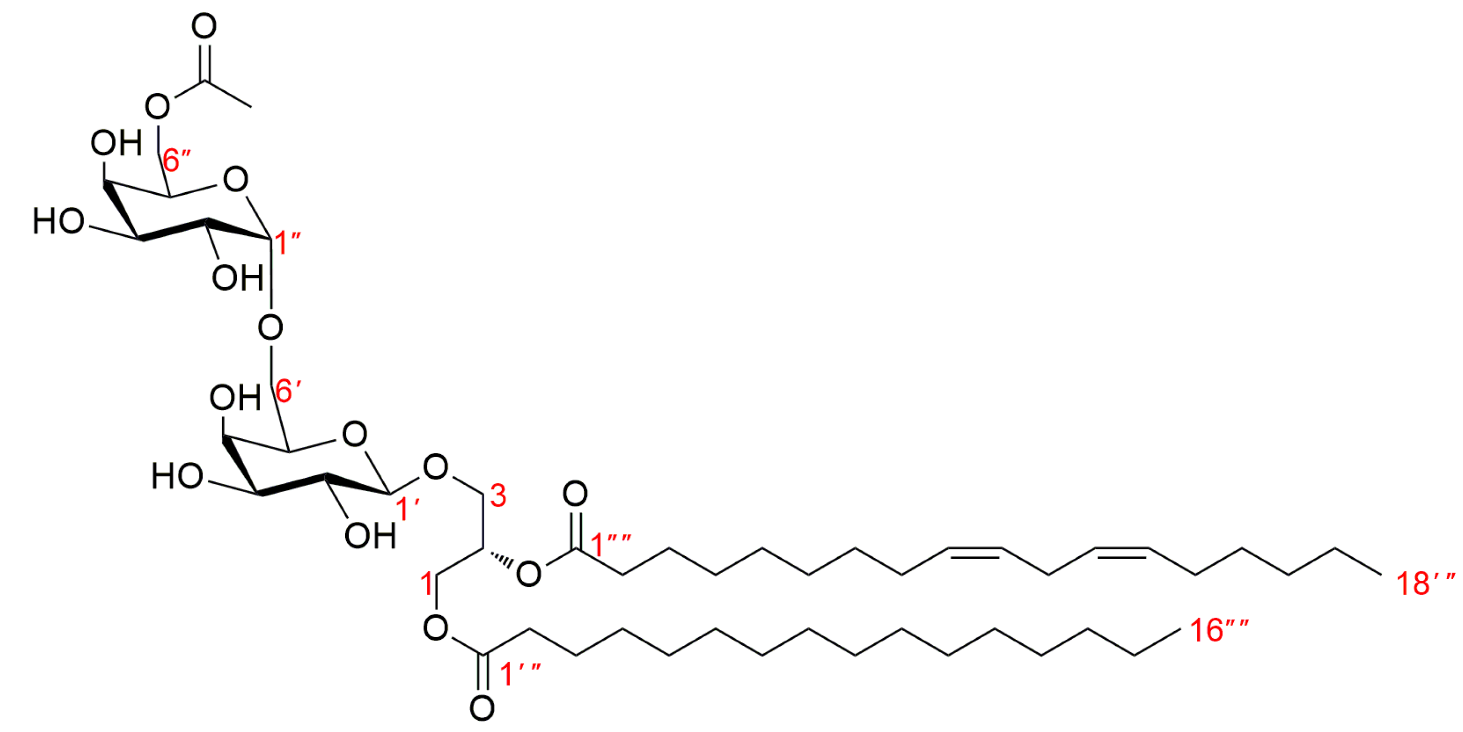
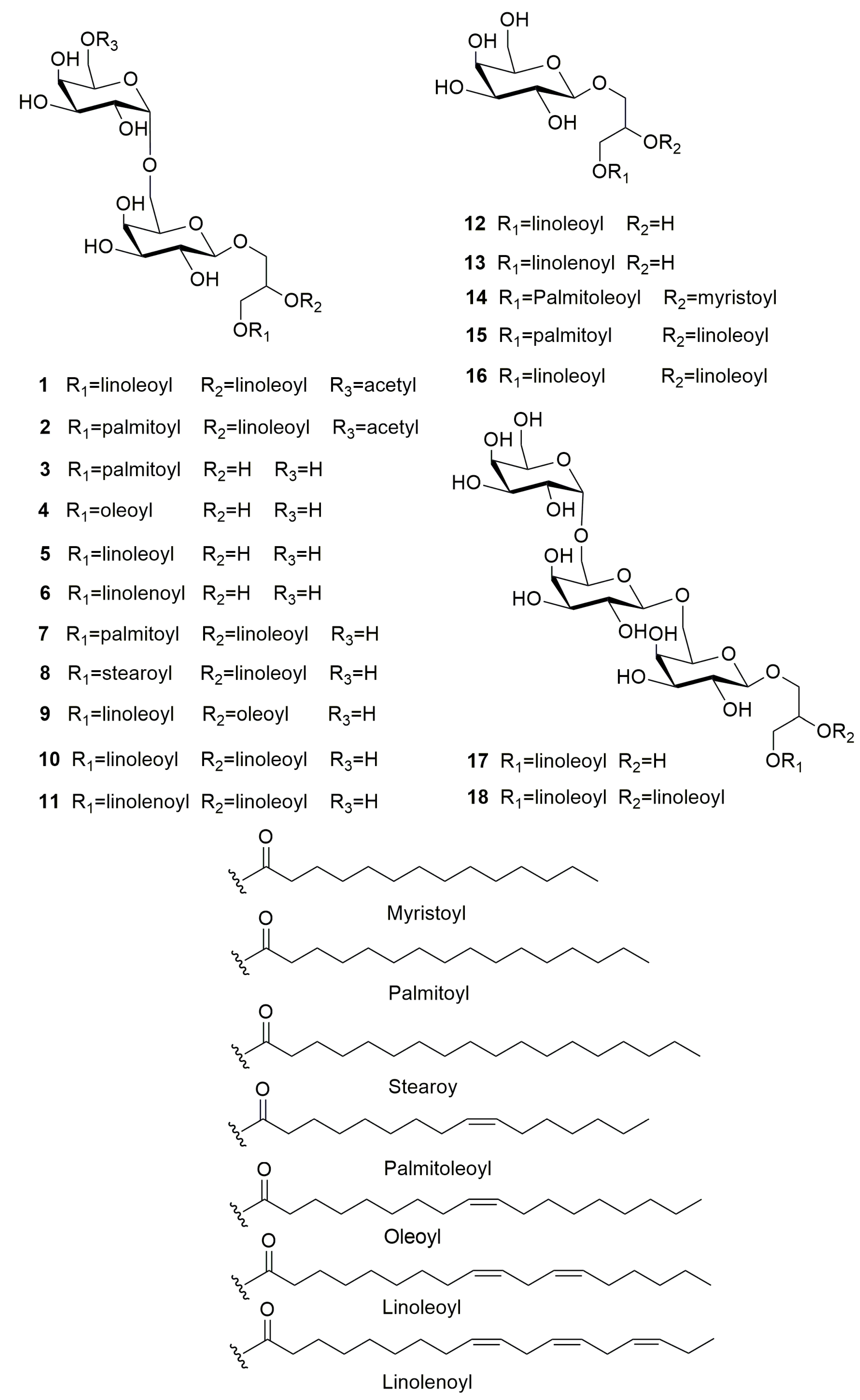
3.1. Structure Identification of Compounds 1–18

Compound (1), namely Rubracin D, was obtained as colorless oil; The structure of compound 1 was shown in Figure 1; [α ] D 28.8 = +62.06 (c 0.13, MeOH) (Figure S1); Its molecular formula was established as C53H90O16 by HR-ESI-MS ([M + Na]+ at m/z 1005.60938) (Figure S2) indicating 9 degrees of unsaturation. The UV (Figure S3) absorption maxima of 1 at 196 nm indicated the presence of double bonds, while the IR (Figure S4) absorption bands at 3400 and 1740 cm−1 revealed the presence of hydroxy and carbonyl functionalities. The 1H NMR (Figure S5), 13C NMR and DEPT spectra (Figure S6) and combined with HSQC spectra (Figure S7) of 1 displayed the presence of sugar and unsaturated long-chain fatty acid ester moieties. the 1D NMR data of compound 1 was shown in Table 1. The 1H-1H COSY (Figure S8) experiment revealed that there are some fragments H-1→H-2→H-3, H-1′→H-2′→H-3′→H-4′→H-5′→H-6′, H-1″→H-2″→H-3″→H-4″→H-5″→H-6″, H-2″′→H-3″′, H-8″′→H-9″′→H-10″′→H-11″′→H-12″′→H-13″′→H-14″′, H-3″″→H-4″″, and H-8″″→H-9″″→H-10″″→H-11″″→H-12″″→H-13″″→H-14″″(see Figure 2). Analysis of the HMBC spectrum (Figure S9), one group of signals at δC 105.4, 72.3, 74.7, 70.1, 74.8, 68.2 and δH 4.24 (1H, d, J = 7.4 Hz) revealed the presence of a 6-O-substituted β-D-galactopyranose unit (Figure S6). Another group of signals at δC 100.5, 70.2, 70.9, 71.2, 70.0, 65.0 and δH 4.85 (1H, br s) indicated the presence of a α-D-galactopyranose unit. Moreover, the other group of signals at δC 64.0, 68.7, 71.7 and δH 4.22 (1H, dd, J = 12.0, 6.6 Hz), 4.44 (1H, dd, J = 12.1, 2.9 Hz), 3.73 (1H, Overlap), 3.91 (1H, dd, J = 10.9, 5.4 Hz), and 5.25 (1H, m) revealed a glycerol moiety. Meanwhile, the anomeric H-1′ of the β-D-galactopyranosyl at δH 4.24 was correlated with δC 68.7, and 72.3 implied the anomeric H-1′ of the β-D-galactopyranosyl connected with the C-3 of the glycerol moiety. Another anomeric H-1″ of the α-D-galactopyranosyl was correlated to the C-6′ of the β-D-alactopyranosyl, which was supported by the correlations of the proton at δH 4.85 to δC 68.2, 70.9, and 71.2 (Figure 3). All of the above findings revealed that compound 1 was a 1, 2-di-O-acyl-3-O-[α-D-galactopyranosyl-(1″→6′)-O-β-D-galactopyranosyl]-glycerol. Furthermore, the 13C NMR spectrum of 1 showed two carbonyl carbon signals at δC 175.0 (C-1′′′) and 174.6 (C-1), and their locations were confirmed by the correlations of H-1 with C-1′′′ and H-2 with C-1′′′′ detected in the HMBC experiment. In the 1H and 13C NMR spectra, the unsaturated long-chain fatty acid ester signals at [14.5 (q), 0.90 (6H, d, J = 6.9 Hz)], [26.6 (t), 2.77 (4H, t, J = 6.6 Hz)], [2.82 (t), 2.05 (8H, m)], and [129.1–131.0 (d), 5.30–5.36 (8H, m)] suggested that compound 1 has two linolenoyl units, which was further verified by the molecular formula of 1. The detailed analysis of 1D NMR and 2D NMR, of the chemical structure of compound 1 were very similar to those of 1, 2-O-(9Z, 12Z-octadecadienoyl)-3-O-[α-D-galactopyranosyl-(1″→6′)-O-β-D-galactopyranosyl]-glycerol [31], except that a hydrogen atom was replaced by acetyl group in compound 1, 2-O-(9Z, 12Z-octadecadienoyl)-3-O-[α-D-galactopyranosyl-(1″→6′)-O-β-D-galactopyranosyl]-glycerol. The acetyl group connected with C-6″ was supported by the correlations of H-6″ with 172.7, 71.2, and 70.9 in the HMBC experiment. Except for β-D-galactopyranose and α-D-galactopyranose, the C-2 configuration of compound 1 was determined to be S by comparison with the optical rotation values and the chemical shift of C-2 reported in the literature [32]. As a result, the structure of 1 was determined as (2S)-1, 2-O-linoleoyl-3-O-[α-D-galactopyranosyl-(1″→6′)- (6″-O-acetyl)-O-β-D-galactopyranosyl]-glycerol.
Compound (2), [α ] D 28.8 = +73.85 (c 0.11, MeOH) (Figure S10), was obtained as a colorless oil and possessed a molecular formula of C51H90O16, established by HR-ESI-MS ([M + Na]+ at m/z 981.61029) (Figure S11). The UV (Figure S12) absorption maxima of 2 at 196 nm indicated the presence of double bands, while the IR spectra (Figure S13) revealed the presence of hydroxy and carbonyl functionalities; the chemical structure of 2 was displayed in Figure 1. In an analysis of 1H NMR (Figure S14), 13C NMR, and DEPT spectra (Figure S15), the 1D NMR data (see Table 1) were very similar to those of 1 except for the difference of another long-chain saturated fatty acid ester unit, This assumption was supported by the signals at δC 175.0, 35.1, 32.7, 30.2–30.8, 26.0, 23.7, 14.5 and δH 2.32 (2H, d, J = 7.3 Hz), 1.60 (2H, m), 0.90 (3H, d, J = 6.9 Hz). The detailed analysis of HSQC (Figure S16), 1H-1H COSY (Figure S17), and HMBC (Figure S18) correlations together with the molecular formula confirmed that compound 2 was (2S)- 1-O-palmitoyl-2-O-linoleoyl-3-O-[α-D-galactopyranosyl-(1′′→6′)- (6″-O-acetyl)-O-β-D-galactopyranosyl]-glycerol, and namely Rubracin E. The key 1H-1H COSY and HMBC correlations of compound 2 were shown in Figure 3.
Furthermore, sixteen known compounds were also isolated. According to the comparison with the reported NMR data and mass data, sixteen compounds [Figure 1] were identified as 1-O-palmitoyl-3-O-[α-D-galactopyranosyl-(1→6)-β-D-galactopyranosyl]- glycerol (3) [33], gingerglycolipid C (4) [34], gingerglycolipid B (5) [35], 3-O-octadeca-9Z,12Z,15Z-trienoylglyceryl-6′-O-(α-D-galactopyranosyl)-β-D-galactopyranoside (6) [35], 1-O-palmitoyl-2-O-linoleoyl-3-O-[α-D-galactopyranosyl-(1→6)-β-D-galactopyranosyl]-glycerol (7) [36], 1-O-octadecanoyl-2-O-(9Z,12Z-octadecadienoyl)-3-O-[α-D-galactopyranosyl-(1′′→6′)-O-β-D-galactopyranosyl] glycerol (8) [37], 1-O-linoleoyl-2-O-oleoyl-3-O-[α-D-galactopyranosyl-(1→6)-β-D-galactopyranosyl]-glycerol (9) [38], 2,3-O-dioctadeca-9Z,12Z-dienoylglyceryl-6′-O-(α-D-galactopyranosyl)-β-D-galactopyranoside (10) [35], 2-O-octadeca-9Z,12Z-dienoyl-9Z,12Z,15Z-trienoylglyceryl-6′-O-(α-D-galactopyranosyl)-β-D-galactopyranoside (11) [39], 1-O-(9Z,12Z-octadecadienoly)-3-O-β-galactopyranosylglycerol (12) [40], 3-O-octadeca-9Z,12Z,15Z-trienoylglyceryl-O-β-D-galactopyranoside (13) [33], 1-O-oleoyl-2-O-myristoyl-glyceryl-O-β-D-galactopyranoside (14) [35], 1,2-O-diacyl-3-O-β-D-galactopyranosyl glycerols (15) [37], 2,3-O-dioctadeca-9Z,12Z-dienoylglyceryl-O-β-D-galactopyranoside (16) [35], 1-O-(9Z, 12Z-octadecadienoyl)-3-O-[β-D-galactopyranosyl-(1→6)-O-β-D-galactopyranosyl-(1→6)-O-β-D-galactopyranosyl] glycerol (17) [41], 1,2-O-(9Z,12Z-octadecadienoyl)-3-O-[α-D-galactopyranosyl-(1′′′′′→6′′′′)-O-β-D-galactopyranosyl-(1′′′′→6′′′)-O-β-D-galactopyranosyl]-glycerol (18) [31]. Their chemical structures were described in Figure 4.

3.2. Results of Bioactivity Assays

3.2.1. Cytotoxicity Evaluation In Vitro

The new compounds 1 (PF70) and 2 (PF52) were subjected to cytotoxicity evaluation on the three human cancer cell lines viz MCF-7/ADM, K562/ADM and A549/ADM using the CCK-8 method. Both compounds 1 and 2 showed cytotoxic activity on the cell lines with an inhibition ratio of less than 20% at a concentration lower than 50 μg/mL. The results indicated that both compounds were applicable for studying MDR reversal activity.

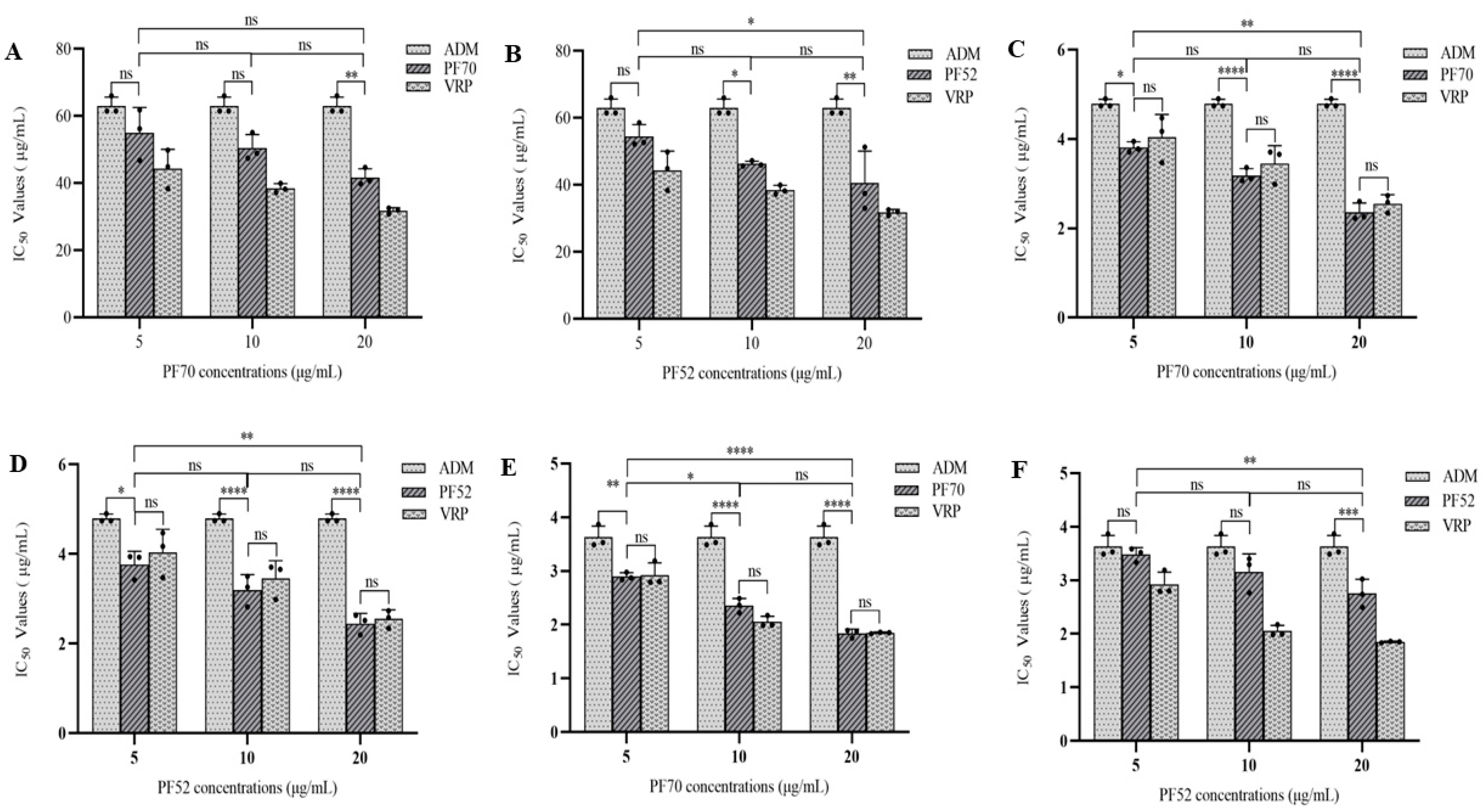
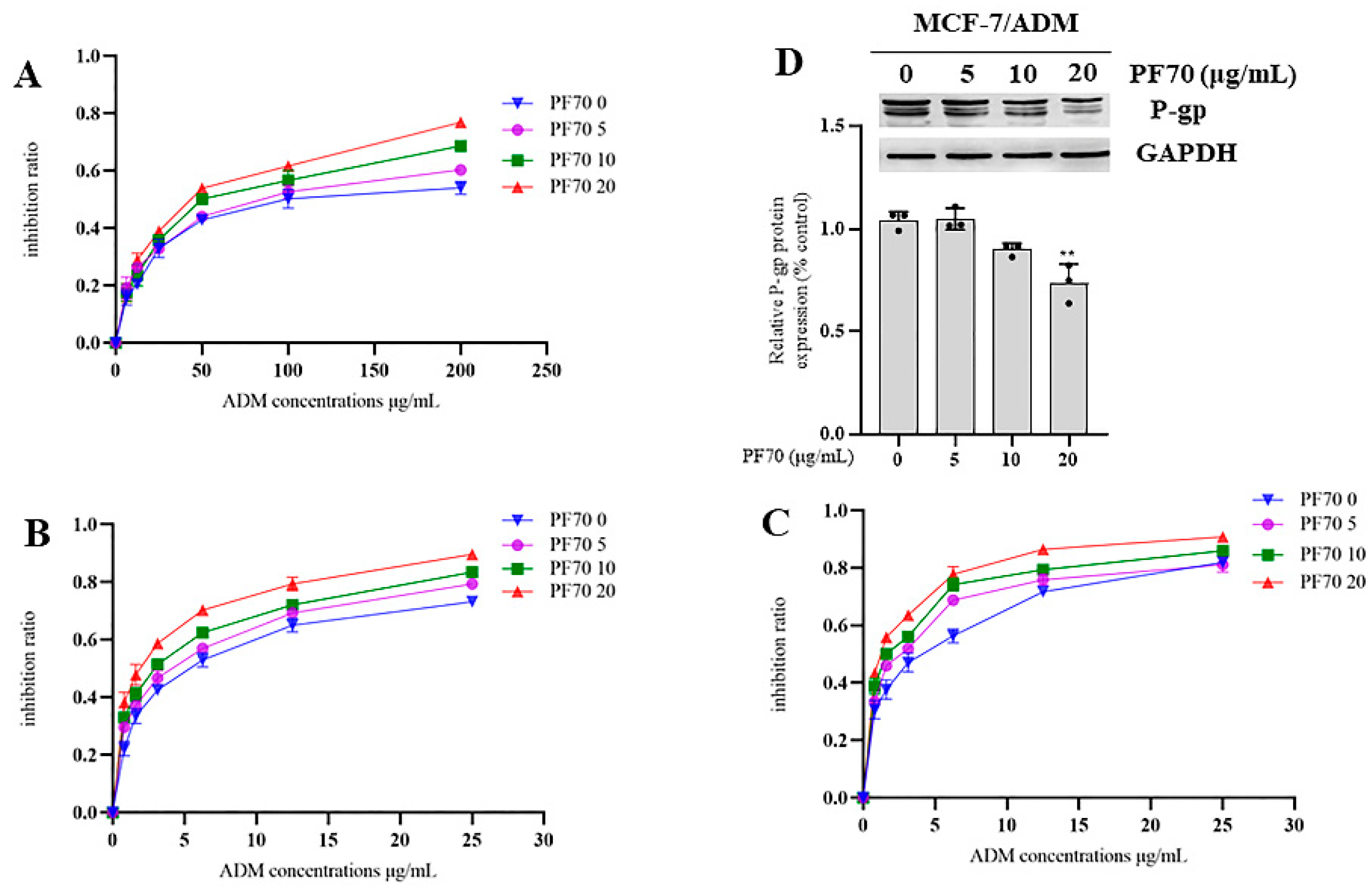
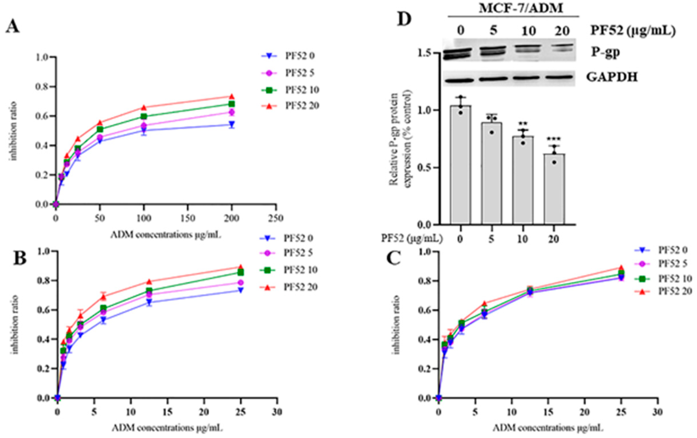
3.2.2. MDR Reversal Effects of Compound 1 and 2 In Vitro

The MDR reversal assay was performed using the anticancer drug adriamycin (ADM) at different concentrations combined with the test compounds at concentrations of 0, 5, 10 and 20 μg/mL, respectively, and Vrp as the positive control. The reversal activities of compound 1 and compound 2 against the three tumor cells were shown in Figure 5 and Table 2. In the assay on MCF-7/ADM, both compounds 1 (See Table 2 and Figure 5A) and 2 (See Table 2 and Figure 5B) showed certain reversal effects, but not as effective as those of Vrp. While in the assay on K562/ADM, both compounds 1 (See Table 2 and Figure 5C) and 2 (See Table 2 and Figure 5D) were proved to have significant MDR reversal effects with IC50 values range from 3.813 ± 0.123 to 2.360 ± 0.208 μg/mL and 3.764 ± 0.297 to 2.448 ± 0.225 μg/mL, respectively, which were higher than that of Vrp with an IC50 value range from 4.038 ± 0.514 to 2.554 ± 0197 μg/mL at 5, 10, 20 μg/mL, respectively. However, one-way variance contrast analysis (ANOVA) showed no statistical difference (p > 0.05). In the assay on A549/ADM, compound 1 (See Table 2 and Figure 5E) displayed significant MDR reversal effects with IC50 value range from 2.897 ± 0.072 to 1.841 ± 0.079 μg/mL, which were higher than those of Vrp at 5 and 20 μg/mL, respectively, with IC50 value ranges from 2.927 ± 0.226 to 1.848 ± 0.015 μg/mL. While the results of the ANOVA indicated no statistical difference (p > 0.05). Compound 2 (See Table 2 and Figure 5F) also showed certain reversal effects, but not as effective as those of Vrp. Meanwhile, as also shown in Figure 6 and Figure 7, the inhibition ratio curves of compound 1 and 2 combined with ADM on the three drug-resistant cell lines indicated that both compounds significantly increased the ADM sensitivities of ADM-resistant cells with significant dose-dependence.
Finally, we tested the P-gp expressions in MCF-7/ADM cells with the treatment of ADM at the concentration of 65 μg/mL combined with compound 1 and 2 at 0, 5, 10, 20 μg/mL, respectively, for 48 h. Our Western blot assays showed that the test with compound 1 at the concentration of 20 μg/mL had a significant suppression effect on the expression level of P-gp (Figure 6D), which proved that compound 1 reversed the MDR of the MCF-7/ADM cell line via hindering the overexpression of P-gp. The Western blot assays also showed that the test of compound 2 at the concentrations of 10 and 20 μg/mL, respectively, had remarkable suppression effects on the expression level of P-gp (Figure 7D) and therefore reversed the MDR of the MCF-7/ADM cell line.

4. Discussion

The strain Tubeufia rubra (PF02-2) was first isolated and identified by Dr. Lu of our research group, so there is no relevant report on the metabolites of the strain. In the paper, two new compounds, viz Rubracin D (1) and E (2), as well as sixteen known constituents (3–18) were isolated from the crude ethyl acetate extract of the fermentation broth of the fungal strain Tubeufia rubra (PF02-2). Their chemical structures were identified on the basis of their spectroscopic spectra data, as well as via comparison with literature data. Compounds (118) were isolated from the fungal genus Tubeufia for the first time. Bioassay results indicated that the new compounds (1, 2) showed significant MDR reversal activities on MCF-7/ADM, K562/ADM and A549/ADM cell lines. It is worth noting that both compounds (1, 2) were proved to have significant MDR reversal effects mostly higher than those of the positive control viz Vrp in the assay on the K562/ADM cell line. Additionally, compound 1 displayed significant MDR reversal effects mostly higher than those of Vrp in the assay on A549/ADM cell line. The results of the Western blot assays proved that both compounds 1 and 2 significantly reversed the MDR of the MCF-7/ADM cell line via hindering the overexpression of P-gp. We, therefore, speculate that the MDR reversal mechanism of both compounds on the other two resistant cancer cell lines viz K562/ADM and A549/ADM was also due to inhibiting the overexpression of P-gp.

Supplementary Materials

The following supporting information can be downloaded at: https://www.mdpi.com/article/10.3390/jof9030309/s1.

Author Contributions

S.Q. conducted principally experiments and wrote the paper. X.Z. and Y.Q. performed partial experiments. Y.L. contributed strain materials. Z.H. and J.K. designed research and revised paper. All authors have read and agreed to the published version of the manuscript.

Funding

This work was supported by the National Natural Science Foundation of China (NSFC Grants Nos. 32170019 and 31670027), the Open Fund Program of Engineering and Research Center for Southwest Bio-Pharmaceutical Resources of National Education Ministry of China, Guizhou University (No. GZUKEY201606) and the Science and Technology Foundation of Guizhou Province ([2020]1Y058).

Institutional Review Board Statement

This article does not contain any studies with human participants or animals performed by any of the authors.

Informed Consent Statement

Not applicable.

Data Availability Statement

Data supporting the reported results are provided in Supplementary Materials. The data from manuscript and Supplementary Materials are available for publication, citation, and use.

Conflicts of Interest

The authors declare that the research was conducted in the absence of any commercial or financial relationships that could be construed as a potential conflict of interest.

References

  1. Siegel, R.L.; Miller, K.D.; Fuchs, H.E.; Jemal, A. Cancer statistics, 2022. CA Cancer J. Clin. 2022, 72, 7–33. [Google Scholar] [CrossRef]
  2. Bray, F.; Ferlay, J.; Soerjomataram, I.; Siegel, R.L.; Torre, L.A.; Jemal, A. Global cancer statistics 2018: GLOBOCAN estimates of incidence and mortality worldwide for 36 cancers in 185 countries. CA Cancer J. Clin. 2018, 68, 394–424. [Google Scholar] [CrossRef] [Green Version]
  3. Li, X.; Li, J.P.; Yuan, H.Y.; Gao, X.; Qu, X.J.; Xu, W.F.; Tang, W. Recent advances in P-glycoprotein-mediated multidrug resistance reversal mechanisms. Methods Find. Exp. Clin. Pharmacol. 2007, 29, 607–617. [Google Scholar] [CrossRef]
  4. Kumar, A.; Jaitak, V. Natural products as multidrug resistance modulators in cancer. Eur. J. Med. Chem. 2019, 176, 268–291. [Google Scholar] [CrossRef]
  5. Wang, Y.P.; Wang, Y.D.; Liu, Y.P.; Cao, J.X.; Yang, M.L.; Wang, Y.F.; Khan, A.; Zhao, T.R.; Cheng, G.G. 6′-O-Caffeoylarbutin from Que Zui tea ameliorates acetaminophen-induced liver injury via enhancing antioxidant ability and regulating the PI3K signaling pathway. Food Funct. 2022, 13, 5299–5316. [Google Scholar] [CrossRef]
  6. Na, G.; Shang, Z.C.; Yu, P.; Jie, L.; Jian, K.L.; Kong, L.Y.; Yang, M.H. Alkaloids from the endophytic fungus Penicillium brefeldianum and their cytotoxic activities. Chin. Chem. Lett. 2017, 28, 1194–1199. [Google Scholar]
  7. Li, F.R.; Liu, L.; Liu, Y.P.; Wang, J.T.; Yang, M.; Khan, A.; Qin, X.; Wang, Y.D.; Cheng, G.G. HRESIMS-guided isolation of aspidosperma-scandine type bisindole alkaloids from Melodinus cochinchinensis and their anti-inflammatory and cytotoxic activities. Phytochemistry 2021, 184, 112673–112679. [Google Scholar] [CrossRef]
  8. Niu, L.T.; Rustamova, N.; Ning, H.X.; Paerhati, P.; Lu, C.F.; Yili, A. Diversity and Biological Activities of Endophytic Fungi from the Flowers of the Medicinal Plant Vernonia anthelmintica. Int. J. Mol. Sci. 2022, 23, 11935. [Google Scholar] [CrossRef]
  9. Nigora, R.; Khayrulla, B.; Niu, L.T.; Rehebati, N.X.; Ahmidin, W.; William, N.S.; Abulimit, Y. Secondary Metabolites and their Biological Activities from Endophytic Fungal Strain Aspergillus terreus XJA8 Associated with Vernonia anthelmintica. J. Biol. Act. Prod. Nat. 2022, 12, 421–435. [Google Scholar]
  10. Kim, S.E.; Kim, H.S.; Hong, Y.S.; Kim, Y.C.; Lee, J.J. Sesquiterpene esters from Celastrus orbiculatus and their structure-activity relationship on the modulation of multidrug resistance. J. Nat. Prod. 1999, 62, 697–700. [Google Scholar] [CrossRef]
  11. Aoki, S.; Okano, M.; Matsui, K.; Itoh, T.; Satari, R.; Akiyama, S.I.; Kobayashi, M. Brianthein A, a novel briarane-type diterpene reversing multidrug resistance in human carcinoma cell line, from the gorgonian Briareum excavatum. Tetrahedron 2001, 57, 8951–8957. [Google Scholar] [CrossRef]
  12. Wan, C.K.; Tse, A.K.; Yu, Z.L.; Wang, H.; Fong, D.W. Inhibition of cytochrome P450 3A4 activity by schisandrol A and gomisin A isolated from Fructus Schisandrae chinensis. Phytomedicine 2010, 17, 702–705. [Google Scholar] [CrossRef]
  13. Paterna, A.; Kincses, A.; Spengler, G.; Mulhovo, S.; Molnar, J.; Ferreira, M.J.U. Dregamine and tabernaemontanine derivatives as ABCB1 modulators on resistant cancer cells. Eur. J. Med. Chem. 2017, 128, 247–257. [Google Scholar] [CrossRef]
  14. Li, S.Z.; Zhao, Q.; Wang, B.; Yuan, S.; Wang, X.Y.; Li, K. Quercetin reversed MDR in breast cancer cells through down-regulating P-gp expression and eliminating cancer stem cells mediated by YB-1 nuclear translocation. Phytother. Res. 2018, 32, 1530–1536. [Google Scholar] [CrossRef]
  15. Pan, Q.G.; Lu, Q.H.; Zhang, K.; Hu, X. Dibenzocyclooctadiene lingnans: A class of novel inhibitors of P-glycoprotein. Cancer Chemother. Pharmacol. 2006, 58, 99–106. [Google Scholar] [CrossRef]
  16. Su, S.; Cheng, X.L.; Wink, M. Natural lignans from Arctium lappa modulate P-glycoprotein efflux function in multidrug resistant cancer cells. Phytomedicine 2015, 22, 301–307. [Google Scholar] [CrossRef]
  17. Ferreira, R.J.; Kincses, A.; Gaidacs, M.; Spengler, G.; Molnar, J.; Ferreira, M.J.U. Terpenoids from Euphorbia pedroi as Multidrug-Resistance Reversers. J. Nat. Prod. 2018, 81, 2032–2040. [Google Scholar] [CrossRef]
  18. Zhu, J.Y.; Wang, R.M.; Lou, L.L.; Li, W.; Tang, G.H.; Bu, X.Z.; Yin, S. Jatrophane Diterpenoids as Modulators of P-Glycoprotein-Dependent Multidrug Resistance (MDR): Advances of Structure-Activity Relationships and Discovery of Promising MDR Reversal Agents. J. Med. Chem. 2016, 59, 6353–6369. [Google Scholar] [CrossRef]
  19. Stierle, A.; Strobel, G.; Stierle, D. Taxol and taxane production by Taxomyces andreanae, an endophytic fungus of Pacific yew. Science 1993, 260, 214–216. [Google Scholar] [CrossRef]
  20. He, D.; Ma, Q.Y.; Yang, L.; Xie, Q.Y.; Zhu, H.J.; Dai, H.F.; Wu, Y.G.; Yang, D.M.; Zhao, Y.X. Two new indole alkaloids isolated from a mangrove-derived fungus Colletotrichum sp. HD-1. Phytochem. Lett. 2023, 54, 81–85. [Google Scholar] [CrossRef]
  21. Shaala, L.A.; Alzughaibi, T.; Gregory, G.J.; Youssef, D.T.A.; Fusaripyridines, A.B. Highly Oxygenated Antimicrobial Alkaloid Dimers Featuring an Unprecedented 1,4-Bis(2-hydroxy-1,2-dihydropyridin-2-yl)-butane-2,3-dione Core from the Marine Fungus Fusarium sp. LY019. Mar. Drugs 2021, 19, 505. [Google Scholar] [CrossRef]
  22. Lin, Z.J.; Zhu, T.J.; Fang, Y.C.; Gu, Q.Q.; Zhu, W.M. Polyketides from Penicillium sp. JP-1, an endophytic fungus associated with the mangrove plant Aegiceras corniculatum. Phytochemistry 2008, 69, 1273–1278. [Google Scholar] [CrossRef]
  23. Bharat, P.B.; Wijeratne, E.M.; Faeth, S.H.; Gunatilaka, A.A. Globosumones A-C, Cytotoxic Orsellinic Acid Esters from the Sonoran Desert Endophytic Fungus Chaetomium globosum. J. Nat. Prod. 2005, 68, 724–728. [Google Scholar]
  24. Isaka, M.; Jaturapat, A.; Rukseree, K.; Danwisetkanjana, K.; Tanticharoen, M.; Thebtaranonth, Y. Phomoxanthones A and B, Novel Xanthone Dimers from the Endophytic Fungus Phomopsis Species. J. Nat. Prod. 2001, 24, 1015–1018. [Google Scholar] [CrossRef]
  25. Liu, Y.; Ding, L.J.; He, J.X.; Zhang, Z.M.; Deng, Y.T.; He, S.; Yan, X.J. A new antibacterial chromone from a marine sponge-associated fungus Aspergillus sp. LS57. Fitoterapia 2021, 154, 105004. [Google Scholar] [CrossRef]
  26. Ohtsu, Y.; Sasamura, H.; Shibata, T.; Nakajima, H.; Hino, M.; Fujii, T. The novel gluconeogenesis inhibitors FR225659 and related compounds that originate from Helicomyces sp. No. 19353 II. Biological profiles. J. Antibiot 2003, 56, 689–693. [Google Scholar] [CrossRef] [Green Version]
  27. Chen, L.Z.; Qian, Y.X.; Kang, J.C.; Wang, L.; Lu, Y.Z.; Fan, C.; He, Z.J. The reversal bioactities of multidrug resistance of MCF-7/ADM cells induced by two Tubeufia fugal strains. Mycosystema 2020, 39, 817–826. [Google Scholar]
  28. Zeng, X.B.; Qian, S.Y.; Lu, Y.Z.; Li, Y.J.; Chen, L.Z.; Qian, Y.X.; He, Z.J.; Kang, J.C. A novel nitrogen-containing glyceride from fungal saprobe Tubeufia rubra reverses MDR of tumor cell lines to doxorubicin. Rec. Nat. Prod. 2022, 16, 622–632. [Google Scholar] [CrossRef]
  29. Lu, Y.Z.; Liu, J.K.; Hyde, K.D.; Rajesh, J.; Kang, J.C.; Fan, C.; Boonmee, S.; Bhat, D.J.; Luo, Z.L.; Lin, C.G.; et al. A taxonomic reassessment of Tubeufiales based on multi-locus phylogeny and morphology. Fungal Divers. 2018, 92, 131–344. [Google Scholar] [CrossRef]
  30. Lau, C.; Kim, Y.; Chia, D.; Spielmann, N.; Eibl, G.; Elashoff, D.; Wei, F.; Lin, Y.L.; Moro, A.; Grogan, T. Role of pancreatic cancer-derive exosomes in salivary biomarker development. J. Biol. Chem. 2013, 288, 26888–26897. [Google Scholar] [CrossRef] [Green Version]
  31. Liu, L.P.; Zhang, J.Q.; Wang, X.Y.; Wang, H.B. Glycoglycerolipids from Stellera chamaejasme. Nat. Prod. Commun. 2012, 7, 1499–1500. [Google Scholar] [CrossRef] [Green Version]
  32. Kim, J.S.; Shim, H.S.; Lee, S.; Chae, S.; Han, S.J.; Kang, S.S.; Lee, Y.S.; Jung, S.H.; Shin, K.H. A Monoacyldigalactosyl Glycerol from the Green Alga Enteromorpha prolifera. Nat. Prod. Sci. 2004, 10, 341–343. [Google Scholar]
  33. Kiem, P.V.; Minh, C.V.; Nhiem, N.X.; Cuong, N.X.; Tai, B.H.; Quang, T.H.; Anh, H.L.T.; Yen, P.H.; Ban, N.K.; Kim, S.H.; et al. Inhibitory Effect on TNF-α-Induced IL-8 Secretion in HT-29 Cell Line by Glyceroglycolipids from the Leaves of Ficus macrocarpa. Arch. Pharm. Res. 2012, 35, 2135–2142. [Google Scholar] [CrossRef]
  34. Chai, X.Y.; Bai, C.C.; Song, Y.L.; Chen, Y.P.; Li, F.F.; Tu, P.F. Chemical Constituents from the Leaves of Itoa orientalis. Chin. J. Nat. Med. 2008, 6, 179–182. [Google Scholar] [CrossRef]
  35. Suedee, A.; Tewtrakul, S.; Panichayupakaranant, P. Anti-HIV-1 integrase compound from Pometia pinnata leaves. Pharm. Biol. 2013, 51, 1256–1261. [Google Scholar] [CrossRef]
  36. Mei, W.L.; Ni, W.; Liu, H.Y.; Chen, C.X. Studied on the constituents of Cinnamomum zeylanicum. Nat. Prod. Res. Dev. 2002, 14, 14–17. [Google Scholar]
  37. Jung, J.H.; Lee, H.; Kang, S.S. Diacylglycerylgalactosides from Arisaema amurense. Phytochemistry 1996, 42, 447–452. [Google Scholar] [CrossRef]
  38. Murakami, N.; Morimoto, T.; Imamura, H.; Ueda, T.; Nagai, S.I.; Sakakibara, J. Studies on Glycolipids Ⅲ Glyceroglycolipids from an Axenically Cultured Cyanobacterium, Phormidium tenue. Chem. Pharm. Bull. 1991, 39, 2277–2281. [Google Scholar] [CrossRef] [Green Version]
  39. Bianco, A.; Mazzei, R.A.; Melchioni, C.; Scarpati, M.L.; Romeo, G.; Uccella, N. Microcomponents of olive oil. Part II: Digalactosyldiacylglycerols from OZea europaea. Food Chem. 1998, 62, 343–346. [Google Scholar] [CrossRef]
  40. Kwon, H.C.; Zee, O.P.; Lee, K.R. Two New Monogalactosylacylglycerols from Hydrocotyle ramiflora. Planta Med. 1998, 64, 477–479. [Google Scholar] [CrossRef]
  41. Jiang, Z.G.; Du, Q.Z. Two new glyceroglycolipids from the fruits of Cucurbita moschata. J. Chem. Res. 2009, 3, 157–159. [Google Scholar] [CrossRef]
Figure 1. Chemical structure of compound 1 from Tubeufia rubra PF02-2.
Jof 09 00309 g001
Figure 2. Key 1H-1H COSY and HMBC correlations of compounds 1 and 2.
Jof 09 00309 g002
Figure 3. Chemical structures of compound 2 from Tubeufia rubra PF02-2.
Jof 09 00309 g003
Figure 4. Chemical structures of compounds 118 from Tubeufia rubra PF02-2.
Jof 09 00309 g004
Figure 5. IC50 values of ADM at different concentrations combined with compound 1 (PF70) and 2 (PF52) at concentrations of 0, 5, 10, 20 μg/mL, respectively: (A) compound 1 (PF70) against MCF-7/ADM; (B) compound 2 (PF52) against MCF-7/ADM; (C) compound 1 (PF70) against K562/ADM; (D) compound 2 (PF52) against K562/ADM; (E) compound 1 (PF70) against A549/ADM; (F) compound 2 (PF52) against A549/ADM. All experiments were performed in triplicate. ns p > 0.05, * p < 0.05, ** p < 0.01, and *** p < 0.001, and **** p < 0.0001.
Jof 09 00309 g005
Figure 6. Inhibition ratios of ADM at different concentrations combined with compound 1 (PF70) at concentrations of 0, 5, 10 and 20 μg/mL, respectively: (A) MCF-7/ADM; (B) K562/ADM; (C) A549/ADM; (D) Effect of 1 (PF 70) on P-gp expression. All experiments were performed in triplicate. ** p < 0.01.
Jof 09 00309 g006
Figure 7. Inhibition ratios of ADM at different concentrations combined with compound 2 (PF52) at concentrations of 0, 5, 10 and 20 μg/mL, respectively: (A) MCF-7/ADM; (B) K562/ADM; (C) A549/ADM; (D) Effect of 2 (PF 52) on P-gp expression. All experiments were performed in triplicate. ** p < 0.01, and *** p < 0.001.
Jof 09 00309 g007
Table 1. 1D NMR data of Rubracin D (1) and E (2) in MeOD (1H-NMR at 500 MHz; 13C-NMR at 125 MHz).
Position1Position2
δCδH, mult (J in Hz)δCδH, mult (J in Hz)
164.0, CH24.44, dd, (12.1, 2.9)
4.22, dd, (12.0, 6.6)
163.9, CH24.44, dd, (12.0, 2.9)
4.22, overlap
271.7, CH5.25, m271.7, CH5.24, m
368.7, CH23.91, dd, (10.9, 5.4)
3.74, overlap
368.7, CH23.91, dd, (10.9, 5.4)
3.74, overlap
1′105.4, CH4.24, d, (7.4)1′105.4, CH4.24, d, (7.3)
2′72.3, CH3.51, dd, (9.7, 7.2)2′72.3, CH3.51, dd, (9.7, 7.3)
3′74.7, CH3.47, dd, (9.7, 3.2)3′74.7, CH3.47, dd, (9.7, 3.2)
4′70.1, CH3.85, dd, (3.1, 0.6)4′70.1, CH3.85, dd, (3.1, 0.6)
5′74.8, CH3.70, overlap5′74.8, CH3.70, overlap
6′68.2, CH23.89, overlap
3.64, dd, (10.0, 5.5)
6′68.2, CH23.89, overlap
3.64, dd, (10.0, 5.5)
1″100.5, CH4.85, br s1″100.5, CH4.85, br s
2″70.2, CH3.76, overlap2″70.2, CH3.76, overlap
3″70.9, CH3.72, overlap3″70.9, CH3.72, overlap
4″71.2, CH3.87, overlap4″71.2, CH3.87, overlap
5″70.0, CH4.04, br t, (6.7)5″70.0, CH4.04, br t, (6.7)
6″65.0, CH24.20, d (6.4)6″65.0, CH24.20, d (6.4)
1′″″172.7, C 1′″″172.7, C
2′″″20.9, CH32.06, s2′″″20.9, CH32.06, s
1′″175.0, C 1′″175.0, C
2′″35.1, CH22.32, t, (7.3)2′″35.1, CH22.32, t, (7.3)
3′″, 3″″26.0, CH21.60, m3′″, 3″″26.0, CH21.60, m
4′″, 4″″-7′″, 7″″30.2–30.8, CH21.28–1.36, m4′″-13′″30.2–30.8, CH21.28–1.36, m
8′″, 8″″, 14′″, 14″″28.2, CH22.05, m14′″, 16″″32.7, CH21.28–1.36, m
9′″, 9″″131.0 a, CH5.34, m15′″, 17″″23.6, CH21.28–1.36, m
10′″, 10″″129.2 b, CH5.32, m16′″, 18″″14.5, CH30.90, t, (6.9)
11′″, 11″″26.6, CH22.77, t, (6.6)1″″174.6, C
12′″, 12″″129.1 b, CH5.32, m2″″35.0, CH22.31, t, (7.3)
13′″, 13″″130.9 a, CH5.34, m4″″-7″″30.2–30.8, CH21.28–1.36, m
15′″, 15″″30.2–30.8, CH21.28–1.36, m8″″, 14″″28.2, CH22.05, m
16′″, 16″″32.7, CH21.28–1.36, m9″″131.0 a, CH5.34, m
17′″, 17″″23.7, CH21.28–1.36, m10″″129.1 b, CH5.32, m
18′″, 18″″14.5, CH30.90, t, (6.9)11″″26.6, CH22.77, t, (6.4)
1′″″174.6, C 12″″129.0 b, CH5.32, m
2′″″35.0, CH22.31, t, (7.3)13″″130.8 a, CH5.34, m
15″″30.2–30.8, CH21.28–1.36, m
Assignments with the same superscript (a, b) and in the same column may be interchangeable.
Table 2. The reversal effects of compounds toward MCF-7/ADM, K562/ADM and A549/ADM.
SamplesIC50 (μg/mL)
MCF-7/ADMK562/ADMA549/ADM
15 μg/mL54.958 ± 7.5953.813 ± 0.1232.897 ± 0.072
10 μg/mL50.456 ± 4.0713.182 ± 0.1572.358 ± 0.133
20 μg/mL41.726 ± 2.5852.360 ± 0.2081.841 ± 0.079
25 μg/mL54.487 ± 3.5383.764 ± 0.2973.480 ± 0.129
10 μg/mL46.289 ± 0.8253.195 ± 0.3473.155 ± 0.338
20 μg/mL40.567 ± 9.5322.448 ± 0.2252.755 ± 0.263
Vrp5 μg/mL44.371 ± 5.7464.038 ± 0.5142.927 ± 0.226
10 μg/mL38.471 ± 1.3973.452 ± 0.4012.051 ± 0.107
20 μg/mL31.799 ± 0.8752.554 ± 0.1971.848 ± 0.015
ADM 63.031 ± 2.6164.797 ± 0.0933.630 ± 0.207
All experiments were performed in triplicate. Each value represents the mean ± SD (n = 3). n represents number of experiments.
Disclaimer/Publisher’s Note: The statements, opinions and data contained in all publications are solely those of the individual author(s) and contributor(s) and not of MDPI and/or the editor(s). MDPI and/or the editor(s) disclaim responsibility for any injury to people or property resulting from any ideas, methods, instructions or products referred to in the content.

