Article

Aflatoxigenic Aspergillus Modulates Aflatoxin-B1 Levels through an Antioxidative Mechanism

1
Laboratory of Genetics, Wageningen University and Research, 6708 PB Wageningen, The Netherlands
2
Mycotoxicology Laboratory, National Institute for Scientific and Industrial Research, Lusaka 310158, Zambia
3
School of Agricultural Sciences, University of Zambia, Lusaka 10101, Zambia
4
Biointeractions and Plant Health, Wageningen University and Research, 6708 PB Wageningen, The Netherlands
*
Author to whom correspondence should be addressed.
J. Fungi 2023, 9(6), 690; https://doi.org/10.3390/jof9060690
Submission received: 28 April 2023 / Revised: 10 June 2023 / Accepted: 12 June 2023 / Published: 20 June 2023

Abstract

:
Aflatoxins (AFs) are considered to play important functions in species of Aspergillus section Flavi including an antioxidative role, as a deterrent against fungivorous insects, and in antibiosis. Atoxigenic Flavi are known to degrade AF-B1 (B1). To better understand the purpose of AF degradation, we investigated the degradation of B1 and AF-G1 (G1) in an antioxidative role in Flavi. Atoxigenic and toxigenic Flavi were treated with artificial B1 and G1 with or without the antioxidant selenium (Se), which is expected to affect levels of AF. After incubations, AF levels were measured by HPLC. To estimate which population would likely be favoured between toxigenic and atoxigenic Flavi under Se, we investigated the fitness, by spore count, of the Flavi as a result of exposure to 0, 0.40, and 0.86 µg/g Se in 3%-sucrose cornmeal agar (3gCMA). Results showed that levels B1 in medium without Se were reduced in all isolates, while G1 did not significantly change. When the medium was treated with Se, toxigenic Flavi significantly digested less B1, while levels of G1 significantly increased. Se did not affect the digestion of B1 in atoxigenic Flavi, and also did not alter levels of G1. Furthermore, atoxigenic strains were significantly fitter than toxigenic strains at Se 0.86 µg/g 3gCMA. Findings show that while atoxigenic Flavi degraded B1, toxigenic Flavi modulated its levels through an antioxidative mechanism to levels less than they produced. Furthermore, B1 was preferred in the antioxidative role compared to G1 in the toxigenic isolates. The higher fitness of atoxigenic over toxigenic counterparts at a plant non-lethal dose of 0.86 µg/g would be a useful attribute for integration in the broader biocontrol prospects of toxigenic Flavi.

1. Introduction

Aflatoxins (AFs) are a group of secondary metabolites produced by Aspergillus section Flavi (Flavi, for short) members such as A. parasiticus and A. flavus. The group of fungi infects important cereals such as maize and groundnuts [1,2,3,4,5,6], resulting in crop AF contamination. Of the known AFs produced by Flavi, AF-B1 (B1) is a highly mutagenic compound and is the most carcinogenic [7,8,9]. Among the known negative effects of B1 in both livestock and humans include growth impairment, reproductive system dysfunction, and immunosuppression [10,11,12]. In addition to this, studies have demonstrated the nephrotoxicity and acute hepatotoxicity of B1 on living tissue [13,14], including an effect on renal dysfunction among the negative effects on organs [15]. The oxidised form of B1 (AFB1-8, 9-epoxide) has been linked to cell injury due to its DNA mutagenicity [16]. The production of AF by Flavi has been demonstrated to be linked to useful biological functions, including the competitive inhibition of other microbes such as fungivores [17,18], antibiosis against bacteria [19], and oxidative stress alleviation [20,21,22,23,24] (Figure 1).
The link of AF to oxidative stress alleviation suggests that AF may be responsible for the scavenging of reactive oxygen species (ROS) [25], which results in the subsequent degradation of the AF molecule in reaction with the ROS. In this regard, it has been suggested that an increase in the antioxidative capacity of a plant could alleviate the increase in AF production by aflatoxigenic species such as A. flavus [22]. Hence, it would be estimated that introducing an antioxidant in the growth environment of the fungus (the crop) would reduce ROS, thereby reducing the need for the fungus to produce AF. A number of antioxidant elements are known, which include copper, manganese, selenium, and zinc. Selenium (Se) is important to human health [26] as well as plays an anti-carcinogenic role [27]. The element is also known to play a defensive role against AF in animals [28,29,30]. In the right amounts, Se can enhance plant growth [31].
Se can be introduced into a plant by biofortification through conventional breeding, genetic modification, mineral fertilisation of the soil, or through foliar application. In crop protection, the biofortification of plants with such an element would play the role of indirect control of AF accumulation in important cereals such as maize and wheat while improving the crop’s micronutrient status. However, it should also be noted that Se can be toxic in elevated levels to the plant itself [31,32] other than inhibiting fungal sporulation at high levels.
Atoxigenic Flavi strains have been demonstrated to degrade B1 [33]. With respect to this, colonisation of a crop by atoxigenic Flavi has been demonstrated to be able to reduce AF contamination in crops such as maize, cotton, and groundnuts [34,35,36]. Although the degradation of AF by atoxigenic A. flavus has been demonstrated [33], the reason behind the phenomenon is still unclear. For example, considering that atoxigenic Flavi do not produce AF, the question arises whether or not the degradation of AF could have a link to antioxidative alleviation in the non-aflatoxin producing Flavi, and if the mechanism would be the same as that for aflatoxin producers.
Some studies have demonstrated that the mechanism of inhibition of AF production involves the downregulation of the regulatory gene aflS, reducing the abundance of the ratio of aflS to aflR [37]. This would imply that there is an actual reduction in the amount of AF produced by the Flavi due to reduced messenger RNA from structural genes such as aflD, and not necessarily the reduction in AF through its degradation. The aflD is a structural gene responsible for the early conversion of AF precursor molecules in the aflatoxin biosynthetic pathway [38,39] and is regulated by the regulatory genes aflR [40,41,42,43] and aflS. To enhance the understanding of the potential role of AF production and its utilisation in Flavi, two related questions need to be answered: (1) is AF degraded by atoxigenic Flavi as part of oxidative stress alleviation, and (2) is AF also degraded by aflatoxin-producing Flavi (toxigenic) as part of the mechanism of oxidative stress alleviation?
Introducing a natural antioxidant such as Se to the environment of toxigenic and atoxigenic members of section Flavi and observing the associated levels of aflatoxin produced in its environment might provide some insights into the potential role aflatoxin plays as an antioxidant in both toxigenic and atoxigenic Flavi. It would also help to understand in which direction the population dynamics between toxigenic and atoxigenic Flavi would likely shift in a Se-fortified environment. It is known that fungi respond to abiotic stimuli, and that the exact nature of the response will depend on the fungal strain and abiotic condition. With respect to an antioxidative environment, if aflatoxin were an antioxidant for both toxigenic and atoxigenic Flavi, then one would expect that toxigenic Flavi would exhibit a higher Darwinian fitness (e.g., produce relatively more spores) in an environment with antioxidants such as Se, compared to atoxigenic Flavi. It would, however, be desirable that atoxigenic strains are fitter in a Se-fortified environment compared to toxigenic counterparts (Figure 1), such that population dynamics are not in favour of the toxigenic over atoxigenic strains.
The aim of this investigation was to explore the involvement of AF as an antioxidant in the growth environment for toxigenic and atoxigenic Flavi. The specific objectives of this study were to (1) determine whether AF is broken down by both atoxigenic and toxigenic Flavi in the role of antioxidation and (2) to establish differences in fitness levels between atoxigenic and toxigenic isolates as a result of antioxidant (Se) fortification. We hypothesise that both toxigenic and atoxigenic Flavi utilise AF in an antioxidative role. We also hypothesise that toxigenic strains are fitter when treated with antioxidant Se compared to atoxigenic counterparts. We base our second hypothesis on grounds that toxigenic isolates expend more energy producing AF, presumably in an antioxidative role, compared to the atoxigenic strains. This would imply that the introduction of a substitute antioxidant (Se) relieves more energy for the aflatoxin-producing fungus to utilise for other purposes, including sporulation (fitness).
Understanding the role of AF in both toxigenic and atoxigenic Flavi from an antioxidative role will better guide efforts aimed at improving the micronutrient status of maize without the risk of promoting toxigenic isolates over atoxigenic counterparts if integrated into the broader biocontrol programme. In addition, such knowledge will help to better understand how antioxidants would help in preventing spikes of AF in maize.

2. Materials and Methods

The Aspergillus section Flavi isolates used were collected as live wild-type laboratory cultures stored in glycerol (−70 °C). They had previously been isolated from maize and soil (data unpublished). Isolates were regenerated on Malt Extract Agar medium (MEA) for 7 days (25 °C, dark). Spores from regenerated cultures were suspended in 0.8% sterile saline solution as stock culture solution. The stocks were diluted with sterile MilliQ water to 1 × 10⁴ spores/mL prior to inoculations. Chromatographic analyses for aflatoxin (AF) were performed by HPLC (Agilent Infinity II 1260 Series, Agilent Technologies, Santa Clara, CA, USA). Technical standards for AF-G2, -G1, -B2, and -B1 were obtained from Merck (Merck Life Science NV, Amsterdam, The Netherlands).

2.1. Determination of Antioxidative Degradation of Aflatoxin by Toxigenic and Atoxigenic Flavi

To determine the antioxidative role of aflatoxin (AF) in its degradation by Flavi, we specifically used an antioxidant (selenium) in lieu of previously used oxidative compound such as H2O2 [22,24]. This would enable the direct determination of the role of AF in antioxidation, by using a substitute antioxidative compound (Se).

2.1.1. Assignment of Atoxigenic and Toxigenic Isolates

From the regenerated lab culture isolates, we selected 12 wild-type laboratory cultures preliminary screened for aflatoxin production ability based on qualitative visualisation of their aflatoxin (AF) bands by Thin Layer Chromatography (TLC) [44]. The first four isolates were initially assigned as non-producers (atoxigenic) based on absence of AF bands on TLC plates. Another four isolates were initially assigned as high producers based on very high intensity of AF bands on TLC plates. Then, four isolates were assigned as low producers based on less intense bands of Aflatoxins by TLC compared to the high producers. All 12 isolates were then verified by HPLC for levels of AF produced. To determine this, a 100 µL spore suspension (1 × 10⁴ spores/mL) per isolate was inoculated into test tube containing 2 mL autoclave sterilised Yeast Extract Sucrose (YES) broth (200 g/L sucrose; 20 g/L Yeast Extract autolysate, pH 6.0) [45]. Tubes were incubated at 31 °C (7 days, dark). Aerobic liquid fermentation was then carried out with shaking at 150 rpm in a temperature-controlled incubator (Innova 4330, New Brunswick Scientific, Enfield, CT, USA). AF was extracted by dichloromethane partitioning, adding 2 mL dichloromethane to the YES broth. Tubes were then vortexed for 45 s on a vortex unit (Genius model VG 3 S000, IKA, Staufen, Germany). Tubes were then centrifuged at 25,000× g (microcentrifuge model 5424, Eppendorf, Hamburg, Germany) for 5 min. The supernatant (aqueous phase) was pipetted off and discarded. Next, 500 µL of the dichloromethane phase (lower phase) was transferred into a 4 mL screw neck vial (45 × 14.7 mm, VWR, cat No. 548-0051). The dichloromethane fraction was then evaporated to dryness and then dried material reconstituted in 250 µL 1:1 methanol/water (v/v). For AF producers, the reconstituted material was diluted 10-fold into an HPLC vial, then 10 µL injected into HPLC. For non-producers, reconstituted material was diluted to 500 µL total volume into HPLC vial, then 20 µL was injected. Reversed-phase separation of injected analyte was achieved with a Cortecs C18 column of 3.5 i.d × 100 mm × 2.7 µm particle size (Waters, Milford, MA, USA) fitted with a guard column. The mobile phase constituted 0.1%-acetic acid/methanol/acetonitrile 70:20:10 (v/v) at a flow rate of 0.45 mL/min. The column oven was set at 35 °C and the eluted AFs were detected on fluorescence detector (λexcit = 365 nm; λemit = 440 nm). Quantification of AFs was achieved with Chemstation software. The isolates were hence assigned as high (>300 ng AF-B1+G1/mL YES medium), low (LOQ—299 ng AF-B1+G1/mL YES), and non-producers (Table 1). Limit of quantitation (LOQ) in AF extracts was assigned as 2.5 ng/mL. All values below LOQ were converted to 0 as unquantifiable noise.

2.1.2. Aflatoxin (AF) Degradation Assay

Artificial AF was first prepared from one aflatoxin-producing isolate, “EKZ10A.” For this, a 100 µL spore suspension (1 × 10⁴ spores/mL) of the isolate EKZ10A was inoculated into test tube containing 2 mL autoclave sterilised YES broth [45] (pH 6.0). Fifty tubes were incubated at 31 °C (7 days, dark) for the liquid fermentation with shaking at 150 rpm in a refrigerated incubator (Innova 4330, New Brunswick Scientific, Enfield, CT, USA). AF was similarly extracted by dichloromethane partitioning as described above, collecting the centrifuged dichloromethane (CH2Cl2) extracts from each tube into one pooled sterile 50 mL tube to ensure homogeneity of AF concentration. Next, 1000 µL of AF extract in CH2Cl2 was transferred per 10 mL glass test tube (×104 tubes) into ×96 test samples, ×4 positive controls, and ×4 negative controls. The CH2Cl2 was vapourised under fume hood to dryness and reconstituted with 300 µL absolute ethanol to re-sterilise artificial AF. Tubes were then placed in laminar flow hood to vaporise the ethanol. Next, 2 mL of sterilised YES agar medium was dispensed warm at 50 °C per tube, then vortexed for 20 s to redissolve the AF extract prior to isolate inoculation. The YES agar constituted 0.2% agar to generate a light paste. One set of YES agar had been fortified with anhydrous sodium selenite (analytical reagent) at intermediate concentration of 0.40 µg/g below a plant lethal dose of 5 mM (equivalent to 0.86 µg/g). In this experiment, the selenite salt was the antioxidant. The YES tubes were set up for the 12 isolates, with each isolate receiving all four of the following treatment permutations: (i) isolate only (“Spp”), (ii) isolate with AF (“Spp+Afl”), (iii) isolate with antioxidant (“Spp+AOx”), and (iv) isolate with AF and antioxidant (“Spp+Afl+AOx”). Each isolate was duplicated (total of 96 test samples). Positive controls to determine base AF levels in tubes were AF extract without isolate (“Afl”) as well as AF extract with antioxidant (“Afl+AOx), in duplicate. The spiking concentration of AF in the tubes was determined based on concentration of the positive controls. This was done post-incubation and determined by HPLC as described in the above section. Hence, the average spiked concentration of the AF in the incubated tubes was G2 = 150 ± 35 ng/mL; G1 = 3244 ± 786 ng/mL; B2 = 86 ± 6 ng/mL; and B1 = 2114 ± 68 ng/mL. Negative controls were YES agar as well as YES agar with antioxidant, in duplicate. Each tube with the YES agar was inoculated with 50 µL of 1 × 10⁴ spores/mL isolate suspension for each of the 12 test isolates. The positive controls were inoculated with 50 µL sterile Milli-Q H2O in place of isolate inoculation. All tubes were incubated at 31 °C (7 days, dark). The liquid fermentation was carried out in an incubator (Innova 4330, New Brunswick Scientific, Enfield, CT, USA) with mild shaking at 100 rpm. After the incubation, AF was extracted by dichloromethane partitioning, as described in the above section. No biomass growth ensued in the positive controls for AF, confirming integrity in sterility of the artificial AF extract medium during preparation. AF was then quantified by HPLC with autosampler as described above.
1.
Gene Expression (AflR and AflD) Response to Antioxidant (Se)
To determine the possible correlation response of gene expression to antioxidant treatment, three randomly selected high-aflatoxin-producing isolates (EKZ10B, ELG33C, and MLV14F) were tested for their gene expression correlation response. Unfortunately, isolate EKZ10B fell out of the experiment. Target genome loci were aflR and aflD (Table 2), which are part of the aflatoxin biosynthetic pathway genes. The β-tubulin was used as the endogenous control to normalise the qPCR cycle threshold (Ct) values.
2.
Generation of RNA
For generation of the genomic RNA with response to antioxidant, the spore suspensions from isolates ELG33C and MLV14F were plated on cellophane discs immersed in YES agar fortified with Se. YES was used as it induces AF production. The antioxidant was introduced as mineral Na2SeO3 at concentrations in µg/g YES of 0 (RNA calibrator), 0.40 (intermediate Se concentration below a plant lethal dose of about 5 mM or 0.86 µg/g), and 5.0 (elevated concentration). Treatments were incubated in triplicate at 31 °C (12 days, dark).
3.
Isolation and Quantification of RNA
Mycelia were aseptically harvested from top of cellophane disc into an RNA-/DNA-ase free (sterile) 1.5 mL screw cap tube containing four sterile 3 mm glass beads. Each tube, upon harvest of mycelia, was immediately placed into liquid N2. The frozen tubes were then clumped on a sideways beater (model MM400, Retsch, Haan, Germany), and beating was performed at 30 beats/second for 30 s for the cell disruption. Immediately, 800 µL TRI lysis solution (RNA lysis buffer) was added per tube and inverted three times to suspend all material in the tube. For the rest of the isolation steps, the Zymo Research Kit (catalogue No. R2073, Zymo Research, Irvine, CA, USA) was used, following the RNA extraction protocol for “Tough-to-Lyse Samples.” The vortex was performed at max speed of 10 (Genius model VG 3 S000, IKA, Staufen, Germany) for 15 s. Centrifuging was performed at 16,000× g (centrifuge model 5424, Eppendorf, Hamburg, Germany) at room temperature. Extracted RNA was eluted from column with 100 µL sterile DNase/RNase-free Milli-Q water. The RNA was stored at −80 °C for long-term storage or −20 °C for short-term storage for downstream analysis.
Purified total RNA concentration was measured using the Qubit fluorometer (Invitrogen, Thermo Fisher Scientific, Waltham, MA, USA) using the RNA BR Assay Kit. The RNA quality was analysed directly on the nanodrop (model 2000, Thermo Fisher Scientific, Wilmington, DE, USA) using 1.0 µL extracted RNA per sample (A260:A280 ratios: ELG33C = 1.91 ± 0.07; MLV14F = 1.67 ± 0.00). Furthermore, the RNA integrity was checked with 1% gel electrophoresis, mixing 8 µL RNA with 2 µL loading dye. Two clear bands were obtained, indicating the ribosomal material had not degraded.
4.
cDNA Synthesis (Reverse-transcriptase PCR)
The complementary DNA (cDNA) was synthesised using the SensiFast® cDNA synthesis kit (Bioline, Luckenwalde, Germany) in a 20 µL reaction mixture as follows: 15 µL (~500 ng ELG33C; ~200 ng MLV14F) of mRNA template was added to a 0.5 µL PCR tube. Next, 4 µL 5X TransAmp buffer was added per 0.5 PCR tube, followed by addition of 1 µL enzyme Reverse Transcriptase. Contents were gently mixed by up–down pipetting. The cDNA was synthesised from the mRNA in a thermal cycler (model T100™, Biorad Laboratories Inc., Hercules, CA, USA) under the following conditions: primer annealing, 25 °C × 10 min; reverse transcription, 42 °C × 15 min; optional step (in case of highly structured RNA), 48 °C × 15 min; and inactivation, 85 °C × 5 min. The synthesised cDNA concentration was then extrapolated from the RNA concentration prior to the cDNA synthesis. The cDNA concentration was then diluted to 3.5 ng/µL.
5.
Quantitative PCR (Real-time PCR)
Real-time PCR (qPCR) was carried out in a Real-Time Thermal Cycler (model CFX96™, Biorad Laboratories Inc., Hercules, CA, USA) using the iQ SYBR Green Supermix kit. For test samples, the qPCR was carried out in triplicate in total reaction volume of 10 µL per sample in MicroAmp optical 96-well reaction plates, sealed with optical adhesive covers (Applied Biosystems, Life Technologies, Waltham, MA, USA). Each reaction was performed using 5 µL of 2X iQ SYBR Green supermix, 2 µL of 2 µM reverse primer (400 nM), 2 µL of 2 uM forward primer (400 nM), and 1 µL of cDNA template of 3.5 ng/µL. For primer efficiency check, DNA from isolate EKA03C, already verified positive for aflR and aflD, was used, which was serial diluted to 3.50 ng/µL, 1.75 ng/µL, and 0.875 ng/µL. Negative control consisted sterile Milli-Q water in place of DNA. The thermal cycling conditions were executed with an initial denaturing step of 95 °C × 2 min followed by 40 cycles of 95 °C × 15 s (denature), 55 °C × 20 s (anneal), and 72 °C × 20 s (elongate). Melting curve analysis of the PCR product was performed by ramp heating from 50 °C to 95 °C in steps of 0.5 °C per increment, continuously measuring the fluorescence. Each ramp per increment was held for 20 s, while Ct values were acquired from instrument. The housekeeping gene β-Tubulin was used as the endogenous expression control against which normalisation of Ct values for aflR and aflD was performed.
6.
Corresponding Aflatoxin Production with Gene Expression
Pieces of YES agar under the cellophane on which RNA generation had been performed were collected into a pre-weighed test tube. The agar was crushed to paste with clean rod and was re-weighed in the test tube. A total of 5 clean glass beads (Ø 3 mm) were added per tube, and 600 µL of 0.05% Triton-X was added per tube. Contents were vortexed for 10 s continuously at max speed of 10 on a vortex unit (Genius model VG 3 S000, IKA, Staufen, Germany). Next, 3 mL dichloromethane was added per gram agar, and the AFs were extracted by dichloromethane partitioning, as described above.

2.2. Fitness Response of Atoxigenic and Toxigenic Isolates under Antioxidant (Se)

2.2.1. Isolates Collection and Preparation of Inoculation Medium

From the retrieved and regenerated isolates of maize and soil, we selected five toxigenic and five atoxigenic isolates on the basis of their high sporulation ability without Se treatment. Spore count was used as the proxy for isolate fitness. CMA amended with 3% w/v sucrose (3gCMA) was used for isolates fitness assays. The 3gCMA was prepared by adding 17 g cornmeal agar per litre of demi water, according to manufacturer specifications. Next, 3% w/v sucrose was added to the medium, configuring the medium to more or less mimic the kernel, including its sucrose-rich endosperm. Three levels of Se concentration in the 3gCMA were prepared by fortifying the medium with Se at 0 µg/g, 0.40 µg/g (intermediate Se concentration below a plant lethal dose of about 5 mM or 0.86 µg/g), and 0.86 µg/g maximum (equivalent maximum non-homeostasis-toxic mineral Se concentration in maize). The medium was then amended to 50 mg/L chloramphenicol and then boiled to dissolve the contents. Medium was then dispensed warm and viscous (50 °C) to 50 mL glass bottles at 3 mL/bottle. Bottles were autoclaved at 120 °C × 15 min.

2.2.2. Fungal Fitness Assay under Antioxidant (Se) Treatment

Isolate spore suspensions, adjusted to 1 × 10⁴ spore/mL, were inoculated in triplicate at 20 µL per sterile 3gCMA bottle with or without Se. The spore suspension was spread on the agar using 3 mm glass beads. Beads were discarded, and bottles were incubated at 30 °C (7 days, dark). Spores were then harvested from the medium with 0.05% Triton-X (surfactant). For the harvest, 3 mL surfactant was added per bottle, and contents were placed on sideways shaker (GFL model 3018, Society for Laboratory Technology, Burgwedel, Germany) and agitated at 200 rpm per minute for 2 min. Next, 1000 µL spore suspension was transferred by pipettor while swirling to a 1.5 mL microcentrifuge tube. Spore concentration was analysed using an automated cell counter (Casy™ TT, Omni Life Sciences, Bremen, Germany). Briefly, 10 µL of the collected spore suspension was added to 10 mL of Casy Ton dilution solution, and contents were gently inverted 10 times, avoiding build-up of bubbles. Immediately, the count was performed on the unit. If spore count was below 200 counts detected, 100 µL spore suspension was used in the dilution in place of 10 µL.

2.3. Data Analysis

To determine the degradation of AF with or without antioxidant treatment, the pairwise Wilcoxon rank sum exact test was used on the treatments. Due to the relatively low yield of the AF variants B2 (86 ng/mL) and G2 (150 ng/mL) in the artificial AF, assays were only performed on B1 (2114 ng/mL) and G1 (3244 ng/mL) in order to generate statistically viable results. G2 and B2 are the more oxidised forms of AF compared to G1 and B1, which are the more reactive forms of aflatoxin for such a redox assay and are the more important in terms of carcinogenicity. Relative gene expression was calculated using the 2−ΔΔCt method, as described by [48]. For gene expression, we considered either the upregulation or downregulation of aflR and aflD by relative number of times the expression increased or reduced in relation to calibrator (0 Se treatment). The simultaneous upregulation or downregulation of aflD and aflR was tested by Spearman rank correlation rho.
Fungal fitness was determined as spore concentration and analysed across treatments by Wilcoxon rank sum exact test, significant if p < 0.05. Mean spore counts between atoxigenic and toxigenic isolates were determined as geometric mean. Data computations were executed in software R [49] version 4.1.0. Visualisations were computed with aid of the R package ggplot2 [50].

3. Results

3.1. Determination of Antioxidative Degradation of Aflatoxin

Without the antioxidant, the results of the change in AF (B1 and G1) are shown across the variables “Afl,” “Spp” and “Spp+Afl” (Figure 2), summarised in Table 3. With the antioxidant, the change in B1 or G1 is shown across the variables “Spp+Afl+AOx” and “Spp+AOx” (Figure 2), summarised in Table 4. “Afl,” is the extrinsically introduced B1 or G1, while “Spp” is the toxigenic or atoxigenic Flavi. “AOx” is the antioxidant selenium (Se).

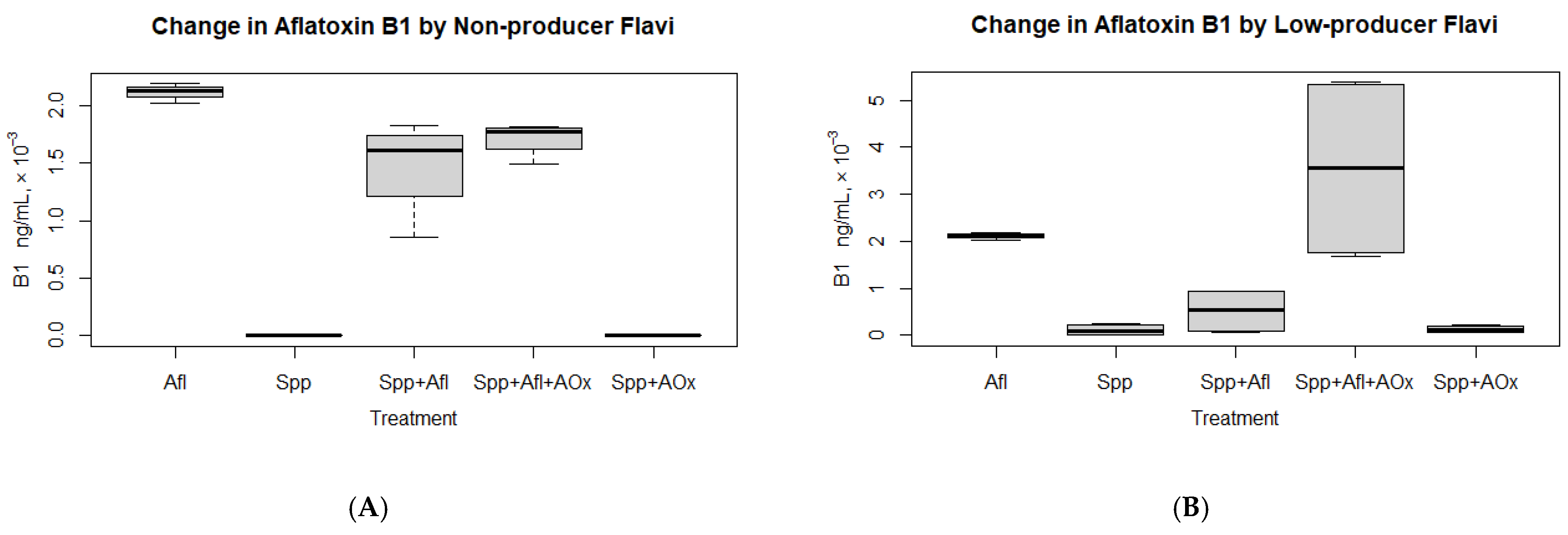
3.1.1. Degradation of Aflatoxin in Non-Aflatoxin Producers without Antioxidant Treatment

There was a significant reduction in added B1 (“Afl”) when exposed to the atoxigenic strains (“Spp+Afl”) (Figure 2A; Table 3), with all isolates digesting the toxin. The greatest reduction was observed with isolate 1MS7 (60%). There was, however, no significant reduction in G1 added. All isolates except 1MS7 did not reduce the added G1. Isolate 1MS7 reduced G1 by 14%.

3.1.2. Change in Aflatoxin in Low-Aflatoxin Producers without Antioxidant Treatment

There was a significant reduction in B1 due to exposure of added AF (“Afl”) to low AF producer strains (“Spp+Afl”) (Figure 2B; Table 3). All isolates reduced the B1. There was no significant overall reduction or change in G1 levels compared to the added amount (“Afl”) due to exposure to low AF producer strains (“Spp+Afl”). Two isolates reduced G1, with ELV13C having the highest G1 reduction (73%). The two other isolates did not reduce G1. Furthermore, the maximally produced levels of G1 (“Spp” = 732 ng/mL) were exceeded when extrinsic AF (G1 “Afl” = 3243 ± 786 ng/mL) was introduced (G1 “Spp+Afl” = 4881 ng/mL).

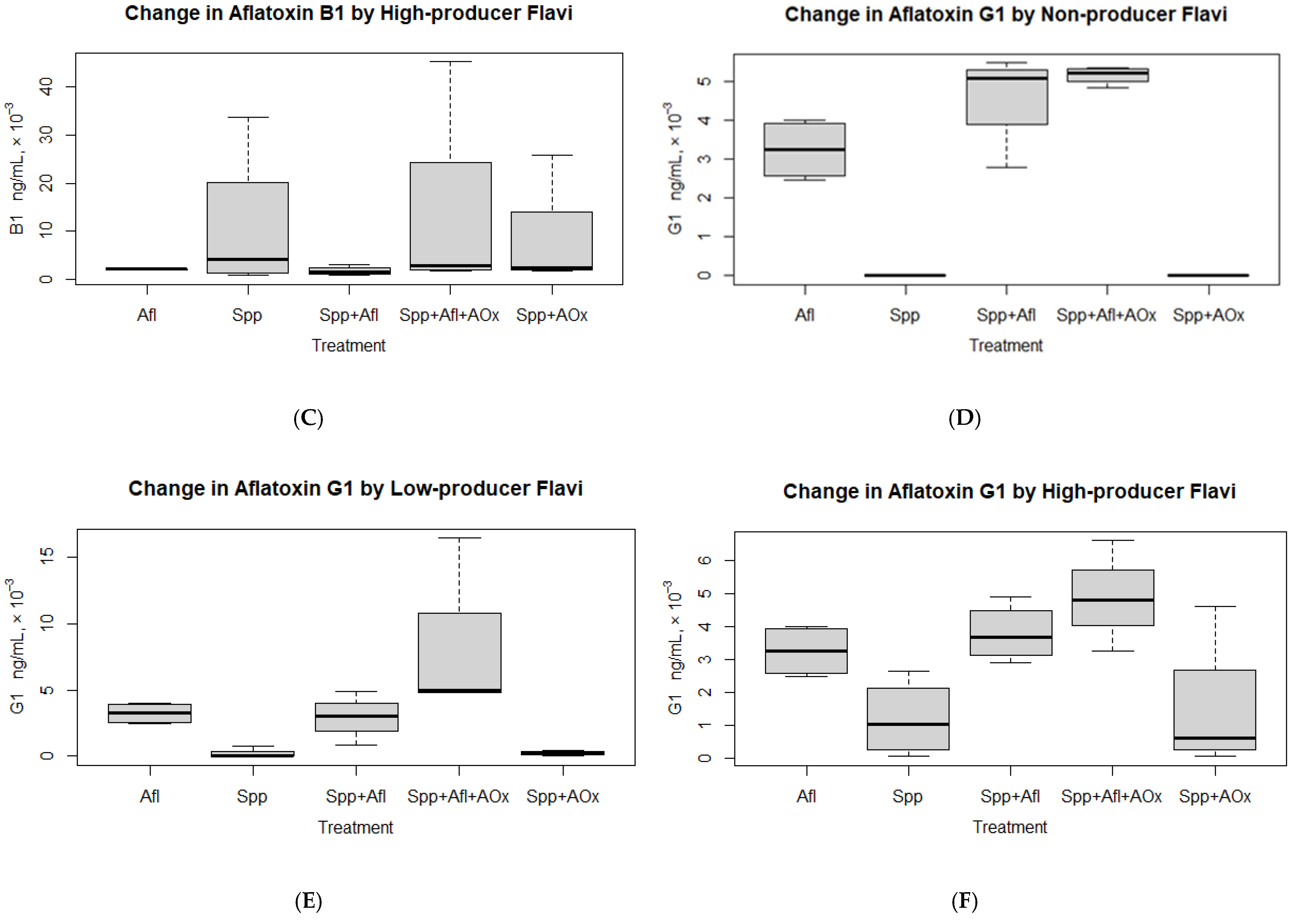
3.1.3. Change in Aflatoxin in High-Aflatoxin Producers without Antioxidant Treatment

There was no significant change in added B1 (“Afl”) due to exposure to high AF producer strains (“Spp+Afl”) (Table 3). However, the B1 maximum levels produced (“Spp+Afl”: 3169 ng/mL) were modulated to levels closer to the added amount (“Afl”: 2114 ± 68 ng/mL) compared to the maximum levels the species (“Spp”) would produce without extrinsic B1 (“Spp”: 33,749 ng/mL) (Figure 2C). (Modulate implies maintaining the level of the aflatoxin variant to within what the Flavi produces.)
With G1, the change was not significant between amount the species produced (“Spp”: 1181 ng/mL) and what it produced together with extrinsically introduced G1 (“Spp+Afl”: 3793 ng/mL) (Table 3). However, the total amount of G1 (“Spp+Afl” = 3793 ng/mL) simulated an augmentation of added G1 amount (“Afl” = 3243 ± 786 ng/mL) with the amount produced by the species (“Spp” = 1181 ng/mL) (Figure 2F).
Overall, the aflatoxin-producing isolates modulated B1 in their environment by reducing the extrinsically added amount or limiting the overall amount to levels added. This was, however, not the case with G1, which tended to increase overall (Table 3).

3.1.4. Degradation of Aflatoxin in Non-Aflatoxin Producers with Antioxidant (Se) Treatment

The antioxidant did not lead to a significant change in extrinsic B1 or G1 reduction levels. (Wilcoxon rank sum exact test, “Spp+Afl” v “Spp+Afl+AOx” p > 0.05). As expected, treatment of the isolates with the antioxidant did not change the AF levels (remained < LOQ) as the isolates were non-producers.

3.1.5. Change in Aflatoxin in Low-Aflatoxin Producers with Antioxidant (Se) Treatment

The antioxidant in the presence of extrinsic B1 led to an increase in the levels of B1 (“Spp+Afl” v “Spp+Afl+AOx”, Table 4; Figure 2B). When antioxidant was introduced to the species without B1 (“Spp+AOx”), the maximum levels of AF the species could produce did not exceed that produced by the species without treatment (“Spp”). With G1, there was no significant difference in levels with or without treatment with antioxidant as long as G1 had been introduced extrinsically, showing ineffectiveness in the overall breakdown of G1 by the “Spp” with or without antioxidant (Table 4, Figure 2E). There was an observed reduction in maximum levels of G1 produced when “Spp” had been treated with antioxidant (“Spp+AOx”), although the overall difference in G1 between “Spp” and “Spp+AOx” was not significant.

3.1.6. Change in Aflatoxin in High-Aflatoxin Producers with Antioxidant (Se) Treatment

There was no effect of antioxidant in the presence of extrinsic B1 on overall levels of B1 (“Spp+Afl+AOx” v “Spp+Afl”). However, the maximum levels of B1 increased in the presence of antioxidant from 3169 to 45,168 ng/mL, showing a reduction in the modulation of the maximum levels of B1 in the presence of antioxidant.
When antioxidant was introduced to the species without extrinsic B1 (“Spp+AOx”), the maximum levels of AF the species could produce did not exceed that produced by the species without treatment (Table 4, Figure 2C). The overall difference in generated B1 between the treatment of species with antioxidant and non-treatment was, however, not significant. With G1, there was no significant difference in levels of G1 with or without treatment with antioxidant (“Spp” v “Spp+AOx”, p > 0.05) as long as G1 had been introduced extrinsically (Figure 2F). This showed that G1 was not effectively broken down by the “Spp” with or without antioxidant. However, there was an observed increase in maximum levels of the G1 produced in the presence of antioxidant (Table 4).
Considering that high producer strains did not significantly change levels of AF produced due to low antioxidant dose (Se = 0.40 µg/g), we investigated the gene expression patterns of structural gene aflD and regulatory gene aflR as well as AF response in two selected high AF producer strains (MLV14F and ELG33C).

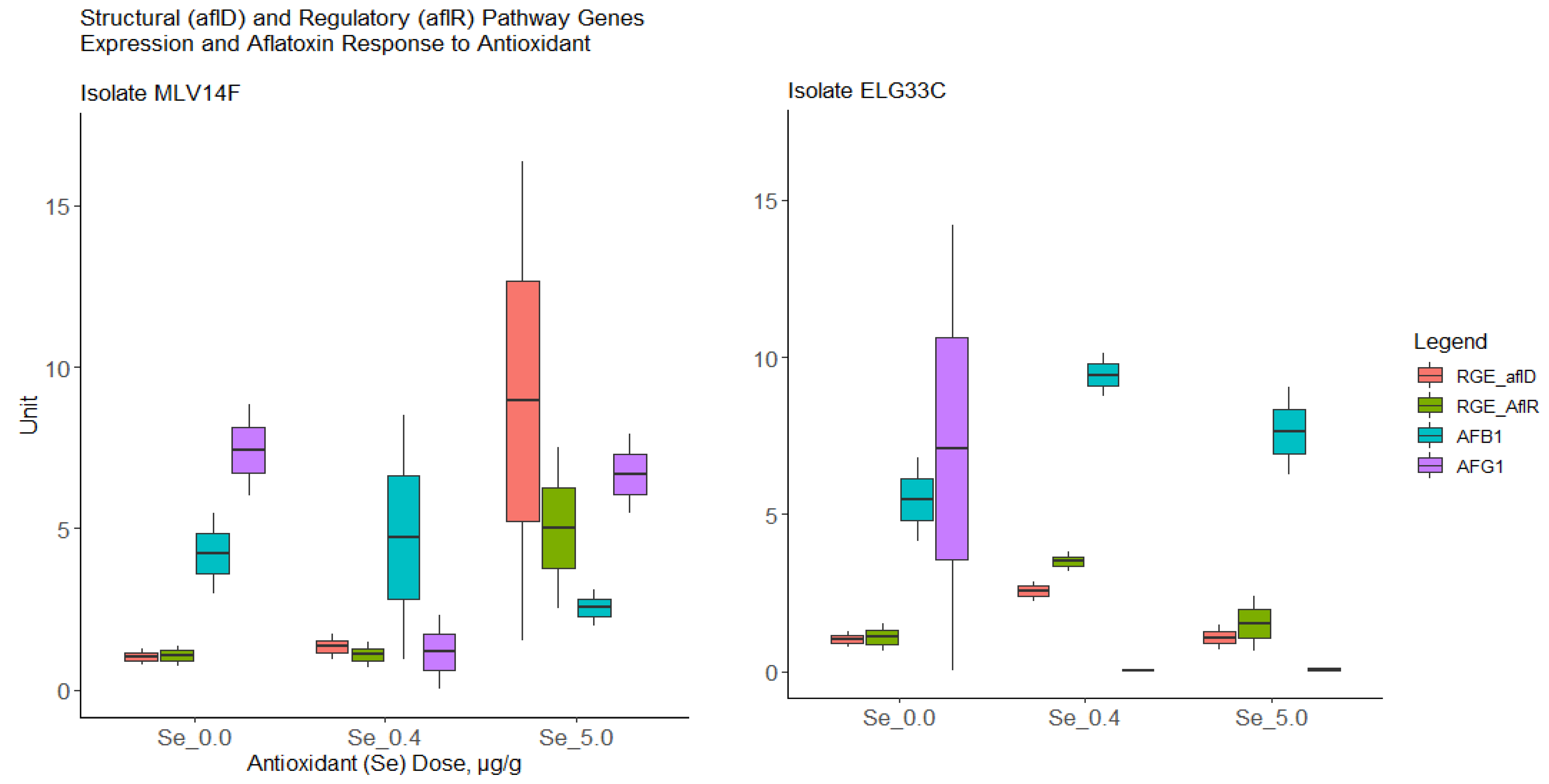
Gene Expression in Aflatoxin Pathway Genes in High Producer Isolates Due to Antioxidant

It was observed from the two randomly tested isolates that in both, the up-regulation of the regulatory gene aflR led to the up-regulation of structural gene aflD (which is involved in AF precursor molecule decoration) and vice versa (Figure 3; Spearman correlation: rho = 0.90; p value < 0.001).
However, the two isolates responded differently to the levels of the antioxidant introduced. Isolate ELG33C showed an upregulation of genes at Se dose 0.4 µg/g and a downregulation at a much higher (8-fold) antioxidant dose. However, the isolate MLV14F upregulated genes at a much higher Se level (8-fold) with no effect at a 0.4 µg/g Se dose. There was no clear correlation between gene expression in the two pathway genes and AF levels. However, it was exceptionally observed that B1 in isolate ELG33C, whose gene upregulation responded to lower Se levels, increased with the upregulation of aflR and aflD and reduced with the downregulation of the two genes.

3.2. Fitness of Isolates Due to Antioxidant Treatment

Without antioxidant (Se) treatment, both atoxigenic and toxigenic fungi were of equal fitness (Wilcoxon rank sum exact test, p = 0.739). Similarly, there was no significant difference in fungal fitness at antioxidant treatment level 0.40 µg/g (p = 0.912). However, a difference in fitness was detected at the treatment level 0.86 µg/g (Figure 4, p = 0.023). The non-producer isolates were fitter than their aflatoxin-producing counterparts (geometric mean: atoxigenic = 0.655 × 10⁶ spores/mL and toxigenic = 0.209 × 10⁶ spores/mL).

4. Discussion

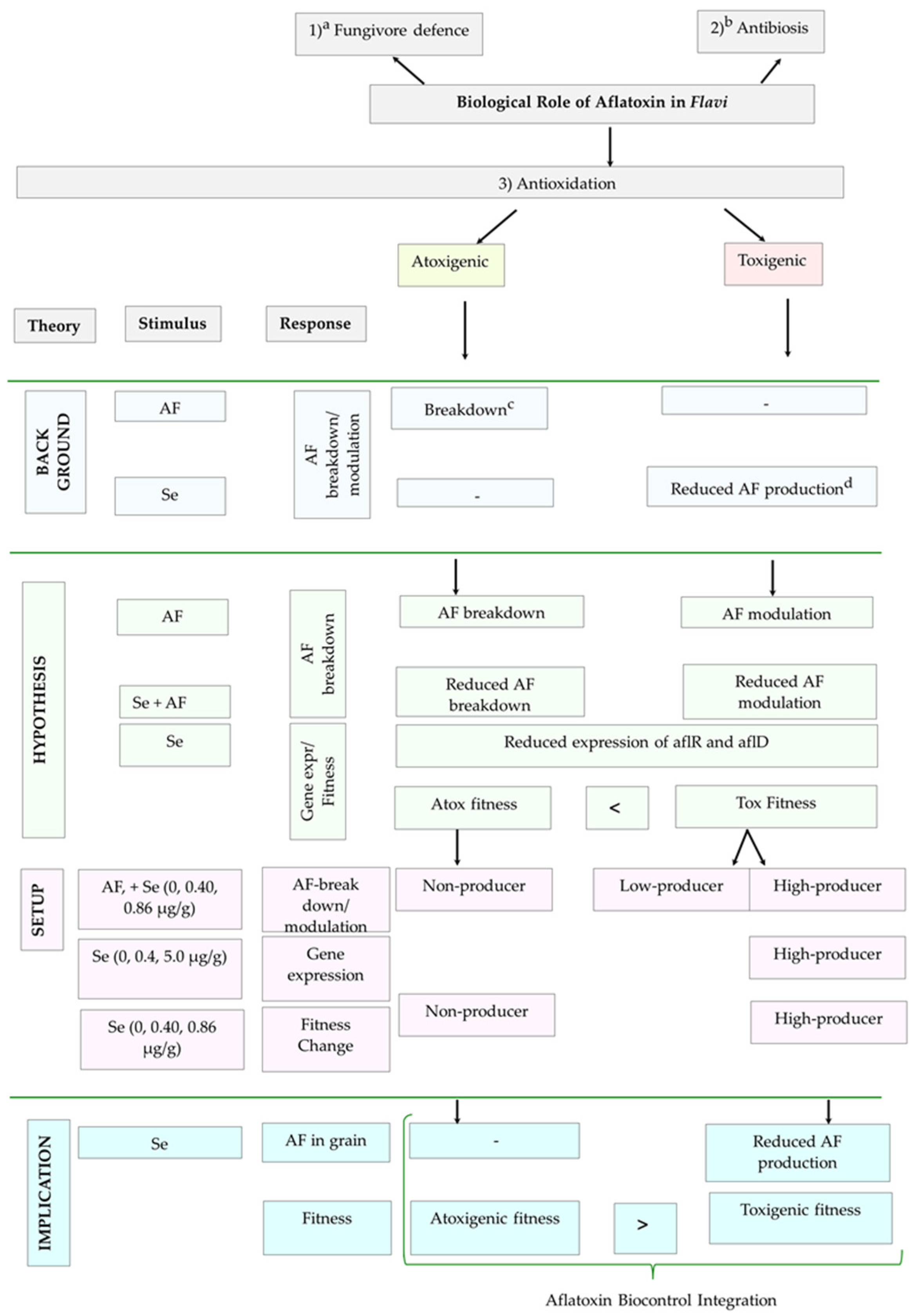
4.1. Degradation of Aflatoxin Levels in Flavi through Antioxidative Mechanism

With the investigated strains, we demonstrate that toxigenic Flavi, in fact, modulate levels of B1 in their environment to within the levels they produce it. To do so, they partly degrade the B1 and also maintain the B1 to within a certain level. The modulation is evidenced by the reduction in the added amount of B1 to medium, to levels within what the species produced (“Afl” v “Spp+Afl” in Figure 2B,C). Furthermore, we demonstrate that this is carried out through an antioxidative mechanism, such that B1 plays an antioxidative role. The antioxidative role is evidenced by the increase in levels of AF in the medium in which both AF and selenium (Se) had simultaneously been added compared to AF alone without the addition of Se (“Spp+Afl” v “Spp+Afl+AOx” in Figure 2B,C). The mechanism in non-producers may, however, not be the same as for producers, as seen by the no effect of Se on the breakdown of extrinsically introduced AF (Figure 2A, Table 4). The observed increase in levels of B1 suggests that the antioxidative role of B1 as a free-radical scavenger was demoted by the presence of the antioxidant Se, with Se presumably taking the more active role in the wiping out of oxidative elements. Examples of such oxidative elements include reactive oxygen species (ROS), as previously described [20,22,24]. Our results also show that B1 is the preferred natural antioxidative biochemical in Flavi compared to G1. This is evidenced by the fact that the addition of B1 and G1 to the medium resulted in the breakdown of B1 for all types of isolates (except MKA01K), whereas G1 was not digested in most isolates (Figure 2D–F). In this regard, overall, the extrinsic introduction of B1 and G1 resulted in higher maximum levels of G1 for low and high producers in contrast to B1, whose average or maximally produced levels by fungi were reduced. The proposed preference of B1 to G1 in the antioxidative AF modulation in the Flavi environment may also be indicative of why B1 is usually found in a significant proportion of total AF in a number of Flavi strains [51,52,53,54,55] (Supplementary Table S1). B1 is also seen to be produced in higher amounts in a good number of strains studied compared to the rest of the AF types [52,53]. The modulation of AF levels can also be noted from the previous investigation in which A. parasiticus degraded AF through peroxidase enzyme [20] despite being a producer of the AF.
As regards the degradation of AF by non-producer strains, in their previous study, [33] showed that atoxigenic strains of Flavi digest B1. In our study, we furthermore show that low producers of B1 similarly do digest B1. Furthermore, we demonstrate that the mechanism of this digestion may not be the same between atoxigenic and toxigenic Flavi. While the low and high producers degraded the B1 in an antioxidative mechanism, the non-producers did not seem to degrade it in such a mechanism. This is explained by the fact that a significant increase in B1 is observed for low and high B1 producers when antioxidant (Se) is introduced to the environment alongside extrinsic B1. This is in comparison to the introduction of extrinsic B1 without Se. The increase is, however, not observed for non-producers between the two treatments (“Spp+Afl” v “Spp+Afl+AOx”, p > 0.05; Table 4). On the contrary, Se had no effect on the digestion of the B1 in non-producers, which sustained its digestion to similar levels as without the addition of Se (Figure 2A).
From a practical perspective, our findings also suggest the presence of antioxidants in the kernel before Flavi infection may deter an increase in AF levels in the kernel above certain limits. This is in comparison to the introduction of the antioxidant when AF and toxigenic Flavi are already present in the kernel (model “Spp”:“Spp+AOx”). This would imply that alteration of the abiotic environment of the kernel at preharvest by the introduction of non-toxic levels of mineral Se-antioxidant may be useful in deterring spikes in AF contamination. The approach would be problematic if the introduction of Se is at postharvest, such that a product that already has AF is biofortified with Se and gets contaminated with toxigenic Flavi. This would equate to the model “Spp”:“Spp+Afl+AOx” in our investigation, potentially leading to a spike in B1 and G1. Furthermore, our findings may also partly explain why a study on Brazilian nuts showed a higher accumulation of AF in nuts with higher levels of Se than those with lower levels of the antioxidant [56]. Considering that nuts are soil-borne, they are likely to get contaminated with aflatoxigenic Flavi and, subsequently, AF in the early stages of seed growth. This then renders the AF produced by Flavi unutilised in an antioxidative role due to the Se in the seed. This resonates with the observed higher levels of B1 and G1 in our study under the scenario “Spp+Afl+AOx.”
It should be noted that for the complete impediment of Flavi, higher levels of the antioxidant Se would have to be administered such that the Flavi fitness is reduced through non-sporulation [57,58] and subsequently prevent the AF production by the Flavi. The increase in the antioxidative capacity of a plant has been suggested previously as a means of reducing the oxidative stress in Flavi and, subsequently, AF production [22]. The antioxidative capacity of a plant by using elements such as Se would have to be high enough. This is as shown by our findings that the non-lethal dose (0.86 µg/g) did not deter AF production (on YES medium) in Flavi, although it reduced the maximum B1 levels produced. Higher Se antioxidative capacity can be achieved through the use of the nanoparticle format of Se. Se nanoparticles in higher levels are less toxic to organisms than mineral Se [59].

Gene Expression in High Aflatoxin B1/G1 Producer Isolates in Response to Antioxidant (Se)

From the results of two randomly selected high-producer strains (soil isolate ELG33C; maize isolate MLV14F), we demonstrated that the two biosynthetic genes aflR (regulatory) and aflD (structural) were consistently either both upregulated or downregulated in response to the abiotic stimulus Se-antioxidant. Furthermore, we observed a positive correlation in isolate ELG33C between expression levels of both aflD and aflR and the levels of B1 produced. The consistency was, however, not observed in isolate MLV14F as well as the G1 levels for both isolates (Figure 3).
Although the transcription factor aflR and structural gene aflD are involved in AF production in Flavi, most studies have shown no correlations between the gene expression levels in the two genes with levels of AF produced [60,61,62,63,64]. While some few studies have shown a level of correlation with AF levels produced, the studies have either been inconsistent with each other or the expression inconsistent with the expected highest production conditions for AF [65,66,67]; for example, high expression of aflD at both lowest B1 levels and lowest aflR expression and vice versa [65]. This may show that response to stimuli may be strain dependent, for example, the observed strain differences in response to CO2 [68] or temperature [64]. The possible strain dependence is also seen in our study on expression patterns of aflD/aflR to Se stimulus (Figure 3). In our study, the levels of available mRNA from aflD and aflR differed from the specific induced levels of Se as an antioxidant. Our preliminary findings show that the soil isolate ELG33C downregulated the two pathway genes at elevated control concentration (5 µg/g) after the preliminary upregulation at 0.40 µg/g. On the other hand, the maize isolate MLV14F only upregulated the two pathway genes at an elevated control concentration of 5 µg/g with no significant change at 0.40 µg/g. This would suggest that the downregulation of the genes in isolate MLV14F may probably occur at a higher Se level than the control-elevated concentration of 5 µg/g. A higher isolate sample size in our study would be required in order to fathom the pattern of gene expression–aflatoxin production correlation with antioxidants. An additional factor most investigators suggest in order to improve the correlation between gene expression and abiotic stimulus is the prevention of oxidative elements that could degrade the AFs in the medium in the course of the experiment, leading to poor or inconsistent correlations.

4.2. Fitness Response of Atoxigenic and Toxigenic Isolates under Antioxidant (Se) Treatment

At the maximum non-lethal dose of mineral Se administered (0.86 µg/g), we demonstrate the increase in fitness of the tested atoxigenic strains of Flavi compared to toxigenic counterparts, rejecting our initial hypothesis. Different fungi can thrive differently under the same abiotic environment or, indeed, under different abiotic environments. As seen in this investigation, introduction of non-lethal dose Se triggered an increase in spore production in the Flavi. We may attribute the relatively higher Darwinian fitness in atoxigenic than toxigenic Flavi to the presumption that the toxigenic Flavi may have channeled the Se to both sporulation and oxidative stress alleviation, whereas the atoxigenic Flavi may have solely channeled the Se towards sporulation. Considering the alteration of the abiotic environment for Flavi in our study, such a comparative increase in fitness of atoxigenic compared to toxigenic counterparts could be an important attribute in promoting the population of toxigenic over atoxigenic strains. This provides the need to further investigate the inhibitory concentration of Se nanoparticles that would trigger a growth inhibition of toxigenic strains, while at the same time allowing some atoxigenic counterparts to grow without complete inhibition. This is considering that biocontrol of AF premised on competitive exclusion relies on atoxigenic Flavi to outgrow toxigenic counterparts and colonise host crops [69,70]. From a practical perspective, integrating pre-harvest Se-biofortification of maize can have prospects to play a useful role as part of a broader AF biocontrol strategy.

5. Conclusions

While atoxigenic Flavi are known to degrade B1 in their environment, as seen in this study, toxigenic Flavi of this investigation modulated B1 in an antioxidative role by doing so to levels less than what they produced. We furthermore see that B1 happened to be a preferred antioxidative biochemical for the Flavi compared to G1. Furthermore, we do observe that atoxigenic Flavi used in this study were fitter than toxigenic counterparts under antioxidant treatment, contrary to expectations but a positive attribute. Hence, work may need to be performed on the prospective use of antioxidative elements, such as selenium (Se) in nanoparticle form, to drive a better fitness of atoxigenic fungi over toxigenic strains. This would be in the form of a broader biocontrol strategy in the competitive exclusion of Flavi. Furthermore, it should also be noted that when considering the use of antioxidants such as Se in substrates such as maize, consideration should be made to the AF contamination status of the substrate. From a practical perspective, Se biofortification may preferably be performed at preharvest on maize, or at postharvest when the product is without Flavi and AF contamination. This is because, while Se could deter potential spikes in B1 and G1 as observed in this study, its application to an environment already harbouring B1 and Flavi can conversely lead to a spike in the dreaded B1 when conditions are conducive for Flavi to proliferate.

Supplementary Materials

The following supporting information can be downloaded at https://www.mdpi.com/article/10.3390/jof9060690/s1. Table S1: Percentage (%) of aflatoxin-producing ability of isolates.

Author Contributions

Conceptualization, B.K., H.N., S.E.S. and S.K.; methodology, B.J.Z., B.K., H.N. and S.E.S.; software, B.K.; validation, A.D.v.D., H.N., P.W.K. and S.E.S.; formal analysis, B.K. and S.K.; investigation, B.K. and S.K.; resources, B.J.Z., H.N. and S.E.S.; data curation, B.K. and S.K.; writing—original draft preparation, B.K. and S.K.; writing—review and editing, B.K., H.N. and P.W.K.; visualization, B.K. and S.K.; supervision, A.D.v.D., B.J.Z., H.N., P.W.K. and S.E.S.; project administration, B.J.Z., H.N. and S.E.S.; funding acquisition, B.J.Z., H.N. and S.E.S. All authors have read and agreed to the published version of the manuscript.

Funding

Funding for the research project was provided by Wageningen University and Research (The Netherlands) and the National Institute for Scientific and Industrial Research (Zambia). No specific grant was received from any funding agency.

Institutional Review Board Statement

Not applicable.

Informed Consent Statement

Not applicable.

Data Availability Statement

Code for the aflatoxin degradation/modulation analyses is found at https://github.com/bkatati/afloxidate (accessed on 27 April 2023). The lab culture isolates used in the experiment were stored at −70 °C, available for re-use.

Acknowledgments

We thank Eric Bastiaans at Laboratory of Genetics for the technical assistance rendered in the fungal fitness assays.

Conflicts of Interest

The authors declare no conflict of interest.

References

  1. Ndisio, B.; Peter, W.; Victor, K.; Sheila, O.; Boaz, N.; Wachira, P.; Kagot, V.; Okoth, S. Susceptibility of locally cultivated groundnut (Arachis hypogaea) varieties to aflatoxin accumulation in Homa Bay County, Kenya. Afr. J. Microbiol. Res. 2017, 11, 1329–1337. [Google Scholar] [CrossRef] [Green Version]
  2. Mallikarjunaiah, N.H.; Jayapala, N.; Puttaswamy, H.; Ramachandrappa, N.S. Characterization of non-aflatoxigenic strains of Aspergillus flavus as potential biocontrol agent for the management of aflatoxin contamination in groundnut. Microb. Pathog. 2017, 102, 21–28. [Google Scholar] [CrossRef] [Green Version]
  3. Kachapulula, P.W.; Akello, J.; Bandyopadhyay, R.; Cotty, P.J. Aspergillus section Flavi community structure in zambia influences aflatoxin contamination of maize and groundnut. Int. J. Food Microbiol. 2017, 261, 49–56. [Google Scholar] [CrossRef]
  4. Munkvold, G.P.; Arias, S.; Taschl, I.; Gruber-Dorninger, C. Chapter 9: Mycotoxins in corn: Occurrence, impacts, and management. In Corn; AACC International Press: St. Paul, MN, USA, 2019; pp. 235–287. [Google Scholar] [CrossRef]
  5. Akello, J.; Ortega-Beltran, A.; Katati, B.; Atehnkeng, J.; Augusto, J.; Mwila, C.M.; Mahuku, G.; Chikoye, D.; Bandyopadhyay, R. Prevalence of Aflatoxin- and Fumonisin-Producing Fungi Associated with Cereal Crops Grown in Zimbabwe and Their Associated Risks in a Climate Change Scenario. Foods 2021, 10, 287. [Google Scholar] [CrossRef]
  6. WHO. Safety Evaluation of Certain Contaminants in Food: Prepared by the Eighty-Third Meeting of the Joint FAO/WHO Expert Committee on Food Additives (JECFA); World Health Organization: Geneva, Switzerland, 2018. [Google Scholar]
  7. IARC. Agents Classified by the IARC Monographs; International Agency for Research on Cancer, vol. 1–104. 2012. Available online: https://monographs.iarc.who.int/list-of-classifications (accessed on 18 June 2023).
  8. Rushing, B.R.; Selim, M.I. Aflatoxin B1: A review on metabolism, toxicity, occurrence in food, occupational exposure, and detoxification methods. Food Chem. Toxicol. 2019, 124, 81–100. [Google Scholar] [CrossRef]
  9. Li, C.; Liu, X.; Wu, J.; Ji, X.; Xu, Q. Research progress in toxicological effects and mechanism of aflatoxin B1 toxin. PeerJ 2022, 10, e13850. [Google Scholar] [CrossRef]
  10. Mohsenzadeh, M.S.; Hedayati, N.; Riahi-Zanjani, B.; Karmi, G. Immunosuppression following dietary aflatoxin B1 exposure: A review of the existing evidence. Toxin Rev. 2016, 35, 121–127. [Google Scholar] [CrossRef]
  11. Zhao, Y.; Wang, T.; Li, P.; Chen, J.; Nepovimova, E.; Long, M.; Wu, W.; Kuca, K. Bacillus amyloliquefaciens B10 can alleviate aflatoxin B1-induced kidney oxidative stress and apoptosis in mice. Ecotoxicol. Environ. Saf. 2021, 218, 112286. [Google Scholar] [CrossRef] [PubMed]
  12. Gallo, A.; Giuberti, G.; Frisvad, J.; Bertuzzi, T.; Nielsen, K. Review on Mycotoxin Issues in Ruminants: Occurrence in Forages, Effects of Mycotoxin Ingestion on Health Status and Animal Performance and Practical Strategies to Counteract Their Negative Effects. Toxins 2015, 7, 3057–3111. [Google Scholar] [CrossRef] [PubMed] [Green Version]
  13. Abdel-Hamid, A.A.; Firgany, A.E.-D.L. Vitamin E supplementation ameliorates aflatoxin B1-induced nephrotoxicity in rats. Acta Histochem. 2015, 117, 767–779. [Google Scholar] [CrossRef] [PubMed]
  14. Rotimi, O.A.; Rotimi, S.O.; Duru, C.U.; Ebebeinwe, O.J.; Abiodun, A.O.; Oyeniyi, B.O.; Faduyile, F.A. Acute aflatoxin B1—Induced hepatotoxicity alters gene expression and disrupts lipid and lipoprotein metabolism in rats. Toxicol. Rep. 2017, 4, 408–414. [Google Scholar] [CrossRef]
  15. Wang, Y.; Liu, F.; Zhou, X.; Liu, M.; Zang, H.; Liu, X.; Shan, A.; Feng, X. Alleviation of Oral Exposure to Aflatoxin B1-Induced Renal Dysfunction, Oxidative Stress, and Cell Apoptosis in Mice Kidney by Curcumin. Antioxidants 2022, 11, 1082. [Google Scholar] [CrossRef]
  16. Engin, A.B.; Engin, A. DNA damage checkpoint response to aflatoxin B1. Environ. Toxicol. Pharmacol. 2019, 65, 90–96. [Google Scholar] [CrossRef]
  17. Drott, M.T.; Debenport, T.; Higgins, S.A.; Buckley, D.H.; Milgroom, M.G. Fitness Cost of Aflatoxin Production in Aspergillus flavus When Competing with Soil Microbes Could Maintain Balancing Selection. mBio 2019, 10, e02782-18. [Google Scholar] [CrossRef] [PubMed] [Green Version]
  18. Drott, M.T.; Lazzaro, B.; Brown, D.; Carbone, I.; Milgroom, M. Balancing selection for aflatoxin in Aspergillus flavus is maintained through interference competition with, and fungivory by insects. Proc. Biol. Sci. 2017, 284, 20172408. [Google Scholar]
  19. Arai, T.; Ito, T.; Koyama, Y. Antimicrobial Activity of Aflatoxins. J. Bacteriol. 1967, 93, 59–64. [Google Scholar] [CrossRef] [Green Version]
  20. Doyle, M.P.; Marth, E.H. Peroxidase activity in mycelia of Aspergillus parasiticus that degrade aflatoxin. Appl. Microbiol. Biotechnol. 1979, 7, 211–217. [Google Scholar] [CrossRef]
  21. Fountain, J.C.; Bajaj, P.; Nayak, S.N.; Yang, L.; Pandey, M.; Kumar, V.; Jayale, A.S.; Chitikineni, A.; Lee, R.D.; Kemerait, R.C.; et al. Responses of Aspergillus flavus to Oxidative Stress Are Related to Fungal Development Regulator, Antioxidant Enzyme, and Secondary Metabolite Biosynthetic Gene Expression. Front. Microbiol. 2016, 7, 2048. [Google Scholar] [CrossRef] [PubMed] [Green Version]
  22. Fountain, J.C.; Bajaj, P.; Pandey, M.; Nayak, S.N.; Yang, L.; Kumar, V.; Jayale, A.S.; Chitikineni, A.; Zhuang, W.; Scully, B.T.; et al. Oxidative stress and carbon metabolism influence Aspergillus flavus transcriptome composition and secondary metabolite production. Sci. Rep. 2016, 6, 38747. [Google Scholar] [CrossRef] [PubMed] [Green Version]
  23. Jayashree, T.; Subramanyam, C. Oxidative stress as a prerequisite for aflatoxin production by Aspergillus parasiticus. Free. Radic. Biol. Med. 2000, 29, 981–985. [Google Scholar] [CrossRef]
  24. Narasaiah, K.V.; Sashidhar, R.B.; Subramanyam, C. Biochemical analysis of oxidative stress in the production of aflatoxin and its precursor intermediates. Mycopathologia 2006, 162, 179–189. [Google Scholar] [CrossRef] [PubMed]
  25. Fountain, J.C.; Scully, B.T.; Ni, X.; Kemerait, R.C.; Lee, R.D.; Chen, Z.-Y.; Guo, B. Environmental influences on maize-Aspergillus flavus interactions and aflatoxin production. Front. Microbiol. 2014, 5, 40. [Google Scholar] [CrossRef] [PubMed] [Green Version]
  26. Rayman, M.P. The importance of selenium to human health. Lancet 2000, 356, 233–241. [Google Scholar] [CrossRef] [PubMed] [Green Version]
  27. Kuršvietienė, L.; Mongirdienė, A.; Bernatonienė, J.; Šulinskienė, J.; Stanevičienė, I. Selenium Anticancer Properties and Impact on Cellular Redox Status. Antioxidants 2020, 9, 80. [Google Scholar] [CrossRef] [Green Version]
  28. Alsuhaibani, A.M.A. Functional role of selenium-fortified yogurt against aflatoxin-contaminated nuts in rats. Agric. Food Secur. 2018, 7, 21. [Google Scholar] [CrossRef] [Green Version]
  29. Mughal, M.J.; Peng, X.; Kamboh, A.A.; Zhou, Y.; Fang, J. Aflatoxin B1 Induced Systemic Toxicity in Poultry and Rescue Effects of Selenium and Zinc. Biol. Trace Element Res. 2017, 178, 292–300. [Google Scholar] [CrossRef]
  30. Wang, J.; Lin, L.; Jiang, Q.; Huang, W.; Liu, N. Effect of supplemental lactic acid bacteria on growth performance, glutathione turnover and aflatoxin B1 removal in lambs. Czech J. Anim. Sci. 2019, 64, 272–278. [Google Scholar] [CrossRef] [Green Version]
  31. Kaur, T.; Vashisht, A.; Prakash, N.T.; Reddy, M.S. Role of Selenium-Tolerant Fungi on Plant Growth Promotion and Selenium Accumulation of Maize Plants Grown in Seleniferous Soils. Water Air Soil Pollut. 2022, 233, 17. [Google Scholar] [CrossRef]
  32. Naseem, M.; Anwar-Ul-Haq, M.; Wang, X.; Farooq, N.; Awais, M.; Sattar, H.; Malik, H.A.; Mustafa, A.; Ahmad, J.; El-Esawi, M.A. Influence of Selenium on Growth, Physiology, and Antioxidant Responses in Maize Varies in a Dose-Dependent Manner. J. Food Qual. 2021, 2021, 6642018. [Google Scholar] [CrossRef]
  33. Maxwell, L.A.; Callicott, K.; Bandyopadhyay, R.; Mehl, H.; Orbach, M.; Cotty, P. Degradation of aflatoxins B1 by atoxigenic Aspergillus flavus biocontrol agents. Plant Dis. 2021, 105, 2343–2350. [Google Scholar] [CrossRef] [PubMed]
  34. Bock, C.H.; Cotty, P.J. Wheat seed colonized with atoxigenic Aspergillus flavus: Characterization and production of a biopesticide for aflatoxin control. Biocontrol Sci. Technol. 1999, 9, 529–543. [Google Scholar] [CrossRef]
  35. Medina, A.; Mohale, S.; Samsudin, N.I.P.; Rodriguez-Sixtos, A.; Rodriguez, A.; Magan, N. Biocontrol of mycotoxins: Dynamics and mechanisms of action. Curr. Opin. Food Sci. 2017, 17, 41–48. [Google Scholar] [CrossRef]
  36. Rao, K.R.; Vipin, A.; Venkateswaran, G. Mechanism of inhibition of aflatoxin synthesis by non-aflatoxigenic strains of Aspergillus flavus. Microb. Pathog. 2020, 147, 104280. [Google Scholar] [CrossRef]
  37. Xing, F.; Wang, L.; Liu, X.; Selvaraj, J.N.; Wang, Y.; Zhao, Y.; Liu, Y. Aflatoxin B 1 inhibition in Aspergillus flavus by Aspergillus niger through down-regulating expression of major biosynthetic genes and AFB 1 degradation by atoxigenic A. flavus. Int. J. Food Microbiol. 2017, 256, 1–10. [Google Scholar] [CrossRef] [PubMed]
  38. Yu, J.; Bhatnagar, D.; Cleveland, T.E. Completed sequence of aflatoxin pathway gene cluster in Aspergillus parasiticus. FEBS Lett. 2004, 564, 126–130. [Google Scholar] [CrossRef] [PubMed] [Green Version]
  39. Yu, J.; Fedorova, N.D.; Montalbano, B.G.; Bhatnagar, D.; Cleveland, T.E.; Bennett, J.W.; Nierman, W.C. Tight control of mycotoxin biosynthesis gene expression in Aspergillus flavus by temperature as revealed by RNA-Seq. FEMS Microbiol. Lett. 2011, 322, 145–149. [Google Scholar] [CrossRef] [PubMed] [Green Version]
  40. Chang, P.-K. Lack of interaction between AFLR and AFLJ contributes to nonaflatoxigenicity of Aspergillus sojae. J. Biotechnol. 2004, 107, 245–253. [Google Scholar] [CrossRef]
  41. Price, M.S.; Conners, S.B.; Tachdjian, S.; Kelly, R.M.; Payne, G.A. Aflatoxin conducive and non-conducive growth conditions reveal new gene associations with aflatoxin production. Fungal Genet. Biol. 2005, 42, 506–518. [Google Scholar] [CrossRef]
  42. Woloshuk, C.P.; Foutz, K.R.; Brewer, J.F.; Bhatnagar, D.; Cleveland, E.T.; Payne, A.G. Molecular characterization of aflR, a regulatory locus for aflatoxin biosynthesis. Appl. Environ. Microbiol. 1994, 60, 2408–2414. [Google Scholar] [CrossRef] [Green Version]
  43. Georgianna, D.R.; Payne, G.A. Genetic regulation of aflatoxin biosynthesis: From gene to genome. Fungal Genet. Biol. 2009, 46, 113–125. [Google Scholar] [CrossRef]
  44. Ono, E.Y.S.; Da Silva, M.; Ribeiro, R.M.R.; Ono, M.A.; Hayashi, L.; Garcia, G.T.; Hirooka, E.Y. Comparison of thin-layer chromatography, spectrofluorimetry and bright greenish-yellow fluorescence test for aflatoxin detection in corn. Braz. Arch. Biol. Technol. 2010, 53, 687–692. [Google Scholar] [CrossRef]
  45. Abdollahi, A.; Buchanan, R.L. Regulation of Aflatoxin Biosynthesis: Induction of Aflatoxin Production by Various Carbohydrates. J. Food Sci. 1981, 46, 633–635. [Google Scholar] [CrossRef]
  46. Medina, A.; Rodríguez, A.; Magan, N. Climate change and mycotoxigenic fungi: Impacts on mycotoxin production. Curr. Opin. Food Sci. 2015, 5, 99–104. [Google Scholar] [CrossRef]
  47. Abdel-Hadi, A.; Carter, D.; Magan, N. Temporal monitoring of the nor-1 (aflD) gene of Aspergillus flavus in relation to aflatoxin B1 production during storage of peanuts under different water activity levels. J. Appl. Microbiol. 2010, 109, 1914–1922. [Google Scholar] [CrossRef] [Green Version]
  48. Livak, K.J.; Schmittgen, T.D. Analysis of relative gene expression data using real-time quantitative PCR and the 2(-Delta Delta C(T)) Method. Methods 2001, 25, 402–408. [Google Scholar] [CrossRef]
  49. R Core Team. R: A Language and Environment for Statistical Computing; R Foundation for Statistical Computing: Vienna, Austria, 2013; Available online: https://www.R-project.org/ (accessed on 18 June 2023).
  50. Wickham, H. Ggplot2: Elegant Graphics for Data Analysis; Springer: New York, NY, USA, 2016. [Google Scholar]
  51. Donner, M.; Atehnkeng, J.; Sikora, R.A.; Bandyopadhyay, R.; Cotty, P.J. Distribution of Aspergillus section Flavi in soils of maize fields in three agroecological zones of Nigeria. Soil Biol. Biochem. 2009, 41, 37–44. [Google Scholar] [CrossRef]
  52. Kachapulula, P.; Akello, J.; Bandyopadhyay, R.; Cotty, P. Aflatoxin contamination of groundnut and maize in Zambia: Observed and potential concentrations. J. Appl. Microbiol. 2017, 122, 1471–1482. [Google Scholar] [CrossRef] [Green Version]
  53. Cotty, P.J.; Cardwell, K.F. Divergence of West African and North American Communities of Aspergillus Section Flavi. Appl. Environ. Microbiol. 1999, 65, 2264–2266. [Google Scholar] [CrossRef] [PubMed] [Green Version]
  54. Mohale, S.; Medina, A.; Rodríguez, A.; Sulyok, M.; Magan, N. Mycotoxigenic fungi and mycotoxins associated with stored maize from different regions of Lesotho. Mycotoxin Res. 2013, 29, 209–219. [Google Scholar] [CrossRef]
  55. Rocha, L.d.O.; Reis, G.M.; Braghini, R.; Kobashigawa, E.; de Araújo, J.; Corrêa, B. Characterization of aflatoxigenic and non-aflatoxigenic strains of Aspergillus section Flavi isolated from corn grains of different geographic origins in Brazil. Eur. J. Plant Pathol. 2012, 132, 353–366. [Google Scholar] [CrossRef]
  56. Pacheco, A.M.; Scussel, V.M. Selenium and Aflatoxin Levels in Raw Brazil Nuts from the Amazon Basin. J. Agric. Food Chem. 2007, 55, 11087–11092. [Google Scholar] [CrossRef] [PubMed]
  57. Asghari-Paskiabi, F.; Imani, M.; Rafii-Tabar, H.; Razzaghi-Abyaneh, M. Physicochemical properties, antifungal activity and cytotoxicity of selenium sulfide nanoparticles green synthesized by Saccharomyces cerevisiae. Biochem. Biophys. Res. Commun. 2019, 516, 1078–1084. [Google Scholar] [CrossRef] [PubMed]
  58. Hassan, A.A.; Iskander, D.; Oraby, N.H. Evaluation of the synergistic antimicrobial activities of selenium nanoparticles and Rosemary oil against Aspergillus fumigatu and Klebsiella pneumoniae recovered from respiratory infection in cattle in Giza governorate, Egypt. Explor. Anim. Med Res. 2022, 12, 24–32. [Google Scholar] [CrossRef]
  59. Bhattacharjee, A.; Basu, A.; Bhattacharya, S. Selenium nanoparticles are less toxic than inorganic and organic selenium to mice in vivo. Nucleus 2019, 62, 259–268. [Google Scholar] [CrossRef]
  60. Accinelli, C.; Abbas, H.; Zablotowicz, R.; Wilkinson, J. Aspergillus flavus aflatoxin occurrence and expression of aflatoxin biosynthesis genes in soil. Can. J. Microbiol. 2008, 54, 371–379. [Google Scholar] [CrossRef] [Green Version]
  61. Al-Saad, L.A.; Al-Badran, A.I.; Al-Jumayli, S.A.; Magan, N.; Rodriguez, A. Impact of bacterial biocontrol agents on aflatoxin biosynthetic genes, aflD and aflR expression, and phenotypic aflatoxin B(1) production by Aspergillus flavus under different environmental and nutritional regimes. Int. J. Food. Microbiol. 2016, 217, 123–129. [Google Scholar] [CrossRef]
  62. Rodrigues, P.; Venâncio, A.; Kozakiewicz, Z.; Lima, N. A polyphasic approach to the identification of aflatoxigenic and non-aflatoxigenic strains of Aspergillus Section Flavi isolated from Portuguese almonds. Int. J. Food Microbiol. 2009, 129, 187–193. [Google Scholar] [CrossRef] [Green Version]
  63. Bernáldez, V.; Córdoba, J.J.; Magan, N.; Peromingo, B.; Rodriguez, A. The influence of ecophysiological factors on growth, aflR gene expression and aflatoxin B 1 production by a type strain of Aspergillus flavus. LWT Food Sci. Technol. 2017, 83, 283–291. [Google Scholar] [CrossRef] [Green Version]
  64. Yunes, N.B.S.; Oliveira, R.C.; Reis, T.A.; Baquião, A.C.; Rocha, L.O.; Correa, B. Effect of temperature on growth, gene expression, and aflatoxin production by Aspergillus nomius isolated from Brazil nuts. Mycotoxin Res. 2020, 36, 173–180. [Google Scholar] [CrossRef]
  65. Gallo, A.; Solfrizzo, M.; Epifani, F.; Panzarini, G.; Perrone, G. Effect of temperature and water activity on gene expression and aflatoxin biosynthesis in Aspergillus flavus on almond medium. Int. J. Food Microbiol. 2016, 217, 162–169. [Google Scholar] [CrossRef]
  66. Obrian, G.R.; Georgianna, D.R.; Wilkinson, J.R.; Yu, J.; Abbas, H.K.; Bhatnagar, D.; Cleveland, T.E.; Nierman, W.; Payne, G.A. The effect of elevated temperature on gene transcription and aflatoxin biosynthesis. Mycologia 2007, 99, 232–239. [Google Scholar] [CrossRef]
  67. Chang, P.-K.; Wilkinson, J.R.; Horn, B.W.; Yu, J.; Bhatnagar, D.; Cleveland, T.E. Genes differentially expressed by Aspergillus flavus strains after loss of aflatoxin production by serial transfers. Appl. Microbiol. Biotechnol. 2007, 77, 917–925. [Google Scholar] [CrossRef]
  68. Baazeem, A.; Rodriguez, A.; Medina, A.; Magan, N. Impacts of climate change Interacting abiotic factors on growth, aflD and aflR gene expression and Aflatoxin B1 production by Aspergillus flavus strains in vitro and on pistachio nuts. Toxins 2021, 13, 385. [Google Scholar] [CrossRef] [PubMed]
  69. Cotty, P.J.; Bhatnagar, D. Variability among atoxigenic Aspergillus flavus strains in ability to prevent aflatoxin contamination and production of aflatoxin biosynthetic pathway enzymes. Appl. Environ. Microbiol. 1994, 60, 2248–2251. [Google Scholar] [CrossRef] [PubMed] [Green Version]
  70. Zanon, M.S.A.; Clemente, M.P.; Chulze, S.N. Characterization and competitive ability of non-aflatoxigenic Aspergillus flavus isolated from the maize agro-ecosystem in Argentina as potential aflatoxin biocontrol agents. Int. J. Food Microbiol. 2018, 277, 58–63. [Google Scholar] [CrossRef] [PubMed]
Figure 1. Schematic representation of the investigation of the role of aflatoxin (AF) degradation in an antioxidative mechanism. The scheme includes the background of what is known/presumed on breakdown of AF by atoxigenic Flavi, and reduction in AF production under increased antioxidant Se (selenium) in environment; hypothesis; experimental setup; implication on control of AF. a [17,18]; b [19]; c [33]; and d [20,21,22,23,24].
Jof 09 00690 g001
Figure 2. Boxplot showing change in quantity of AFs B1 (AC) and G1 (DF) exposed to toxigenic and atoxigenic Flavi with or without the antioxidant (Se). (A,D) is change in B1 and G1 in none-producers; (B,E) is change in B1 and G1 in low-producers; and (C,F) is change in B1 and G1 in high-producers.
Jof 09 00690 g002aJof 09 00690 g002b
Figure 3. Relative gene expression change at different selenium (antioxidant, Se) treatment levels and change in B1 and G1 production in Flavi lab culture isolates. AFB1 = Aflatoxin-B1, µg/g; AFG1 = Aflatoxin G1, µg/g. The aflatoxin levels (µg/g) are concentrations in agar upon elapse of 12-day incubation. Abbreviations: RGE = relative gene expression; Se = selenium.
Jof 09 00690 g003
Figure 4. Boxplot of the change in Flavi fitness as a result of Se antioxidant treatment levels in µg/g 3% sucrose CMA. The difference in the fitness between toxigenic and atoxigenic Flavi at Se treatment level 0.86 µg/g was significant (p = 0.023).
Jof 09 00690 g004
Table 1. Flavi isolates used in determination of B1 and G1 degradation.
IsolateAflatoxin-ProductionMean Aflatoxin Produced, ng/mL
B1G1
1MS7 aNone00
125GF8 aNone00
ESF24BNone00
MLV12BNone00
ESF62A bLow00
ELV13CLow3.28.0
MKZ06BLow33.80
EKZ10ALow52.1146.3
MKA01KHigh342.483.9
EKW36BHigh164.6323.3
EKW40AHigh1340.2527.6
ELG33CHigh6749.810.4
a Obtained as lab culture reference isolates from other experiments. b Was assigned as low producer, considering that AF was produced in presence of antioxidant. Strain coding first letter is ‘E’ for soil and ‘M’ for maize. The aflatoxins B1 and G1 were determined by High-Pressure Liquid Chromatography.
Table 2. Primers used in quantitative PCR assays for determination of gene expression in aflD and aflR.
GenePrimer NamePrimer Pair Nucleotide SequencePosition *GenBank Accession No.Reference
aflRAflR taq1(F)—TCG TCC TTA TCG TTC TCA AGG1646AF441435.2[46]
AflR taq2(R)—ACT GTT GCT ACA GCT GCC ACT1735
aflDNor taq1(F)—GTC CAA GCA ACA GGC CAA GT516XM_002379908.1[47]
Nor taq2(R)—TCG TGC ATG TTG GTG ATG GT562
β-tubulinBen taq 1(F)—CTT GTT GAC CAG GTT GTC GAT65AF036803.1[46]
Ben taq 2(R)—GTC GCA GCC CTC AGC CT99
* Positions are according to the published sequences of the above genes of A. flavus.
Table 3. Aflatoxin digestion in Flavi according to levels of B1 and G1, ng/mL (without antioxidant treatment).
ToxigenicityAF Digestion:
[A] “Afl” v “Spp+Afl”
[B] Effect on AF Max Levels
If AF was Not Reduced in [A]:
[“Spp” v “Spp+Afl”]
B1G1B1G1
NoneReduced,
p = 0.029
No reduction,
p = 0.114
Not applicableNot applicable
LowReduced,
p = 0.029
No reduction,
p ~ 0.99
Not applicableNot applicable
HighNo reduction,
p = 0.34
No reduction,
p = 0.34
Max produced reduced (33,749 to 3169)Max produced exceeded (2638 to 4914)
Table 4. Aflatoxin digestion in Flavi according to levels of B1 and G1, ng/mL due to antioxidant treatment.
Aflatoxin-ProductionAF Utilisation:
[C] “Spp+Afl” v “Spp+Afl+AOx”
AF Suppression:
[D] Effect on Se on Max Levels
[“Spp” v “Spp+AOx”]
B1G1B1G1
NoneSe no effect, B1 reduction same
p = 0.69
Se no effect, No G1 reduction same
p = 0.69
Not applicableNot applicable
LowB1 utilisation reduced by Se,
p = 0.03
G1 non-utilisation unaffected by Se, p = 0.11No effect,
p = 0.89; B1
Max produced not exceeded (260 to 214)
No effect,
p = 0.66; G1
Max produced not exceeded (732 to 463)
HighB1 utilisation not affected by Se,
p = 0.11
G1 non-utilisation unaffected by Se, p = 0.49No effect,
p = 0.89; B1 Max produced not exceeded (33,749 to 25,842)
No effect,
p ~ 0.99; G1 Max produced is exceeded (2638 to 4612)
Disclaimer/Publisher’s Note: The statements, opinions and data contained in all publications are solely those of the individual author(s) and contributor(s) and not of MDPI and/or the editor(s). MDPI and/or the editor(s) disclaim responsibility for any injury to people or property resulting from any ideas, methods, instructions or products referred to in the content.

