Article

Metabolic Plasticity and Virulence-Associated Factors of Sporothrix brasiliensis Strains Related to Familiar Outbreaks of Cat-to-Human Transmitted Sporotrichosis

by
Dario Corrêa-Junior
1,
Iara Bastos de Andrade
1,
Vinicius Alves
1,
Igor Avellar-Moura
1,
Tânia Rodrigues-Alves
1,
Vanessa Brito de Souza Rabello
2,
Glauber R. de S. Araújo
1,
Luana Pereira Borba-Santos
2,
Rosely Maria Zancopé-Oliveira
3,
Rodrigo Almeida-Paes
3,4 and
Susana Frases
1,4,*
1
Laboratório de Biofísica de Fungos, Instituto de Biofísica Carlos Chagas Filho, Universidade Federal do Rio de Janeiro, Rio de Janeiro 21941-902, Brazil
2
Laboratório de Biologia Celular de Fungos, Instituto de Biofísica Carlos Chagas Filho, Universidade Federal do Rio de Janeiro, Rio de Janeiro 24020-141, Brazil
3
Laboratório de Micologia, Instituto Nacional de Infectologia Evandro Chagas, Fundação Oswaldo Cruz, Rio de Janeiro 21041-210, Brazil
4
Rede Micologia RJ, FAPERJ, Rio de Janeiro 20020-000, Brazil
*
Author to whom correspondence should be addressed.
J. Fungi 2023, 9(7), 724; https://doi.org/10.3390/jof9070724
Submission received: 14 June 2023 / Revised: 30 June 2023 / Accepted: 30 June 2023 / Published: 4 July 2023
(This article belongs to the Special Issue Sporothrix Emerging Investigators)

Abstract

:
Sporothrix brasiliensis is the main agent of zoonotic sporotrichosis transmitted by domestic cats in South America. In humans, sporotrichosis commonly presents with cutaneous or lymphocutaneous lesions, and in cats, with multiple ulcerated skin lesions associated with enlarged lymph nodes and respiratory signs. Fungal virulence factors may affect the clinical presentation of the mycoses. Sporothrix spp. present some virulence factors. This study aims to compare 24 S. brasiliensis strains from 12 familiar outbreaks of cat-to-human transmitted sporotrichosis. Fungal growth in different substrates, thermotolerance, resistance to oxidative stress, and production of enzymes were evaluated. An invertebrate model of experimental infection was used to compare the virulence of the strains. The strains grew well on glucose and N-acetyl-D-glucosamine but poorly on lactate. Their thermotolerance was moderate to high. All strains were susceptible to hydrogen peroxide, and the majority produced hemolysins but not phospholipase and esterase. There was no significant difference in the putative virulence-associated factors studied among the different hosts. Moreover, strains isolated from a human and a cat from four familiar outbreaks presented a very similar profile of expression of these factors, reinforcing the zoonotic transmission of S. brasiliensis in Brazil and demonstrating the plasticity of this species in the production of virulence factors.

1. Introduction

Species of the genus Sporothrix include dimorphic fungi, which inhabit the soil and decaying plants and can also be pathogenic to animals [1]. They belong to the Kingdom Fungi, Division Ascomycota, Class Pyrenomycetes, Order Ophiostomatales, and Family Ophiostomataceae, with about 53 species, including several species of medical and environmental relevance, such as Sporothrix schenckii, Sporothrix luriei Sporothrix albicans, S. brasiliensis, Sporothrix globosa, Sporothrix mexicana, Sporothrix pallida, Sporothrix Brunneoviolacea, and Sporothrix dimorphospora [2]. It is known that contact with decaying plant material is frequently observed in sporotrichosis cases due to S. globosa or S. schenckii, while S. brasiliensis-associated sporotrichosis is more related to zoonotic transmission by naturally infected cats [1].
The saprophytic filamentous form, also observed in in vitro cultures incubated between 25 °C and 30 °C, presents hyaline septate hyphae, 1–2 μm wide, with conidiogenous cells that arise from differentiated hyphae and form conidia in groups, in small denticles, in a so-called sympodial arrangement. These unicellular conidia are teardrop-shaped to clavate [3] and do not form chains [4]. In their parasitic form, yeast cells are oval, unicellular, globular and/or cigar-shaped, between 2 and 8 µm in size, and may have one or more buds. They can be observed in direct tissue and/or histological examinations or from cultivation at 35 °C to 37 °C in an enriched culture medium, such as brain heart infusion agar (BHI) [5].
The largest known sporotrichosis epidemic associated with a common environmental source occurred in South Africa in the 1950s, involving 3000 mineworkers. In the 1980s, in the United States, especially in the Mississippi Valley, there was an outbreak of reforestation workers infected by pine trees and moss seedlings. In the early 1990s, a micro epidemic occurred with people infected by hay stored in an abandoned house [6]. The zoonotic transmission of sporotrichosis has been sporadically described as involving accidents with snakes and birds, as well as bites from mosquitoes, rats, horses, squirrels, and fish [7]. The importance of the cat in zoonotic transmission was first noted in a report involving five people exposed to a sick animal [8]. In Brazil, there are currently two important disease transmission routes for humans: a sapronotic route that involves direct contact with the soil and the decomposition of organic matter; and a zoonotic route, in which domestic cats actively participate in the transmission [9]. In the state of Rio de Janeiro, the emergence of zoonotic sporotrichosis was noticed in the late 1990s, and continues to this day, with hyperendemic levels [10]. Cases have been described throughout Brazil [11] and more recently in other South American countries [12,13] and imported cases in Europe [14].
In humans, sporotrichosis mostly presents as cutaneous or lymphocutaneous lesions, including papules, nodules, and ulcers. These lesions usually appear at the site of trauma a few days to several months after exposure [15]. In addition to the cutaneous presentations, S. brasiliensis can be associated with ocular involvement, disseminated disease, central nervous system disease, and hypersensitivity reactions [16]. In cats, the most common clinical manifestations include multiple ulcerated skin lesions associated with enlarged lymph nodes and the presence of respiratory signs [17]. Although human cases of disseminated sporotrichosis occur more commonly among people with immunosuppressive conditions [18], a similar association does not occur in feline sporotrichosis, since the sporotrichosis incidence does not increase in cats with feline immunodeficiency virus or feline leukemia virus [19].
Virulence is the relative capacity of a microbe to cause damage in a host. This definition broadly includes the enormous diversity of microbe–host interactions. Host damage can drive microbial processes, host immune response, or both, and disease occurs when host damage interferes with homeostasis [20]. Virulence factors enable and amplify the capacity for fungal growth into the host [21]. The main fungal virulence factors include metabolic and structural plasticity, which can cause clinical alterations in the mycoses caused by them. Thermotolerance, production of hydrolytic enzymes, and factors related to adhesion and initiation of infection are the main virulence factors observed in these pathogens. Enzymatic activities in fungi are essential for their nutrition, maintenance of their metabolic pathways, and are directly related to virulence [22]. Hydrolytic enzymes play a central role in fungal metabolism, causing damage to host cells and providing nutrients in a restricted and harsh environment. In addition, they act in the adhesion and survival of the pathogen on mucosal surfaces and during the invasion of host tissues [23].
It is presumed that the origin of the Sporothrix spp. virulence arose through inter-microbial interactions in their natural habitat [24]. Sporothrix spp. present some virulence factors described in the literature, such as glycoproteins, secreted proteins, extracellular vesicles, thermotolerance, ability to produce melanin, ergosterol peroxide, dimorphism [25], and more recently, the ability to form biofilm both in the filamentous and yeast forms [26]. Pathogenicity is closely related to virulence factors since they allow the establishment and development of the fungus in the host tissue. Therefore, it is necessary to study the metabolic plasticity of these agents to clarify questions related to their biology and participation in the infectious process. Here, we explored some virulence-associated factors of 24 S. brasiliensis strains from 12 familiar outbreaks of cat-to-human transmitted sporotrichosis that occurred in Rio de Janeiro, Brazil. This study assessed the relationships between virulence factors of strains isolated from humans and cats, the similarity of these factors on strains from a single outbreak, and whether all S. brasiliensis strains equally produce high levels of virulence-associated factors.

2. Materials and Methods

2.1. Paired Samples of Cats and Humans

Twenty-four S. brasiliensis strains from human patients and their respective cats with sporotrichosis treated at the Evandro Chagas National Institute of Infectious Diseases (INI-Fiocruz), previously identified by a species specific-PCR [27], were used (Table 1). Strains were obtained from the Coleção de Fungos Patogênicos, Fiocruz, Rio de Janeiro, Brazil (WDCM 951). All humans reported a history of contact with the sick cat before the onset of symptoms, as previously reported [28].

2.2. Inoculum Preparation

All S. brasiliensis strains were previously cultivated at 25 °C on potato dextrose agar (PDA—Difco, Franklin Lakes, NJ, USA) to obtain conidia, and the filamentous colonies were washed with phosphate-buffered saline (PBS) pH 7.2 ± 0.1, containing 15% glycerol. To obtain yeast cells, strains were cultivated on BHI agar (Difco, Franklin Lakes, NJ, USA) at 35 °C. Both incubations occurred for 7 days and yeasts were used in all experiments.

2.3. Evaluation of Fungal Growth

2.3.1. Growth Assay on Different Carbon Sources

The growth of strains was evaluated in the presence of glucose, lactate, and N-acetyl-glucosamine (GlcNAc) as carbon sources, with an initial inoculum of 1 × 10⁶ yeasts/mL prepared in yeast nitrogen base medium (Difco, Franklin Lakes, NJ, USA) supplemented with 0.5% w/v lactate, 0.5% w/v GlcNAc, 0.5%, 1%, or 2% glucose (all from Sigma-Aldrich, Burlington, MA, USA). Aliquots of 100 µL were inoculated in triplicate in 96-well flat bottom plates (Jet Biofil, Guangzhou, China) and incubated at 35 °C for 7 days. The optical density of the wells at 530 nm was measured in a spectrophotometer (SpectraMax Plus 384, Molecular Devices, San Jose, CA, USA) every 24 h during the incubation period for the construction of growth curves [18].

2.3.2. Yeast Growth Assay in Powdered Claw Suspension

Healthy cat claws were crushed with a mortar and pestle, sterilized by autoclavation, and subsequently mixed with a minimum medium (MM) containing 15 mM glucose, 10 mM MgSO47·H2O, 29 mM KH2PO4, 13 mM glycine, and 3 µM thiamine (all compounds from Merck Millipore, Darmstadt, Germany) to form a suspension of 2% w/v powdered claws. An inoculum of 1 × 10⁶ yeast cells/mL was prepared in MM with or without (control) powdered claws and added to the wells of a 96-well plate in a 200 μL/well volume. Plates were incubated at 35 °C for 7 days, and the growth was analyzed by the absorbance measurement at 530 nm using a microplate reader (SpectraMax M3; Molecular Devices, San Jose, CA, USA). Mitochondrial activity was determined with the CyQUANT XTT assay (Thermo Fisher, Waltham, MA, USA). A mixture of XTT reagent and electron coupling reagent was prepared at a ratio of 6 to 1. Seventy microliters of the mixture were added to each well. Then, the plate was incubated at 37 °C for 4 h in the dark in a 5% CO2 incubator. The optical densities (ODs) were determined, in two technical replicates, using the SpectraMax® Plus 384 Microplate Reader (SpectraMax M3; Molecular Devices, San Jose, CA, USA) at 450 nm and 660 nm. Mitochondrial activity was expressed by the decrease in OD (450–660 nm), discounting the average of the ODs of the controls [29].

2.4. Thermotolerance Evaluation

An inoculum of 1 × 10⁶ yeasts/mL was prepared from all strains. Then, 5 µL aliquots of the cell suspension were inoculated in triplicate in two Petri dishes (90 mm) containing PDA medium and incubated at 25 °C and 35 °C for a total period of 7 days. After the incubation, the diameter of the colonies was measured in millimeters, and then the percentage of growth inhibition (%GI) was calculated, as described [30]. The thermotolerance of each strain was classified as %GI from 0 to 33%—high thermotolerance, %GI from 34 to 65%—moderate thermotolerance, and %GI from 66 to 100%—low thermotolerance.

2.5. Resistance to Oxidative Stress

For the analysis of oxidative stress in S. brasiliensis, 57.5 mL of BHI medium (Difco, Franklin Lakes, NJ, USA), sterilized in an autoclave and cooled to 50 °C, was supplemented with 2.5 mL of a cell suspension (1 × 10⁶ yeasts/mL) in 0.45% NaCl solution. This suspension was added to a Petri dish (15 × 150 mm) and then gently shaken to obtain a homogeneous medium. After solidification, four wells of 5 mm in diameter and distributed equidistantly were made on each plate. In each well, 65 µL of 5% v/v hydrogen peroxide (Proquimios, Rio de Janeiro, Brazil) was applied. The assessment of resistance to oxidative stress was carried out by measuring the diameter of the fungal growth inhibition halo formed around each well, with the aid of a pachymeter, after 7 days of incubation at 35 °C [18].

2.6. Production of Extracellular Enzymes

2.6.1. Urease Production

In a final volume of 2 mL of Christensen urea broth (peptone 0.1%, NaCl 0.5%, KH2PO4 0.2%, urea 2%, glucose 0.1%, phenol red 0.0016%), 200 µL of a cell suspension containing 1 × 10⁶ yeasts/mL was added. These suspensions were incubated at 35 °C for 7 days, then centrifuged at 10,000× g for 10 min, and 100 µL of the supernatant was transferred in triplicate to a sterile 96-well flat-bottom polystyrene plate (Jet Biofil, Guangzhou, China). Candida krusei (ATCC 6258) and Candida parapsilosis (ATCC 22019) were used as negative controls and Cryptococcus neoformans (H99) as a positive control of urease production. The absorbance of the samples was measured in an Epoch Biotek spectrophotometer at 559 nm [31].

2.6.2. Hemolytic, Phospholipase, and Esterase Production

Enzymatic activity was measured in Petri dishes (90 mm) containing specific media for testing each enzyme. Each enzymatic activity was determined through the Pz value P z = A B , where A is the colony diameter in millimeters (mm) and B is the colony diameter (mm) plus the halo of enzymatic activity around the fungal colony. To prepare the inoculum, a cell suspension of 1 × 10⁶ yeasts/mL was used, where 5 µL aliquots were inoculated in triplicate on the surface of the undermentioned media and incubated for 7 days at 35 °C. Positive samples showed a halo around the colonies. Candida albicans (ATCC 18804) was used as a positive control to assess the quality of the culture media [22]. The enzymatic activity was classified into four categories as described: Pz: 1—Negative; Pz between 0.70 and 0.99—low activity; Pz between 0.40 and 0.69—moderate activity; and Pz lower than 0.39—high activity [32]. The hemolytic activity was determined by 5% Sheep Blood Agar (PlastLabor, Rio de Janeiro, Brazil) [22]. Egg Yolk Agar, composed of 2% glucose (Sigma–Aldrich, St Louis, MO, USA), 1% peptone, 0.5% yeast extract (Difco, Franklin Lakes, NJ, USA), 4% NaCl, 0.074% CaCl2 (Sigma–Aldrich, Burlington, MA, USA), 1.5% Agar (Difco, Franklin Lakes, NJ, USA), plus 8% organic egg yolk emulsion at pH 7.0, was used to determine phospholipase activity [32]. Tween Agar (1% peptone; 0.5% yeast extract; 0.01% CaCl2; 1.5% agar), plus 0.1% sterile Tween 80 (Sigma–Aldrich, Burlington, MA, USA), pH 7.0 was used to determine esterase activity [33].

2.7. Survival Tests on Molitor

For survival studies, we use the adapted methodology proposed by Lozoya-Pérez and collaborators [34], where larvae of at least 1 cm of uniform color, without dark spots or grayish marks, were selected. The animals were infected in the last left proleg with 10 μL of S. brasiliensis yeast cell suspensions (1 × 10⁶ yeasts/mL), using a Hamilton syringe (701 N, 26 caliber, 10 μL capacity), into the hemocoel. Then, animals were kept in Petri dishes of 14 cm at 37 °C. Mortality changes were recorded for 10 days. A total of 40 animals were included in each experimental group: 10 infected, 10 control, 10 animals untouched, and 10 inoculated only with PBS. The animal groups were under observation for up to 10 days, and live animals at the end of this period, along with those killed by the fungal inoculum, were decapitated with a sterile scalpel. The hemolymph was used to calculate the colony-forming units (CFUs) by serial dilutions that were incubated on Mycosel plates (Difco, Franklin Lakes, NJ, USA), at 28 °C for 72 h. The experiment was performed in triplicate.

2.8. Statistical Analyses

All experiments were performed in triplicate, in three independent experimental sets, except when strictly described before, and the results were expressed as mean ± standard deviation. The results were analyzed using the GraphPad Prism 9 software (La Jolla, San Diego, CA, USA), with p-values less than 0.05 to determine the significance. Non-parametric tests were used as appropriate and necessary in the different analyses and comparisons. The overall analysis of all virulence-related phenotypes was performed through a heat map constructed with the Heatmapper tool [35]. The correlation between the expression of different phenotypes by the same strain was assessed using Spearman’s correlation in GraphPad Prism 9 software.

3. Results

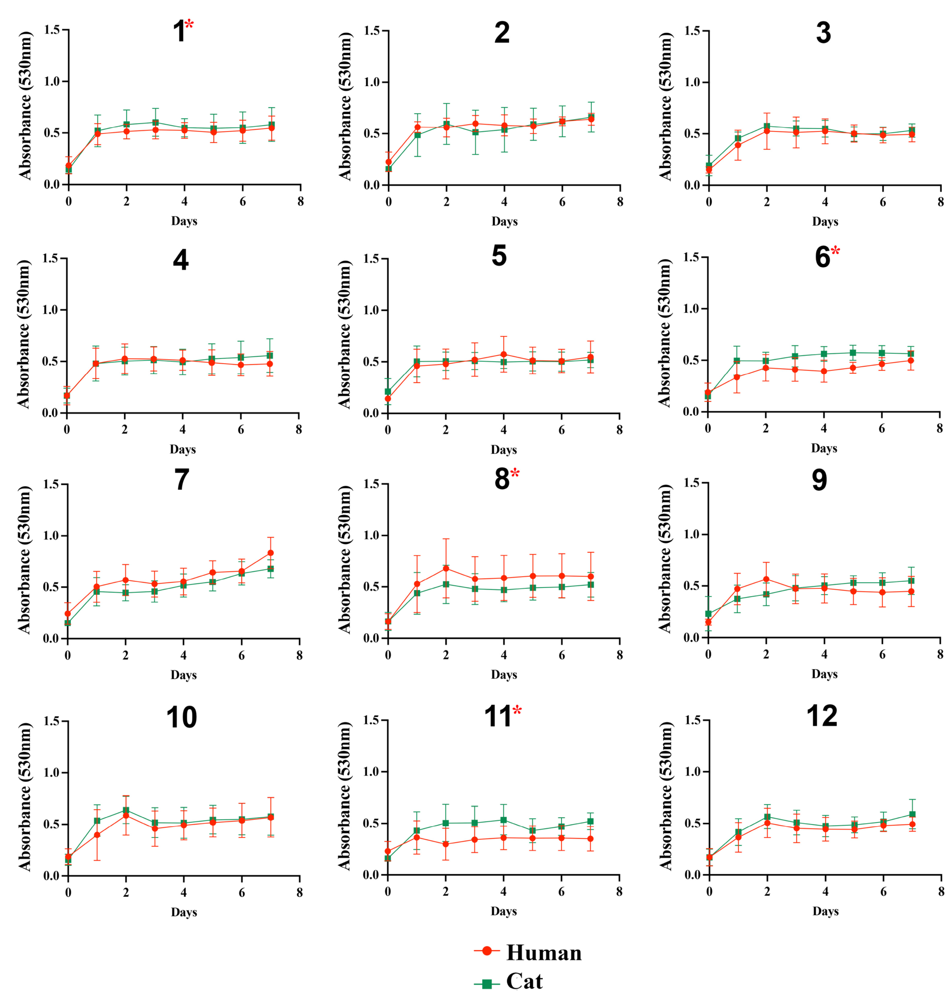
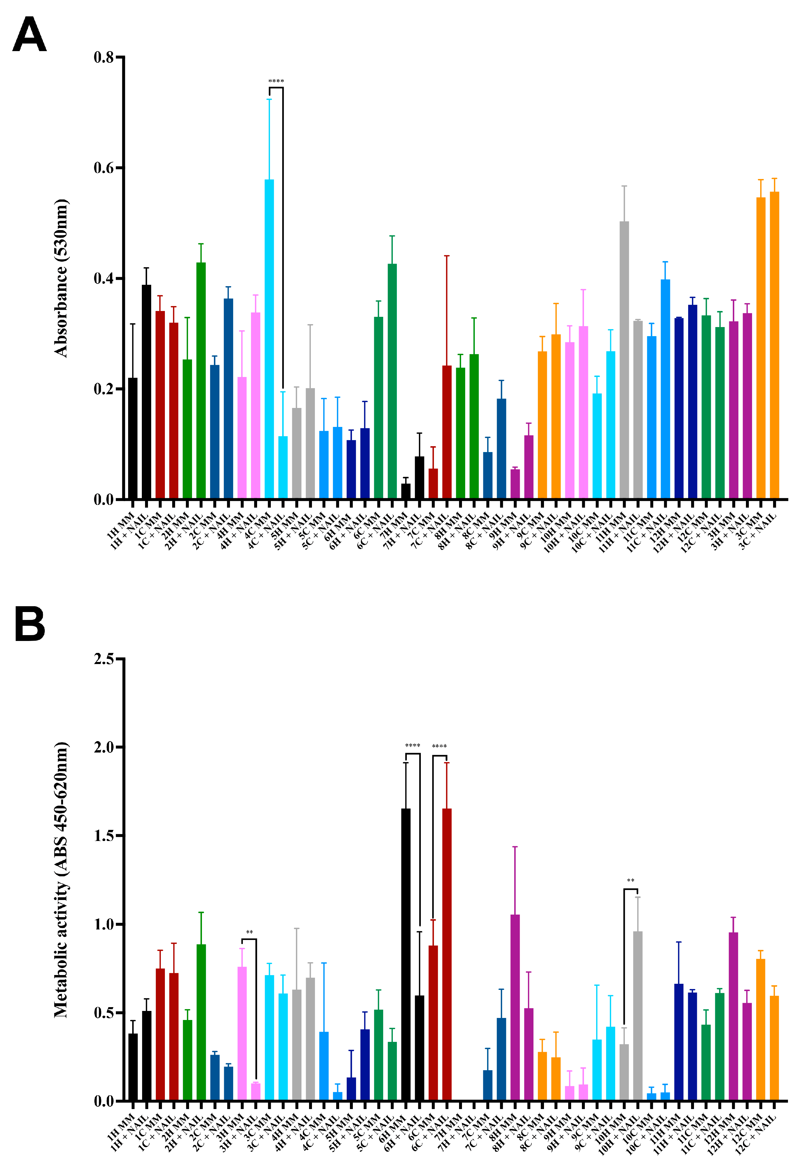
3.1. Fungal Growth on Different Substrates

The growth profile of the isolates in the presence of glucose was similar among the 24 S. brasiliensis, regardless of glucose concentrations (Figure 1). In general, all isolates grew poorly in the presence of lactate (Figure 2). The growth of the S. brasiliensis strains herein studied was more abundant in the presence of GlcNAc than lactate (Figure 3). Growth curves for human- and cat-derived S. brasiliensis strains from the familiar outbreaks were similar for all carbon sources evaluated; cases 1, 3, 4, 6, 11, and 12 presented significant growth differences on lactate and cases 1, 6, 8, and 11 on GlcNAc (p-value ≤ 0.05, Mann–Whitney test).
The majority of S. brasiliensis strains exhibited better growth in the MM supplemented with powdered claws than in MM without any supplementation after 7 days of incubation (Figure 4A). Besides growth, metabolic activity after this incubation time was quantified by XTT-reaction assay (Figure 4B). The human S. brasiliensis strain from outbreak 7 did not have metabolic activity after this incubation period. The metabolic activity of human- and cat-derived strains was similar on both MM and MM supplemented with cat claw powder. In addition, outbreaks 3, 4, and 6 presented differences between growth in MM of human and cat strains (p-values = 0.0059, =0.0042, and <0.0001, respectively, Tukey’s multiple comparisons test).

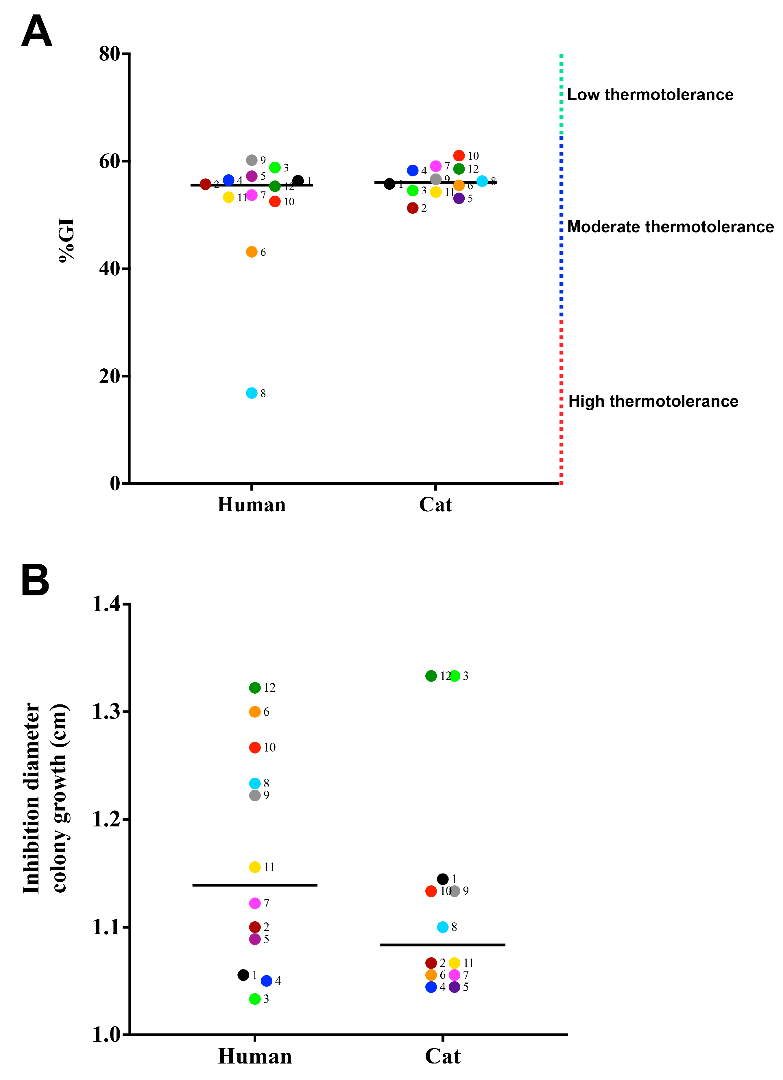
3.2. Susceptibility to Physical and Chemical Stressors

All strains analyzed showed thermotolerance, ranging from high (n = 1) (%GI 0 to 33%) to moderate thermotolerance (n = 23) (%GI 34 to 65%). Cases 6 and 8 showed significant differences between human and animal isolates (Figure 5A) (p-values = 0.0313 and <0.0001, respectively, Mann–Whitney test). Figure 5B presents data about the oxidative stress of the clinical S. brasiliensis isolates herein studied. The oxygen-derived oxidant susceptibility profile of strains isolated from cats and humans varied (p-value ≤ 0.0001, Mann–Whitney test). Moreover, the paired strains from outbreak 6 showed a significant difference between them (p-value = 0.0370, Mann–Whitney test).

3.3. Production of Extracellular Enzymes

All samples produced urease, as visually determined by the change of the medium color to pink, like that observed for the C. neoformans strain and contrasting with that observed for the C. parapsilosis and C. krusei control strains that did not change the color of the Christensen’s urea broth. Outbreaks 1, 4, 5, 8, and 9 showed significant differences between human- and cat-derived strains (Figure 6A) (cases 1, 8, and 9 p-value ≤ 0.0001, 4 = 0.0238, and 5 = 0.0001, Tukey’s multiple comparisons test).
For the enzymes evaluated on agar plates, the control C. albicans strain yielded Pz values of 0.4, 0.6, and 0.6 for phospholipase, esterase, and hemolysin, respectively. There was no enzymatic production of phospholipase and esterase by the 24 S. brasiliensis strains of this study. Among these strains, four (16.7%), three isolated from humans and one isolated from a cat, from four distinct outbreaks (6H, 8H, 10C, and 11H) were unable to produce hemolysin (Figure 6B). The production of hemolysin varied from low enzymatic activity (n= 17) (Pz between 0.70 and 0.99) to moderate (n = 3) (Pz between 0.40 and 0.699). Differences between human- and cat-derived strains were not significant (p-value < 0.05, Tukey’s multiple comparisons test).

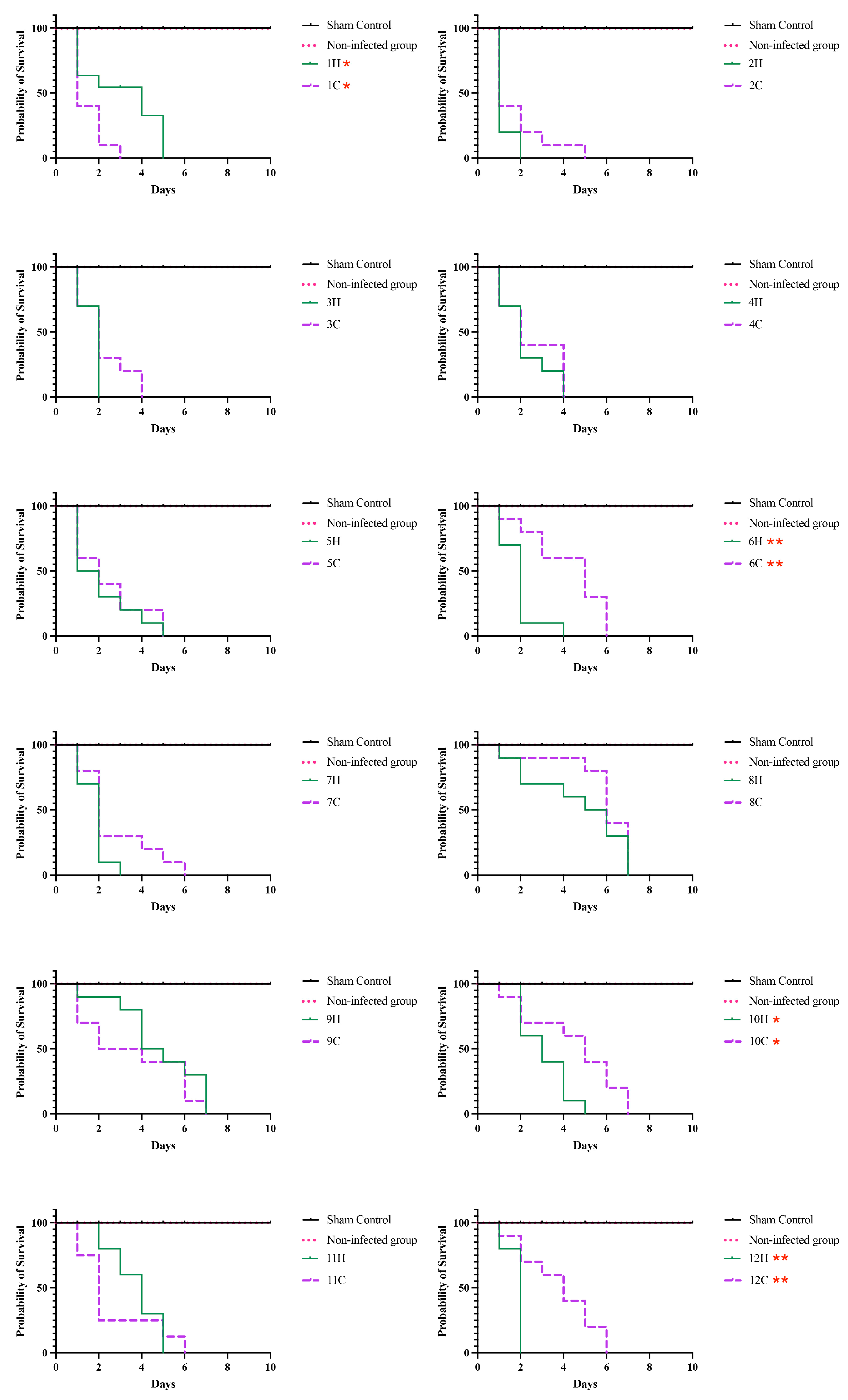
3.4. Survival Tests on Tenebrio Molitor

Figure 7 illustrates the survival of larvae infected with the studied S. brasiliensis strains and incubated at 37 °C. T. molitor larvae showed median survival values ranging from 2 to 7 days. The Mantel–Cox test revealed a difference in survival patterns of human and cat-derived strains from outbreaks 1 (p-value = 0.0129), 6 (p-value = 0.0019), 10 (p-value = 0.0307), and 12 (p-value = 0.0042) (Log-rank Mantel–Cox test).
To demonstrate fungal growth in inoculated animals, the colony-forming units (CFUs) were quantified in infected animals (Figure 8). Cases 2, 6, and 8 show differences in CFU compared to corresponding paired cases (p = 0.0022, Mann–Whitney test).

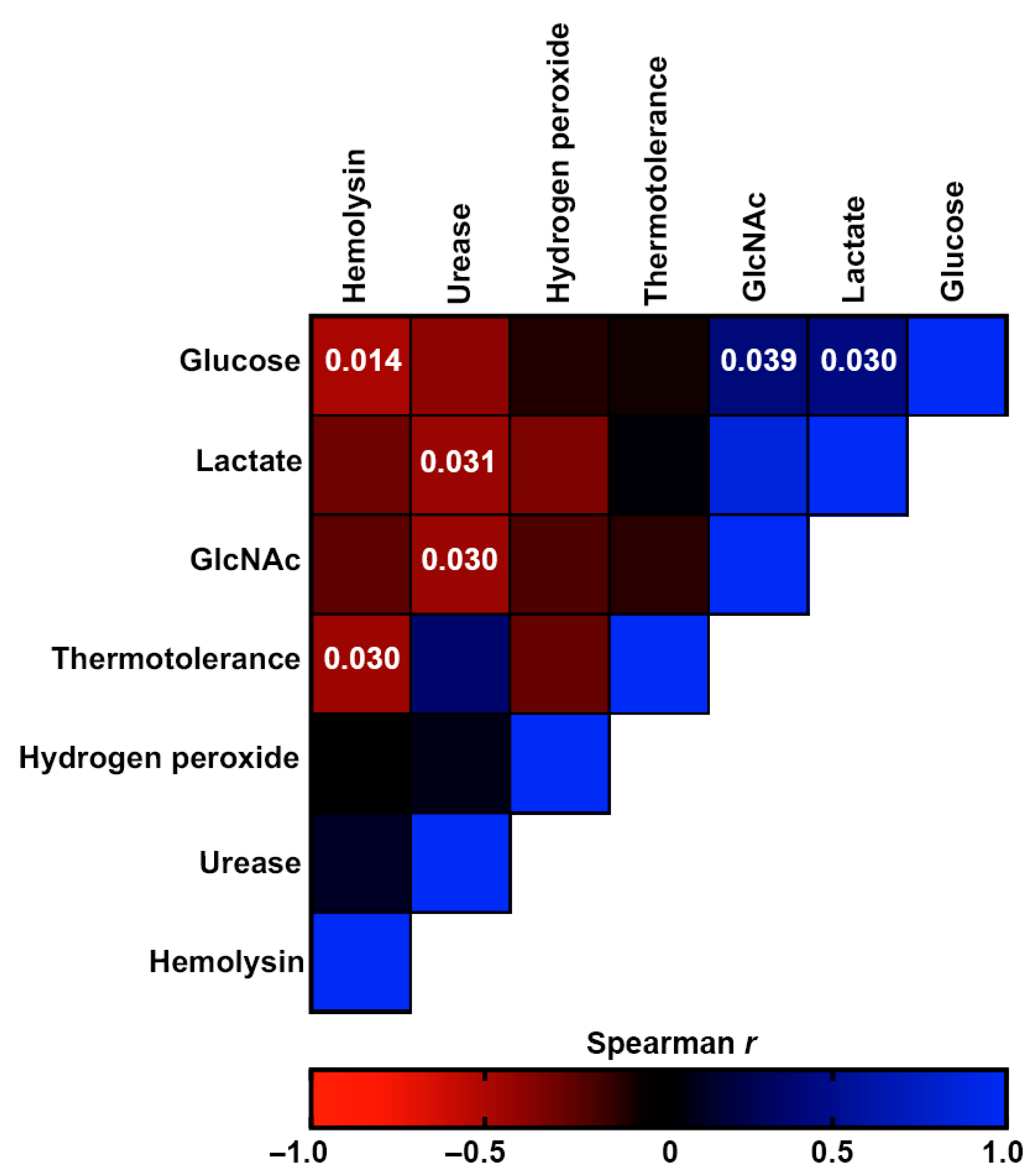
3.5. Comparative Analysis of Virulence-Related Factors

As depicted in Figure 9, none of the 24 S. brasiliensis strains presented a high level of all herein-studied putative virulence-associated factors. In general, the major phenotypic features potentially related to fungal virulence among the studied S. brasiliensis strains were urease production and susceptibility to hydrogen peroxide. The dendrogram clustered the strains according to their global similarity in the production of those studied factors. It is interesting to note that human- and cat-derived strains from outbreaks 2 and 4 clustered together, while strains from outbreaks 7 and 9 clustered very closely.
Possible correlations between the different fungal phenotypes were also investigated. Figure 10 shows a heatmap representing the Spearman’s correlation coefficients of the associations between different phenotypes of the studied isolates. The red color represents negative associations, whereas the blue color represents positive associations. Growth on glucose was negatively monotonically related to growth on GlcNAc (p-value = 0.039) and lactate (p-value = 0.030). Hemolysin production was positively monotonically related to growth on glucose and thermotolerance (p-values = 0.014 and =0.030, respectively), while urease production was positively monotonically related to growth on lactate and GlcNAc (p-values = 0.031 and =0.030, respectively).
Finally, a regression analysis was conducted to estimate the relative contributions of the studied virulence factors to the survival of T. molitor. As shown in Table 2, none of the herein-studied virulence-associated factors presented a significant association with larvae survival.

4. Discussion

The S. brasiliensis strains herein studied showed differences in some phenotypical characteristics that have been described as related to virulence in other human pathogenic fungi [36,37,38,39,40]. These aspects should be studied from a “One Health” perspective since this species continuously passes through different mammalian hosts and environmental sources [41]. This was the reason that motivated us to compare these phenotypes on strains obtained from humans and the cats that have infected them. In general, no differences were observed on human- and cat-derived S. brasiliensis strains, and for some outbreaks, phenotypes were extremely close for the related strains, suggesting that one passage through a mammalian host does not significantly affect the studied phenotypes.
Adaptations to environmental stressors may compel some fungi to acquire abilities that can increase their virulence and survival, driving the emergence of new threats to human, animal, and environmental health [42]. To survive the adverse conditions during parasitism, pathogenic fungi must adapt to the harsh microenvironments of the host. They may display altered phenotypic characteristics due to the selection pressure, in a process known as microevolution, as demonstrated in fungi of medical interest, such as Aspergillus fumigatus [43], C. neoformans [44], and C. albicans [45]. The present study also shows that S. brasiliensis has metabolic plasticity and may also suffer microevolution depending on the host. This plasticity to adapt to various stress conditions imposed during protracted infections on different patients was also observed previously for patients with cryptococcosis [46] and sporotrichosis [18].
Pathogenic fungi could use various carbon sources for development and survival [47,48]. Disturbances in host homeostasis can cause differences in glucose concentrations in the body [48], impacting the pathogenesis of the mycoses. For instance, colonization by Candida spp. is associated with glycemic control in patients with diabetes because hyperglycemia increases salivary glucose levels, promoting pathogen growth in the oral cavity [48]. Studies indicate that C. albicans can colonize immunocompetent individuals, and its cells are able to quickly adapt to available carbon sources in the host. Lactate, glucose, and GlcNAc are some of the carbon sources available for pathogens in the human body [49,50]. The S. brasiliensis growth studied here was noticeable in glucose since this is the main carbon source used by fungi. Concerning alternative carbon sources, the growth was slightly greater in the presence of GlcNAc than lactate, like that observed previously by our group in a group of S. brasiliensis strains isolated from human patients with chronic disseminated disease [18]. The skin tropism of S. brasiliensis may explain its preferential use of GlcNAc over lactate. GlcNAc is a glucosamine derivative that is naturally produced by humans and, together with D-glucuronate, forms hyaluronic acid, which is present in several body tissues, such as joints and skin. On the other hand, lactate is a widely abundant compound in most host niches, and it is a carboxylic acid that, in the body, is mainly found in its ionic form. Lactate is a product of glycolysis and plays fundamental roles in skeletal muscle tissue [48].
The production of extracellular enzymes allows the hydrolysis of skin components and promotes the invasion of the pathogen into the host tissues [51]. Moreover, when fungi are phagocytosed, they induce the release of oxidative chemical mediators, such as hydrogen peroxide, that cause oxidative stress in the microbe. Fungi, as an escape mechanism, produce catalases promoting fungal multiplication within macrophages [52]. The enzyme converts H2O2 into water and oxygen, allowing the fungus to remain viable after the host’s immune response, triggering its multiplication process [53]. The S. brasiliensis strains studied here were inhibited by hydrogen peroxide. Our results do not allow quantifying the catalase amount produced by the strains, but they show that they produce high amounts of catalase, due to the halo of growth inhibition lower than that described for A. fumigatus [54] and S. brasiliensis strains from chronic disease [18]. S. brasiliensis is an extracellular pathogen; therefore, its survival does not strongly depend on resistance to phagocytosis.
Phospholipase and esterase activities were not detected on the S. brasiliensis strains under the current study conditions. It is interesting to note that we previously detected esterase production in patients with chronic sporotrichosis [18], indicating that this enzyme may have a role in fungal maintenance in hosts with compromised immunity over extended periods of time. All isolates were able to produce urease. This enzyme is responsible for the use of urea as a source of nitrogen, causing the release of ammonia, resulting in a partial increase in the acidic pH [55]. In C. neoformans, urease is important for the invasion of the central nervous system since it facilitates the crossing of the blood–brain barrier [56]. It is known that S. brasiliensis produces higher levels of urease when compared to S. schenckii [31], and that S. brasiliensis could cause meningitis in severely immunosuppressed patients [57], which may be linked to the production of this enzyme. Interestingly, the urease levels produced by our strains were similar to that produced by the control C. neoformans strain, a traditional agent of fungal meningitis. Fungi can also secrete hemolysins responsible for the lysis of nucleated and red blood cells [58]. Four strains of the present study were unable to produce hemolysin. The production of this enzyme by S. brasiliensis isolates from human disseminated disease is useful during hematogenic dissemination [18]. Sporotrichosis in cats is progressive and blood dissemination will likely occur without proper management [59]. These proteins are important for the survival of microorganisms and are related to iron acquisition; therefore, their production is essential for the establishment of an infectious process [60].
Sporothrix spp. can be isolated from claw surfaces of naturally infected cats [61] and in an ex vivo biofilm model using the filamentous form of Sporothrix species and cat claws [26]. Our results show that the yeast-like form of S. brasiliensis can also use nutrients from cat claws to improve fungal growth, like that described in the literature [29]. This is important information, supporting that this species can survive on cat claws in its parasitic form, facilitating the zoonotic transmission of sporotrichosis. It is not possible to conclude that the added keratin facilitated the formation of biofilms, as the nail powder used may contain various nutrients for fungi. Furthermore, the low metabolic activity observed in certain Sporothrix strains may be related to this. To explore this further, future experiments may focus on identifying components present in cat nails that may promote the growth of S. brasiliensis.
The mealworm, also known as T. molitor, is an invertebrate animal model used to study fungal virulence [62]. T. molitor has been used to assess the virulence of entomopathogenic fungi such as Metarhizium robertsii [63], Metarhizium brunneum [64], and Beauveria bassiana [65] and human pathogens such as C. albicans [62], C. neoformans [62], and Fonsecaea spp. [66]. This study demonstrated the virulence of S. brasiliensis in these animals, with the median survival ranging from 2 to 7 days at 37 °C. The results that we have obtained for the yeast form are like that previously described for this species [34]. The observed outcomes concerning the yeast form align with previous reports on this particular species’ behavior.
The present study suggests that S. brasiliensis has a complex phenotype expression profile to adapt to the conditions found in the hosts. The data obtained in this study demonstrated that the S. brasiliensis isolates could grow in different nutrient sources available in hosts, present thermotolerance, susceptibility to the stress generated by hydrogen peroxide, and produce hemolysin. These metabolic factors may be associated with the installation/infection of S. brasiliensis in humans and other animals, demonstrating the plasticity of S. brasiliensis strains. The clinical signs of human and cat patients infected by this fungus are most probably linked to the host immunity or to other enzymes and virulence factors not evaluated in this study because they did not demonstrate significance with the survival of the experimental model herein used.

Author Contributions

Conceptualization, D.C.-J., R.A.-P. and S.F.; methodology, D.C.-J., I.B.d.A., V.A., I.A.-M., T.R.-A., V.B.d.S.R., G.R.d.S.A., L.P.B.-S., R.M.Z.-O., R.A.-P. and S.F.; formal analysis, D.C.-J., I.B.d.A., V.A., I.A.-M., V.B.d.S.R., G.R.d.S.A., L.P.B.-S., R.M.Z.-O., R.A.-P. and S.F.; resources, R.A.-P. and S.F.; writing—original draft preparation, D.C.-J., R.A.-P. and S.F.; writing—review and editing and funding acquisition, R.A.-P. and S.F. All authors have read and agreed to the published version of the manuscript.

Funding

This work was supported by the Brazilian agencies Conselho Nacional de Desenvolvimento Científico e Tecnológico (CNPq), Coordenação de Aperfeiçoamento de Pessoal de Nível Superior—Brasil (CAPES), Finance Code 001, and Fundação de Amparo à Pesquisa do Rio de Janeiro (FAPERJ). The authors have no other relevant affiliations or financial involvement with any organization or entity with a financial interest in or financial conflict with the subject matter or materials discussed in the manuscript apart from those disclosed. No writing assistance was utilized in the production of this manuscript.

Institutional Review Board Statement

Not applicable.

Informed Consent Statement

Not applicable.

Data Availability Statement

The data in this study are available in the presented manuscript.

Acknowledgments

We express our gratitude to Gepeto for providing valuable suggestions on improving the English language of the manuscript. We thank the Brazilian agencies Conselho Nacional de Desenvolvimento Científico e Tecnológico (CNPq), Coordenação de Aperfeiçoamento de Pessoal de Nível Superior—Brasil (CAPES), Finance Code 001, and Fundação de Amparo à Pesquisa do Rio de Janeiro (FAPERJ) for the financial support.

Conflicts of Interest

The authors declare that the research was conducted in the absence of any commercial or financial relationships that could be construed as a potential conflict of interest.

References

  1. Gremião, I.D.F.; Miranda, L.H.M.; Reis, E.G.; Rodrigues, A.M.; Pereira, S.A. Zoonotic Epidemic of Sporotrichosis: Cat to Human Transmission. PLoS Pathog. 2017, 13, e1006077. [Google Scholar] [CrossRef] [PubMed] [Green Version]
  2. Rodrigues, A.M.; Della Terra, P.P.; Gremião, I.D.; Pereira, S.A.; Orofino-Costa, R.; de Camargo, Z.P. The Threat of Emerging and Re-Emerging Pathogenic Sporothrix Species. Mycopathologia 2020, 185, 813–842. [Google Scholar] [CrossRef] [PubMed]
  3. De Hoog, G.S.; Guarro, J. Atlas of Clinical Fungi. Mycoses 1996, 39, 323. [Google Scholar] [CrossRef]
  4. Sigler, L.; Harris, J.L.; Dixon, D.M.; Flis, A.L.; Salkin, I.F.; Kemna, M.; Duncan, R.A. Microbiology and Potential Virulence of Sporothrix Cyanescens, a Fungus Rarely Isolated from Blood and Skin. J. Clin. Microbiol. 1990, 28, 1009–1015. [Google Scholar] [CrossRef] [PubMed] [Green Version]
  5. Tamez-Castrellón, A.; Romeo, O.; García-Carnero, L.; Lozoya-Pérez, N.; Mora-Montes, H. Virulence Factors in Sporothrix Schenckii, One of the Causative Agents of Sporotrichosis. Curr. Protein Pept. Sci. 2020, 21, 295–312. [Google Scholar] [CrossRef]
  6. Hay, R.J.; Morris-Jones, R. Outbreaks of Sporotrichosis. Curr. Opin. Infect. Dis. 2008, 21, 119–121. [Google Scholar] [CrossRef]
  7. Kwon-Chung, K.J.; Bennett, J.E. Medical Mycology. Rev. Inst. Med. Trop. Sao Paulo 1992, 34, 504. [Google Scholar] [CrossRef] [Green Version]
  8. Read, S.I.; Sperling, L.C. Feline Sporotrichosis: Transmission to Man. Arch. Dermatol. 1982, 118, 429–431. [Google Scholar] [CrossRef]
  9. Orofino-Costa, R.; Rodrigues, A.M.; de Macedo, P.M.; Bernardes-Engemann, A.R. Sporotrichosis: An Update on Epidemiology, Etiopathogenesis, Laboratory and Clinical Therapeutics. An. Bras. Dermatol. 2017, 92, 606–620. [Google Scholar] [CrossRef] [Green Version]
  10. Falcão, E.M.M.; Pires, M.C.D.S.; Andrade, H.B.; Gonçalves, M.L.C.; Almeida-Paes, R.; Do Valle, A.C.F.; Bastos, F.I.; Gutierrez-Galhardo, M.C.; Freitas, D.F.S. Zoonotic Sporotrichosis with Greater Severity in Rio de Janeiro, Brazil: 118 Hospitalizations and 11 Deaths in the Last 2 Decades in a Reference Institution. Med. Mycol. 2020, 58, 141–143. [Google Scholar] [CrossRef]
  11. Gremião, I.D.F.; Oliveira, M.M.E.; Monteiro de Miranda, L.H.; Saraiva Freitas, D.F.; Pereira, S.A. Geographic Expansion of Sporotrichosis, Brazil. Emerg. Infect. Dis. 2020, 26, 621–624. [Google Scholar] [CrossRef] [Green Version]
  12. Etchecopaz, A.; Toscanini, M.A.; Gisbert, A.; Mas, J.; Scarpa, M.; Iovannitti, C.A.; Bendezú, K.; Nusblat, A.D.; Iachini, R.; Cuestas, M.L. Sporothrix Brasiliensis: A Review of an Emerging South American Fungal Pathogen, Its Related Disease, Presentation and Spread in Argentina. J. Fungi 2021, 7, 170. [Google Scholar] [CrossRef] [PubMed]
  13. Thomson, P.; González, C.; Blank, O.; Ramírez, V.; del Río, C.; Santibáñez, S.; Pena, P. Sporotrichosis Outbreak Due to Sporothrix Brasiliensis in Domestic Cats in Magallanes, Chile: A One-Health-Approach Study. J. Fungi 2023, 9, 226. [Google Scholar] [CrossRef]
  14. Rachman, R.; Ligaj, M.; Chinthapalli, S.; Wani, R.S. Zoonotic Acquisition of Cutaneous Sporothrix Braziliensis Infection in the UK. BMJ Case Rep. 2022, 15, e248418. [Google Scholar] [CrossRef] [PubMed]
  15. Orofino-Costa, R.; Freitas, D.F.S.; Bernardes-Engemann, A.R.; Rodrigues, A.M.; Talhari, C.; Ferraz, C.E.; Veasey, J.V.; Quintella, L.; de Sousa, M.S.L.A.; Vettorato, R.; et al. Human Sporotrichosis: Recommendations from the Brazilian Society of Dermatology for the Clinical, Diagnostic and Therapeutic Management. Bras Dermatol. 2022, 97, 757–777. [Google Scholar] [CrossRef] [PubMed]
  16. Silva-Bailão, M.G.; de Lima, P.S.; Oliveira, M.M.E.; Oliveira, L.C.; Almeida-Paes, R.; Borges, C.L.; Bailão, A.M.; Coelho, A.S.G.; de Soares, C.M.A.; Zancopé-Oliveira, R.M. Comparative Proteomics in the Three Major Human Pathogenic Species of the Genus Sporothrix. Microbes Infect. 2021, 23, 104762. [Google Scholar] [CrossRef]
  17. Boechat, J.S.; Oliveira, M.M.E.; Almeida-Paes, R.; Gremião, I.D.F.; de Machado, A.C.S.; de Oliveira, R.V.C.; Figueiredo, A.B.F.; de Rabello, V.B.S.; Silva, K.B.D.L.; Zancopé-Oliveira, R.M.; et al. Feline Sporotrichosis: Associations between Clinical-Epidemiological Profiles and Phenotypic-Genotypic Characteristics of the Etiological Agents in the Rio de Janeiro Epizootic Area. Mem. Inst. Oswaldo Cruz 2018, 113, 185–196. [Google Scholar] [CrossRef] [Green Version]
  18. Cruz, I.L.R.; Freitas, D.F.S.; de Macedo, P.M.; Gutierrez-Galhardo, M.C.; do Valle, A.C.F.; de Almeida, M.A.; Coelho, R.A.; Brito-Santos, F.; Figueiredo-Carvalho, M.H.G.; Zancopé-Oliveira, R.M.; et al. Evolution of Virulence-Related Phenotypes of Sporothrix Brasiliensis Isolates from Patients with Chronic Sporotrichosis and Acquired Immunodeficiency Syndrome. Braz. J. Microbiol. 2020, 52, 5. [Google Scholar] [CrossRef]
  19. Schubach, T.M.P.; de Schubach, A.O.; Okamoto, T.; Figueiredo, F.B.; Pereira, S.A.; Leme, L.R.P.; dos Santos, I.B.; dos Reis, R.S.; de Paes, R.A.; de Perez, M.A.; et al. Utilidade Do Coágulo Sangüíneo Para o Isolamento de Sporothrix Schenckii de Gatos Naturalmente Infectados. Braz. J. Vet. Res. Anim. Sci. 2004, 41, 404–408. [Google Scholar] [CrossRef]
  20. Casadevall, A.; Pirofski, L.A. Host-Pathogen Interactions: Redefining the Basic Concepts of Virulence and Pathogenicity. Infect. Immun. 1999, 67, 3703–3713. [Google Scholar] [CrossRef] [Green Version]
  21. Casadevall, A. Fungi and the Rise of Mammals. PLoS Pathog. 2012, 8, e1002808. [Google Scholar] [CrossRef] [PubMed]
  22. Figueiredo-Carvalho, M.H.G.; Ramos, L.D.S.; Barbedo, L.S.; De Oliveira, J.C.A.; Dos Santos, A.L.S.; Almeida-Paes, R.; Zancopé-Oliveira, R.M. Relationship between the Antifungal Susceptibility Profile and the Production of Virulence-Related Hydrolytic Enzymes in Brazilian Clinical Strains of Candida Glabrata. Mediat. Inflamm. 2017, 2017, 8952878. [Google Scholar] [CrossRef] [PubMed] [Green Version]
  23. López, M.S.; Villavicencio, L.L.F.; Arredondo, K.S.; Sabanero, G.B.; Villagómez-Castro, J.C.; Jiménez, G.C.; Bernal, G.S.; Guerrero, H.T. Proteases of Sporothrix Schenckii: Cytopathological Effects on a Host-Cell Model. Rev. Iberoam. De Micol. 2018, 35, 32–38. [Google Scholar] [CrossRef]
  24. Almeida-Paes, R.; Brito-Santos, F.; Oliveira, M.M.E.; Bailão, A.M.; Borges, C.L.; de Araújo, G.R.S.; Frases, S.; de Soares, C.M.A.; Zancopé-Oliveira, R.M. Interaction with Pantoea Agglomerans Modulates Growth and Melanization of Sporothrix Brasiliensis and Sporothrix Schenckii. Mycopathologia 2019, 184, 367–381. [Google Scholar] [CrossRef]
  25. Carnero, L.C.G.; Pérez, N.E.L.; Hernández, S.E.G.; Álvarez, J.A.M. Immunity and Treatment of Sporotrichosis. J. Fungi 2018, 4, 100. [Google Scholar] [CrossRef] [Green Version]
  26. Brilhante, R.S.N.; Fernandes, M.R.; Pereira, V.S.; da Costa, A.C.; de Oliveira, J.S.; de Aguiar, L.; Rodrigues, A.M.; de Camargo, Z.P.; Pereira-Neto, W.A.; Sidrim, J.J.C.; et al. Biofilm Formation on Cat Claws by Sporothrix Species: An Ex Vivo Model. Microb. Pathog. 2021, 150, 104670. [Google Scholar] [CrossRef]
  27. Rodrigues, A.M.; de Hoog, G.S.; de Camargo, Z.P. Molecular Diagnosis of Pathogenic Sporothrix Species. PLoS Negl. Trop. Dis. 2015, 9, e0004190. [Google Scholar] [CrossRef] [Green Version]
  28. Reis, R.S.; Almeida-Paes, R.; de Muniz, M.M.; Tavares, P.M.e.S.; Monteiro, P.C.F.; Schubach, T.M.P.; Gutierrez-Galhardo, M.C.; Zancopé-Oliveira, R.M.; Santos Reis, R.; Almeida-Paes, R.; et al. Molecular Characterisation of Sporothrix Schenckii Isolates from Humans and Cats Involved in the Sporotrichosis Epidemic in Rio de Janeiro, Brazil. Mem. Inst. Oswaldo Cruz 2009, 104, 769–774. [Google Scholar] [CrossRef] [Green Version]
  29. Dos Santos, G.M.P.; Borba-Santos, L.P.; Vila, T.; Gremião, I.D.F.; Pereira, S.A.; De Souza, W.; Rozental, S. Sporothrix Spp. Biofilms Impact in the Zoonotic Transmission Route: Feline Claws Associated Biofilms, Itraconazole Tolerance, and Potential Repurposing for Miltefosine. Pathogens 2022, 11, 206. [Google Scholar] [CrossRef] [PubMed]
  30. Freitas, D.F.S.F.F.S.; Santos, S.S.; Almeida-Paes, R.; De Oliveira, M.M.E.; Do Valle, A.C.F.; Gutierrez-Galhardo, M.C.; Zancopé-Oliveira, R.M.; Nosanchuk, J.D.; Me De Oliveira, M.; Cf Do Valle, A.; et al. Increase in Virulence of Sporothrix Brasiliensis over Five Years in a Patient with Chronic Disseminated Sporotrichosis. Virulence 2015, 6, 112–120. [Google Scholar] [CrossRef] [PubMed] [Green Version]
  31. Almeida-Paes, R.; De Oliveira, L.C.; Oliveira, M.M.E.; Gutierrez-Galhardo, M.C.; Nosanchuk, J.D.; Zancopé-Oliveira, R.M. Phenotypic Characteristics Associated with Virulence of Clinical Isolates from the Sporothrix Complex. Biomed Res. Int. 2015, 2015, 212308. [Google Scholar] [CrossRef] [Green Version]
  32. Price, M.F.; Wilkinson, I.D.; Gentry, L.O. Plate Method for Detection of Phospholipase Activity in Candida Albicans. Sabouraudia 1982, 20, 7–14. [Google Scholar] [CrossRef] [PubMed]
  33. Aktaz, E.; Yigit, N.; Ayyildiz, A. Esterase Activity in Various Candida Species. J. Int. Med. Res. 2002, 30, 322–324. [Google Scholar] [CrossRef]
  34. Lozoya-Pérez, N.E.; García-Carnero, L.C.; Martínez-Álvarez, J.A.; Martínez-Duncker, I.; Mora-Montes, H.M. Tenebrio Molitor as an Alternative Model to Analyze the Sporothrix Species Virulence. Infect. Drug Resist. 2021, 14, 2059–2072. [Google Scholar] [CrossRef]
  35. Babicki, S.; Arndt, D.; Marcu, A.; Liang, Y.; Grant, J.R.; Maciejewski, A.; Wishart, D.S. Heatmapper: Web-Enabled Heat Mapping for All. Nucleic Acids Res. 2016, 44, W147–W153. [Google Scholar] [CrossRef]
  36. Kaerger, K.; Schwartze, V.U.; Dolatabadi, S.; Nyilasi, I.; Kovács, S.A.; Binder, U.; Papp, T.; de Hoog, S.; Jacobsen, I.D.; Voigt, K. Adaptation to Thermotolerance in Rhizopus Coincides with Virulence as Revealed by Avian and Invertebrate Infection Models, Phylogeny, Physiological and Metabolic Flexibility. Virulence 2015, 6, 395–403. [Google Scholar] [CrossRef] [PubMed] [Green Version]
  37. Tamayo, D.; Muñoz, J.F.; Almeida, A.J.; Puerta, J.D.; Restrepo, Á.; Cuomo, C.A.; McEwen, J.G.; Hernández, O. Paracoccidioides Spp. Catalases and Their Role in Antioxidant Defense against Host Defense Responses. Fungal Genet Biol. 2017, 100, 22–32. [Google Scholar] [CrossRef] [PubMed] [Green Version]
  38. Mroczyńska, M.; Brillowska-Dabrowska, A. Virulence of Clinical Candida Isolates. Pathogens 2021, 10, 466. [Google Scholar] [CrossRef]
  39. de Andrade, I.B.; Figueiredo-Carvalho, M.H.G.; da Silva Chaves, A.L.; Coelho, R.A.; Almeida-Silva, F.; Zancopé-Oliveira, R.M.; Frases, S.; Brito-Santos, F.; Almeida-Paes, R. Metabolic and Phenotypic Plasticity May Contribute for the Higher Virulence of Trichosporon Asahii over Other Trichosporonaceae Members. Mycoses 2022, 66, 430–440. [Google Scholar] [CrossRef]
  40. Miao, J.; Regan, J.; Cai, C.; Palmer, G.E.; Williams, D.L.; Kruppa, M.D.; Peters, B.M. Glycogen Metabolism in Candida Albicans Impacts Fitness and Virulence during Vulvovaginal and Invasive Candidiasis. mBio 2023, 14. [Google Scholar] [CrossRef]
  41. Cabañes, F.J. Sporotrichosis in Brazil: Animals + humans = one Health. Rev. Iberoam. Micol. 2020, 37, 73–74. [Google Scholar] [CrossRef] [PubMed]
  42. Araújo, G.R.D.S.; De Souza, W.; Frases, S. The Hidden Pathogenic Potential of Environmental Fungi. Future Microbiol. 2017, 12, 1533–1540. [Google Scholar] [CrossRef]
  43. Ballard, E.; Melchers, W.J.G.; Zoll, J.; Brown, A.J.P.; Verweij, P.E.; Warris, A. In-Host Microevolution of Aspergillus Fumigatus: A Phenotypic and Genotypic Analysis. Fungal Genet. Biol. 2018, 113, 1–13. [Google Scholar] [CrossRef] [PubMed]
  44. Hong, N.; Chen, M.; Xu, J. Molecular Markers Reveal Epidemiological Patterns and Evolutionary Histories of the Human Pathogenic Cryptococcus. Front. Cell Infect. Microbiol. 2021, 11, 683670. [Google Scholar] [CrossRef] [PubMed]
  45. Gambhir, N.; Harris, S.D.; Everhart, S.E. Evolutionary Significance of Fungal Hypermutators: Lessons Learned from Clinical Strains and Implications for Fungal Plant Pathogens. mSphere 2022, 7, e0008722. [Google Scholar] [CrossRef] [PubMed]
  46. Iyengar, Y.; Xu, J. Phenotypic Plasticity in the Productions of Virulence Factors Within and Among Serotypes in the Cryptococcus Neoformans Species Complex. Mycopathologia 2022, 187, 65–83. [Google Scholar] [CrossRef]
  47. Zeng, L.; Huang, Y.; Tan, J.; Peng, J.; Hu, N.; Liu, Q.; Cao, Y.L.; Zhang, Y.; Chen, J.; Huang, X. QCR7 Affects the Virulence of Candida Albicans and the Uptake of Multiple Carbon Sources Present in Different Host Niches. Front. Cell Infect. Microbiol. 2023, 13, 1136698. [Google Scholar] [CrossRef]
  48. Al-Attas, S.A.; Amro, S.O. Candidal Colonization, Strain Diversity, and Antifungal Susceptibility among Adult Diabetic Patients. Ann. Saudi Med. 2010, 30, 101–108. [Google Scholar] [CrossRef] [Green Version]
  49. Ballou, E.R.; Avelar, G.M.; Childers, D.S.; Mackie, J.; Bain, J.M.; Wagener, J.; Kastora, S.L.; Panea, M.D.; Hardison, S.E.; Walker, L.A.; et al. Lactate Signalling Regulates Fungal β-Glucan Masking and Immune Evasion. Nat. Microbiol. 2016, 2, 16238. [Google Scholar] [CrossRef] [Green Version]
  50. Barelle, C.J.; Priest, C.L.; MacCallum, D.M.; Gow, N.A.R.; Odds, F.C.; Brown, A.J.P. Niche-Specific Regulation of Central Metabolic Pathways in a Fungal Pathogen. Cell Microbiol. 2006, 8, 961–971. [Google Scholar] [CrossRef] [Green Version]
  51. CHINNAPUN, D. Virulence Factors Involved in Pathogenicity of Dermatophytes. J. Sci. Tech. 2015, 12, 573–580. [Google Scholar]
  52. Hansberg, W.; Salas-Lizana, R.; Domínguez, L. Fungal Catalases: Function, Phylogenetic Origin and Structure. Arch. Biochem. Biophys. 2012, 525, 170–180. [Google Scholar] [CrossRef]
  53. Román, E.; Prieto, D.; Martin, R.; Correia, I.; Mesa Arango, A.C.; Alonso-Monge, R.; Zaragoza, O.; Pla, J. Role of Catalase Overproduction in Drug Resistance and Virulence in Candida Albicans. Future Microbiol. 2016, 11, 1279–1297. [Google Scholar] [CrossRef]
  54. Shakesheff, K.M.; Evora, C.; Soriano, I.; Langer, R. The Adsorption of Poly(Vinyl Alcohol) to Biodegradable Microparticles Studied by X-Ray Photoelectron Spectroscopy (XPS). J. Colloid Interface Sci. 1997, 185, 538–547. [Google Scholar] [CrossRef] [PubMed]
  55. Dubreuil, J.D.; Del Giudice, G.; Rappuoli, R. Helicobacter Pylori Interactions with Host Serum and Extracellular Matrix Proteins: Potential Role in the Infectious Process. Microbiol. Mol. Biol. Rev. 2002, 66, 617–629. [Google Scholar] [CrossRef] [PubMed] [Green Version]
  56. Olszewski, M.A.; Noverr, M.C.; Chen, G.-H.; Toews, G.B.; Cox, G.M.; Perfect, J.R.; Huffnagle, G.B. Urease Expression by Cryptococcus Neoformans Promotes Microvascular Sequestration, Thereby Enhancing Central Nervous System Invasion. Am. J. Pathol. 2004, 164, 1761. [Google Scholar] [CrossRef] [Green Version]
  57. Lima, M.A.; Freitas, D.F.S.; Oliveira, R.V.C.; Fichman, V.; Varon, A.G.; Freitas, A.D.; Lamas, C.C.; Andrade, H.B.; Veloso, V.G.; Almeida-Paes, R.; et al. Meningeal Sporotrichosis Due to Sporothrix Brasiliensis: A 21-Year Cohort Study from a Brazilian Reference Center. J. Fungi 2022, 9, 17. [Google Scholar] [CrossRef]
  58. Nayak, A.P.; Green, B.J.; Beezhold, D.H. Fungal Hemolysins. Med. Mycol. 2013, 51, 1–16. [Google Scholar] [CrossRef] [Green Version]
  59. Lloret, A.; Hartmann, K.; Pennisi, M.G.; Ferrer, L.; Addie, D.; Belák, S.; Boucraut-Baralon, C.; Egberink, H.; Frymus, T.; Gruffydd-Jones, T.; et al. Sporotrichosis in Cats: ABCD Guidelines on Prevention and Management. J. Feline Med. Surg. 2013, 15, 619–623. [Google Scholar] [CrossRef]
  60. Nevitt, T.; Thiele, D.J. Host Iron Withholding Demands Siderophore Utilization for Candida Glabrata to Survive Macrophage Killing. PLoS Pathog. 2011, 7, e1001322. [Google Scholar] [CrossRef]
  61. De Lima Barros, M.B.; Pacheco Schubach, T.M.; Gutierrez Galhardo, M.C.; De Oliveira Schubach, A.; Fialho Monteiro, P.C.; Santos Reis, R.; Zancopé-Oliveira, R.M.; Dos Santos Lazéra, M.; Cuzzi-Maya, T.; Moita Blanco, T.C.; et al. Sporotrichosis: An Emergent Zoonosis in Rio de Janeiro. Mem. Inst. Oswaldo Cruz 2001, 96, 777–779. [Google Scholar] [CrossRef] [PubMed] [Green Version]
  62. de Souza, P.C.; Morey, A.T.; Castanheira, G.M.; Bocate, K.P.; Panagio, L.A.; Ito, F.A.; Furlaneto, M.C.; Yamada-Ogatta, S.F.; Costa, I.N.; Mora-Montes, H.M.; et al. Tenebrio Molitor (Coleoptera: Tenebrionidae) as an Alternative Host to Study Fungal Infections. J. Microbiol. Methods 2015, 118, 182–186. [Google Scholar] [CrossRef] [PubMed] [Green Version]
  63. Rantala, M.J.; Dubovskiy, I.M.; Pölkki, M.; Krama, T.; Contreras-Garduño, J.; Krams, I.A. Effect of Juvenile Hormone on Resistance against Entomopathogenic Fungus Metharizium Robertsii Differs between Sexes. J. Fungi 2020, 6, 298. [Google Scholar] [CrossRef] [PubMed]
  64. Bharadwaj, A.; Stafford, K.C. Potential of Tenebrio Molitor (Coleoptera: Tenebrionidae) as a Bioassay Probe for Metarhizium Brunneum (Hypocreales: Clavicipitaceae) Activity against Ixodes Scapularis (Acari: Ixodidae). J. Econ. Entomol. 2011, 104, 2095–2098. [Google Scholar] [CrossRef]
  65. Jirakkakul, J.; Roytrakul, S.; Srisuksam, C.; Swangmaneecharern, P.; Kittisenachai, S.; Jaresitthikunchai, J.; Punya, J.; Prommeenate, P.; Senachak, J.; So, L.; et al. Culture Degeneration in Conidia of Beauveria Bassiana and Virulence Determinants by Proteomics. Fungal Biol. 2018, 122, 156–171. [Google Scholar] [CrossRef]
  66. Fornari, G.; Gomes, R.R.; Degenhardt-Goldbach, J.; Do sSantos, S.S.; De Almeida, S.R.; Dos Santos, G.D.; Muro, M.D.; Bona, C.; Scola, R.H.; Trindade, E.S.; et al. A Model for Trans-Kingdom Pathogenicity in Fonsecaea Agents of Human Chromoblastomycosis. Front. Microbiol. 2018, 9, 2211. [Google Scholar] [CrossRef] [Green Version]
Figure 1. Growth curves of Sporothrix brasiliensis strains from 12 familiar outbreaks of sporotrichosis under 0.5%, 1%, and 2% glucose. The optical density (530 nm) of cultured strains was measured daily for 7 days. Results are expressed as the mean and standard deviation of readings obtained from three independent experiments performed in technical triplicates.
Jof 09 00724 g001
Figure 2. Growth curves of Sporothrix brasiliensis strains from 12 familiar outbreaks of sporotrichosis under 0.5% lactate. The optical density (530 nm) of cultured strains was measured daily for 7 days. Results are expressed as the mean and standard deviation of readings obtained from three independent experiments performed in technical triplicates. The asterisk symbol indicates a statistically significant difference between the growth curves of paired cases, p-value (Mann–Whitney test) ≤ 0.05.
Jof 09 00724 g002
Figure 3. Growth curves of Sporothrix brasiliensis strains from 12 familiar outbreaks of sporotrichosis under 0.5% GlcNAc. The optical density (530 nm) of cultured isolates was measured daily for 7 days. Results are expressed as the mean and standard deviation of readings obtained from three independent experiments performed in technical triplicates. Asterisks indicate significant differences between growth curves of human- and cat-derived strains from the familiar outbreak. The asterisk symbol indicates a statistically significant difference between the growth curves of paired cases, p-value (Mann–Whitney test) ≤ 0.05.
Jof 09 00724 g003
Figure 4. Growth of Sporothrix brasiliensis strains from 12 familiar outbreaks of sporotrichosis using cat claws as a nutrient. (A) Growth of S. brasiliensis in minimal medium (MM) and MM supplemented with 2% cat claws powder. (B) Metabolic activity of S. brasiliensis strains grown in MM and MM supplemented with 2% claws powder. Results are expressed as the mean plus standard deviation of three independent experiments performed in technical triplicates. Bar colors represent the different familiar outbreaks. The asterisk symbol indicates a statistically significant difference between the growth curves of paired cases, p-value (Tukey’s multiple comparisons test) ≤ 0.05.
Jof 09 00724 g004
Figure 5. Resistance of Sporothrix brasiliensis strains from 12 familiar outbreaks of sporotrichosis against physical and chemical stressors. (A) Thermotolerance of S. brasiliensis, the smaller the percentage of growth inhibition (%GI), the greater the thermotolerance of the isolates. (B) Diameters in millimeters of growth inhibition of S. brasiliensis against exposure of yeast cells to 5% hydrogen peroxide. Each dot represents the mean value of three independent experiments performed in technical triplicates. Dot colors and numbers after dots represent the different familiar outbreaks. Black lines represent the median value of each group of strains.
Jof 09 00724 g005
Figure 6. Production of extracellular enzymes by Sporothrix brasiliensis strains from 12 familiar outbreaks of sporotrichosis. Results are expressed as the mean and standard deviation of (A) urease and (B) hemolytic activity. Urease activity is expressed as the optical density (559 nm) of yeast S. brasiliensis cultures in Christensen’s urea broth after 7 days of growth at 35 °C. Hemolytic activity is expressed as the Pz of the enzyme of yeast S. brasiliensis cultures on sheep blood agar. Each dot represents the mean value of three independent experiments performed in technical triplicates. Dot colors and numbers after dots represent the different familiar outbreaks. Black lines represent the median value of each group of strains.
Jof 09 00724 g006
Figure 7. Survival curves of Tenebrio molitor larvae infected with Sporothrix brasiliensis strains from 12 familiar outbreaks of sporotrichosis. Larvae were infected with a 10 µL suspension containing 1 × 10⁶ yeast-like cells and incubated at 37 °C and observed until 10 days after inoculation. A total of 40 animals were included in each experimental group, and these were monitored daily to assess mortality, which was defined as a lack of irritability and the presence of extensive body melanization. The asterisk symbol indicates a statistically significant difference between the growth curves of paired cases, p-value (Log-rank (Mantel-Cox) test) ≤ 0.05.
Jof 09 00724 g007
Figure 8. Colony-forming units (CFUs/mL) of Sporothrix brasiliensis in Tenebrio molitor. Results are expressed as the mean plus standard deviation of three independent experiments performed in technical triplicates. Bar colors represent the different familiar outbreaks. ** p = 0.0022, Mann–Whitney test.
Jof 09 00724 g008
Figure 9. Global expression of putative virulence-associated factors by Sporothrix brasiliensis strains from 12 familiar outbreaks of sporotrichosis. The gray scale in the heatmap ranges from low (white) to high virulence (black). Different strains are represented in the lines of the heatmap, and the different virulence factors herein are studied in the columns of the heat map. Strains were grouped in a dendrogram reflecting the similarity between the virulence-related phenotypes of each strain. Colors of the strain identification numbers represent the different familiar outbreaks.
Jof 09 00724 g009
Figure 10. Correlation analyses of the virulence-associated factors produced by the Sporothrix brasiliensis strains from 12 familiar outbreaks of sporotrichosis. The heat map represents the Spearman’s correlation coefficients of the associations. The red color represents negative monotonic correlations, whereas the blue color represents positive monotonic correlations. Probability values of statistically significant correlations (p < 0.05) are displayed within the respective squares of the heat map.
Jof 09 00724 g010
Table 1. Paired samples of Sporothrix brasiliensis obtained at Fiocruz (Rio de Janeiro, Brazil), between 1998 and 2001 [28].
CaseOriginal NumberHostStrain Identification
115485-1Human1H
34-1Cat1C
215647-1Human2H
47Cat2C
319536Human3H
856Cat3C
416393-2Human4H
92-2Cat4C
516415Human5H
99-2Cat5C
616459Human6H
75-1Cat6C
716672Human7H
142-1Cat7C
817878Human8H
98-2Cat8C
916910-1Human9H
195-1Cat9C
1017500Human10H
165-1Cat10C
1119182Human11H
721-1Cat11C
1219481Human12H
792-1Cat12C
Table 2. Hierarchical regression analysis results for initial 24-strain data set.
Virulence Factor(s)Correlation with Time to Death (r)R2 ChangedfF Valuep Value
Glucose47650.3199147650.0443
Lactate−74340.2641142020.0571
NAG−39960.2695118790.1894
Thermotolerance11440.475410.059340.8106
Hydrogen peroxide−0.52220.186610.015360.9029
Urease−81500.4145112200.2858
Hemolysin−34540.3832114800.2399
Disclaimer/Publisher’s Note: The statements, opinions and data contained in all publications are solely those of the individual author(s) and contributor(s) and not of MDPI and/or the editor(s). MDPI and/or the editor(s) disclaim responsibility for any injury to people or property resulting from any ideas, methods, instructions or products referred to in the content.

