Article

In Vivo Microevolutionary Analysis of a Fatal Case of Rhinofacial and Disseminated Mycosis Due to Azole-Drug-Resistant Candida Species

1
Bioinformatics Center of AMMS, Beijing Key Laboratory of New Molecular Diagnosis Technologies for Infectious Diseases, Beijing Institute of Microbiology and Epidemiology, Beijing 100850, China
2
TEDA Institute of Biological Sciences and Biotechnology, Nankai University, Tianjing 300457, China
3
Division of Dermatology and Mycological Lab, Peking University Third Hospital, Beijing 100191, China
4
School of Life Science & Technology, China Pharmaceutical University, Nanjing 210009, China
*
Authors to whom correspondence should be addressed.
These authors contributed equally to this work.
J. Fungi 2023, 9(8), 815; https://doi.org/10.3390/jof9080815
Submission received: 27 June 2023 / Revised: 26 July 2023 / Accepted: 31 July 2023 / Published: 2 August 2023
(This article belongs to the Section Fungal Pathogenesis and Disease Control)

Abstract

:
Ten Candida species strains were isolated from the first known fatal case of rhinofacial and rhino–orbital–cerebral candidiasis. Among them, five strains of Candida parapsilosis complex were isolated during the early stage of hospitalization, while five strains of Candida tropicalis were isolated in the later stages of the disease. Using whole-genome sequencing, we distinguished the five strains of C. parapsilosis complex as four Candida metapsilosis strains and one Candida parapsilosis strain. Antifungal susceptibility testing showed that the five strains of C. parapsilosis complex were susceptible to all antifungal drugs, while five C. tropicalis strains had high minimum inhibitory concentrations to azoles, whereas antifungal-drug resistance gene analysis revealed the causes of azole resistance in such strains. For the first time, we analyzed the microevolutionary characteristics of pathogenic fungi in human hosts and inferred the infection time and parallel evolution of C. tropicalis strains. Molecular clock analysis revealed that azole-resistant C. tropicalis infection occurred during the first round of therapy, followed by divergence via parallel evolution in vivo. The presence/absence variations indicated a potential decrease in the virulence of genomes in strains isolated following antifungal drug treatment, despite the absence of observed clinical improvement in the conditions of the patient. These results suggest that genomic analysis could serve as an auxiliary tool in guiding clinical diagnosis and treatment.

1. Introduction

Fungal infections affect more than 1 billion people worldwide and cause approximately 1.5 million deaths annually [1]. More than 90% of invasive fungal infections are caused by Candida species [2,3]. Candida albicans is the most common cause of candidiasis, but infections attributed to other non-Candida albicans Candida (NCAC) are of increasing concern. Candida tropicalis is one of the most common pathogenic NCAC species in Asia and Latin America [4,5]. Candida tropicalis is inherently resistant to azoles [6], one of the main classes of antifungal agents, particularly fluconazole [7,8], and can develop cross-resistance to other antifungal drugs [9]. The molecular mechanisms of azole resistance have been extensively studied in Candida species [10,11].
Candida species are notorious for causing chronic mucocutaneous candidiasis (CMC) but have never been reported to cause rhinofacial and rhino–orbital–cerebral mycosis (ROCM). CMC is a heterogeneous group of overlapping syndromes characterized by persistent, refractory, and recurrent infections caused by Candida species and may include resistant strains [12]. CMC usually occurs when a patient presents with complex deficiencies in immunology, endocrinology, and autoimmunity [13]. This includes a persistent or recurrent infection, and nearly all patients with CMC have inherited or acquired T-cell deficiencies [14]. Immune system deficiencies often render this infection incurable. ROCM is a life-threatening disease that causes facial swelling, ulceration, and disfigurement, with the sudden or progressive onset of symptoms [15]. This infection is associated with high mortality and severe morbidities, such as blindness, eyeball extraction, and facial disfigurement, particularly when the pathogen is drug-resistant or when drug resistance occurs due to the discontinuation of treatment [12,16]. Therefore, caring for patients with ROCM presents significant challenges to physicians because of severe outcomes when the infection is unresponsive to aggressive antifungal therapy.
Whole-genome sequencing (WGS) is gaining increasing importance in the identification and characterization of fungal pathogens [17]. WGS can assist in understanding the genetic diversity and evolution of fungal species, as well as their virulence factors and drug-resistance mechanisms [18]. WGS is currently being implemented in routine diagnostic microbiology, allowing for the identification of antimicrobial resistance genes and genes associated with virulence in bacterial genomes [19], which can be extended to fungal pathogens [20].
Herein, we report a fatal case of CMC with rhinofacial involvement due to the C. parapsilosis complex and C. tropicalis infection. To further explore the effect of drug therapy on strain evolution, we analyzed the microevolutionary characteristics of multi-drug-resistant C. tropicalis in a human host. Analyses of single nucleotide polymorphisms (SNPs), indels, and copy number variants of resistance-associated genes revealed the resistance of the C. tropicalis strains to azoles and identified new variants that may lead to antifungal drug resistance. Analysis of mating type locus (MTL) and heterozygous sites demonstrated asexual reproduction and single ancestor and parallel evolution of C. tropicalis strains. Molecular clock analysis showed that a change in treatment regimen was consistent with the differentiation time of the strain. We further analyzed presence/absence variations (PAVs) among the C. tropicalis strains and found that the strains were evolving toward reduced virulence.

2. Materials and Methods

2.1. All Candida Strains Culture and Antifungal Susceptibility Assays

All strains used in this study were grown on Sabouraud dextrose agar (SDA) solid medium and cultured in an incubator at 25 °C for 48–72 h. Single colonies were selected and cultured at 37 °C and 200 rpm and activated twice for 16 h each time to ensure that the fungus was in the late exponential growth phase. To investigate in vitro antifungal susceptibility, a broth microdilution method was used according to the M27 [21] and M38 [22] methods of the Clinical and Laboratory Standards Institute (CLSI) for susceptibility testing of nine antifungal drugs (MedChemExpress, Monmouth Junction, NJ, USA). A small number of strains were inoculated into 1 mL of liquid Sabouraud dextrose agar (SDA) medium. The concentration of the strain was determined using a hemocytometer and subsequently diluted to a concentration of 2 × 10³ CFU/mL in RPMI 1640 medium (Procell Life Science & Technology Co., Ltd., Wuhan, China). The nine antifungal drugs were gradient diluted and added to 96-well plates. The final concentration gradients are shown in Supplementary Table S9. Next, 100 μL of the cell suspension was added to each well at a final concentration of 1 × 10³ CFU/mL. After incubation at 37 °C for 24 h, the OD600 of each well was measured using an Epoch Microplate Spectrophotometer (Agilent Technologies, Santa Clara, CA, USA). The CLSI resistance breakpoints of the strains were listed in Supplementary Table S1 according to CLSI M27 [21] and M59 [23] methods.

2.2. Genome Sequencing and Assembly

The methods for library construction, genomic sequencing, and assembly were based on our previous publication [24]. A combined sequencing platform strategy was used for all 10 strains. A 20 kbp single-molecule real-time (SMRT) bell library was prepared from sheared genomic DNA according to the library preparation workflow, and the PacBio Sequel system was used to conduct SMRT sequencing using C3 sequencing chemistry and P5 polymerase with 16 SMRT cells (PacBio, Menlo Park, CA, USA). A Nextera XT DNA Sample Preparation Kit (Illumina Inc., San Diego, CA, USA) was used to prepare the Illumina 250 bp paired-end library with an insert size of approximately 400 bp, and the read inserts were sequenced using Illumina HiSeqXTen (Illumina Inc.). PacBio yielded a total of 22.77 G data with an average coverage of 152× per strain. Illumina yielded a total of 25.56 G data with an average coverage of 170× per strain.
The combination of second- and third-generation sequencing technology can effectively improve the integrity of genome assembly [25]. De novo assembly of PacBio sequencing reads was carried out using the Hierarchical Genome Assembly Process workflow within the SMRT Analysis v2.1 package (www.pacb.com/support/software-downloads, accessed on 26 June 2023). First, error correction of the raw data generated by the PacBio platform was performed using the PacBioRs_PreAssembler with one module, a default minimum subread length of 500 bp, a minimum read quality of 0.80, and a minimum subread length of 7500 bp. Next, the short reads generated by the Illumina HiSeqXTen were mapped to the longer-read PacBio assembly to increase the resolution and quality of the sequences.

2.3. Genomic Prediction and Annotation

RepeatModeler [26] and RepeatMasker [27] were used to perform de novo identification and mask repeats. Protein-coding genes were predicted using AUGUSTUS [28], and their functional annotations were assessed according to their homologs in the NCBI non-redundant (nr) database using E-value cut-offs of 1 × 10⁻⁵ and >40% amino acid identity.

2.4. Phylogenetic Analysis

Similar to our previous study [29], the phylogeny of the internal transcribed spacer (ITS) region was analyzed using MEGA v7.0.21 [30] with the neighbor-joining (NJ) method with 1000 bootstraps. Orthofinder [31] was used to identify and cluster the orthologous genes. Furthermore, we used Mafft v7.508 [32] to align the multiple sequences. A maximum-likelihood phylogenetic tree was constructed with single-copy orthologs using RAxML v8.2.12 [33], with PROTCATWAG model and 1000 bootstrap replicates. Whole-genome SNPs were used to construct an unrooted neighbor-joining tree with 100 bootstrap replicates using SNPhylo v20180901 [34]. Lastly, ancestral state reconstruction and molecular clock analysis were performed using TreeTime [35] v0.9.0. When the 0/0 and 0/1 genotypes were present in the five C. tropicalis strains, the reference allele was identified as the ancestral genotype. When the 1/1 and 0/1 genotypes were present, the alternate allele was identified as the ancestral genotype.

2.5. Variant Calling

Low-quality bases from paired-end reads were trimmed using Trimmomatic v0.33 [36], with the following parameters: LEADING:3 TRAILING:3 SLIDINGWINDOW:4:15 MINLEN:80. Paired-end resequencing reads were mapped to the assembly of the 2016_Ct 3 strains with BWA v0.7.10-r789 [37] using the default parameters. SAMtools v1.3.1 [38] was used to convert the mapping results from SAM to the BAM format. Duplicated reads were filtered using the Picard package (Picard. sourceforge. net, v2.1.1) (Broad Institute, 2018, Cambridge, MA, USA). After BWA alignment, the reads around the indels were realigned using the Genome Analysis Toolkit (GATK) v3.8 [39]. Next, the SNP and indel variants for each accession were called using GATK Unified Genotype. Then, the PacBio reads of each sample were mapped to the assembly of the 2016_Ct 3 strains using NGMLR v0.2.7 [40]. The structural variant is called Sniffles. Lastly, functional regions were annotated using the SnpEff v5.1 [41].

2.6. Verification of SNPs and Indels

Primer sequences of the resistance genes and DNA mismatch repair genes that were designed using Primer Premier5 (http://www.premierbiosoft.com/primerdesign/, accessed on 26 June 2023) are shown in Supplementary Table S9. Sanger sequencing was performed by Beijing Biomed Gene Technology Co., Ltd. (Beijing, China).

2.7. Quantification and Statistical Analysis

All details of the statistical analyses are provided alongside the corresponding methods. As described in our previous work [42], PHI database annotation was carried out using blastp, with identity >50% and query coverage >50%. SAMtools v1.3.1 [38] was used to calculate copy numbers. Gene copy number was obtained from gene depth/genome depth. GC skew and gene density were calculated using TBtools v1.098769 [43].

2.8. Ethical Declaration

The study was reviewed and approved by Peking University Third Hospital Medical Science Research Ethics Committee IRB (approval: #00006761-2015025).

3. Results

3.1. Patient Course and Candida Strains

We summarized the disease course of the patient and listed the time points when different antifungal drugs were used and when strains were obtained (Figure 1). C. tropicalis strains have been designated as 2015_Ct 1, 2015_Ct 2, 2016_Ct 1, 2016_Ct 2, and 2016_Ct 3. C. parapsilosis complex strains have been designated as 2015_Cp, 2015_Cm 1, 2015_Cm 2, 2015_Cm 3, and 2015_Cm 4.
In April 2015, the patient was diagnosed with CMC (ROCM type) based on repeated positive cultures, observation of hyphal forms on direct KOH microscopic examinations, and pathology. Voriconazole was prescribed and resulted in the alleviation and disappearance of symptoms after a 50-day treatment period. However, following the discontinuation of voriconazole and the initiation of corticosteroid treatment, the skin lesions of the patient relapsed. Despite the administration of multiple antifungal drugs, including itraconazole, fluconazole, and voriconazole, the infections worsened, leading to complete damage to the patient’s face. Subsequently, the patient decided to discontinue treatment.
Before the relapse, five C. parapsilosis complex strains were isolated, and following the relapse, five C. tropicalis strains were isolated. Originally, these strains were characterized as five C. parapsilosis and five C. tropicalis strains, respectively, using ITS sequencing. Moreover, antifungal susceptibility testing showed that all strains were susceptible to amphotericin B and 5-flucytosine. However, the C. tropicalis strains were resistant to the azoles tested, including fluconazole, itraconazole, posaconazole, and voriconazole (Supplementary Table S1).

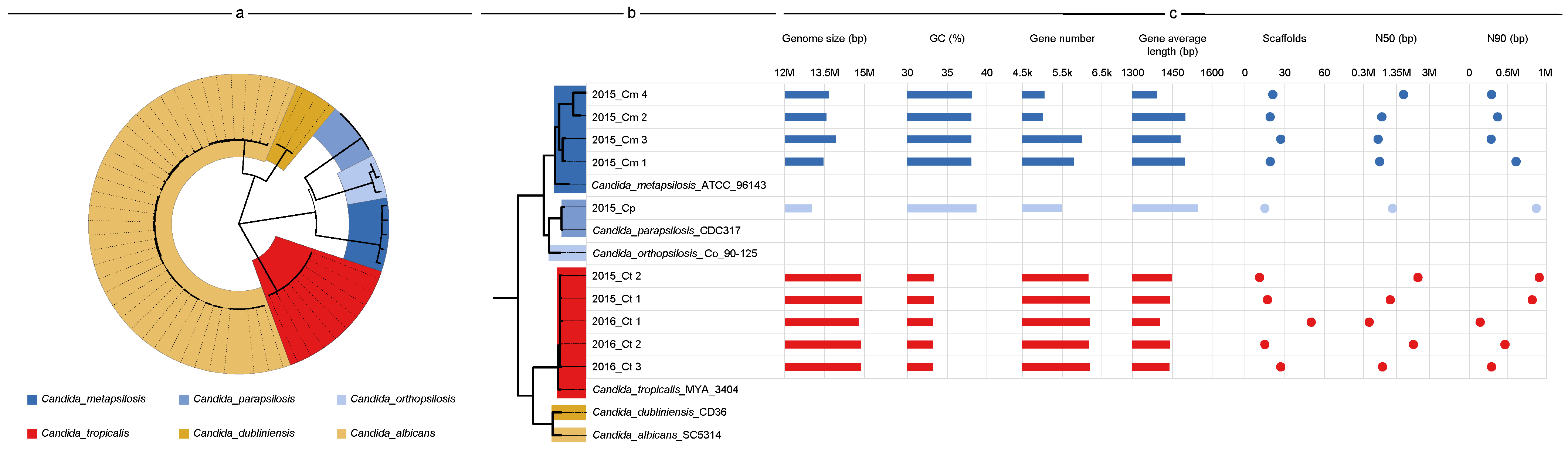
3.2. Genome Features and Species Identification

The genomes of all 10 strains were sequenced, and approximately 1.4–5.4 G and 1.3–3.6 G raw data were generated by the PacBio Sequel system and Illumina MiSeq sequencer, respectively. Each genome assembly displayed high continuity, from 11–50 scaffolds with 0.5–2.5 Mb of scaffold N50. The genomic features of all five C. tropicalis strains were consistent with those of C. tropicalis reference strain MYA-3404 (genome size, <15 Mb; gene number, ~6300; GC content, 33%). However, the genome size of five C. parapsilosis strains ranged from 13 Mb (2015_Cp) to 13.9 Mb (2015_Cm 3), and the annotated gene number from 5046 (2015_Cm 2) to 6081 (2015_Cm 3), a difference of approximately 20%. Although strain 2015_Cp possessed the smallest genome, its gene number was intermediate (5548), and it showed the largest gene length (1561 bp), particularly with the GC content (38.69) being slightly higher than that of its partners (Figure 2c).
A maximum-likelihood phylogenetic tree was constructed (Figure 2a) based on 407 single-copy orthologs from 63 Candida genomes. All the ITS-characterized C. tropicalis strains were clustered with MYA-3404. However, among the ITS-characterized C. parapsilosis strains, four belonged to a single clade adjacent to C. metapsilosis, whereas 2015_Cp was clustered into another clade comprising C. parapsilosis strain CDC317 (Figure 2b). These two clades, along with C. orthopsilosis, formed a separate cluster. Previously, C. parapsilosis strains were characterized as belonging to three groups (I–III), which were later designated as separate species: Group I remained C. parapsilosis, and groups II and III were renamed C. orthopsilosis and C. metapsilosis, respectively; however, this classification still reflects the close evolutionary relationship among them. Based on this phylogeny, all ITS-characterized C. parapsilosis strains were C. metapsilosis except 2015_Cp.

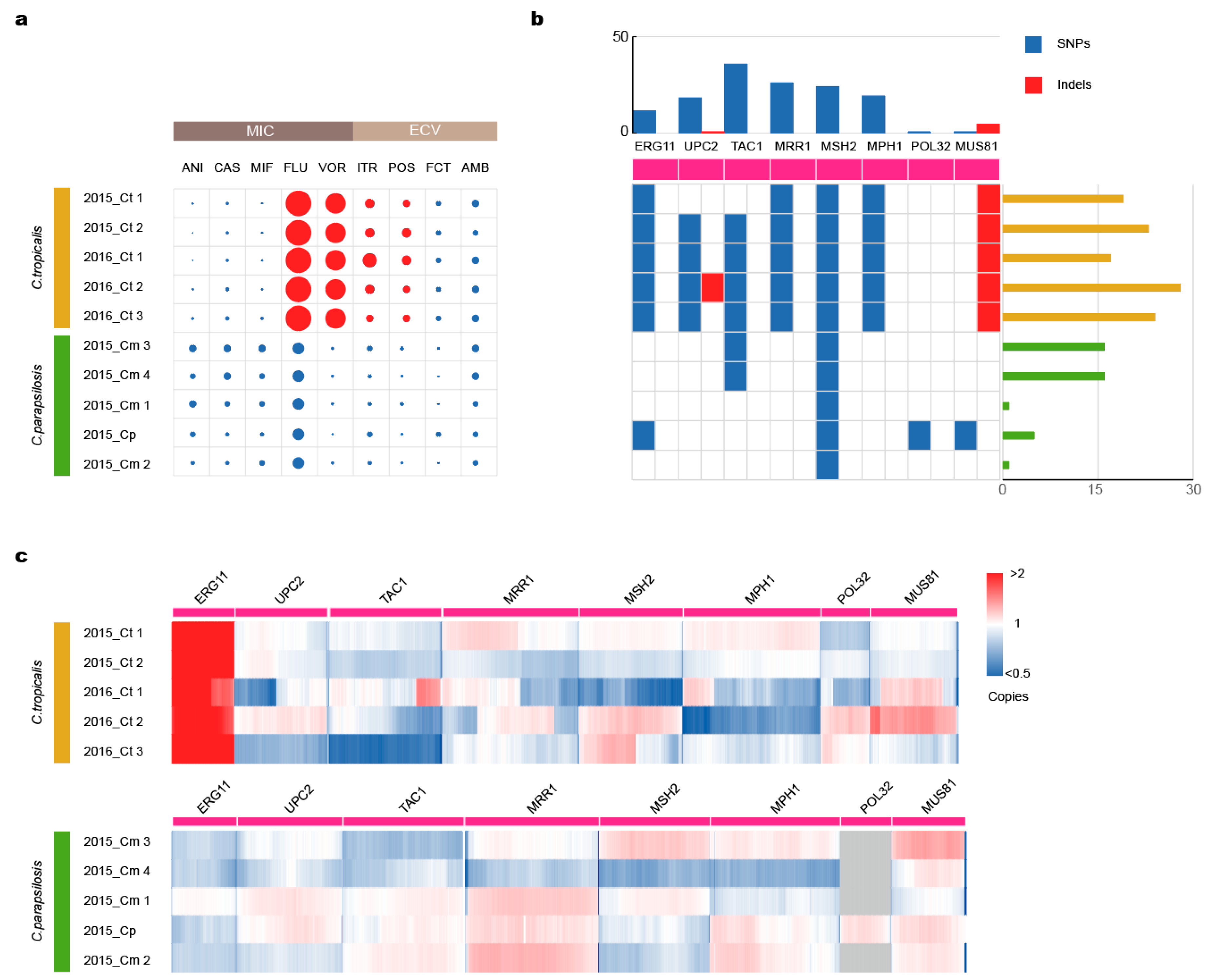
3.3. Mutations in Drug-Resistance Genes Lead to Azole Resistance

Drug susceptibility testing showed that five C. tropicalis strains had high minimum inhibitory concentrations (MICs) to azole antifungal agents, whereas C. metapsilosis strains and C. parapsilosis strains were susceptible to all antifungal drugs (Figure 3a). We counted the gene mutations (Figure 3b, Supplementary Table S2) and copy numbers (Figure 3c) of antifungal resistance genes and their regulators to characterize the relationship between resistance and these genes.
ERG11 encodes a major component in the membranes of Candida species. The C. parapsilosis strain 2015_Cp had two single-nucleotide variants (SNVs); however, this mutation did not lead to drug resistance. The two SNVs that lead to amino acid changes (Y132F and S154F), which have been associated with azole resistance in C. tropicalis, existed in all our C. tropicalis strains. Multiple copies of ERG11 appeared in all five C. tropicalis strains.
UPC2 is a transcription factor that contributes to fluconazole resistance development by inducing ERG11 overexpression. In contrast to C. metapsilosis strains and C. parapsilosis strains containing no mutations in UPC2, each of our C. tropicalis strains harbored A251T and Q289S amino acid substitutions, except strain 2015_Ct 1, as seen in two fluconazole-resistant strains [44]. Additionally, 2016_Ct 1 and 2016_Ct 2 shared an L168P substitution, which has not previously been recorded. Moreover, strain 2016_Ct 2 harbored a TTAA insertion at position 2334 of the UPC2 gene, thereby introducing a stop codon and resulting in translation termination at amino acid position 799.
TAC1 and MMR1 are two transcription factors that play a role in drug resistance in Candida species. Strains 2015_Cm 3 and 2015_Cm 4 had some missense SNPs in TAC1, but those did not lead to drug resistance. No SNP mutations were found within MRR1 in any of the C. metapsilosis and C. parapsilosis strains. Furthermore, during the screening for TAC1 (50% protein identity with TAC1C. albicans) and MRR1 (57% identity with coding protein of MRR1C. albicans) in all C. tropicalis, we observed 0–4 SNPs resulting in amino acid changes in TAC1 and 3–11 in MRR1, compared with each counterpart of the reference MYA-3404.
We calculated the raw data coverage for each contig to determine whether any large aneuploidy occurred. However, each contig exhibited similar data coverage distribution, indicating that no large aneuploidies occurred (Supplementary Figure S1).

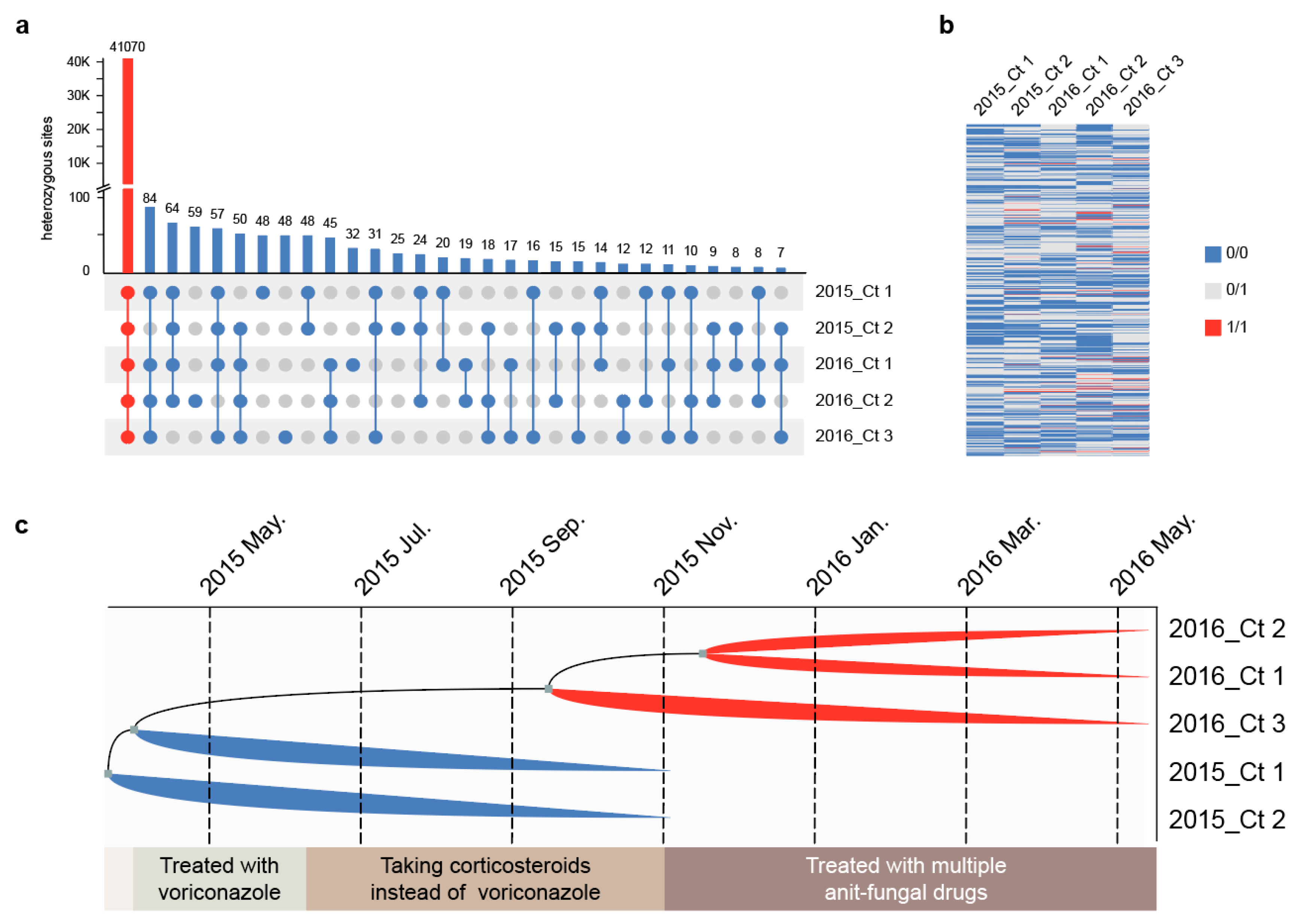
3.4. Strain Differentiation Is Associated with Antifungal Treatment

We analyzed the in vivo microevolution of five multi-drug-resistant C. tropicalis strains. K-mer analysis showed that all five strains were diploid (Supplementary Figure S2), and further heterozygosity analysis revealed that nearly all heterozygous loci (41,070 of 41,892) were shared by all the C. tropicalis genomes (Figure 4a), indicating that these strains originated from the same clone.
We identified the MTL to determine the reproduction type of five C. tropicalis strains. We found that 2015_Ct 1 and 2016_Ct 2 have the same MTL (a/α), whereas 2015_Ct 2, 201605214, and 2016_Ct 3 are a/a mating types. In C. tropicalis, a/α reproduces asexually; however, α-secreted pheromones can promote homosexual reproduction of a/a strains to form four ploidy, and such offspring return to diploidy due to instability [45]. All heterozygous loci showed two genotypes, and no allele separation of 0/0, 0/1, and 1/1 occurred, indicating that the strains were propagated asexually (Figure 4b).
Compared with the reconstructed ancestral sequence, the number of “private mutations/total mutations” of the strains, except for 2016_Ct 1, was >50% (Supplementary Table S3). Therefore, we identified five C. tropicalis strains that underwent parallel evolution in vivo, indicating that Candida did not undergo adaptive evolution in the host.

3.5. Multi-Drug Resistant Strains Originated from the Same Clone and Underwent Parallel Evolution In Vivo

We determined the haplotype of C. tropicalis strains based on the molecular infinite locus model because the strain did not undergo genetic recombination [46]. Based on 822 SNVs among the C. tropicalis strains (Supplementary Table S4), a maximum-likelihood phylogenetic tree was constructed to determine the ancestral states for each SNV site at the internal nodes (Figure 4c). There are only two-time nodes for calibration; hence, the inferred time will inevitably exhibit some deviation. The putative common ancestor of C. tropicalis strains was proposed to have emerged between March 2015 and May 2015 at the start of voriconazole treatment. Within these two months, ancestors quickly diverged into 2015_Ct 2, 2015_Ct 1, and another group. Around September 2015, during corticosteroid treatment, the strains were divided into 2016_Ct 3 and another group. Next, around November 2015, the strains 2016_Ct 1 and 2016_Ct 2 were obtained when the patient started receiving multiple antifungal medications. Through ancestral state reconstruction, we determined that C. tropicalis infection developed after the patient was discharged from the hospital rather than having been lurking in the patient until it became the dominant strain.

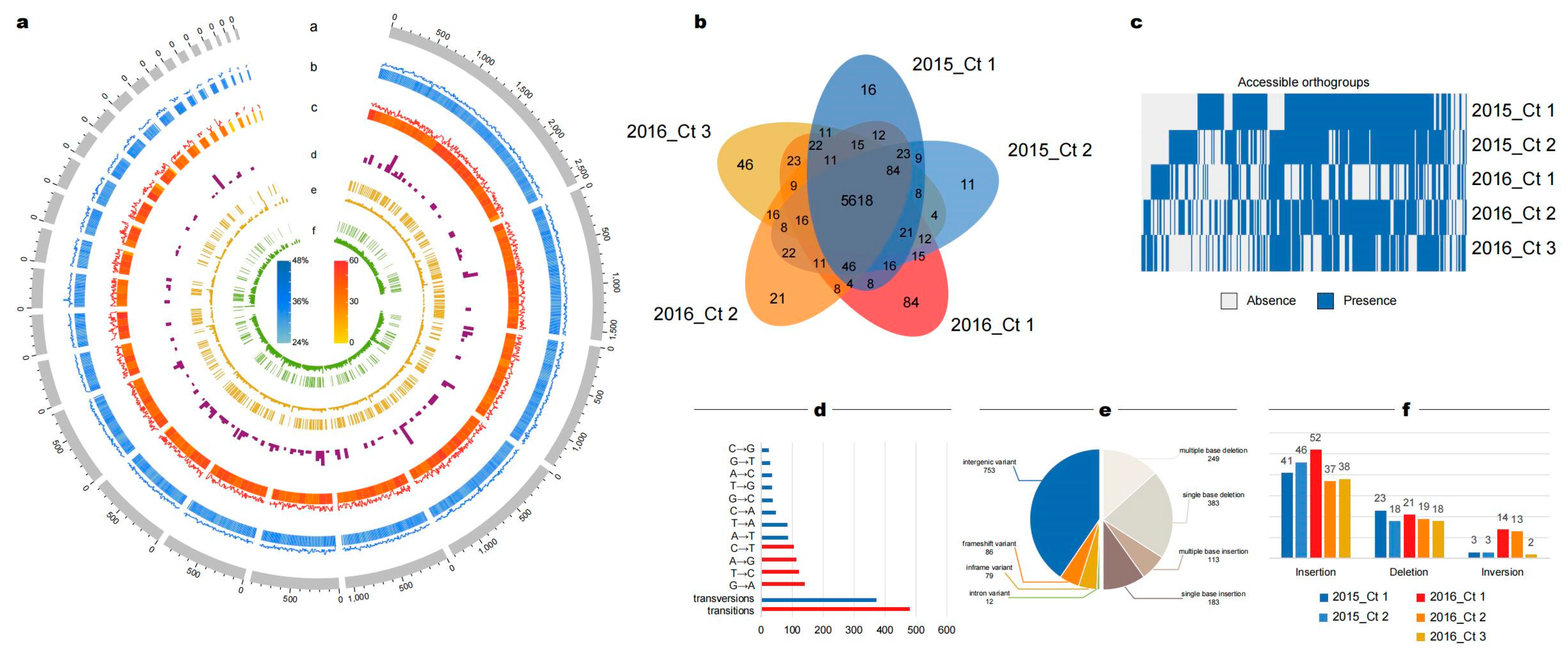
3.6. Abnormal Number of Insertions and Deletions May Be Caused by DNA Mismatch Repair Genes

We analyzed genome-wide variation in the five C. tropicalis strains to explore functional changes (Figure 5a). Individual SNV frequency was first investigated, and a high transition rate was observed therein compared to that of transversions. Within transversions, A→T and T→A mutations exhibited a higher frequency, consistent with the comparatively low GC content of the C. tropicalis genome (Figure 5d). It is uncommon for more insertions and deletions (indels) than SNVs to be called (Figure 5e). There were 930 indel events, and the number of single-base indels accounted for slightly more than those of multiple indels (Supplementary Table S5); 72% of single-base insertions (81/113) and 88% of single-base deletions (335/383) occurred in or near homopolymers.
The deletion of the DNA mismatch repair gene MSH2 has been confirmed to cause an increase in the occurrence of Indel [47]. The MSH2 homolog in each of our strains had no mutations except for some synonymous SNVs (Figure 3b). We further investigated other DNA mismatch repair genes and discovered a single-base insertion (2015_Ct 1) or deletion (other C. tropicalis strains) in MUS81 (Supplementary Table S6). We found that 2016_Ct 1 had the fewest mutations in DNA mismatch repair genes, and the number of unique indels was significantly lower than that of other C. tropicalis strains (Supplementary Figure S3). Therefore, we speculated that the abnormal number of indels was related to mutations in the DNA mismatch repair genes.

3.7. Strains Isolated after Multiple Antifungal Administrations Exhibited Decreased Virulence

Among the multi-drug-resistant C. tropicalis strains, the 2015 and 2016 strains were isolated before and after administering multiple antifungals, respectively. We investigated the differences between the strains before and after multiple antifungal administrations.
We characterized the private PAVs of the 2016 strains, defined as the number of structural variations (SVs) that occur in coding sequences and accessible genes (present only in partial strains). In C. tropicalis strains, 160 SVs were allocated across 71 genes, including 32 deletions, 100 insertions, and 28 inversions (Figure 5f, Supplementary Table S7). The number of accessible genes in 2016_Ct 1 and 2016_Ct 3 was significantly higher than that in the other three C. tropicalis strains (Figure 5b). Notably, each C. tropicalis strain had a similarly small number of private presence genes (2–4), indicating that the difference in the number of private genes was due to the private absence genes. Among them, 2016_Ct 1 had a maximum of 80 privately absent genes, followed by 2016_Ct 3 with 45 (Figure 5c).
We further annotated strain genomes using the PHI database (Supplementary Table S8) to explore changes in the strains after multiple antifungal administrations. We counted PAV events present in the 2016 strains but absent in the 2015 strains. Common PAVs of the three 2016 strains did not show virulence; however, a few depleted genes were strain-specific and closely pathogenicity-related. The four genes depleted in 2016_Ct 1 were ERG3, SOD1, BIG1, and ADA2. Uniquely, 2016_Ct 2 deleted two genes, EAP1 and CDC42, which we propose to be virulence-related in C. tropicalis. Furthermore, the deletion of PSY4 appeared only in 2016_Ct 3. Our results indicate that although each strain generated unique absence genes, the strains isolated after multiple antifungal administrations likely generated variants with attenuated pathogenicity under parallel evolution in vivo.

4. Discussion

Here, we report the first case of CMC with ROCM involvement due to Candida infection. During the early stage of hospitalization, five strains belonging to the C. parapsilosis complex were isolated. Using whole-genome sequencing, we successfully differentiated C. metapsilosis and C. parapsilosis, which could not be distinguished by ITS sequencing. Therefore, we emphasize the importance of utilizing whole-genome sequencing to differentiate the C. parapsilosis complex, as it allows for the identification of pathogenic C. metapsilosis that arises from mating events between non-pathogenic parental strains [48].
In the later stages of the disease, five azole-drug-resistant strains of C. tropicalis were isolated. Although CMC is frequently associated with primary or secondary immunodeficiency, research has shown that drug resistance, innate or acquired, of the strains involved cannot be ignored [12,49]. Multiple copies of ERG11 appeared in all five C. tropicalis strains in this study, which increased the mRNA level, thereby increasing ERG11 expression. ERG11 overexpression associated with nonsynonymous point mutations is the most common azole resistance mechanism in C. tropicalis [44,50]. Gain-of-function mutations in UPC2 attributed to certain SNVs have been repeatedly reported to confer azole resistance in C. albicans [51,52,53]. The TTAA insertion of strain 2016_Ct 2 is in the extreme C-terminal ligand-binding domain of UPC2, the key region for sensing ergosterol levels in the cell [54]. Constitutive upregulation of efflux pumps in azole-resistant C. albicans is mediated by gain-of-function point mutations in the zinc cluster transcription factor genes MRR1 [55,56]. Mutations in MRR1 may enable C. tropicalis to escape an attack from the host, which is likely another role of MRR1 (C. albicans) in increasing C. albicans resistance against the innate host defense system by upregulating MDR1 expression, thereby allowing it to better adapt to certain host niches [57].
We conducted a further microevolutionary analysis of five azole-drug-resistant C. tropicalis strains. Sexual reproduction drives the evolution of new traits and adaptation to new environments in eukaryotic organisms, including fungi, allowing for the adoption of different strategies for sexual reproduction [58,59,60]. Analysis of the MTL and heterozygous loci suggests that the strains undergo asexual reproduction, thereby excluding the influence of sexual reproduction on the microevolutionary processes within these strains. We further investigated the parallel evolution of C. tropicalis strains and found that the acquisition of azole resistance occurred independently under the drug pressure or was present in the ancestral strains.
Applying molecular clock analysis, we revealed a significant concurrence between the timing of C. tropicalis strain differentiation and the change in treatment modality, suggesting a potential impact of drug treatment on the C. tropicalis strains. In addition, the ancestral C. tropicalis strains emerged around the time of voriconazole treatment, indicating that they were likely drug-resistant and invaded patients during the treatment process, which has recently raised concerns [61]. These C. tropicalis strains may have been shed by other patients [62] or have colonized the hospital environment [63]. We thus emphasize the importance of environmental fungal screening and disinfection.
MUS81 encodes a resolvase for resolving Holliday junctions and has been identified for homologous recombination-mediated DNA repair in C. albicans [64]. Indel in MUS81 results in a frameshift at position 225 and premature termination of translation in all C. tropicalis strains, suggesting a loss of MUS81 function. The absence of DNA mismatch repair genes can enhance genetic diversity and antifungal resistance in fungal strains [65]; however, it can also result in an elevated mortality rate in fungal cells [66].
PAV analysis revealed an absence of virulence genes in C. tropicalis strains isolated in 2016 compared to those in C. tropicalis strains isolated in 2015. Loss-of-function mutations in ERG3 increase azole antifungal resistance [67] and attenuate pathogenesis [68] in C. albicans. SOD1-mutated C. albicans is more susceptible to macrophage attachment than the wild type and is less virulent in mice [69]. The BIG1 gene is involved in beta-1,6 glucan synthesis, and its deletion could reduce synthesis and filamentation, thereby leading to decreased adhesion and virulence in C. albicans [70]. ADA2 is required for drug tolerance and virulence in C. albicans and C. neoformans, and loss of ADA2 leads to attenuated virulence. This result contradicts that observed in C. glabrata, suggesting the divergent role of ADA2 in governing pathogenicity between different fungi [71]. EAP1 helps C. albicans to bind to human epithelial cells [72]. The CDC42-mutant C. albicans cannot form invasive hyphal filaments and germ tubes, resulting in avirulence in a mouse model of systemic infection [73]. Deletion of PSY4 has been shown to enhance the virulence of C. albicans in mice [74]. Overall, our study indicates an evolutionary trend of reduced virulence in the C. tropicalis strains.
The absence of DNA mismatch repair genes increases the mortality rate of fungal strains, and the absence of virulence genes leads to microevolution toward reduced virulence. However, clinical observations did not show improvement in patient conditions. This may be attributed to the combination of deep colonization of the strains [75] and limited drug delivery efficiency [10], meaning that longer-term treatment is required.
There were some limitations to this study. Strains representing key clinical time points are scarce in our study, such as the absence of isolates before antifungal treatment, which affects the accuracy of our microevolution analysis. Despite the wealth of research on C. albicans, further gene functional experiments are necessary to validate the influence of variations in drug resistance and virulence genes on antifungal drug susceptibility and invasiveness in C. tropicalis.
In conclusion, our findings demonstrate a potential evolutionary trend toward reduced virulence in genomes of C. tropicalis strains during treatment, despite the absence of clinical improvement in patients’ conditions. Therefore, we suggest that genomic analysis can serve as complementary evidence in guiding clinical treatment decisions.

Supplementary Materials

The following supporting information can be downloaded at: https://www.mdpi.com/article/10.3390/jof9080815/s1, Figure S1: Sequencing depth distribution of Candida tropicalis strains; Figure S2: Analysis of ploidy and heterozygosity of Candida tropicalis strains; Figure S3: Distribution of Indels of Candida tropicalis strains; Table S1: Antifungal susceptibility of 10 strains; Table S2: Mutations in resistance genes in 10 strains; Table S3: Single-nucleotide polymorphisms in Candida tropicalis strains compared to the reconstructed ancestral sequences; Table S4: Single-nucleotide polymorphisms in Candida tropicalis; Table S5: Indels (≤50 bp) within Candida tropicalis strains; Table S6: Mutations in DNA mismatch repair genes in 10 strains; Table S7: Structural variations (50 bp) within Candida tropicalis strains; Table S8: PHI annotation within Candida tropicalis strains; Table S9: Drug concentration gradient of antifungal drug and primer sequences for Sanger sequencing.

Author Contributions

Conceptualization, Y.Y. and D.L.; data curation, Y.W., T.M. and X.Z. (Xingchen Zhou); formal analysis, Y.W., X.G., X.Z. (Xinran Zhang) and S.H.; funding acquisition, Y.Y. and D.L.; investigation, Y.W., X.G., X.Z. (Xinran Zhang), P.C. and W.W.; methodology, Y.Y., Y.W., X.G., X.Z. (Xinran Zhang) and S.H.; project administration, Y.Y. and D.L.; resources, D.L., P.C. and W.W.; software, X.Z. (Xingchen Zhou); supervision, Y.Y. and D.L.; validation, Y.W., X.Z. (Xinran Zhang), P.C. and W.W.; visualization, Y.W. and X.G.; writing—original draft, Y.W., X.G., X.Z. (Xinran Zhang) and S.H.; writing—review and editing, Y.W., X.G. and X.Z. (Xinran Zhang). All authors have read and agreed to the published version of the manuscript.

Funding

This research was supported by the Ministry of Science and Technology of the People’s Republic of China (2018ZX10101003, 2018ZX10712001) and the National Natural Science Foundation of China (81871734, 81471994, 32070019, 32270002). The authors are grateful for funding from the National Key Research and Development Program of China (2018YFB2001204) and the Natural Science Foundation of China (51771226).

Institutional Review Board Statement

The study was reviewed and approved by Peking University Third Hospital Medical Science Research Ethics Committee IRB (approval: #00006761-2015025).

Data Availability Statement

Genome assemblies were deposited into the National Center for Biotechnology Information’s Sequence read Archive under BioProject PRJNA1000740.

Acknowledgments

We thank the Division of Dermatology and Mycological Lab, Peking University Third Hospital, Beijing, China, for providing all strains used in this study.

Conflicts of Interest

The authors declare no conflict of interest.

References

  1. Brown, G.D.; Denning, D.W.; Gow, N.A.; Levitz, S.M.; Netea, M.G.; White, T.C. Hidden killers: Human fungal infections. Sci. Transl. Med. 2012, 4, 165rv13. [Google Scholar] [CrossRef] [PubMed] [Green Version]
  2. Bassetti, M.; Peghin, M.; Timsit, J.F. The current treatment landscape: Candidiasis. J. Antimicrob. Chemother. 2016, 71 (Suppl. 2), ii13–ii22. [Google Scholar] [CrossRef] [PubMed] [Green Version]
  3. Fu, J.; Ding, Y.; Jiang, Y.; Mo, S.; Xu, S.; Qin, P. Persistent candidemia in very low birth weight neonates: Risk factors and clinical significance. BMC Infect. Dis. 2018, 18, 558. [Google Scholar] [CrossRef]
  4. Xiao, M.; Chen, S.C.; Kong, F.; Xu, X.L.; Yan, L.; Kong, H.S.; Fan, X.; Hou, X.; Cheng, J.W.; Zhou, M.L.; et al. Distribution and antifungal susceptibility of Candida species causing candidemia in China: An update from the CHIF-NET study. J. Infect. Dis. 2020, 221 (Suppl. 2), S139–S147. [Google Scholar] [CrossRef]
  5. Da Matta, D.A.; Souza, A.C.R.; Colombo, A.L. Revisiting Species Distribution and Antifungal Susceptibility of Candida Bloodstream Isolates from Latin American Medical Centers. J. Fungi 2017, 3, 24. [Google Scholar] [CrossRef] [PubMed] [Green Version]
  6. Silva, L.N.; de Mello, T.P.; de Souza Ramos, L.; Branquinha, M.H.; Dos Santos, A.L.S. New and promising chemotherapeutics for emerging infections involving drug-resistant non-albicans Candida species. Curr. Top. Med. Chem. 2019, 19, 2527–2553. [Google Scholar] [CrossRef] [PubMed]
  7. Hendrickson, J.A.; Hu, C.; Aitken, S.L.; Beyda, N. Antifungal resistance: A concerning trend for the present and future. Curr. Infect. Dis. Rep. 2019, 21, 47. [Google Scholar] [CrossRef]
  8. Ksiezopolska, E.; Gabaldón, T. Evolutionary emergence of drug resistance in candida opportunistic pathogens. Genes 2018, 9, 461. [Google Scholar] [CrossRef] [Green Version]
  9. Arastehfar, A.; Daneshnia, F.; Hafez, A.; Khodavaisy, S.; Najafzadeh, M.-J.; Charsizadeh, A.; Zarrinfar, H.; Salehi, M.; Shahrabadi, Z.Z.; Sasani, E.; et al. Antifungal susceptibility, genotyping, resistance mechanism, and clinical profile of Candida tropicalis blood isolates. Mycology 2020, 58, 766–773. [Google Scholar] [CrossRef]
  10. Fisher, M.C.; Alastruey-Izquierdo, A.; Berman, J.; Bicanic, T.; Bignell, E.M.; Bowyer, P.; Bromley, M.; Brüggemann, R.; Garber, G.; Cornely, O.A.; et al. Tackling the emerging threat of antifungal resistance to human health. Nat. Rev. Microbiol. 2022, 20, 557–571. [Google Scholar] [CrossRef]
  11. Arendrup, M.C.; Patterson, T.F. Multidrug-resistant Candida: Epidemiology, molecular mechanisms, and treatment. J. Infect. Dis. 2017, 216 (Suppl. 3), S445–S451. [Google Scholar] [CrossRef] [PubMed] [Green Version]
  12. Ceballos-Garzon, A.; Wintaco-Martínez, L.M.; Velez, N.; Hernandez-Padilla, C.; De la Hoz, A.; Valderrama-Beltrán, S.L.; Alvarez-Moreno, C.A.; Pape, P.L.; Ramírez, J.D.; Parra-Giraldo, C.M. Persistence of Clonal Azole-Resistant Isolates of Candida albicans from a Patient with Chronic Mucocutaneous Candidiasis in Colombia. J. Glob. Infect. Dis. 2020, 12, 16–20. [Google Scholar] [CrossRef]
  13. Khosravi, A.R.; Mansouri, P.; Saffarian, Z.; Vahedi, G.; Nikaein, D. Chronic mucocutaneous candidiasis, a case study and literature review. J. Mycol. Med. 2018, 28, 206–210. [Google Scholar] [CrossRef]
  14. Okada, S.; Puel, A.; Casanova, J.L.; Kobayashi, M. Chronic mucocutaneous candidiasis disease associated with inborn errors of IL-17 immunity. Clin. Transl. Immunol. 2016, 5, e114. [Google Scholar] [CrossRef]
  15. Vaughan, C.; Bartolo, A.; Vallabh, N.; Leong, S.C. A meta-analysis of survival factors in rhino-orbital-cerebral mucormycosis-has anything changed in the past 20 years? Clin. Otolaryngol. 2018, 43, 1454–1464. [Google Scholar] [CrossRef] [PubMed]
  16. Zhou, Y.B.; Li, D.M.; Houbraken, J.; Sun, T.T.; de Hoog, G.S. Fatal Rhinofacial Mycosis Due to Aspergillus nomiae: Case Report and Review of Published Literature. Front. Microbiol. 2020, 11, 595375. [Google Scholar] [CrossRef] [PubMed]
  17. Cuomo, C.A. Harnessing Whole Genome Sequencing in Medical Mycology. Curr. Fungal Infect. Rep. 2017, 11, 52–59. [Google Scholar] [CrossRef] [Green Version]
  18. Schikora-Tamarit, M.À.; Gabaldón, T. Using genomics to understand the mechanisms of virulence and drug resistance in fungal pathogens. Biochem. Soc. Trans. 2022, 50, 1259–1268. [Google Scholar] [CrossRef]
  19. Tagini, F.; Greub, G. Bacterial genome sequencing in clinical microbiology: A pathogen-oriented review. Eur. J. Clin. Microbiol. Infect. Dis. 2017, 36, 2007–2020. [Google Scholar] [CrossRef] [PubMed]
  20. Salem-Bango, Z.; Price, T.K.; Chan, J.L.; Chandrasekaran, S.; Garner, O.B.; Yang, S. Fungal Whole-Genome Sequencing for Species Identification: From Test Development to Clinical Utilization. J. Fungi 2023, 9, 183. [Google Scholar] [CrossRef]
  21. Clinical and Laboratory Standards Institute. Reference Method for Broth Dilution Antifungal Susceptibility Testing of Yeasts, 4th ed.; Approved standard M27; Clinical and Laboratory Standards Institute: Wayne, PA, USA, 2017. [Google Scholar]
  22. Clinical and Laboratory Standards Institute. Reference Method for Broth Dilution Antifungal Susceptibility Testing of Filamentous Fungi; Approved Standard; CLSI Document m38-A2; Clinical and Laboratory Standards Institute: Wayne, PA, USA, 2017. [Google Scholar]
  23. Clinical and Laboratory Standards Institute. Epidemiological Cutoff Values for Antifungal Susceptibility Testing, 2nd ed.; CLSI supplement M59; Clinical and Laboratory Standards Institute: Wayne, PA, USA, 2018. [Google Scholar]
  24. Liu, J.; Guo, L.; Li, Z.; Zhou, Z.; Li, Z.; Li, Q.; Bo, X.; Wang, S.; Wang, J.; Ma, S.; et al. Genomic analyses reveal evolutionary and geologic context for the plateau fungus Ophiocordyceps sinensis. Chin. Med. 2020, 15, 107. [Google Scholar] [CrossRef] [PubMed]
  25. Yang, Y.; Chen, M.; Li, Z.; Al-Hatmi, A.M.; de Hoog, S.; Pan, W.; Ye, Q.; Bo, X.; Li, Z.; Wang, S.; et al. Genome sequencing and comparative genomics analysis revealed pathogenic potential in Penicillium capsulatum as a novel fungal pathogen belonging to Eurotiales. Front. Microbiol. 2016, 7, 1541. [Google Scholar] [CrossRef] [PubMed] [Green Version]
  26. Flynn, J.M.; Hubley, R.; Goubert, C.; Rosen, J.; Clark, A.G.; Feschotte, C.; Smit, A.F. RepeatModeler2 for automated genomic discovery of transposable element families. Proc. Natl. Acad. Sci. USA 2020, 117, 9451–9457. [Google Scholar] [CrossRef] [PubMed]
  27. Tarailo-Graovac, M.; Chen, N. Using RepeatMasker to identify repetitive elements in genomic sequences. Curr. Protoc. Bioinform. 2009, 5, 4–10. [Google Scholar] [CrossRef] [PubMed]
  28. Keller, O.; Kollmar, M.; Stanke, M.; Waack, S. A novel hybrid gene prediction method employing protein multiple sequence alignments. Bioinformatics 2011, 27, 757–763. [Google Scholar] [CrossRef] [Green Version]
  29. Chen, M.; Hong, N.; Hu, S.; Wang, P.; Guan, H.; Xiao, M.; Zhu, X.; Al-Hatmi, A.M.S.; Zhou, Z.; Gao, L.; et al. Molecular identification of Cryptococcus gattii from cerebrospinal fluid using single-cell sequencing: A case study. J. Infect. 2020, 81, 634–638. [Google Scholar] [CrossRef]
  30. Kumar, S.; Stecher, G.; Tamura, K. MEGA7: Molecular Evolutionary Genetics Analysis, version 7.0 for Bigger Datasets. Mol. Biol. Evol. 2016, 33, 1870–1874. [Google Scholar] [CrossRef] [Green Version]
  31. Emms, D.M.; Kelly, S. OrthoFinder: Phylogenetic orthology inference for comparative genomics. Genome Biol. 2019, 20, 238. [Google Scholar] [CrossRef] [Green Version]
  32. Katoh, K.; Standley, D.M. MAFFT multiple sequence alignment software version 7: Improvements in performance and usability. Mol. Biol. Evol. 2013, 30, 772–780. [Google Scholar] [CrossRef] [Green Version]
  33. Stamatakis, A. RAxML version 8: A tool for phylogenetic analysis and post-analysis of large phylogenies. Bioinformatics 2014, 30, 1312–1313. [Google Scholar] [CrossRef] [Green Version]
  34. Lee, T.H.; Guo, H.; Wang, X.; Kim, C.; Paterson, A.H. SNPhylo: A pipeline to construct a phylogenetic tree from huge SNP data. BMC Genom. 2014, 15, 162. [Google Scholar] [CrossRef] [Green Version]
  35. Sagulenko, P.; Puller, V.; Neher, R.A. TreeTime: Maximum-likelihood phylodynamic analysis. Virus Evol. 2018, 4, vex042. [Google Scholar] [CrossRef] [Green Version]
  36. Bolger, A.M.; Lohse, M.; Usadel, B. Trimmomatic: A flexible trimmer for Illumina sequence data. Bioinformatics 2014, 30, 2114–2120. [Google Scholar] [CrossRef] [PubMed] [Green Version]
  37. Li, H.; Durbin, R. Fast and accurate short read alignment with Burrows-Wheeler transform. Bioinformatics 2009, 25, 1754–1760. [Google Scholar] [CrossRef] [Green Version]
  38. Danecek, P.; Bonfield, J.K.; Liddle, J.; Marshall, J.; Ohan, V.; Pollard, M.O.; Whitwham, A.; Keane, T.; McCarthy, S.A.; Davies, R.M.; et al. Twelve years of SAMtools and BCFtools. GigaScience 2021, 10, giab008. [Google Scholar] [CrossRef]
  39. McKenna, A.; Hanna, M.; Banks, E.; Sivachenko, A.; Cibulskis, K.; Kernytsky, A.; Garimella, K.; Altshuler, D.; Gabriel, S.; Daly, M.; et al. The Genome Analysis Toolkit: A MapReduce framework for analyzing next-generation DNA sequencing data. Genome Res. 2010, 20, 1297–1303. [Google Scholar] [CrossRef] [Green Version]
  40. Sedlazeck, F.J.; Rescheneder, P.; Smolka, M.; Fang, H.; Nattestad, M.; von Haeseler, A.; Schatz, M.C. Accurate detection of complex structural variations using single-molecule sequencing. Nat. Methods 2018, 15, 461–468. [Google Scholar] [CrossRef] [PubMed] [Green Version]
  41. Cingolani, P. Variant annotation and functional prediction: SnpEff. Methods Mol. Biol. 2022, 2493, 289–314. [Google Scholar] [CrossRef] [PubMed]
  42. Yang, Y.; Ye, Q.; Li, K.; Li, Z.; Bo, X.; Li, Z.; Xu, Y.; Wang, S.; Wang, P.; Chen, H.; et al. Genomics and comparative genomic analyses provide insight into the taxonomy and pathogenic potential of novel Emmonsia pathogens. Front. Cell. Infect. Microbiol. 2017, 7, 105. [Google Scholar] [CrossRef] [Green Version]
  43. Chen, C.; Chen, H.; Zhang, Y.; Thomas, H.R.; Frank, M.H.; He, Y.; Xia, R. TBtools: An integrative toolkit developed for interactive analyses of big Biological Data. Mol. Plant 2020, 13, 1194–1202. [Google Scholar] [CrossRef]
  44. Jiang, C.; Dong, D.; Yu, B.; Cai, G.; Wang, X.; Ji, Y.; Peng, Y. Mechanisms of azole resistance in 52 clinical isolates of Candida tropicalis in China. J. Antimicrob. Chemother. 2013, 68, 778–785. [Google Scholar] [CrossRef] [PubMed]
  45. Du, H.; Zheng, Q.; Bing, J.; Bennett, R.J.; Huang, G. A coupled process of same- and opposite-sex mating generates polyploidy and genetic diversity in Candida tropicalis. PLoS Genet. 2018, 14, e1007377. [Google Scholar] [CrossRef] [PubMed] [Green Version]
  46. Kimura, M. The number of heterozygous nucleotide sites maintained in a finite population due to steady flux of mutations. Genetics 1969, 61, 893–903. [Google Scholar] [CrossRef] [PubMed]
  47. Gregory, I. Lang and others, Mutation Rates, Spectra, and Genome-Wide Distribution of Spontaneous Mutations in Mismatch Repair Deficient Yeast. G3 Genes Genomes Genet. 2013, 3, 1453–1465. [Google Scholar] [CrossRef] [Green Version]
  48. Pryszcz, L.P.; Németh, T.; Saus, E.; Ksiezopolska, E.; Hegedűsová, E.; Nosek, J.; Wolfe, K.H.; Gacser, A.; Gabaldón, T. The Genomic Aftermath of Hybridization in the Opportunistic Pathogen Candida metapsilosis. PLoS Genet. 2015, 11, e1005626. [Google Scholar] [CrossRef]
  49. Kamai, Y.; Maebashi, K.; Kudoh, M.; Makimura, K.; Naka, W.; Uchida, K.; Yamaguchi, H. Characterization of mechanisms of fluconazole resistance in a Candida albicans isolate from a Japanese patient with chronic mucocutaneous candidiasis. Microbiol. Immunol. 2004, 48, 937–943. [Google Scholar] [CrossRef]
  50. Vandeputte, P.; Larcher, G.; Bergès, T.; Renier, G.; Chabasse, D.; Bouchara, J.P. Mechanisms of azole resistance in a clinical isolate of Candida tropicalis. Antimicrob. Agents Chemother. 2005, 49, 4608–4615. [Google Scholar] [CrossRef] [Green Version]
  51. Heilmann, C.J.; Schneider, S.; Barker, K.S.; Rogers, P.D.; Morschhäuser, J. An A643T mutation in the transcription factor Upc2p causes constitutive ERG11 upregulation and increased fluconazole resistance in Candida albicans. Antimicrob. Agents Chemother. 2010, 54, 353–359. [Google Scholar] [CrossRef] [Green Version]
  52. Hoot, S.J.; Smith, A.R.; Brown, R.P.; White, T.C. An A643V amino acid substitution in Upc2p contributes to azole resistance in well-characterized clinical isolates of Candida albicans. Antimicrob. Agents Chemother. 2011, 55, 940–942. [Google Scholar] [CrossRef] [Green Version]
  53. Flowers, S.A.; Barker, K.S.; Berkow, E.L.; Toner, G.; Chadwick, S.G.; Gygax, S.E.; Morschhäuser, J.; Rogers, P.D. Gain-of-function mutations in UPC2 are a frequent cause of ERG11 upregulation in azole-resistant clinical isolates of Candida albicans. Eukaryot. Cell 2012, 11, 1289–1299. [Google Scholar] [CrossRef] [Green Version]
  54. Yang, H.; Tong, J.; Lee, C.W.; Ha, S.; Eom, S.H.; Im, Y.J. Structural mechanism of ergosterol regulation by fungal sterol transcription factor Upc2. Nat. Commun. 2015, 6, 6129. [Google Scholar] [CrossRef] [PubMed] [Green Version]
  55. Morschhäuser, J.; Barker, K.S.; Liu, T.T.; BlaB-Warmuth, J.; Homayouni, R.; Rogers, P.D. The transcription factor Mrr1p controls expression of the MDR1 efflux pump and mediates multidrug resistance in Candida albicans. PLoS Pathog. 2007, 3, e164. [Google Scholar] [CrossRef] [Green Version]
  56. Dunkel, N.; Blass, J.; Rogers, P.D.; Morschhäuser, J. Mutations in the multi-drug resistance regulator MRR1, followed by loss of heterozygosity, are the main cause of MDR1 overexpression in fluconazole-resistant Candida albicans strains. Mol. Microbiol. 2008, 69, 827–840. [Google Scholar] [CrossRef] [Green Version]
  57. Hampe, I.A.I.; Friedman, J.; Edgerton, M.; Morschhäuser, J. An acquired mechanism of antifungal drug resistance simultaneously enables Candida albicans to escape from intrinsic host defenses. PLoS Pathog. 2017, 13, e1006655. [Google Scholar] [CrossRef] [Green Version]
  58. Lin, X.; Hull, C.M.; Heitman, J. Sexual reproduction between partners of the same mating type in Cryptococcus neoformans. Nature 2005, 434, 1017–1021. [Google Scholar] [CrossRef]
  59. Alby, K.; Schaefer, D.; Bennett, R.J. Homothallic and heterothallic mating in the opportunistic pathogen Candida albicans. Nature 2009, 460, 890–893. [Google Scholar] [CrossRef] [PubMed] [Green Version]
  60. Wang, Y.; Xu, J. Population genomic analyses reveal evidence for limited recombination in the superbug Candida auris in nature. Comput. Struct. Biotechnol. J. 2022, 20, 3030–3040. [Google Scholar] [CrossRef]
  61. Tanne, J.H. Drug resistant fungus is spreading in US long term care facilities and hospitals, warns CDC. BMJ 2023, 380, 709. [Google Scholar] [CrossRef]
  62. Rocchi, S.; Sewell, T.R.; Valot, B.; Godeau, C.; Laboissiere, A.; Millon, L.; Fisher, M.C. Molecular Epidemiology of Azole-Resistant Aspergillus fumigatus in France Shows Patient and Healthcare Links to Environmentally Occurring Genotypes. Front. Cell Infect. Microbiol. 2021, 11, 729476. [Google Scholar] [CrossRef] [PubMed]
  63. Chen, M.; Xu, Y.; Hong, N.; Yang, Y.; Lei, W.; Du, L.; Zhao, J.; Lei, X.; Xiong, L.; Cai, L.; et al. Epidemiology of fungal infections in China. Front. Med. 2018, 12, 58–75. [Google Scholar] [CrossRef]
  64. Marton, T.; Chauvel, M.; Feri, A.; Maufrais, C.; D’enfert, C.; Legrand, M. Factors that influence bidirectional long-tract homozygosis due to double-strand break repair in Candida albicans. Genetics 2021, 218, iyab028. [Google Scholar] [CrossRef]
  65. Billmyre, R.B.; Applen Clancey, S.; Li, L.X.; Doering, T.L.; Heitman, J. 5-fluorocytosine resistance is associated with hypermutation and alterations in capsule biosynthesis in Cryptococcus. Nat. Commun. 2020, 11, 127. [Google Scholar] [CrossRef] [PubMed] [Green Version]
  66. Steenwyk, J.L. Evolutionary Divergence in DNA Damage Responses among Fungi. MBio 2021, 16, e03348-20. [Google Scholar] [CrossRef] [PubMed]
  67. Whaley, S.G.; Berkow, E.L.; Rybak, J.M.; Nishimoto, A.T.; Barker, K.S.; Rogers, P.D. Azole antifungal resistance in Candida albicans and emerging non-albicans Candida species. Front. Microbiol. 2016, 7, 2173. [Google Scholar] [CrossRef] [PubMed] [Green Version]
  68. Zhou, Y.; Liao, M.; Zhu, C.; Hu, Y.; Tong, T.; Peng, X.; Li, M.; Feng, M.; Cheng, L.; Ren, B.; et al. ERG3 and ERG11 genes are critical for the pathogenesis of Candida albicans during the oral mucosal infection. Int. J. Oral Sci. 2018, 10, 9. [Google Scholar] [CrossRef] [Green Version]
  69. Hwang, C.S.; Rhie, G.E.; Oh, J.H.; Huh, W.K.; Yim, H.S.; Kang, S.O. Copper- and zinc-containing superoxide dismutase (Cu/ZnSOD) is required for the protection of Candida albicans against oxidative stresses and the expression of its full virulence. Microbiology 2002, 148, 3705–3713. [Google Scholar] [CrossRef] [Green Version]
  70. Umeyama, T.; Kaneko, A.; Watanabe, H.; Hirai, A.; Uehara, Y.; Niimi, M.; Azuma, M. Deletion of the CaBIG1 gene reduces β-1,6-Glucan synthesis, filamentation, adhesion, and virulence in Candida albicans. Infect. Immun. 2006, 74, 2373–2381. [Google Scholar] [CrossRef] [Green Version]
  71. Yu, S.J.; Chang, Y.L.; Chen, Y.L. Deletion of ADA2 Increases antifungal Drug Susceptibility and Virulence in Candida glabrata. Antimicrob. Agents Chemother. 2018, 62, e01924-17. [Google Scholar] [CrossRef] [Green Version]
  72. Li, F.; Palecek, S.P. EAP1, a Candida albicans Gene involved in binding human epithelial cells. Eukaryot. Cell 2003, 2, 1266–1273. [Google Scholar] [CrossRef] [Green Version]
  73. Bassilana, M.; Blyth, J.; Arkowitz, R.A. Cdc24, the GDP-GTP exchange factor for Cdc42, is required for invasive hyphal growth of Candida albicans. Eukaryot. Cell 2003, 2, 9–18. [Google Scholar] [CrossRef] [Green Version]
  74. Feng, J.; Duan, Y.; Qin, Y.; Sun, W.; Zhuang, Z.; Zhu, D.; Jiang, L. The N-terminal pY33XL motif of CaPsy2 is critical for the function of protein phosphatase 4 in CaRad53 deactivation, DNA damage-induced filamentation and virulence in Candida albicans. Int. J. Med. Microbiol. 2017, 307, 471–480. [Google Scholar] [CrossRef] [PubMed]
  75. Pappas, P.G.; Lionakis, M.S.; Arendrup, M.C.; Ostrosky-Zeichner, L.; Kullberg, B.J. Invasive candidiasis. Nat. Rev. Dis. Primers 2018, 4, 18026. [Google Scholar] [CrossRef] [PubMed]
Figure 1. Map of the patient course and strain isolation. The black dotted line represents the turning point in the disease course and the time to obtain the clinical strains. The blue line represents the time before the relapse, and the red line represents the time after the relapse. Isolated strains are labeled above the timeline.
Jof 09 00815 g001
Figure 2. Phylogeny and genome statistics of strains. (a) A maximum-likelihood phylogenetic tree of Candida strains. Bootstrap was iterated 1000 times. (b) Partial representative of the phylogenetic tree. The colors represent different branches, with one reference being selected within each branch. (c) General features of genome and sequencing data. Histograms show the genome size, GC content, gene number, and average length. The scatter plots show the number of scaffolds, N50 and N90.
Jof 09 00815 g002
Figure 3. Drug resistance of the strains. (a) The MIC or ECV values of nine antifungal drugs. The red circles indicate strains classified as antifungal drug-resistant, and blue circles indicate strains classified as antifungal drug susceptible dose-dependent/intermediate. (b) Mutation map of antifungal resistance-associated genes in the strains. The X-axis represents the total number of mutations per strain on eight resistance-related genes, and the Y-axis represents the number of mutations in each resistance-related gene for 10 strains. (c) Copy numbers of antifungal resistance-associated genes in the strains. Copy numbers were obtained by comparing the sequencing depth of genomes and single genes using PacBio subreads. The heat map displays upper and lower limits of 2 and 0.5 copies, respectively, corresponding to the changes in the integer copy number.
Jof 09 00815 g003
Figure 4. Microevolution of Candida tropicalis strains in vivo. (a) The number and distribution of common (red) and non-common (blue) heterozygous sites. (b) Genotypic distribution of strains at non-common heterozygous loci. A “0/1” represents a heterozygous genotype, and “0/0” or “1/1” means that the individual is homozygous for the reference allele. (c) Ancestral state reconstruction of C. tropicalis strains. The branch length represents the time scale, gray squares represent divergence nodes, and the bottom box represents the patient course.
Jof 09 00815 g004
Figure 5. Genome-wide variant events of Candida tropicalis strains. (a) Distribution of genome features and genetic variations of C. tropicalis strains. (a.) Contigs of 2016_Ct 3. (b.) GC content and skew. (c.) Gene density and numbers. (d.) Distribution of SVs. (e.) The yellow vertical bar represents the accessible indel, and the yellow histogram represents the core indel. (f.) The green vertical bar represents the accessible SNVs, and the green histogram represents core SNVs. (b) Distribution of orthogroups. Color represents different strains; there are 5618 core orthogroups in the C. tropicalis strains. The outermost number represents each strain’s presence/absence of private genes. (c) Presence/absence variation among the C. tropicalis strains. Blue represents presence, and gray represents absence. (d) SNVs distribution. Blue represents transversions, and red transitions. (e) Indels distribution. The colors represent different types. (f) Differences in structural variation numbers among C. tropicalis strains.
Jof 09 00815 g005
Disclaimer/Publisher’s Note: The statements, opinions and data contained in all publications are solely those of the individual author(s) and contributor(s) and not of MDPI and/or the editor(s). MDPI and/or the editor(s) disclaim responsibility for any injury to people or property resulting from any ideas, methods, instructions or products referred to in the content.

