


Development and Physicochemical Characterization of Edible Chitosan–Casein Hydrogel Membranes for Potential Use in Food Packaging

 , ,
, ,  ,
,  ,
,  , and
, and
Abstract
1. Introduction
2. Results and Discussion
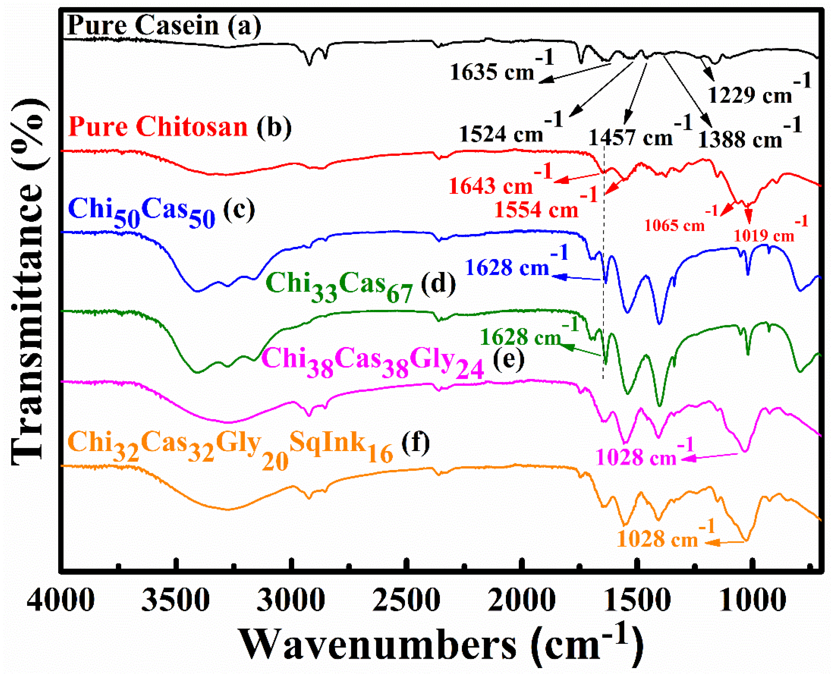
2.1. ATR-FTIR
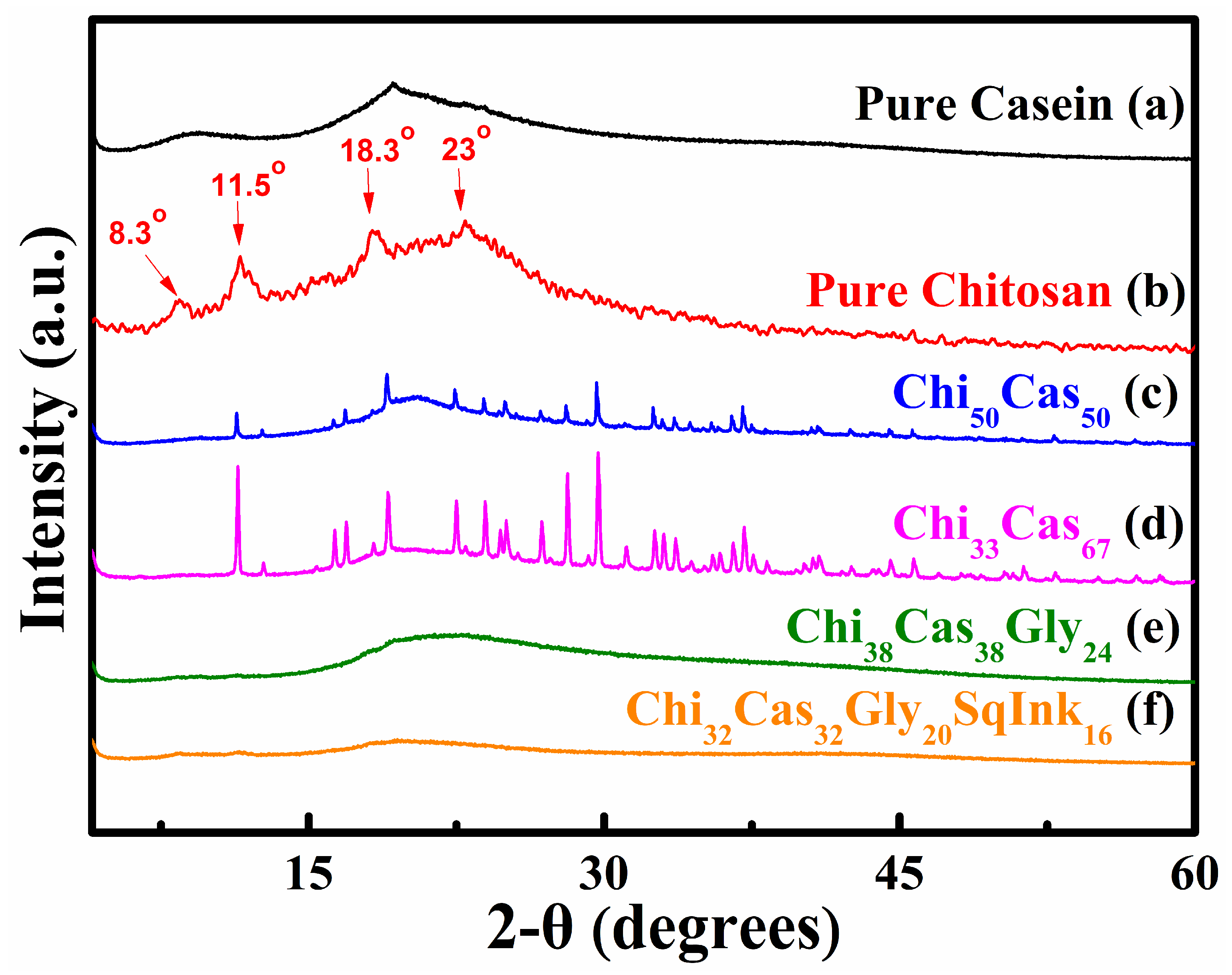
2.2. XRD Analysis
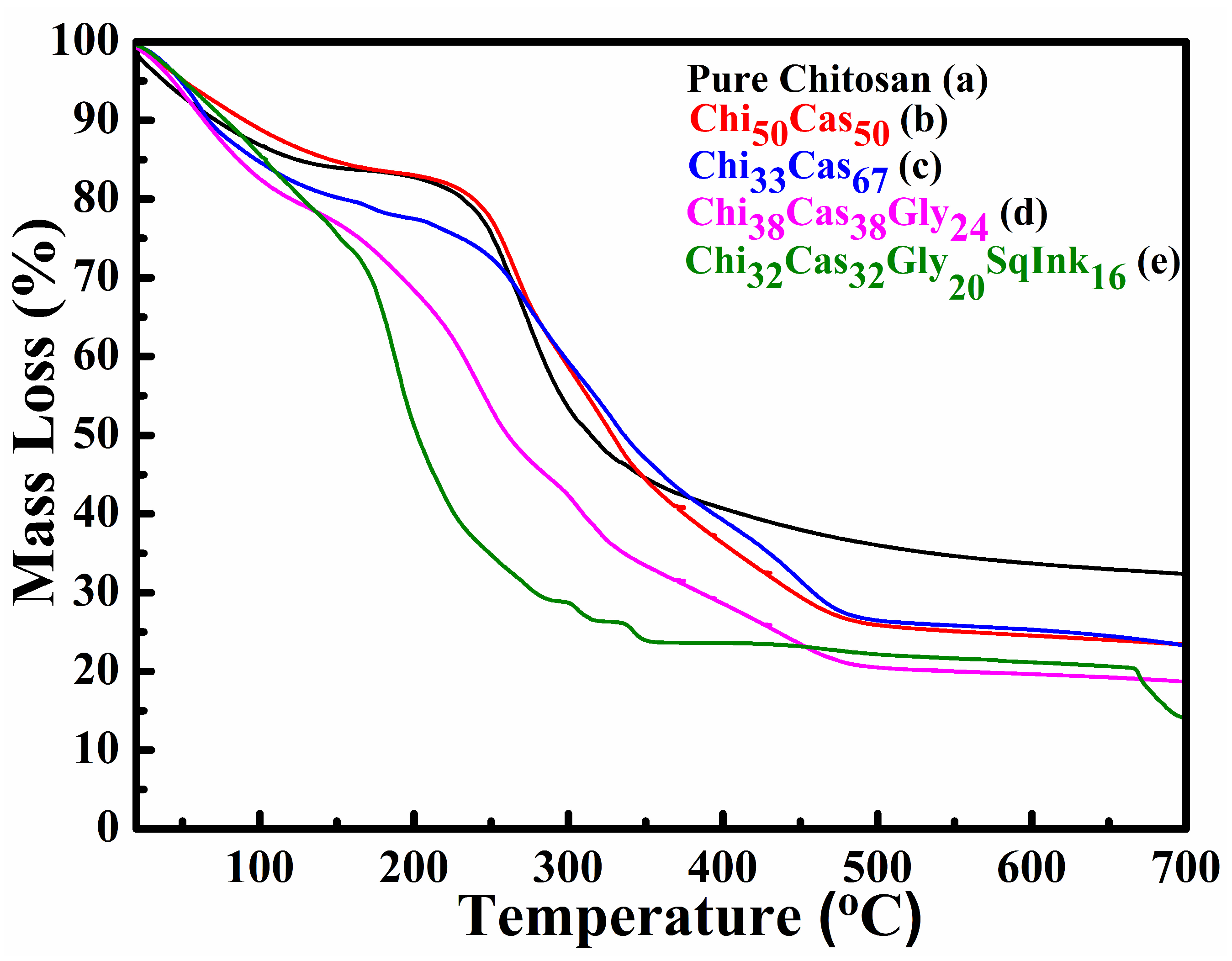
2.3. TGA
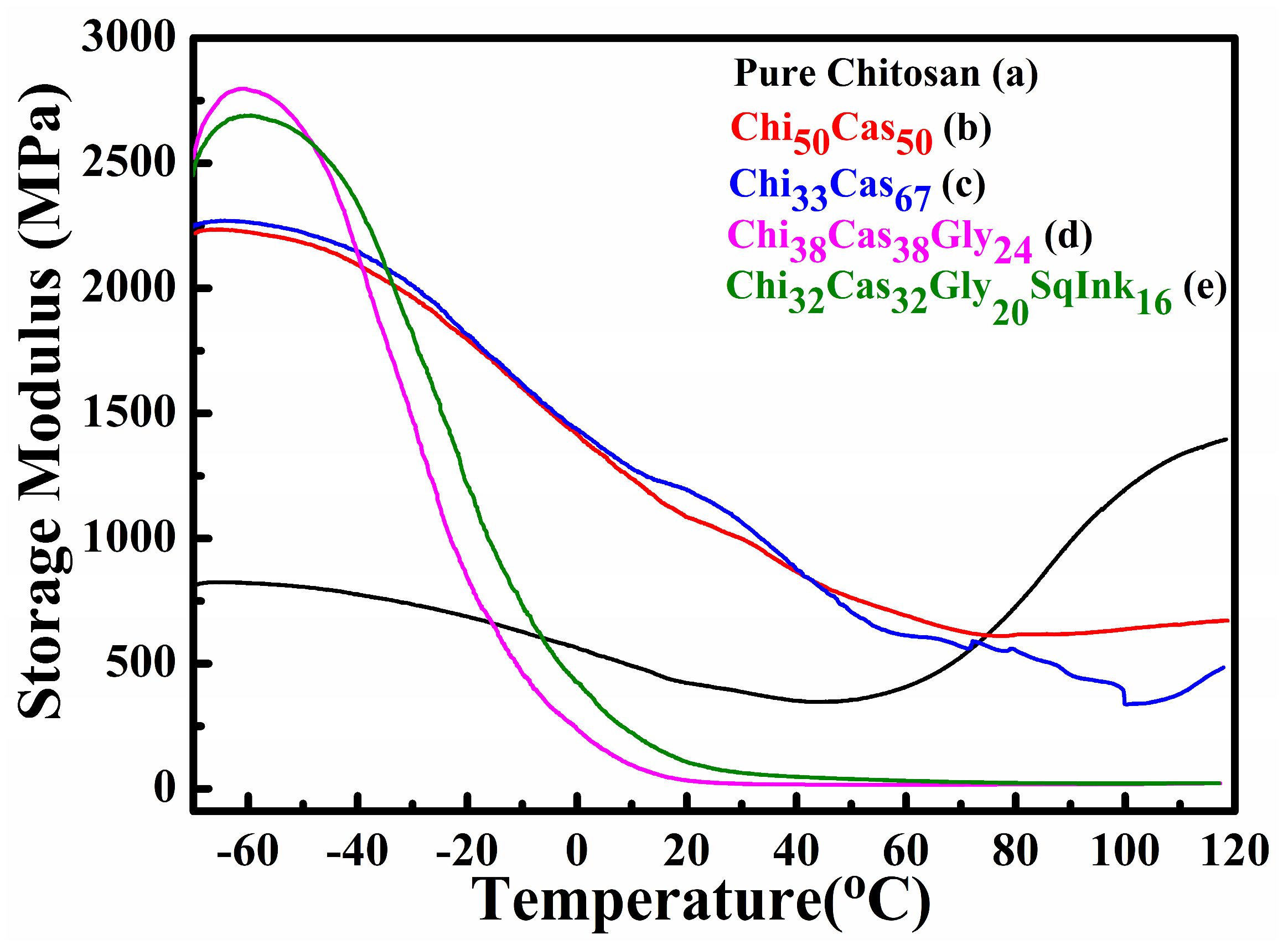
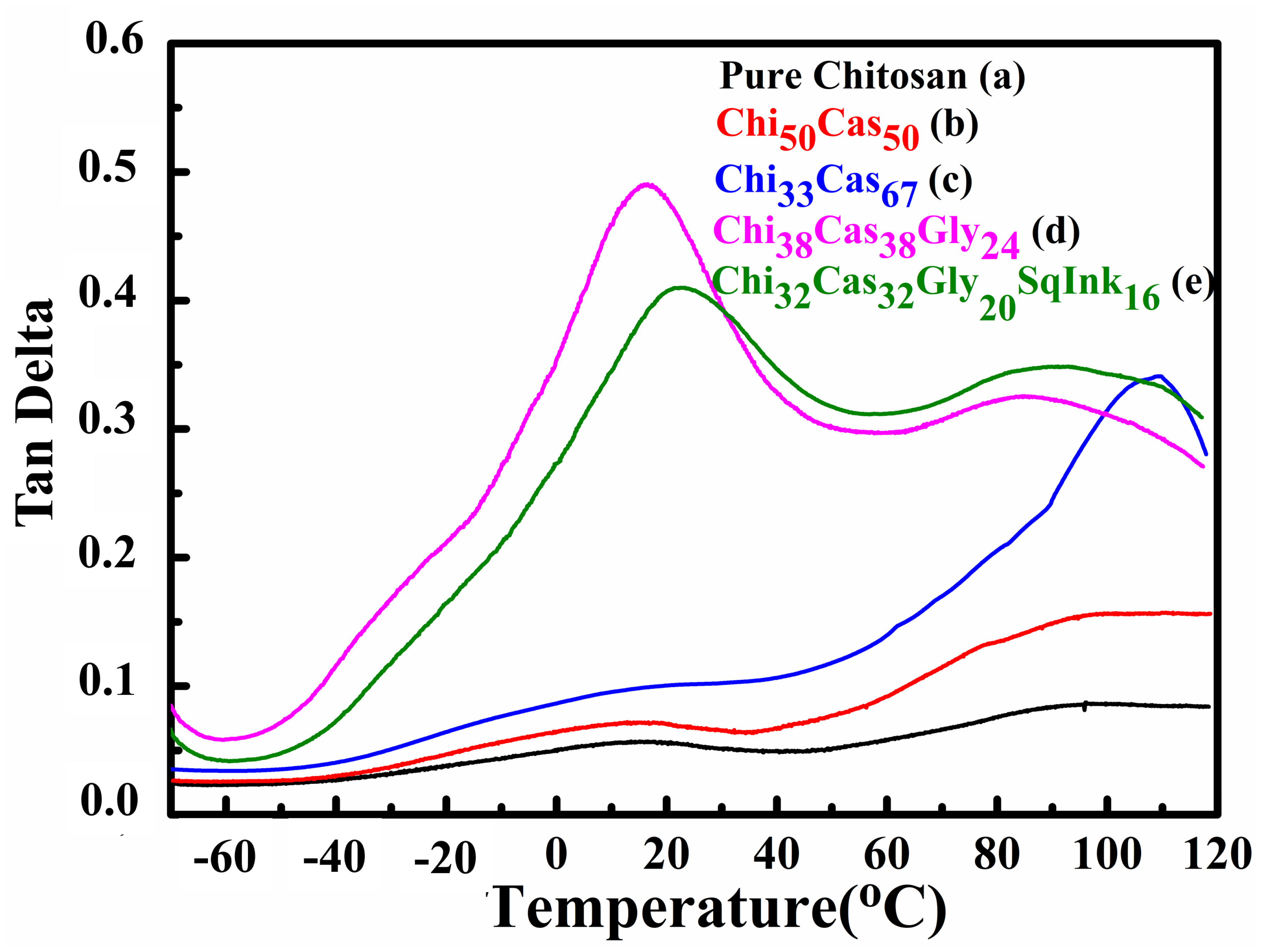
2.4. DMA
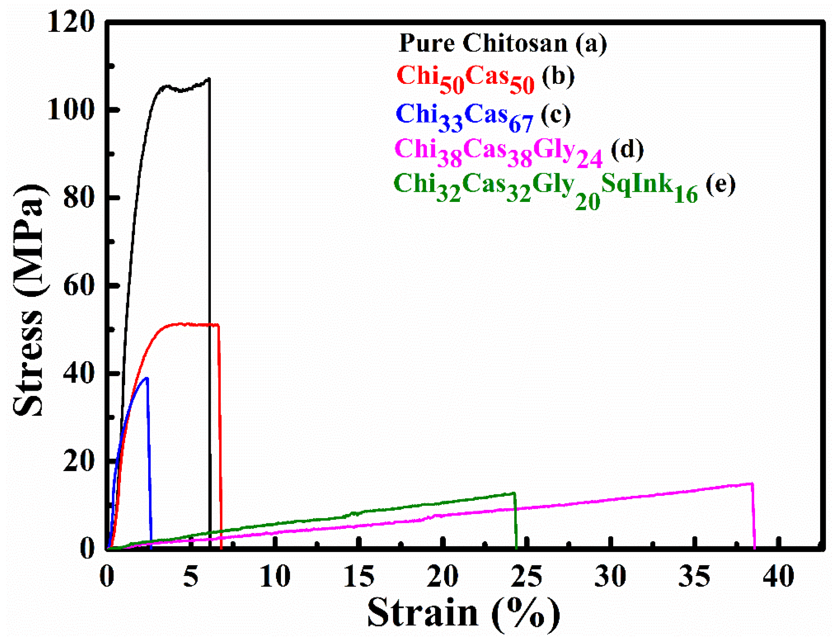
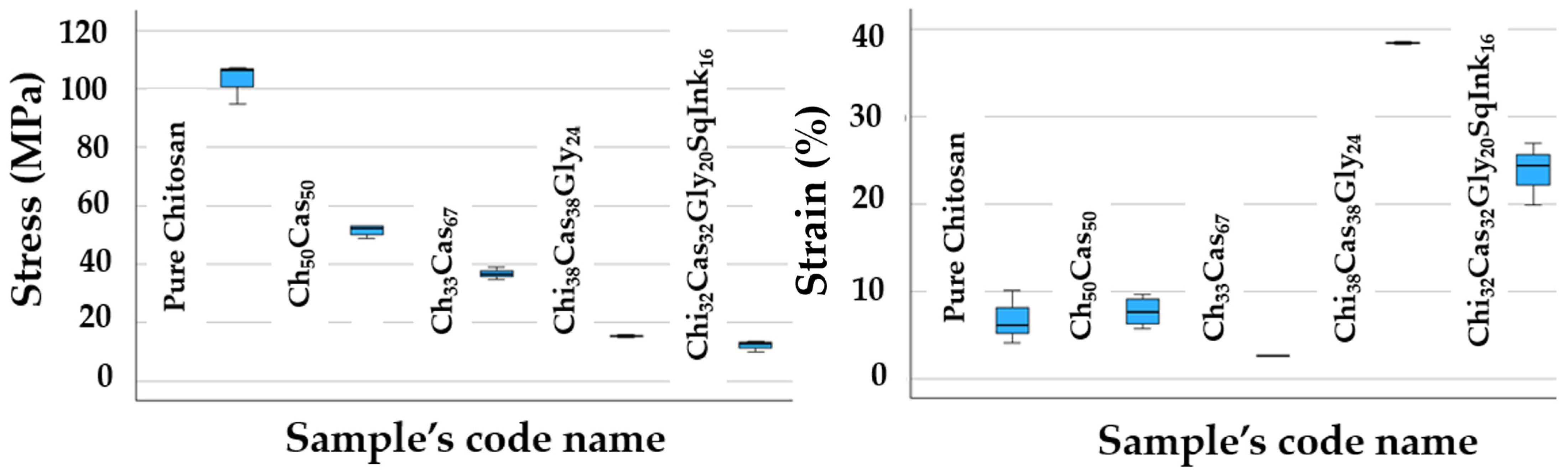
2.5. Tensile Properties
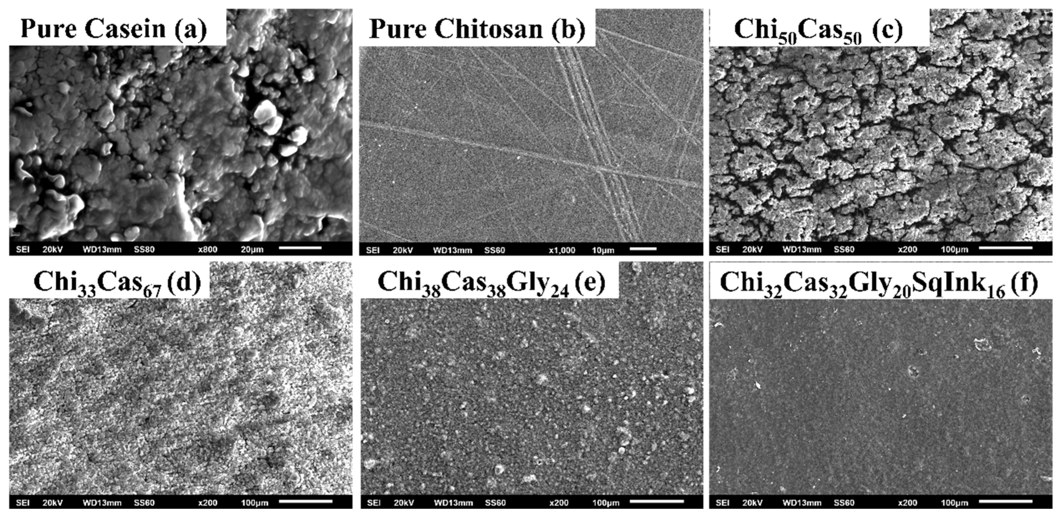
2.6. SEM Measurements
2.7. WVTR—Water/Vapor Diffusion Coefficient Calculation
2.8. OTR—Oxygen Permeability Calculation
3. Conclusions
4. Materials and Methods
4.1. Materials
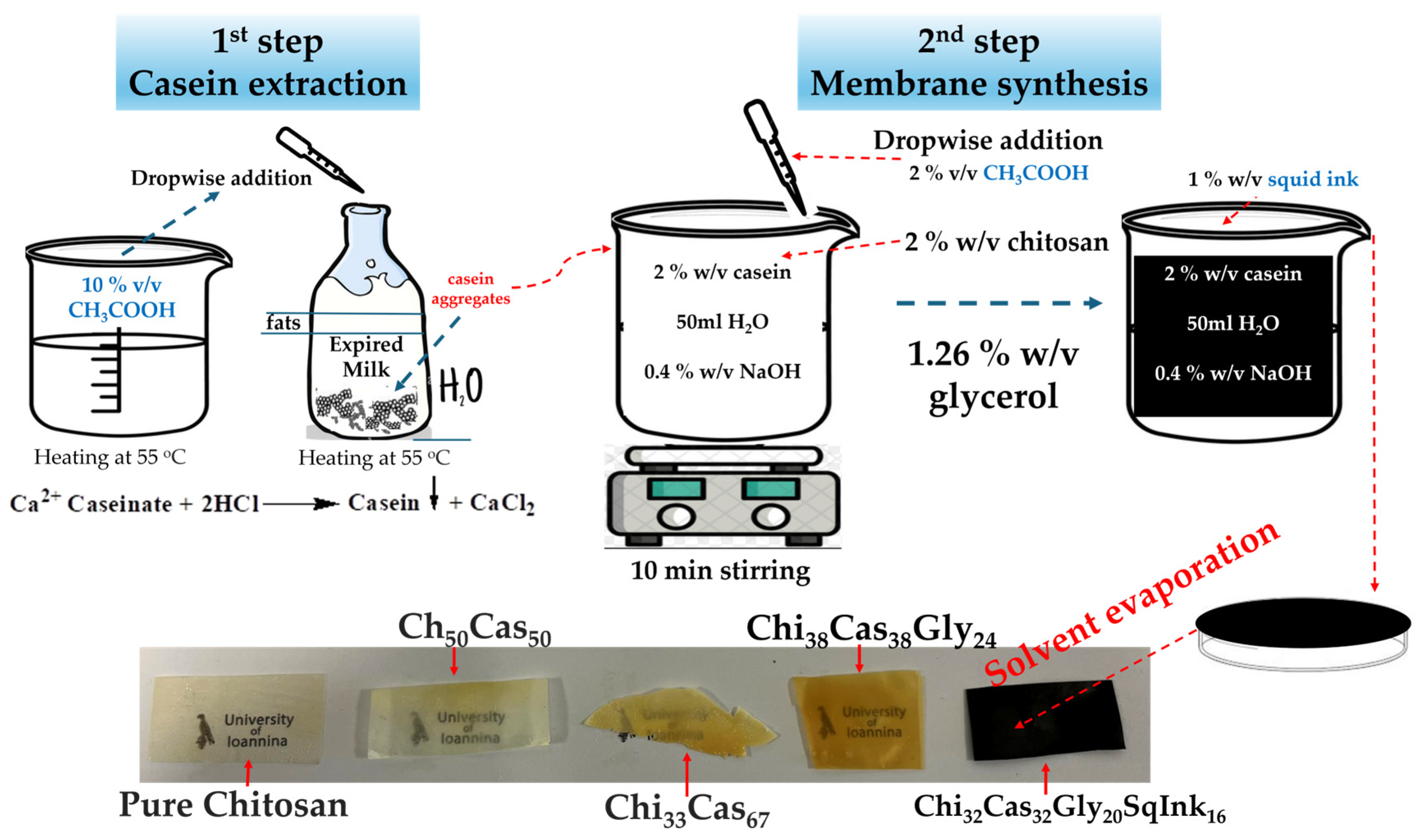
4.2. Extraction of Casein
4.3. Membrane Synthesis
4.4. Attenuated Total Reflectance-Fourier Transform Infrared Spectroscopy (ATR-FTIR)
4.5. X-ray Diffraction (XRD)
4.6. Thermogravimetric Analysis (TGA)
4.7. Dynamic Mechanical Analysis (DMA)
4.8. Mechanical Properties
4.9. Scanning Electron Microscopy (SEM)
4.10. Water Vapor Transmission Rate Measurements—Water Diffusion Coefficient Calculation
4.11. Oxygen Transmission Rate Measurements—Oxygen Permeability Calculation
4.12. Statistical Analysis
Author Contributions
Funding
Data Availability Statement
Acknowledgments
Conflicts of Interest
References
- Kummu, M.; de Moel, H.; Porkka, M.; Siebert, S.; Varis, O.; Ward, P.J. Lost food, wasted resources: Global food supply chain losses and their impacts on freshwater, cropland, and fertiliser use. Sci. Total Environ. 2012, 438, 477–489. [Google Scholar] [CrossRef] [PubMed]
- Ravindran, R.; Jaiswal, A.K. Exploitation of Food Industry Waste for High-Value Products. Trends Biotechnol. 2016, 34, 58–69. [Google Scholar] [CrossRef] [PubMed]
- Rosenboom, J.-G.; Langer, R.; Traverso, G. Bioplastics for a circular economy. Nat. Rev. Mater. 2022, 7, 117–137. [Google Scholar] [CrossRef] [PubMed]
- Rabnawaz, M.; Wyman, I.; Auras, R.; Cheng, S. A roadmap towards green packaging: The current status and future outlook for polyesters in the packaging industry. Green Chem. 2017, 19, 4737–4753. [Google Scholar] [CrossRef]
- Luzi, F.; Torre, L.; Kenny, J.M.; Puglia, D. Bio- and Fossil-Based Polymeric Blends and Nanocomposites for Packaging: Structure-Property Relationship. Materials 2019, 12, 471. [Google Scholar] [CrossRef] [PubMed]
- Guillard, V.; Gaucel, S.; Fornaciari, C.; Angellier-Coussy, H.; Buche, P.; Gontard, N. The Next Generation of Sustainable Food Packaging to Preserve Our Environment in a Circular Economy Context. Front. Nutr. 2018, 5, 121. [Google Scholar] [CrossRef] [PubMed]
- Aguirre-Joya, J.A.; De Leon-Zapata, M.A.; Alvarez-Perez, O.B.; Torres-León, C.; Nieto-Oropeza, D.E.; Ventura-Sobrevilla, J.M.; Aguilar, M.A.; Ruelas-Chacón, X.; Rojas, R.; Ramos-Aguiñaga, M.E.; et al. Chapter 1—Basic and Applied Concepts of Edible Packaging for Foods. In Food Packaging and Preservation; Grumezescu, A.M., Holban, A.M., Eds.; Academic Press: Cambridge, MA, USA, 2018; pp. 1–61. [Google Scholar]
- Hamed, I.; Jakobsen, A.N.; Lerfall, J. Sustainable edible packaging systems based on active compounds from food processing byproducts: A review. Compr. Rev. Food Sci. Food Saf. 2022, 21, 198–226. [Google Scholar] [CrossRef] [PubMed]
- Trajkovska Petkoska, A.; Daniloski, D.; D’Cunha, N.M.; Naumovski, N.; Broach, A.T. Edible packaging: Sustainable solutions and novel trends in food packaging. Food Res. Int. 2021, 140, 109981. [Google Scholar] [CrossRef]
- Sar, T.; Harirchi, S.; Ramezani, M.; Bulkan, G.; Akbas, M.Y.; Pandey, A.; Taherzadeh, M.J. Potential utilization of dairy industries by-products and wastes through microbial processes: A critical review. Sci. Total Environ. 2022, 810, 152253. [Google Scholar] [CrossRef] [PubMed]
- Gheorghita, R.; Gutt, G.; Amariei, S.J.C. The Use of Edible Films Based on Sodium Alginate in Meat Product Packaging: An Eco-Friendly Alternative to Conventional Plastic Materials. Coatings 2020, 10, 166. [Google Scholar] [CrossRef]
- Mellinas, C.; Valdés, A.; Ramos, M.; Burgos, N.; Garrigós, M.d.C.; Jiménez, A. Active edible films: Current state and future trends. J. Appl. Polym. Sci. 2016, 133. [Google Scholar] [CrossRef]
- Restrepo, A.E.; Rojas, J.D.; García, O.R.; Sánchez, L.T.; Pinzón, M.I.; Villa, C.C. Mechanical, barrier, and color properties of banana starch edible films incorporated with nanoemulsions of lemongrass (Cymbopogon citratus) and rosemary (Rosmarinus officinalis) essential oils. Food Sci. Technol. Int. 2018, 24, 705–712. [Google Scholar] [CrossRef] [PubMed]
- Gobbetti, M.; Minervini, F.; Rizzello, C.G. Angiotensin I-converting-enzyme-inhibitory and antimicrobial bioactive peptides. Int. J. Dairy Technol. 2004, 57, 173–188. [Google Scholar] [CrossRef]
- Korhonen, H.; Marnila, P.; Gill, H.S. Bovine milk antibodies for health. Br. J. Nutr. 2000, 84 (Suppl. S1), S135–S146. [Google Scholar] [CrossRef] [PubMed]
- Ryder, K.; Ali, M.A.; Carne, A.; Billakanti, J. The potential use of dairy by-products for the production of nonfood biomaterials. Crit. Rev. Environ. Sci. Technol. 2017, 47, 621–642. [Google Scholar] [CrossRef]
- Mazorra-Manzano, M.A.; Robles-Porchas, G.R.; González-Velázquez, D.A.; Torres-Llanez, M.J.; Martínez-Porchas, M.; García-Sifuentes, C.O.; González-Córdova, A.F.; Vallejo-Córdoba, B. Cheese Whey Fermentation by Its Native Microbiota: Proteolysis and Bioactive Peptides Release with ACE-Inhibitory Activity. Fermentation 2020, 6, 19. [Google Scholar] [CrossRef]
- Campos, C.A.; Gerschenson, L.N.; Flores, S.K. Development of Edible Films and Coatings with Antimicrobial Activity. Food Bioprocess Technol. 2011, 4, 849–875. [Google Scholar] [CrossRef]
- Bonnaillie, L.M.; Zhang, H.; Akkurt, S.; Yam, K.L.; Tomasula, P.M. Casein Films: The Effects of Formulation, Environmental Conditions and the Addition of Citric Pectin on the Structure and Mechanical Properties. Polymers 2014, 6, 2018–2036. [Google Scholar] [CrossRef]
- Khan, M.R.; Volpe, S.; Valentino, M.; Miele, N.A.; Cavella, S.; Torrieri, E. Active Casein Coatings and Films for Perishable Foods: Structural Properties and Shelf-Life Extension. Coatings 2021, 11, 899. [Google Scholar] [CrossRef]
- Babaei-Ghazvini, A.; Acharya, B.; Korber, D.R. Antimicrobial Biodegradable Food Packaging Based on Chitosan and Metal/Metal-Oxide Bio-Nanocomposites: A Review. Polymers 2021, 13, 2790. [Google Scholar] [CrossRef] [PubMed]
- Chaudhary, V.; Kajla, P.; Kumari, P.; Bangar, S.P.; Rusu, A.; Trif, M.; Lorenzo, J.M. Milk protein-based active edible packaging for food applications: An eco-friendly approach. Front. Nutr. 2022, 9, 942524. [Google Scholar] [CrossRef] [PubMed]
- Nadarajah, S.K.; Vijayaraj, R.; Mani, J. Therapeutic Significance of Loligo vulgaris (Lamarck, 1798) ink Extract: A Biomedical Approach. Pharmacogn. Res. 2017, 9, S105–S109. [Google Scholar] [CrossRef] [PubMed]
- Celli, G.B.; Ravanfar, R.; Kaliappan, S.; Kapoor, R.; Abbaspourrad, A. Annatto-entrapped casein-chitosan complexes improve whey color quality after acid coagulation of milk. Food Chem. 2018, 255, 268–274. [Google Scholar] [CrossRef] [PubMed]
- Chakrapani, V.; Ayaz Ahmed, K.B.; Kumar, V.V.; Ganapathy, V.; Anthony, S.P.; Anbazhagan, V. A facile route to synthesize casein capped copper nanoparticles: An effective antibacterial agent and selective colorimetric sensor for mercury and tryptophan. RSC Adv. 2014, 4, 33215–33221. [Google Scholar] [CrossRef]
- Gebhardt, R.; Takeda, N.; Kulozik, U.; Doster, W. Structure and Stabilizing Interactions of Casein Micelles Probed by High-Pressure Light Scattering and FTIR. J. Phys. Chem. B 2011, 115, 2349–2359. [Google Scholar] [CrossRef] [PubMed]
- Karydis-Messinis, A.; Moschovas, D.; Markou, M.; Gkantzou, E.; Vasileiadis, A.; Tsirka, K.; Gioti, C.; Vasilopoulos, K.C.; Bagli, E.; Murphy, C.; et al. Development, physicochemical characterization and in vitro evaluation of chitosan-fish gelatin-glycerol hydrogel membranes for wound treatment applications. Carbohydr. Polym. Technol. Appl. 2023, 6, 100338. [Google Scholar] [CrossRef]
- Ma, Y.; Xin, L.; Tan, H.; Fan, M.; Li, J.; Jia, Y.; Ling, Z.; Chen, Y.; Hu, X. Chitosan membrane dressings toughened by glycerol to load antibacterial drugs for wound healing. Mater. Sci. Eng. C 2017, 81, 522–531. [Google Scholar] [CrossRef] [PubMed]
- Fernández, C.; Ausar, S.F.; Badini, R.G.; Castagna, L.F.; Bianco, I.D.; Beltramo, D.M. An FTIR spectroscopy study of the interaction between αs-casein-bound phosphoryl groups and chitosan. Int. Dairy J. 2003, 13, 897–901. [Google Scholar] [CrossRef]
- Astbury, W.T.; Lomax, R. X-Ray Photographs of Crystalline Pepsin. Nature 1934, 133, 795. [Google Scholar] [CrossRef]
- Clark, G.L.; Schaad, J.A. X-ray Diffraction Studies of Tendon and Intestinal Wall Collagen. Radiology 1936, 27, 339–356. [Google Scholar] [CrossRef]
- Arvanitoyannis, I.S.; Nakayama, A.; Aiba, S.-I. Chitosan and gelatin based edible films: State diagrams, mechanical and permeation properties. Carbohydr. Polym. 1998, 37, 371–382. [Google Scholar] [CrossRef]
- Karydis-Messinis, A.; Moschovas, D.; Markou, M.; Tsirka, K.; Gioti, C.; Bagli, E.; Murphy, C.; Giannakas, A.E.; Paipetis, A.; Karakassides, M.A.; et al. Hydrogel Membranes from Chitosan-Fish Gelatin-Glycerol for Biomedical Applications: Chondroitin Sulfate Incorporation Effect in Membrane Properties. Gels 2023, 9, 844. [Google Scholar] [CrossRef] [PubMed]
- Bengoechea, C.; Arrachid, A.; Guerrero, A.; Hill, S.E.; Mitchell, J.R. Relationship between the glass transition temperature and the melt flow behavior for gluten, casein and soya. J. Cereal Sci. 2007, 45, 275–284. [Google Scholar] [CrossRef]
- Murrieta-Martínez, C.L.; Soto-Valdez, H.; Pacheco-Aguilar, R.; Torres-Arreola, W.; Rodríguez-Felix, F.; Márquez Ríos, E. Edible protein films: Sources and behavior. Packag. Technol. Sci. 2018, 31, 113–122. [Google Scholar] [CrossRef]
- Shah, Y.A.; Bhatia, S.; Al-Harrasi, A.; Afzaal, M.; Saeed, F.; Anwer, M.K.; Khan, M.R.; Jawad, M.; Akram, N.; Faisal, Z. Mechanical Properties of Protein-Based Food Packaging Materials. Polymers 2023, 15, 1724. [Google Scholar] [CrossRef] [PubMed]
- Volpe, S.; Cavella, S.; Masi, P.; Torrieri, E. Effect of solid concentration on structure and properties of chitosan-caseinate blend films. Food Packag. Shelf Life 2017, 13, 76–84. [Google Scholar] [CrossRef]
- Kneifel, W.; Paquin, P.; Abert, T.; Richard, J.P. Water-Holding Capacity of Proteins with Special Regard to Milk Proteins and Methodological Aspects—A Review. J. Dairy Sci. 1991, 74, 2027–2041. [Google Scholar] [CrossRef]
- Chevalier, E.; Assezat, G.; Prochazka, F.; Oulahal, N. Development and characterization of a novel edible extruded sheet based on different casein sources and influence of the glycerol concentration. Food Hydrocoll. 2018, 75, 182–191. [Google Scholar] [CrossRef]
- Giannakas, A.E.; Salmas, C.E.; Moschovas, D.; Zaharioudakis, K.; Georgopoulos, S.; Asimakopoulos, G.; Aktypis, A.; Proestos, C.; Karakassides, A.; Avgeropoulos, A.; et al. The Increase of Soft Cheese Shelf-Life Packaged with Edible Films Based on Novel Hybrid Nanostructures. Gels 2022, 8, 539. [Google Scholar] [CrossRef]


| Specimen | Stress (MPa) | Strain (%) | Elongation at Break (mm) | % Change in Stress * | % Change in Strain * |
|---|---|---|---|---|---|
| Pure Chitosan | 102.82 ± 6.97 a | 6.77 ± 3.01 a | 1.79 ± 0.79 | Reference system | |
| Chi50Cas50 | 51.60 ± 2.00 a,b | 7.66 ± 1.72 a | 2.01 ± 0.40 | −49.82 | +13.15 |
| Chi33Cas67 | 36.84 ± 2.02 a,b,c | 2.61 ± 0.07 a | 0.69 ± 0.02 | −64.17 | −61.45 |
| Chi38Cas38Gly24 | 15.36 ± 0.45 b,c | 38.42 ± 0.16 b | 10.34 ± 0.22 | −85.06 | +467.50 |
| Chi32Cas32Gly20SqInk16 | 12.18 ± 1.85 c | 23.74 ± 3.56 b | 6.33 ± 1.09 | −88.15 | +250.66 |
| Samples | WVTR [×10−7 gr/(cm2*s)] | Dwv (×10−4 cm2/s) |
|---|---|---|
| Pure Chitosan | 7.69367 ± 0.64878 | 1.10 ± 0.198 a |
| Chi50Cas50 | 9.55637 ± 4.27094 | 3.33 ± 0.790 a,b |
| Chi33Cas67 | 7.79506 ± 5.03817 | 5.16 ± 1.040 a,b |
| Chi38Cas38Gly24 | 19.2141 ± 3.35570 | 8.20 ± 1.690 b |
| Chi32Cas32Gly20SqInk16 | 15.3178 ± 3.21761 | 8.51 ± 0.876 b |
| Samples | OTR (mL.*m−2*day−1) | PeO2 (cm2/s) |
|---|---|---|
| Pure Chitosan | <0.5 * | impermeable |
| Chi50Cas50 | <0.5 * | impermeable |
| Chi33Cas67 | <0.5 * | impermeable |
| Chi38Cas38Gly24 | <0.5 * | impermeable |
| Chi32Cas32Gly20SqInk16 | <0.5 * | impermeable |
| Sample Code | Chitosan (w/v %) | Casein (w/v %) | Glycerol (w/v %) | Squid Ink (w/v %) |
|---|---|---|---|---|
| Chi32Cas32Gly20SqInk16 (%wt: 32/32/20/16) | 2 | 2 | 1.26 | 1 |
| Chi38Cas38Gly24 (%wt: 38/38/24) | 2 | 2 | 1.26 | - |
| Chi33Cas67 (%wt: 33/67) | 2 | 4 | - | - |
| Chi50Cas50 (%wt: 50/50) | 2 | 2 | - | - |
| Pure Chitosan (%wt: 100) | 2 | - | - | - |
| Pure Casein (powder) | - | 100 | - | - |
Disclaimer/Publisher’s Note: The statements, opinions and data contained in all publications are solely those of the individual author(s) and contributor(s) and not of MDPI and/or the editor(s). MDPI and/or the editor(s) disclaim responsibility for any injury to people or property resulting from any ideas, methods, instructions or products referred to in the content. |
© 2024 by the authors. Licensee MDPI, Basel, Switzerland. This article is an open access article distributed under the terms and conditions of the Creative Commons Attribution (CC BY) license (https://creativecommons.org/licenses/by/4.0/).
Share and Cite
Karydis-Messinis, A.; Kyriakaki, C.; Triantafyllou, E.; Tsirka, K.; Gioti, C.; Gkikas, D.; Nesseris, K.; Exarchos, D.A.; Farmaki, S.; Giannakas, A.E.; et al. Development and Physicochemical Characterization of Edible Chitosan–Casein Hydrogel Membranes for Potential Use in Food Packaging. Gels 2024, 10, 254. https://doi.org/10.3390/gels10040254
Karydis-Messinis A, Kyriakaki C, Triantafyllou E, Tsirka K, Gioti C, Gkikas D, Nesseris K, Exarchos DA, Farmaki S, Giannakas AE, et al. Development and Physicochemical Characterization of Edible Chitosan–Casein Hydrogel Membranes for Potential Use in Food Packaging. Gels. 2024; 10(4):254. https://doi.org/10.3390/gels10040254
Chicago/Turabian StyleKarydis-Messinis, Andreas, Christina Kyriakaki, Eleni Triantafyllou, Kyriaki Tsirka, Christina Gioti, Dimitris Gkikas, Konstantinos Nesseris, Dimitrios A. Exarchos, Spyridoula Farmaki, Aris E. Giannakas, and et al. 2024. "Development and Physicochemical Characterization of Edible Chitosan–Casein Hydrogel Membranes for Potential Use in Food Packaging" Gels 10, no. 4: 254. https://doi.org/10.3390/gels10040254
APA StyleKarydis-Messinis, A., Kyriakaki, C., Triantafyllou, E., Tsirka, K., Gioti, C., Gkikas, D., Nesseris, K., Exarchos, D. A., Farmaki, S., Giannakas, A. E., Salmas, C. E., Matikas, T. E., Moschovas, D., & Avgeropoulos, A. (2024). Development and Physicochemical Characterization of Edible Chitosan–Casein Hydrogel Membranes for Potential Use in Food Packaging. Gels, 10(4), 254. https://doi.org/10.3390/gels10040254