
Advances in Hydrogel-Based Drug Delivery Systems

1
Division of Hematology/Oncology, Boston Children’s Hospital, Boston, MA 02115, USA
2
Department of Pediatrics, Harvard Medical School, Boston, MA 02115, USA
3
Department of Polymer Science and Engineering, University of Massachusetts, Amherst, MA 01003, USA
*
Authors to whom correspondence should be addressed.
Gels 2024, 10(4), 262; https://doi.org/10.3390/gels10040262
Submission received: 19 March 2024
/
Revised: 5 April 2024
/
Accepted: 10 April 2024
/
Published: 13 April 2024
(This article belongs to the Special Issue Advanced Hydrogels for Regenerative Medicine and Tissue Engineering (2nd Edition))
Abstract
:Hydrogels, with their distinctive three-dimensional networks of hydrophilic polymers, drive innovations across various biomedical applications. The ability of hydrogels to absorb and retain significant volumes of water, coupled with their structural integrity and responsiveness to environmental stimuli, renders them ideal for drug delivery, tissue engineering, and wound healing. This review delves into the classification of hydrogels based on cross-linking methods, providing insights into their synthesis, properties, and applications. We further discuss the recent advancements in hydrogel-based drug delivery systems, including oral, injectable, topical, and ocular approaches, highlighting their significance in enhancing therapeutic outcomes. Additionally, we address the challenges faced in the clinical translation of hydrogels and propose future directions for leveraging their potential in personalized medicine and regenerative healthcare solutions.
1. Introduction
Hydrogels, characterized by their unique three-dimensional networks of hydrophilic polymers, have emerged as a cornerstone in the advancement of biomaterial science, revolutionizing applications across a broad spectrum of biomedical fields [1,2,3,4,5,6,7,8]. These networks, capable of absorbing and retaining substantial volumes of water, are distinguished by their remarkable ability to swell without dissolution, maintaining structural integrity through chemical or physical cross-linking mechanisms [9,10]. This intrinsic property allows hydrogels to mimic the physicochemical aspects of the natural extracellular matrix, making them particularly suited for applications in drug delivery systems [7,9,11,12,13,14,15,16,17,18,19], tissue engineering [20,21,22,23,24], wound healing [25,26,27,28], and beyond, as illustrated in Figure 1. The initiation of hydrogel research and its expansion into biomedical sciences exemplify a trajectory of innovation, highlighting the versatility of these materials in solving complex biological challenges and their role in advancing biomedical solutions.
The foundation of hydrogel technology was laid through the pioneering work of Wichterle and Lim, who produced poly(hydroxyethylmethacrylate) (pHEMA) hydrogel in the 1960s [29], marking the inception of hydrogels as biomaterials. Since then, the field has witnessed exponential growth, driven by advancements in polymer science and a deepening understanding of biological interfaces [1,30]. In addition to the comprehensive experimental investigation of hydrogels in biomedical applications, particularly in drug delivery, theoretical frameworks and computational modeling have been instrumental in shedding light on the structural dynamics of hydrogels [31,32,33,34,35,36,37]. They provide valuable insights into characteristics such as elasticity, porosity, and mesh size. These predictive models, based on thermodynamic principles, have enabled the design of hydrogels with customized properties for targeted biomedical uses. Hydrogels can be broadly categorized into natural, synthetic, and semisynthetic, based on their origin and the nature of their cross-linking mechanisms [38,39,40]. This classification highlights the need to balance mechanical strength with biodegradation rates for optimal biocompatibility and functionality. To be biocompatible, hydrogels must be non-toxic and not trigger adverse immune reactions. Functionality requires adjusting physical properties like porosity, swelling, and strength to suit specific applications, from drug delivery to tissue engineering. The evolution of hydrogel polymerization techniques, including the development of homopolymers, copolymers, and interpenetrating networks, further demonstrates the versatility of these materials, enabling the customization of hydrogel properties to meet the demands of specific biomedical applications.
The ability of hydrogels to respond to external stimuli, such as chemical, physical, or biological factors, has unveiled novel opportunities for the creation of intelligent materials [9,14,39,41]. This characteristic is pivotal in the conception of dynamic hydrogel systems that can adjust to varying physiological conditions, paving the way for advanced applications in smart drug delivery systems, adaptive tissue scaffolds, and responsive biomedical devices. The introduction of injectable hydrogels, characterized by their shear thinning and self-healing properties, represents significant progress towards the development of less invasive therapeutic modalities [42,43]. These hydrogels present a promising alternative to conventional surgical techniques, offering targeted drug delivery and tissue repair directly at the site of action. Nonetheless, the path towards the clinical translation of these materials is beset with challenges, particularly the imperative to mitigate adverse immunogenic responses and ensure the removal of deleterious by-products from the cross-linking process [44]. Recent advancements in nanotechnology have precipitated the emergence of nanogels, a novel class of hydrogel-based nanomaterials with promising prospects in drug delivery and tissue engineering [45,46]. Alongside progress in 3D bioprinting [47,48], hydrogels are now at the vanguard of fabricating tissue constructs with unprecedented precision and complexity, heralding a new epoch in regenerative medicine and tissue engineering.
In the realm of drug delivery systems, hydrogels encounter limitations pertaining to biocompatibility, safe assembly, and controlled drug release [49]. These materials must be constructed from biocompatible constituents to circumvent immune reactions and assembled utilizing non-toxic chemistries in aqueous conditions at physiological pH. Despite the availability of suitable synthetic and natural polymers, the assurance of controlled therapeutic release, whether through physical constraints or drug-material affinity, remains a formidable challenge. Advances in polymer science have led to the creation of biopolymers that offer a biodegradable and biocompatible framework for hydrogels, augmenting their utility in targeted and efficient drug delivery. However, achieving the requisite mechanical stability, biodegradability, and target specificity while ensuring safety and efficacy in the physiological milieu highlights the ongoing challenges in the design of hydrogel-based drug delivery systems.
In this review, we begin by categorizing hydrogels based on their cross-linking methods, then proceed to explore the critical properties of hydrogels that influence drug delivery, integrating both theoretical insights and experimental results. This encompasses assessments of mesh size, swelling behavior, porosity, microstructures, mechanical properties, and the degradation features of biodegradable hydrogels. Subsequently, we address recent advancements in hydrogel applications, particularly in drug delivery, underscoring their significance in enhancing therapeutic outcomes. Furthermore, we delve into the current challenges and future directions in the utilization of hydrogels for drug delivery. In summary, this review underscores the transformative potential of hydrogels in biomedical engineering, spotlighting their evolving applications in drug delivery and the critical need for ongoing innovation to address existing challenges and unlock new avenues for therapeutic intervention.
2. Classification of Hydrogels
The classification of hydrogels can be based on various criteria, including their source, cross-linking methods, composition, degradability, stimuli responsiveness, and ionic charge. In this context, we categorize them into physical and chemical hydrogels, distinguished by their cross-linking formation mechanisms, as illustrated in Figure 2. Hybrid gels featuring both physical and chemical cross-linking are not individually classified here. We proceed to select and review various representative hydrogels characterized by diverse gelation mechanisms.
2.1. Physcial Hydrogel
Physical hydrogels are synthesized from low-molecular-weight compounds or polymers through non-covalent interactions such as hydrogen bonding [50,51,52,53], van der Waals forces [11], electrostatic attractions [54,55,56], and specific host-guest interactions [57,58,59]. These reversible interactions allow physical gels to undergo gel-to-sol transitions in response to environmental stimuli like temperature, pH, and ionic strength, endowing them with adaptability and responsiveness. In contrast to chemical gels, which rely on permanent covalent bonds, physical gels form transient networks through weak, albeit numerous, physicochemical bonds, granting them properties such as softness, reversibility, and sensitivity to external stimuli. These features make physical gels particularly suitable for applications requiring sensitivity to environmental changes, such as in drug delivery and tissue engineering scenarios. Numerous physical hydrogels have been designed for use in drug delivery applications, with several notable examples listed below.
Yoshimura et al. introduced biodegradable hydrogels synthesized by reacting starch with succinic anhydride (SA), using 4-dimethylaminopyridine as an esterification catalyst in DMSO or water, followed by NaOH neutralization [51]. The formation of the starch-SA hydrogel is attributed primarily to the regeneration of hydrogen bonds during the dialysis process. These hydrogels, with substitution degrees ranging from 0.1 to 1.4, demonstrate maximum water absorbencies of up to 120 g-water/g-dry gel. Notably, hydrogels produced in DMSO showed superior substitution degrees and absorbencies due to the reduction of SA hydrolysis, suggesting their suitability for biomedical and agricultural uses given their biodegradability and efficient water absorption.
Lu et al. crafted an injectable hydrogel through the physical blending of carboxymethyl hexanoyl chitosan and hyaluronic acid, exploiting supermolecular interactions to form a network capable of sustained therapeutic release [58]. This injectable modality, paired with its controlled release capabilities, is poised for localized drug delivery and regenerative medicine applications. Tran et al. devised a syringeable hydrogel from -cyclodextrin and mixed micelles for methotrexate delivery [59], utilizing host-guest interactions for hydrogel formation, which enhances drug delivery efficiency, showcasing the innovative use of host-guest chemistry in developing functional biomedical materials.
Sun et al. explored a novel hydrogel formulation integrating nanostructured lipid carriers (NLC) with chitosan-tripolyphosphate (chitosan-TPP) hydrogel beads, leveraging hydrophobic interactions for efficient encapsulation of hydrophobic active substances [60]. The chitosan-TPP hydrogel forms through electrostatic interactions between the phosphate groups of TPP and the protonated amino groups of chitosan. This strategy not only improves bioavailability and controlled drug release but also holds significant promise for enhancing topical drug delivery systems, illustrating the potential of hydrophobic interactions in innovative hydrogel designs.
2.2. Chemical Hydrogel
Chemical hydrogels, synthesized through covalent bonding of polymers, leverage chemically active motifs for crosslinking, utilizing a variety of methods such as carbodiimide chemistry [61,62,63], free radical polymerization [29,64,65,66,67,68,69,70,71,72,73], and click chemistry [74,75,76,77,78,79,80]. These approaches offer chemical hydrogels enhanced matrix stabilization and greater control over gel formation, enabling a higher degree of flexibility and spatiotemporal precision than physical gels. Specifically, enzymatic crosslinking, utilizing biocompatible enzymes like peroxidases and transglutaminases, presents a favorable method for creating biocompatible, nonimmunogenic hydrogels suitable for tissue engineering, drug delivery, and regenerative medicine (TERM) applications due to its mild reaction conditions and minimal cytotoxicity [81,82]. Additionally, electron irradiation techniques have emerged, providing efficient and precise crosslinking capabilities that expand the possibilities for hydrogel customization [83,84].
The pioneering synthesis of pHEMA hydrogels by Wichterle and Lim [29], achieved through free radical polymerization, highlights the potential for integrating acrylate derivatives into biopolymers such as dextran [64], albumin [69,70], starch [68,71], and hyaluronic acid [72,73], transforming them into cross-linked hydrogels. Similarly, high-energy radiation, including gamma rays and electron beams, facilitates the transformation of vinyl polymer solutions into hydrogels or initiates the polymerization of monofunctional acrylates with cross-linkers, forming hydrogels [65,66,67]. This process involves radical generation from carbon-hydrogen bond breaking or water molecule radicals, which then recombine to create covalent cross-links. Specifically, polyvinyl alcohol, polyethylene glycol, and polyacrylic acid are capable of undergoing such radical reactions, facilitating the formation of hydrogels.
Furthermore, the Click chemistry reaction, catalyzed by monovalent copper to form triazole rings from azides and alkynes, stands out for its efficiency and by-product-free nature, making it ideal for in situ hydrogel formation [74,76,77,78]. This reaction is particularly suited for modifying polymers like polyethylene glycol, hyaluronic acid, gelatin, and peptides with azido or alkynyl groups for cross-linking, facilitating the creation of enzyme-degradable and cell-adhesive hydrogels. To avoid the toxicity associated with copper ions, copper-free Click reactions utilizing cyclooctyne derivatives have been developed, offering safer cross-linking alternatives [75,79,80].
Sperinde et al. used enzymes for hydrogel synthesis, developing a system based on tetrahydroxyl polyethylene glycol (PEG) functionalized with glutamine [85]. By incorporating glutaminase into a solution of glutamine-functionalized tetrahydroxyl PEG and poly(lysine phenylalanine), they catalyzed the formation of amide bonds between the glutamine and lysine groups, effectively cross-linking the polymers. This enzymatic approach, particularly with the calcium ion dependence of transglutaminase, allows for the design of stimulus-responsive gelation systems, featuring the depth of chemical versatility in hydrogel synthesis for targeted applications.
3. Characterization of Hydrogels
This section delves into the crucial properties of hydrogels, such as mesh size, swelling behavior, porosity, microstructure, mechanical strength, and degradability. We start by examining why these characteristics are vital for the effectiveness of hydrogel-based drug delivery systems. Next, we investigate the common techniques used to measure these physical properties. We then highlight how the manipulation of these properties can enhance hydrogel performance. The pros and cons of these characterization techniques are neatly summarized in Table 1. Recently, Denzer et al. have offered an extensive overview of the methods employed in hydrogel characterization [86].
3.1. Mesh Size and Swelling Behavior
The swelling behavior of hydrogels in drug delivery significantly impacts drug release and diffusion, making it a crucial factor in their application. Hydrogels, with their crosslinked polymer networks, facilitate the movement of liquids and solutes through open spaces or meshes. The mesh size () of hydrogel used in drug delivery, typically less than 200 nm, is pivotal in determining the interaction between the drug and the polymer network, thereby controlling the diffusion of drugs [12]. However, it is important to note that hydrogels often display a wide range of mesh sizes due to network heterogeneity and polymer polydispersity. This variability is especially pronounced in hydrogels formed through free-radical polymerization. Achieving a uniform mesh size is possible by using symmetrical tetrahedron-like macromeres of identical size for gelation, leading to hydrogels with a more uniform mesh size [87,88,89]. For smaller drug molecules, release is primarily governed by diffusion, allowing for their free movement within the network. This process is influenced by factors such as the drug size and the viscosity of a medium, as dictated by the Stokes–Einstein equation. Typically, drugs are composed of small molecules, resulting in their entropic effects being relatively minor when trapped within the gel network, especially in comparison to the more complex dynamics of the embedded polymers [90,91,92]. When mesh and drug sizes are similar, steric hindrance slows diffusion, enabling prolonged release. A small ratio between mesh size and drug leads to entrapment until the network degrades or expands.
Characterizing the swelling ratio (Q) of hydrogels can be approached through various methodologies, each with its own set of advantages and challenges. A prevalent and straightforward method is to dry or lyophilize the hydrogel sample with a dry weight, , and immerse it in a substantial volume of water or specific buffer solutions for predetermined durations such as 16, 24, or 48 h [10,25,26,51,62,64,65,68,72,78,93,94], followed by filtration and measurement of the swollen hydrogel’s weight () to determine the degree of swelling using the formula:
The immersion time is critical and varies based on the time to reach swelling equilibrium, which presents a limitation due to the difficulty in precisely determining this point. Furthermore, the filtration step introduces variability based on the mesh used and the operator’s technique, potentially complicating measurements for very soft hydrogels that might obstruct the mesh. An alternative method, particularly suitable for hydrogels with isotropic network structures [54,85,95], calculates the volumetric swelling ratio from the thickness swelling ratio as , where t is the thickness of the swollen gel and is its original thickness. This method directly correlates isotropic swelling behavior with volumetric changes, emphasizing the importance of precise measurement techniques for thickness determination.
The mesh size () of the hydrogel can be characterized through two distinct methods [65,96,97,98,99,100]. The first method assesses the cross-link density, or the number of monomers positioned between cross-linkers. This measurement can be derived from mechanical or rheological testing and incorporates the swelling ratio into the estimation. This integration leverages principles from equilibrium-swelling theory and rubber-elasticity theory, particularly relevant for highly swollen hydrogels:
where is the Flory characteristic ratio, N is the number of monomers between two adjunct cross-linkers, and l is C-C bond length. The effective cross-linking density could be determined by the elasticity or compressive modulus from mechanical or rheological testing, which were well described in entensive studies [101]. Rheological analysis, notable for its independence from sample size or shape, emerges as more precise and reproducible than mechanical testing. The second method involves determining the correlation length through various scattering techniques, such as static light scattering, small-angle X-ray scattering, and small-angle neutron scattering, which combines the Lorentzian and power law model fitting to the scattering curves and provides the network calculated as [88,89,90,91,98,99]:
where q is the scattering vector, I(0) is the intensity at q = 0, A is a constant, and n is the power law exponent in the low-q region. While incident light in scattering techniques, encompassing laser, X-ray, and neutron, spans a broad range of scattering vectors or detection lengthscales from 1 to 1000 nm, the utilization of these methods for determining mesh size necessitates sophisticated instrumentation and relies heavily on fitting models.
Controlled swelling of hydrogels, where the mesh size expands, offers another avenue for drug release. Swelling is influenced by a balance between network deformation forces and osmotic water absorption, responsive to external conditions like temperature, pH, and ionic strength. Lots of hydrogels have been engineered to leverage these environmental changes for effective drug delivery. Recently, Han et al. have introduced a dual pH-responsive hydrogel actuator tailored for the delivery of lipophilic drugs, drawing inspiration from the movement of Drosera leaves [102]. This system encapsulates drugs within a capsule comprising two pH-responsive hydrogel layers that release drugs via a “turn on” motion in specific pH settings, thereby increasing both the scope of lipophilic drugs and their loading efficiency. Similarly, Wang et al. have developed a biomimetic system that triggers insulin release in response to glucose, employing a hydrogel infused with -cyclodextrin and insulin [103]. This innovative setup specifically reacts to D-glucose, offering precise and on-demand insulin release, and differentiates D-glucose from similar isomers, enhancing blood glucose control for up to 12 h in Type I diabetic mice. However, the effectiveness of swelling-controlled systems is limited by the slow diffusion of water, particularly in larger hydrogels. Strategies to enhance response times include reducing hydrogel size or incorporating quick-swelling layers.
The theoretical analysis of hydrogel swelling, incorporating models from Brannon-Peppas and Peppas [31], leverages the Flory-Huggins theory and rubber elasticity theory, including ionic interactions, to describe pH-sensitive hydrogels’ equilibrium swelling behavior. Recent work by Jia and Muthukumar further elucidates the theory of charged hydrogels [37], emphasizing the significance of electrostatic and hydrodynamic interactions in determining hydrogel properties. These theories and models facilitate the design of hydrogels with tailored swelling behaviors and drug release profiles, essential for optimizing their biomedical applications.
3.2. Porosity and Microstructures
Porosity (P) and microstructure are foundational to the biomedical utility of hydrogels, as they dictate the internal void spaces and structural arrangement [12,104,105,106]. These properties are crucial for fluid dynamics, including drug delivery and nutrient-waste exchange in tissue engineering contexts, and are influenced by polymerization techniques and cross-linking density. The microstructure, encompassing pore size, shape, and distribution, is shaped by synthesis conditions and affects the mechanical properties, biocompatibility, and biodegradability of hydrogels. Optimizing porosity and microstructure is therefore vital for designing hydrogels that mimic the extracellular matrix, promoting cell growth and migration, enabling precise drug release, and ensuring predictable degradation for seamless tissue integration. The creation of interconnected porosity within polymeric hydrogels represents a significant innovation, improving performance through enhanced solvent transport via convective flow rather than simple diffusion [105]. This feature is especially critical in the biomedical application of hydrogel, where tailored porosity and pore size distribution facilitate drug release. Various fabrication methods, including foaming, phase separation, in situ crosslinking polymerization, particulate leaching, freeze-drying, and reverse casting, have been employed to achieve hydrogels with precise porosity, significantly impacting their porosity and microstructure. Research indicates that the swelling and degradation behaviors of hydrogel are profoundly influenced by its porous characteristics [94,107], such as void fraction and pore interconnectivity. Moreover, the drug delivery efficacy of hydrogel is largely determined by its water content and porosity, which affect solute absorption and diffusion. Advanced production techniques, like solvent casting, electrospinning, fused deposition modeling, and 3D printing, have been investigated for crafting porous scaffolds, underscoring the necessity for innovative approaches in hydrogel development to optimize their porosity and microstructure for biomedical applications [47,48].
The common method used in the calculation of the porosity of the hydrogel is based on the weight of the hydrogel before and after drying [101,108], incorporating the previously mentioned swelling ratio as follows:
where is density of water and is density of polymer. This approach to assessing porosity is more straightforward than alternative techniques, which include the diffusion of probes within the hydrogel, measuring water content through differential scanning calorimetry (DSC), and testing the permeation of probe solutes. Other complex methods such as mercury porosimetry, gas pycnometry [104], and liquid extrusion porosimetry also fall into this category. The microstructure of hydrogels is typically characterized using a variety of microscopy techniques, including optical microscopy, scanning electron microscopy (SEM) [10,22,25,26,58,59,62,63,78,106,108], transmission electron microscopy (TEM) [22,86], and atomic force microscopy (AFM) [86], which elucidate surface morphology and topographical details. For a more in-depth internal analysis, micro-computed tomography (Micro-CT), or X-ray microtomography, serves as a high-resolution, nondestructive method to evaluate pore size, distribution, and the directional orientation of pores, providing a comprehensive view of the internal structure. The advantages and disadvantages of these methods are detailed in Table 1.
3.3. Mechanical Properties
The mechanical properties of hydrogels, such as tensile strength, elasticity, and viscoelasticity, play a pivotal role in their application for drug delivery, enabling them to mimic the mechanical environment of natural tissues, support cellular functions, and withstand dynamic physiological stresses. These properties are essential for the successful integration of hydrogels into biological systems, facilitating sustained and controlled drug release. Mechanical deformation of hydrogels, through methods like mechanical force, ultrasound, and magnetic fields, offers a strategic avenue for drug release [109,110,111]. This approach can alter the network structure, increase mesh size, and induce convective flow within the hydrogel, enabling pulsatile release patterns that can mimic biological signaling processes, such as the release of insulin postprandially.
To accurately assess the mechanical properties of hydrogels, researchers utilize rheological measurements and uniaxial compression tests [10,25,26,50,53,58,59,63,64,66,68,72,78,88,95,100,101,106,110]. Micro-rheology and mechanophores [86,112,113], used to probe the local mechanical properties within the heterogeneous hydrogel environment, also fall into this category. Rheological measurements, involving deformation and frequency sweep tests, determine the viscoelastic properties of the hydrogel. This process includes estimating the linear viscoelastic region and subsequently determining the elastic () and viscous () moduli. From these values, the aforementioned effective cross-link density can be calculated. Complementary to this, uniaxial compression tests measure the hydrogel’s response to force, providing data on stress, strain, and the compression modulus (G). Compared to uniaxial tensile mechanical tests, rheological tests require much less preparation of samples, especially for extremely soft gels. However, preparing uniformly sized samples for mechanical testing can be very challenging and varies significantly from one researcher to another, leading to less consistent reproducibility in experiments.
However, challenges such as the high water content and network heterogeneity of hydrogels often compromise their mechanical stability, posing limitations to their practical utility. To address these issues, research has been directed towards enhancing the mechanical performance of hydrogels through innovative strategies, including the development of double-network hydrogels and the introduction of chemically or ionically cross-linked hydrogels. Noteworthy is the work by Gong et al. [114], which introduced “sacrificial weak bonds” in double network hydrogels to improve mechanical durability. This approach combines a densely cross-linked polyelectrolyte network with a loosely cross-linked polyacrylamide network. The sacrificial breakdown of the former absorbs energy and prevents crack propagation, significantly boosting the toughness of hydrogels.
Advancements continue with the exploration of reversible physical interactions—such as ionic bonds, crystallization, hydrophobic interactions, and hydrogen bonding—to further enhance the resilience of hydrogel networks. A significant breakthrough by Suo et al. involved creating a dual cross-linked double network hydrogel through a one-pot process [115], incorporating sodium alginate and acrylamide with N,N′-methylenebisacrylamide and calcium ions. This innovation leads to a hydrogel that, after photoinitiated polymerization, showcases remarkable extensibility and toughness, is able to stretch more than 20 times its original length, and achieves a tear energy of nearly 9000 J/m2. Its notable stretchability, even in the presence of notches allowing for a 17-fold extension from its original length, exemplifies the potential of integrating physical interactions to significantly bolster the mechanical properties of hydrogels for drug delivery and other biomedical applications.
3.4. Degradability
Degradability in hydrogels is a key attribute tailored for biomedical applications, allowing these materials to break down into biocompatible by-products that can be safely metabolized or excreted by the human body [12]. This property ensures that hydrogel-based devices, such as drug delivery systems, tissue engineering scaffolds, or temporary implants, perform their intended therapeutic functions and then degrade at a controlled rate to minimize long-term adverse effects. The degradability rate and mechanism—often achieved through hydrolysis or enzymatic degradation—are influenced by the chemical composition of hydrogel, including the nature of the polymeric backbone and the presence of degradable linkages. For example, hydrogels incorporating poly(lactic-co-glycolic acid) or peptides designed to be cleaved by specific enzymes can provide precise control over degradation timescales, aligning with tissue healing processes or drug release profiles [116,117]. Optimizing degradability for biomedical hydrogels is crucial for their successful integration and function in medical treatments, promoting efficient treatment while avoiding the need for surgical removal.
The standard method for quantitatively characterizing the degradability of hydrogels in vitro involves measuring weight loss during the degradation process [25,26,58,62,63,64,65,75,94,118,119]. This encompasses mechanisms such as enzymatic hydrolysis, photolytic cleavage, and ester hydrolysis, or a combination thereof. To conduct this assessment, a hydrogel sample with an initial dry mass () is incubated in a buffer solution under specified degradation conditions for a defined period. Subsequently, the sample is dried to remove water and degraded monomers or oligomers, and then weighed to ascertain the final mass (). The percentage of mass loss is calculated using the formula:
Moreover, analyzing the change in content of specific components within the gel network before and after degradation offers further insight into the degree of degradation. Comparative imaging of hydrogel sizes is also qualitatively utilized to estimate the degradability of hydrogels [120,121].
Different biodegradability strategies in hydrogels have been explored for biomedical applications, with two representative examples illustrating this trend. Shmidov et al. conducted a study on how the topology of crosslinkers affects the enzymatic degradation of hyaluronic acid (HA) hydrogels [122]. By employing dendritic and linear elastin-like peptides (ELPs) as crosslinkers and examining their degradation with trypsin, it was found that hydrogels crosslinked with dendritic ELPs degrade more slowly than those with linear ELPs. This difference is attributed to the steric hindrance and unique structure of dendritic peptides, highlighting the influence of crosslinker topology on hydrogel degradation rates. Such insights pave the way for customizing hydrogel properties for drug delivery applications. On another front, Ashley et al. introduced a hydrogel-based drug delivery system engineered for adjustable drug release and degradation rates, employing -eliminative linkers [123]. These linkers facilitate drug attachment to PEG hydrogels, granting precise control over drug half-lives from mere hours to beyond a year. The method presents a novel way to prolong the therapeutic action of drugs, particularly peptides and proteins, by circumventing the typical renal elimination challenges of circulating carriers. Their work showcases the innovative use of biodegradable hydrogels for developing subcutaneous implants as drug carriers, aiming for significantly extended half-lives for various therapeutics.
4. Hydrogel in Advanced Drug Delivery System
Contemporary research employs a variety of delivery methodologies, such as nanoparticles, liposomes, ethosomes, nanocomposites, and more, for the targeted administration of drugs to efficaciously address diseases [124,125,126]. Parallel to these developments, hydrogels have emerged as a focal point of interest due to their exceptional utility in devices designed for therapeutic agent targeting, bioadhesion, and controlled release [4,7]. The distinctive properties of hydrogels, such as their ability to retain substantial amounts of water, their biocompatibility, and their controlled swelling behavior, have significantly contributed to their prominence in drug delivery systems [2,11]. These hydrogel formulations are engineered to facilitate the gradual elution of drugs, thereby sustaining elevated concentrations of the medication in the target area and adjacent tissues for extended periods. This attribute underscores their potential for the systemic administration of a myriad of therapeutic drugs and bioactive compounds [49,127]. Furthermore, hydrogel-based delivery mechanisms have been innovatively devised for oral, injectable, topical, and ocular applications.
4.1. Oral Hydrogel-Based Drug Delivery
Oral drug delivery is instrumental for administering a broad spectrum of therapeutic agents aimed at treating both local and systemic diseases [128]. Nevertheless, the oral route presents challenges for the delivery of certain categories of drugs. Peptide and protein therapeutics, for example, are particularly vulnerable to acidic denaturation and enzymatic degradation and face issues related to stability, solubility, and absorption in the gastrointestinal tract (GIT) [129]. Hydrogels offer a promising solution for site-specific delivery within the GIT, safeguarding therapeutic molecules through their complex milieu and facilitating controlled, site-specific drug release.
Ji et al. engineered an innovative platform for oral protein delivery by modifying the zeolitic imidazole framework-90 (ZIF-90) with medium-chain lipids (C10) and encapsulating these nanoparticles within sodium alginate [130]. This approach not only efficiently encapsulates proteins, safeguarding them from gastrointestinal degradation, but also enhances mucosal penetration and cellular uptake. Moreover, the modified ZIF-90 nanoparticles are designed to release their payload in response to adenosine triphosphate, enabling targeted delivery to disease-afflicted cells. Ouyang et al. devised a novel strategy for the oral administration of selenoproteins bluethough in situ synthesis of hydrogel microbeads and modulation of cytokine levels and immune cell distribution [131], as shown in Figure 3. These microbeads, created by enveloping hyaluronic acid-modified selenium nanoparticles in a calcium alginate (SA) hydrogel shell, circumvent the obstacles traditionally associated with oral protein delivery. Demonstrated in mouse models of inflammatory bowel disease (IBD), this methodology significantly attenuated proinflammatory cytokine levels, modulated immune cell distributions, and altered the gut microbiota, suggesting its therapeutic potential for diseases linked to intestinal immunity and microbiota.
Hydrogels have also been explored for the precise delivery of antigens, vaccines, DNA, proteins, or peptides [132,133]. Li et al. bluepresent a glucose-responsive nanocarrier system for insulin delivery, encapsulated within a three-dimensional hyaluronic acid hydrogel, demonstrating enhanced oral bioavailability and prolonged hypoglycemic effects in diabetic rats compared to insulin-loaded nanocarriers alone [134]. Zhu et al. reported the development of bluezein/sodium alginate-based core-shell microspheres (Zein/SA/BG) for oral delivery of bioactive glass (BG), which helps prevent premature dissolution of BG in the stomach and significantly reduces intestinal inflammation, promotes epithelial tissue regeneration, and partially restores microbiota homeostasis [135]. Miller et al. presented a self-assembling, pH-responsive nanoparticle systemblue, which were synthesized via nanoprecipitation using pH-responsive copolymers based on poly(methacrylic acid-co-methyl methacrylate)-block-poly(ethylene glycol), offering a promising non-invasive alternative to injections [136]. Touzout et al. proposed a pH-sensitive calcium alginate/polyvinyl alcohol hydrogel bead system for the controlled oral delivery of curcumin, demonstrating its antibacterial and antioxidant capabilities [137]. Despite the inherent challenges of oral drug administration, an array of hydrogel-based systems have been developed to enhance the performance and efficacy of oral drug delivery strategies. Andretto et al. developed hybrid lipid-polymer nanocomposites by integrating bioadhesive peptide-based hydrogels with nanoemulsions [138], encapsulating 100 nm mucosal-penetrating nanoemulsions within a self-assembling peptide hydrogel scaffold known as PuraStat. Administered orally, this nanocomposite acts as a reservoir in the stomach, enabling the controlled release of nanoemulsions into the intestine, thereby effectively alleviating intestinal inflammation.
4.2. Injectable Hydrogel-Based Drug Delivery
Injectable hydrogels represent a cutting-edge drug delivery system that facilitates administration through minimally invasive techniques. These hydrogels offer precise control over the kinetics and localization of drug release, making them exceptionally suited for targeted treatments across various medical conditions [139]. Injectable hydrogels require rheological properties tailored for easy administration and effective performance. They must demonstrate shear thinning behavior, reducing in viscosity under the shear stress of injection but quickly recovering once injected, facilitating both the ease of passage through needles and immediate stability within the target site [140]. Additionally, an optimal balance of viscosity and elasticity is essential—viscosity allows the hydrogel to flow smoothly during injection, while elasticity ensures it retains its structure once in place. The formulation must also account for gelation time and temperature sensitivity, ensuring the hydrogel transitions from liquid to solid at body temperature swiftly after injection. This rapid gelation at 37 °C is critical for providing enough time for injection and securing the hydrogel’s position within the target area immediately afterward. Recent advancements in hydrogel technology have demonstrated their capacity for high drug encapsulation efficiency and the simultaneous delivery of multiple therapeutic agents, leading to the effective management of cancer and other diseases [141,142].
One notable study by Kang et al. detailed the development of an injectable thermoresponsive hydrogel nanocomposite for the post-surgical management of glioblastoma multiforme (GBM) [143]. This nanocomposite incorporates drug-laden micelles and ferrimagnetic iron oxide nanocubes (wFIONs), which, upon injection into the site of tumor resection, solidify into a gel at body temperature, creating a deep intracortical depot for drug delivery. The micelles are engineered to release the drug directly to residual GBM cells, minimizing premature dispersion, while the wFIONs, under an alternating magnetic field, enhance drug permeation. Tested in an orthotopic mouse model of GBM, this hydrogel nanocomposite significantly impeded tumor progression and extended survival, showcasing its potential for effective GBM postoperative care. Lin et al. introduced a chitosan micellar self-healing hydrogel (CM hydrogel) tailored for brain tissue regeneration following intracerebral hemorrhage (ICH) stroke [144]. Formulated from phenolic chitosan (PC) and a micellar cross-linker (DPF), this hydrogel matches the mechanical properties of brain tissue and administers two model drugs with asynchronous release patterns to ICH rats, fostering behavioral recovery and equilibrium in brain movements. The CM hydrogel emerges as a novel therapeutic avenue for ICH stroke, promoting neurogenesis and angiogenesis. Wang et al. disclosed an injectable, self-reinforcing blueantagomir-21-loaded nanogel-encapsulated hydrogel (NG@antagomir-21) designed for gene delivery to repair degenerated nucleus pulposus [145], blue, which can provide sufficient mechanical support and maintain the stability of the spinal segment. Hu et al. developed an injectable hydrogel for delivering selenium nanoparticles (SeNPs) aimed at treating osteoarthritis [146]. As illustrated in Figure 4, by incorporating SeNPs into a hydrogel composed of oxidized hyaluronic acid (OHA) and hyaluronic acid-adipic acid dihydrazide (HA-ADH), they crafted a platform with minimal toxicity, self-healing capabilities, and sustained drug release properties. The hydrogel facilitates cartilage repair by scavenging reactive oxygen species (ROS) and reducing apoptosis, mainly through targeting glutathione peroxidase-1 (GPX1), a key enzyme in redox homeostasis. Their findings, demonstrated in an osteoarthritis rat model, highlight the therapeutic potential of this approach for osteoarthritis treatment, emphasizing its innovative mechanism for addressing selenium imbalances in biomaterial development for osteoarthritis therapy. Tian et al. proposed an injectable hydrogel nanostructure that enables near-infrared-controlled drug release for the photothermal and endocrine synergistic management of endometriosis [147]. Gregorio et al. developed soft, injectable, and biocompatible hydrogels, Ac-K1 and Ac-K2, incorporated with iopamidol—an iodinated contrast agent authorized for X-ray computed tomography. These hydrogels exhibit efficiency for CEST-MRI, highlighting their potential as smart MRI-detectable hydrogels [148]. Furthermore, Li et al. designed a pH-responsive injectable hydrogel composed of the octapeptide FOE, which disintegrates within the tumor microenvironment. This disintegration enhances the cellular uptake of doxorubicin through morphological transformations, thereby potentially advancing the clinical application of anti-cancer drugs [149].
Moreover, injectable hydrogels have facilitated the delivery of immunomodulatory agents, extending the duration of drug presence at the target site and thereby enhancing the immune response [150]. Wang et al. crafted a novel hydrogel vaccine, amalgamating nucleic acids (NA) to address challenges such as insufficient antigen encapsulation, inadequate immune activation, and the immunosuppressive tumor microenvironment in cancer treatment [151]. This innovative vaccine merges the chemotherapeutic agent 7-ethyl-10-hydroxycamptothecin (SN38), the immune stimulant CpG fragment, and programmed cell death ligand-1 (PD-L1) siRNA, serving as an immune checkpoint inhibitor. The hydrogel prompts immunogenic cell death, enhances antigen presentation, and fosters the maturation of dendritic cells and the infiltration of effector T lymphocytes while alleviating the immunosuppressive tumor milieu. Dai et al. introduced an ultrasound-mediated hydrogel delivery platform, HFTiDP, encapsulating a sonosensitizer (Ti-MOF-Au), a chemotherapy prodrug (PEG-TK-DOX), and the extracellular matrix solubilizing drug pirfenidone (PFD), achieving high efficacy and biosafety in localized cancer therapy by overcoming dense ECM and immunosuppressive environments in malignant solid tumors [152]. Collectively, these studies underscore the broad applicability and substantial promise of injectable hydrogels in medical treatment and drug delivery.
4.3. Topical Hydrogel-Based Drug Delivery
Topical delivery systems enable the active drug to make direct contact with target organs such as the skin and eyes, establishing it as a preferred route for the local administration of active compounds due to its cost-effectiveness and convenience. This method is particularly advantageous in treating conditions like wound healing and skin cancer [153]. In recent decades, a variety of topical drug delivery systems, including creams, aerosols, lotions, and powders, have been developed [154]. Hydrogel dressings, in particular, have shown promise in delivering therapeutic agents, such as biosignaling molecules and antibacterial agents, for wound care and the management of chronic wounds [155].
Chronic wounds frequently harbor biofilm-forming bacteria and exhibit elevated levels of oxidative stress. Existing dressings aimed at facilitating the healing of chronic wounds often necessitate supplementary interventions like photothermal irradiation or result in the accumulation of substantial, unwanted residues. Pranantyo et al. engineered a hydrogel dressing for topical application with dual functionality, featuring intrinsic antibiofilm and antioxidative properties, through a crosslinked network with integrated antibacterial cationic polyimidazolium and antioxidative N-acetylcysteine [156]. This dressing facilitated wound closure in murine diabetic wounds infected with methicillin-resistant Staphylococcus aureus or carbapenem-resistant Pseudomonas aeruginosa biofilms. Additionally, in a human skin equivalent model, the dressing promoted keratinocyte differentiation and re-epithelialization, offering a versatile and contaminant-free solution for the treatment of chronic wounds. Tan et al. reported on hydrogel dressings laden with dandelion-derived vesicles, capable of neutralizing Staphylococcus aureus exotoxins for invasive wound care [157]. Surgical site infection (SSI) is a feared complication, and developing wound dressings that effectively combat bacterial infection and promote tissue regeneration is clinically significant. Wang et al. introduced a near-infrared (NIR) light-responsive multifunctional system (PDA/Mup@DA-HA) consisting of mupirocin-loaded polydopamine nanoparticles (PDA) and dopamine-modified hyaluronic acid (DA-HA) hydrogel dressing [158]; it induces the destruction of bacterial integrity and enhances the effective release of the antibacterial drug mupirocin under near-infrared light irradiation, thereby synergistically leading to bacterial inactivation and accelerate bacteria-infected wound healing. Surgery is the main treatment modality for malignant melanoma, but the worsened hypoxic microenvironment after surgery is the source of tumor recurrence/metastasis and delayed wound healing. Chen et al. developed a sprayable hydrogel [159], that encapsulates tumor-targeted nanodrugs and photosynthetic cyanobacteria (PCC 7942) for the dual purpose of preventing tumor recurrence/metastasis and promoting wound healing after surgery as shown in Figure 5. The hydrogel works by disrupting cellular redox homeostasis in tumor cells via photodynamic therapy-induced reactions, while the photosynthetically generated oxygen from PCC 7942 not only potentiate the oxidative stress-triggered cell death to prevent local recurrence of residual tumor cells, but also block the signaling pathway of hypoxia-inducible factor 1 to inhibit their distant metastasis. Additionally, the oxygen supply and extracellular vesicles from PCC 7942 promote angiogenesis and accelerating wound healing, showing significant potential for post-surgical cancer therapy. Bao et al. crafted a multifunctional biomimetic hydrogel dressing that offers anti-infection treatment and enhances immunotherapy by reprogramming the infection-related wound microenvironment [160]. Additionally, there is some research focus on natural composites in wound healing for topical drug therapy. Zmejkoski et al. used gamma rays to synthesize nanoscale chitosan dots (ChiD) and integrate them into a bacterial cellulose (BC) polymer matrix to form a novel photosensitive protective hydrogel by using methicillin-resistant Staphylococcus aureus, demonstrating the potential against biofilm-associated infections of hydrogel, making it highly beneficial for wound healing purposes [161]. Zmejkoski et al. also developed a novel composite hydrogel for potential use as a chronic wound dressing, comprising bacterial cellulose (BC) and chitosan polymer (Chi-BC-Chi), along with chitosan nanoparticles (nChiD-BC-nChiD) [162]. Their research demonstrated excellent dressing properties, including higher porosity, increased wound fluid absorption, and accelerated cell migration, highlighting the hydrogel’s potential as an effective agent for chronic wound healing.
4.4. Ocular Hydrogel-Based Drug Delivery
When delivering drugs to the eye, numerous physiological challenges, including low corneal permeability, rapid tear drainage, and frequent blinking, can impede effective delivery. Consequently, conventional eye drops are swiftly expelled from the eye, leading to restricted drug absorption and diminished ocular bioavailability [163].
Additionally, hydrogels show potential as topical medications for eye diseases, with extensive ongoing research in this area. Ou et al. introduced a novel approach for treating dry eye disease using aldehyde-functionalized F127 hydrogel eye drops delivering antioxidant Cu2-xSe nanoparticles [164]. These nanoparticles, acting as superoxide dismutase and glutathione peroxidase mimics, scavenge reactive oxygen species, mitigating oxidative damage. In a dry eye mouse model, the Cu2-xSe nanoparticles showed therapeutic promise by modulating the NRF2 and p38 MAPK pathways, reducing apoptosis and inflammation, and the AF127 hydrogel eye drops demonstrating effective ocular surface adherence. This suggests a highly efficacious therapeutic strategy for dry eye disease and reactive oxygen species-related disorders. Shi et al. developed a nanozyme-thixotropic anionic hydrogel with multi-enzyme-mimicking activity for fungal keratitis treatment [165], further illustrating the promising future of topical delivery. Shi et al. developed a multi-enzyme-mimicking nanozyme-thixotropic anionic hydrogel coating (NHC) by reacting a self-synthesized polyaldehyde oligomer (PAO) with amino-functionalized hyaluronic acid (AHA) via the Schiff base reaction [165]. This hydrogel, embedding voriconazole and copper-proanthocyanidins (CuPC) nanozyme, targets the treatment of fungal keratitis. Meanwhile, Liu et al. demonstrated the use of cationic peptides [166], like Nap-FFKK, as molecular hydrogelators that spontaneously form supramolecular hydrogels within a pH range of 5–7. These hydrogels, notable for their high ocular tolerance, biocompatibility, and non-toxicity, improve corneal surface retention and adhesion through ionic interactions with ocular surface mucins, making them promising for ocular drug delivery.
In summary, the application of hydrogels in drug delivery systems has been extensively explored across oral, injectable, and topical modalities, demonstrating their unique advantages and potential for improving therapeutic outcomes. Table 2 summarizes the representative hydrogel-based drug delivery systems across various applications, detailing the delivery route, hydrogel formulation, active agent, and specific applications. These developments not only highlight the versatility of hydrogels as drug delivery vehicles but also point towards their significant role in advancing patient care and treatment efficacy across a variety of medical conditions.
5. Challenges and Perspectives
In the domain of drug delivery, the evolution of hydrogels from traditional chemical-based compositions to advanced supramolecular structures represents a paradigm shift. This transition has been facilitated by significant advancements in material chemistry and polymer science and complemented by cutting-edge fabrication techniques such as three-dimensional (3D) printing and microfluidics. These advanced hydrogels are engineered to possess diverse functional properties, including the ability to respond to specific stimuli, be injected directly into target sites, and offer controlled drug release kinetics tailored to individual patient needs. The capacity to engineer complex microscale and nanoscale architectures not only augments the versatility of hydrogels but also substantially amplifies their applicability in surmounting intricate delivery challenges. Despite these technological advances, the translation of hydrogel-based products from research laboratories to clinical settings has been notably slow, with 16 commercial products available for oral drug delivery, 17 for vaginal drug delivery, 16 for buccal drug delivery, and 7 for transdermal drug delivery [167]. This discrepancy underscores a prevalent gap between laboratory innovations and their practical implementation.
One of the primary obstacles impeding the clinical integration of hydrogels pertains to the regulatory and manufacturing complexities inherent to their sophisticated nature. Challenges related to storage, degradation, sterilization, and the nuanced equilibrium between material complexity and regulatory compliance have stymied their transition from laboratory settings to clinical practice. Nevertheless, the integration of hydrogels with precision medicine and the emerging field of biofabrication—particularly in developing bioinks for 3D bioprinting—heralds novel avenues of opportunity. Such advancements, aimed at forging personalized tissue constructs and refining drug delivery systems, emphasize the imperative for design simplification to enable regulatory endorsement and commercial viability while maintaining functional integrity.
As we stand at the threshold of revolutionary breakthroughs in hydrogel technology for drug delivery, the establishment of explicit design principles and the enhancement of theoretical models are paramount to optimizing drug release mechanisms and improving the predictability of therapeutic efficacy. Further exploration into novel material combinations and the mitigation of biocompatibility and immunological concerns are essential for broadening the clinical application of hydrogels. Through a concerted effort to address these challenges and fully exploit the capabilities of hydrogels, the horizon looks promising for advancements in therapeutic delivery and regenerative medicine. This evolution heralds a significant leap towards the realization of customized and more efficacious healthcare interventions.
6. Conclusions
Hydrogels have revolutionized the field of drug delivery, offering versatile and sophisticated platforms for targeted therapy and regenerative medicine. Despite their promising attributes, such as high water content, biocompatibility, and controlled release capabilities, the path toward their clinical adoption is fraught with regulatory, manufacturing, and biological challenges. To bridge the gap between laboratory research and clinical applications, future endeavors should focus on simplifying hydrogel designs to meet regulatory standards, developing improved theoretical models for predictable therapeutic outcomes, and exploring new material combinations to enhance biocompatibility. Embracing these strategies will propel the advancement of hydrogel technologies, paving the way for their integration into precision medicine and opening new avenues for customized and effective healthcare solutions.
Author Contributions
K.C. proposed the topic of the review. B.L. and K.C. investigated the literature andprepared the manuscript. K.C. revised the manuscript. All authors have read and agreed to the published version of the manuscript.
Funding
This research received no external funding.
Institutional Review Board Statement
Not applicable.
Informed Consent Statement
Not applicable.
Data Availability Statement
No new data were created.
Conflicts of Interest
The authors declare no conflicts of interest.
References
- Lim, F.; Sun, A.M. Microencapsulated islets as bioartificial endocrine pancreas. Science 1980, 210, 908–910. [Google Scholar] [CrossRef] [PubMed]
- Kopeček, J. Hydrogel biomaterials: A smart future? Biomaterials 2007, 28, 5185–5192. [Google Scholar] [CrossRef] [PubMed]
- Van Vlierberghe, S.; Dubruel, P.; Schacht, E. Biopolymer-based hydrogels as scaffolds for tissue engineering applications: A review. Biomacromolecules 2011, 12, 1387–1408. [Google Scholar] [CrossRef] [PubMed]
- Hoffman, A.S. Hydrogels for biomedical applications. Adv. Drug Deliv. Rev. 2012, 64, 18–23. [Google Scholar] [CrossRef]
- Brown, T.E.; Anseth, K.S. Spatiotemporal hydrogel biomaterials for regenerative medicine. Chem. Soc. Rev. 2017, 46, 6532–6552. [Google Scholar] [CrossRef] [PubMed]
- Dreiss, C.A. Hydrogel design strategies for drug delivery. Curr. Opin. Colloid Interface Sci. 2020, 48, 1–17. [Google Scholar] [CrossRef]
- Kesharwani, P.; Bisht, A.; Alexander, A.; Dave, V.; Sharma, S. Biomedical applications of hydrogels in drug delivery system: An update. J. Drug Deliv. Sci. Technol. 2021, 66, 102914. [Google Scholar] [CrossRef]
- Patel, D.K.; Jung, E.; Priya, S.; Won, S.Y.; Han, S.S. Recent advances in biopolymer-based hydrogels and their potential biomedical applications. Carbohydr. Polym. 2023, 323, 121408. [Google Scholar] [CrossRef] [PubMed]
- Qiu, Y.; Park, K. Environment-sensitive hydrogels for drug delivery. Adv. Drug Deliv. Rev. 2001, 53, 321–339. [Google Scholar] [CrossRef] [PubMed]
- Zhang, Y.; Liu, Y.; Liu, J.; Guo, P.; Heng, L. Super water absorbency OMMT/PAA hydrogel materials with excellent mechanical properties. RSC Adv. 2017, 7, 14504–14510. [Google Scholar] [CrossRef]
- Tang, Y.; Heaysman, C.L.; Willis, S.; Lewis, A.L. Physical hydrogels with self-assembled nanostructures as drug delivery systems. Expert Opin. Drug Deliv. 2011, 8, 1141–1159. [Google Scholar] [CrossRef] [PubMed]
- Li, J.; Mooney, D.J. Designing hydrogels for controlled drug delivery. Nat. Rev. Mater. 2016, 1, 1–17. [Google Scholar] [CrossRef] [PubMed]
- Pérez-Luna, V.H.; González-Reynoso, O. Encapsulation of biological agents in hydrogels for therapeutic applications. Gels 2018, 4, 61. [Google Scholar] [CrossRef] [PubMed]
- Mayr, J.; Saldías, C.; Díaz, D.D. Release of small bioactive molecules from physical gels. Chem. Soc. Rev. 2018, 47, 1484–1515. [Google Scholar] [CrossRef] [PubMed]
- Vigata, M.; Meinert, C.; Hutmacher, D.W.; Bock, N. Hydrogels as drug delivery systems: A review of current characterization and evaluation techniques. Pharmaceutics 2020, 12, 1188. [Google Scholar] [CrossRef] [PubMed]
- Bernhard, S.; Tibbitt, M.W. Supramolecular engineering of hydrogels for drug delivery. Adv. Drug Deliv. Rev. 2021, 171, 240–256. [Google Scholar] [CrossRef] [PubMed]
- Ho, T.C.; Chang, C.C.; Chan, H.P.; Chung, T.W.; Shu, C.W.; Chuang, K.P.; Duh, T.H.; Yang, M.H.; Tyan, Y.C. Hydrogels: Properties and applications in biomedicine. Molecules 2022, 27, 2902. [Google Scholar] [CrossRef] [PubMed]
- Thang, N.H.; Chien, T.B.; Cuong, D.X. Polymer-based hydrogels applied in drug delivery: An overview. Gels 2023, 9, 523. [Google Scholar] [CrossRef] [PubMed]
- Shan, B.H.; Wu, F.G. Hydrogel-based growth factor delivery platforms: Strategies and recent advances. Adv. Mater. 2024, 36, 2210707. [Google Scholar] [CrossRef] [PubMed]
- Lee, K.Y.; Mooney, D.J. Hydrogels for tissue engineering. Chem. Rev. 2001, 101, 1869–1880. [Google Scholar] [CrossRef] [PubMed]
- Drury, J.L.; Mooney, D.J. Hydrogels for tissue engineering: Scaffold design variables and applications. Biomaterials 2003, 24, 4337–4351. [Google Scholar] [CrossRef] [PubMed]
- Abouzeid, R.E.; Salama, A.; El-Fakharany, E.M.; Guarino, V. Mineralized polyvinyl alcohol/sodium alginate hydrogels incorporating cellulose nanofibrils for bone and wound healing. Molecules 2022, 27, 697. [Google Scholar] [CrossRef] [PubMed]
- Zhao, Y.; Song, S.; Ren, X.; Zhang, J.; Lin, Q.; Zhao, Y. Supramolecular adhesive hydrogels for tissue engineering applications. Chem. Rev. 2022, 122, 5604–5640. [Google Scholar] [CrossRef] [PubMed]
- Rana, M.M.; De la Hoz Siegler, H. Evolution of Hybrid Hydrogels: Next-Generation Biomaterials for Drug Delivery and Tissue Engineering. Gels 2024, 10, 216. [Google Scholar] [CrossRef]
- Liang, Y.; Zhao, X.; Hu, T.; Chen, B.; Yin, Z.; Ma, P.X.; Guo, B. Adhesive hemostatic conducting injectable composite hydrogels with sustained drug release and photothermal antibacterial activity to promote full-thickness skin regeneration during wound healing. Small 2019, 15, 1900046. [Google Scholar] [CrossRef] [PubMed]
- Thomas, J.; Sharma, A.; Panwar, V.; Chopra, V.; Ghosh, D. Polysaccharide-based hybrid self-healing hydrogel supports the paracrine response of mesenchymal stem cells. ACS Appl. Bio Mater. 2019, 2, 2013–2027. [Google Scholar] [CrossRef] [PubMed]
- Deptuła, M.; Zawrzykraj, M.; Sawicka, J.; Banach-Kopeć, A.; Tylingo, R.; Pikuła, M. Application of 3D-printed hydrogels in wound healing and regenerative medicine. Biomed. Pharmacother. 2023, 167, 115416. [Google Scholar] [CrossRef] [PubMed]
- Zheng, Y.; Pan, C.; Xu, P.; Liu, K. Hydrogel-mediated extracellular vesicles for enhanced wound healing: The latest progress, and their prospects for 3D bioprinting. J. Nanobiotechnol. 2024, 22, 57. [Google Scholar] [CrossRef] [PubMed]
- Wichterle, O.; Lim, D. Hydrophilic gels for biological use. Nature 1960, 185, 117–118. [Google Scholar] [CrossRef]
- Soon-Shiong, P.; Heintz, R.; Merideth, N.; Yao, Q.; Yao, Z.; Zheng, T.; Murphy, M.; Moloney, M.; Schmehl, M.; Harris, M. Insulin independence in a type 1 diabetic patient after encapsulated islet transplantation. Lancet 1994, 343, 950–951. [Google Scholar] [CrossRef] [PubMed]
- Brannon-Peppas, L.; Peppas, N.A. Equilibrium swelling behavior of pH-sensitive hydrogels. Chem. Eng. Sci. 1991, 46, 715–722. [Google Scholar] [CrossRef]
- Sengers, B.G.; Taylor, M.; Please, C.P.; Oreffo, R.O. Computational modelling of cell spreading and tissue regeneration in porous scaffolds. Biomaterials 2007, 28, 1926–1940. [Google Scholar] [CrossRef]
- Ganji, F.; Vasheghani, F.S.; Vasheghani, F.E. Theoretical description of hydrogel swelling: A review. Iran. Polym. J. 2010, 19, 375–398. [Google Scholar]
- Caccavo, D. An overview on the mathematical modeling of hydrogels’ behavior for drug delivery systems. Int. J. Pharm. 2019, 560, 175–190. [Google Scholar] [CrossRef] [PubMed]
- Axpe, E.; Chan, D.; Offeddu, G.S.; Chang, Y.; Merida, D.; Hernandez, H.L.; Appel, E.A. A multiscale model for solute diffusion in hydrogels. Macromolecules 2019, 52, 6889–6897. [Google Scholar] [CrossRef] [PubMed]
- Vernerey, F.J.; Lalitha Sridhar, S.; Muralidharan, A.; Bryant, S.J. Mechanics of 3D cell–hydrogel interactions: Experiments, models, and mechanisms. Chem. Rev. 2021, 121, 11085–11148. [Google Scholar] [CrossRef] [PubMed]
- Jia, D.; Muthukumar, M. Theory of charged gels: Swelling, elasticity, and dynamics. Gels 2021, 7, 49. [Google Scholar] [CrossRef] [PubMed]
- Ullah, F.; Othman, M.B.H.; Javed, F.; Ahmad, Z.; Akil, H.M. Classification, processing and application of hydrogels: A review. Mater. Sci. Eng. C 2015, 57, 414–433. [Google Scholar] [CrossRef] [PubMed]
- Bustamante-Torres, M.; Romero-Fierro, D.; Arcentales-Vera, B.; Palomino, K.; Magaña, H.; Bucio, E. Hydrogels classification according to the physical or chemical interactions and as stimuli-sensitive materials. Gels 2021, 7, 182. [Google Scholar] [CrossRef]
- Lee, C.S.; Hwang, H.S. Starch-Based Hydrogels as a Drug Delivery System in Biomedical Applications. Gels 2023, 9, 951. [Google Scholar] [CrossRef] [PubMed]
- Malta, R.; Marques, A.C.; Costa, P.C.d.; Amaral, M.H. Stimuli-Responsive Hydrogels for Protein Delivery. Gels 2023, 9, 802. [Google Scholar] [CrossRef] [PubMed]
- Norouzi, M.; Nazari, B.; Miller, D.W. Injectable hydrogel-based drug delivery systems for local cancer therapy. Drug Discov. Today 2016, 21, 1835–1849. [Google Scholar] [CrossRef] [PubMed]
- Dimatteo, R.; Darling, N.J.; Segura, T. In situ forming injectable hydrogels for drug delivery and wound repair. Adv. Drug Deliv. Rev. 2018, 127, 167–184. [Google Scholar] [CrossRef] [PubMed]
- Chen, G.; Roy, I.; Yang, C.; Prasad, P.N. Nanochemistry and nanomedicine for nanoparticle-based diagnostics and therapy. Chem. Rev. 2016, 116, 2826–2885. [Google Scholar] [CrossRef] [PubMed]
- Oh, J.K.; Lee, D.I.; Park, J.M. Biopolymer-based microgels/nanogels for drug delivery applications. Prog. Polym. Sci. 2009, 34, 1261–1282. [Google Scholar] [CrossRef]
- Sultana, F.; Imran-Ul-Haque, M.; Arafat, M.; Sharmin, S. An overview of nanogel drug delivery system. J. Appl. Pharm. Sci. 2013, 3, S95–S105. [Google Scholar] [CrossRef]
- Xing, J.F.; Zheng, M.L.; Duan, X.M. Two-photon polymerization microfabrication of hydrogels: An advanced 3D printing technology for tissue engineering and drug delivery. Chem. Soc. Rev. 2015, 44, 5031–5039. [Google Scholar] [CrossRef] [PubMed]
- Wang, J.; Zhang, Y.; Aghda, N.H.; Pillai, A.R.; Thakkar, R.; Nokhodchi, A.; Maniruzzaman, M. Emerging 3D printing technologies for drug delivery devices: Current status and future perspective. Adv. Drug Deliv. Rev. 2021, 174, 294–316. [Google Scholar] [CrossRef] [PubMed]
- Hoare, T.R.; Kohane, D.S. Hydrogels in drug delivery: Progress and challenges. Polymer 2008, 49, 1993–2007. [Google Scholar] [CrossRef]
- Kimura, M.; Fukumoto, K.; Watanabe, J.; Takai, M.; Ishihara, K. Spontaneously forming hydrogel from water-soluble random-and block-type phospholipid polymers. Biomaterials 2005, 26, 6853–6862. [Google Scholar] [CrossRef] [PubMed]
- Yoshimura, T.; Yoshimura, R.; Seki, C.; Fujioka, R. Synthesis and characterization of biodegradable hydrogels based on starch and succinic anhydride. Carbohydr. Polym. 2006, 64, 345–349. [Google Scholar] [CrossRef]
- Wang, D.; Xia, Y.; Zhang, D.; Sun, X.; Chen, X.; Oliver, S.; Shi, S.; Lei, L. Hydrogen-bonding reinforced injectable hydrogels: Application as a thermo-triggered drug controlled-release system. ACS Appl. Polym. Mater. 2020, 2, 1587–1596. [Google Scholar] [CrossRef]
- You, Y.; Yang, J.; Zheng, Q.; Wu, N.; Lv, Z.; Jiang, Z. Ultra-stretchable hydrogels with hierarchical hydrogen bonds. Sci. Rep. 2020, 10, 11727. [Google Scholar] [CrossRef] [PubMed]
- Katayama, S.; Hirokawa, Y.; Tanaka, T. Reentrant phase transition in acrylamide-derivative copolymer gels. Macromolecules 1984, 17, 2641–2643. [Google Scholar] [CrossRef]
- Puttipipatkhachorn, S.; Nunthanid, J.; Yamamoto, K.; Peck, G. Drug physical state and drug–polymer interaction on drug release from chitosan matrix films. J. Control. Release 2001, 75, 143–153. [Google Scholar] [CrossRef] [PubMed]
- Shi, X.; Du, Y.; Sun, L.; Zhang, B.; Dou, A. Polyelectrolyte complex beads composed of water-soluble chitosan/alginate: Characterization and their protein release behavior. J. Appl. Polym. Sci. 2006, 100, 4614–4622. [Google Scholar] [CrossRef]
- Webber, M.J.; Langer, R. Drug delivery by supramolecular design. Chem. Soc. Rev. 2017, 46, 6600–6620. [Google Scholar] [CrossRef] [PubMed]
- Lu, K.Y.; Lin, Y.C.; Lu, H.T.; Ho, Y.C.; Weng, S.C.; Tsai, M.L.; Mi, F.L. A novel injectable in situ forming gel based on carboxymethyl hexanoyl chitosan/hyaluronic acid polymer blending for sustained release of berberine. Carbohydr. Polym. 2019, 206, 664–673. [Google Scholar] [CrossRef] [PubMed]
- Tran, T.T.; Nguyen, M.T.; Luong, T.H.; Nguyen, D.T.; Truong, M.D.; Le Thi, P.; Le, H.K.; Dang, T.T.; Tran, N.Q. Syringeable hydrogel based β-cyclodextrin and mixed micelles for methotrexate delivery. J. Drug Deliv. Sci. Technol. 2024, 92, 105299. [Google Scholar]
- Sun, R.; Xia, Q.; Sun, Y. A Novel Strategy for Topical Administration by Combining Chitosan Hydrogel Beads with Nanostructured Lipid Carriers: Preparation, Characterization, and Evaluation. Gels 2024, 10, 160. [Google Scholar] [CrossRef] [PubMed]
- Liang, H.C.; Chang, W.H.; Liang, H.F.; Lee, M.H.; Sung, H.W. Crosslinking structures of gelatin hydrogels crosslinked with genipin or a water-soluble carbodiimide. J. Appl. Polym. Sci. 2004, 91, 4017–4026. [Google Scholar] [CrossRef]
- Lu, P.L.; Lai, J.Y.; Ma, D.H.K.; Hsiue, G.H. Carbodiimide cross-linked hyaluronic acid hydrogels as cell sheet delivery vehicles: Characterization and interaction with corneal endothelial cells. J. Biomater. Sci. Polym. Ed. 2008, 19, 1–18. [Google Scholar] [CrossRef] [PubMed]
- Wang, Y.; Niu, C.; Shi, J.; Yu, W.; Zhu, C.; Zhang, Q.; Mizuno, M. A Carbodiimide Cross-Linked Silk Fibroin/Sodium Alginate Composite Hydrogel with Tunable Properties for Sustained Drug Delivery. Macromol. Mater. Eng. 2021, 306, 2100470. [Google Scholar] [CrossRef]
- Hovgaard, L.; Brøndsted, H. Dextran hydrogels for colon-specific drug delivery. J. Control. Release 1995, 36, 159–166. [Google Scholar] [CrossRef]
- Andreopoulos, F.M.; Beckman, E.J.; Russell, A.J. Light-induced tailoring of PEG-hydrogel properties. Biomaterials 1998, 19, 1343–1352. [Google Scholar] [CrossRef] [PubMed]
- Martens, P.; Anseth, K. Characterization of hydrogels formed from acrylate modified poly (vinyl alcohol) macromers. Polymer 2000, 41, 7715–7722. [Google Scholar] [CrossRef]
- Ward, J.H.; Peppas, N.A. Preparation of controlled release systems by free-radical UV polymerizations in the presence of a drug. J. Control. Release 2001, 71, 183–192. [Google Scholar] [CrossRef] [PubMed]
- Elvira, C.; Mano, J.; San Roman, J.; Reis, R. Starch-based biodegradable hydrogels with potential biomedical applications as drug delivery systems. Biomaterials 2002, 23, 1955–1966. [Google Scholar] [CrossRef] [PubMed]
- Iemma, F.; Spizzirri, U.G.; Puoci, F.; Muzzalupo, R.; Trombino, S.; Cassano, R.; Leta, S.; Picci, N. pH-Sensitive hydrogels based on bovine serum albumin for oral drug delivery. Int. J. Pharm. 2006, 312, 151–157. [Google Scholar] [CrossRef] [PubMed]
- Satish, C.; Satish, K.; Shivakumar, H. Hydrogels as controlled drug delivery systems: Synthesis, crosslinking, water and drug transport mechanism. Indian J. Pharm. Sci. 2006, 68, 2. [Google Scholar]
- Saboktakin, M.R.; Maharramov, A.; Ramazanov, M.A. pH-sensitive starch hydrogels via free radical graft copolymerization, synthesis and properties. Carbohydr. Polym. 2009, 77, 634–638. [Google Scholar] [CrossRef]
- Bencherif, S.A.; Siegwart, D.J.; Srinivasan, A.; Horkay, F.; Hollinger, J.O.; Washburn, N.R.; Matyjaszewski, K. Nanostructured hybrid hydrogels prepared by a combination of atom transfer radical polymerization and free radical polymerization. Biomaterials 2009, 30, 5270–5278. [Google Scholar] [CrossRef] [PubMed]
- Vasi, A.M.; Popa, M.I.; Butnaru, M.; Dodi, G.; Verestiuc, L. Chemical functionalization of hyaluronic acid for drug delivery applications. Mater. Sci. Eng. C 2014, 38, 177–185. [Google Scholar] [CrossRef] [PubMed]
- Lallana, E.; Fernandez-Trillo, F.; Sousa-Herves, A.; Riguera, R.; Fernandez-Megia, E. Click chemistry with polymers, dendrimers, and hydrogels for drug delivery. Pharm. Res. 2012, 29, 902–921. [Google Scholar] [CrossRef] [PubMed]
- Takahashi, A.; Suzuki, Y.; Suhara, T.; Omichi, K.; Shimizu, A.; Hasegawa, K.; Kokudo, N.; Ohta, S.; Ito, T. In situ cross-linkable hydrogel of hyaluronan produced via copper-free click chemistry. Biomacromolecules 2013, 14, 3581–3588. [Google Scholar] [CrossRef] [PubMed]
- Jiang, Y.; Chen, J.; Deng, C.; Suuronen, E.J.; Zhong, Z. Click hydrogels, microgels and nanogels: Emerging platforms for drug delivery and tissue engineering. Biomaterials 2014, 35, 4969–4985. [Google Scholar] [CrossRef] [PubMed]
- Kharkar, P.M.; Rehmann, M.S.; Skeens, K.M.; Maverakis, E.; Kloxin, A.M. Thiol–ene click hydrogels for therapeutic delivery. ACS Biomater. Sci. Eng. 2016, 2, 165–179. [Google Scholar] [CrossRef] [PubMed]
- García-Astrain, C.; Avérous, L. Synthesis and evaluation of functional alginate hydrogels based on click chemistry for drug delivery applications. Carbohydr. Polym. 2018, 190, 271–280. [Google Scholar] [CrossRef] [PubMed]
- Kim, E.; Koo, H. Biomedical applications of copper-free click chemistry: In vitro, in vivo, and ex vivo. Chem. Sci. 2019, 10, 7835–7851. [Google Scholar] [CrossRef] [PubMed]
- Yoon, H.Y.; Lee, D.; Lim, D.K.; Koo, H.; Kim, K. Copper-Free Click Chemistry: Applications in Drug Delivery, Cell Tracking, and Tissue Engineering. Adv. Mater. 2022, 34, 2107192. [Google Scholar] [CrossRef]
- Teixeira, L.S.M.; Feijen, J.; van Blitterswijk, C.A.; Dijkstra, P.J.; Karperien, M. Enzyme-catalyzed crosslinkable hydrogels: Emerging strategies for tissue engineering. Biomaterials 2012, 33, 1281–1290. [Google Scholar] [CrossRef] [PubMed]
- Wang, X.; Wang, Q. Enzyme-laden bioactive hydrogel for biocatalytic monitoring and regulation. Accounts Chem. Res. 2021, 54, 1274–1287. [Google Scholar] [CrossRef] [PubMed]
- Rosiak, J.M. Radiation formation of hydrogels for drug delivery. J. Control. Release 1994, 31, 9–19. [Google Scholar] [CrossRef]
- Raza, M.A.; Jeong, J.O.; Park, S.H. State-of-the-art irradiation technology for polymeric hydrogel fabrication and application in drug release system. Front. Mater. 2021, 8, 769436. [Google Scholar] [CrossRef]
- Sperinde, J.J.; Griffith, L.G. Synthesis and characterization of enzymatically-cross-linked poly (ethylene glycol) hydrogels. Macromolecules 1997, 30, 5255–5264. [Google Scholar] [CrossRef]
- Denzer, B.R.; Kulchar, R.J.; Huang, R.B.; Patterson, J. Advanced methods for the characterization of supramolecular hydrogels. Gels 2021, 7, 158. [Google Scholar] [CrossRef] [PubMed]
- Sakai, T.; Matsunaga, T.; Yamamoto, Y.; Ito, C.; Yoshida, R.; Suzuki, S.; Sasaki, N.; Shibayama, M.; Chung, U.i. Design and fabrication of a high-strength hydrogel with ideally homogeneous network structure from tetrahedron-like macromonomers. Macromolecules 2008, 41, 5379–5384. [Google Scholar] [CrossRef]
- Matsunaga, T.; Sakai, T.; Akagi, Y.; Chung, U.i.; Shibayama, M. Structure characterization of tetra-PEG gel by small-angle neutron scattering. Macromolecules 2009, 42, 1344–1351. [Google Scholar] [CrossRef]
- Chen, K.; Muthukumar, M. Substantial slowing of electrophoretic translocation of DNA through a nanopore using coherent multiple entropic traps. ACS Nano 2023, 17, 9197–9208. [Google Scholar] [CrossRef] [PubMed]
- Jia, D.; Muthukumar, M. Topologically frustrated dynamics of crowded charged macromolecules in charged hydrogels. Nat. Commun. 2018, 9, 2248. [Google Scholar] [CrossRef]
- Chen, K.; Muthukumar, M. Entropic barrier of topologically immobilized DNA in hydrogels. Proc. Natl. Acad. Sci. USA 2021, 118, e2106380118. [Google Scholar] [CrossRef] [PubMed]
- Chen, K.; Li, S.F.; Muthukumar, M. Boundaries of the topologically frustrated dynamical state in polymer dynamics. ACS Macro Lett. 2022, 11, 699–705. [Google Scholar] [CrossRef] [PubMed]
- Nagasawa, N.; Yagi, T.; Kume, T.; Yoshii, F. Radiation crosslinking of carboxymethyl starch. Carbohydr. Polym. 2004, 58, 109–113. [Google Scholar] [CrossRef]
- Sun, X.F.; Wang, H.h.; Jing, Z.x.; Mohanathas, R. Hemicellulose-based pH-sensitive and biodegradable hydrogel for controlled drug delivery. Carbohydr. Polym. 2013, 92, 1357–1366. [Google Scholar] [CrossRef] [PubMed]
- Hoshino, K.i.; Nakajima, T.; Matsuda, T.; Sakai, T.; Gong, J.P. Network elasticity of a model hydrogel as a function of swelling ratio: From shrinking to extreme swelling states. Soft Matter 2018, 14, 9693–9701. [Google Scholar] [CrossRef] [PubMed]
- Shibayama, M.; Tanaka, T.; Han, C.C. Small-angle neutron scattering study on weakly charged temperature sensitive polymer gels. J. Chem. Phys. 1992, 97, 6842–6854. [Google Scholar] [CrossRef]
- Hickey, A.S.; Peppas, N.A. Mesh size and diffusive characteristics of semicrystalline poly (vinyl alcohol) membranes prepared by freezing/thawing techniques. J. Membr. Sci. 1995, 107, 229–237. [Google Scholar] [CrossRef]
- Bastide, J.; Leibler, L. Large-scale heterogeneities in randomly cross-linked networks. Macromolecules 1988, 21, 2647–2649. [Google Scholar] [CrossRef]
- Bode, F.; Da Silva, M.A.; Smith, P.; Lorenz, C.D.; McCullen, S.; Stevens, M.M.; Dreiss, C.A. Hybrid gelation processes in enzymatically gelled gelatin: Impact on nanostructure, macroscopic properties and cellular response. Soft Matter 2013, 9, 6986–6999. [Google Scholar] [CrossRef] [PubMed]
- Raghuwanshi, V.S.; Garnier, G. Characterisation of hydrogels: Linking the nano to the microscale. Adv. Colloid Interface Sci. 2019, 274, 102044. [Google Scholar] [CrossRef] [PubMed]
- Ammar, N.E.B.; Barbouche, M.; Hamzaoui, A.H. Historical view of hydrogel characterization. In Hydrogels Based on Natural Polymers; Elsevier: Amsterdam, The Netherlands, 2020; pp. 459–479. [Google Scholar]
- Han, Z.; Wang, P.; Mao, G.; Yin, T.; Zhong, D.; Yiming, B.; Hu, X.; Jia, Z.; Nian, G.; Qu, S.; et al. Dual pH-responsive hydrogel actuator for lipophilic drug delivery. ACS Appl. Mater. Interfaces 2020, 12, 12010–12017. [Google Scholar] [CrossRef] [PubMed]
- Wang, R.; Tian, Y.; Wang, J.; Song, W.; Cong, Y.; Wei, X.; Mei, Y.; Miyatake, H.; Ito, Y.; Chen, Y.M. Biomimetic Glucose Trigger-Insulin Release System Based on Hydrogel Loading Bidentate β-Cyclodextrin. Adv. Funct. Mater. 2021, 31, 2104488. [Google Scholar] [CrossRef]
- Yanez, F.; Gomez-Amoza, J.L.; Magarinos, B.; Concheiro, A.; Alvarez-Lorenzo, C. Hydrogels porosity and bacteria penetration: Where is the pore size threshold? J. Membr. Sci. 2010, 365, 248–255. [Google Scholar] [CrossRef]
- Salerno, A.; Borzacchiello, R.; Netti, P.A. Pore structure and swelling behavior of porous hydrogels prepared via a thermal reverse-casting technique. J. Appl. Polym. Sci. 2011, 122, 3651–3660. [Google Scholar] [CrossRef]
- Song, X.; Zhu, C.; Fan, D.; Mi, Y.; Li, X.; Fu, R.Z.; Duan, Z.; Wang, Y.; Feng, R.R. A novel human-like collagen hydrogel scaffold with porous structure and sponge-like properties. Polymers 2017, 9, 638. [Google Scholar] [CrossRef] [PubMed]
- Feng, W.; Wang, Z. Tailoring the Swelling-Shrinkable Behavior of Hydrogels for Biomedical Applications. Adv. Sci. 2023, 10, 2303326. [Google Scholar] [CrossRef] [PubMed]
- Zhao, C.; Liu, X.; Nomizu, M.; Nishi, N. Preparation of DNA-loaded polysulfone microspheres by liquid–liquid phase separation and its functional utilization. J. Colloid Interface Sci. 2004, 275, 470–476. [Google Scholar] [CrossRef] [PubMed]
- Liu, T.Y.; Hu, S.H.; Liu, T.Y.; Liu, D.M.; Chen, S.Y. Magnetic-sensitive behavior of intelligent ferrogels for controlled release of drug. Langmuir 2006, 22, 5974–5978. [Google Scholar] [CrossRef] [PubMed]
- Zhao, X.; Kim, J.; Cezar, C.A.; Huebsch, N.; Lee, K.; Bouhadir, K.; Mooney, D.J. Active scaffolds for on-demand drug and cell delivery. Proc. Natl. Acad. Sci. USA 2011, 108, 67–72. [Google Scholar] [CrossRef] [PubMed]
- Huebsch, N.; Kearney, C.J.; Zhao, X.; Kim, J.; Cezar, C.A.; Suo, Z.; Mooney, D.J. Ultrasound-triggered disruption and self-healing of reversibly cross-linked hydrogels for drug delivery and enhanced chemotherapy. Proc. Natl. Acad. Sci. USA 2014, 111, 9762–9767. [Google Scholar] [CrossRef]
- Stratigaki, M.; Baumann, C.; van Breemen, L.C.; Heuts, J.P.; Sijbesma, R.P.; Göstl, R. Fractography of poly (N-isopropylacrylamide) hydrogel networks crosslinked with mechanofluorophores using confocal laser scanning microscopy. Polym. Chem. 2020, 11, 358–366. [Google Scholar] [CrossRef]
- Chen, J.; Peng, Q.; Peng, X.; Han, L.; Wang, X.; Wang, J.; Zeng, H. Recent advances in mechano-responsive hydrogels for biomedical applications. ACS Appl. Polym. Mater. 2020, 2, 1092–1107. [Google Scholar] [CrossRef]
- Gong, J.P.; Katsuyama, Y.; Kurokawa, T.; Osada, Y. Double-network hydrogels with extremely high mechanical strength. Adv. Mater. 2003, 15, 1155–1158. [Google Scholar] [CrossRef]
- Sun, J.Y.; Zhao, X.; Illeperuma, W.R.; Chaudhuri, O.; Oh, K.H.; Mooney, D.J.; Vlassak, J.J.; Suo, Z. Highly stretchable and tough hydrogels. Nature 2012, 489, 133–136. [Google Scholar] [CrossRef] [PubMed]
- Makadia, H.K.; Siegel, S.J. Poly lactic-co-glycolic acid (PLGA) as biodegradable controlled drug delivery carrier. Polymers 2011, 3, 1377–1397. [Google Scholar] [CrossRef] [PubMed]
- Alexander, A.; Khan, J.; Saraf, S.; Saraf, S. Poly (ethylene glycol)–poly (lactic-co-glycolic acid) based thermosensitive injectable hydrogels for biomedical applications. J. Control. Release 2013, 172, 715–729. [Google Scholar] [CrossRef] [PubMed]
- Alves, M.; Young, C.; Bozzetto, K.; Poole-Warren, L.; Martens, P. Degradable, click poly (vinyl alcohol) hydrogels: Characterization of degradation and cellular compatibility. Biomed. Mater. 2012, 7, 024106. [Google Scholar] [CrossRef] [PubMed]
- Kharkar, P.M.; Kiick, K.L.; Kloxin, A.M. Designing degradable hydrogels for orthogonal control of cell microenvironments. Chem. Soc. Rev. 2013, 42, 7335–7372. [Google Scholar] [CrossRef] [PubMed]
- Yang, J.; Jacobsen, M.T.; Pan, H.; Kopeček, J. Synthesis and characterization of enzymatically degradable PEG-based peptide-containing hydrogels. Macromol. Biosci. 2010, 10, 445–454. [Google Scholar] [CrossRef] [PubMed]
- Hu, J.; Chen, Y.; Li, Y.; Zhou, Z.; Cheng, Y. A thermo-degradable hydrogel with light-tunable degradation and drug release. Biomaterials 2017, 112, 133–140. [Google Scholar] [CrossRef] [PubMed]
- Shmidov, Y.; Zhu, Y.; Matson, J.B.; Bitton, R. Effect of crosslinker topology on enzymatic degradation of hydrogels. Biomacromolecules 2020, 21, 3279–3286. [Google Scholar] [CrossRef] [PubMed]
- Ashley, G.W.; Henise, J.; Reid, R.; Santi, D.V. Hydrogel drug delivery system with predictable and tunable drug release and degradation rates. Proc. Natl. Acad. Sci. USA 2013, 110, 2318–2323. [Google Scholar] [CrossRef] [PubMed]
- Tibbitt, M.W.; Dahlman, J.E.; Langer, R. Emerging frontiers in drug delivery. J. Am. Chem. Soc. 2016, 138, 704–717. [Google Scholar] [CrossRef] [PubMed]
- Li, C.; Wang, J.; Wang, Y.; Gao, H.; Wei, G.; Huang, Y.; Yu, H.; Gan, Y.; Wang, Y.; Mei, L.; et al. Recent progress in drug delivery. Acta Pharm. Sin. B 2019, 9, 1145–1162. [Google Scholar] [CrossRef] [PubMed]
- Gao, J.; Karp, J.M.; Langer, R.; Joshi, N. The future of drug delivery. Chem. Mater. 2023, 35, 359–363. [Google Scholar] [CrossRef] [PubMed]
- Ahmadi, A.; Hosseini-Nami, S.; Abed, Z.; Beik, J.; Aranda-Lara, L.; Samadian, H.; Morales-Avila, E.; Jaymand, M.; Shakeri-Zadeh, A. Recent advances in ultrasound-triggered drug delivery through lipid-based nanomaterials. Drug Discov. Today 2020, 25, 2182–2200. [Google Scholar] [CrossRef] [PubMed]
- Lou, J.; Duan, H.; Qin, Q.; Teng, Z.; Gan, F.; Zhou, X.; Zhou, X. Advances in oral drug delivery systems: Challenges and opportunities. Pharmaceutics 2023, 15, 484. [Google Scholar] [CrossRef] [PubMed]
- Ensign, L.M.; Cone, R.; Hanes, J. Oral drug delivery with polymeric nanoparticles: The gastrointestinal mucus barriers. Adv. Drug Deliv. Rev. 2012, 64, 557–570. [Google Scholar] [CrossRef]
- Ji, W.; Zhang, P.; Zhou, Y.; Zhou, X.; Ma, X.; Tan, T.; Cao, H. Hydrogel-encapsulated medium chain lipid-modified zeolite imidazole framework-90 as a promising platform for oral delivery of proteins. J. Control. Release 2024, 367, 93–106. [Google Scholar] [CrossRef] [PubMed]
- Ouyang, J.; Deng, B.; Zou, B.; Li, Y.; Bu, Q.; Tian, Y.; Chen, M.; Chen, W.; Kong, N.; Chen, T.; et al. Oral hydrogel microbeads-mediated in situ synthesis of selenoproteins for regulating intestinal immunity and microbiota. J. Am. Chem. Soc. 2023, 145, 12193–12205. [Google Scholar] [CrossRef] [PubMed]
- des Rieux, A.; Fievez, V.; Théate, I.; Mast, J.; Préat, V.; Schneider, Y.J. An improved in vitro model of human intestinal follicle-associated epithelium to study nanoparticle transport by M cells. Eur. J. Pharm. Sci. 2007, 30, 380–391. [Google Scholar] [CrossRef] [PubMed]
- O’Neill, M.J.; Bourre, L.; Melgar, S.; O’Driscoll, C.M. Intestinal delivery of non-viral gene therapeutics: Physiological barriers and preclinical models. Drug Discov. Today 2011, 16, 203–218. [Google Scholar] [CrossRef] [PubMed]
- Li, L.; Jiang, G.; Yu, W.; Liu, D.; Chen, H.; Liu, Y.; Huang, Q.; Tong, Z.; Yao, J.; Kong, X. A composite hydrogel system containing glucose-responsive nanocarriers for oral delivery of insulin. Mater. Sci. Eng. C 2016, 69, 37–45. [Google Scholar] [CrossRef] [PubMed]
- Zhu, Y.; Wang, Y.; Xia, G.; Zhang, X.; Deng, S.; Zhao, X.; Xu, Y.; Chang, G.; Tao, Y.; Li, M.; et al. Oral Delivery of Bioactive Glass-Loaded Core–Shell Hydrogel Microspheres for Effective Treatment of Inflammatory Bowel Disease. Adv. Sci. 2023, 10, 2207418. [Google Scholar] [CrossRef] [PubMed]
- Miller, M.K.; Chapa-Villarreal, F.A.; Oldenkamp, H.F.; Elder, M.G.; Venkataraman, A.K.; Peppas, N.A. Stimuli-responsive self-assembled polymer nanoparticles for the oral delivery of antibodies. J. Control. Release 2023, 361, 246–259. [Google Scholar] [CrossRef] [PubMed]
- Touzout, Z.; Abdellaoui, N.; Hadj-Hamou, A.S. Conception of pH-sensitive calcium alginate/poly vinyl alcohol hydrogel beads for controlled oral curcumin delivery systems. Antibacterial and antioxidant properties. Int. J. Biol. Macromol. 2024, 263, 130389. [Google Scholar] [CrossRef] [PubMed]
- Andretto, V.; Rosso, A.; Zilio, S.; Sidi-Boumedine, J.; Boschetti, G.; Sankar, S.; Buffier, M.; Miele, A.E.; Denis, M.; Choffour, P.A.; et al. Peptide-Based Hydrogel for Nanosystems Encapsulation: The Next Generation of Localized Delivery Systems for the Treatment of Intestinal Inflammations. Adv. Healthc. Mater. 2024, 1, 2303280. [Google Scholar] [CrossRef] [PubMed]
- Yu, L.; Ding, J. Injectable hydrogels as unique biomedical materials. Chem. Soc. Rev. 2008, 37, 1473–1481. [Google Scholar] [CrossRef] [PubMed]
- Alonso, J.M.; Andrade del Olmo, J.; Perez Gonzalez, R.; Saez-Martinez, V. Injectable hydrogels: From laboratory to industrialization. Polymers 2021, 13, 650. [Google Scholar] [CrossRef] [PubMed]
- Ishii, S.; Kaneko, J.; Nagasaki, Y. Development of a long-acting, protein-loaded, redox-active, injectable gel formed by a polyion complex for local protein therapeutics. Biomaterials 2016, 84, 210–218. [Google Scholar] [CrossRef] [PubMed]
- Ren, Y.; Zhao, X.; Liang, X.; Ma, P.X.; Guo, B. Injectable hydrogel based on quaternized chitosan, gelatin and dopamine as localized drug delivery system to treat Parkinson’s disease. Int. J. Biol. Macromol. 2017, 105, 1079–1087. [Google Scholar] [CrossRef]
- Kang, T.; Cha, G.D.; Park, O.K.; Cho, H.R.; Kim, M.; Lee, J.; Kim, D.; Lee, B.; Chu, J.; Koo, S.; et al. Penetrative and sustained drug delivery using injectable hydrogel nanocomposites for postsurgical brain tumor treatment. ACS Nano 2023, 17, 5435–5447. [Google Scholar] [CrossRef] [PubMed]
- Lin, S.H.; Huang, A.P.H.; Hsu, S.h. Injectable, Micellar Chitosan Self-Healing Hydrogel for Asynchronous Dual-Drug Delivery to Treat Stroke Rats. Adv. Funct. Mater. 2023, 33, 2303853. [Google Scholar] [CrossRef]
- Wang, Y.; Zheng, C.; Wu, Y.; Zhang, B.; Hu, C.; Guo, C.; Kong, Q.; Wang, Y. An injectable and self-strengthening nanogel encapsuled hydrogel gene delivery system promotes degenerative nucleus pulposus repair. Compos. Part B Eng. 2023, 250, 110469. [Google Scholar] [CrossRef]
- Hu, W.; Yao, X.; Li, Y.; Li, J.; Zhang, J.; Zou, Z.; Kang, F.; Dong, S. Injectable hydrogel with selenium nanoparticles delivery for sustained glutathione peroxidase activation and enhanced osteoarthritis therapeutics. Mater. Today Bio 2023, 23, 100864. [Google Scholar] [CrossRef] [PubMed]
- Tian, W.; Wang, C.; Chu, R.; Ge, H.; Sun, X.; Li, M. Injectable hydrogel nanoarchitectonics with near-infrared controlled drug delivery for in situ photothermal/endocrine synergistic endometriosis therapy. Biomater. Res. 2023, 27, 100. [Google Scholar] [CrossRef] [PubMed]
- Di Gregorio, E.; Rosa, E.; Ferrauto, G.; Diaferia, C.; Gallo, E.; Accardo, A.; Terreno, E. Development of cationic peptide-based hydrogels loaded with iopamidol for CEST-MRI detection. J. Mater. Chem. B 2023, 11, 7435–7441. [Google Scholar] [CrossRef] [PubMed]
- Li, J.; Wang, Z.; Han, H.; Xu, Z.; Li, S.; Zhu, Y.; Chen, Y.; Ge, L.; Zhang, Y. Short and simple peptide-based pH-sensitive hydrogel for antitumor drug delivery. Chin. Chem. Lett. 2022, 33, 1936–1940. [Google Scholar] [CrossRef]
- Lee, J.H. Injectable hydrogels delivering therapeutic agents for disease treatment and tissue engineering. Biomater. Res. 2018, 22, 27. [Google Scholar] [CrossRef] [PubMed]
- Wang, F.; Xie, M.; Huang, Y.; Liu, Y.; Liu, X.; Zhu, L.; Zhu, X.; Guo, Y.; Zhang, C. In Situ Vaccination with An Injectable Nucleic Acid Hydrogel for Synergistic Cancer Immunotherapy. Angew. Chem. Int. Ed. 2024, 63, e202315282. [Google Scholar] [CrossRef] [PubMed]
- Dai, Z.; Li, X.; Chen, Q.; Zhu, Y.; Shi, Z.; Deng, X.; Wang, C.; Chen, H. Injectable Responsive Hydrogel Delivery Platform: Enabling High Tissue Penetration and Sonogenetic-Like Potentiating Anti-Tumor Immunotherapy. Adv. Funct. Mater. 2024, 1, 2313723. [Google Scholar] [CrossRef]
- Weiss, S.C. Conventional topical delivery systems. Dermatol. Ther. 2011, 24, 471–476. [Google Scholar] [CrossRef] [PubMed]
- Singh Malik, D.; Mital, N.; Kaur, G. Topical drug delivery systems: A patent review. Expert Opin. Ther. Patents 2016, 26, 213–228. [Google Scholar] [CrossRef] [PubMed]
- Koehler, J.; Brandl, F.P.; Goepferich, A.M. Hydrogel wound dressings for bioactive treatment of acute and chronic wounds. Eur. Polym. J. 2018, 100, 1–11. [Google Scholar] [CrossRef]
- Pranantyo, D.; Yeo, C.K.; Wu, Y.; Fan, C.; Xu, X.; Yip, Y.S.; Vos, M.I.G.; Mahadevegowda, S.H.; Lim, P.L.K.; Yang, L.; et al. Hydrogel dressings with intrinsic antibiofilm and antioxidative dual functionalities accelerate infected diabetic wound healing. Nat. Commun. 2024, 15, 954. [Google Scholar] [CrossRef] [PubMed]
- Tan, S.; Liu, Z.; Cong, M.; Zhong, X.; Mao, Y.; Fan, M.; Jiao, F.; Qiao, H. Dandelion-derived vesicles-laden hydrogel dressings capable of neutralizing Staphylococcus aureus exotoxins for the care of invasive wounds. J. Control. Release 2024, 368, 355–371. [Google Scholar] [CrossRef] [PubMed]
- Wang, W.; Sun, C.; Jing, L.; Zhou, M.; Gao, J.; Shi, Y.; Zhang, S. Multifunctional nanocomposite hydrogel dressing with low-temperature photothermal and controlled antibiotics release for combating bacterial infection. Mater. Des. 2024, 239, 112812. [Google Scholar] [CrossRef]
- Chen, S.; Luo, Y.; He, Y.; Li, M.; Liu, Y.; Zhou, X.; Hou, J.; Zhou, S. In-situ-sprayed therapeutic hydrogel for oxygen-actuated Janus regulation of postsurgical tumor recurrence/metastasis and wound healing. Nat. Commun. 2024, 15, 814. [Google Scholar] [CrossRef] [PubMed]
- Bao, X.; Huo, S.; Wang, Z.; Yang, S.; Dou, L.; Liu, Y.; Huang, J.; Cai, C.; Fang, B.; Xu, G. Multifunctional biomimetic hydrogel dressing provides anti-infection treatment and improves immunotherapy by reprogramming the infection-related wound microenvironment. J. Nanobiotechnol. 2024, 22, 80. [Google Scholar] [CrossRef]
- Zmejkoski, D.Z.; Zdravković, N.M.; Budimir Filimonović, M.D.; Pavlović, V.B.; Butulija, S.V.; Milivojević, D.D.; Marković, Z.M.; Todorović Marković, B.M. Reduction in Pathogenic Biofilms by the Photoactive Composite of Bacterial Cellulose and Nanochitosan Dots under Blue and Green Light. J. Funct. Biomater. 2024, 15, 72. [Google Scholar] [CrossRef] [PubMed]
- Zmejkoski, D.Z.; Zdravković, N.M.; Trišić, D.D.; Budimir, M.D.; Marković, Z.M.; Kozyrovska, N.O.; Marković, B.M.T. Chronic wound dressings–pathogenic bacteria anti-biofilm treatment with bacterial cellulose-chitosan polymer or bacterial cellulose-chitosan dots composite hydrogels. Int. J. Biol. Macromol. 2021, 191, 315–323. [Google Scholar] [CrossRef] [PubMed]
- Wei, G.; Xu, H.; Ding, P.T.; Li, S.M.; Zheng, J.M. Thermosetting gels with modulated gelation temperature for ophthalmic use: The rheological and gamma scintigraphic studies. J. Control. Release 2002, 83, 65–74. [Google Scholar] [CrossRef] [PubMed]
- Ou, L.; Wu, Z.; Hu, X.; Huang, J.; Yi, Z.; Gong, Z.; Li, H.; Peng, K.; Shu, C.; Koole, L.H. A tissue-adhesive F127 hydrogel delivers antioxidative copper-selenide nanoparticles for the treatment of dry eye disease. Acta Biomater. 2024, 175, 353–368. [Google Scholar] [CrossRef] [PubMed]
- Shi, D.; Qi, X.; Ma, L.; Zhao, L.; Dou, S.; Wang, Y.; Zhou, Q.; Zhang, Y.; Yang, C.; Wang, H.; et al. Fabrication of nanozyme-thixotropic anionic hydrogel coating with multi-enzyme-mimicking activity for the treatment of fungal keratitis. Chem. Eng. J. 2024, 486, 150264. [Google Scholar] [CrossRef]
- Liu, H.; Bi, X.; Wu, Y.; Pan, M.; Ma, X.; Mo, L.; Wang, J.; Li, X. Cationic self-assembled peptide-based molecular hydrogels for extended ocular drug delivery. Acta Biomater. 2021, 131, 162–171. [Google Scholar] [CrossRef] [PubMed]
- Cascone, S.; Lamberti, G. Hydrogel-based commercial products for biomedical applications: A review. Int. J. Pharm. 2020, 573, 118803. [Google Scholar] [CrossRef] [PubMed]
Figure 1.
Illustration of hydrogel classification based on cross-linking methods and their biomedical applications.
Figure 1.
Illustration of hydrogel classification based on cross-linking methods and their biomedical applications.

Figure 2.
Classification of hydrogels based on the cross-linking formation mechanisms.

Figure 3.
Schematic of SHSe microbead delivery: Upon reaching the intestine, they release HA-Se to target colon inflammation, modulating immune responses and optimizing gut microbiota by altering cytokines, immune cells, and bacterial communities. Adapted with permission from [131]. Copyright © 2023, American Chemical Society.

Figure 4.
Scheme of the hydrogel-based delivery system described by Hu et al. [146]. (A) Synthesis of OHA/HA-ADH-gels, highlighting their chemical versatility and ability to encapsulate SeNPs. (B) Osteoarthritis is characterized by selenium deficiency, oxidative stress, cartilage damage, and increased inflammatory cytokines. (C) The gels serve as an intra-articular platform for sustained SeNPs delivery to the inflamed joint. (D) They mitigate cartilage degeneration by restoring redox balance and inhibiting apoptosis via GPX1 activation. Copyright © 2023, Elsevier Ltd.

Figure 5.
Scheme of the hydrogel-based delivery system described by Chen et al. [159]. Copyright © 2024, Springer Nature.
Figure 5.
Scheme of the hydrogel-based delivery system described by Chen et al. [159]. Copyright © 2024, Springer Nature.


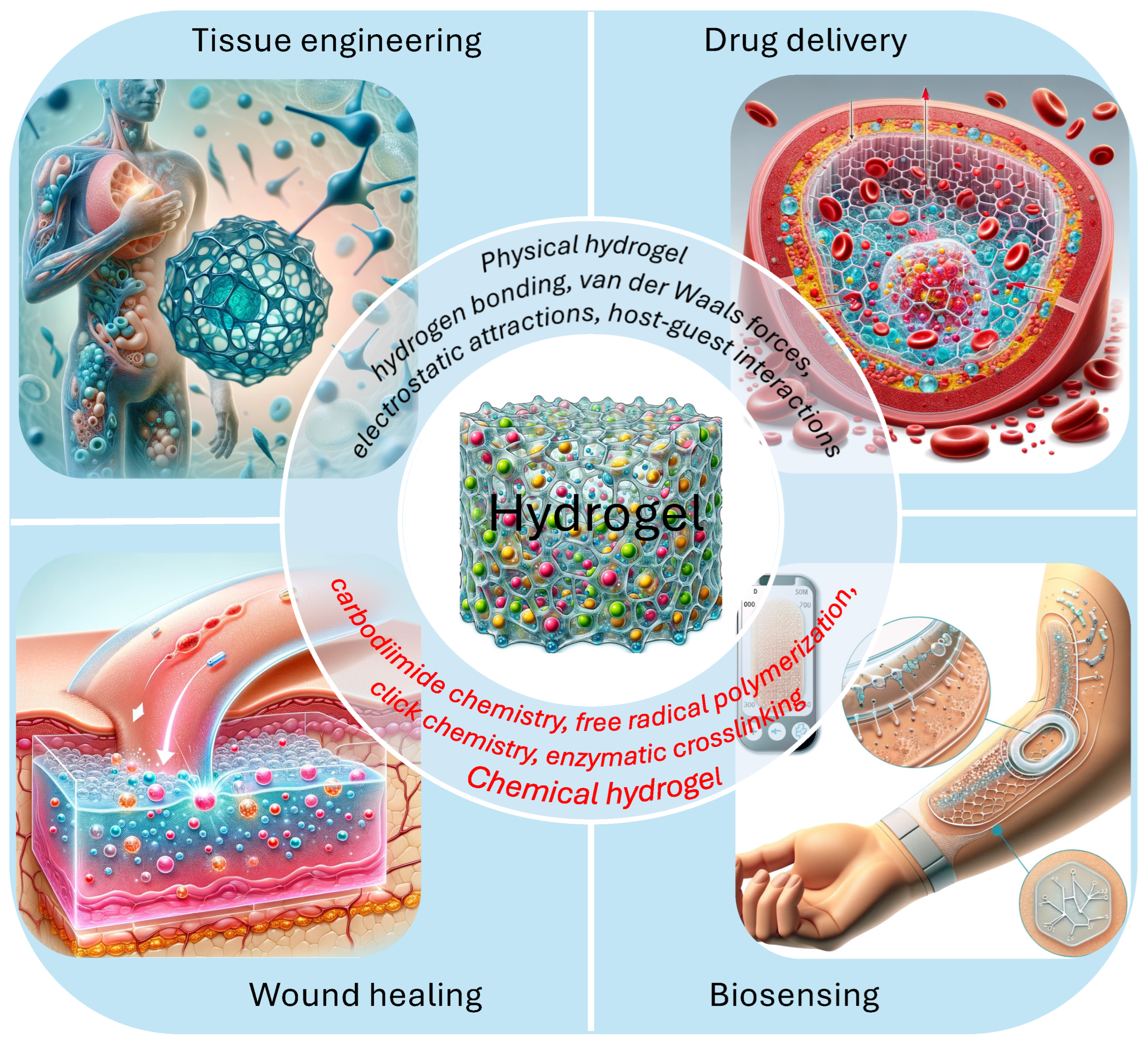
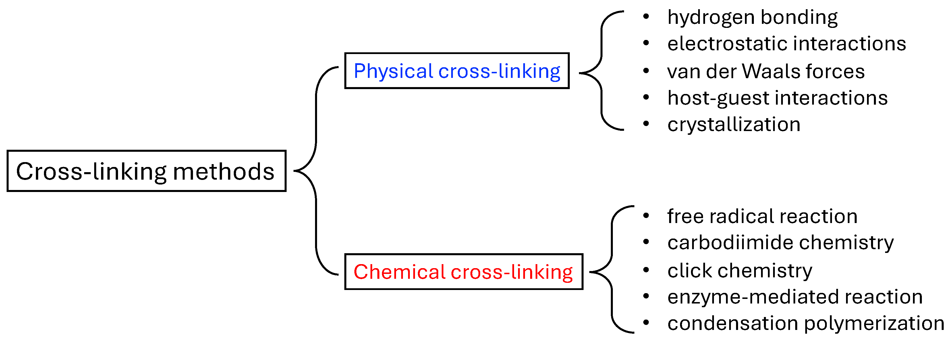
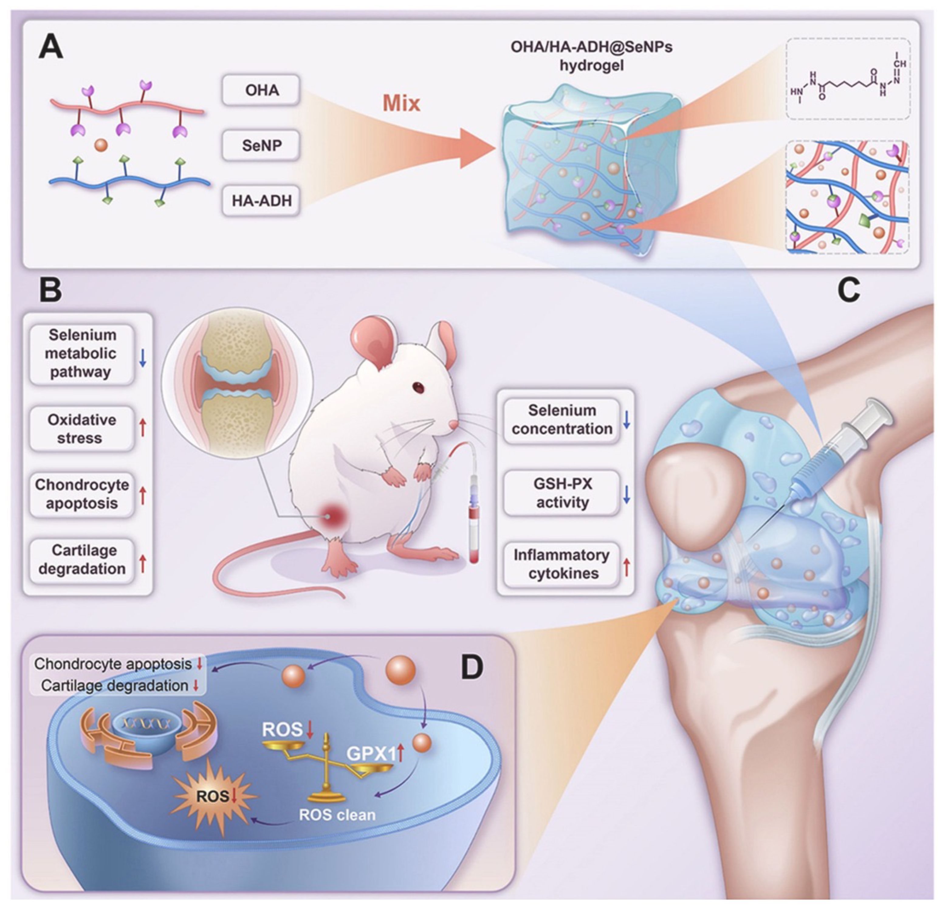
Table 1.
Comparison of common methods used in hydrogel characterization.
| Property | Methods | Advantages | Disadvantages |
|---|---|---|---|
| Mesh size | Rheological testing | Non-destructive, suitable for varied hydrogels, time-dependence analysis. | Specialized equipment needed, complex interpretation. |
| Estimation from swelling ratio | Straightforward, informative. | Limited by the drawbacks of swelling ratio methods. | |
| Mechanical testing | Correlates with mechanical properties, diverse application. | Indirect estimation, sample preparation can be complex. | |
| Scattering techniques | Broad applicability, non-destructive, in-situ analysis possible. | Advanced equipment required, sensitive to sample preparation. | |
| Swelling ratio | Weight change | Simple, cost-effective, broadly applicable. | Influenced by environmental factors, not for fast-swelling gels. |
| Volumetric change | Direct measurement, effective for significant swelling. | Challenging for irregular shapes or small samples. | |
| Differential scanning calorimetry (DSC) | Quantitative, non-destructive. | Specialist interpretation needed, higher equipment cost. | |
| Porosity | Estimation from swelling ratio | Straightforward, informative. | Limited by the drawbacks of swelling ratio methods. |
| Mercury intrusion porosimetry | Accurate pore size and distribution, reproducible. | May alter structure, mercury is hazardous. | |
| SEM | Detailed surface imagery, pore size and distribution. | Dry samples only, surface-level. | |
| Gas adsorption | Non-destructive, good for surface area and microporosity. | Limited to surface, not suitable for all types. | |
| Capillary flow porometry | Provides thorough porosity data. | Requires careful choice of phases. | |
| Microstructure | SEM | Detailed surface imagery, pore size and distribution. | Dry samples only, surface-level. |
| TEM | High-resolution internal images. | Requires meticulous preparation, small coverage. | |
| NMR | Molecular insights, non-destructive. | Expensive, complex data interpretation. | |
| AFM | Nanoscale surface topology. | Surface-specific, complex sample prep. | |
| Confocal microscopy | 3D imaging, non-destructive. | Limited penetration, fluorescence needed. | |
| X-ray CT | Non-destructive 3D internal imaging, high resolution. | High cost, requires contrast agents for some hydrogels. | |
| Mechanical strength | Dynamic Mechanical Analysis (DMA) | Frequency-dependent properties. | Requires specific equipment and sample shapes. |
| Rheological testing | Measures viscoelastic properties, non-destructive. | Complex interpretation, condition-sensitive. | |
| Tensile and compression testing | Direct strength and elasticity measurement. | Destructive, specific sample shapes necessary. | |
| Degradability | Mass loss measurement | Direct, simple quantification. | May overlook subtle changes. |
| Gel Permeation Chromatography (GPC) | Detailed profile, molecular weight insight. | Complex, requires solubilization. | |
| NMR | Degradation pathways at the molecular level. | Requires expensive equipment, expertise for data interpretation. | |
| Viscosity measurement | Indicates molecular weight changes. | Indirect, requires careful interpretation. |
Table 2.
Overview of representative hydrogel-based drug delivery systems and their applications.
| Delivery Route | Hydrogels | Formulation | Active Reagent | Application |
|---|---|---|---|---|
| Oral | Sodium alginate hydrogel | Attach medium-chain lipids (C10) onto the surface of zeolitic imidazole framework-90 (ZIF-90), then encapsulated the nanoparticles with sodium alginate [130]. | Protein. | Protein therapeutics. |
| Calcium alginate hydrogel | Coating hyaluronic acid-modified selenium nanoparticles with a protective shell of calcium alginate (SA) hydrogel [131]. | Selenoprotein. | Inflammatory bowel disease. | |
| Hyaluronic acid hydrogel | Insulin-loaded glucose-responsive nanocarriers were encapsulated into a three-dimensional hyaluronic acid hydroge [134]. | Insulin. | Diabetes. | |
| Sodium alginate hydrogel | Zein/sodium alginate-based core-shell microspheres (Zein/SA/BG) are developed for oral delivery of Bioactive glass [135]. | Bioactive glass. | Inflammatory bowel disease. | |
| PEG hydrogel | Synthesized via nanoprecipitation using the pH-responsive copolymers based on poly(methacrylic acid-co-methyl methacrylate)-block-poly(ethylene glycol) [136]. | Antibody. | Antibody therapies. | |
| Sodium alginate biopolymer and poly vinyl alcohol (PVA) hydrogel | Pristine and curcumin loaded calcium alginate/poly vinyl alcohol beads (CA/PVA and CA/PVA/Cur) were prepared by an ionotropic gelation method of SA followed by crosslinking of PVA [137]. | Curcumin. | Colon cancer. | |
| Peptide-based hydrogel | Mucopenetrating nanoemulsions of 100 nm are embedded in a scaffold constituted of the self-assembling peptide hydrogel product [138]. | Cytokine. | Inflammatory bowel diseases. | |
| Inject | Hyaluronic acid hydrogel | Oxidized hyaluronic acid (OHA) cross-linked with hyaluronic acid-adipic acid dihydrazide (HA-ADH), further incorporated with SeNPs [146]. | Selenium. | Osteoarthritis. |
| PLA-PEG-PLA hydrogel | Drug-loaded micelles mixed with water-dispersed ferrimagnetic iron oxide nanocubes (wFIONs) [143]. | Doxorubicin. | Glioblastoma multiforme. | |
| Chitosan hydrogel | Chitosan micellar self-healing hydrogel (CM hydrogel) with comparable modulus to brain [144]. | Curcumin. | Stroke. | |
| Peptide-based hydrogel | Cross-linking oxidized dextran (Ox-Dex) with MMP-2-cleavable peptide [145]. | Antagomir-21. | Intervertebral disc degeneration. | |
| Agarose (AG) hydrogels | Polydopamine (PDA), letrozole (LTZ), and agarose (AG) hydrogels were combined to construct an near-infrared controlled drug delivery [147]. | Letrozole. | Endometriosis. | |
| Peptide-based hydrogel | Iopamidol was loaded into Ac-K1/Ac-K2 HGs [148]. | Iopamidol. | Imaging agents. | |
| Peptide-based hydrogel | pH-responsive ionic-complementary octapeptide FOE to delivery DOX [149]. | Doxorubicin. | Cancer. | |
| Nucleic acid hydrogel | Crosslinking PD-L1 siRNA with a SN38- and CpG-containing Y-motif [151]. | PD-L1 siRNA. | Cancer. | |
| Hyaluronic acid hydrogel | HA-F127@Ti-MOF-Au/PEG-TK-DOX/PFD (abbr. HFTiDP) encapsulates sonosensitizer (Ti-MOF-Au), chemotherapeutic prodrug (PEG-TK-DOX), and ECM-solubilizing drug pirfenidone (PFD) [152]. | Doxorubicin. | Triple-negative breast cancer. | |
| Topical | PEG hydrogel | Crosslinked polyethylene glycol (PEG) hydrogel tethered with highly potent antibacterial cationic polymer, polyimidazolium (PIM), and the antioxidant N-acetylcysteine (NAC) [156]. | N-acetylcysteine. | Wound healing. |
| Gelatin methacryloyl hydrogel | Gelatin methacryloyl hydrogel loaded dandelion-derived extracellular vesicle-like nanoparticles [157]. | Dandelion-derived extracellular vesicle-like nanoparticles. | Wound healing. | |
| Hyaluronic acid hydrogel | Near-infrared (NIR) light-responsive multifunctional hydrogel system (PDA/Mup@DA-HA) consisting of mupirocin-loaded polydopamine nanoparticles (PDA) and dopamine-modified hyaluronic acid (DA-HA) hydrogel [158]. | Mupirocin. | Bacterial infection and tissue regeneration. | |
| Calcium alginate hydrogel | Sprayable calcium alginate hydrogel encapsulating HIL@Z nanodrug and photosynthetic cyanobacteria [159]. | Photosynthetic cyanobacteria. | Tumor recurrence/metastasis and wound healing. | |
| Porcine acellular dermal matrix | Hydrogel matrix is derived from porcine acellular dermal matrix and is loaded with bioactive glass nanoparticles doped with magnesium and loaded with Curcumin [160]. | Curcumin. | Antimicrobial and wound healing. | |
| Chitosan hydrogel | Nanochitosan dots (ChiDs) were synthesized using gamma rays and encapsulated in bacterial cellulose (BC) polymer matrix [161]. | Nanochitosan dots. | Chronic infections. | |
| Chitosan hydrogel | Hydrogels composed of bacterial cellulose (BC) with chitosan polymer (Chi)-BC-Chi and chitosan nanoparticles (nChiD) [162]. | Bacterial cellulose with chitosan polymer. | Chronic infections. | |
| Ocular | Aldehyde-functionalized F127 (AF127) hydrogel | Combines copper-selenium nanoparticles (Cu2-xSe NPs) AF127 [164]. | Cu2-xse nanoparticles. | Dry eye disease. |
| Hyaluronic acid hydrogel | Nanozyme-thixotropic hydrogel coating (NHC) incorporated voriconazole and copper-proanthocyanidins, self-synthesized polyaldehyde oligomer with amino functionalized hyaluronic acid [165]. | Voriconazole and copper-proanthocyanidins. | Fungal keratitis. | |
| Peptide-based hydrogel | Nap-FFKK generate supramolecular hydrogels spontaneously in a pH value of 5-7 [166]. | Nap-FFKK. | Ocular disorders. |
Disclaimer/Publisher’s Note: The statements, opinions and data contained in all publications are solely those of the individual author(s) and contributor(s) and not of MDPI and/or the editor(s). MDPI and/or the editor(s) disclaim responsibility for any injury to people or property resulting from any ideas, methods, instructions or products referred to in the content. |
© 2024 by the authors. Licensee MDPI, Basel, Switzerland. This article is an open access article distributed under the terms and conditions of the Creative Commons Attribution (CC BY) license (https://creativecommons.org/licenses/by/4.0/).
Share and Cite
MDPI and ACS Style
Liu, B.; Chen, K. Advances in Hydrogel-Based Drug Delivery Systems. Gels 2024, 10, 262. https://doi.org/10.3390/gels10040262
AMA Style
Liu B, Chen K. Advances in Hydrogel-Based Drug Delivery Systems. Gels. 2024; 10(4):262. https://doi.org/10.3390/gels10040262
Chicago/Turabian StyleLiu, Boya, and Kuo Chen. 2024. "Advances in Hydrogel-Based Drug Delivery Systems" Gels 10, no. 4: 262. https://doi.org/10.3390/gels10040262
Note that from the first issue of 2016, this journal uses article numbers instead of page numbers. See further details here.