Article

Lactic Acid Bacteria-Fermented Diet Containing Bacterial Extracellular Vesicles Inhibited Pathogenic Bacteria in Striped Beakfish (Oplegnathus fasciatus)

1
Department of Aquaculture, National Taiwan Ocean University, Keelung 202301, Taiwan
2
Department of Aquaculture, National Pingtung University of Science and Technology, Pingtung 912301, Taiwan
3
Department of Food Safety/Hygiene and Risk Management, National Cheng Kung University, Tainan 701401, Taiwan
*
Authors to whom correspondence should be addressed.
Fermentation 2024, 10(1), 49; https://doi.org/10.3390/fermentation10010049
Submission received: 7 December 2023 / Revised: 19 December 2023 / Accepted: 2 January 2024 / Published: 9 January 2024
(This article belongs to the Special Issue Recent Trends in Lactobacillus and Fermented Food, 2nd Edition)

Abstract

:
In recent years, probiotics have received considerable attention for improving the health of aquaculture organisms, such as fish and shrimp, by stimulating immune activity and increasing growth rates. Oplegnathus fasciatus is a common and economically important cultured fish species in Asia. In this study, we aimed to investigate the potential of lactic acid bacteria (LAB; Limosilactobacillus reuteri)-fermented feed to promote growth and enhance immune function in O. fasciatus. The feed contained the highest proportion of LAB after L. reuteri fermentation for 3 days in anaerobic conditions. Oplegnathus fasciatus was fed LAB-fermented feed for 30 days. The administration of LAB-fermented feed (live bacteria > 109 CFU/g) significantly increased the growth rate (weight gain = 174.8%; FCR = 4.23) and intestinal probiotic levels of O. fasciatus. After LAB-fermented feeding, the immunity index was evaluated by superoxide anion production, the phagocytic activity of leukocytes, and bactericidal and lysozyme activities in the serum of O. fasciatus. We found that LAB-fermented feed treatment potentially elevated the proportions of intestinal Bifidobacterium, Blautia, and Dorea species and reduced pathogenic bacterial growth (Acinetobacter, Escherichia_Shigella, and Megasphaera) in O. fasciatus. This study demonstrated that LAB-fermented feed containing extracellular vesicles improves growth performance and the inhibition of pathogenic Acinetobacter baumannii.

1. Introduction

Several factors (host selection, diet, environment, antibiotics, and chemicals) have been found to affect intestinal microbiota imbalance [1]. A study also reported that starvation influenced microbial abundance (elevation of Vibrio; reduction of Brevibacillus, Bifidobacterium, and Alloprevotella) and diversity [2]. Diverse environmental conditions result in variations in the gut and skin microbiota, thereby affecting the health of cultivated fish and shrimp. The microbial flora in hosts prevents pathogenic infection, promotes growth, and lowers antibiotic use.
An increase in the proportion of intestinal pathogenic Pelomonas and Fusobacterium spp. is associated with increased infection and disease risk in fish [3]. Moreover, Dhler et al. reported that variations in cultivation environments markedly affected the abundance of Firmicutes, Proteobacteria, Bacteroidetes, and Tenericutes species in salmon [4]. Studies have demonstrated an association between harmful microbial flora and reduced immunity in various aquaculture organisms such as fish [5] and shrimp [6]. These findings suggest that gut health could be influenced by the gut microbiota, which determines the immunity of cultured organisms.
Aquaculture organisms fed probiotics or probiotic supernatants showed increased growth of beneficial intestinal bacteria [7]. Probiotics are live microorganisms that exert beneficial effects on the host when administered properly. As an alternative to antibiotics, probiotics are commonly used in aquaculture because of their beneficial effects on immunology, nutrition, and the environment [8]. Among the available probiotics, some types of lactic acid bacteria (LAB) are widely used owing to their beneficial functions in the digestive tract of the host, such as improving immune response, modulating the bacterial community, and antagonizing opportunistic pathogens. Dietary Lactobacillus and Lactococcus supplementation has been shown to promote growth, maintain the gut microbiota, and stimulate immune responses in red sea bream (Pagrus major) [9]. Furthermore, it has been found that dietary probiotic supplementation (containing Bacillus sp., Pediococcus sp., Enterococcus sp., and Lactobacillus sp.) modulates the gut microbiota and improves the growth of juvenile rainbow trout [10]. In addition, the exopolysaccharide–protein complex obtained from LAB has been found to enhance the immune response of zebrafish against Aeromonas veronii infection [11]. These results indicate that LAB have the potential to be developed as feed additives to promote the growth of shrimp and fish by modulating the gut microbiota and immunity. Extracellular vesicles (EVs) are membrane-based structures, which can carry various types of cellular components (lipids, proteins, and nucleic acids). EVs with a diameter ranging between 40 and 200 nm are called exosomes, whereas those with a diameter ranging between 300 and 1000 nm are called microvesicles [12]. Recently, we found that EVs isolated from lactic acid bacteria have the potential to inhibit pathogenic bacteria and regulate microbiota [12,13]. Recently, the functional activities of EVs obtained from different species have been reported and for the promotion of physiological health; moreover, we investigated the proteomics of EVs isolated from different lactic acid bacteria [14]. These results reveal that Lactobacillus-derived EVs may have potential in marine technology applications. The striped beakfish Oplegnathus fasciatus (Oplegnathidae) is an economically important fishery resource in Asia which is widely distributed in the Pacific Ocean, around southern parts of the Korean Peninsula, in Japan, and in Taiwan. As the growth rate of O. fasciatus is slower than that of other ocean fishes among the family Oplegnathidae, the culture yield and economic benefits of O. fasciatus are limited. Cost is a crucial consideration for aquaculture operators, and therefore research should focus on more economical methods. LAB fermentation is a promising approach for development. This study mainly explores the effects of non-fermented feed and LAB-fermented feed (containing EVs) on the growth and intestinal microbial community of O. fasciatus.

2. Materials andMethods

2.1. Cultivation of LAB and Preparation of Fermented Dietary Feed

Limosilactobacillus reuteri was isolated from lamb feces, and Lactobacillus acidophilus (BCRC10695) and Lactobacillus paracasei subsp. paracasei (BCRC14023) were purchased from the Bioresource Collection and Research Center (BCRC) in Taiwan (Hsinchu, Taiwan). These three LAB were inoculated into de Man, Rogosaand Sharpe (MRS) broth (BD Biosciences, San Jose, CA, USA) or MRS agar at 37 °C and subcultured monthly. LAB-fermented feed was produced according to the following procedure. Briefly, the MRS broth re-inoculated with 1% L. reuteri was inoculated in 400 mL of MRS broth and cultured at 37 °C for 24 h under anaerobic conditions (Oxoid, Basingstoke, Hampshire, UK). The commercial diet (crude protein > 50.0%, crude lipid > 8.0%, crude ash < 15.0%, crude fiber < 3.0%, moisture < 11.0%) (Tung Li Feed Industrial Co., LTD, PingTung, Taiwan) was sterilized and inoculated with pre-cultured L. reuteri for 1, 3, or 5 days to carry out fermentation (which is fermented feed). The number of L. reuteri in the feed diet was determined via MRS plate counting. Finally, the LAB-fermented dietary feed was freeze-dried and stored at −20 °C. In addition, the sterilized commercial diet was used as control diet without fermentation, and the formula is shown in Supplementary Table S1.

2.2. Fish and Husbandry Conditions

Oplegnathus fasciatus individuals were obtained after breeding and fry hatching (animal experiments were approved by National Cheng Kung University: IACUC No. 108317). These fish were fed diets with or without fermentation. A total of 120 O. fasciatus fish (initial body weight: 75.79 ± 4.11 g) were evenly distributed into four groups (30 fish/group), and they were stocked in a net cage (10 fish per cage; length 100 cm × width 130 cm × height 66 cm) in an indoor cement pool with a flow-through sea water system with continuous aeration. The indoor cement pool was maintained under a natural light/dark regime. The monitored water quality parameters were as follows: water temperature 25.8 ± 2.3 °C, pH 8.4 ± 0.5, and salinity 32.1 ± 0.2 ppt.

2.3. Feeding Trial

The fish were hand-fed to apparent satiation three times daily (6:00, 17:00, and 22:00 h) for 30 days. The remaining feed was removed after feeding and dried using a freeze drier(FDU-1200, Eyela, Tokyo, Japan) to subtract the weight from the total feed intake. Body weight was recorded by measuring the basket weight containing water and then subtracting the water weight to calculate body weight every week.

2.4. Assays for Superoxide Anion Production and Phagocytotic Activity of Leucocytes Isolated from O. fasciatus

After 30 days of experiment, the head kidney was removed and passed through a 100 mm nylon mesh after suspension in Hanks’ balanced salt solution (HBSS) (Sigma, St. Louis, MO, USA). The suspended cells were placed in a 30%–50% Percoll density gradient medium and centrifuged at 400× g for 40 min at 4 °C. Subsequently, the isolated cells were harvested at the Percoll interface, washed three times with HBSS, and centrifuged at 400× g for 10 min. Respiratory burst activity of phagocytes in the head kidney was measured using a previously described method [15]. Briefly, 100 μL of leukocyte suspension (5 × 10⁶ cells/mL) was placed in 96-well plates and incubated at 37 °C for 1 h. The nonadherent cells were removed by washing with HBSS. Zymosan (Sigma, St. Louis, MO, USA) in 100 μL of HBSS was added to leukocytes at 1 mg/mL and the samples in the wells were incubated at 25 °C for 30 min. Thereafter, 100 μL of nitrobluetetrazolium (NBT) (Sigma, St. Louis, MO, USA) was added into each well, and the plate was incubated at 25 °C for 30 min. Finally, 100 μL of 100% methanol was added into each well to cease the reaction; the formazan formed in each well was dissolved by adding 120 μL of 2 M KOH and 140 μL of dimethylsulfoxide (DMSO).
For phagocytic activity, the 50 μL of kidney leukocyte suspension was placed on glass slides with 5 × 10⁶ cells/mL HBSS for 20 min at 25 °C in a moist incubation chamber to allow for cell adhesion. Furthermore, 50 μL of latex bead suspension (107/mL) (Sigma, St. Louis, MO, USA) was added to the leukocyte monolayer, and slides were incubated for 30 min at 25 °C. The slides were rinsed with saline, fixed with methanol for 5 min, and stained with Giemsa solution for 15 min. The percentage of phagocytes ingesting the beads and the number of beads ingested per phagocyte were calculated by enumerating 100 phagocytes under a microscope (Becton-Dickinson, San Jose, CA, USA).

2.5. Assays for Serum Bactericidal and Lysozyme Activity

Bactericidal activity was measured as previously described [16,17]. The samples were diluted 3, 4, and 5 times with Tris buffer (pH 7.5). The diluted samples were mixed with Escherichia coli suspension and incubated at 25 °C for 24 h. Thereafter, 50 μL of the reaction solutions was incubated on tryptic soy broth at 25 °C for 24 h. Colony forming units (CFUs) were determined using the plate counting method, and the bactericidal activity was calculated for each group and compared with that of the control group. Blood was collected from the caudal vein, and serum was isolated after centrifugation at 5000 rpm for 30 min and stored at −80 °C until use. Samples or standard (10 μL) of samples were added into the wells of a microplate and 190 μL of substrate (0.2 mg/mL Micrococcuslysodeikticus, lyophilized cell; Sigma, St. Louis, MO, USA) was added. The microplate was incubated with gentle shaking at room temperature and the absorbance of the samples was measured at 520 nm after 1 and 6 min of incubation. The lysozyme activity was calculated using a standard curve.

2.6. Assay for Microbiota

Intestinal samples with feces were collected after scarification by anesthetization treatment with 2-phenoxyethanol (0.1 mL/L) and were immediately frozen in liquid nitrogen and stored at −80 °C. Total genomic DNA from the samples was extracted (QIAamp PowerFecal DNA Kit, Qiagen, Hilden, Germany). The V3–V4 region was amplified using specific primers (319F:5′-CCTACGGGNGGCWGCAG-3,’ 806R:5′-GACTACHVGGGTATCTAATCC-3′) according to the Illumina MiSeq PE300 platform. Microbiota analysis was performed using the QIIME2 alignment platform (v2019.7.0; https://qiime2.org/, accessed on 31 December 2023) and SILVA132 annotation database.

2.7. Isolation of Extracellular Vesicles (EVs)

The bacteria in MRS medium and LAB-fermented feed were removed by centrifugation at 3000× g for 15 min. Bacterial debris and intact organelles in the culture medium were further removed by centrifugation at 35,000× g for 60 min. The supernatant was then subjected to ultracentrifugation at 100,000× g for 60 min. The pellets were resuspended in PBS. The particle number of EVs and their size distribution were detected by a nanoparticle tracking analyzer (Nano-ZS 90 dynamic light scattering, Malvern Panalytical, Malvern, UK). The morphology of EVs was observed using transmission electron microscopy (JEOL, Tokyo, Japan) [12,13,14]. Total RNA containing small RNA was isolated from extracellular vesicles using an miRNeasy mini kit (Qiagen, Hilden, Germany) according to the manufacturer’s instructions. The small RNA and microRNA content in EVs were assayed by Agilent Bioanalyzer (Santa Clara, CA, USA).

2.8. Inhibition of Pathogenic Bacteria

Acinetobacter baumannii (17106) was purchased from Bioresource Collection and Research Center (BCRC, Hsinchu, Taiwan) and cultured in a medium (formula: 10 g meat extract and 5 g NaCl in 1 L distilled water) with or without EV treatment in an aerobic environment at 30 °C. Three independent samples were analyzed for each experiment, and the OD600 was measured after culturing the cells for different times [12].

2.9. Statistical Analysis

The data were recorded as mean ± SD. Statistical significance was determined using a one-way analysis of variance (ANOVA) with the SAS general linear model procedure (SAS Inc., Cary, NC, USA), followed by ANOVA with Duncan’s test. Results were considered statistically significant at p < 0.05. For β diversity of gut microbiota analysis, different distance matrices were evaluated using Principal coordinate analyses (PCA) assay. The value corresponding to the heatmap represents the Z Score obtained by the abundance of each species in all groups. The Z score of a sample on a certain classification is the value of the average abundance of the sample on the category and all samples in the classification.

3. Results

3.1. Feeding Fermentation with LAB

The feed was fermented with LAB, and the L. reuteri strain was isolated from lamb feces. Growth curves were created and tolerance against low pH and bile salt conditions was determined to evaluate the optimal LAB strains. We found that L. reuteri performed better than L. acidophilus (BCRC10695) and L. paracasei subsp. paracasei (BCRC14023) in terms of tolerance and survival rate at pH 2 and 0.3% bile salt conditions, whereas the adsorption capacity of the intestinal cells was lower than that of L. acidophilus (BCRC10695) (Supplementary Table S1).
In addition, the growth rate of LAB in MRS medium after 48 h of cultivation was investigated. The growth rate of L. paracasei subsp. paracasei (BCRC14023) was higher than that of L. acidophilus (BCRC10695) and L. reuteri (Figure 1A). In summary, based on the results shown in Supplementary Table S1, L. reuteri was used in this study owing to its tolerance to low pH and bile salt conditions. As L. reuteri presented a good survival rate and tolerance to adverse environments, this strain was selected to ferment feed for 1, 3, and 5 days. The highest count of L. reuteri was reached after 3 days of fermentation, and it decreased after 5 days of fermentation (Figure 1B).

3.2. Effects of LAB-Fermented Feed for Fish Growth

The initial body weight of O. fasciatus was between 74.8 and 78.8 g; the fish were fed LAB-fermented feed (1-, 3-, or 5-day fermentation) for 30 days. The body weight and weight gain (%) were evaluated (Table 1). The growth of fish was significantly increased in the group administered LAB-fermented feed (3-day and 5-day fermentation), whereas no significant difference was observed between the group administered 1-day-fermented feed and the control groups. The food conversion rate (FCR) (Table 2) was 4.23 (3-day-fermentation feed), 5.03 (5-day-fermentation feed), 6.05 (1-day-fermentation feed), and 6.15 (control) for O. fasciatus.

3.3. Effect of LAB-Fermented Feed on Immune Activity of O. fasciatus

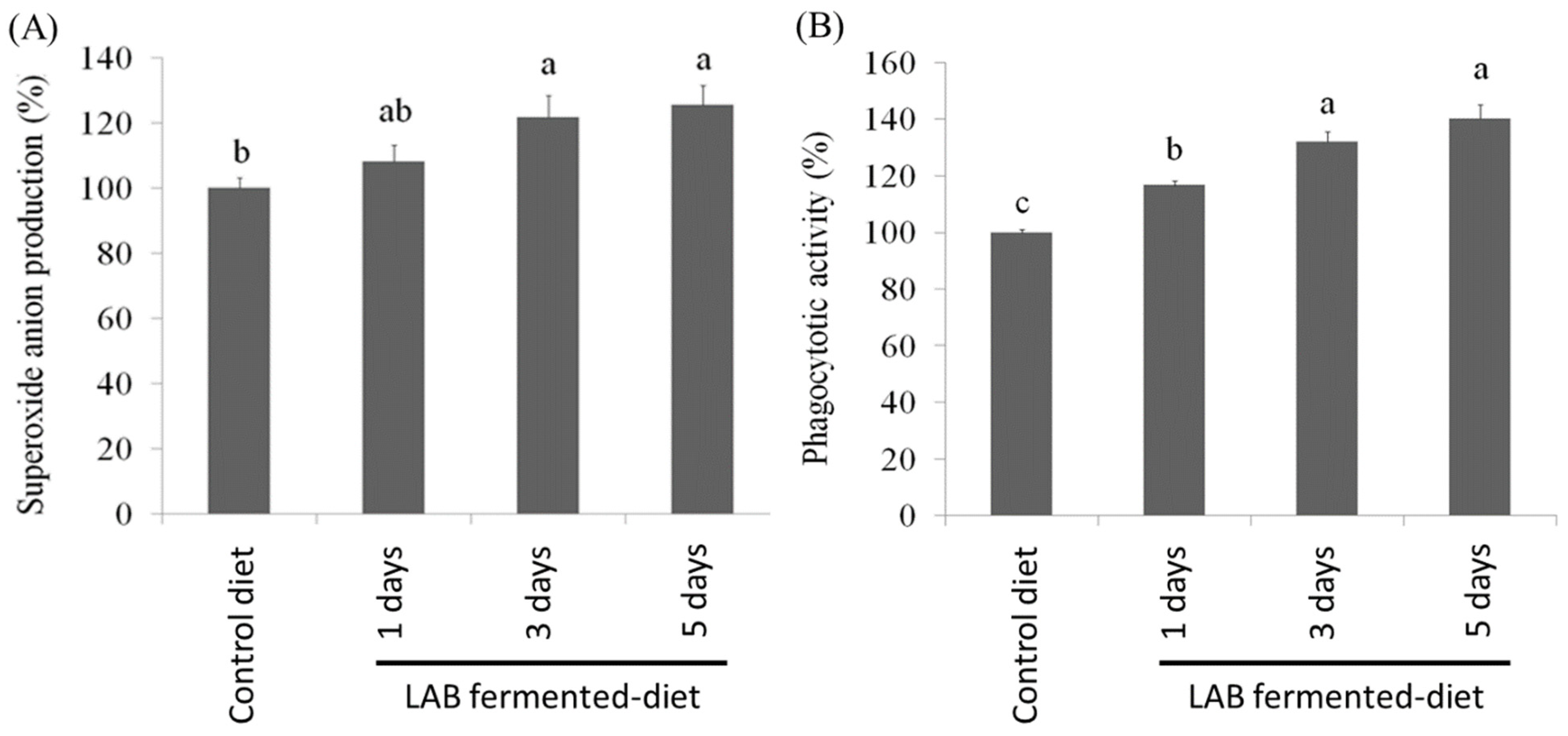
In this study, the fish were fed LAB-fermented feed for 30 days, and then head-kidney leukocytes were isolated. We found that phagocytosis and respiratory burst reactions were elevated in the group administered LAB-fermented feed compared with those in the untreated group. After the administration of LAB-fermented feed for 30 days, leukocytes were isolated from O. fasciatus and stimulated with zymosan to analyze superoxide anion production. The results showed that the production of superoxide anions was markedly increased in the leukocytes isolated from fish fed LAB-fermented feed compared with that in the control group (Figure 2A). The phagocytic activity of the leukocytes isolated from O. fasciatus was also investigated. The phagocytic activity of leukocytes isolated from O. fasciatus was elevated by the administration of LAB-fermented feed (Figure 2B).
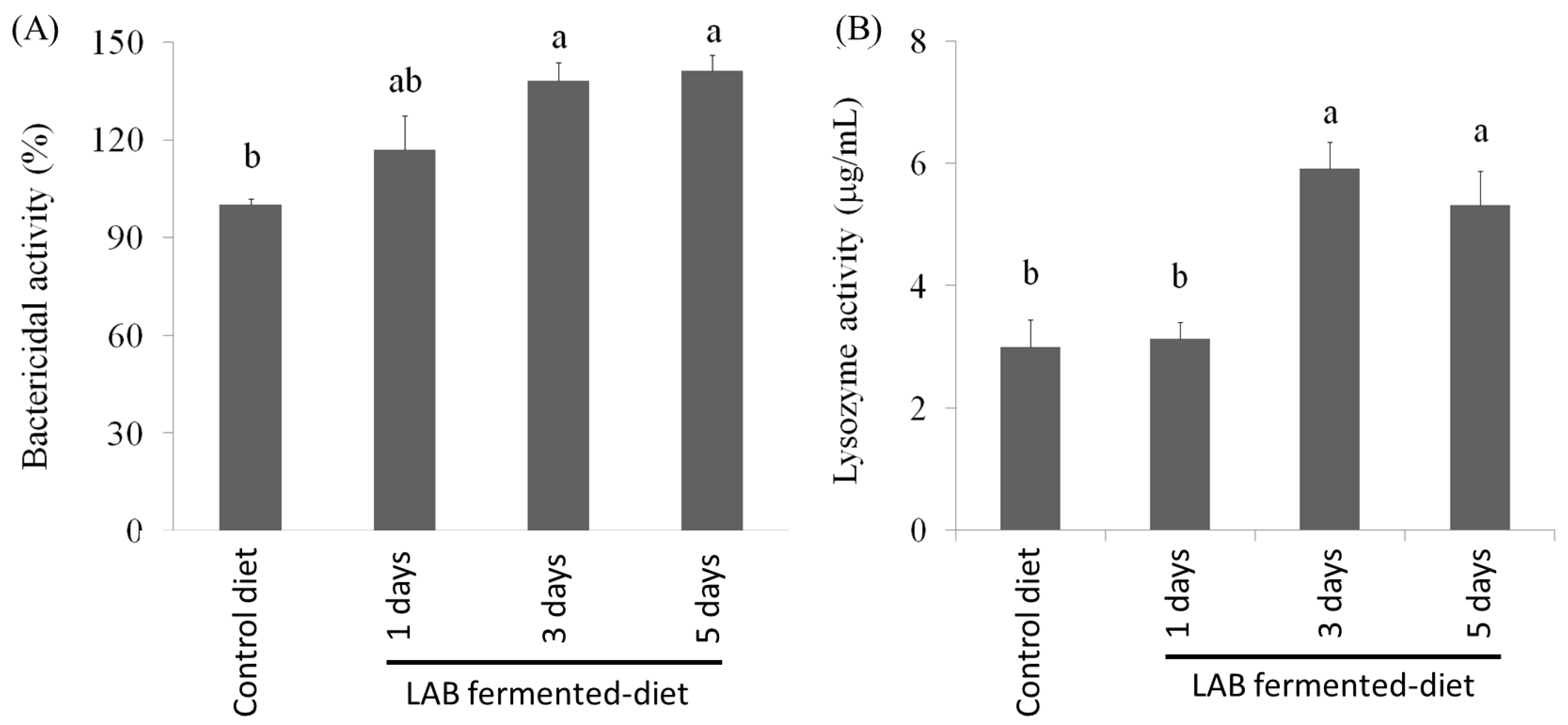
The bactericidal and lysozyme activities in the serum of O. fasciatus were investigated. The highest serum bactericidal activity was observed in O. fasciatus fed the LAB-fermented feed. Similar to the serum bactericidal activity, the serum lysozyme activity in O. fasciatus administered LAB-fermented feed was also elevated. The results indicate that the administration of LAB-fermented feed significantly increased the serum bactericidal and lysozyme activities compared with those in the control group (Figure 3A,B).

3.4. Effect of LAB-Fermented Feed on Intestinal Microbiota in Fish

From the above results, it can be seen that in addition to the highest number of LAB population being obtained after fermentation for 3 days, it also had a significant effect on improving fish growth and immune status, and no significant difference was found between fish administered LAB-fermented feed for 3 days and for 5 days. The intestinal microbiota was analyzed in LAB-fermented feed (fermentation for 3 days) compared to the control diet in the present study. There were 30 fish in each group, and we selected the 6 fish with the best growth rate and immune status among them for intestinal microbial analysis.
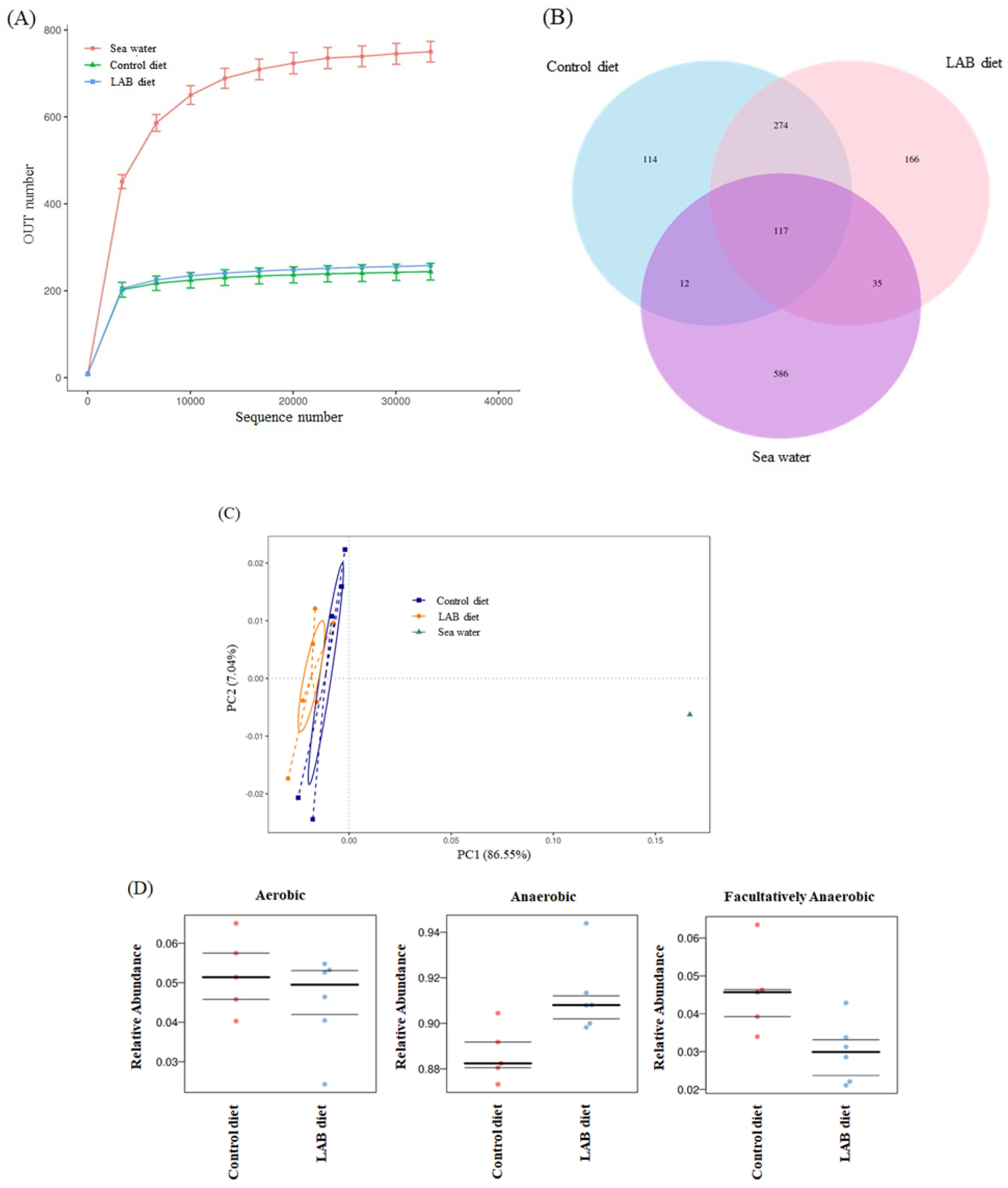
Intestinal contents were collected from fish fed LAB-fermented feed (fermented with L. reuteri for 3 days), and the intestinal microbiota was analyzed using 16S rRNA sequencing. As shown in Figure 4A, the OTU count in the intestinal stool samples of O. fasciatus fed LAB-fermented feed was similar to that of O. fasciatus fed the control diet. We generated over 30,000 sequences for each sample obtained from the intestinal content of O. fasciatus, and these unique sequences were classified and grouped. In contrast, 117 different types of bacteria were found in all samples, including seawater and the intestinal stool of O. fasciatus fed the control diet or LAB-fermented diet. However, the specific OTU counts were 114 and 166 in the intestinal content of O. fasciatus fed the control and LAB-fermented diet, respectively (Figure 4B). But there are a very large number of microorganisms (586 OTUs) that have only been analyzed in seawater, and these bacteria were not found in the intestinal content. The results of PCoA uniFrac_Weight analysis suggest that the diet fermented with L. reuteri may potentially regulate microbial beta-diversity in the intestine (Figure 4C). This phenomenon was mainly due to the administration of the LAB-fermented diet, which resulted in an increase in the proportion of anaerobic bacteria and a decrease in the proportion of facultatively anaerobic bacteria (Figure 4D).
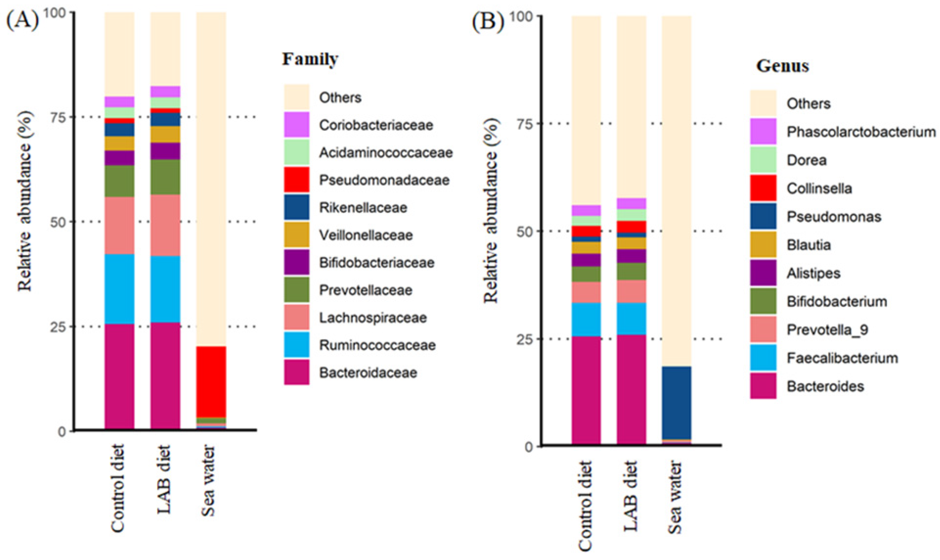
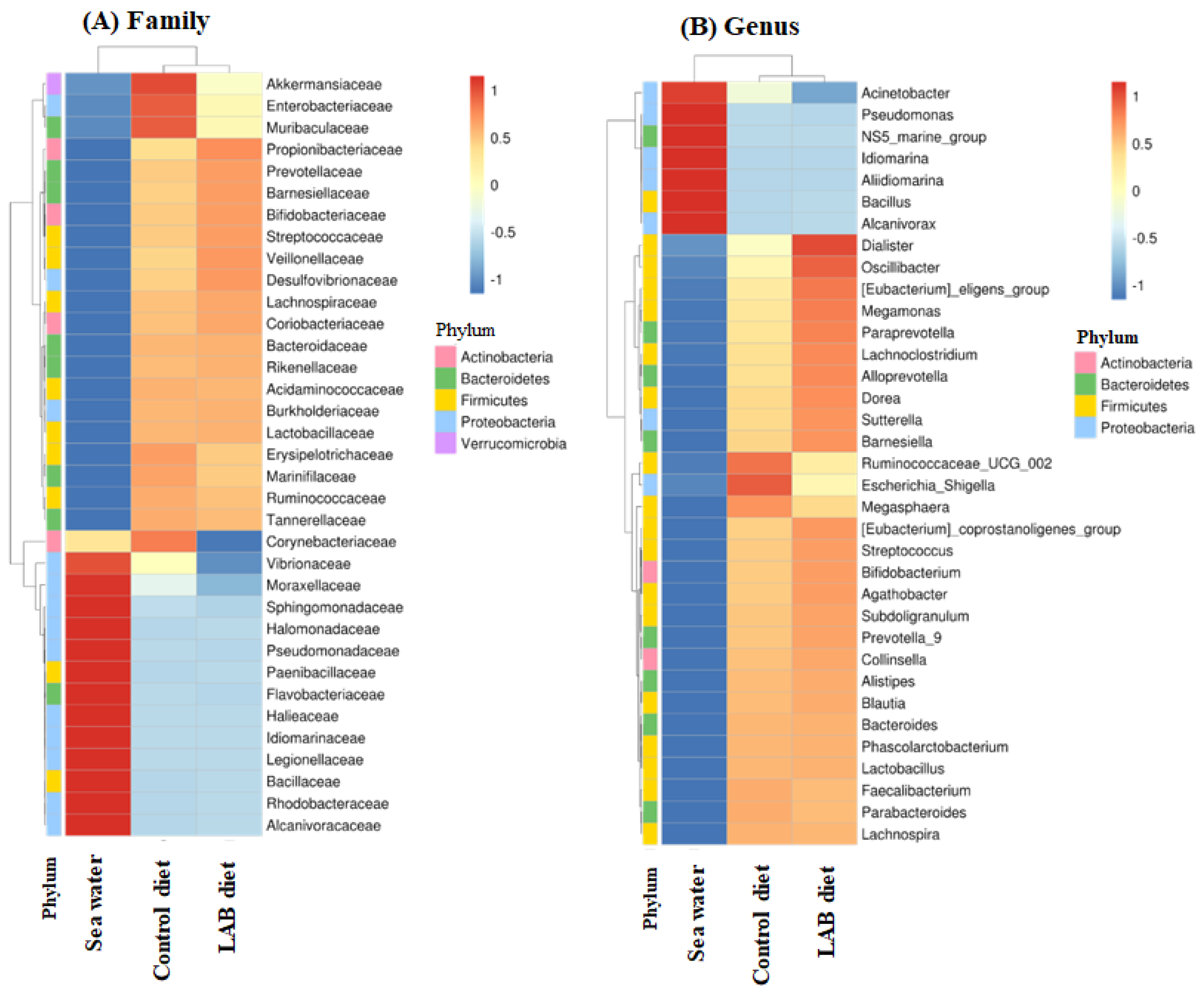
We analyzed the most abundant microbes (top 10) in the stool of O. fasciatus fed the control diet or LAB-fermented diet. The results indicated that the LAB-fermented diet potentially elevated the proportions of Prevotellaceae, Bifidobacteriaceae, and Veillonellaceae members at the family level (Figure 5A) and genus level (Figure 5B). Interestingly, a large number of Pseudomonas species were found in seawater, but they were not present in large numbers in the intestinal stool of O. fasciatus fed the control diet or the LAB-fermented diet. These results indicate that the top 10 members were present at similar levels in the intestinal stools of O. fasciatus fed the control diet and the LAB-fermented diet. The bacterial species that showed the greatest variation between the control and LAB-fermented diet groups at the family level are shown in Figure 6A. We found that the LAB-fermented diet increased the abundance of Propionibacteriaceae, Prevotellaceae, Barnesiellaceae, Bifidobacteriaceae, Streptococcaceae, Veillonellaceae, and Desulfovibrionaceae, but decreased the abundance of Akkermansiaceae, Enterobacteriaceae, and Muribaculaceae compared with that in the group fed the control diet. In addition, Corynebacteriaceae and Vibrionaceae members were found in the seawater and intestinal stool of O. fasciatus, but the growth of these pathogenic bacteria could be suppressed by the LAB-fermented diet (Figure 6A). At the genus level, Escherichia-shigella, Ruminococcaceae_UCG_002, and Megasphaera were found at considerable proportions in the intestinal stool of O. fasciatus fed the control diet. However, the LAB-fermented diet suppressed the growth of these microbes and elevated the proportion of Dialister, Oscillibacter, Megamonas, Paraprevotella, Lachnoclostridium, Alloprevotella, Dorea, Sutterella, and Bamesiella in the intestinal stool of O. fasciatus (Figure 6B).
We performed statistical analysis of the microbiota in the intestinal stool of O. fasciatus. Feeding the LAB-fermented diet decreased the proportion of Enterobacteriaceae members in O. fasciatus compared with that in the control group (Supplementary Figure S1A). The family Enterobacteriaceae includes several pathogens, such as Escherichia, Shigella, Salmonella, Enterobacter, Klebsiella, Citrobacter, Proteus, and Serratia. In addition, LAB-fermented diet administration decreased the proportion of the pathogenic genus Escherichia_Shigella in the intestine of O. fasciatus compared with that in the group fed the control diet (Supplementary Figure S1B).
We also found that the LAB-fermented diet suppressed the growth of Ruminococcaceae_UCG_002, Succinivibrio, Eggerthella, Dolosigranulum, Anaerotruncus, and Ruminococcus_gauvreauii_group, and increased the abundance of Cutibacterium, Anaerobacillus, and Mucispinllum in the intestinal stool of O. fasciatus (Supplementary Figure S1C). The microbial phenotype of relative abundance was predicted and mapped from the sequencing data using BugBase. Figure 7 shows that LAB-fermented diet administration decreased the relative proportion of potentially pathogenic bacteria and the formation of biofilm compared with the control.
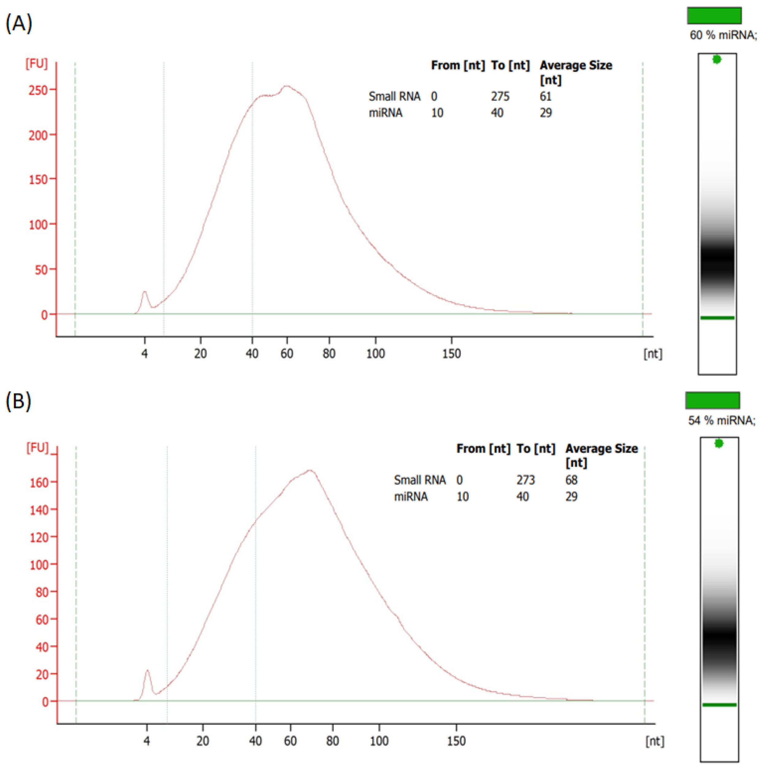
This study further explored the characteristics of EVs secreted by L. reuteri in MRS medium and LAB-fermented feed. The L. reuteri-derived EVs had symmetrical and circular structures by TEM observation, and the size distribution of EVs (NTA evaluation) was 168.1 ± 60.2 nm and 147.6 ± 64.5 nm in MRS medium (Figure 8A) and LAB-fermented feed (Figure 8B), respectively. Moreover, the NTA results indicated that the numbers of EVs were 1.2 × 10⁹ particles/mL and 1.7 × 10⁹ particles/mL in MRS medium (Figure 8A) and LAB-fermented feed (Figure 8B), respectively. Recently, small RNA/microRNA rich-EVs have shown functional abilities for regulating bacterial growth [18]. We found that the average sizes of small RNA and microRNA (60%), respectively, were 61 nt and 29 nt in EVs obtained from MRS medium. However, the average sizes of small RNA and microRNA (54%), respectively, were 68 nt and 29 nt in EVs obtained from LAB-fermented feed (Figure 9). These results indicate that as long as L. reuteri grows well, different culture methods will not significantly affect the small RNA and microRNA contents in EVs.
Acinetobacter baumannii is a pathogenic bacterium (Gram-negative, strictly aerobic, non-fermentative, rod-shaped bacteria) that frequently causes infections in aquaculture organisms [19]. The suppression of L. reuteri-derived EVs against A. baumannii growth was investigated. The inhibitory abilities of L. reuteri-derived EVs for A. baumannii obtained from cultured medium (MRS) and fermented feed were found in dosage-dependent manners (106–1010 particles/mL) as shown in Figure 10A,B. Moreover, similar antibacterial potential was observed in L. reuteri-derived EVs obtained through different culture methods (Figure 10C). From these results, it can be inferred that microRNA in L. reuteri-derived EVs should be the main antibacterial substance, and the antibacterial ability obtained from L. reuteri-fermented feed is not affected compared to that obtained from MRS medium.

4. Discussion

The positive effects of supplementing LAB such as the Lactobacillus species in feed on the growth of fish, including Rachycentron canadum [20], Oreochromis niloticus [21], Labeo rohita [22], and Pagrus major [23], have been reported. Moreover, L. lactis promotes growth and improves the health of the grouper E. coioides [24] and olive flounder Paralichthys olivaceus [25]. Currently, we prepared feed fermented with L. reuteri for 3 days in this study. We found that this LAB-fermented feed has the potential to improve the growth of O. fasciatus. Our results are consistent with the hypothesis that LAB-fermented feed administration improves the FCR.
Several studies have demonstrated that the intestinal microbiota affects health and immunity, as well as protects against pathogenic infections in shrimp and fish. A study found that probiotic or probiotic supernatant supplementation results in the positive regulation of the intestinal microbiota in Litopenaeus vannamei [7]. The supplementation of LAB has been reported to regulate intestinal microbial flora in juvenile pike perch and rainbow trout [10,26]. On the contrary, studies have reported that the supplementation of LAB resulted in the promotion of digestive enzyme activities and the elevation of feed utilization in E. coioides and P. major [9,24]. Lactobacillus spp. administration leads to improved nutrient digestibility [23]. The activity of digestive enzymes is positively correlated with the digestive capacity of the fish [27], and it facilitates their ability to obtain nutrients from food [28]. Improvements in growth performance and feed utilization have also been attributed to the enhanced intestinal digestive functions of fish due to probiotic supplementation, including increased digestive enzyme activity, protease activity, and digestibility coefficients of dry matter, protein, and lipid content [9]. Numerous strains of LAB such as L. lactis and L. plantarum have been reported to promote immune activity in Labeo rohita, E. coioides, and P. olivaceus [22,24,25]. The relationship between the gut microbiota and host immunity has been investigated, including probiotics such as Lacticaseibacillus rhamnosus, L. lactis, and Enterococcus faeciumin and hosts such as P. major, E. coioides, and Oreochromis niloticus [9,24,29]. Importantly, both prebiotics and L. plantarum have been reported to protect against pathogenic infections in O. niloticus [21] and elevate disease/stress resistance in E. coioides and P. major [18,23]. In addition, Latilactobacillus sakei BK19-enriched diet administration enhanced immune activity and protected against Streptococcosis infection in Epinephelus bruneus [19]. Dietary probiotics also up-regulated the non-specific immunity of Rachycentron canadum, including elevated serum lysozyme activity and activated the serum alternative complement pathway; they affect phagocytosis and the respiratory burst reaction in head-kidney leukocytes [30]. Bactericidal and lysozyme activities are important indices of infections caused by pathogens. Bactericidal activity is one of the important factors in hosts to resist pathogenic bacteria. Lysozyme activity in fish is modulated by several probiotic species; for example, the administration of L. plantarum or L. rhamnosus enhances lysozyme activity in rainbow trout [18,31]. Poor bacterial phases and environmental stress may reduce the immune activity of fish, which in turn may lead to an increase in the proportion of pathogenic bacteria and the risk of food spoilage. In addition, harsh environments lead to the downregulation of the gut microbiota and health state in aquaculture organisms [32]. Recently, we found that the supplementation of LAB could improve the gut microbiota, decrease pathogen growth, and promote immunity against infection in white shrimp [33,34,35].
The results of this study showed that the LAB-fermented diet increased the proportion of Propionibacteriaceae, Prevotellaceae, Barnesiellaceae, Bifidobacteriaceae, Streptococcaceae, Veillonellaceae, and Desulfovibrionaceae members at the family level, but decreased the proportion of Akkermansiaceae, Enterobacteriaceae, and Muribaculaceae compared with the control diet. In addition, Corynebacteriaceae and Vibrionaceae were found in both seawater and the intestinal stool of O. fasciatus fed the control diet, but these pathogenic bacteria could be suppressed by the LAB-fermented diet. Corynebacterium is a Gram-positive (non-spore-forming) pathogenic aerobic bacterium that occasionally and opportunistically affects immunity and defense in the host with systemic infection [36,37]. The family Vibrionaceae includes several pathogenic species, such as Vibrio cholerae, which cause diseases in humans and marine animals [38]. At the genus level, Escherichia-shigella, Ruminococcaceae_UCG_002, and Megasphaera were found at considerable proportions in the intestinal stool of O. fasciatus fed the control diet; however, the LAB-fermented diet suppressed the growth of these microbes and elevated the proportion of Dialister, Oscillibacter, Megamonas, Paraprevotella, Lachnoclostridium, Alloprevotella, Dorea, Sutterella, and Bamesiella in the intestinal stool of O. fasciatus. Shigella species, closely related to Escherichia coli, produce a potent toxin termed shigatoxin during infection [39]. Megasphaera, belonging to Veillonellaceae [40], is an obligate anaerobe and inhabits the intestinal environment. Megasphaera species are susceptible to metronidazole and tinidazole [41]. Our results revealed that the LAB-fermented diet potentially attenuated the growth of pathogens (including Escherichia-shigella, Megasphaera, Vibrionaceae, and Corynebacterium) in O. fasciatus.
In recent years, a specific strain of lactic acid bacteria (L. reuteri P16) has been used to improve growth rates, immune responses, and disease protection in aquatic species [42]. L. reuteri isolated from the gut of Tor putitora fish was found to generate exopolysaccharides with probiotic properties and to have the potential to improve gut microbiota [43]. The supplementation of probiotics in the diet of Labeo rohita resulted in enhanced growth and feed efficiency by up-regulating digestive enzymes, indicating the potential application of probiotics in aquaculture [44]. Probiotics have gained increasing importance in recent years due to their ability to improve the gastrointestinal environment by promoting the growth of beneficial bacterial groups such as Pandoraea, Christensenellaceae R-7 group, and Weissella, which can effectively decrease the population of pathogenic bacteria, including Streptococcus and Acinetobacter, in the gastrointestinal tract of aquaculture species [45]. Probiotics and L. reuteri were reported to show protection ability and immunity up-regulation against infection [46,47]. This study demonstrated that using L. reuteri as a feed fermenter resulted in an increased number of live bacteria in the feed and improved the intestinal ability.
A recent study indicated that miR-515-5p and miR-1226-5p in EVs regulated the growth of Fusobacterium nucleatum and Escherichia coli, respectively. These EVs can enter microorganisms and specifically regulate bacterial gene transcripts affected by microRNA bacterial growth [18]. Another study reported that plant-derived EVs could affect bacterial growth and the intestinal microbiota [48]. In our recent results, we found that LAB-derived EVs exerted antibacterial potential for Shewanella putrefaciens [12]. This study is the first to explore the use of feed for lactic acid bacteria fermentation. In addition to exploring the impact of fermented feed on intestinal bacteria, it further evaluates the difference between traditional MRS culture medium and fermented feed on L. reuteri-derived EVs. In summary, L. reuteri-fermented feed can regulate the intestinal flora of O. fasciatus and reduce intestinal pathogenic bacteria (A. baumannii). We further found that the inhibitory ability of L. reuteri-fermented feed against pathogenic bacteria is mainly through L. reuteri-derived EVs (small RNA/microRNA). The above results can be used as the basis for aquaculture operations.

5. Conclusions

The microbiota is an important factor affecting the health of aquaculture species. Here, LAB (L. reuteri)-fermented feed had beneficial effects on the growth, intestinal bacterial flora, and immunity of O. fasciatus, suggesting that L. reuteri could be developed as a feed additive for dietary supplementation in aquaculture. However, the detailed mechanism of the modulation of microbiota composition in O. fasciatus by L. reuteri requires further study.

Supplementary Materials

The following supporting information can be downloaded at: https://www.mdpi.com/article/10.3390/fermentation10010049/s1, Figure S1: Statistical analysis of the gut microbiota of O. fasciatus. Bar chart of species differences between groups; Table S1: Formula of commercial diet; Table S2: The characteristics of different lactic acid bacteria.

Author Contributions

Conceptualization, B.-H.L. and Y.-S.W.; Methodology, B.-H.L.; Validation, B.-H.L., Y.-T.C. and W.-H.H.; Investigation, Y.-F.H. and W.-H.H.; Data curation, B.-H.L.; Writing—review & editing, Y.-F.H., Y.-T.C., Y.-S.W. and W.-H.H.; Visualization, B.-H.L. and Y.-F.H.; Supervision, W.-H.H.; Project administration, F.-H.N.; Funding acquisition, F.-H.N. All authors have read and agreed to the published version of the manuscript.

Funding

This research received no external funding.

Institutional Review Board Statement

Animal experiments were approved by National Cheng Kung University: IACUC No. 108317.

Informed Consent Statement

Not applicable.

Data Availability Statement

Data are contained within the article and supplementary materials.

Conflicts of Interest

The authors declare no conflicts of interest.

References

  1. Luan, Y.; Li, M.; Zhou, W.; Yao, Y.; Yang, Y.; Zhang, Z.; Ringo, E.; Olsen, R.E.; Clarke, J.L.; Xie, S.; et al. The fish microbiota: Research progress and potential applications. Engineering 2023. [Google Scholar] [CrossRef]
  2. Liu, X.; Shi, H.; He, Q.; Lin, F.; Wang, Q.; Xiao, S.; Dai, Y.; Zhang, Y.; Yang, H.; Zhao, H. Effect of starvation and refeeding on growth, gut microbiota and non-specific immunity in hybrid grouper (Epinephelus fuscoguttatus♀ × E. lanceolatus♂. Fish Shell. Immunol. 2020, 97, 182–193. [Google Scholar] [CrossRef] [PubMed]
  3. Rasheeda, M.K.; Rangamaran, V.R.; Srinivasan, S.; Ramaiah, S.K.; Gunasekaran, R.; Jaypal, S.; Gopal, D.; Ramalingam, K. Comparative profiling of microbial community of three economically important fishes reared in sea cages under tropical offshore environment. Mar. Genom. 2017, 34, 57–65. [Google Scholar] [CrossRef] [PubMed]
  4. Dhler, C.E.; Secombes, C.J.; Martin, S.A. Environmental and physiological factors shape the gut microbiota of Atlantic salmon parr (Solmo salar L.). Aquaculture 2017, 467, 149–157. [Google Scholar] [CrossRef] [PubMed]
  5. Sylvain, F.E.; Nicolas, D. Vertically and horizontally transmitted microbial symbionts shape the gut microbiota ontogenesis of a skin-mucus feeding discus fish progeny. Sci. Rep. 2017, 7, 5263. [Google Scholar] [CrossRef] [PubMed]
  6. He, W.; Rahimnejad, S.; Wang, L.; Song, K.; Lu, K.; Zhang, C. Effects of organic acids and essential oils blend on growth, gut microbiota, immune response and disease resistance of Pacific white shrimp (Litopenaeus vannamei) against Vibro parahaemolyticus. Fish Shell. Immunol. 2017, 70, 164–173. [Google Scholar] [CrossRef] [PubMed]
  7. Sha, Y.; Liu, M.; Wang, B.; Jiang, K.; Qi, C.; Wang, L. Bacterial population in intestines of Litopenaeus vannamei fed different probiotics or probiotic supernatant. J. Microbiol. Biotechnol. 2016, 26, 1736–1745. [Google Scholar] [CrossRef]
  8. Hai, N.V. The use of probiotics in aquaculture. J. Appl. Microbiol. 2015, 119, 917–935. [Google Scholar] [CrossRef]
  9. Dawood, M.A.; Koshio, S.; Ishikawa, M.; Yokoyama, S.; El-Basuini, M.F.; Hossain, M.S.; Nhu, T.H.; Dossou, S.; Moss, A.S. Effects of dietary supplementation of Lactobacillus rhamnosus or/and Lactococcus lactis on the growth, gut microbiota and immune responses of red sea bream, Pagrus major. Fish Shell. Immunol. 2016, 49, 275–285. [Google Scholar] [CrossRef]
  10. Ramos, M.A.; Weber, B.; Concalves, J.F.; Santos, G.A.; Rema, P.; Ozorio, R.O.A. Dietary probiotic supplementation modulated gut microbiota and improved growth of juvenile rainbow trout (Oncorhynchus mykiss). Comp. Biochem. Physiol. A 2013, 166, 302–307. [Google Scholar] [CrossRef]
  11. Qin, C.; Zhang, Z.; Wang, Y.; Li, S.; Ran, C.; Hu, J.; Xie, Y.; Li, W.; Zhou, Z. EPSP of L. casei BL23 protected against the infection caused by Aeromonas veronii via enhancement of immune response in zebrafish. Front. Microbiol. 2017, 8, 2406. [Google Scholar] [CrossRef] [PubMed]
  12. Lee, B.H.; Wu, S.C.; Shen, T.L.; Hsu, Y.Y.; Chen, C.H.; Hsu, W.H. The applications of Lactobacillus plantarum-derived extracellular vesicles as a novel natural antibacterial agent for improving quality and safety in tuna fish. Food Chem. 2021, 340, 128104. [Google Scholar] [CrossRef] [PubMed]
  13. Wang, L.M.; Lee, B.H.; Hou, C.Y.; Hsu, W.H.; Tai, C.J. Probiotics-derived extracellular vesicles protect oxidative stress against H2O2 induction in placental cells. Fermentation 2022, 8, 74. [Google Scholar] [CrossRef]
  14. Lee, B.H.; Chen, Y.Z.; Shen, T.L.; Pan, T.M.; Hsu, W.H. Proteomic characterization of extracellular vesicles derived from lactic acid bacteria. Food. Chem. 2023, 427, 136685. [Google Scholar] [CrossRef] [PubMed]
  15. Fujiki, K.; Yano, T. Effect of sodium alginate on the non-specific defense system of the common carp (Cyprinuscarpio L.). Fish Shell. Immunol. 1997, 7, 417–427. [Google Scholar] [CrossRef]
  16. Iida, T.; Takahashi, T.; Wakabayashi, H. Decrease in the bactericidal activity of normal serum during the spawning period of rainbow trout. Nippon SuisanGakkaishi 1989, 55, 463–465. [Google Scholar] [CrossRef]
  17. Chen, H.C.; Huang, Y.R.; Hsu, H.H.; Lin, C.S.; Chen, W.C.; Lin, C.M. Determination of histamine and biogenic amines in fish cubes (Tetrapturus angustirostris) implicated in a food borne poisoning. Food Control 2010, 21, 13–18. [Google Scholar] [CrossRef]
  18. Liu, S.; da Cunha, A.P.; Rezende, R.M.; Cialic, R.; Wei, Z.; Bry, L.; Comstock, L.E.; Gandhi, R.; Weiner, H.L. The host shapes the gut microbiota via fecal microRNA. Cell Host Microb. 2016, 19, 32–43. [Google Scholar] [CrossRef]
  19. Behera, B.K.; Paria, P.; Das, A.; Bhowmick, S.; Sahoo, A.K.; Das, B.K. Molecular characterization and pathogenicity of a virulent Acinetobacter baumannii associated with mortality of farmed Indian major carp Labeo rohita (Hamilton 1822). Aquaculture 2017, 471, 157–162. [Google Scholar] [CrossRef]
  20. Villegas-Plazas, M.; Villamil, L.; Martinez-Silva, M.A.; Gonzalez-Jimenez, T.; Salazar, M.; Guiza, L.; Mendoza, M.; Junca, H. Microbiome composition and autochthonous probiotics from contrasting probiosis/dysbiosis states in cobia (Rachycentron canadum) fish epitheliocystis. Access Microbiol. 2022, 4, acmi000405. [Google Scholar] [CrossRef]
  21. Aly, S.M.; Mohamed, M.F.; John, G. Effect of probiotics on the survival, growth and challenge infection in Tilapia nilotica (Oreochromis niloticus). Aquac. Res. 2008, 39, 647–656. [Google Scholar] [CrossRef]
  22. Giri, S.S.; Sukumaran, V.; Sen, S.S.; Jena, P.K. Effects of dietary supplementation of potential probiotic Bacillus subtilis VSG1 singularly or in combination with Lactobacillus plantarum VSG3 or/and Pseudomonas aeruginosa VSG2 on the growth, immunity and disease resistance of Labeorohita. Aquac. Nutr. 2014, 20, 163–171. [Google Scholar] [CrossRef]
  23. Dawood, M.A.; Koshio, S.; Ishikawa, M.; Yokoyama, S. Effects of heat killed Lactobacillus plantarum (LP20) supplemental diets on growth performance, stress resistance and immune response of red sea bream, Pagrus major. Aquaculture 2015, 442, 29–36. [Google Scholar] [CrossRef]
  24. Sun, Y.Z.; Yang, H.L.; Ma, R.L.; Song, K.; Li, J.S. Effect of Lactococcus lactis and Enterococcus faecium on growth performance, digestive enzymes and immune response of grouper Epinephelus coioides. Aquac. Nutr. 2012, 18, 281–289. [Google Scholar] [CrossRef]
  25. Beck, B.R.; Kim, D.; Jeon, J.; Lee, S.M.; Kim, H.K.; Kim, O.J.; Lee, J.I.; Suh, B.S.; Do, H.K.; Lee, K.H.; et al. The effects of combined dietary probiotics Lactococcus lactis BFE920 and Lactobacillus plantarum FGL0001 on innate immunity and disease resistance in olive flounder (Paralichthys olivaceus). Fish Shell. Immunol. 2015, 42, 177–183. [Google Scholar] [CrossRef] [PubMed]
  26. Ljuboratovic, U.; Kosanovic, D.; Vukotic, G.; Molnar, Z.; Stanisavljevic, N.; Ristovic, T.; Peter, G.; Lukic, J.; Jeney, G. Supplementation of lactobacilli improves growth, regulates microbiota composition and suppresses skeletal anomalies in juvenile pike-perch (Sander lucioperca) reared in recirculating aquaculture system (RAS): A pilot study. Res. Vet. Sci. 2017, 115, 451–462. [Google Scholar] [CrossRef] [PubMed]
  27. Hakima, Y.; Unib, Z.; Hulataa, G.; Harpaz, S. Relationship between intestinal brush border enzymatic activity and growth rate in tilapias fed diets containing 30% or 48% protein. Aquaculture 2006, 257, 420–428. [Google Scholar] [CrossRef]
  28. Furne, M.; Hidalgo, M.C.; Lopez, A.; García-Gallegoa, M.; Moralesa, A.E.; Domezainb, A.; Domezaine, J.; Sanza, A. Digestive enzyme activities in Adriatic sturgeon Acipenser naccarii and rainbow trout Oncorhynchus mykiss. A comparative study. Aquaculture 2005, 250, 391–398. [Google Scholar] [CrossRef]
  29. Xia, Y.; Lu, M.; Chen, G.; Gao, J.; Gao, F.; Wang, M.; Liu, Z.; Zhang, D.; Zhu, H.; Yi, M. Effects of dietary Lactobacillus rhamnosus JCM1136 and Lactococcus lactis subsp. lactis JCM5805 on the growth, intestinal microbiota, morphology, immune response and disease resistance of juvenile Nile tilapia Oreochromis niloticus. Fish Shell. Immunol. 2018, 76, 368–379. [Google Scholar] [CrossRef]
  30. Geng, X.; Dong, X.H.; Tan, B.P.; Yang, Q.H.; Chi, S.Y.; Liu, H.Y.; Liu, X.Q. Effects of dietary probiotic on the growth performance, non-specific immunity and disease resistance of cobia, Rachycentron canadum. Aquac. Nutr. 2012, 18, 46–55. [Google Scholar] [CrossRef]
  31. Panigrahi, A.; Kiron, V.; Kobayashi, T.; Puangkaew, J.; Satoh, S.; Sugita, H. Immuneresponses in rainbow trout Oncorhynchus mykiss induced by a potential probiotic bacteria Lactobacillus rhamnosus JCM 1136. Vet. Immunol. Immunopathol. 2004, 102, 379–388. [Google Scholar] [CrossRef] [PubMed]
  32. Suo, Y.; Li, E.; Li, T.; Jia, Y.; Qin, J.G.; Gu, Z.; Chen, L. Response of gut health and microbiota to sulfide exposure in Pacific white shrimp Litopenaeus vannamei. Fish Shell. Immunol. 2017, 63, 87–96. [Google Scholar] [CrossRef] [PubMed]
  33. Lee, B.H.; Hsu, W.H.; Chen, Y.Z.; Hsu, K.T.; Pan, T.M. Limosilactobacillus fermentum SWP-AFFS-2 improves the growth and survival rate of white shrimp via regulating immunity and intestinal microbiota. Fermentation 2021, 7, 179. [Google Scholar] [CrossRef]
  34. Wu, Y.S.; Chu, Y.T.; Chen, Y.Y.; Chang, C.S.; Lee, B.H.; Nan, F.H. Effects of dietary Lactobacillus reuteri and Pediococcus acidilactici on the cultured water qualities, the growth and non-specific immune responses of Penaeus vannamei. Fish Shell. Immunol. 2022, 127, 176–186. [Google Scholar] [CrossRef] [PubMed]
  35. Huang, H.T.; Hu, Y.F.; Lee, B.H.; Huang, C.Y.; Lin, Y.R.; Huang, S.N.; Chen, Y.Y.; Chang, J.J.; Nan, F.H. Dietary of Lactobacillus paracasei and Bifidobacterium longum improve nonspecific immune responses, growth performance, and resistance against Vibrio parahaemolyticus in Penaeus vannamei. Fish Shell. Immunol. 2022, 128, 307–315. [Google Scholar] [CrossRef] [PubMed]
  36. Ott, L. Adhesion properties of toxigenic corynebacterial. AIMS Microbiol. 2018, 4, 85–103. [Google Scholar] [CrossRef] [PubMed]
  37. Puliti, M.; von Hunolstein, C.; Marangi, M.; Bistoni, F.; Tissi, L. Experimental model of infection with non-toxigenic strains of Corynebacterium diphtheriae and development of septic arthritis. J. Med. Microbiol. 2006, 55, 229–235. [Google Scholar] [CrossRef]
  38. Le Roux, F.; Wegner, K.M.; Baker-Austin, C.; Vezzulli, L.; Osorio, C.R.; Amaro, C.; Ritchie, J.M.; Defoirdt, T.; Destoumieux-Garzon, D.; Blokesch, M.; et al. The emergence of Vibrio pathogens in Europe: Ecology, evolution, and pathogenesis (Paris, 11–12th March 2015). Front. Microbiol. 2015, 6, 830. [Google Scholar]
  39. Anderson, M.; Sansonetti, P.J.; Marteyn, B.S. Shigella diversity and changing landscape: Insights for the twenty-first century. Front. Cell. Infect. Microbiol. 2016, 6, 45. [Google Scholar] [CrossRef]
  40. Srinivasan, S.; Beamer, M.A.; Fiedler, T.L.; Austin, M.N.; Sizova, M.V.; Strenk, S.M.; Agnew, K.J.; Nagana Gowda, G.A.; Raftery, D.; Epstein, S.S.; et al. Megasphaera lornae sp. nov., Megasphaera hutchinsoni sp. nov., and Megasphaera vaginalis sp. nov.: Novel bacteria isolated from the female genital tract. Int. J. Syst. Evol. Microbiol. 2021, 71, 004702. [Google Scholar] [CrossRef]
  41. Petrina, M.A.B.; Cosentino, L.A.; Rabe, L.K.; Hillier, S.L. Susceptibility of bacterial vaginosis (BV)-associated bacteria to secnidazole compared to metronidazole, tinidazole and clindamycin. Anaerobe 2017, 47, 115–119. [Google Scholar] [CrossRef] [PubMed]
  42. Giri, S.S.; Kim, H.J.; Kim, S.G.; Kim, S.W.; Kwon, J.; Lee, S.B.; Woo, K.J.; Jung, W.J.; Kim, M.J.; Sukumaran, V.; et al. Effects of dietary Lactiplantibacillus plantarum subsp. plantarum L7, alone or in combination with Limosilactobacillus reuteri P16, on growth, mucosal immune responses, and disease resistance of Cyprinus carpio. Probiotics Antimicrob. Proteins 2021, 13, 1747–1758. [Google Scholar] [CrossRef] [PubMed]
  43. Ahmed, W.; Nasir, A.; Sattar, F.; Ashfaq, I.; Chen, M.H.; Hayat, A.; Ur Rehman, M.; Zhao, S.; Khaliq, S.; Ghauri, M.A.; et al. Production of biomodal molecular weight levan by a Lactobacillus reuteri isolate from fish gut. Folia Microbiol. 2022, 67, 21–31. [Google Scholar] [CrossRef] [PubMed]
  44. Ghori, I.; Tubassam, M.; Ahmad, T.; Zuberi, A.; Imran, M. Gut microbiome modulation mediated by probiotics: Positive impact on growth and health status of Labeo rohita. Front. Physiol. 2022, 13, 949559. [Google Scholar] [CrossRef] [PubMed]
  45. Zeng, S.; Duan, Y.; Li, X.; Hu, Y.; Mo, Z.; Dan, X.; Li, Y. Effects of Cryptocaryon irritans infection on the histopathology, oxidative stress, immune response, and intestinal microbiota in the orange-spotted grouper Epinephelus coioides. Fish Shell. Imunol. 2023, 133, 108562. [Google Scholar] [CrossRef]
  46. Kewcharoen, W.; Srisapoome, O. Probiotic effects of Bacillus spp. From pacific white shrimp (Litopenaeus vannamei) on water quality and shrimp growth, immune responses, and resistance to Vibrio parahaemolyticus (AHPND strains). Fish Shell. Immunol. 2019, 94, 175–189. [Google Scholar] [CrossRef]
  47. Lee, J.W.; Chiu, S.T.; Wang, S.T.; Liao, Y.C.; Chang, H.T.; Ballantyne, R.; Lin, J.S.; Liu, C.H. Dietary SYNSEA probiotic improves the growth of white shrimp, Litopenaeus vannamei and reduces the risk of Vibrio infection via improving immunity and intestinal microbiota of shrimp. Fish Shell. Immunol. 2022, 127, 482–491. [Google Scholar] [CrossRef]
  48. Teng, Y.; Ren, Y.; Sayed, M.; Hu, X.; Lei, C.; Kumar, A.; Hutchins, E.; Mu, J.; Deng, Z.; Luo, C.; et al. Plant-derived exosomal microRNAs shape the gut microbiota. Cell Host Microb. 2018, 24, 637–652. [Google Scholar] [CrossRef]
Figure 1. (A) The growth curve of various lactic acid bacteria after 48 h of cultivation. (B) The bacterial numbers of L. reuteri on feed stuff after 1, 3, and 5 days of fermentation. Data are shown as mean ± SD (n = 3).
Fermentation 10 00049 g001
Figure 2. Effect of LAB-fermented feed on (A) superoxide anion production and (B) phagocytotic activity in the leukocytes of O. fasciatus after administration for 30 days. Data are shown as mean ± SD (n = 10 in each group with three repeats). Significant differences are indicated with different letters (p < 0.05).
Fermentation 10 00049 g002
Figure 3. Effects of LAB-fermented feed on (A) serum bactericidal activity, (B) serum lysozyme activity in O. fasciatus. Data are show as mean ± SD (n = 10 in each group with three repeats). Significant differences are indicated with different letters (p < 0.05).
Fermentation 10 00049 g003
Figure 4. Effect of LAB-fermented diet (3 days) treatment on the gut microbiota in O. fasciatus. (A) Rarefaction curve reflecting the alpha diversity index of microbiota. (B) Venn diagram was drawn according to the number of common and unique OTUs among different groups by OTU cluster analysis. Numbers indicate the number of species that overlapped between groups and blocks that do not overlap with each other represent species unique to the group. (C) Principal co-ordinates analysis (PCoA) was used to determine the beta diversity of the gut microbiota. (D) Relative abundance of aerobic, anaerobic, and facultatively anaerobic bacteria.
Fermentation 10 00049 g004
Figure 5. Difference in the gut microbiota composition between O. fasciatus fed the control and LAB-fermented (3 days) diets. Top 10 classifications at the (A) family and (B) genus levels. Taxa not in the top 10 are grouped together in the others category.
Fermentation 10 00049 g005
Figure 6. Heatmap of the relative abundance from taxa analysis of the gut microbiota composition in O. fasciatus. The top 35 most abundant species at the (A) family and (B) genus levels are shown, and the color gradient represents the relative abundance of taxa. The data were accorded by 2-fold changes (p < 0.05).
Fermentation 10 00049 g006
Figure 7. Microbial phenotype prediction from microbiota analysis. (A) Box plots of relative abundance distribution of species. Three horizontal lines are the quartiles; each point represents one sample. (B) Histogram of relative abundance of species. The different colors indicate different phyla and show the relative abundance of microbiota in each group.
Fermentation 10 00049 g007
Figure 8. The morphology (by transmission electron microscopy; TEM) and size distribution (by nanoparticle tracking analyzer; NTA) of EVs isolated from (A) L. reuteri-cultured medium and (B) LAB-fermented feed (3 days).
Fermentation 10 00049 g008
Figure 9. The small RNA/microRNA content (Agilent Bioanalysis) of EVs isolated from (A) L. reuteri-cultured medium and (B) LAB-fermented feed (3 days).
Fermentation 10 00049 g009
Figure 10. The inhibition of L. reuteri-derived EVs against A. baumannii growth. (A) L. reuteri-fermented MRS-derived EVs inhibit the growth of A. baumannii. (B) L. reuteri-fermented feed diet-derived EVs inhibit the growth of A. baumannii. (C) A comparison of L. reuteri-fermented MRS-derived EVs and L. reuteri-fermented feed diet-derived EVs suppressed A. baumannii.
Fermentation 10 00049 g010
Table 1. Growth of O. fasciatus fed the LAB-fermented diet.
GroupsInitial Weight (g)Final Weight (g)Weight Gain (%)
Control74.8 ± 2.61152.3 ± 1.6 b103.7 ± 5.7 b
1 day74.87 ± 6.38153.9 ± 4.4 b106.9 ± 12.5 b
3 days74.67 ± 1.15205.1 ± 4.1 a174.8 ± 9.4 a
5 days78.83 ± 5.03188.1 ± 4.3 a139.5 ± 20.3 ab
Weight gain (%) = [(Final body weight − Initial body weight)/Initial body weight] × 100. Significant differences (p < 0.05) are indicated with different letters (a, ab, b).
Table 2. Effect of LAB-fermented diet on food conversion rate (FCR) in O. fasciatu.
GroupsFeed Intake (g)Body Weight (g)FCR
Control476.5 ± 0.3 b77.5 ± 1.7 c6.15 ± 0.14 a
1 day471.4 ± 7.2 b79.1 ± 10.8 c6.05 ± 0.9 a
3 days550.6 ± 20.6 a130.5 ± 5.2 a4.23 ± 0.28 b
5 days547.2 ± 16.6 a109.3 ± 9.3 b5.03 ± 0.36 ab
FCR: [Feed intake/(Final body weight − initial body weight)]. Significant differences (p < 0.05) are indicated by different letters (a, ab, b, c).
Disclaimer/Publisher’s Note: The statements, opinions and data contained in all publications are solely those of the individual author(s) and contributor(s) and not of MDPI and/or the editor(s). MDPI and/or the editor(s) disclaim responsibility for any injury to people or property resulting from any ideas, methods, instructions or products referred to in the content.