Article

Construction of the Pseudomonas putida Strain with Low Motility and Reduced Biofilm Formation for Application in Fermentation

by
Mikhail Frolov
1,
Galim Alimzhanovich Kungurov
1,
Emil Elmirovich Valiakhmetov
1,
Artur Sergeyevich Gogov
2,
Natalia Viktorovna Trachtmann
1 and
Shamil Zavdatovich Validov
1,*
1
Laboratory of Molecular Genetics and Microbiology Methods, Kazan Scientific Center of Russian Academy of Sciences, Orenburgskiy Tract 20A, 420059 Kazan, Russia
2
National Research Center Kurchatov Institute, po. Akademika Kurchatova, v. 1, 123182 Moscow, Russia
*
Author to whom correspondence should be addressed.
Fermentation 2024, 10(12), 606; https://doi.org/10.3390/fermentation10120606
Submission received: 24 October 2024 / Revised: 16 November 2024 / Accepted: 22 November 2024 / Published: 27 November 2024
(This article belongs to the Section Industrial Fermentation)

Abstract

:
Pseudomonas putida PCL1760 is a well-studied bacterium that can be used for a wide range of biotechnological applications. In our study we investigated the effect of deletion of the genes involved in alginate accumulation (algA), flagellar export (flhA), and pili formation pilQ on the behavior of the strain in bioreactors. We obtained the knockout mutant P. putida LN6160 with the deletion of these genes and showed that the absence of these genes reduces mobility and biofilm formation (40% lower after 72 h) in the mutant. At the same time, we noted the positive effect of these deletions on the growth of the mutant strain on rich medium (1.39 × 10¹⁰ CFU/mL in the mutant and 6.4 × 10⁹ CFU/mL in the wild type) and on mineral medium (6.11 × 10⁹ CFU/mL in the mutant and 1.36 × 10⁹ CFU/mL in the wild type) by growing them in small-volume bioreactors. A significant decrease in the biofilm and the foam formation was also observed for LN6160 in a small-volume bioreactor. Most probably, the rapid growth of the deletion strain occurs due to a decrease in the energy load on the bacterial apparatus.

1. Introduction

Pseudomonas putida PCL1760 belongs to a group of well-studied non-pathogenic bacteria that live in soil, water, and on plant surfaces. It is a Gram-negative, rod-shaped bacterium that is commonly found in nature [1,2,3]. A wide range of its properties and biotechnological applications are described in Koehorst at al., where the results of a comparison of the pangenomes of 432 strains of the genus Pseudomonas are presented [4]. There are numerous studies demonstrating the use of the Pseudomonas putida KT2440 strain in small-scale bioreactors, as well as showing the potential for optimizing this process by the genetic modification of a wild-type strain [5,6,7].
The most common issues in the cultivation of microorganisms in bioreactors concern the formation of biofilms, which significantly complicates the fermentation process, leading to an increased energy consumption and difficulty in cleaning equipment [8]. Several approaches are used to control biofilms formation during fermentation. They can be categorized into the modification of the material properties used for bioreactors, the surface characteristics, and the use of physical and chemical interventions [9]. Genetic optimization of the strains for fermentation seems to be a promising approach, which improves the strains themselves, instead of the modification of bioreactors or the use of additives.
The biofilm formation process has been extensively studied in the case of the pathogenic bacterium Pseudomonas aeruginosa, which forms biofilms. Bacterial biofilms are composed of an extracellular matrix, which includes various polymeric substances such as carbohydrate-binding proteins, pili, flagella, and adhesive fibers. Alginates, which are anionic hydrophilic heteropolysaccharides, are essential components of biofilms. There are more than 30 known genes involved in the entire cycle of bacterial biofilm formation, including the synthesis, polymerization, and export of these substances [10]. A specific set of genes, including algD, alg8, alg44, algK, algE, algG, algX, algL, algI, algJ, algF, and algA, have been identified in Pseudomonas that are involved in the biosynthesis and modification of alginates.
The synthesis of alginates includes bifunctional protein coding with an algA gene (mannose-1-phosphate guanylyl transferase/mannose-6-phosphate isomerase), which is involved in converting fructose-6-phosphate into mannose-6-phosphate, followed by the GDP-mannose synthesis [10,11,12]. In fact, the genes algA, algB, algC, and algD have no counterparts, and none of them can be removed without a decrease in the ability of the microorganism to form biofilms. Bacterial flagella play a significant role in the motility of bacteria but can also act as adhesive structures, contributing to the effectiveness of biofilm formation [13]. For pathogenic microorganisms, such as P. aeruginosa, flagella play an important role in increasing the virulence [14]. The flagellum is a complex structure that consists of a flexible hook and flagellin filaments, as described by [15]. It is encoded with 50 genes distributed across 10 different operons, as reported by [16]. In Gram-negative bacteria, flagella are located throughout the cell wall and are referred to as peritrichous [17]. The flagellar systems of Pseudomonas, which are known as polar flagella, on the other hand, are located at one of the poles of the cell and provide them with the fastest directional movement [18]. The flhA gene encodes protein (FlhA), and is believed to play a role in the export process of flagellum assembly and, along with the FlhB protein, involved in the switching of substrate specificity to regulate the secretion for flagellum proteins [19]. There are studies suggesting that the FlhA protein may function as an ion channel, utilizing proton propulsion to export the protein [20].
In addition, the bacteria of the genus Pseudomonas possess type IV pili (T4Ps), which allow them to adhere to surfaces and thus facilitate colonization [21]. The formation of these pili is dependent on a number of proteins, including the cytoplasmic motor subcomplex (PilBTUCD), inner membrane alignment subcomplex (PilMNOP), outer membrane secretory pore formation subcomplex (PilQF), and the pilus protein itself (PilA). In this complex, PilQ performs the central assembly function, interacting with minor pilins [22] and exiting through the outer membrane pore formed by PilQ [23].
Since biofilm formation is a complex process, some regulatory systems can influence it. For example, the deletion of genes coding quorum sensing regulators LuxI and LuxR in P. fluorescens influenced the secondary metabolism and reduced biofilm formation [24].
In this study, we proposed that the deletion of the algA, pilQ and flhA genes could significantly reduce biofilm formation and decrease the mobility of the P. putida strain PCL1760. This, in turn, would presumably have a positive impact on the growth of the mutant strain’s culture in bioreactors.

2. Materials and Methods

2.1. Bacterial Strains, Plasmids, and Growth Conditions

The bacterial strains and recombinant plasmids used in this work are presented in the Table 1. The E. coli DH5α strain was used for plasmid construction and cultivated in aerobic conditions at 37 °C in LB medium (10 g/L triptone, 10 g/L NaCl and 5 g/L yeast extract). Optionally, antibiotics (50 µg/mL kanamycin and 100 µg/mL ampicillin) were added to maintain the plasmids and clone selection. The strain E. coli S17-1 was used as a donor for the conjugation transfer of plasmid DNA into recipient strains. The P. putida strains were cultivated in aerobic conditions in a 30 °C LB medium. For the growth experiment in the bioreactor, the mineral medium (NaH2PO4—4.7 g/L, K2HPO4—11.5 g/L, (NH4)2SO4—2.64 g/L, MgSO4 7 H2O—74 mg/L, CaCl2∙2 H2O—1.47 mg/L, ZnCl2—0.135 mg/L, FeSO4 7 H2O—0.28 mg/L pH 7.2) was used.

2.2. Construction of Recombinant Plasmids for Deletion of algA, pilQ, flhA Genes

To create genetic constructs, DNA fragments containing sequences of about 1000 bp long flanking target genes (algA, pilQ, flhA) were cloned into the pK18mobSacB vector. Fragments were amplified using PhantaMax polymerase (Vazyme, Nanjing, China). Primers used for fragments amplification are listed in Table 2. These primers were designed based on the complete genome sequence of P. putida PCL1760 (NCBI CP099727.1) [2]. Cloning of fragments in SmaI restriction site of the vector pK18mobSacB was performed with a ligase-free cloning method using the ClonExpress Ultra One Step Cloning Kit (Vazyme, Nanjing, China). The clones were selected in the DH5α strain on LB medium plates containing kanamycin at a concentration of 50 µ/mL, and IPTG (0.5 mM) and X-gal (50 µg/mL) for blue/white selection. Plasmid DNAs isolated from selected clones were verified by sequencing. The synthesis of primers, as well as the verification of the obtained plasmids, was carried out by the company “Evrogen” (Moscow, Russia).

2.3. Creating of the Mutant Strain P. putida PCL1760 with Knockout of the algA, pilQ and flhA Genes

Deletion mutations into the strain P. putida PCL1760 were introduced sequentially. At the first step, a mutant strain with an algA deletion was obtained. Then, a deletion of the flhA gene was introduced, and finally, the pilQ gene was deleted (Table 1). Deletions were generated by the homologous recombination method, employing the conjugative transfer of plasmid constructs based on the pK18mobSacB vector. This vector is unable to replicate within Pseudomonas sp. cells and carries the kanamycin resistance gene (nptII). In addition, the plasmid contains the sacB gene, which encodes levansaccharase. This enzyme synthesizes levans, a type of polysaccharide, in the presence of sucrose in the nutrient medium. The levans accumulate inside cells and are lethal to most bacteria. These features of the plasmid allow for the selection of primary and secondary crossovers on plates with appropriate selective media. The E. coli strain S17-1, acting as a donor during conjugative transfer, was transformed by recombinant plasmids (pK18mobSacB-∆algA, pK18mobSacB-∆pilQ or pK18mobSacB-∆flhA) (Table 1). The obtained transformants were mixed in a 1:1 ratio with cells of the recipient P. putida PCL1760 strain. The cell suspension was then incubated at 30 °C for 18 h and transferred to plates containing LB medium with kanamycin (50 µ/mL) and ampicillin (100 µg/mL). The P. putida PCL1760 strain has been shown to be resistant to ampicillin [2]. This property allowed the selection of trans-conjugates on plates with two antibiotics, which excluded E. coli cells containing the recombinant plasmids. The plates were incubated at 30 °C for 12 h, and single clones were plated on the LB agar medium supplemented with 10% sucrose. At this stage, the secondary crossover and removal of the plasmid from the chromosomal DNA occurred in the cells of donor strain. Colonies selected on plates containing 10% sucrose were tested using PCR with the test primers shown in Table 2. The PCR reaction was performed using cell lysates from the obtained mutants. Lysates were prepared by heating a suspension of cells to a temperature of 98 °C for 10 min before use. The fragments obtained during the PCR were analyzed by electrophoresis in 0.8% agarose gel in a TAE buffer (40 mM Tris base, 20 mM acetic acid, 1 mM EDTA pH 8.2–8.4 at 25 °C).

2.4. Comparison of the Growth Rate of the P. putida LN6160 and PCL1760 Strains

The cells of the P. putida PCL1760 wild-type strain and its LN6160 mutant were inoculated in LB medium and incubated overnight at 30 °C. The resulting cell suspension was then diluted with a fresh LB medium to an optical density OD600nm ≈ 0.1. Cell cultures were prepared in a volume of 200 μL and incubated in 96-well plates (Costar, New York, NY, USA), with constant stirring, for 24 h at 30 °C. The growth rate was estimated by measuring the optical density (OD600) of each culture once per well per hour at a wavelength (λ) of 600 nm using the spectrophotometer (Feyond A400, Allsheng, China).

2.5. Comparison of the Motility of P. putida PCL1760 Wild Type Strain and Its Mutants

The motility test was conducted on Petri dishes containing an LB medium with a concentration of 0.3% agar. Using sterile toothpicks, the bacterial strains were applied to the dishes, piercing the soft agar. The dishes were then incubated at 30 °C for 18 h [28]. Following incubation, the diameter of the bacterial growth area was measured (mm).

2.6. Quantitative Comparison of P. putida PCL1760 Wild Type Strain and Its Mutants

The formation of biofilms by PCL1760 and the mutant LN6160 strain was analyzed after 24, 48, and 72 h cultivation of cells in 96-well plates, using the previously described method with some modifications [29]. The cell suspension (200 μL) was added to the wells of a 96-well plate containing LB medium with an initial optical density of approximately 0.1 at 600 nm. As the control, 200 µL of LB medium without cells was used. The plates were incubated at 30 °C for 24, 48 and 72 h without agitation. After incubation, a biofilm formation assay was performed. Cells were removed with a pipette and the wells were washed three times with 250 μL of sterile saline solution (0.9% NaCl) to remove planktonic cells. Then, the biofilms were fixed with 200 μL of methanol for 15 min. After fixing, the methanol was removed, and the samples were dried at room temperature until all the methanol had completely evaporated (approximately 15 min). Staining of biofilms was performed using a 0.1% gentian violet solution for 5 min (200 μL/well). After the staining, the wells were washed 5 times with sterile water (300 µL) to remove any excess unbound dye. The excess dye was completely removed, and the wells were then dried at room temperature. The dried, dye-bound biofilms were eluted with 200 µL of 33% acetic acid for 5 min, and the optical density was measured at 560 nm.

2.7. Comparing Analysis of the Growth Rate of the Mutant and Wild-Type Strains in the Small-Scale Parallel Bioreactors

Pre-cultivation was used to obtain an inoculum for the 1400 mL cultivation process. 50 mL of LB medium (pH 7.2) was inoculated with a single colony from an agarized LB plate and incubated for 12 h at 30 °C with a shaking speed of 180 RPM. The resulting cell suspension was used to inoculate a 1400 mL bioreactor. The fermentation process was conducted in parallel using KV-108 bioreactors (Prointech Bio, Moscow, Russia), with maximum working volume of 2000 mL. Before sterilization (120 °C, 20 min), the reactor was filled with a complete LB nutrient medium and then cooled to a temperature of 30 °C. For inoculation, 50 mL of preculture was pumped into the reactor from a sterile bottle. During cultivation, the pH was maintained at 7.0 by adding 0.1 M NaOH or 0.1 M HCl. The concentration of dissolved oxygen was maintained at a level above 20% air saturation by changing the mixing rate (180–300 min−1). The aeration rate was maintained at 3 L min−1. For the cultivation in bioreactor the mineral medium (see materials and methods) was supplemented with 5 g/L glucose and performed batch culture fermentation. Culture samples from the reactor were collected 5 min after inoculation, and then at 2, 4, 6, 8 and 24 h of fermentation process. A series of dilutions and plating were performed to determine the number of viable cells (CFU/mL) using the EasySpiral instrument (Interscience, Puycapel, France). Colony counts were carried out according to the manufacturer’s instructions using the Scan 1200 system (Interscience, Puycapel, France).

3. Results

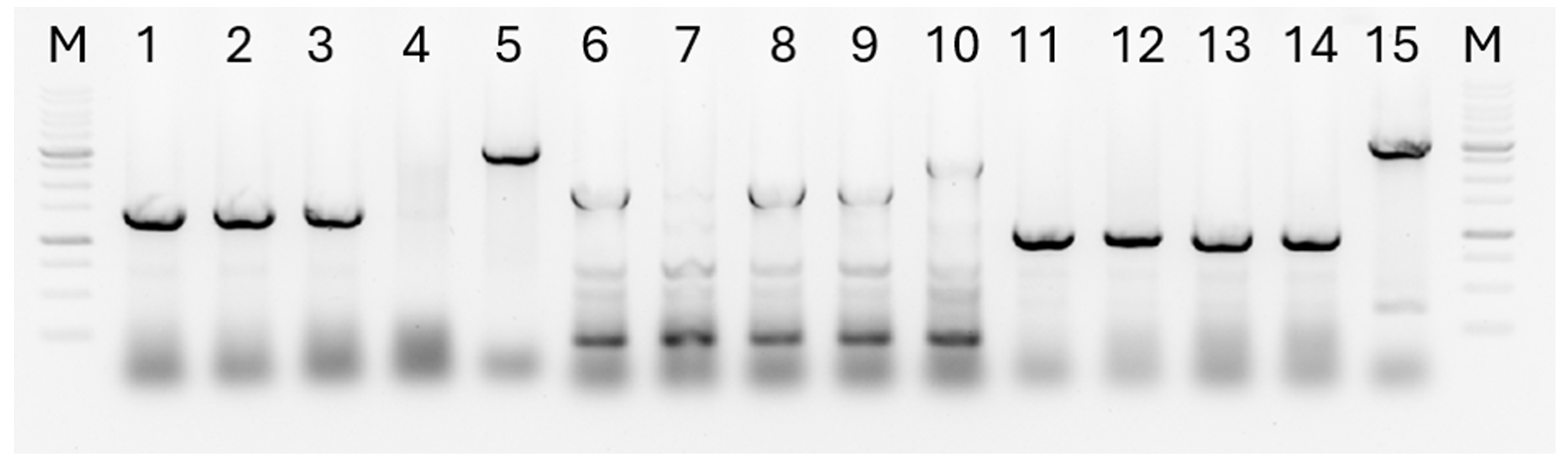
3.1. Construction of Recombinant Plasmids for Deletion of the algA, pilQ and flhA Genes

Based on the pK8mobSacB suicidal vector carrying the sacB gene, the genetic constructs pK18mobSacB-∆algA, pK18mobSacB-∆pilQ, and pK18mobSacB-∆flhA containing the corresponding flanking regions were obtained. All recombinant plasmids were verified by sequencing. The obtained plasmid DNAs were used to conduct deletion mutations in the P. putida PCL1760. Full sequences with description and maps of plasmids are presented in section Supplementary materials (Figure S1).

3.2. Construction of the P. putida LN6160 Strain

Using the homologous recombination method described in the above Section 2, a strain of P. putida LN6160 was created. The deletion of key genes encoding the formation of pili and flagella (pilQ and flhA), as well as the gene of the first stage of biofilm formation (algA), was successfully performed. The clones chosen, which were on dishes with a selective medium and showed the Km Sucrose+ phenotype, were verified by PCR using test-primers (Table 2). The analysis of the PCR fragments obtained after PCR was performed using 0.8% TAE agarose gel electrophoresis. Figure 1 shows the results of the PCR test of four clones obtained after introducing all three mutations into the P. putida PCL1760 strain. The wild-type strain was used as a control for this experiment. Cell lysates were tested for the presence of the algA deletion (lines 1–5), the pilQ deletion (lines 6–10), and the flhA deletion (lines 11–15). Lines 5, 10, 15 showed responses to the control (wild type cells). As can be seen in Figure 1, two of the four clones selected at the last stage showed bands corresponding to the expected size of the deleted gene fragments. This way, the strain contained deletion of the pilQ, flhA and algA gene (LN6160) was selected. The resulting deletion mutant LN6160 was used in subsequent experiments.

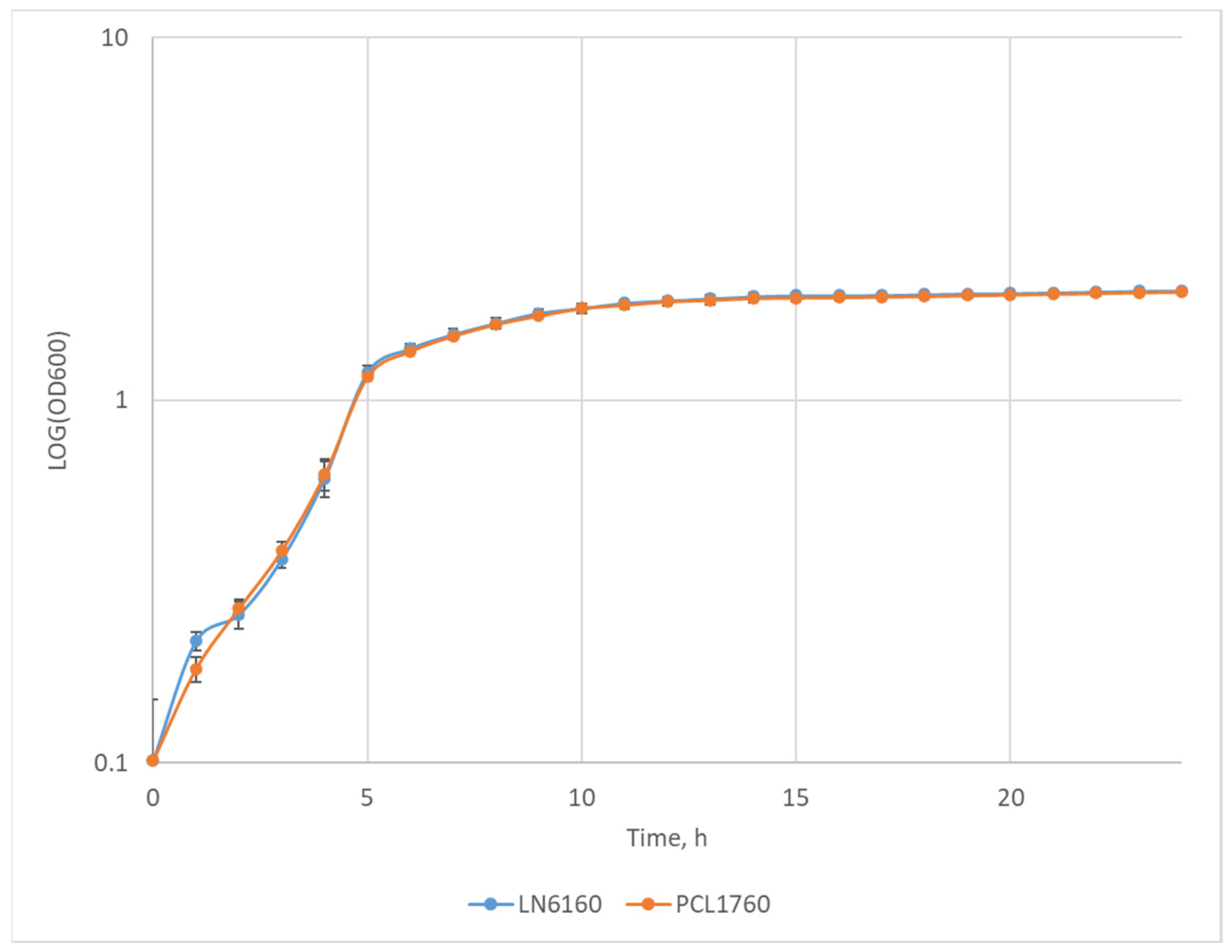
3.3. Comparison of the Growth Rate of the P. putida LN6160 and PCL1760 Strains

The growth curves of LN6160 and PCL1760 strains are shown in Figure 2. The results show that the deletions of the algA, pilQ, and flhA genes in the LN6160 strain had no effect on the growth dynamics compared to the wild-type strain under the experimental conditions and performed on 96-wells plates. The final optical density for PCL1760 was 1.98 ± 0.03, and for LN6160 it was 2.005 ± 0.02.
The growth rate (μ) and doubling time (Td) of PCL1760 strain were μ1760 = 0.74 ± 0.04 and Td1760 = 0.94 ± 0.05, respectively. For the LN6160 strain, the growth rate during the exponential phase was μ6160 = 0.79 ± 0.03 and the doubling time Td6160 was 0.90 ± 0.04. Thus, we demonstrated that gene deletions do not lead to a reduction in the rate of cell growth in a nutrient-rich medium in the experiment conditions.

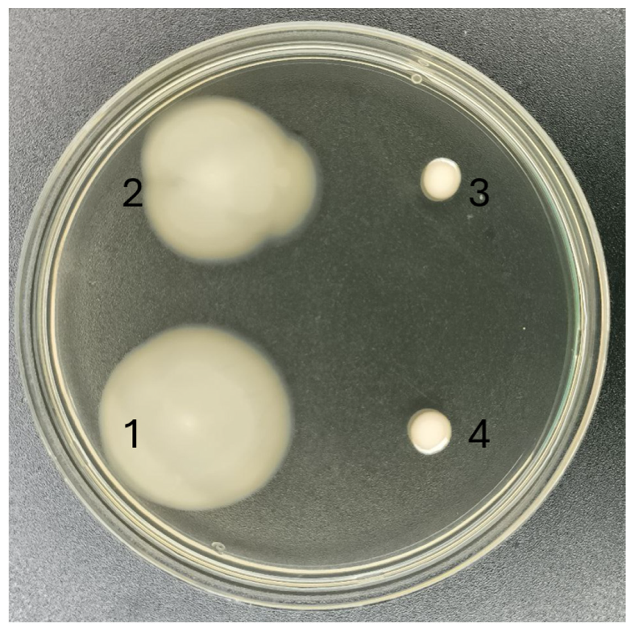
3.4. Motility of P. putida PCL1760 Wild Type Strain and Its Mutants

Figure 3 shows the results of a mobility test on a semi-liquid LB agar dish (0.3% agar). As can be seen from Figure 3, the deletion of algA did not decrease the motility of the cells. The motility of LN1610 (ΔalgA, ΔflhA) was severely reduced, whereas additional deletion of pilQ did not additionally decrease the motility of the cells. The distribution diameter of P. putida PCL1760 was 32 ± 2 mm, and the LN616 strain was 28 ± 6 mm, whereas that of the LN1610 and LN6160 strains was 5 ± 1 mm.

3.5. Quantitative Comparison of P. putida PCL1760 Wild Type Strain and Its Mutants Biofilm Formation

To quantify the difference in biofilm formation, we measured the optical density at 570 nm of the colored solution that was washed off the wells after the cultivation of microorganisms. The measurements were taken after 24, 48 and 72 h of incubation. The data obtained after subtracting the background values (the control without cells) from the measurements are shown in Figure 4. We observed a decrease in biofilm formation for all mutant strains. The graph shows that after 24 h of incubation, biofilm formation in all deletion strains was 20–30% less intense than the wild-type strain. After 48 and 72 h, the difference between the wild type and the LN6160 strain was 33.3 and 40%, respectively. Moreover, after 72 h of incubation, there was a difference in the intensity of biofilm formation between all mutants.
When comparing the average values using the Tukey test for four replicates, we found that there were statistically significant differences between the samples (p < 0.05). The variance values were <0.0002 and the standard deviation (SD) did not exceed 0.015. This indicates that the deletions were effective in significantly reducing the formation of biofilms in the LN6160 strain.

3.6. Comparative Cultivation of the LN6160 Mutant Strain and the Wild-Type LN6160 Strain in Small-Scale Bioreactors in Rich and Mineral Mediua

The initial concentration of cells in the bioreactor with LB medium for the P. putida LN6160 strain was slightly lower (9.5 × 10⁶ CFU/mL) than that of PCL1760 (1.36 × 10⁷ CFU/mL). During the experiment, the lag phase of growth for the mutant strain was observed to be slightly slower than for the wild-type strain. However, between 2 and 4 h of culture, the number of cells in the mutant increased almost 9-fold, while in the wild type, the increase was only 1.5 times (Figure 5A). With further culture, the growth rates of the mutant and wild type did not significantly differ, although one day later, the LN6160 strain had about 1.39 × 10¹⁰ CFU/mL, while the wild-type concentration of cells was 6.4 × 10⁹ CFU/mL. Figure 5B shows the growth curves of P. putida LN6160 and P. putida PCL1760 strains in low-volume bioreactors on a mineral medium. The initial cell concentration for P. putida strain LN6160 was slightly higher (2.83 × 10⁸ CFU/mL) than for PCL1760 (2.25 × 10⁸ CFU/mL). Rapid growth of the mutant strain was observed during the experiment, while slow growth was observed for the wild type throughout the experiment. After 24 h, there were 6.11 × 10⁹ CFU/mL of LN6160 and only 1.36 × 10⁹ CFU/mL for the wild-type strain PCL1760.

4. Discussion

Pseudomonas putida is frequently referred to as the “work horse” of a microbial laboratory, since many elaborate techniques for its cultivation and genetic manipulation are available [30]. This has resulted in the engineering of many P. putida strains for industrial biocatalysis [31].
Another waste application field for P.putida includes environmental biotechnologies, such as soil remediation and plant protection [32]. One of the most used strains is P. putida KT2440, which provided a way for soilborne bacteria to become a synthetic biology tool, due to the extensive use of gene engineering [33].
Non-pathogenic soilborne P. putida PCL1760 was isolated due to its advanced colonizing ability [1] and used for the biocontrol of plant diseases [34]. The genomic organization shows that it has the potential to be used as a cell factory for the genetic manipulation or biosynthesis of desirable organic compounds [2]. Good colonizing abilities, which help P. putida PCL1760 to protect the plants, are based on motility, root exudate consumption [1] and persistence [3] are not required for applications in bioreactors. To become a useful tool for fermentation, P. putida PCL1760 needs to lose, to some extent, its environmental fitness.
For example, it is known that biofilm formation, which is important for survival of the strains in natural habitats, is undesirable when cultures grow in bioreactors, as well as the flagellar biosynthesis which requires 2% of the cell’s biosynthetic resources, and flagellar rotation which consumes 0.1% of the cell’s energy [35]. In our work, we have deleted specific genes of the P. putida strain PCL1760 from the three operons involved in the accumulation of alginates, formation of pili, and flagella, which represent energy-intensive processes that are necessary for biofilm formation.
Hay et al. [10] provide information that the encoded-with-algA gene (Phosphomannose isomerase/pyrophosphorylase) has two unique functions during alginates formation: it converts fructose-6-phosphate to mannose-6-phosphate, and converts mannose-1-phosphate to GDP-mannose. These data indicate the importance of this gene in the alginate’s accumulation process. Therefore, it can be assumed that deleting the algA gene would completely prevent biofilm formation. However, in our experiments, we found that, despite the deletion of the algA gene, the cells were still able to form biofilms (Figure 4 and Figure 5). This is likely due to the production of other extracellular exopolysaccharides. For example, in the work by Ma et al. [36], the possibility of producing an exopolysaccharide called Psl, which consists of d-glucose, d-mannose, and L-rhamnose, was demonstrated for the P. aeruginosa strain. In addition, an exopolysaccharide called Pel, composed of N-acetyl-d-glucosamine and N-acetyl-d-galactosamine, was discovered by Colvin et al. [37], and its role in biofilm formation has been demonstrated.
PilM, PilN, and PilQ are the key enzymes involved in the formation of the flagellar system in P. aeruginosa [22]. Qi et al. [38] stated that the coordinated expression of various genes involved in mobility systems formation is necessary, and showed the importance of surface appendages on the flagellum and type IV pili, which are encoded by genes from the flh cluster. We assumed that deleting genes from these two clusters would significantly reduce or prevent the formation of flagella and pili. However, we did not find any data in the literature showing the effectiveness of joint deletion of pilQ and flhA genes on reducing motility in Gram-negative bacteria.
In our study, we deleted the pilQ and flhA genes in a P. putida PCL1760 strain, and observed the almost complete suppression of its cell motility on semi-liquid agar plates (Figure 3).
Many studies on bacterial cultivation have focused on finding the optimal growth conditions for microorganisms in bioreactors [6,39]. Some studies have shown the effectiveness of using automatic feeding systems, while others have focused on the rate of oxygen transfer during microbial processes. Ramadhan et al. [39] showed that adding a nonpolar solvent can decrease the evaporation of the substrate and increase product production. Although these methods are effective for optimizing cultivation, they require significant resources and time without guaranteeing success. Additionally, researchers often face challenges with biofilm formation and foam formation when culturing bacterial strains in bioreactors. Using genetic engineering techniques, we have developed a mutant strain, LN6160, by deleting genes involved in flagella formation, alginate production, and biofilm formation. In parallel cultivation experiments in bioreactors using a rich medium and a minimal medium, this strain showed several advantages compared to the wild-type strain. The mutant had generated less foam during cultivation (Figure S2). The mutant strain LN6160 formed less biofilm (Figure S3). The residual biofilm formation, at a smaller extent, in mutant strain LN6160 might be the result of undisturbed production of extracellular polymeric substances (EPSs) other than the alginate. It was shown that branched pentasaccharide Psl in P. aeruginosa can take part in biofilm formation [40]. Residual biofilm formation may be due to the synthesis of the cationic polymer Pel, which is able to mediate intercellular interactions and is associated with non-mucoid biofilms [41].
It has been shown that flagellar biosynthesis consumes about 2% of the cell’s energy [35]. Alginate biosynthesis is also an energy-consuming process, requiring the presence of carbon in large quantities and the fine regulation of metabolic processes [42]. In our study mutant LN6160 grew better than wild type strain PCL1760, which might be attributed to relatively unloaded energy apparatus of the mutant in comparison to wild-type strain (Figure 5). Due to the absence of flagella and pili alginates, the mutant also has reduced directional movement towards the walls of the bioreactor and decreased adhesion to their surface.
We constructed P. putida strain LN6160 that can be used as a platform for developing producer strains of various fine chemical, as well as a basis for the expression of specific proteins.

5. Conclusions

The biofilm formation in bacteria is a complex multicomponent process, which involves regulation of many operons with different sets of genes. The genes algA, pilQ, and flhA perform unique functions in alginate accumulation, pilus, and flagellum formation, respectively. The deletion of these genes dramatically reduces the biofilm formation in P. putida strain PCL1760 and increases the cell concentration in the bioreactor. The former might be a result of the energy load alleviation on the bacterial biosynthesis apparatus. Taking all of this together, we can state that the deletion of these three genes leads to a significant improve of the fermentation of P. putida strain PCL1760 in bioreactors. We assume that there are many other functions of free-living bacteria cells, which are dispensable for the bacterial cultures developing in bioreactors. Removal of these functions from bacterial metabolism may further improve fermentation processes, “focusing” cells on desirable production.

Supplementary Materials

The following supporting information can be downloaded at: https://www.mdpi.com/article/10.3390/fermentation10120606/s1, Figure S1: Nucleotide sequences of constructed plasmids; Figure S2: Foam formation in bioreactors during fermentation in Tanaka Medium; Figure S3: Biofilms formatiom in bioreactors after fermentation in LB medium.

Author Contributions

Conceptualization, S.Z.V. and N.V.T.; methodology, E.E.V., M.F. and G.A.K.; validation, S.Z.V. and N.V.T.; data curation, M.F. and G.A.K.; writing—original draft preparation, M.F.; writing—review and editing, N.V.T.; visualization, A.S.G. All authors have read and agreed to the published version of the manuscript.

Funding

This work was supported by Ministry of Science and Higher Education of the Russian Federation. Agreement #075-15-2022-254, 17 June 2022 “Development of recombinant Pseudomonas putida PCL1760 for biocatalytic valorization of lignin-derived aromatic compounds”.

Institutional Review Board Statement

Not applicable.

Informed Consent Statement

Not applicable.

Data Availability Statement

Data are contained within the article and supplementary materials.

Conflicts of Interest

The authors have no conflicts of interest to declare.

References

  1. Validov, S. Biological Control of Tomato Foot and Root Rot by Pseudomonas bacteria in Stonewool; Leiden University: Leiden, The Netherlands, 2007. [Google Scholar]
  2. Afordoanyi, D.M.; Diabankana, R.G.C.; Miftakhov, A.K.; Kuchaev, E.S.; Validov, S.Z. Genomic Features of Pseudomonas putida PCL1760: A Biocontrol Agent Acting via Competition for Nutrient and Niche. Appl. Microbiol. 2022, 2, 749–765. [Google Scholar] [CrossRef]
  3. Miftakhov, A.K.; Diabankana, R.G.C.; Frolov, M.; Yusupov, M.M.; Validov, S.Z.; Afordoanyi, D.M. Persistence as a Constituent of a Biocontrol Mechanism (Competition for Nutrients and Niches) in Pseudomonas putida PCL1760. Microorganisms 2023, 11, 19. [Google Scholar] [CrossRef] [PubMed]
  4. Koehorst, J.J.; van Dam, J.C.J.; van Heck, R.G.A.; Saccenti, E.; Martins dos Santos, V.A.P.; Suarez-Diez, M.; Schaap, P.J. Comparison of 432 Pseudomonas Strains through Integration of Genomic, Functional, Metabolic and Expression Data. Sci. Rep. 2016, 6, 38699. [Google Scholar] [CrossRef] [PubMed]
  5. Brehl, C.; Brass, H.U.C.; Lüchtrath, C.; Böckmann, L.; Ihling, N.; Classen, T.; Pietruszka, J.; Büchs, J. Optimized prodigiosin production with Pseudomonas putida KT2440 using parallelized noninvasive online monitoring. Biotechnol. Prog. 2022, 38, e3245. [Google Scholar] [CrossRef] [PubMed]
  6. Davis, R.; Duane, G.; Kenny, S.T.; Cerrone, F.; Guzik, M.W.; Babu, R.P.; Casey, E.; O’Connor, K.E. High cell density cultivation of Pseudomonas putida KT2440 using glucose without the need for oxygen enriched air supply. Biotechnol. Bioeng. 2015, 112, 725–733. [Google Scholar] [CrossRef] [PubMed]
  7. Rodriguez, A.; Escobar, S.; Gomez, E.; Santos, V.E.; Garcia-Ochoa, F. Behavior of several Pseudomonas putida strains growth under different agitation and oxygen supply conditions. Biotechnol. Prog. 2018, 34, 900–909. [Google Scholar] [CrossRef]
  8. Prabhukhot, G.S.; Eggleton, C.D.; Patel, J. Multispecies Bacterial Biofilms and Their Evaluation Using Bioreactors. Foods 2023, 12, 4495. [Google Scholar] [CrossRef]
  9. Yin, W.; Xu, S.; Wang, Y.; Zhang, Y.; Chou, S.-H.; Galperin, M.Y.; He, J. Ways to Control Harmful Biofilms: Prevention, Inhibition, and Eradication. Crit. Rev. Microbiol. 2021, 47, 57–78. [Google Scholar] [CrossRef]
  10. Hay, I.D.; Rehman, Z.U.; Moradali, M.F.; Wang, Y.; Rehm, B.H.A. Microbial alginate production, modification and its applications. Microb. Biotechnol. 2013, 6, 637. [Google Scholar] [CrossRef]
  11. Shinabarger, D.; Berry, A.; May, T.B.; Rothmel, R.; Fialho, A.; Chakrabarty, A.M. Purification and characterization of phosphomannose isomerase-guanosine diphospho-D-mannose pyrophosphorylase. A bifunctional enzyme in the alginate biosynthetic pathway of Pseudomonas aeruginosa. J. Biol. Chem. 1991, 266, 2080–2088. [Google Scholar] [CrossRef]
  12. Tatnell, P.J.; Russell, N.J.; Gacesa, P. GDP-mannose dehydrogenase is the key regulatory enzyme in alginate biosynthesis in Pseudomonas aeruginosa: Evidence from metabolite studies. Microbiology 1994, 140, 1745–1754. [Google Scholar] [CrossRef] [PubMed]
  13. Haiko, J.; Westerlund-Wikström, B. The Role of the Bacterial Flagellum in Adhesion and Virulence. Biology 2013, 2, 1242. [Google Scholar] [CrossRef]
  14. Feldman, M.; Bryan, R.; Rajan, S.; Scheffler, L.; Brunnert, S.; Tang, H.; Prince, A. Role of flagella in pathogenesis of Pseudomonas aeruginosa pulmonary infection. Infect. Immun. 1998, 66, 43–51. [Google Scholar] [CrossRef] [PubMed]
  15. Bouteiller, M.; Dupont, C.; Bourigault, Y.; Latour, X.; Barbey, C.; Konto-ghiorghi, Y.; Merieau, A. Pseudomonas Flagella: Generalities and Specificities. Int. J. Mol. Sci. 2021, 22, 3337. [Google Scholar] [CrossRef] [PubMed]
  16. Liu, R.; Ochman, H. Origins of Flagellar Gene Operons and Secondary Flagellar Systems. J. Bacteriol. 2007, 189, 7098. [Google Scholar] [CrossRef] [PubMed]
  17. Osterman, I.A.; Dikhtyar, Y.Y.; Bogdanov, A.A.; Dontsova, O.A.; Sergiev, P.V. Regulation of flagellar gene expression in Bacteria. Biochemistry 2015, 80, 1447–1456. [Google Scholar] [CrossRef] [PubMed]
  18. Kojima, S.; Terashima, H.; Homma, M. Regulation of the Single Polar Flagellar Biogenesis. Biomolecules 2020, 10, 533. [Google Scholar] [CrossRef] [PubMed]
  19. Minamino, T.; Inoue, Y.; Kinoshita, M.; Namba, K. FLIK-driven conformational rearrangements of FlhA and FlhB are required for export switching of the flagellar protein export apparatus. J. Bacteriol. 2020, 202, e00637-19. [Google Scholar] [CrossRef] [PubMed]
  20. Terashima, H.; Kawamoto, A.; Tatsumi, C.; Namba, K.; Minamino, T.; Imada, K. In vitro reconstitution of functional type III protein export and insights into flagellar assembly. Mbio 2018, 9, e00988-18. [Google Scholar] [CrossRef]
  21. Bradley, D.E. Basic Characterization of a Pseudomonas aeruginosa Pilus-Dependent Bacteriophage with a Long Noncontractile. Tail. J. Virol. 1973, 12, 1139. [Google Scholar] [CrossRef]
  22. Tammam, S.; Sampaleanu, L.M.; Koo, J.; Manoharan, K.; Daubaras, M.; Burrows, L.L.; Howell, P.L. PilMNOPQ from the Pseudomonas aeruginosa Type IV Pilus System Form a Transenvelope Protein Interaction Network That Interacts with PilA. J. Bacteriol. 2013, 195, 2126. [Google Scholar] [CrossRef] [PubMed]
  23. McCallum, M.; Tammam, S.; Rubinstein, J.L.; Burrows, L.L.; Howell, P.L. CryoEM map of Pseudomonas aeruginosa PilQ enables structural characterization of TsaP. Structure 2021, 29, 457–466.e4. [Google Scholar] [CrossRef] [PubMed]
  24. Tang, R.; Zhu, J.; Feng, L.; Li, J.; Liu, X. Characterization of LuxI/LuxR and Their Regulation Involved in Biofilm Formation and Stress Resistance in Fish Spoilers Pseudomonas fluorescens. Int. J. Food Microbiol. 2019, 297, 60–71. [Google Scholar] [CrossRef] [PubMed]
  25. Norrander, J.; Kempe, T.; Messing, J. Construction of improved M13 vectors using oligodeoxynucleotide-directed mutagenesis. Gene 1983, 26, 101–106. [Google Scholar] [CrossRef]
  26. Simon, R.; Priefer, U.; Pühler, A. A Broad Host Range Mobilization System for In Vivo Genetic Engineering: Transposon Mutagenesis in Gram Negative Bacteria. Nat. Biotechnol. 1983, 1, 784–791. [Google Scholar] [CrossRef]
  27. Kvitko, B.H.; Collmer, A. Construction of Pseudomonas syringae pv. tomato DC3000 mutant and polymutant strains. Methods Mol. Biol. 2011, 712, 109–128. [Google Scholar] [CrossRef]
  28. Ha, D.G.; Kuchma, S.L.; O’Toole, G.A. Plate-based assay for swimming motility in Pseudomonas aeruginosa. Methods Mol. Biol. 2014, 1149, 59–65. [Google Scholar] [CrossRef]
  29. Stepanović, S.; Vuković, D.; Dakić, I.; Savić, B.; Švabić-Vlahović, M. A modified microtiter-plate test for quantification of staphylococcal biofilm formation. J. Microbiol. Methods 2000, 40, 175–179. [Google Scholar] [CrossRef]
  30. Loeschcke, A.; Thies, S. Pseudomonas putida-a versatile host for the production of natural products. Appl. Microbiol Biotechnol. 2015, 99, 6197. [Google Scholar] [CrossRef]
  31. Nikel, P.I.; de Lorenzo, V. Pseudomonas putida as a functional chassis for industrial biocatalysis: From native biochemistry to trans-metabolism. Metab. Eng. 2018, 50, 142–155. [Google Scholar] [CrossRef]
  32. Weimer, A.; Kohlstedt, M.; Volke, D.C.; Nikel, P.I.; Wittmann, C. Industrial biotechnology of Pseudomonas putida: Advances and prospects. Appl. Microbiol. Biotechnol. 2020, 104, 7745–7766. [Google Scholar] [CrossRef] [PubMed]
  33. de Lorenzo, V.; Pérez-Pantoja, D.; Nikel, P.I. Pseudomonas putida KT2440: The long journey of a soil-dweller to become a synthetic biology chassis. J. Bacteriol. 2024, 206, e0013624. [Google Scholar] [CrossRef]
  34. Validov, S.Z.; Kamilova, F.; Lugtenberg, B.J.J. Pseudomonas putida strain PCL1760 controls tomato foot and root rot in stonewool under industrial conditions in a certified greenhouse. Biol. Contr. 2009, 48, 6–11. [Google Scholar] [CrossRef]
  35. Fontaine, F.; Stewart, E.J.; Lindner, A.B.; Taddei, F. Mutations in two global regulators lower individual mortality in Escherichia coli. Mol. Microbiol. 2008, 67, 2–14. [Google Scholar] [CrossRef]
  36. Ma, L.; Conover, M.; Lu, H.; Parsek, M.R.; Bayles, K.; Wozniak, D.J. Assembly and development of the Pseudomonas aeruginosa biofilm matrix. PLoS Pathog. 2009, 5, e1000354. [Google Scholar] [CrossRef]
  37. Colvin, K.M.; Alnabelseya, N.; Baker, P.; Whitney, J.C.; Howell, P.L.; Parsek, M.R. PelA deacetylase activity is required for Pel polysaccharide synthesis in Pseudomonas aeruginosa. J. Bacteriol. 2013, 195, 2329–2339. [Google Scholar] [CrossRef]
  38. Qi, Y.H.; Huang, L.; Liu, G.F.; Leng, M.; Lu, G.T. PilG and PilH antagonistically control flagellum-dependent and pili-dependent motility in the phytopathogen Xanthomonas campestris pv. campestris. BMC Microbiol. 2020, 20, 37. [Google Scholar] [CrossRef]
  39. Ramadhan, S.H.; Matsui, T.; Nakano, K.; Minami, H. High cell density cultivation of Pseudomonas putida strain HKT554 and its application for optically active sulfoxide production. Appl. Microbiol. Biotechnol. 2013, 97, 1903–1907. [Google Scholar] [CrossRef]
  40. Chung, J.; Eisha, S.; Park, S.; Morris, A.J.; Martin, I. How Three Self-Secreted Biofilm Exopolysaccharides of Pseudomonas aeruginosa, Psl, Pel, and Alginate, Can Each Be Exploited for Antibiotic Adjuvant Effects in Cystic Fibrosis Lung Infection. Int. J. Mol. Sci. 2023, 24, 8709. [Google Scholar] [CrossRef]
  41. Friedman, L.; Kolter, R. Genes Involved in Matrix Formation in Pseudomonas aeruginosa PA14 Biofilms. Mol. Microbiol. 2004, 51, 675–690. [Google Scholar] [CrossRef] [PubMed]
  42. Núñez, C.; López-Pliego, L.; Ahumada-Manuel, C.L.; Castañeda, M. Genetic Regulation of Alginate Production in Azotobacter vinelandii a Bacterium of Biotechnological Interest: A Mini-Review. Front. Microbiol. 2022, 13, 845473. [Google Scholar] [CrossRef] [PubMed]
Figure 1. The 0.8% TAE agarose gel electrophoresis of fragments obtained after PCR analysis of selected clones of the deletion mutant strain. M-1kb DNA Ladder (Evrogen, Moscow, Russia); lines 5, 10, 15—response to the control (wild type strain); Lines 1, 2, 3, 4—clones tested with primers specific to the algA gene region; Lines 6, 7, 8, 9—clones tested with primers specific to the flhA gene region; Lines 11, 12, 13, 14—clones tested with primers specific to the pilQ gene region.
Fermentation 10 00606 g001
Figure 2. Growth curve of the P. putida PCL1760 and LN6160 strains in the LB medium in 96-well plates.
Fermentation 10 00606 g002
Figure 3. Petri dishes with 0.3% LB agar medium. Cultures 1—P. putida PCL1760, 2—P. putida LN616, 3—P. putida LN1610, and 4—P. putida LN6160 were plated in soft agar by piercing with the sterile toothpicks.
Fermentation 10 00606 g003
Figure 4. The optical density values obtained from measurements of violet gentian-stained solutions that had been washed off from biofilm at a wavelength of 570 nanometers. PCL1760 is the wild-type strain, LN616 is an algA deletion mutant, LN1610 is an algA flhA deletion mutant, and LN6160 is an algA flhA pilQ deletion mutant.
Fermentation 10 00606 g004
Figure 5. Growth curve of the P. putida LN6160 и P. putida PCL1760 strains in (A) parallel small-volume bioreactors in LB medium (B) and in mineral medium. The X-axis shows the time of cultivation, and the Y-axis shows the absolute amount of the live cells in ml (CFU/mL).
Fermentation 10 00606 g005
Table 1. The bacterial strains and plasmids used in this work.
Strain/PlasmidFeaturesReference/Source
E. coli DH5αF-80ΔlacZ M15 (lacZYA-argF) U169 recA1 endA1hsdR17(rk−, mk+) phoA supE44-thi-1 gyrA96 relA1[25]
E. coli S17-1recA pro (RP4-2Tet: Mu)[26]
P. putida PCL1760Wild typeLab stock
P. putida LN616ΔalgA,This work
P. putida LN1610ΔalgA, ΔflhAThis work
P. putida LN6160ΔalgA, ΔpilQ, ΔflhAThis work
pK18mobSacBsacB lacZα Kmr; cloning vector for allelic exchange[27]
pK18mobSacB-∆algApK18mobsacB carrying a modified algA gene regionThis work
pK18mobSacB-∆pilQpK18mobsacB carrying a modified pilQ gene regionThis work
pK18mobSacB-∆flhApK18mobsacB carrying a modified flhA gene regionThis work
Table 2. Primers used for construction and verification of knockout mutant of P. putida PCL1760 strain.
PrimerNucleotide Sequence 5′-3′
algA-fl1-forCGAATTCGAGCTCGGTACCCACCTGGCCCTCGGTATCG
algA-fl1-revCCACCCACCACCCTGAAAGGTCGCTGCAAATC
algA-fl2-forCCTTTCAGGGTGGTGGGTGGCCCATTCG
algA-fl2-revGTCGACTCTAGAGGATCCCCCCCGAACGTTATCTGCCAATGAAAAAC
test-algA-forCTTGGCTGGGTAACGCAACAACAACAC
test-algA-revGGACAGACCACTGACTGATAGAGAGG
pilQ-fl1-forCGAATTCGAGCTCGGTACCCGCTGCAAGCGCTCGAGAATATCG
pilQ-fl1-revACCAGGGTGAAAAAAAGCGGCAACGGCGAGATC
pilQ-fl2-forCCGCTTTTTTTCACCCTGGTAAGGCGTCGC
pilQ-fl2-revGTCGACTCTAGAGGATCCCCGCTCGCTTGGCTGGCGATG
test-pilQ-forGGCTTGCTTGTCGAACGCCTCGATG
test-pilQ-revCGGCGACCTGCGCGGTAACG
flhA-fl1-forCGAATTCGAGCTCGGTACCCTGTTGCAGCCAAAATTCAGC
flhA-fl1-revATCCCCTACCCGCGAGTCCTCTTGATGC
flhA-fl2-forAGGACTCGCGGGTAGGGGATAATGCAAGTTAAGC
flhA-fl2-revGTCGACTCTAGAGGATCCCCAGGTAGTTCTTCGCGGCAATG
test-flhA-forGCAACCCGACGCATTATGCGGTGG
test-flhA-revGCCAGCGAACCGAGTTGCACTTCC
Disclaimer/Publisher’s Note: The statements, opinions and data contained in all publications are solely those of the individual author(s) and contributor(s) and not of MDPI and/or the editor(s). MDPI and/or the editor(s) disclaim responsibility for any injury to people or property resulting from any ideas, methods, instructions or products referred to in the content.

