Article

New Solutions in Single-Cell Protein Production from Methane: Construction of Glycogen-Deficient Mutants of Methylococcus capsulatus MIR

by
Sergey Y. But
1,2,
Ruslan Z. Suleimanov
1,
Igor Y. Oshkin
1,
Olga N. Rozova
1,2,
Ildar I. Mustakhimov
1,2,
Nikolai V. Pimenov
1,
Svetlana N. Dedysh
1,* and
Valentina N. Khmelenina
2
1
Winogradsky Institute of Microbiology, Research Center of Biotechnology, Russian Academy of Sciences, 119071 Moscow, Russia
2
G. K. Skryabin Institute of Biochemistry and Physiology of Microorganisms, Scientific Center for Biological Research, Russian Academy of Sciences, 142290 Pushchino, Russia
*
Author to whom correspondence should be addressed.
Fermentation 2024, 10(5), 265; https://doi.org/10.3390/fermentation10050265
Submission received: 4 December 2023 / Revised: 15 May 2024 / Accepted: 17 May 2024 / Published: 19 May 2024

Abstract

:
The biotechnology of converting methane to single-cell protein (SCP) implies using fast-growing thermotolerant aerobic methanotrophic bacteria. Among the latter, members of the genus Methylococcus received significant research attention and are used in operating commercial plants. Methylococcus capsulatus MIR is a recently discovered member of this genus with the potential to be used for the purpose of SCP production. Like other Methylococcus species, this bacterium stores carbon and energy in the form of glycogen, particularly when grown under nitrogen-limiting conditions. The genome of strain MIR encodes two glycogen synthases, GlgA1 and GlgA2, which are only moderately related to each other. To obtain glycogen-free cell biomass of this methanotroph, glycogen synthase mutants, ΔglgA1, ΔglgA2, and ΔglgA1ΔglgA2, were constructed. The mutant lacking both glycogen synthases exhibited a glycogen-deficient phenotype, whereas the intracellular glycogen content was not reduced in strains defective in either GlgA1 or GlgA2, thus suggesting functional redundancy of these enzymes. Inactivation of the glk gene encoding glucokinase also resulted in a sharp decrease in glycogen content and accumulation of free glucose in cells. Wild-type strain MIR and the mutant strain ΔglgA1ΔglgA2 were also grown in a bioreactor operated in batch and continuous modes. Cell biomass of ΔglgA1ΔglgA2 mutant obtained during batch cultivation displayed high protein content (71% of dry cell weight (DCW) compared to 54% DCW in wild-type strain) as well as a strong reduction in glycogen content (10.8 mg/g DCW compared to 187.5 mg/g DCW in wild-type strain). The difference in protein and glycogen contents in biomass of these strains produced during continuous cultivation was less pronounced, yet biomass characteristics relevant to SCP production were slightly better for ΔglgA1ΔglgA2 mutant. Genome analysis revealed the presence of glgA1-like genes in all methanotrophs of the Gammaproteobacteria and Verrucomicrobia, while only a very few methanotrophic representatives of the Alphaproteobacteria possessed these determinants of glycogen biosynthesis. The glgA2-like genes were present only in genomes of gammaproteobacterial methanotrophs with predominantly halo- and thermotolerant phenotypes. The role of glycogen in terms of energy reserve is discussed.

1. Introduction

Microbial protein, also known as single-cell protein (SCP), has been recognized as one of the feasible and sustainable alternatives to animal products in meeting the growing global protein demand [1,2]. A wide variety of fast-growing microorganisms can be cultivated for SCP production (reviewed in [1]). Among all potential SCP producers, aerobic methanotrophic bacteria that use methane (CH4) as a growth substrate, are of particular importance. Methane (or natural gas) is a relatively cheap and readily available source of carbon. Gas fermenters that produce methane-derived biomass can be built at different scales, with a typical commercial plant size producing 10,000 to 20,000 tons of protein per year [2,3]. In addition, methane-based SCP production technology is highly attractive on the grounds of sustainability and; therefore, is currently on the verge of large-scale commercialization [2,4].
The biotechnology of converting methane to SCP and other value-added products implies using aerobic methanotrophic bacteria [5,6,7,8,9]. A unique trait of these prokaryotes is the use of methane monooxygenase enzymes to catalyze the oxidation of methane to methanol [10,11,12]. Currently, described aerobic methanotrophs form several coherent phylogenetic clades within Gamma- and Alphaproteobacteria as well as Verrucomicrobia. Among these bacteria, fast-growing thermotolerant members of the genus Methylococcus have been extensively studied as producers of methane SCP [13,14,15,16]. A number of currently operating commercial plants, such as those owned by UniBio and Calysta Inc., use strains of Methylococcus capsulatus as key components of the industrial methane-utilizing microbial consortia.
Despite the long history of fundamental research and industrial applications, some metabolic features of these bacteria remain poorly understood. Like other gammaproteobacterial methanotrophs, Methylococcus species store glycogen, especially under imbalanced growth conditions [17,18,19]. The accumulation of glycogen in cells may result in a loss of protein content, which could affect the biomass quality as a feed source. Despite the long history of using Methylococcus species in biotechnology, little is known about the regulation of metabolic routes leading to glycogen biosynthesis in these bacteria. This knowledge may offer new solutions for constructing biotechnologically relevant strains.
Glycogen is a branched homopolysaccharide of α-1,4-linked glucose subunits with α-1,6-linked glucose at the branching points. In bacteria, glycogen synthase (GlgA) and branching enzyme (GlgB) are responsible for glycogen biosynthesis. Glycogen synthesis is also dependent on the activity of ADP glucose pyrophosphorylase (GlgC, EC 2.7.7.27) [20,21]. Anabolic phosphoglucomutase (Pgm) and catabolic glycogen phosphorylase were also demonstrated to play an important role in glycogen accumulation [22,23,24]. Enterobacteria, including Escherichia coli and Salmonella enterica, were found to store glycogen under limited growth conditions in excess of a carbon source [21]. Glycogen accumulation of up to 30% of dry cell weight (DCW) was reported for the thermotolerant methanotroph Methylococcus sp. NCIB 11083 and the halotolerant Methylotuvimicrobium alcaliphilum 20Z [17,18,19]. Nowadays, the metabolic engineering of methanotrophs represents a rapidly evolving field, offering new opportunities for constructing producer strains tailored for use in methane-based biotechnology [25,26,27,28,29].
Mc. capsulatus MIR is a fast-growing thermotolerant gammaproteobacterial methanotroph, which was isolated from the activated sludge of a wastewater treatment plant [30]. Unlike many other Methylococcus bacteria, strain MIR exhibits the ability to grow on methanol in a concentration range of 0.05 to 3.5% (vol/vol), which provides additional flexibility in biotechnological applications [30]. With this advantage, strain MIR emerges as a promising candidate for studying carbon metabolism in relation to glycogen biosynthesis. The genome of strain MIR encodes both particulate and soluble membrane monooxygenases, MxaFI and XoxF methanol dehydrogenases as well as the ribulose monophosphate pathway (RuMP), the serine pathway and the Calvin–Benson–Bassham (CBB) cycle for carbon assimilation. Strain MIR harbors two glycogen synthase-encoding gene clusters involved in directing carbon excess into glycogen synthesis.
This study was initiated in order to construct glycogen-deficient strains of Mc. capsulatus MIR by inactivating the genes encoding glycogen synthases or the gene encoding glucokinase, and evaluating the characteristics of the mutant strains with respect to their potential use for the purposes of SCP production.

2. Materials and Methods

2.1. Strains Used in This Study

Mc. capsulatus MIR was maintained in 200-mL flasks filled with 30 mL mineral medium P of the following composition (g L−1): KNO3 (1), MgSO4 (0.2), CaCl2 (0.02), Na2-EDTA (0.005), FeSO4×7H2O (0.002), ZnSO4×7H2O (0.0001), MnCl2×4H2O (0.00003), CuSO4×5H2O (0.0001), CoCl2×6H2O (0.0002), NiCl2×6H2O (0.00002), Na2MoO4 (0.00003), H3BO3 (0.0003). To create nitrogen-limited conditions, the concentration of KNO3 in the medium was reduced to 0.29 g L−1. If needed, gentamycin (10 μg mL−1), 100 μg/mL spectinomycin (100 μg mL−1), or kanamycin (50 μg mL−1) were added to the medium. After inoculation, the flasks were hermetically closed with silicone rubber septa, 50 mL of methane was injected into the headspace, and the flasks were incubated on a shaker (120 rpm) at 42 °C. Escherichia coli strains Top 10 and S-17-1 were maintained at 37 °C in a selective LB broth or agar-solidified medium LB (1.5% Difco agar) [31]. Gentamycin (4 μg mL−1) and kanamycin (50 μg mL−1) were added if required.

2.2. Construction of Mutant Strains Defective in Glycogen Biosynthesis

To knock out the glgA1 (M3M30_03055; https://mage.genoscope.cns.fr/, accessed on 20 May 2023) and glgA2 (M3M30_12400) genes, the ~700 bp flanking fragments were amplified from the genomic DNA of strain MIR using primers listed in Table 1. The glgA1 flanking fragments were cloned into pK18mob vector between XbaI, SphI, and SphI, HindIII sites, while glgA2 flanking fragments were cloned between EcorI, Acc65I and XbaI, HindIII sites [32]. The gentamycin or spectinomycin resistance cassettes were cloned between the fragments at the BamHI site resulting in the generation of plasmids pk18glgA1-Gm and pk18glgA2-Sp. These vectors were introduced into the cells of strain MIR via conjugation with E. coli S17-1. The latter was grown on LB plates, while strain MIR was grown on agar plates with P medium. Cells of these two strains were mixed on the surface of agar plates containing P medium supplemented with 3% (v/v) LB, and incubated in a methane-air atmosphere at 37 °C for 2 days. The biomass was then spread on agar plates with medium P containing gentamycin or spectinomycin. The plates were incubated in a methane-air atmosphere at 42 °C until colonies became visible (10–14 days). The clones were selected by resistance to either gentamycin or spectinomycin and sensitivity to kanamycin. The mutant genotype was further confirmed by polymerase chain reaction PCR analysis.
A similar procedure was carried out to generate the ΔglgA1ΔglgA2 strain. The pk18glgA1-Gm vector was introduced to the ΔglgA2 strain, or alternatively, the pk18glgA2-Sp vector was introduced to the ΔglgA1. Colonies were selected on plates with P medium supplemented with gentamycin and spectinomycin. However, as this approach did not allow us to generate mutants, we constructed a vector providing an inducible expression of glycogen synthase. This involved amplifying the glgA2 gene from the genomic DNA of Mm. alcaliphilum 20Z and cloning it between EcorI and BamHI sites into pCAH01 [5,19]. The resulting vector was introduced into the ΔglgA1 cells by conjugation as described above. The cells of ΔglgA1 strain harboring this vector were used for glgA2 inactivation using a pk18glgA2-Sp plasmid. The mating media and selective media also contained 500 ng/mL anhydrotetracycline. Clones were selected by resistance to gentamycin, spectinomycin, and kanamycin. The genotype was tested by PCR. The obtained mutant strain ΔglgA1ΔglgA2 was cured of plasmid by growth in a kanamycin-free medium, followed by plating on agar media and selecting kanamycin-sensitive colonies. The colonies were additionally screened by PCR for the absence of plasmid using the primers designed for specific amplification of glgA2 from Mm. alcaliphilum 20Z.
To generate the Δglk mutant strain, the complete sequence (1011 bp) of glucokinase-encoding glk gene (M3M30_07505) was amplified using GLK_MIR-F and GLK_MIR-R primers (Table 1) and subsequently cloned into the pK18mob plasmid. The 652 bp internal fragment of the glk gene was excised using BshTI (AgeI) and PstI restriction endonucleases, and the resulting gap was filled with an 840 bp gentamicin cassette from plasmid p34S-Gm using SacI restriction endonuclease, followed by T4 polymerase treatment. The resulting vector was used for glk gene inactivation as described above.

2.3. Glycogen and Glucose Extraction and Quantification

Cells of Mc. capsulatus MIR and the mutant strains, grown in 200 mL of liquid P medium, were collected using a centrifuge (Beckman Coulter, Pasadena, CA, USA) at 8000× g for 10 min and freeze-dried. Low molecular weight metabolites were extracted from the cells as described earlier [33] and the extracts were used for glucose quantification by ABTS assay [34]. Glycogen was extracted from cells as described elsewhere [19] and quantified using an anthrone reagent (60 mg of anthrone dissolved in 40 mL of 70% sulfuric acid). A total of 100 μL of the sample was mixed with 1 mL of the anthrone reagent and incubated at 100 °C for 15 min. Optical density was measured at 620 nm. Glycogen concentrations were calculated using a calibration curve for glucose standards, with a correction coefficient of 0.9 applied.

2.4. Protein Content Assay

A total of 10 mg of dried cell material was suspended in 1 mL of 1 M NaOH and incubated for 5 min at 100 °C. The protein concentration in the resulting solution was measured using the Lowry method [35].

2.5. Electron Microscopy

Examination of cell ultrastructure was performed using cell suspensions of wild-type strain MIR and ΔglgA1ΔglgA2 mutant strain collected at the very beginning of the stationary phase. Collected cells were pre-fixed with 2.5% glutaraldehyde in 0.05 M cacodylate buffer (pH 7.3) at 4 °C for 2 h, and post-fixed in in the same buffer containing 1% OsO4 at 4 °C [18]. Fixed and dehydrated cells were embedded in epoxy resin Epon-812 (Sigma-Aldrich, Burlington, MA, USA). Ultrathin sections were placed on copper grids coated with formvar film, post-stained with uranyl acetate for 30 min and with lead citrate for 15 min [36]. The prepared cell specimens were examined using a JEM-1400 transmission electron microscope (JEOL, Tokyo, Japan) under an accelerating voltage of 80 kV.

2.6. RNA Isolation and Real-Time PCR

The cultures of Mc. capsulatus MIR and Δglk mutant were grown up to OD600 ~0.4. 10 mL of stop solution (5% water-saturated phenol in ethanol) was added to the cultures and cells were collected by centrifugation (3500× g for 10 min) using Eppendorf MiniSpin plus centrifuge (Eppendorf, Hamburg, Germany). The precipitated cells were washed with DEPC-treated water, centrifuged, and dissolved in 1 mL Trizol® Reagent (Sigma-Aldrich, Burlington, MA, USA). Lysate was stored on ice for 5 min, and then 200 μL chloroform was added. After 15 min incubation on ice, the samples were centrifuged at 10,000× g for 15 min and water fractions were transferred into clean tubes. After addition of 0.5 mL isopropanol and 1 h incubation at −20 °C, the samples were centrifuged at 1,0000× g for 15 min, and the supernatant was discarded. The precipitate was washed twice with 80% ethanol, dried, and then dissolved in 50 μL of RNase-free water (Evrogen, Moscow, Russia). To remove any remaining DNA, 5U of RNase-free DNaseI (Thermo Fisher Scientific, Waltham, MA, USA) and 50 U of Thermo Scientific RiboLock RNase Inhibitor (Thermo Fisher Scientific, Waltham, MA, USA) were added to each tube, and the latter were incubated for 30 min at 37 °C. A total of 5 μL of 50 mM EDTA was added followed by DNase temperature inactivation (10 min at 65 °C). Finally, the integrity of the RNA samples was estimated by electrophoresis in 1% agarose gel.
cDNA was built using 0.5 μg of total RNA, 20 pmol of the specific primers (Table 1), and an MMLV Reverse Transcriptase kit (Evrogen, Moscow, Russia). Gene expression levels were determined via quantitative real-time polymerase chain reaction using a qPCRmix-HS SYBR Kit (Evrogen, Moscow, Russia) in the DTlite Real-Time PCR system (DNA-technology, Moscow, Russia). The reaction consisted of pre-incubation at 95 °C for 5 min, followed by 40 cycles at 95 °C for 20 s, 58 °C for 15 s, and 72 °C for 20 s. The data were analyzed according to the Pfaffl method [37] using rpoB as a reference gene. The reaction without reverse transcription served as a control for the absence of DNA in RNA extracts.

2.7. Cultivation in a Bioreactor

Growth characteristics of Mc. capsulatus MIR and mutant strains were also assessed by cultivating them in a 1.5 L bioreactor (GPC BIO, Perigny, France) on natural gas. These experiments were performed with the following process parameters: temperature, 42 °C; agitation, 1000 rpm; gas flow rate, 6000 cm3 h−1; air flow rate, 18000 cm3 h−1. The pH level of 6.3 was controlled by titration with 0.5% NH4OH solution. The cultivation was performed in a mineral medium of the following composition (mg L−1): (NH4)2SO4, 200; KCl, 250; MgSO4×7H2O, 250; CuSO4×5H2O, 4.4; ZnSO4×7H2O, 1.8; MnSO4×5H2O, 1.2; FeSO4×7H2O, 4.2; CoSO4×6H2O, 0.58; Na2MoO4×2H2O, 0.7; H3BO3, 0.6; NiSO4×6H2O, 0.5, and 0.5 mL/L 85% H3PO4. A freshly grown seed culture of the examined methanotroph was used to inoculate the bioreactor at the initial OD600 0.3. Every two hours, an aliquot of the cell suspension was taken from the bioreactor to determine the OD600 value on a Spectroquant Prove 300 spectrophotometer (Merck, Darmstadt, Germany). Batch cultivation was carried out until OD600 of 17–19 was reached, after which the bioreactor was switched to a continuous cultivation with a dilution rate of ~0.25 h−1. To determine cell dry weight (DCW) and protein content, cells were collected (centrifugation at 14,000 × g for 7 min), frozen at −76 °C overnight, and subsequently lyophilized at −70 °C. Protein concentration in lyophilized biomass was determined by the Kjeldahl method using a Kjeldahl analyzer (FOSS, Stockholm, Sweden). Ammonia concentrations in culture aliquots were determined using a commercial kit (MedEcoTest, Moscow, Russia).

3. Results

3.1. Identification of Glycogen Synthase Genes in the Genome of Mc. capsulatus MIR

To generate a glycogen-deficient strain, we searched the genome of Mc. capsulatus MIR for sequences resembling known glycogen synthases. Two putative glycogen synthase genes, glgA1 (M3M30_03055) and glgA2 (M3M30_12400), were identified using the GlgA sequence from E. coli K12 as the query in a blast search [38]. Both putative glycogen synthases belong to the GT1 glycosyl transferase family and are only moderately related to each other (23% of amino acid sequence identity) (Figure 1). The GlgA1 from strain MIR and the corresponding enzyme from E. coli K12 display 46% sequence identity.
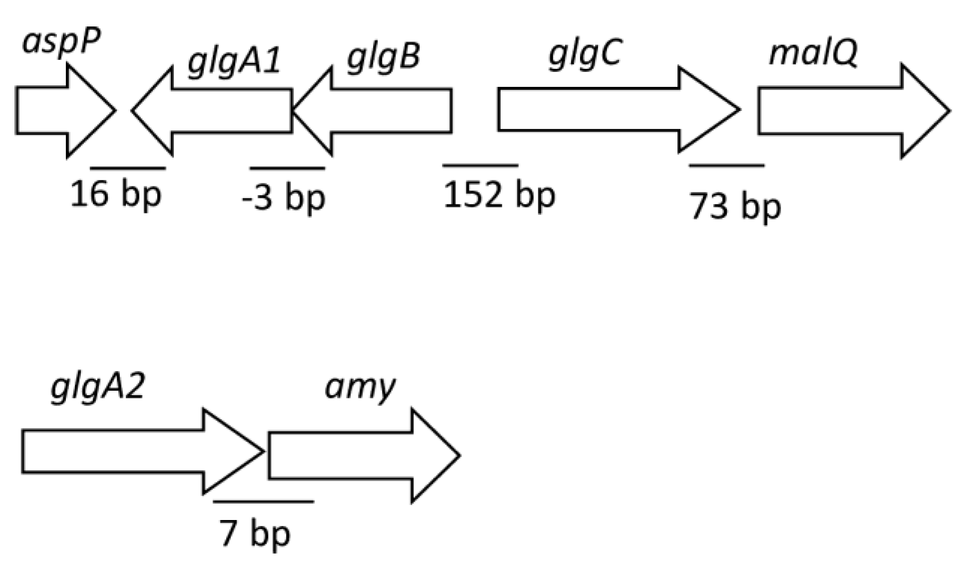
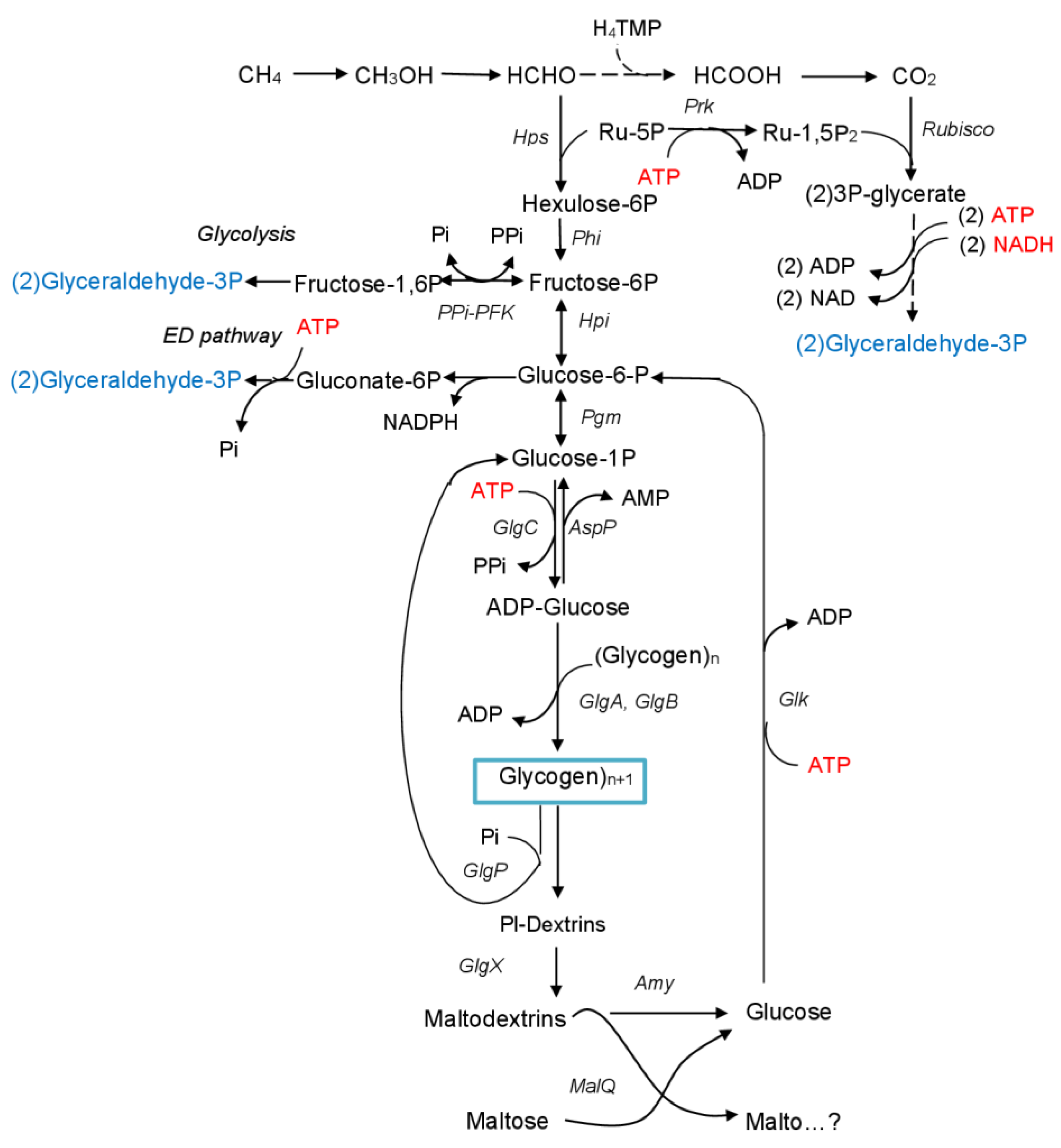
In Mc. capsulatus MIR, the glgA1 gene is located in a cluster containing the glgB gene, encoding the putative 1,4-α-glucan branching protein (GlgB), and the glgC gene, encoding putative ADP-glucose pyrophosphorylase (GlgC, EC 2.7.7.27). GlgC catalyzes the formation of glycogen precursor ADP-glucose from glucose-1-phosphate and ATP, releasing PPi as another product. This cluster includes the malQ gene encoding 4-α-glucanotransferase (MalQ, EC 2.4.1.25) (Figure 2), which is known to preferentially remove free glucose from the reducing end of maltose (or small maltodextrins). The cluster also contains aspP gene, coding for the homologue of ADP-sugar pyrophosphatase (AspP, E.C. 3.6.1.21), known to catalyze the hydrolytic breakdown of ADP-glucose to AMP and glucose-1-phosphate [23,39,40]. In Mc. capsulatus MIR, the glgA2-like gene clusters together with amy gene that encodes α-amylase (Figure 2).

3.2. Construction of Mutant Strains and Phenotypic Characterization

Deletion mutagenesis was successfully applied to obtain ΔglgA1 and ΔglgA2 single mutants, as well as ΔglgA1ΔglgA2 double mutant. Previously, the chromosomal deletion of glk genes in Mm. alcaliphilum 20Z resulted in a significant reduction in glycogen content [41]. Therefore, the glucokinase-deficient strain of Mc. capsulatus MIR was also generated.
In the early stationary phase, cells of wild-type (WT) strain MIR grown in the standard medium (with an initial KNO3 concentration of 1 g/L) contained approximately 1.5% glycogen of DCW (Table 2). Under nitrogen-limited conditions, the WT strain accumulated an order of magnitude more glycogen compared to the standard conditions while its protein content decreased. On the contrary, the biomass of ΔglgA1ΔglgA2 strain demonstrated similar protein content regardless of the growth conditions (Table 2).
The ΔglgA1 and ΔglgA2 single mutants accumulated nearly the same amounts of glycogen as compared to WT strain, but exhibited slightly slower growth rates (Table 2; Figure 2). The protein levels in the dry biomass of the two single mutants were ~85% of those in the WT strain, which suggests that both glycogen synthases were functional and interchangeable. These data also imply that a partial defect in glycogen synthesis in the single glgA mutants triggers the rearrangement of metabolism, possibly directing carbon to the synthesis of other intermediates. A double gene deletion mutant, ΔglgA1ΔglgA2, showed a growth rate nearly equal to that of the WT strain. Both ΔglgA1ΔglgA2 and glucokinase-deficient Δglk strain exhibited only negligible levels of glycogen while maintaining protein contents comparable to that in WT strain. However, the Δglk strain displayed a slightly higher growth rate than the WT strain (Figure 3). Intracellular glucose concentration in Δglk strain cells was two-fold higher than in the WT strain and in the mutants lacking glycogen synthases.

3.3. Cell Ultrastructure of WT and Mutant Strains under Nitrogen-Limited and Nitrogen-Sufficient Growth Conditions

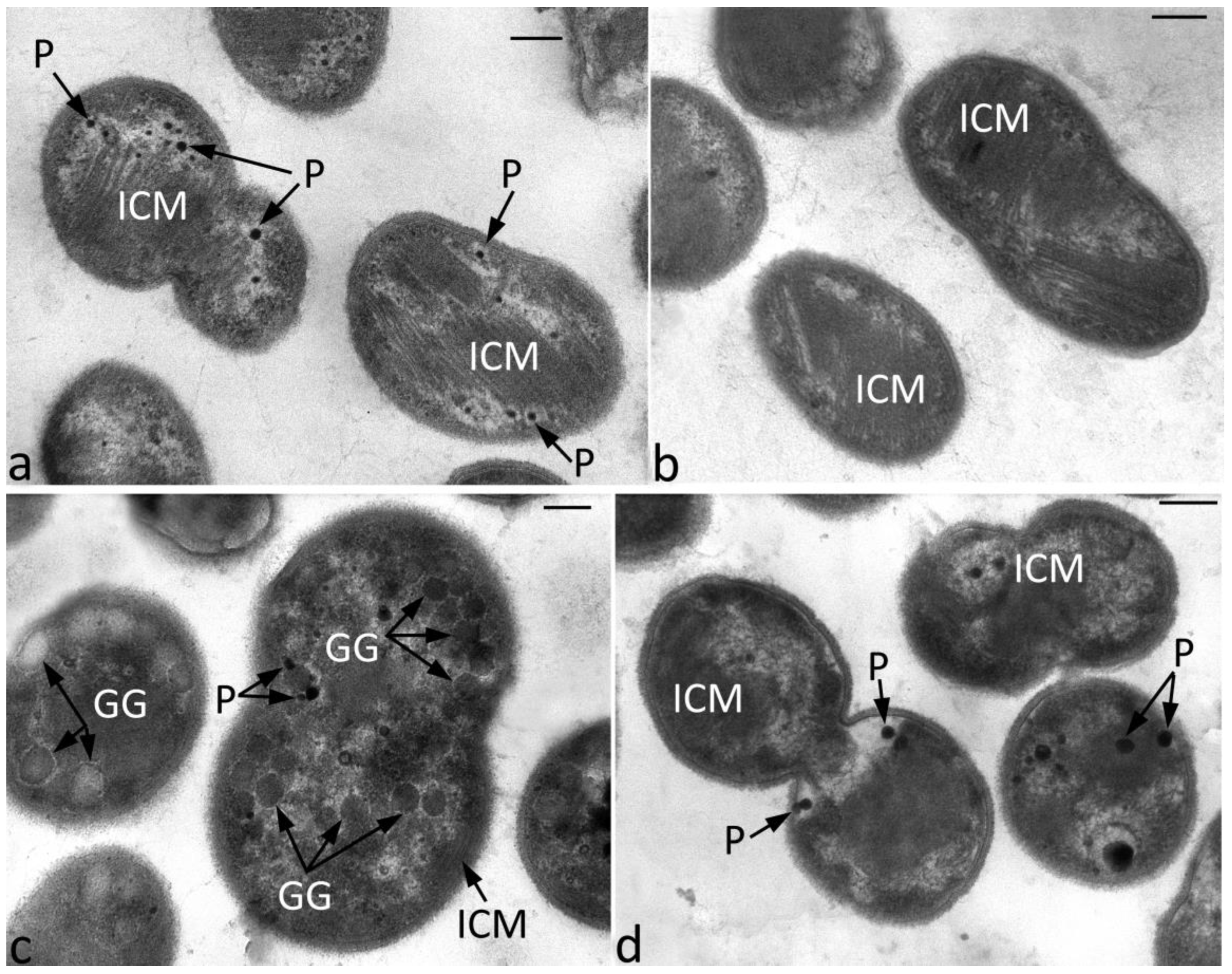
The cell ultrastructure of the WT strain and the ΔglgA1glgA2 mutant was examined using transmission electron microscopy. Under nitrogen-sufficient conditions, cells of these strains exhibited similar ultrastructure and contained numerous stacks of intracytoplasmic membranes (Figure 4a,b). Cells of the WT strain grown under nitrogen limitation were filled with numerous glycogen granules (Figure 4c). By contrast, no glycogen granules were observed in electron micrographs of thin sections prepared with cells of strain ΔglgA1ΔglgA2 grown under nitrogen-limited conditions (Figure 4d).

3.4. Real-Time PCR Analysis of the Genes Responsible for Glycogen Synthesis

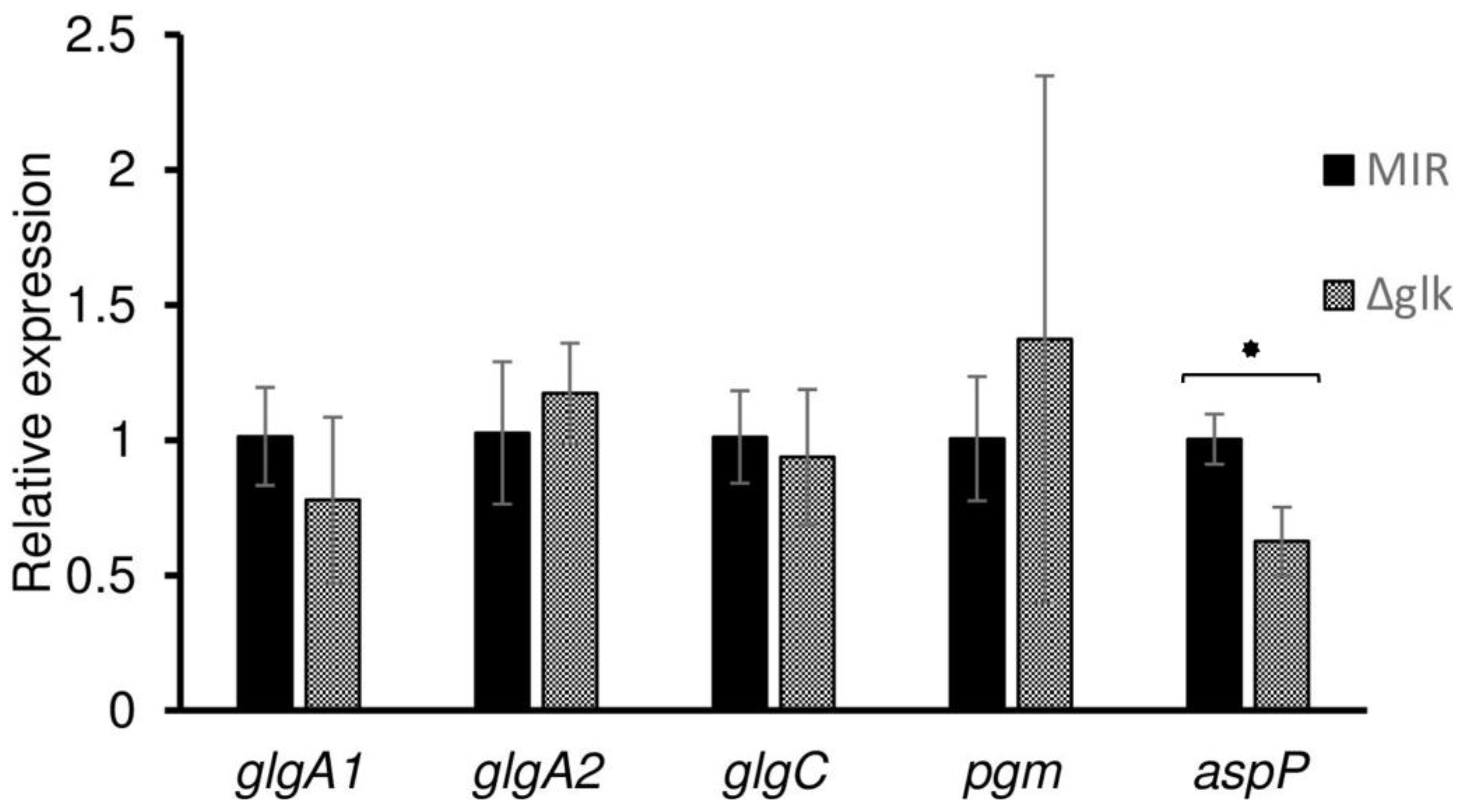
The observed decrease in glycogen content in Δglk mutant cells could be attributed to a drop in the expression of genes directly involved in the polymer synthesis (i.e., glycogen synthases) or genes whose products supply precursors (e.g., ADP-glucose pyrophosphorylase, GlgC; phosphoglucomutase, Pgm). Real-time PCR was employed to assess the expression of these genes. However, experimental results did not confirm this assumption, as the expression levels of glgA1, glgA2, glgC, and pgm genes showed no significant variation between the WT strain MIR and the Δglk mutant (Figure 5). The transcript level of AspP, which catalyzes the hydrolysis of ADP-glucose to AMP and glucose-1-phosphate, thus preventing glycogen biosynthesis, was not enhanced. In E. coli cells, the activity of AspP was inversely correlated with the intracellular glycogen content and the glucose concentration in the culture medium [23].

3.5. Growth Experiments in a Bioreactor

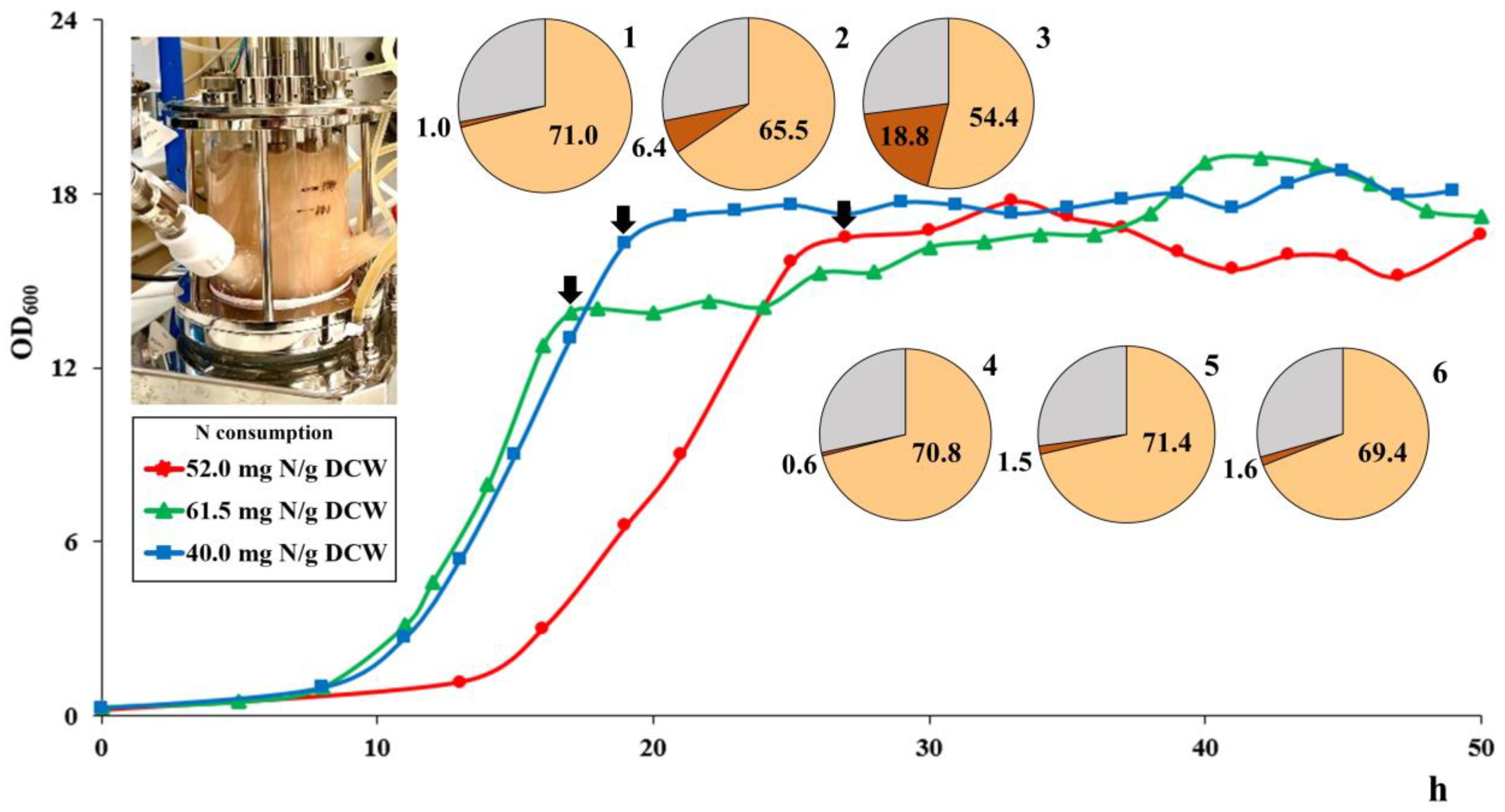
Growth of the WT, ΔglgA1glgA2, and Δglk strains was also examined in a bioreactor operated in batch and continuous modes (Figure 6).
During batch cultivation, the specific growth rate of WT strain MIR was 0.27 h−1, while this parameter reached 0.29 h−1 for the ΔglgA1ΔglgA2 and Δglk mutants. Notably, the lag phase was nearly twice as long for the WT strain. The highest biomass yield observed in batch cultures of the WT, ΔglgA1ΔglgA2, and Δglk strains constituted 4.12, 4.72, and 3.15 g DCW L−1, respectively. During continuous cultivation, all strains achieved an almost identical biomass concentration of 3.4–3.5 g DCW L−1 at a dilution rate of 0.23–0.25 h−1. Culture aliquots were sampled at the early stationary phase before switching to continuous mode and at the steady state of continuous growth. Protein content in the dry biomass of the WT strain increased from 54% to 71% upon reaching steady-state conditions in a continuous culture. Under the same operating conditions, the protein content remained constant at 71% during both batch and continuous growth of strain ΔglgA1ΔglgA2. In batch and continuous cultures, cells of the Δglk strain contained 66% and 63% of protein, respectively. Specific uptake rates of N by the batch cultures of mutant strains were 61.5 (ΔglgA1ΔglgA2) and 40.0 (Δglk) mg/g DCW, while the corresponding value determined for WT strain was 52.0 mg/g DCW. Batch culture of WT accumulated a large amount of glycogen (187.5 mg/g DCW) compared to 10.8 mg/g DCW in ΔglgA1ΔglgA2 cells and 60.4 mg/g DCW in Δglk cells. Glycogen content in the biomass of continuously grown WT, ΔglgA1ΔglgA2, and Δglk strains constituted 1.6, 0.6, and 4.6 mg/g DCW, respectively.

4. Discussion

As concluded in specialized assessment studies, SCP produced from methane has a high potential as a resilient food source for global catastrophic food shocks [2]. The product would be affordable at an expected retail cost between US$3–5/kg dry. The technology of SCP production requires three main inputs: (1) methane, which acts as both a carbon source and an electron donor, (2) a nitrogen source, and (3) an oxygen source. Additionally, some minerals are also needed in smaller quantities. As concluded in the analysis by Garcia-Martinez and co-authors [2], most or all of the methane required to fulfill the global protein requirements could be sourced exclusively from a combination of biogas and natural gas associated with oil which is currently being flared or reinjected. Other types of natural gas reserves can also be exploited. Ammonia is used as the source of nitrogen. Notably, an imbalance of C and N availability leads to the accumulation of glycogen in methanotrophic cells which, in turn, results in a loss of protein content and affects the biomass quality as a feed source. A low oxygen-to-methane ratio in the bioreactor can also result in excess carbon flow, potentially leading to glycogen accumulation in the cells. In such cases, employing glycogen-deficient mutants may serve as a strategy to uphold the high quality of SCP by preventing losses in both biomass production rates and protein content.
In this study, thermotolerant methanotroph Mc. capsulatus MIR synthesized glycogen when cultivated in a low-nitrogen medium, which aligns with its role as a carbon and energy storage compound. Even the biomass produced by this methanotroph under nitrogen-sufficient conditions contained small amounts of glycogen (~1.5% of dry cell weight) (Table 2). Apparently, due to the low glycogen content, blocking its synthesis did not have an impact on protein synthesis in strain ΔglgA1ΔglgA2 under nitrogen excess. In addition, a restructuring of carbohydrate metabolism cannot be ruled out, specifically the redirection of carbon flow towards the synthesis of other sugars in response to the knockout of the glgA genes. Some examples of such alterations in carbohydrate metabolism have been described previously. Thus, glycogen synthase null mutant obtained from Synechococcus sp. PCC7002 synthesized no glycogen but instead produced more soluble sugars excreted into the growth medium via membrane vesicles [42]. Mc. capsulatus VSB-874 synthesized large amounts of glycogen (27% of DCW) when cultivated under nutrient excess conditions and excreted exopolysaccharide consisting mostly of glucose and galactose during growth at oxygen or nitrogen limitation [43]. Intracellular polyglucose synthesis was hypothesized as an additional mechanism for formaldehyde binding [43].
These data suggest that glycogen in Methylococcus bacteria may serve both as an intermediate metabolite and a long-term storage compound. Recent studies demonstrated that glycogen can be simultaneously synthesized and degraded during bacterial growth [44]. In bioreactor experiments involving WT and ΔglgA1ΔglgA2 strains, a profound difference in glycogen accumulation was observed only during batch cultivation. Glycogen content in biomass of continuously grown ΔglgA1ΔglgA2 mutant was lower only by 1%, which correlated with a slight increase in protein content compared to the WT strain. The mutant strain required more nitrogen for growth during the batch phase, suggesting an increased demand for protein synthesis. Interestingly, the WT strain exhibited a longer lag phase than the glycogen-deficient strain during growth in the bioreactor. This was also observed in flask cultivations. The shorter lag phase demonstrated by glycogen-deficient strain offers a reduction in production cycle time, which is particularly beneficial for bioprocesses utilizing batch, fed-batch, and fill-and-draw cultivation modes for SCP production from methane or natural gas.
Genome analysis showed that the glycogen biosynthesis pathway in Mc. capsulatus MIR is generally similar to that in heterotrophic bacteria (Figure 7). The first product of formaldehyde assimilation in the RuMP cycle, fructose-6-phosphate, is transformed into glycogen via glucose-6-phosphate, glucose-1-phosphate, and ADP-glucose as intermediates. This conversion proceeds via sequential reactions catalyzed by hexose phosphate isomerase (Hpi), phosphoglucomutase (Pgm), ADP-glucose pyrophosphorylase (GlgC), glycogen synthases (GlgA1 and GlgA2), and branching enzyme (GlgB). Glycogen catabolism can be controlled by glycogen phosphorylase (GlgP) and debranching enzyme (GlgX).
Further metabolism of glycogen molecules involves α-amylase (Amy) and 4-α-glucanotransferase (MalQ). MalQ is known for its role in removing free glucose from the reducing ends of maltose (or small maltodextrins) and transferring the remaining enzyme-bound dextrinyl residue to other maltodextrins [45,46]. Thus, the breakdown of glycogen results in the formation of free glucose. Glucokinase (Glk) activates glucose by converting it to glucose-6-phosphate that can enter the central metabolic pathways or the new glycogen biosynthesis cycle. Glycogen elongation from glucose-6-phosphate, along with the recapture of this phosphosugar with the participation of Glk, may represent a cycle where two molecules of ATP are transformed into two molecules of ADP and one molecule of PPi.
ADP-sugar pyrophosphatase (AspP) hydrolyzes ADP-glucose to AMP and glucose-1-phosphate, thus preventing glycogen synthesis. The simultaneous activity of GlgC and AspP can create a futile cycle that results in the dissipation of energy stored in ATP. The futile ATP consumption seems to be a reasonable strategy to adapt to nitrogen-limited environments by many bacteria [47,48].
As revealed in our study, the genes encoding glycogen biosynthesis are widely distributed among methanotrophs (Figure 1). Phylogenetic analysis demonstrated that the glgA genes from evolutionarily distant methanotrophs formed separate clades, suggesting the vertical inheritance of these genes. Gammaproteobacterial methanotrophs, as well as methanotrophic representatives of the Verrucomicrobia possess glgA1 genes encoding a bacterial-type glycogen synthase GlgA1. The additional (starch-type) glycogen synthase, GlgA2, is found in some gammaproteobacterial methanotrophs, predominantly in those with halo- and thermotolerant phenotypes (Figure 1).
Inhibition of glycogen synthesis in Mc. capsulatus MIR was achieved by simultaneous inactivation of both glycogen synthases. An intriguing finding of our studies was the inhibition of glycogen synthesis by mutating glucokinase in two methanotrophs (see also [41]). Mm. alcaliphilum strain Δglk exhibited slower growth compared to the WT strain, while Mc. capsulatus strain Δglk grew faster than the WT strain. We believe that the main function of GIK is the removal of intracellular glucose formed during the degradation of glycogen through the mal system. Free glucose may have an inhibitory effect on the process of glycogen degradation. In E. coli, glucose at concentrations above 0.1 mM was found to block the activity of amylomaltase (MalQ), an enzyme essential for the breakdown of glycogen [49]. In addition, all mal genes are thought to be under the positive control of the activator protein MalT [49]. It has been shown that in E. coli glucokinase per se is a regulatory protein, forming a complex with MalT. The latter becomes inactive with respect to the initiation of transcription of mal genes. Thus, we do not exclude that free glucose may act as a negative regulator of activity or expression of enzymes involved in glycogen biosynthesis and degradation in the methanotroph. However, quantitative real-time PCR did not reveal significant changes in the transcription levels of the glgA1, glgA2, glgC, pgm, or aspP genes in cells of the WT and Δglk mutant of Mc. capsulatus MIR. Therefore, further studies are required to analyze the effect of glucose on the properties of enzymes involved in glycogen biosynthesis. Differences in the pathways of carbohydrate metabolism may specify different responses upon glk elimination in two methanotrophs. In addition to glucokinase, NAD-glucose dehydrogenase (GDH) participates in removing free glucose in cells of strain 20Z, whereas Mc. capsulatus MIR lacks genes for GDH [33]. The halotolerant Mm. alcaliphilum 20Z accumulated more glycogen than strain MIR [33]. Obviously, this reflects the distinct nitrogen requirements of these bacteria given that Mm. alcaliphilum 20Z synthesizes the nitrogen-containing osmoprotector ectoine to maintain the water balance of the cytoplasm [18].
In summary, we demonstrated that the construction of glycogen-deficient mutants of Methylococcus species opens new avenues for the production of single-cell protein from methane. Single deletions of glgA genes did not significantly impact growth rate and glycogen content in Mc. capsulatus MIR, thus suggesting functionality and interchangeability of these two GlgA isoenzymes. Simultaneous inactivation of both glgA genes or disruption of the sole glk gene resulted in a glycogen-deficient phenotype. Cell biomass of ΔglgA1ΔglgA2 mutant obtained during batch cultivation in a bioreactor displayed high protein content (71% DCW compared to 54% DCW in wild-type strain) as well as a strong (18-fold) reduction in glycogen content. The difference in protein and glycogen contents in biomass of these strains produced during continuous cultivation was less pronounced, yet biomass characteristics relevant to SCP production were slightly better for ΔglgA1ΔglgA2 mutant. Further experiments under controlled conditions are necessary to assess the potential for large-scale applications of these modified methanotrophic strains.

Author Contributions

Conceptualization, S.Y.B. and O.N.R.; resources, I.I.M. and N.V.P.; investigation, S.Y.B., O.N.R., R.Z.S. and I.Y.O.; data curation, S.Y.B. and O.N.R.; writing—original draft preparation, V.N.K., S.Y.B., O.N.R. and I.Y.O.; writing—review and editing, V.N.K. and S.N.D.; funding acquisition, S.N.D. and N.V.P. All authors have read and agreed to the published version of the manuscript.

Funding

This study was supported by a grant for the Development of genomic editing technologies for innovation in industrial biotechnology (grant no. 075-15-2021-1071) from the Ministry of Science and Higher Education of the Russian Federation.

Institutional Review Board Statement

Not applicable.

Informed Consent Statement

Not applicable.

Data Availability Statement

The raw data and material of the present study are available upon request.

Acknowledgments

The authors thank Natalia E. Suzina for the help with electron microscopy studies.

Conflicts of Interest

The authors declare no conflict of interest.

References

  1. Ritala, A.; Häkkinen, S.T.; Toivari, M.; Wiebe, M.G. Single Cell Protein-State-of-the-Art, Industrial Landscape and Patents 2001–2016. Front. Microbiol. 2017, 8, 2009. [Google Scholar] [CrossRef] [PubMed]
  2. García Martínez, J.B.; Pearce, J.M.; Throup, J.; Cates, J.; Lackner, M.; Denkenberger, D.C. Methane Single Cell Protein: Potential to Secure a Global Protein Supply Against Catastrophic Food Shocks. Front. Bioeng. Biotechnol. 2022, 10, 906704. [Google Scholar] [CrossRef] [PubMed]
  3. Matassa, S.; Papirio, S.; Pikaar, I.; Hülsen, T.; Leijenhorst, E.; Esposito, G.; Pirozzi, F.; Verstraete, W. Upcycling of Biowaste Carbon and Nutrients in Line with Consumer Confidence: The “Full Gas” Route to Single Cell Protein. Green Chem. 2020, 22, 4912–4929. [Google Scholar] [CrossRef]
  4. Risso, C.; Choudhary, S.; Johannessen, A.; Silverman, J. Methanotrophy Goes Commercial: Challenges, Opportunities, and Brief History BT. In Methane Biocatalysis: Paving the Way to Sustainability; Kalyuzhnaya, M.G., Xing, X.-H., Eds.; Springer International Publishing: Cham, Switzerland, 2018; pp. 293–298. ISBN 978-3-319-74866-5. [Google Scholar]
  5. Henard, C.A.; Smith, H.; Dowe, N.; Kalyuzhnaya, M.G.; Pienkos, P.T.; Guarnieri, M.T. Bioconversion of Methane to Lactate by an Obligate Methanotrophic Bacterium. Sci. Rep. 2016, 6, 21585. [Google Scholar] [CrossRef]
  6. Strong, P.J.; Kalyuzhnaya, M.; Silverman, J.; Clarke, W.P. A Methanotroph-Based Biorefinery: Potential Scenarios for Generating Multiple Products from a Single Fermentation. Bioresour. Technol. 2016, 215, 314–323. [Google Scholar] [CrossRef] [PubMed]
  7. Strong, P.J.; Xie, S.; Clarke, W.P. Methane as a Resource: Can the Methanotrophs Add Value? Environ. Sci. Technol. 2015, 49, 4001–4018. [Google Scholar] [CrossRef]
  8. Le, H.T.Q.; Lee, E.Y. Methanotrophs: Metabolic Versatility from Utilization of Methane to Multi-Carbon Sources and Perspectives on Current and Future Applications. Bioresour. Technol. 2023, 384, 129296. [Google Scholar] [CrossRef]
  9. Mühlemeier, I.M.; Speight, R.; Strong, P.J. Biogas, Bioreactors and Bacterial Methane Oxidation BT. In Methane Biocatalysis: Paving the Way to Sustainability; Kalyuzhnaya, M.G., Xing, X.-H., Eds.; Springer International Publishing: Cham, Switzerland, 2018; pp. 213–235. ISBN 978-3-319-74866-5. [Google Scholar]
  10. Hanson, R.; Hanson, T. Methanotrophic Bacteria. Microbiol. Rev. 1996, 60, 439–471. [Google Scholar] [CrossRef]
  11. Hakemian, A.S.; Rosenzweig, A.C. The Biochemistry of Methane Oxidation. Annu. Rev. Biochem. 2007, 76, 223–241. [Google Scholar] [CrossRef]
  12. Rosenzweig, A.C. Biochemistry: Breaking Methane. Nature 2015, 518, 309–310. [Google Scholar] [CrossRef]
  13. Hamer, G.; Harrison, D.E.F. Single Cell Protein: The Technology, Economics and Future Potential. In Hydrocarbons in Biotechnology; Harrison, D.E.F., Higgins, I.J., Watkinson, W., Eds.; Heyden: London, UK, 1980; pp. 59–73. ISBN 9789811305016. [Google Scholar]
  14. Zhivotchenko, A.G.; Nikonova, E.S.; Jørgensen, M.H. Copper Effect on the Growth Kinetics of Methylococcus capsulatus (Bath). Biotechnol Technol. 1995, 9, 163–168. [Google Scholar] [CrossRef]
  15. Øverland, M.; Tauson, A.H.; Shearer, K.; Skrede, A. Evaluation of Methane-Utilising Bacteria Products as Feed Ingredients for Monogastric Animals. Arch. Anim. Nutr. 2010, 64, 171–189. [Google Scholar] [CrossRef] [PubMed]
  16. Nunes, J.J.; Aufderheide, B.; Ramjattan, D.M.; Dass, R. Enhanced Production of Single Cell Protein from M. capsulatus (Bath) Growing in Mixed Culture. J. Microbiol. Biotechnol. Food Sci. 2016, 6, 894–899. [Google Scholar] [CrossRef]
  17. Linton, J.D.; Cripps, R.E. The Occurrence and Identification of Intracellular Polyglucose Storage Granules in Methylococcus NCIB 11083 Grown in Chemostat Culture on Methane. Arch. Microbiol. 1978, 117, 41–48. [Google Scholar] [CrossRef]
  18. Khmelenina, V.N.; Kalyuzhnaya, M.G.; Sakharovsky, V.G.; Snzina, N.E.; Trotsenko, Y.A.; Gottschalk, G. Osmoadaptation in Halophilic and Alkaliphilic Methanotrophs. Arch. Microbiol. 1999, 172, 321–329. [Google Scholar] [CrossRef] [PubMed]
  19. But, S.Y.; Dedysh, S.N.; Popov, V.O.; Pimenov, N.V.; Khmelenina, V.N. Construction of a Type-I Metanotroph with Reduced Capacity for Glycogen and Sucrose Accumulation. Appl. Biochem. Microbiol. 2020, 56, 538–543. [Google Scholar] [CrossRef]
  20. Ballicora, M.A.; Iglesias, A.A.; Preiss, J. ADP-Glucose Pyrophosphorylase, a Regulatory Enzyme for Bacterial Glycogen Synthesis. Microbiol. Mol. Biol. Rev. 2003, 67, 213–225. [Google Scholar] [CrossRef]
  21. Preiss, J.; Romeo, T. Molecular Biology and Regulatory Aspects of Glycogen Biosynthesis in Bacteria. Prog. Nucleic Acid Res. Mol. Biol. 1994, 47, 299–329. [Google Scholar] [CrossRef]
  22. Morán-Zorzano, M.T.; Alonso-Casajús, N.; Muñoz, F.J.; Viale, A.M.; Baroja-Fernández, E.; Eydallin, G.; Pozueta-Romero, J. Occurrence of More than One Important Source of ADPglucose Linked to Glycogen Biosynthesis in Escherichia Coli and Salmonella. FEBS Lett. 2007, 581, 4423–4429. [Google Scholar] [CrossRef]
  23. Morán-Zorzano, M.T.; Viale, A.M.; Muñoz, F.J.; Alonso-Casajús, N.; Eydallín, G.G.; Zugasti, B.; Baroja-Fernández, E.; Pozueta-Romero, J. Escherichia Coli AspP Activity Is Enhanced by Macromolecular Crowding and by Both Glucose-1,6-Bisphosphate and Nucleotide-Sugars. FEBS Lett. 2007, 581, 1035–1040. [Google Scholar] [CrossRef]
  24. Eydallin, G.; Morán-Zorzano, M.T.; Muñoz, F.J.; Baroja-Fernández, E.; Montero, M.; Alonso-Casajús, N.; Viale, A.M.; Pozueta-Romero, J. An Escherichia Coli Mutant Producing a Truncated Inactive Form of GlgC Synthesizes Glycogen: Further Evidences for the Occurrence of Various Important Sources of ADPglucose in Enterobacteria. FEBS Lett. 2007, 581, 4417–4422. [Google Scholar] [CrossRef] [PubMed]
  25. Liu, Y.; He, X.; Zhu, P.; Cheng, M.; Hong, Q.; Yan, X. PheSAG Based Rapid and Efficient Markerless Mutagenesis in Methylotuvimicrobium. Front. Microbiol. 2020, 11, 441. [Google Scholar] [CrossRef] [PubMed]
  26. Lee, H.M.; Ren, J.; Yu, M.S.; Kim, H.; Kim, W.Y.; Shen, J.; Yoo, S.M.; Eyun, S.I.; Na, D. Construction of a Tunable Promoter Library to Optimize Gene Expression in Methylomonas sp. DH-1, a Methanotroph, and Its Application to Cadaverine Production. Biotechnol. Biofuels 2021, 14, 228. [Google Scholar] [CrossRef] [PubMed]
  27. Kalyuzhnaya, M.G.; Kumaresan, D.; Heimann, K.; Caetano, N.S.; Visvanathan, C.; Parthiba Karthikeyan, O. Editorial: Methane: A Bioresource for Fuel and Biomolecules. Front. Environ. Sci. 2020, 8, 9. [Google Scholar] [CrossRef]
  28. Nguyen, A.D.; Hwang, I.Y.; Lee, O.K.; Kim, D.; Kalyuzhnaya, M.G.; Mariyana, R.; Hadiyati, S.; Kim, M.S.; Lee, E.Y. Systematic Metabolic Engineering of Methylomicrobium alcaliphilum 20Z for 2,3-Butanediol Production from Methane. Metab. Eng. 2018, 47, 323–333. [Google Scholar] [CrossRef] [PubMed]
  29. Nguyen, D.T.N.; Lee, O.K.; Hadiyati, S.; Affifah, A.N.; Kim, M.S.; Lee, E.Y. Metabolic Engineering of the Type I Methanotroph Methylomonas sp. DH-1 for Production of Succinate from Methane. Metab. Eng. 2019, 54, 170–179. [Google Scholar] [CrossRef] [PubMed]
  30. Oshkin, I.Y.; Suleimanov, R.Z.; Khmelenina, V.N.; Mardanov, A.V.; Pimenov, N.V.; Dedysh, S.N. Complete Genome Sequence of Methylococcus capsulatus MIR, a Methanotroph Capable of Growth on Methanol. Microbiol. Resour. Announc. 2022, 11, 97–98. [Google Scholar] [CrossRef] [PubMed]
  31. Mamiatis, T.; Fritsch, E.F.; Sambrook, J.; Engel, J. Molecular Cloning–A Laboratory Manual. New York: Cold Spring Harbor Laboratory. 1982, 545 S., 42 $. Acta Biotechnol. 1985, 5, 104. [Google Scholar] [CrossRef]
  32. Schäfer, A.; Tauch, A.; Jäger, W.; Kalinowski, J.; Thierbach, G.; Pühler, A. Small Mobilizable Multi-Purpose Cloning Vectors Derived from The. Gene 1994, 145, 69–73. [Google Scholar] [CrossRef] [PubMed]
  33. Rozova, O.N.; Ekimova, G.A.; Molochkov, N.V.; Reshetnikov, A.S.; Khmelenina, V.N.; Mustakhimov, I.I. Enzymes of an Alternative Pathway of Glucose Metabolism in Obligate Methanotrophs. Sci. Rep. 2021, 11, 8795. [Google Scholar] [CrossRef]
  34. Yee, Y.C.; Hashim, R.; Mohd Yahya, A.R.; Bustami, Y. Colorimetric Analysis of Glucose Oxidase-magnetic Cellulose Nanocrystals (CNCS) for Glucose Detection. Sensors 2019, 19, 2511. [Google Scholar] [CrossRef]
  35. Schacterle, G.R.; Pollack, R.L. A Simplified Method for the Quantitative Assay of Small Amounts of Protein in Biologic Material. Anal. Biochem. 1973, 51, 654–655. [Google Scholar] [CrossRef] [PubMed]
  36. Reynolds, E.S. The Use of Lead Citrate at High PH as an Electron-Opaque Stain in Electron Microscopy. J. Cell Biol. 1963, 17, 208–212. [Google Scholar] [CrossRef] [PubMed]
  37. Pfaffl, M.W. A New Mathematical Model for Relative Quantification in Real-Time RT–PCR. Nucleic Acids Res. 2001, 29, e45. [Google Scholar] [CrossRef]
  38. Altschul, S.F.; Gish, W.; Miller, W.; Myers, E.W.; Lipman, D.J. Basic Local Alignment Search Tool. J. Mol. Biol. 1990, 215, 403–410. [Google Scholar] [CrossRef] [PubMed]
  39. Bessman, M.J.; Frick, D.N.; O’Handley, S.F. The MutT Proteins or “Nudix” Hydrolases, a Family of Versatile, Widely Distributed, “Housecleaning” Enzymes. J. Biol. Chem. 1996, 271, 25059–25062. [Google Scholar] [CrossRef] [PubMed]
  40. Moreno-Bruna, B.; Baroja-Fernández, E.; Muñoz, F.J.; Bastarrica-Berasategui, A.; Zandueta-Criado, A.; Rodríguez-López, M.; Lasa, I.; Akazawa, T.; Pozueta-Romero, J. Adenosine Diphosphate Sugar Pyrophosphatase Prevents Glycogen Biosynthesis in Escherichia coli. Proc. Natl. Acad. Sci. USA 2001, 98, 8128–8132. [Google Scholar] [CrossRef] [PubMed]
  41. Mustakhimov, I.I.; Rozova, O.N.; Solntseva, N.P.; Khmelenina, V.N.; Reshetnikov, A.S.; Trotsenko, Y.A. The Properties and Potential Metabolic Role of Glucokinase in Halotolerant Obligate Methanotroph Methylomicrobium alcaliphilum 20Z. Antonie Van Leeuwenhoek 2017, 110, 375–386. [Google Scholar] [CrossRef] [PubMed]
  42. Xu, Y.; Tiago Guerra, L.; Li, Z.; Ludwig, M.; Charles Dismukes, G.; Bryant, D.A. Altered Carbohydrate Metabolism in Glycogen Synthase Mutants of Synechococcus sp. Strain PCC 7002: Cell Factories for Soluble Sugars. Metab. Eng. 2013, 16, 56–67. [Google Scholar] [CrossRef]
  43. Khmelenina, V.N.; Gayazov, R.R.; Suzina, N.E.; Doronina, N.V.; Mshenskii, Y.N.; Trotsenko, Y.A. The Synthesis of Polysaccharides by Methylococcus capsulatus under Various Conditions of Cultivation. Mikrobiologiya 1992, 61, 404–410. [Google Scholar]
  44. Hernández, M.A.; Alvarez, H.M. Glycogen Formation by Rhodococcus Species and the Effect of Inhibition of Lipid Biosynthesis on Glycogen Accumulation in Rhodococcus opacus PD630. FEMS Microbiol. Lett. 2010, 312, 93–99. [Google Scholar] [CrossRef]
  45. Palmer, T.N.; Wöber, G.; Whelan, W.J. The Pathway of Exogenous and Endogenous Carbohydrate Utilization in Escherichia coli: A Dual Function for the Enzymes of the Maltose Operon. Eur. J. Biochem. 1973, 39, 601–612. [Google Scholar] [CrossRef] [PubMed]
  46. Park, J.T.; Shim, J.H.; Tran, P.L.; Hong, I.H.; Yong, H.U.; Oktavina, E.F.; Nguyen, H.D.; Kim, J.W.; Lee, T.S.; Park, S.H.; et al. Role of Maltose Enzymes in Glycogen Synthesis by Escherichia coli. J. Bacteriol. 2011, 193, 2517–2526. [Google Scholar] [CrossRef] [PubMed]
  47. Russell, J.B. The Energy Spilling Reactions of Bacteria and Other Organisms. J. Mol. Microbiol. Biotechnol. 2007, 13, 1–11. [Google Scholar] [CrossRef] [PubMed]
  48. Nakajima, T.; Yoshikawa, K.; Toya, Y.; Matsuda, F.; Shimizu, H. Metabolic Flux Analysis of the Synechocystis sp. PCC 6803 ΔnrtABCD Mutant Reveals a Mechanism for Metabolic Adaptation to Nitrogen-Limited Conditions. Plant Cell Physiol. 2017, 58, 537–545. [Google Scholar] [CrossRef]
  49. Lengsfeld, C.; Schönert, S.; Dippel, R.; Boos, W. Glucose- and Glucokinase-Controlled Mal Gene Expression in Escherichia coli. J. Bacteriol. 2009, 191, 701–712. [Google Scholar] [CrossRef]
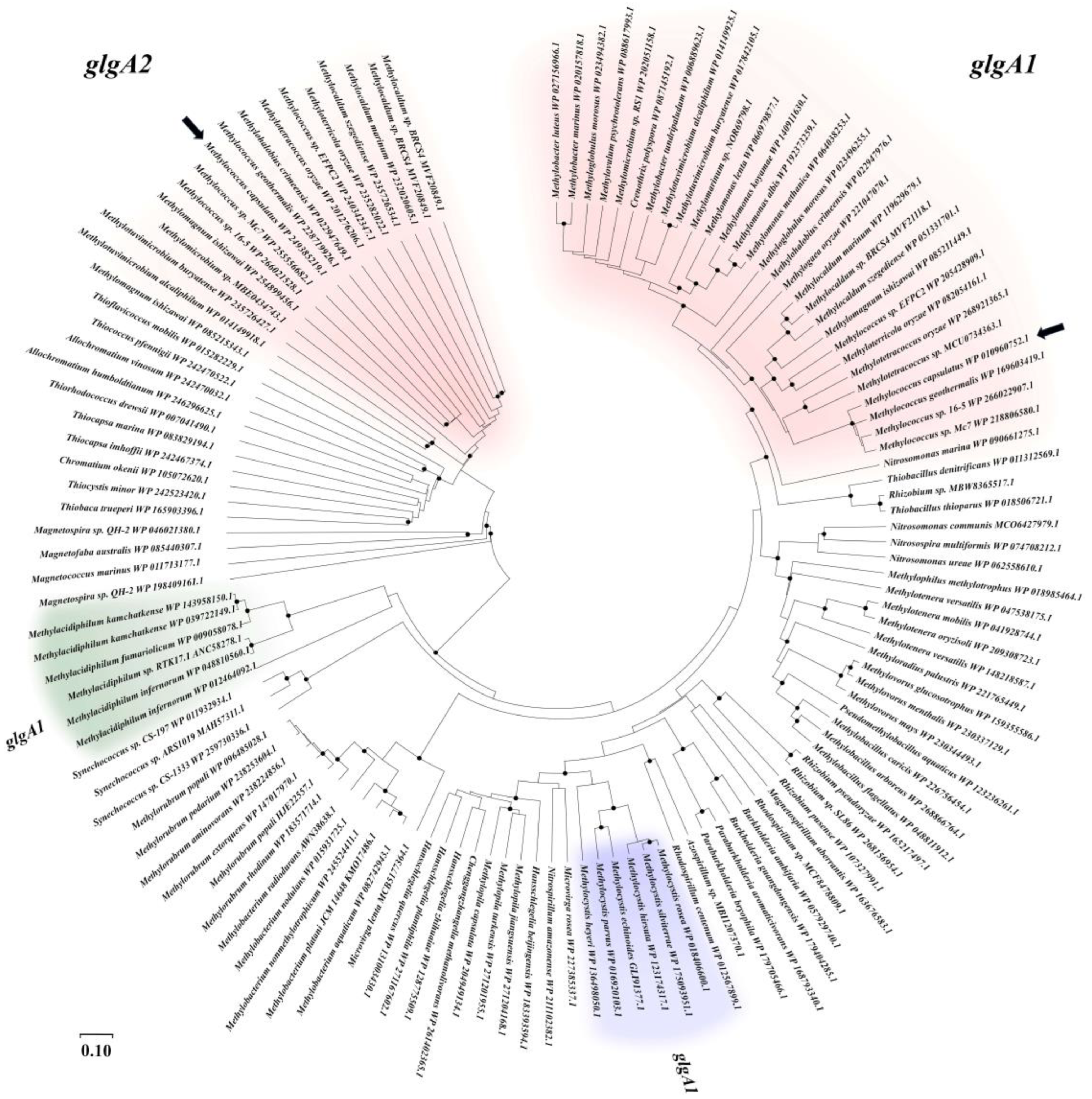
Figure 1. Unrooted phylogenetic tree of the glycogen synthase-encoding genes in aerobic methanotrophic bacteria. Methanotrophs of the Gammaproteobacteria, Alphaproteobacteria, and Verrucomicrobia are highlighted in red, blue, and green, respectively. Black arrows point to glgA1 and glgA2 genes from Mc. capsulatus MIR. Bootstrap values of >80% are shown. Marker, 0.1 substitutions per nucleotide position.
Fermentation 10 00265 g001
Figure 2. Scheme of gene clusters encoding glycogen synthesis in Mc. capsulatus MIR. Asp, gene for ADP-sugar pyrophosphatase; glgA1, glgA2, glycogen synthase genes; glgB, 1,4--α-glucan branching protein; glgC, ADP-glucopyrophosphorylase; malQ, gene for MalQ protein (4-α-glucantransferase); amy, α-amylase gene.
Fermentation 10 00265 g002
Figure 3. Growth rates of WT strain MIR and glycogen-deficient mutants under nitrogen-sufficient conditions (1 g/L KNO3).
Fermentation 10 00265 g003
Figure 4. Electron micrographs of thin cell sections of Mc. capsulatus MIR (a,c) and Δglg1Δglg2 mutant (b,d). Cells were grown in minimal medium in exponential growth phase with 1 g/L KNO3 (1, 2) or 0.29 g/L KNO3 (3, 4). GG, glycogen granules; ICM, intracytoplasmic membranes; P, inclusions of polyphosphates. Thin sections were stained with uranyl acetate/lead citrate. Bars, 200 nm.
Fermentation 10 00265 g004
Figure 5. Real-time PCR analysis of gene expression in WT strain MIR and glucokinase-deficient mutant strain (Δglk). Examined genes included glgA1 (glycogen synthase 1), glgA2 (glycogen synthase 2), glgC (ADP-glucose pyrophosphorylase), pg. (phosphoglucomutase) and aspP (ADP-sugar pyrophosphatase). The rpoB gene was used as a reference gene. Three independent experiments were carried out. Bars represent standard deviation. Result with a p-value < 0.05 is marked by an asterisk (*).
Fermentation 10 00265 g005
Figure 6. Growth dynamics of WT strain MIR (red line), ΔglgA1ΔglgA2 mutant (green line), and Δglk mutant (blue line) during growth in a bioreactor operated in batch and continuous modes. Growth curves represent three independent runs. Transition points from batch to continuous phase are indicated by black arrows. Growth was monitored by measuring OD600 values every 2 h. Pie charts show the contents of protein (beige), glycogen (brown), and other cellular components (grey) in dry biomass of ΔglgA1ΔglgA2 (1, 4), Δglk (2, 5), and WT (3, 6) strains during batch (1, 2, 3) and continuous (4, 5, 6) growth. Nitrogen consumption per g DCW for each of the three strains is shown in the frame.
Fermentation 10 00265 g006
Figure 7. Schematic pathways of glycogen metabolism in Mc. capsulatus MIR. Pgm, phosphoglucomutase; GlgA, glycogen synthase; GlgB, 4-α-glucan branching protein; AspP, ADP-sugar pyrophosphatase; GlgP, glycogen phosphorylase; GlgC, ADP glucose pyrophosphorylase; GlgX, debranching enzyme; MalQ, 4-alpha-glucanotransferase; Hps, hexulosephosphate synthase; Phi, phosphohexulose isomerase; Hpi, hexosephosphate isomerase; Prk, phosphoribulokinase; Rubisco, ribulosebisphosphate carboxylase/oxygenase; PPi-PFK, pyrophosphate dependent phosphofructokinase.
Fermentation 10 00265 g007
Table 1. Primers used in this work.
NamePrimer (5′-3′)Target
Primers for cloning
MIR glg1-up-FATATCTAGAACCGGCATTACCATCACGAHomology region (HR) upstream of glgA1 gene
MIR glg1-up-RCAAGCATGCAGGAGTGGCGGACGGTGCGA
MIR glg1-dw-FCAAGCATGCGCTGCTCCATCGCCGAC HR downstream of glgA1 gene
MIR glg1-dw-RTCAAAGCTTGCCAAGGAGATCGTGAATTA
MIR glg2-up-FAGTGAATTCGACCACGACCAGGCCGAGCAHR upstream of glgA2 gene
MIR glg2-up-RTCAGGTACCTCCAGTACCGCCGACACCTA
MIR glg2-dw-FCAAGCATGCCGCTACGACTATTCCTGGAHR downstream of glgA2 gene
MIR glg2-dw-RCTTAAGCTTTGAGCCTGGGCGTGTCGTG
20Z-glg2C-FAGTGAATTCGGAGGAGACACATGGCAAAGCAAACTACAACglgA2 gene from Mm. alcaliphilum 20Z
20Z-glg2C-RCAAGGATCCTTATTTGTTACGAATATAGTCATAGAT
GLK_MIR-FTTGGATCCTGATCGGCGGCTATCGCATglk gene
GLK_MIR-RTTCGATCGTCCTGCGGCTTTTCGTAGT
Primers for real-time PCR
MIR qrpoB-FCTGGATGCCCTGGTGGAAATrpoB gene
MIR qrpoB-RATTCTCCACCATCTCCCCCA
q-pgmMIR-FCTACGCAAGAAGGTGAAGGTTpgm gene
q-pgmMIR-RTGTTTACGGATTACGCAGGA
q-glgCMIR-FACTTCCCGCTGTCCAACTGglgC gene
q-glgCMIR-RCCAAGTTCTGATACACCGCAT
q-glgA1MIR-FTACCCCTCTGGCTGCTGGAglgA1 gene
q-glgA1MIR-RTTCGGCTCGTCGCTCAAAA
q-glgA2MIR-FGCCAAGAGCCCCCCAGTglgA2 gene
Table 2. Glycogen, glucose, and total protein contents in cells of WT and mutant strains of Mc. capsulatus MIR recorded during shake flask cultivation. nd – not determined.
Growth ConditionsStrainGlycogen, mg/g DCWGlucose, µg/g DCWProtein, mg/g DCW
Nitrogen excess
(1 g L−1 KNO3)
WT14.3 ± 3.832 ± 3496 ± 15
ΔglgA19.4 ± 2.526 ± 3420 ±22
ΔglgA217.6 ± 3.228 ± 1420 ± 17
ΔglgA1ΔglgA21.1 ± 0.435 ± 2477 ± 16
Δglk1.3 ± 0.175 ± 19498 ± 27
Nitrogen limit
(0.29 g L−1 KNO3)
WT202 ± 54nd297 ± 63
ΔglgA161.0 ± 9nd510 ± 24
ΔglgA221.0 ± 3nd511 ± 52
ΔglgA1ΔglgA23.0 ± 0.4nd532 ± 13
Δglk142.0 ± 5nd482 ± 36
Disclaimer/Publisher’s Note: The statements, opinions and data contained in all publications are solely those of the individual author(s) and contributor(s) and not of MDPI and/or the editor(s). MDPI and/or the editor(s) disclaim responsibility for any injury to people or property resulting from any ideas, methods, instructions or products referred to in the content.