Article

The Impact of Lactobacillus delbrueckii Hepatic Metabolism in Post-Weaning Piglets

1
College of Animal Science and Technology, Hunan Agricultural University, Changsha 410128, China
2
College of Animal Science and Technology, Hunan Biological and Electromechanical Polytechnic, Changsha 410128, China
*
Authors to whom correspondence should be addressed.
Fermentation 2024, 10(6), 286; https://doi.org/10.3390/fermentation10060286
Submission received: 30 April 2024 / Revised: 22 May 2024 / Accepted: 22 May 2024 / Published: 28 May 2024
(This article belongs to the Special Issue Application of Fermentation Technology in Animal Nutrition)

Abstract

:
Lactobacillus delbrueckii garners interest for its contributions to gut microecological balance, diarrheal prevention and treatment, immune modulation, growth promotion, and meat quality enhancement in livestock. However, its impact on the gut microbiota and liver metabolism in weaned piglets is less documented. This study involved 80 Duroc-Landrace-Yorkshire weaned piglets aged 28 days, randomized into two groups with four replicates each and ten piglets per replicate. Over a 28-day period, the piglets were fed either a basal diet (control group) or the same diet supplemented with 0.1% Lactobacillus delbrueckii microcapsules (≥1.0 × 10¹⁰ CFU/g) (Lactobacillus delbrueckii group). The principal findings are as follows: During the initial phase of the experiment, supplementation with Lactobacillus delbrueckii increased the levels of L-phenylalanine and L-lysine in the liver while reducing the L-alanine levels, thereby enhancing the aminoacyl–tRNA synthesis pathway in weaned piglets. In the later phase, Lactobacillus delbrueckii supplementation boosted the liver arachidonic acid content, strengthening the arachidonic acid metabolic pathway in the piglets. The gut microbiota and their metabolites likely play a role in regulating these processes. These results indicate that, compared to the control group, Lactobacillus delbrueckii reduced weaning stress-induced liver damage and metabolic disorders, increased liver glycogen content, and enhanced liver antioxidant function by improving the metabolism of lipids and carbohydrates. Consequently, the liver functioned more healthily.

1. Introduction

Lactobacillus delbrueckii was first discovered and studied in 1901 and was subsequently named after the renowned bacteriologist M. Delbruck [1]. Lactobacillus delbrueckii, one of the most extensively utilized lactobacilli, encompasses the following three subspecies: Lactobacillus delbrueckii subsp. bulgaricus, Lactobacillus delbrueckii subsp. lactis, and Lactobacillus delbrueckii subsp. delbrueckii; it belongs to the facultative anaerobic Gram-positive bacteria. It can be sourced from a diverse range of origins, including human and animal oral cavities and intestines, as well as dairy products. Numerous scholars have demonstrated that Lactobacillus delbrueckii plays a pivotal role in regulating intestinal microecological balance [2,3,4], immune response modulation [5], and antioxidant activity enhancement [6], thereby facilitating healthy intestinal development while preventing diarrhea occurrence [7,8]. Moreover, it also contributes to growth promotion and improvements in meat quality. Consequently, this probiotic exhibits immense potential with significant advantages over antibiotics [2,4,6,7,8,9,10,11,12].
Weaning is a critical event in pig production, involving significant changes in feeding, management, and the environment. The abrupt transition from predominantly breast milk to solid feed often leads to reduced daily energy and nutrient intake by piglets [13]. Moreover, piglets experience stress due to separation from sows and alterations in the piggery environment, which can further elevate their stress levels. Dietary modifications may disrupt the balance of the gastrointestinal microbiota and increase susceptibility to intestinal pathogen colonization [14]. Additionally, weaning induces substantial morphological and functional changes in the small intestine, such as villi atrophy and crypt proliferation, that result in decreased digestion and absorption capacity [15,16,17,18]. Probiotics offer a potential alternative for enhancing the health and productivity of weaned piglets.
Lactobacillus delbrueckii is a probiotic strain with superior characteristics. Our group’s prior research indicates its ability to colonize the animal gut, modulate microecological balance, enhance gut immunity, facilitate nutrient absorption, regulate metabolism, and counteract oxidative stress, thus improving animal productivity. However, the mechanisms by which it affects the gut microbiota to regulate liver metabolism and enhance productivity are not yet clear.
The weaning stress experienced by piglets results in a sudden decline in feed intake, necessitating the increased consumption of fat, sugar, and protein to meet the body’s metabolic demands [19]. The liver, as a vital organ involved in energy supply and nutrient metabolism regulation, plays a crucial role in animal growth, metabolism, anti-stress capacity, and overall health. Weaning stress can reduce the glycogen content in piglet livers; however, the liver can compensate for this by enhancing gluconeogenesis and inhibiting glycolysis function to maintain glucose metabolic balance [20,21]. Lactobacillus delbrueckii has been found to modulate intestinal bile acid absorption and liver cholesterol metabolic enzyme activity while also influencing the gut microbiota composition. Consequently, it can regulate liver health and metabolism in weaned piglets [22]. In our preliminary experiment with Lactobacillus delbrueckii supplementation on weaned piglets’ serum indexes related to liver function—alanine aminotransferase and aspartate aminotransferase—we observed certain effects. To further investigate the mechanism underlying these effects on the nutritional metabolism of weaned piglets supplemented with Lactobacillus delbrueckii, we conducted a PAS staining analysis to observe the changes in liver morphology and glycogen content, along with a non-targeted full spectrum analysis of liver metabolites using liquid chromatogram–mass spectrometry (LC-MS).
This experiment employs metabolomics to study the impact of Lactobacillus delbrueckii on the liver metabolism of weaned piglets. The objective is to elucidate how Lactobacillus delbrueckii influences liver metabolism changes in weaned piglets, explore the mechanisms by which it affects their productive performance, and provide crucial reference data and theoretical support for the application of Lactobacillus delbrueckii formulations in antibiotic-free diets and production practices for weaned piglets.

2. Materials and Methods

2.1. Testing Materials and Sources

The protocol of this study was approved by the Institutional Animal Care and Use Committee of the College of Animal Science and Technology, Hunan Agricultural University (Changsha, China), and was conducted in accordance with the National Institutes of Health (Changsha, China) guidelines for the care and use of experimental animals (No. 2017-09). The strain utilized in the experiment was Lactobacillus delbrueckii, which was acquired from the Laboratory of Animal Science at Hunan Agricultural University and authenticated and preserved by the Strain Preservation Center at Wuhan University (storage number M207096). Hunan Perfly Biotechnology Co., Ltd. (Changsha, China) was entrusted with preparing a microencapsulated granule formulation of Lactobacillus delbrueckii (vital cell counting ≥1.0 × 10¹⁰ CFU/g).

2.2. Design of Experiments and Formulation of Dietary Composition

The experiment was approved by the Experimental Animal Welfare Ethics Committee of Hunan Agricultural University (Changsha, China). A single-factor design was employed in this study. Eighty 28-day-old Duroc×Landrace×Yorkshire trihybrid weaned piglets (half male and half female) with similar parity, body weight (approximately 7.5 kg), and good health were selected and randomly divided into two groups with four replicates per group and ten piglets per replicate. The duration of the experiment was 28 days. The control group received a basal diet, while the Lactobacillus delbrueckii group received a basal diet supplemented with 0.1% Lactobacillus delbrueckii (vital cell counting ≥1.0 × 10⁷ CFU/g feed). The basic diet consisted of powder without any antibiotics, and its nutritional level followed the NRC (2012) standard for nutritional requirements (Table 1).

2.3. Management of Animal Experiments

The experiment was conducted at Taiping Pig farm in Changsha County, Hunan Province, under the supervision of designated personnel. All pigs were provided with unrestricted access to food and water and immunized following the conventional protocols of the pig farm. Feeding occurred three times daily (at 8:00, 12:00, and 18:00) using a dry mix diet that ensured no residual material remained in the troughs. Huts were cleaned twice daily to maintain cleanliness standards. Natural ventilation was employed throughout the entire hut. Daily records were maintained for pig feeding patterns, the occurrence of diarrhea, disease incidence, and medication administration.

2.4. Detection Indices and Methodologies

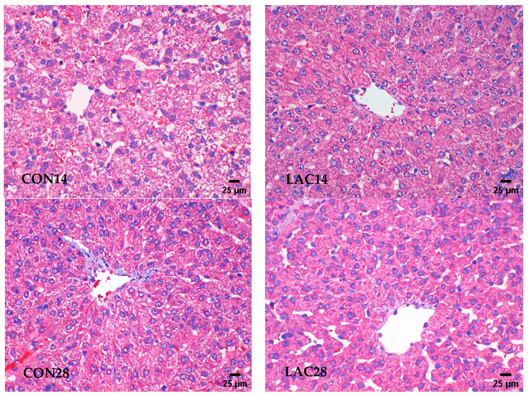
2.4.1. The Liver Tissue Was Examined Using Hematoxylin and Eosin (HE) Staining

On the 14 d and 28 d of the experiment, piglets were slaughtered under anesthesia after venous blood collection, and part of the liver was fixed in 4% paraformaldehyde with the size of about 1 cm3 and divided into two pieces for section preparation. The steps of hematoxylin–eosin (HE) staining after a paraffin section of liver tissue are as follows: bake the slices at 60 °C for 1–2 h; after dewaxing to water, dye with hematoxylin for 5–10 min, rinse with distilled water, return to blue with PBS, slice into eosin dye solution for 3–5 min, and rinse with distilled water; and dehydrate the slices successively with gradient alcohol (95–100%) for 5 min per stage. After removal, the liver was placed in xylene for 10 min, sealed with neutral gum twice, examined by a microscope, and photographed to observe the histological changes.

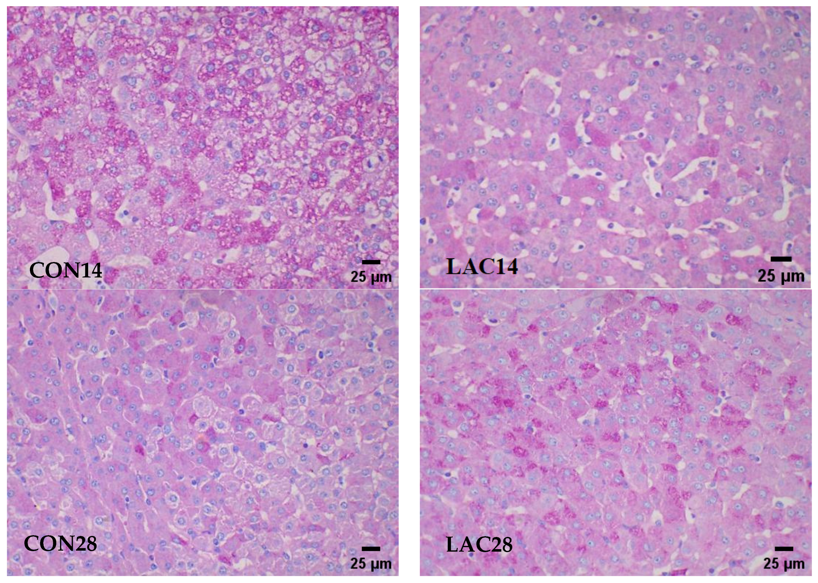
2.4.2. Revealing Hepatic Glycogen Accumulation through PAS Staining in Liver Tissue

To guarantee the integrity of the data and the reliability of the experimental outcomes, adherence to the established protocols for sample collection and processing was of paramount importance. As outlined in Section 2.4.1, these procedures were meticulously followed to the letter. This section provides a detailed explanation of the steps for sample collection, required materials, environmental conditions, and precautions to maintain the quality of the samples. Following paraffin sectioning, the liver tissue was subjected to periodic acid-Schiff (PAS) staining using the following protocol: The sections were baked at 60 °C for 30–60 min. Dewaxing of the slices was performed by immersing them in xylene twice for 10 min each time, followed by sequential immersion in 100%, 95%, 85%, and 75% ethanol for 5 min at each stage. Subsequently, the sections were soaked in distilled water for an additional 5 min. To initiate the PAS reaction, a quick application of 50 μL periodic acid onto the tissue was carried out, allowing it to stand for a duration of 10 min. After rinsing with tap water for another period of ten minutes, Schiff’s solution was applied to facilitate dye binding during a subsequent incubation step lasting ten minutes. The sections were then thoroughly rinsed with water until no further color change occurred. Hematoxylin staining (for nuclear counterstaining) was performed by immersing the sections in a hematoxylin solution for a duration ranging from five to ten minutes, subsequently returning them to blue using phosphate-buffered saline (PBS), followed by drying with a hair dryer. Gradual dehydration through graded alcohols (from low concentration to high) ensued before two rounds of immersion in xylene for ten-minute intervals each time took place prior to sealing with neutral gum and microscopic examination. The evaluation revealed that PAS-positive structures exhibited purplish-red coloration accompanied by blue nuclei.

2.4.3. Comprehensive Analysis of Hepatic Metabolites across the Entire Spectrum

After administering the anesthesia and conducting the slaughter procedure, venous blood samples were collected from piglets on days 14 and 28 of the trial. Subsequently, a portion of the liver was swiftly excised using surgical scissors and transferred into a diethyl pyrocarbonate (DEPC)-treated Eppendorf tube. To ensure sample integrity, the liver specimen was pre-cooled with liquid nitrogen and stored at −80 °C for comprehensive spectrum analysis aimed at investigating liver metabolites.
For metabolomics determination, the following procedure was employed:
(a) Sample pretreatment: The liver sample was slowly thawed on ice, and then 50 mg of freeze-dried sample was placed in a 1.5 mL centrifuge tube containing 800 μL of 80% methanol. The mixture was ground for 90 s at 60 Hz and thoroughly mixed using vortex oscillations. Subsequently, ultrasound treatment was performed at 4 °C for 30 min followed by standing at −40 °C for 1 h, vortexing for an additional 30 s, and standing again at 4 °C for another half an hour. Next, centrifugation was carried out at 4 °C and 12,000 rpm for 15 min. All the supernatant in the centrifuge tube was collected and allowed to stand at −40 °C for 1 hour before undergoing another round of centrifugation (4 °C and 12,000 rpm; 15 min). Finally, 200 μL of supernatant was withdrawn from the resulting solution and combined with the dichlorophenylalanine internal standard (140 μg/mL; 5 μL) in a vial.
(b) Instrumental analysis conditions: Chromatographic conditions: The chromatographic column used was a Hyper gold C18 liquid chromatography column, maintained at a temperature of 40 °C, with a sample size of 4 μL. For the positive ion mode test, the mobile phase consisted of water +5% acetonitrile +0.1% formic acid (A) and acetonitrile +0.1% formic acid (B). For the negative ion mode tests, water +5% acetonitrile +0.05% acetic acid (A) and acetonitrile +0.05% acetic acid (B) were employed as the mobile phases. The elution gradient is presented in Table 2.
Mass spectrum detection parameters for positive mode were set as follows: heater temperature at 300 °C; sheath gas flow rate at 45 arb; auxiliary gas flow rate at 15 arb; tail gas flow rate at 1 arb; electric spray voltage at 3.0 KV; capillary temperature at 350 °C; and S-Lens RF Level set to 30%.
For the negative mode, mass spectrum detection parameters included heater temperature set to 300 °C; sheath gas flow rate adjusted to 45 arb; auxiliary gas flow rate maintained at 15 arb; tail gas flow rate kept constant at 1 arb; electric spray voltage set to 3.2 KV; capillary temperature held steady at 350 °C; and S-Lens RF Level adjusted to 60%.
Scanning was performed using a primary full scan from m/z 70 to 1050 and a data-dependent secondary mass spectrometry scan (dd-MS2, TopN = 10); resolution settings were configured as 70,000 (primary mass spectrometry) and 17,500 (secondary mass spectrometry). Collision mode utilized high-energy collision dissociation (HCD).
(c) Data extraction: The Compound Discoverer software 3.0 (Thermo Company, Waltham, MA, USA) was utilized for the extraction and pre-processing of LC/MS detection data, which were subsequently organized into a two-dimensional data matrix format. This matrix encompasses essential information such as retention time (RT), molecular weight, observed quantity (sample name), and peak intensity, among others.

2.5. Revealing the Identification of Metabolic Pathways

Enrichment analysis of metabolite differences was performed using the KEGG function and Metaboanalyst (http://www.metaboanalyst.ca/ (accessed on 29 April 2024)), based on the Bos Taurus pathway enrichment library. The significance of enrichment pathways was determined by evaluating the P-value and influence factor, with pathways having a P < 0.05 or influence factor ≥1.0 considered key metabolic pathways for differential metabolite enrichment.

2.6. Data Processing and Analysis Procedures

The principal component analysis (PCA) and partial least squares discriminant analysis (PLS-DA) were conducted using SIMCA-P+ software 14.1.0. The univariate statistical analysis (UVA) model was employed to screen the variables, where a Student’s t-test with a p-value less than 0.05 and a variable importance in the projection (VIP) greater than 1 for the first principal component of the PLS-DA model were considered significant screening conditions.

3. Results and Analysis

3.1. The Impact of Lactobacillus delbrueckii on Hepatic Morphology in Post-Weaning Piglets

The liver histopathology observed through HE staining is presented in Figure 1. On day 14 of the experiment, the control group exhibited slight hepatic damage, characterized by well-organized hepatocyte cords with a few nuclei displaying dissolution and shrinkage, the absence of noticeable inflammatory cell infiltration, and an increased presence of lipid vacuoles. In contrast, the Lactobacillus delbrueckii group displayed clear liver architecture with a radial arrangement of hepatocytes around the central vein and distinct hepatocyte cords. A minimal number of liver nuclei showed signs of dissolution and contraction without any evident inflammatory cell infiltration or lipid vacuole accumulation. By day 28 of the experiment, there was significant improvement in the liver morphology for the control group compared to day 14, as evidenced by a well-defined arrangement of hepatocyte cords, with only a small fraction showing dissolution and shrinkage along with reduced inflammatory cell infiltration and diminished lipid vacuoles. The liver structure in the Lactobacillus delbrueckii group remained clear, resembling that observed on day 14.

3.2. Impact of Lactobacillus delbrueckii on Hepatic Glycogen Content in Post-Weaning Piglets

The morphological observation of PAS glycogen staining in the liver is presented in Figure 2.
On the 14th day of the experiment, the control group exhibited a higher abundance of liver glycogen, characterized by glycogen aggregation and fat vacuole accumulation. In contrast, the Lactobacillus delbrueckii group displayed a sporadic distribution of liver glycogen with reduced levels.
By day 28 of the experiment, the liver glycogen content decreased in the control group, along with a significant reduction in fat vacuoles. Conversely, there was a substantial increase in the liver glycogen content within the Lactobacillus delbrueckii group, distributed as granules and focal points.

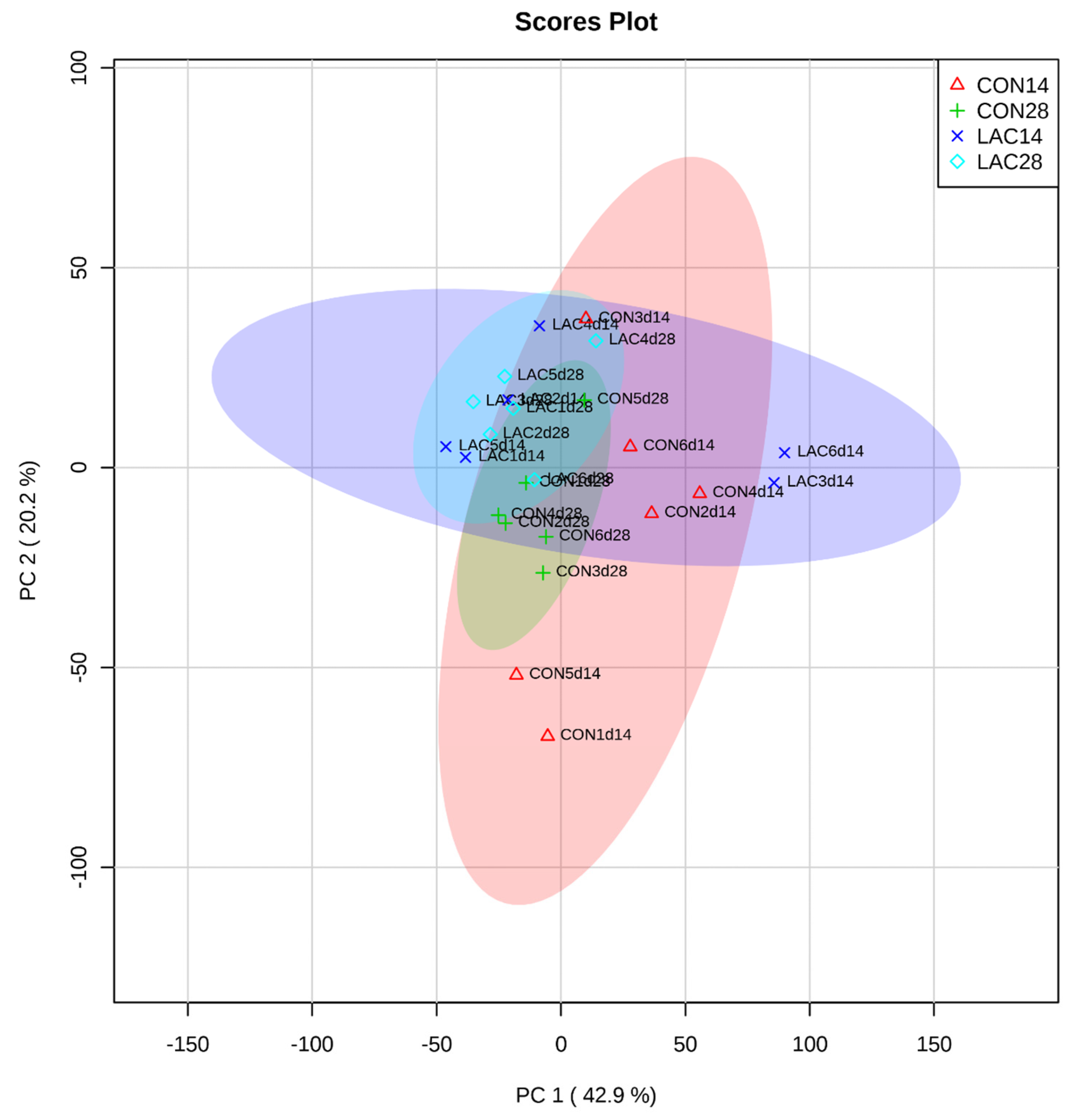
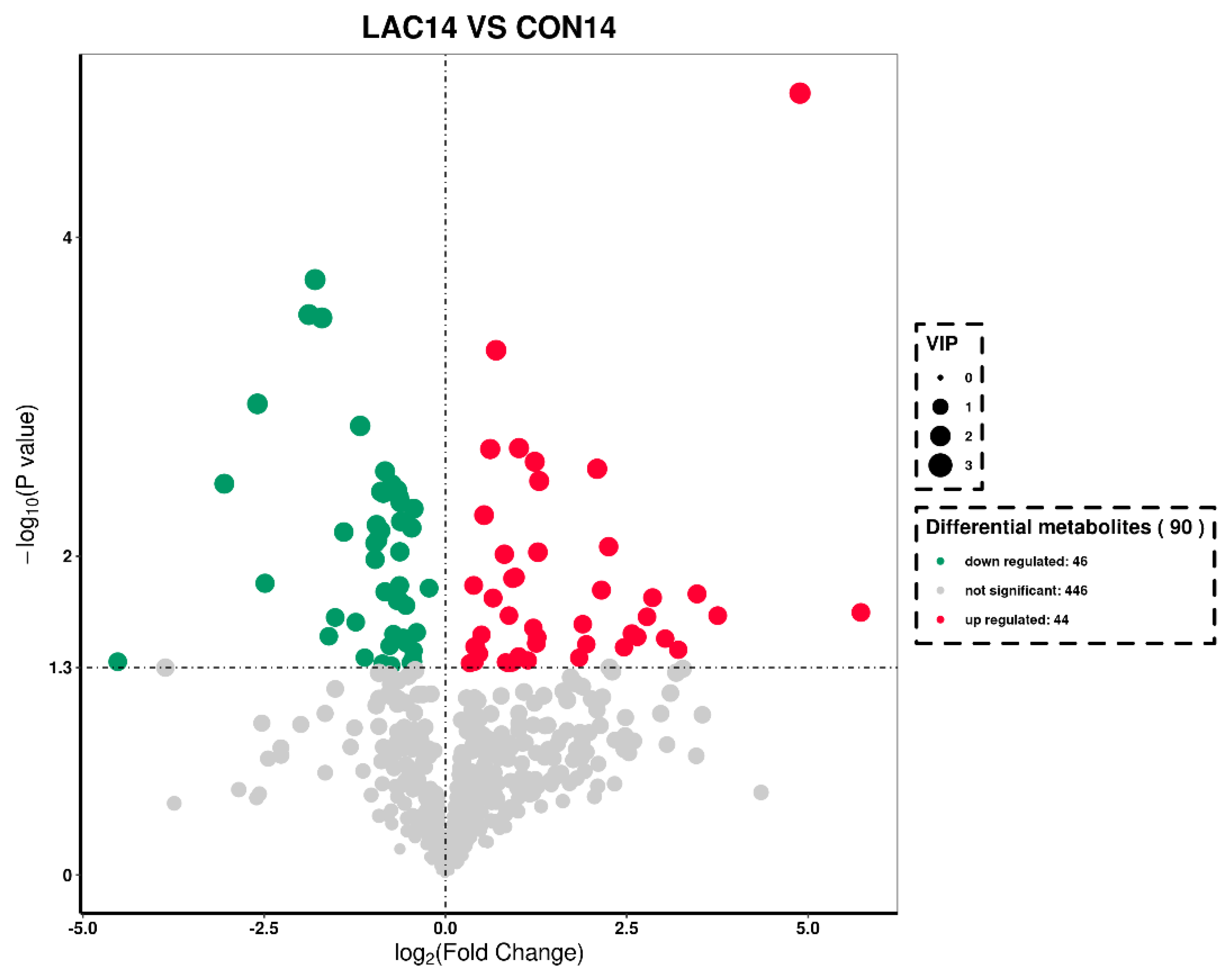
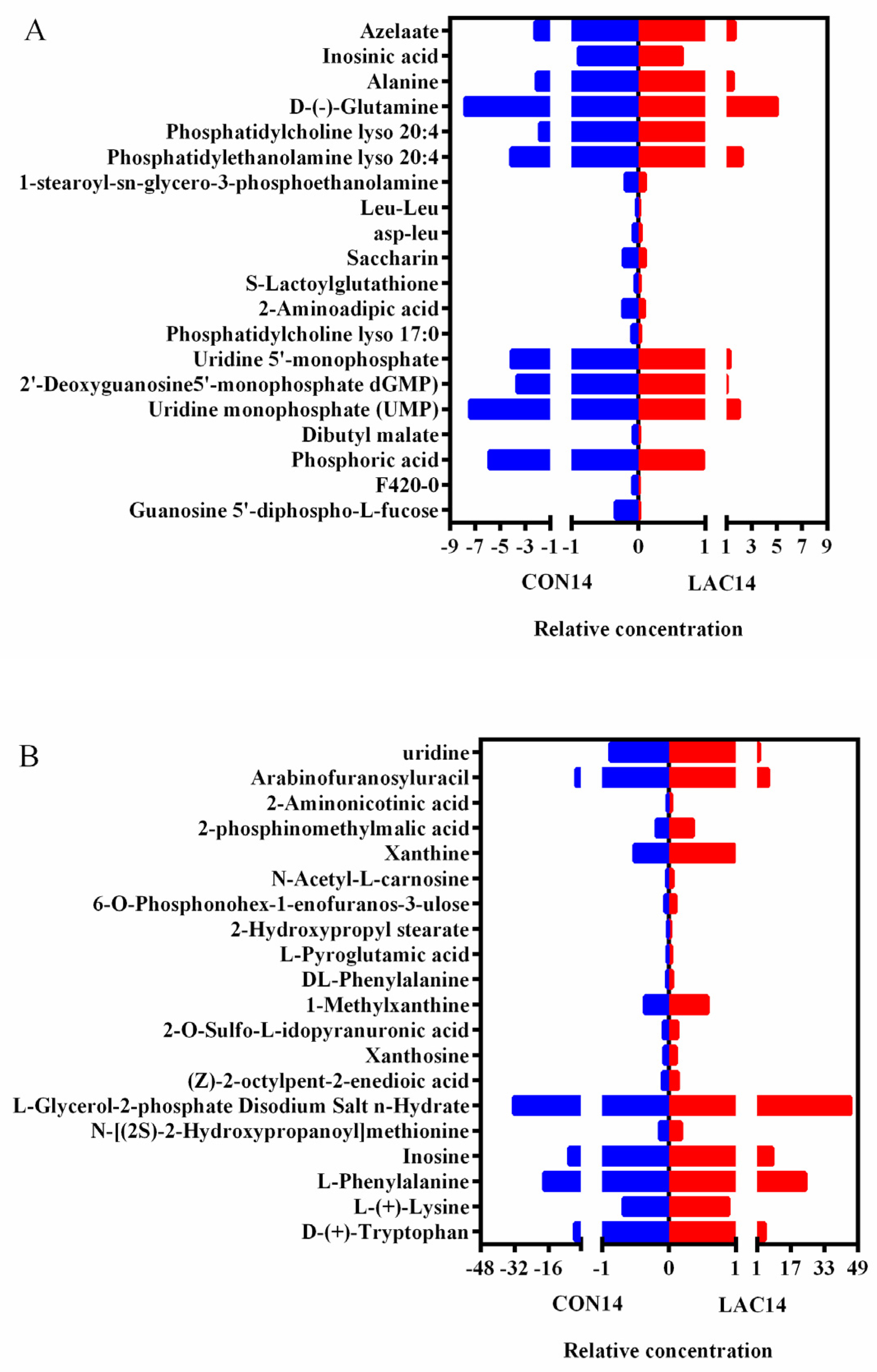
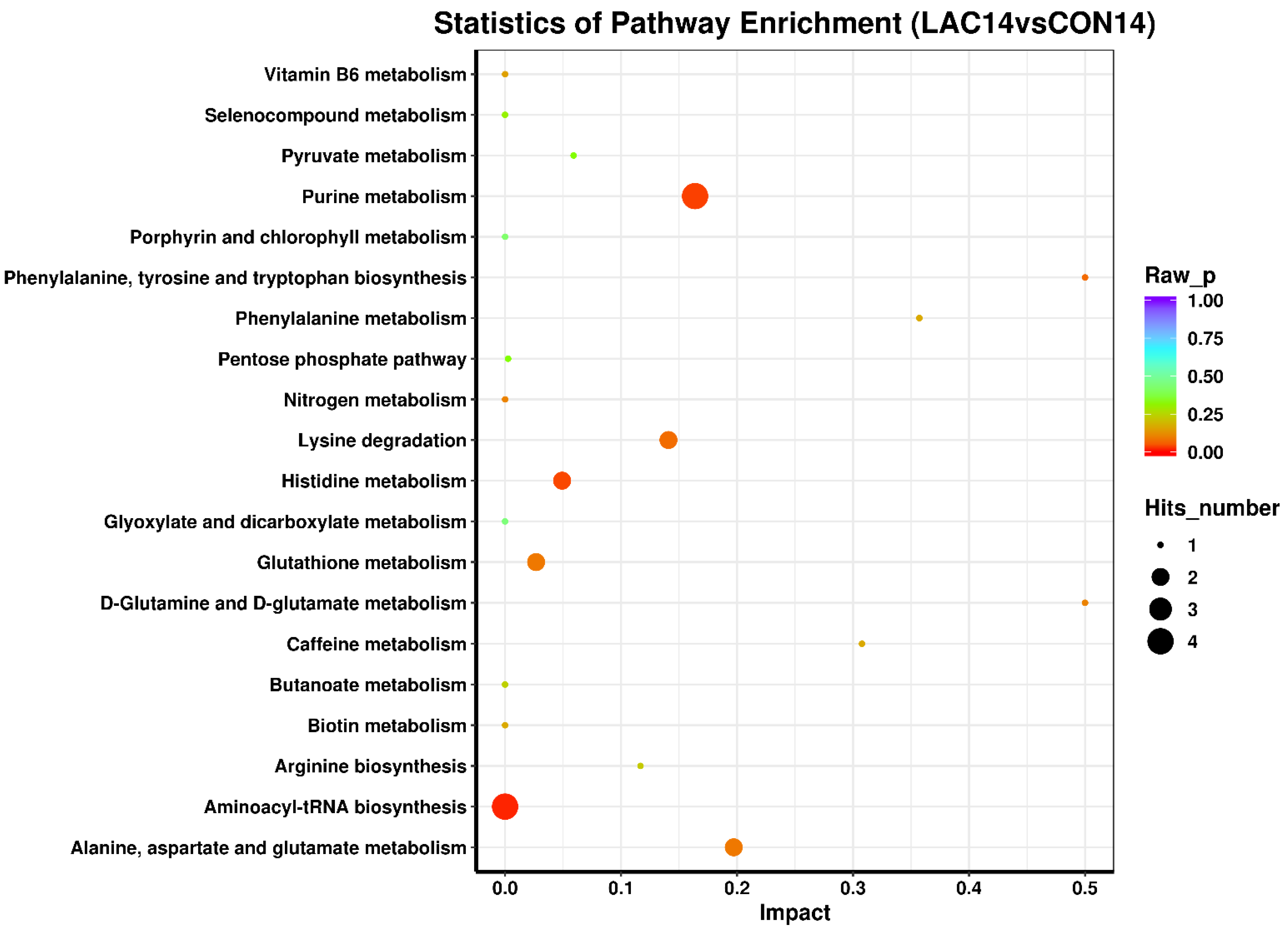
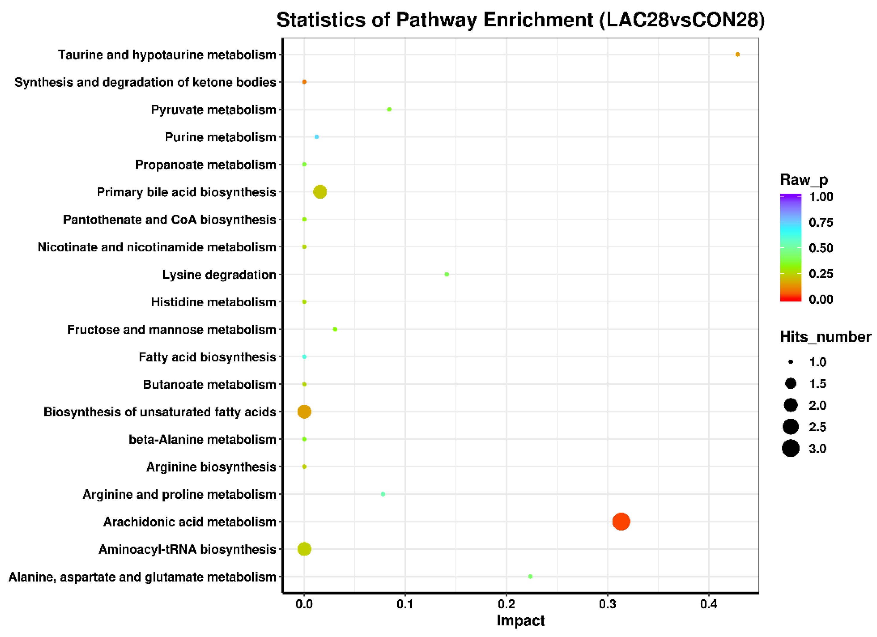
3.3. The Impact of Lactobacillus delbrueckii on Hepatic Metabolism in Post-Weaning Piglets

Observations from the principal component analysis (PCA) graph in Figure 3 reveal an interesting phenomenon. On days 14 and 28 of the experiment, the liver metabolite profiles within the control group appeared consistent, without significant differentiation. In contrast, the Lactobacillus de brueckii group displayed a distinct trend, with clear separation of the liver metabolites on days 14 and 28, suggesting that the addition of Lactobacillus de brueckii may have modulated the liver metabolic activity. This variance indicates that Lactobacillus de brueckii could have a positive effect on liver function, particularly in terms of metabolic regulation.
According to the volcano plot in Figure 4, under a positive ion mode, we observed the differential metabolites identified in the liver of weaned piglets. By day 14 of the experiment, compared to the control group, the Lactobacillus delbrueckii group identified 90 known differential metabolites. Specifically, 44 of these metabolites showed an increase in the relative concentration, while the relative concentration of the other 46 metabolites decreased. This distribution indicates that the addition of Lactobacillus delbrueckii significantly impacted the metabolites in the piglets’ liver, altering not only the types of metabolites but also their concentration levels, which may have significant implications for the health and growth development of the piglets.

3.4. Impact of Lactobacillus delbrueckii on Hepatic Glycogen Content in Post-Weaning Piglets

According to the fold change ranking, the top 20 differential metabolites that are upregulated and downregulated are shown in Figure 5. Compared with the control group, Guanosine 5’-diphospho-L-fucose, F420-0, Phosphoric acid, Dibutyl malate, and guanosine 5’ -diphospho-l-fucose in the liver of the Lactobacillus deli group, Uridine monophosphate (UMP), 2-deoxyguanosine 5’-monophosphate (dGMP), Uridine 5’-monophosphate, Phosphatidylcholine lyso 17:0, 2-Aminoadipic acid (2-Aminoadipic acid), S-Lactoylglutathione (S-Lactoylglutathione), Saccharin, aspartate leucine (asp-leu), leu-leu, Alanine, Inosinic acid 1-stearoyl-sn-glycero-3-phosphoethanolamine, D-glutamine (D-(-)-Glutamine), and Phosphatidylethanolamine lyso 20:4. The relative concentrations of Phosphatidylcholine lyso 20:4 and Azelaate were significantly decreased (VIP ≥ 1.0 and P < 0.05).
Compared to the control group, the liver of the Lactobacillus delsoni group exhibited detectable levels of L-tryptophan (D-(+)Tryptophan), L-lysine (L-(+) Lysine), L-phenylalanine, Inosine, and Xanthosine. Additionally, N-[(2S)-2-hydroxypropanoyl]methionine, DL-Phenylalanine, L-Glycerol-2-phosphate Disodium Salt n-Hydrate, (Z)-2-octylpent-2-enedioic acid, (Z)-2-octylp-2-enendioic acid, Sulfo-l-idopyranuronic acid, and 2-O-Sulfo-L-idopyranuronic acid were detected. Furthermore, an increase in the relative concentrations of 1-Methylxanthine, L-Pyroglutamic acid, 6-O-phosphonohexa-1-enofuranos-3-ulose, N-Acetyl-L-carnosine(N-acetyl-L-Carnosine), Xanthine, and 6-O-phosphonohexa -1-enofuranos-3-ulose was observed, along with significantly elevated levels of 2-Aminonicotinic acid and Arabinofuranosyluracil (VIP ≥ 1.0 and P < 0.05).
The differential metabolite enrichment analysis of KEGG results is visualized using a bubble plot. As depicted in Figure 6, on day 14 of the experiment, the key metabolic pathways were enriched with differential metabolites, including Aminoacyl tRNA biosynthesis, purine metabolism, histidine metabolism, phenylalanine, tyrosine and tryptophan biosynthesis, lysine degradation, glutathione metabolism, and other metabolic pathways. Additionally, the identified pathways are alanine, aspartate, and glutamate metabolism; nitrogen metabolism; D-glutamine and D-glutamate metabolism; vitamin B6 metabolism; biotin metabolism; caffeine metabolism; phenylalanine metabolism; arginine biosynthesis; butyrate metabolism; selenium compound metabolism; pentose phosphate pathway; pyruvate metabolism; porphyrin and chlorophyll metabolism; glyoxylic acid and dicarboxylic acid metabolism; arginine and proline metabolism; and pyrimidine metabolism. The supplementation of Lactobacillus delbrueckii significantly enhanced the Aminoacyl tRNA biosynthesis pathway on day 14 of weaning piglets, with significant increases observed in intermediate products such as L-phenylalanine and L-lysine (P < 0.05).
The volcanic map in Figure 7 clearly shows the metabolite differences between the Lactobacillus delbrueckii group and the control group on day 28 of the trial. The data showed that a total of 121 known differential metabolites were identified in the Lactobacillus delbrueckii group. Among these different metabolites, the relative concentration of 73 showed an increasing trend, while the relative concentration of the other 48 metabolites showed a decreasing trend. This result further confirms the significant influence of Lactobacillus delbrueckii on the liver metabolic activity of piglets, suggesting that this strain may influence the physiological status and health of piglets by regulating the concentration of specific metabolites. This difference in metabolic regulation may have important biological implications for the growth and development of piglets.
The top 20 differential metabolites upregulated and downregulated are shown in Figure 8. Compared with the control group, the liver Phosphatidylethanolamine lyso 20:4, Phosphatidylcholine lyso 16:1, Cyclohexanecarboxylate, Myristoleic acid, Phosphatidylcholine lyso, L-Aspartic acid, 4-Methylene L-glutamic acid, L-(+)-lactic acid, 1-stearoyl-SN-glycero-3-phosphoethanolamine, D-Ribulose-5-phosphate sodium salt, 2-Acetamido-2-deoxy-D-glucono-1,5-lactone, phosphoenol (1-O-Phosphonopentitol), glycine phenylalanine (Gly-Phe) and the relative concentrations of Leucylproline and 2-Acetamido-2-deoxy-D-glucono-1,5-lactone were significantly decreased (VIP ≥ 1.0 and P < 0.05). Compared with the control group, the Lactobacillus delbrueckii group had higher levels of Oleic acid, Ethyl myristate, Lauric acid, Arachidonic acid, and ethyl myristate in the liver. gamma-Glutamylleucine, Reduced Glutathione, L-Proline, DL-TYROSINE, DL-4-Hydroxyphenyllactic acid, Docosapentaenoic acid, Taurine, N-Isovalerylglycine, Xanthine, 2-O-Sulfo-L-idopyranuronic acid, (10S)-Juvenile hormone III diol, GLUTATHIONE, 3-Hydroxybutyric acid, and N-arachidonoylglycine significantly increased the relative concentrations of S-(Formylmethyl) glutathione and 11,12-Epoxy-(5Z,8Z,11Z)-icosatrienoic acid (VIP ≥ 1.0 and P < 0.05).
The enrichment analysis of differential metabolites in the KEGG results is visualized using a bubble plot. As depicted in Figure 9, on the 28th day of the experiment, the significant metabolic pathways associated with differential metabolite enrichment include arachidonic acid metabolism, ketone body synthesis and degradation, taurine and hypotaurine metabolism, unsaturated fatty acid biosynthesis, primary bile acid biosynthesis, metabolic pathways, Aminoacyl tRNA biosynthesis, arginine biosynthesis, butyrate metabolism, niacin and niacinamide metabolism, histidine metabolism, pantothenic acid and Coenzyme A biosynthesis, fructose and mannose metabolism, beta-alanine metabolism, pyruvate metabolism, propionic acid metabolism, alanine, aspartic acid and glutamate metabolism, arginine and proline metabolism, fatty acid biosynthesis, and purine metabolism. The supplementation of Lactobacillus delbrueckii significantly enhanced the arachidonic acid metabolic pathway on the 28th day of the weaning experiment by significantly increasing the levels of intermediate products such as arachidonic acid and eicosatrienoic acid within this pathway (P < 0.05).

4. Discussion

Lipopolysaccharide (LPS) and enterotoxic Escherichia coli are commonly employed to establish immune or enterotoxic stress models in piglets. Previous studies have demonstrated that stimulation by LPS or enterotoxic Escherichia coli can induce intestinal immune stress and enteritis, subsequently impacting liver energy supply deficiency, liver damage, and metabolic dysfunction through the “enterohepatic axis” [23,24]. Numerous investigations have indicated that lipopolysaccharide-induced immune stress can alter the hepatic morphological structure of piglets [25,26]. Oxidative stress can trigger inflammatory responses and apoptosis, accelerate cellular oxidative damage, and enhance triglyceride (TG) accumulation within the liver, leading to lipid deposition [27,28]. Intestinal probiotics such as lactic acid bacteria and their metabolites modulate host the NF-κB signaling pathway or the Nrf2/ARE signaling pathway through various mechanisms to alleviate host immune stress and oxidative stress [29,30,31]. Studies suggest that Lactobacillus delbrueckii may initially ameliorate immunity and oxidative stress in weaned piglets by regulating immune antibodies, enzymes (GSH-Px, HO-1), or cytokines (8-OHdG) in intestinal tissues, thereby mitigating liver damage [6,32].
This study revealed that piglets in the control group exhibited mild hepatic morphological damage and increased liver fat content on day 14, indicating impaired hepatic lipid metabolism, possibly due to weaning stress. However, by day 28 of the experiment, hepatic morphology had recovered and the effects of weaning stress had been mitigated. Furthermore, intervention with Lactobacillus delbrueckii alleviated weaning stress-induced immune and oxidative stress in weaned piglets 14 days prior to the trial period, thereby safeguarding liver morphology and function.
Impaired liver function in animals can adversely affect their production performance and disease resistance. Previous studies have demonstrated that weaning stress in piglets leads to the increased consumption of liver glycogen and decreased glycogen content [21]. Chen Xiaole et al. [33] observed inhibitory phosphorylation of GSK-3 in rats following LPS pretreatment, which promotes liver glycogen synthesis and storage. Additionally, oxidative stress induced by diquat injection has been found to reduce the expression of the GLUT2 transporter, resulting in slowed glycogen decomposition and increased liver glycogen content in weaned piglets [34]. Our study revealed higher levels of liver glycogen and fat content on the 14th day among the control group weaned piglets, indicating impaired metabolism of hepatic sugar and fat affecting their growth. On the 28th day, there was a sharp decrease in the liver glycogen levels, suggesting the alleviation of stress but excessive consumption of glycogen. However, intervention with Lactobacillus delbrueckii prior to the experiment mitigated weaning stress and metabolic disorders among piglets, as evidenced by the increased glycogen content on the 28th day along with the enhanced ability to maintain energy metabolism balance.
Lactobacillus delbrueckii exerts a significant impact on liver metabolites. On day 14 of the experiment, the differentially enriched metabolites were primarily involved in the aminoacyl tRNA biosynthesis pathway, including L-phenylalanine, L-alanine, and L-lysine. The supplementation of Lactobacillus delbrueckii increased the levels of hepatic L-phenylalanine and L-lysine while decreasing the content of L-alanine, thereby enhancing the synthesis pathway of aminoacyl tRNA. Correlation analysis revealed a significant positive association between bifidobacterium and lactobacillus in the intestinal tract with phenylalanine and uridine content in the liver. Phenylalanine is an aromatic amino acid that undergoes hydroxylase-catalyzed oxidation to tyrosine within the body, playing a crucial role in sugar metabolism and fat metabolism [35]. The liver plays a vital role in amino acid metabolism as it contains most of the reaction enzymes involved in phenylalanine metabolism. Additionally, glutamic acid biosynthesis, along with tyrosine and tryptophan synthesis pathways, glutathione metabolism, and the pentose phosphate pathway, are major routes for differential metabolite enrichment, encompassing compounds such as erythritos-4-phosphate, D-glutamine ester derivative (4-pyridoxinol), uridine, etc. Supplementation with Lactobacillus delbrueckii resulted in decreased levels of D-glutamine and γ-glutamate while increasing uridine content within the liver metabolite pool. D-Erythrose 4-phosphate serves as an intermediate compound within the pentose phosphate pathway responsible for generating five-carbon sugar phosphates, whereas uridine contributes to the nucleic acid composition within animal cells, ultimately improving antibody levels. Simultaneously, on day 14 of the experiment, supplementation with Lactobacillus delsoni resulted in a reduction in guanosine diphosphate, mannose, and fucose levels in liver metabolites. Guanosine diphosphate mannose serves as a donor for mannose and is involved in the biosynthesis of mannosides, mannose-containing lipids, and glycoproteins in vivo. Fucose is predominantly found at the non-reducing end of complex hepatic sugar chains, such as glycoproteins, sugars, and lipids, playing a crucial role in physiological processes like cell transformation through sugar binding interactions. Fucosylation can reflect certain aspects of disease states within the body; moreover, L-fucosylation may facilitate pathogen transmission under specific circumstances [36]. Consequently, patients with cholangiocarcinoma, liver cancer, cirrhosis, or pancreatic cancer exhibit significantly higher levels of L-fucose compared to healthy individuals [37]. In this study involving weaned piglet livers, supplementation with Lactobacillus delbrueckii led to reduced guanosine diphosphate, mannose, and fucose contents. This suggests an improvement in liver damage caused by weaning stress and overall liver health enhancement. The observed differences in early-stage liver metabolism among weaned piglets might be associated with alterations in aminoacyl tRNA biosynthesis pathways. Supplementation with Lactobacillus delbrueckii promoted aminoacyl tRNA biosynthesis while improving liver amino acid metabolism among weaned piglets.
On the 28th day of the experiment, the main pathways enriched with differential metabolites were arachidonic acid metabolism, unsaturated fatty acid biosynthesis, and primary bile acid biosynthesis. The key differential metabolites identified were arachidonic acid, docosapentenoic acid, and eicosahexaenoic acid. Arachidonic acid serves as a precursor for various bioactive substances such as prostaglandins and leukotrienes, thereby exhibiting diverse biological functions, including lipid-lowering effects, anti-inflammatory properties, and the inhibition of lipid peroxidation [38]. Dietary supplementation with arachidonic acid has been shown to enhance liver function and lipid metabolism in eels [39]. Moreover, this study observed an increase in the levels of arachidonic acid, docosapentaenoic acid, and eicosahexaenoic acid, along with the improved biosynthesis of unsaturated fatty acids and saturated fatty acids in the liver. Additionally, there was a significant elevation in the relative contents of glutathione (GSH), reduced glutathione (GSH-R), and S-(formyl methyl) glutathione (FM-GSH) within the Lactobacillus deli group’s liver samples. Glutathione is abundantly present in animal livers and red blood cells, where it plays crucial roles in detoxification processes as well as antioxidant defense mechanisms. Reduced glutathione contributes to hepatic protection by participating in carboxymethyl reactions and transpropylamino reactions [40]. In conclusion, differences observed during early-stage liver metabolism among weaned piglets may be associated with the aminoacyl tRNA biosynthesis pathway, while variations detected during late-stage liver metabolism could be linked to the arachidonic acid metabolism pathway specific to weaned piglets. Intestinal microorganisms, along with their metabolites, are likely involved in regulating these metabolic processes.

5. Conclusions

Lactobacillus delbrueckii effectively preserves the hepatic morphology in weaned piglets and alleviates the liver damage and metabolic disorders caused by weaning stress.
During the initial phase of the trial, Lactobacillus delbrueckii enhanced the aminoacyl–tRNA synthesis in weaned piglets, and in the later phase, it boosted the metabolism of arachidonic acid in the piglets.

Author Contributions

Conceptualization, Z.L. and X.H.; Investigation, Z.L., X.W. and L.M.; Methodology, Z.L. and X.W.; Writing—original draft, Z.L. and X.W.; Writing—review and editing, Z.L. and L.M. All authors have read and agreed to the published version of the manuscript.

Funding

This study was supported by the Hunan Province Natural Science Foundation key project (2024JJ3020), National Natural Science Foundation of China (U20A2055), and Scientific Research Project of Education Department of Hunan Province (22C1048).

Institutional Review Board Statement

Not applicable.

Informed Consent Statement

Not applicable.

Data Availability Statement

The original contributions presented in the study are included in the article, further inquiries can be directed to the corresponding author.

Conflicts of Interest

The authors declare no conflicts of interest.

References

  1. Rogosa, M.; Hansen, P.A. Nomenclatural Considerations of Certain Species of Lactobacillus Beijerinck. Int. J. Syst. Evol. Microbiol. 1971, 21, 177–186. [Google Scholar] [CrossRef]
  2. Wen, W.; Yang, T.; Wei, L.; Huang, X. Biological Function of Lactobacillus delbrueckii and Its Application in Animal Production. Chin. J. Anim. Nutr. 2019, 31, 1533–1539. [Google Scholar] [CrossRef]
  3. Toba, T.; Yoshioka, E.; Itoh, T. Lacticin, a bacteri-ocin produced by Lactobacillus delbrueckii subsp. Lactis. Lett. Appl. Microbiol. 1991, 12, 43–45. [Google Scholar] [CrossRef]
  4. Dai, Y.; Tian, Z.; Nan, X. Progress of the Lactobacillus and Its Physiological Function. Agric. Prod. Process. (J. Agric. Sci.) 2009, 7, 24–26+29. [Google Scholar]
  5. Hansen, P.A. Type Strains of Lactobacillus Species. A Report by the Taxonomic Subcommittee on Lactobacilli and Closely Related Organisms. R. A Subcommittee of the International Committee on Nomenclature of Bacteria of the International Association of Microbiological Societies; American Culture Collection: Rockville, MD, USA, 1968; Corpus ID: 89670364. [Google Scholar]
  6. Li, Y.; Hou, S.; Peng, W.; Lin, Q.; Chen, F.; Yang, L.; Li, F.; Huang, X. Oral Administration of Lactobacillus delbrueckii during the Suckling Phase Improves Antioxidant Activities and Immune Responses after the Weaning Event in a Piglet Model. Oxidative Med. Cell Longev. 2019, 2019, 6919803. [Google Scholar] [CrossRef] [PubMed]
  7. Guo, S.; Tian, S.; Bai, H. Lactic acid bacteria and lactic acid bacteria fermented food. J. Food Food Ind. 2005, 12, 3. [Google Scholar] [CrossRef]
  8. Giang, H.H.; Viet, T.Q.; Ogle, B.; Lindberg, J.E. Effects of Supplementation of Probiotics on the Performance, Nutrient Digestibility and Faecal Microflora in Growing-finishing Pigs. Asian-Australas. J. Anim. Sci. 2011, 24, 655–661. [Google Scholar] [CrossRef]
  9. Liu, H.; Zhang, J.; Zhang, S.; Yang, F.; Thacker, P.A.; Zhang, G.; Qiao, S.; Ma, X. Oral Administration of Lactobacillus fermentum I5007 Favors Intestinal Development and Alters the Intestinal Microbiota in Formula-Fed Piglets. J. Agric. Food Chem. 2014, 62, 860–866. [Google Scholar] [CrossRef] [PubMed]
  10. Giang, H.H.; Viet, T.Q.; Ogle, B.; Lindberg, J.E. Growth performance, digestibility, gut environment and health status in weaned piglets fed a diet supplemented with potentially probiotic complexes of lactic acid bacteria. Livest. Sci. 2010, 129, 95–103. [Google Scholar] [CrossRef]
  11. He, T.; Zhu, Y.-H.; Yu, J.; Xia, B.; Liu, X.; Yang, G.-Y.; Su, J.-H.; Guo, L.; Wang, M.-L.; Wang, J.-F. Lactobacillus johnsonii L531 reduces pathogen load and helps maintain short-chain fatty acid levels in the intestines of pigs challenged with Salmonella enterica Infantis. Veter Microbiol. 2019, 230, 187–194. [Google Scholar] [CrossRef]
  12. Li, Y.; Hou, S.; Chen, J.; Peng, W.; Wen, W.; Chen, F.; Huang, X. Oral administration of Lactobacillus delbrueckii during the suckling period improves intestinal integrity after weaning in piglets. J. Funct. Foods 2019, 63, 103591. [Google Scholar] [CrossRef]
  13. Pluske, J.R.; Thompson, M.J.; Atwood, C.S.; Bird, P.H.; Williams, I.H.; Hartmann, P.E. Maintenance of villus height and crypt depth, and enhancement of disaccharide digestion and monosaccharide absorption, in piglets fed on cows’ whole milk after weaning. Br. J. Nutr. 1996, 76, 409–422. [Google Scholar] [CrossRef] [PubMed]
  14. Fuller, R. Probiotics in man and animals. J. Appl. Bacteriol. 1989, 66, 365–378. [Google Scholar] [PubMed]
  15. Meale, S.J.; Li, S.C.; Azevedo, P.; Derakhshani, H.; DeVries, T.J.; Plaizier, J.C.; Steele, M.A.; Khafipour, E. Weaning age influences the severity of gastrointestinal microbiome shifts in dairy calves. Sci. Rep. 2017, 7, 198. [Google Scholar] [CrossRef] [PubMed]
  16. Stokes, C.R. The development and role of microbial-host interactions in gut mucosal immune development. J. Anim. Sci. Biotechnol. 2017, 8, 307–316. [Google Scholar] [CrossRef] [PubMed]
  17. Yang, H.; Xiong, X.; Wang, X.; Tan, B.; Li, T.; Yin, Y. Effects of Weaning on Intestinal Upper Villus Epithelial Cells of Piglets. PLoS ONE 2016, 11, e0150216. [Google Scholar] [CrossRef] [PubMed]
  18. Gresse, R.; Chaucheyras-Durand, F.; Fleury, M.A.; Van de Wiele, T.; Forano, E.; Blanquet-Diot, S. Gut Microbiota Dysbiosis in Postweaning Piglets: Understanding the Keys to Health. Trends Microbiol. 2017, 25, 851–873. [Google Scholar] [CrossRef] [PubMed]
  19. Zhang, W.; Liu, P.; Guan, W.; Chen, F.; Deng, Y. Effects of Dietary Supplementation of Immuno-globulin of Yolk on Growth Performance, Serum Biochemical Parameters, Intestinal Morphology and Microbial Flora of Weaned Piglets. Chin. J. Anim. Nutr. 2017, 29, 271–279. [Google Scholar] [CrossRef]
  20. Nafikov, R.A.; Beitz, D.C. Carbohydrate and Lipid Metabolism in Farm Animals. J. Nutr. 2007, 137, 702–705. [Google Scholar] [CrossRef] [PubMed]
  21. Yang, H.; Yi, S.; Xu, E.; Chen, X.; Dai, B.; Xiao, Y. Effects of Early Weaning on Glycogen Concent and Expression of Hepatic Genes Associated with Gluconeogenesis and Glycolysis in Piglets. Chin. J. Anim. Nutr. 2018, 30, 1019–1026. [Google Scholar]
  22. Li, R. Effects of Lactobacillus delbrueckii on Cholesterol Metabolism in Fattening Pigs; Hunan Agricultural University: Changsha, China, 2014. [Google Scholar]
  23. Nielsen, S.S.; Grøfte, T.; Tygstrup, N.; Vilstrup, H. Synthesis of acute phase proteins in rats with cirrhosis exposed to lipopolysaccharide. Comp. Hepatol. 2006, 5, 3. [Google Scholar] [CrossRef] [PubMed]
  24. Wang, L.M.; Wang, X.Y.; Wu, H.T.; Wang, H.B.; Chen, S.K.; Zhu, H.L.; Liu, Y.L. Effects of glutamate on energy metabolism and mRNA expression of key enzymes and related regulatory factors in liver of piglets. Chin. J. Anim. Sci. 2016, 52, 38–43. [Google Scholar]
  25. Li, Q.; Liu, Y.; Che, Z.; Zhu, H.; Meng, G.; Hou, Y.; Ding, B.; Yin, Y.; Chen, F. Dietary L-arginine supplementation alleviates liver injury caused by Escherichia coli LPS in weaned pigs. J. Endotoxin Res. 2012, 18, 804–814. [Google Scholar] [CrossRef] [PubMed]
  26. Wang, H.B. Regulative Effect of Flaxseed Oil on Intestinal and Liver Injury of Piglets after Lipopolysaccharide Challenge; Wuhan University of Light Industry: Wuhan, China, 2016. [Google Scholar]
  27. Baiceanu, A.; Mesdom, P.; Lagouge, M.; Foufelle, F. Endoplasmic reticulum proteostasis in hepatic steatosis. Nat. Rev. Endocrinol. 2016, 12, 710–722. [Google Scholar] [CrossRef] [PubMed]
  28. He, Q.; Shao, Y.; Yao, W.; Wang, T.; Xia, J.; Huang, F. Effects of Garcinol Supplementation on Growth Performance, Antioxidant Function and Liver Lipid Synthesis of Piglets under Oxidative Stress. Chin. J. Anim. Nutr. 2019, 31, 5834–5845. [Google Scholar]
  29. Sokol, H.; Seksik, P.; Furet, J.; Firmesse, O.; Mariat, D.; Beaugerie, L.; Dore, J.M. T1238 fecal microbiota alterations in inflammatory bowel disease (IBD): Decrease of Faecali-bacterium Prausnitzii but no quantitative variation of Escherichia coli. Gastroenterology 2008, 134, A-513. [Google Scholar] [CrossRef]
  30. Kobatake, E.; Nakagawa, H.; Seki, T.; Miyazaki, T. Protective effects and functional mechanisms of Lactobacillus gasseri SBT2055 against oxidative stress. PLoS ONE 2017, 12, e0177106. [Google Scholar] [CrossRef] [PubMed]
  31. Lin, X.; Xia, Y.; Wang, G.; Xiong, Z.; Zhang, H.; Lai, F.; Ai, L. Lactobacillus plantarum AR501 alleviates the oxidative stress of D-Galactose-induced aging mice liver by up regulation of Nrf2-Mediated antioxidant enzyme expression. J. Food Sci. 2018, 83, 1990–1998. [Google Scholar] [CrossRef]
  32. Liu, M. Study on the Alleviating Effect of Lactobacillus desseri on Inflammatory Bowel Disease of Weaned Piglets; Hunan Agricultural University: Changsha, China, 2016. [Google Scholar]
  33. Chen, X.L.; Gong, J.P.; Xu, F.L. Effects and mechanisms of glycogen synthase kinase-3 inhibition induced by lipopolysaccharide pretreatment on liver glycogen. J. South. Med. Univ. 2014, 2, 201–205. [Google Scholar]
  34. Huang, L.H. Effects of Chitooligosaccharides on Alleviating Hepatic Oxidative Damage in Weaned Piglets; Sichuan Agricultural University: Chengdu, China, 2016. [Google Scholar]
  35. Li, L.Z.; Li, L.Y. Key Technologies of Modern Biochemical Drug Production; Chemical Industry Press: Beijing, China, 2006; pp. 145–150. [Google Scholar]
  36. Song, Y.H.; Feng, T. Clinical application of determination of free L-fucose in urine. Prog. Biochem. Biophys. 1999, 26, 513–516. [Google Scholar]
  37. Dong, Z.N.; Shen, W.M. Clinical significance of urinary L-fucose determination. Proc. Acad. Mil. Med. Sci. 1997, 4, 257. [Google Scholar]
  38. Fu, S.E.; Han, W.; Bian, J. Research progress of arachidonic acid. Yunnan Chem. Ind. 2004, 31, 31–32. [Google Scholar]
  39. Zhong, J. Effects of Arachidonic Acid (ARA) on the Growth and Hepatoprotective of Monopterus albus; Jiangxi Agricultural University: Nanchang, China, 2018. [Google Scholar]
  40. Winterbour, N.; Christine, C. Regulation of intracellular glutathione. Redox Biol. 2018, 22, 101086. [Google Scholar]
Figure 1. Effects of LAC on liver morphology of weaned piglets (original magnification 400×, hematoxylin–eosin staining). Note: CON: control group; LAC: basal diet group with 0.1% Lactobacillus delbrueckii.
Fermentation 10 00286 g001
Figure 2. Effects of LAC on the liver glycogen content of weaned piglets (original magnification 400×, periodic acid Schiff staining). Note: CON: control group; LAC: basal diet group with 0.1% Lactobacillus delbrueckii.
Fermentation 10 00286 g002
Figure 3. PCA analysis of weaned piglets liver metabolites. Note: CON: control group; LAC: basal diet group with 0.1% Lactobacillus de brueckii.
Fermentation 10 00286 g003
Figure 4. Volcano plot of liver differences in metabolites on the 14th day of the trial period. Note: CON: control group; LAC: basal diet group with 0.1% Lactobacillus delbrueckii. The horizontal axis of the volcano plot represents the fold change in metabolite levels (as log2 multiples), while the vertical axis indicates the level of statistical significance. Each point on the plot corresponds to an individual metabolite, with its position determined by the magnitude of the metabolite’s effect and its significance. Points to the right signify an increase in metabolite levels (upregulation), whereas points to the left indicate a decrease (downregulation). The higher the position of a point, the greater its statistical significance, which means a smaller p-value. Typically, points that are situated far from the center line and towards the top of the volcano plot represent metabolites that are statistically significant and exhibit notable changes.
Fermentation 10 00286 g004
Figure 5. On day 14 of the trial period, through metabolite analysis of the weaned piglets’ livers, we successfully identified 20 downregulated (A) and 20 upregulated (B) metabolites. These changes reveal significant differences in liver function and metabolic status. Note: CON: control group; LAC: basal diet group with 0.1% Lactobacillus delbrueckii. The identification of these differential metabolites is crucial, as it provides a snapshot of the dynamic metabolic adjustments within the liver. Group A metabolites showcase a marked decrease in concentration, suggesting a diminished role or a lower demand for these compounds within the physiological processes of the liver. Conversely, Group B metabolites have shown a significant increase in levels, indicating an elevated significance or enhanced utilization in the liver’s metabolic activities. This categorization helps in pinpointing specific pathways that may be responsible for critical liver functions and health, offering valuable insights into how the liver adapts during the trial period’s advanced stage.
Fermentation 10 00286 g005
Figure 6. On the 14th day of the trial, an analysis was conducted on the changes in metabolites within the weaned piglets’ livers, and their enrichment in KEGG pathways was investigated. Note: CON: control group; LAC: basal diet group with 0.1% Lactobacillus delbrueckii. This enrichment highlights the crucial biochemical routes that are either upregulated or downregulated in response to the physiological changes occurring in the piglets post-weaning. The pathways identified provide a comprehensive view of the metabolic adjustments and adaptations that the liver undergoes during this critical developmental phase. Enrichment in these pathways might indicate shifts in energy production, detoxification processes, or the biosynthesis of essential biomolecules, all of which are vital for the growth, health, and overall wellbeing of the piglets. Understanding these enriched pathways offers invaluable insights into the metabolic strategies employed by the liver to cope with the nutritional and environmental challenges encountered post-weaning.
Fermentation 10 00286 g006
Figure 7. On the 28th day of the trial period, a volcano plot was meticulously crafted to showcase the differential metabolites present in the liver samples. Note: CON: control group; LAC: basal diet group with 0.1% Lactobacillus delbrueckii.
Fermentation 10 00286 g007
Figure 8. On the 28th day of the trial period, comprehensive data analysis revealed a list of the top 20 downregulated (Group (A)) and upregulated (Group (B)) liver metabolites. Note: CON: control group; LAC: basal diet group with 0.1% Lactobacillus delbrueckii. The identification of these differential metabolites is crucial, as it provides a snapshot of the dynamic metabolic adjustments within the liver. Group A metabolites showcase a marked decrease in concentration, suggesting a diminished role or a lower demand for these compounds within the physiological processes of the liver. Conversely, Group B metabolites have shown a significant increase in levels, indicating an elevated significance or enhanced utilization in the liver’s metabolic activities. This categorization helps in pinpointing specific pathways that may be responsible for critical liver functions and health, offering valuable insights into how the liver adapts during the trial period’s advanced stage.
Fermentation 10 00286 g008
Figure 9. By the 28th day of the trial period, an extensive analysis revealed that the liver metabolites exhibiting differential expression in weaned piglets were significantly enriched in specific KEGG (Kyoto Encyclopedia of Genes and Genomes) pathways. Note: CON: control group; LAC: basal diet group with 0.1% Lactobacillus delbrueckii.
Fermentation 10 00286 g009
Table 1. Composition and nutrition levels of basal diets.
Diet IngredientContent (%)Nutrition Levels 2Content
Corn62.00DE (MJ/Kg)14.11
Extruded soybean10.00CP (%)18.47
Soybean meal14.00EE (%)4.40
Whey powder5.00Lys (%)1.30
Fermented soybean meal3.00Met (%)0.39
Fish meal2.50Thr (%)0.80
Calcium hydrophosphate1.40Met + Cys (%)0.70
Calcium carbonate0.40Ca (%)0.70
Choline0.10P (%)0.63
Salt0.20AP (%)0.44
Lysine0.42
Methionine0.08
Threonine0.10
Premix 10.80
Total100.00
Note: 1 Each kilogram of the diet contains 90 mg of iron (Fe), 80 mg of zinc (Zn), 100 mg of copper (Cu), 40 mg of manganese (Mn), 0.3 mg of selenium (Se), 0.6 mg of iodine (I), 9000 IU of vitamin A (VA), 2800 IU of vitamin D (VD), 22 IU of vitamin E (VE), 3 mg of vitamin K3, 3 mg of vitamin B1, 7 mg of vitamin B2, 4 mg of vitamin B6, 0.03 mg of vitamin B12, 30 mg of niacin, 10 mg of pantothenic acid, 0.32 mg folate, and 0.20 mg of biotin. 2 Crude protein, calcium, and total phosphorus in nutrients are measured values, while other indexes are calculated values.
Table 2. The gradient of the mobile phase.
Time (min)Flow Rate (mL/min)A (%)B (%)
00.31000
00.31000
1.50.38020
9.50.30100
14.50.30100
14.60.31000
180.31000
Disclaimer/Publisher’s Note: The statements, opinions and data contained in all publications are solely those of the individual author(s) and contributor(s) and not of MDPI and/or the editor(s). MDPI and/or the editor(s) disclaim responsibility for any injury to people or property resulting from any ideas, methods, instructions or products referred to in the content.

