Article

Cloning, Expression, Enzymatic Characterization and Mechanistic Studies of M13 Mutant Acetohydroxyacid Synthase That Rescues Valine Feedback Inhibition

1
Department of Biological Engineering, College of Life Sciences, Yantai University, Yantai 264005, China
2
College of Pharmacy and Chemistry & Chemical Engineering, Taizhou University, Taizhou 225300, China
3
MabPlex International, Ltd., Yantai 264005, China
*
Author to whom correspondence should be addressed.
Fermentation 2024, 10(6), 311; https://doi.org/10.3390/fermentation10060311
Submission received: 7 April 2024 / Revised: 27 May 2024 / Accepted: 7 June 2024 / Published: 12 June 2024
(This article belongs to the Section Microbial Metabolism, Physiology & Genetics)

Abstract

:
Acetohydroxyacid synthase (AHAS) is a key enzyme in the first step of the branched-chain amino acid synthesis pathway, and the production of acetohydroxybutyrate from one molecule of 2-ketobutyric acid and one molecule of pyruvate. AHAS is inhibited by feedback from L-valine, L-leucine, and L-isoleucine, and the expression of ilvBN, the gene encoding AHAS, is regulated by all three branched-chain amino acids. A change in amino acids 20–22 on the regulatory subunit (M13 mutation) removes the feedback inhibition by valine. We cloned the gene encoding AHAS (ilvBN) into a vector and then transfected it into Escherichia coli BL21 for expression with targeted changes in amino acids 20–22 on the regulatory subunit, and then determined the activity of the mutated AHAS and its inhibitory effects on valine, isoleucine, and leucine. The enzyme containing the M13 mutation was feedback resistant to all three amino acids. Previous studies have suggested that the binding sites for the three branched-chain amino acids may be at the same variable center. We investigated the enzymatic properties of wild-type and mutant AHAS, modeled their crystal structures, and resolved the mechanism of feedback inhibition induced by mutant M13, which will be useful for continuing the modification of AHAS and the design of broad-spectrum herbicides.

1. Introduction

Acetohydroxyacid synthase (AHAS) belongs to the decarboxylase subgroup of the thiamine diphosphate-dependent family of enzymes and is a type of acetolactamase, a key enzyme in the synthesis of branched-chain amino acids (L-valine, L-leucine, and L-isoleucine) [1]. AHAS is found in plants, fungi, bacteria, and archaea, but it is not present in animals or humans. It consists of large and small subunits that form a multimeric group, with the large subunit performing its catalytic function and containing the binding sites of thiamine diphosphate, flavin adenine dinucleotide, and magnesium (Mg2+) [2,3] and the small subunit acting as a regulator, containing binding sites for branched-chain amino acids and enhancing catalytic subunit (CSU) activity [4,5]. The CSUs of prokaryotes and eukaryotes have relatively conserved amino acid sequences and peptide lengths, which results in their identical sizes (approximately 60 kDa) [6,7]. The regulatory subunits (RSUs) of AHAS vary in size, with the RSU of AHAS reaching 55 kDa [4] in eukaryotes and ranging from 9 to 17 kDa [7] in prokaryotes.
ACT structural domains, binding sites for branched-chain amino acids, are present in AHAS RSU. The number of ACT structural domains varies among species, with two ACT structural domains per polypeptide in plant AHAS and varying from one to two in microbial AHAS [7,8,9,10]. Microbial ACT-like structural domains, which appear at the C-terminus of the RSU, are also present in microorganisms. The catalytic process of AHAS is subject to feedback inhibition by the product, resulting in reduced enzyme activity. Branched-chain amino acids act as feedback inhibitors, binding to the ACT structural domains of RSUs and thereby inhibiting their response [4]. However, the mechanisms by which RSUs exert their functions remain unknown.
Studies have been conducted to reveal various AHAS-related structures, and the AHAS of Saccharomyces cerevisiae AHAS (ScAHAS), Arabidopsis thaliana AHAS (AtAHAS), and Mycobacterium tuberculosis AHAS (MtAHAS) have similar subunit arrangements [11]. ScAHAS consists of eight CSUs and eight RSUs, with the eight CSUs forming a four-dimer structure attached at the end and the eight RSUs having ATP binding sites and forming the central core for ATP pair binding. ScAHAS is approximately 800 kDa and has an overall Maltese cross shape. AtAHAS is approximately 710 kDa in size and has a structure similar to that of ScAHAS, but only four RSUs of AtAHAS are located in the same position as the eight RSUs of ScAHAS [11]. MtAHAS has a similar shape [11], suggesting that the arrangement of the subunits in the structure of AHAS is similar among different species.
AHAS is an important rate-limiting enzyme that catalyzes the first step in branched-chain amino acid synthesis in both plants and bacteria. AHAS is present in all plants, and the loss of its enzymatic activity results in the blockage of branched-chain amino acid synthesis in plants, leading to metabolic disorders, growth retardation, and even death. However, the synthesis pathway of branched-chain amino acids does not exist in humans and animals, so AHAS inhibitors are relatively safe for humans and animals. This makes AHAS an important target for herbicides. Triazolopyrimidine, sulfonylurea, imidazolidinone, pyrimidinyl thiobenzoate, and sulfonylaminocarbonyl triadimefon are five herbicides used globally, with AHAS as a common target for all of them. In addition, branched-chain amino acids catalyzed by AHAS account for 35–40% of the total essential amino acids in the human body, which not only play crucial roles in protein and energy metabolism but are also important substances in the glucose metabolism pathway in living organisms. However, the catalytic effect of AHAS is hindered by feedback inhibition of the reaction products of branched-chain amino acids. The three branched-chain amino acids bind to the RSU of AHAS and inhibit its holoenzymatic activity. The feedback inhibition of these three amino acids significantly reduces the yield of branched-chain amino acids. However, the mechanism of feedback inhibition of AHAS by the three branched-chain amino acids remains unclear.
There have been studies to obtain M13 mutant AHAS that lifts the feedback inhibition of L-valine. The crystal structures of brewer’s yeast AHAS and Arabidopsis thaliana AHAS have also been resolved, and the L-valine binding site is located at an inflection in the first α-structural domain of the regulatory subunit. On this basis, we explored the feedback inhibition of M13 mutant AHAS by L-leucine and L-isoleucine, as well as the feedback inhibition of M13 mutant AHAS when any two branched-chain amino acids and three branched-chain amino acids act simultaneously. At the same time, we explored the enzymatic properties of M13 mutant AHAS to determine its optimal conditions of action. We also simulated the structures of wild-type and M13-type AHAS to analyze the reasons for the release of L-valine feedback inhibition by M13 mutant AHAS, which will provide alternative directions for the subsequent continuation of the modification of AHAS to obtain an AHAS that releases the feedback inhibition of all branched-chain amino acids.

2. Materials and Methods

2.1. Bacterial Strains, Plasmids, Oligonucleotide Primers, Media, and Growth Conditions

Escherichia coli DH5α and E. coli BL21 were used for cloning and expression in this experiment. The strains and plasmids used are listed in Table 1, and the primers used are listed in Table 2. E. coli was cultured in Luria Bertani medium at 37 °C, and Corynebacterium glutamicum was cultured in LBG medium at 30 °C. Kanamycin (100 mg/mL) was added to the medium of the strain containing plasmid pET-28a such that the medium always contained 30 mg/mL kanamycin.

2.2. Acquisition of the Target Gene and Construction of the Expression Vector

Genomic DNA was extracted from C. glutamicum ATCC 13032 using the Bacterial Genome Extraction Kit (Tengen Biochemical Technology Co., Beijing, China). The ilvBN gene was amplified by polymerase chain reaction (PCR) using the primers ilvBN-F and ilvBN-R containing Bam HI and Hind III restriction enzyme cleavage sites, and the whole genome of C. glutamicum ATCC 13032 was used as the template. The PCR product and plasmid pET-28a were digested with Bam HI and Hind III, and T4 ligase was ligated into the PCR product with the vector pET-28a to obtain pET-28a-ilvBN. Sequencing was performed by Sangong Biological Engineering Co. (Shanghai, China). Sequencing-free pET-28a-ilvBN was used as a template to amplify the upstream fragment using ilvBN-F and ilvBNM13-R primers. The downstream fragment was obtained using primers ilvBNM13-F and ilvBN-R. Finally, the M13 mutation was completed by obtaining overlapping fragments using the ilvBN-F and ilvBN-R primers with the upstream and downstream fragments as templates. The overlapping fragment was ligated into plasmid pET-28a using the nucleic acid endonucleases Bam HI and Hind III to obtain plasmid pET-28a-ilvBNM13. ilvBNM13C1 and ilvBNM13N, which contain the enterokinase(EK) enzyme site at both ends, were obtained by amplification using the primers ilvBNM13C1 and ilvBNM13N, and ilvBNM13CN, which contains the EK enzyme site at both ends, was obtained by amplification using the nucleic acid endonucleases Bam HI and Hind III, which were used to cut the overlapping fragments and was ligated with plasmid pET-28a to obtain plasmid pET-28a-ilvBNM13CN after sequencing using BioWorks Sangong Biological Engineering Co. (Shanghai, China). The homologous recombination method was used to obtain the plasmids pET-28a-ilvBNM13C and pET-28a-ilvBNM13N, which each contained a His tag and the EK enzyme locus at one end only (Figure 1); these were checked for errors and verified by junluoPCR, after which the plasmids were extracted for sequencing.

2.3. Protein Expression and Purification

The gene ilvBNCNM13 adds hexahistidine (6 × His) and a pre-shear protease cleavage site at the N- and C-termini, respectively; the gene ilvBNCM13 adds 6 × His and a pre-shear protease cleavage site at the C-terminus; the gene ilvBNNM13 adds 6 × His and a pre-shear protease cleavage site at the N-terminus. The gene pET-28a-ilvBN adds 6 × His to the C-terminus. Each of these four plasmids was then transferred into E. coli BL21 (DE3) to produce three AHAS holoenzyme single-stranded polypeptides and overexpressed by induction with 0.5 mM (final concentration) isopropyl β-d-thiogalactopyranoside for 14–16 h at 18 °C.
After induction of expression, the organisms were collected by centrifugation at 10,000 rpm for 10 min at 4 °C, resuspended in buffer A (20 mM Tris-HCl pH 7.5 and 500 mM NaCl), and lysed at a low temperature using a high-pressure homogenizer. After centrifugation at 11,000 rpm for 45 min at 4 °C, the supernatant was loaded onto an ATAK nickel column at a flow rate of 3 mL/min, and the protein peaks were collected by washing and elution with different ratios of buffers A and B (20 mM Tris-HCl pH 7.5, 500 mM NaCl, and 500 mM imidazole).

2.4. Determination of AHAS Activity

The purified enzyme was subjected to an enzyme activity assay after washing and desalting. The reaction mixture (67 μL) contained 50 mM phosphate buffer (pH 7.5, 47.4 μL), 10 mM MgCl2 (6 μL), 3.33 mM thiamine pyrophosphate (6 μL), 10 μM flavin adenine dinucleotide (0.6 μL), and 49.31 M pyruvate (7 μL). In this case, pyruvate is the substrate required for the reaction, and Mg2+, thiamine pyrophosphate, and flavin adenine dinucleotide are the cofactors required for the AHAS-catalyzed reaction. After the addition of 23 μL of purified AHAS, the reaction starts and AHAS catalyzes the formation of acetolactic acid from pyruvate, and at the end of the reaction the decarboxylation of acetolactic acid to form 3-hydroxy 2-butanone occurs after the addition of 10 μL of 3 M H2SO4. The pyruvate concentration can be determined using the method described by Westerfeld [12]. The specific activity of AHAS is expressed as units per milligram of protein.

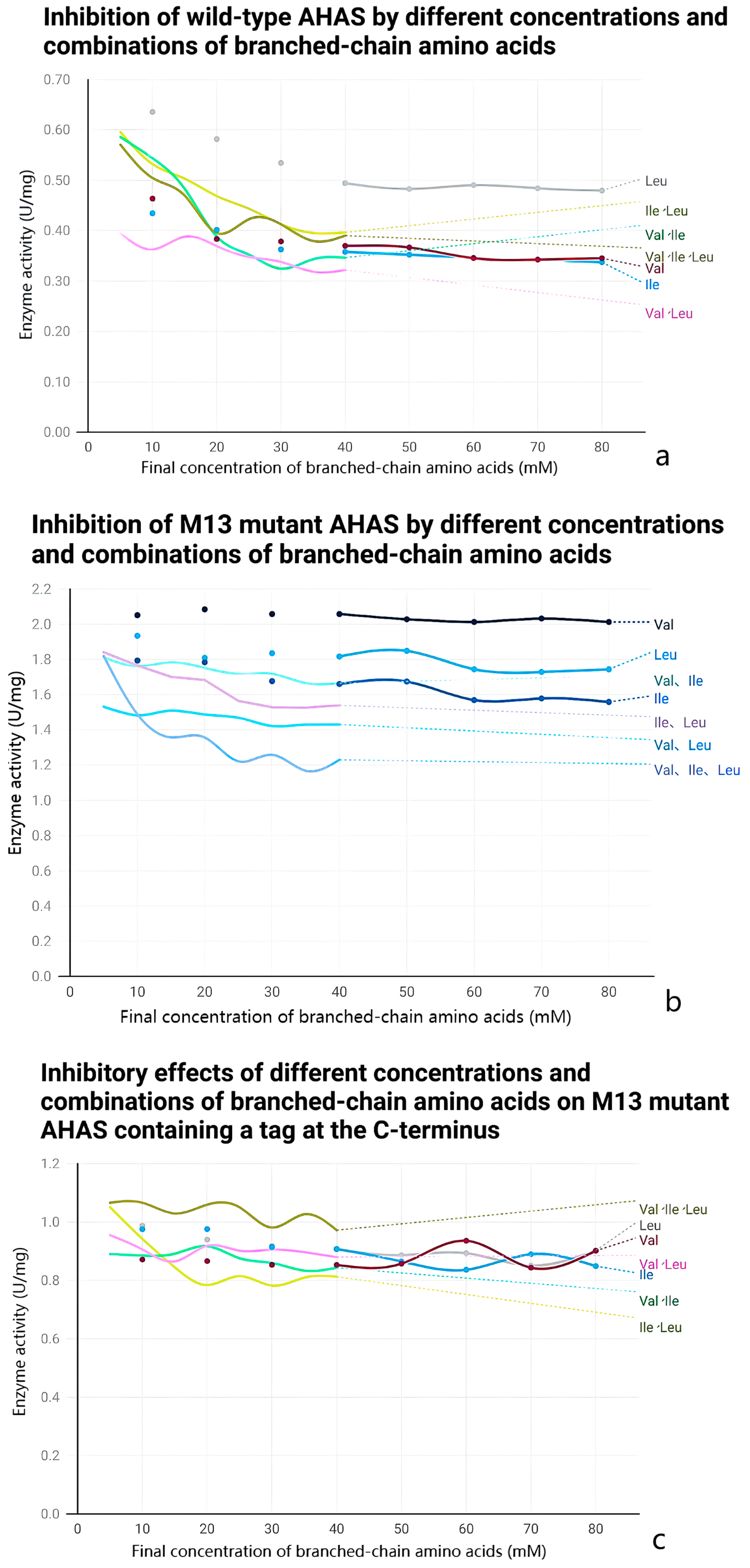
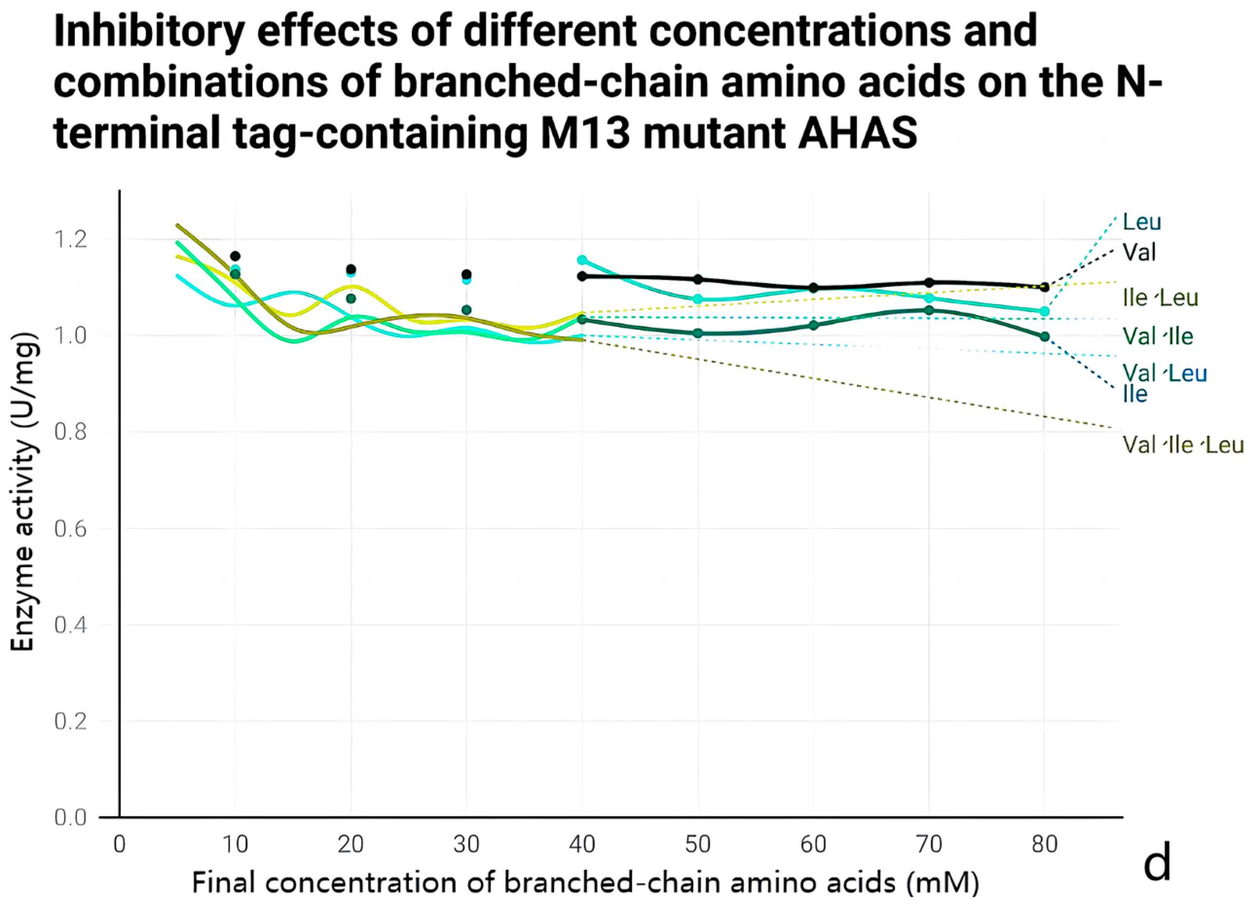
2.5. Anti-Feedback Inhibition Analysis

The enzyme activity was determined in the presence of different concentrations and types of branched-chain amino acids. In the enzyme reaction system, three branched-chain amino acids with different concentration gradients were added, and the final concentrations of the three amino acids were 10, 20, 30, 40, 50, 60, 70, and 80 mM when the three amino acids were added separately; meanwhile, the final concentrations of the three amino acids when the three amino acids were added simultaneously were 5, 10, 15, 20, 25, 30, 35, 40, and 30 mM.

2.6. Wild-Type and M13 Mutant AHAS

The maximum conversion of AHAS was determined at 37 °C, and the concentration of ethyl diphosgene needed to reach 10% of the maximum conversion was calculated to achieve a reduction in reaction time and amount of enzyme required. A pyruvate final concentration gradient of 1, 2.5, 5, 7.5, 10, 15, 20, 30, 40, and 50 mM was set to determine the steady-state kinetic parameters of AHAS under these conditions. Km and Vmax were calculated by fitting the data to the Michaelis–Menten equation V = Vmax × S/(Km + S) in Origin Pro 2022 (OriginLab, Northampton, MA, USA), where S is the concentration of the substrate pyruvate and V is the rate of the enzymatic reaction corresponding to the concentration of the substrate, and each analysis consisted of three sets of replicates.
Citrate buffer (pH 3, 4, 5, 6, and 6.5), potassium phosphate buffer (pH 6, 6.5, 7, 7.5, and 8), Tris-HCl buffer (pH 7, 8, and 9), and Gly-NaOH buffer (pH 9, 10, and 11) were formulated and substituted for pH 7.5 potassium phosphate buffer (pH 7.5, 7.5, 7.5, and 8), and pH 10.5 potassium phosphate buffer (pH 9, 10, and 11) was used in the enzyme activity measurement system to determine the optimal pH of wild-type and M13 mutant AHAS in the enzyme activity reaction system. Each analysis consisted of three sets of replicates.
A preparation of 100 mM Co2+, Ca2+, Cu2+, Na+, Mn2+, K+, Fe2+, Zn2+, Mg2+, and Fe3+ solution, replacing MgCl2 in the enzyme activity assay system, was used to determine the most suitable metal ions for wild-type and M13 mutant AHAS in the enzyme activity reaction system. Each analysis consisted of three sets of replicates.
The time and temperature of the enzyme-activated reaction varied, and time (5, 10 min, 15, 30, 45, 60, 75, 90, 105, and 120 min) and temperature gradients (0, 4, 10, 15, 20, 25, 30, 35, 40, 45, 50, and 55 °C) were used to determine the optimal time and temperature for the enzymatic reaction. Each analysis consisted of three sets of replicates.

2.7. Molecular Modeling

The crystal structures of wild-type ilvN and mutant ilvNM13 were modeled using AlphaFold2 (DeepMind, London, UK), and protein structure comparison was performed using RCSB PDB (https://www.rcsb.org/, accessed on 20 November 2023) to determine the changes in the structure of ilvN before and after the mutation. PyMOL 2.4 was used to obtain the changes in the specific amino acids before and after the mutation and the position of hydrogen bonds within the chain and length changes. Similarly, the ilvB and ilvN complexes were modeled using AlphaFold2, and the CSU and RSU docking sites were calculated using PyMOL 2.4.

3. Results and Discussion

3.1. Construction of AHAS Mutants Resistant to Feedback Inhibition

According to a previous study, mutating glycine at position 20 and isoleucine at position 21 to aspartic acid and isoleucine at position 22 to the phenylalanine of the CSU of C. glutamicum AHAS can completely release the feedback inhibition of L-valine by AHAS [13]. We mutated amino acids at positions 20–22 of C. glutamicum ilvN (G20D, I21D, and I22F) and introduced them into E. coli for expression to obtain an AHAS that rescued the feedback inhibition of L-valine, which was then sequenced after validation by PCR, and the correctly sequenced mutant was named ilvBNM13. Selective addition of EKase sites and His tags at both ends based on the correctly sequenced AHAS yielded M13CN that had EKase sites and His tags at both ends, the C-terminus M13C with EKase site and His tag, and M13N with EKase site and His tag at the N-terminus.

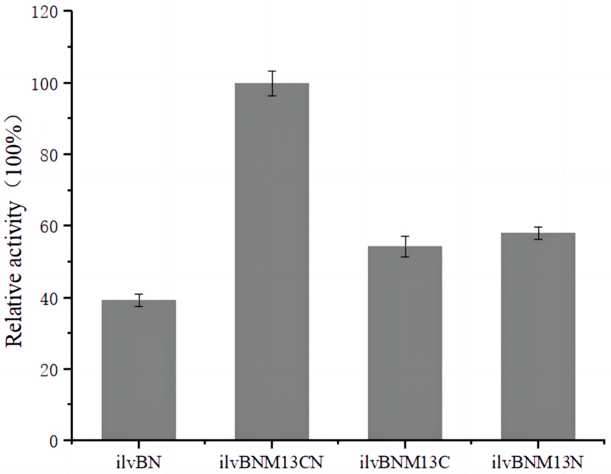
3.2. Determination of AHAS Activity

In the E. coli isoenzyme AHAS III, the binding site for valine and the RSU is in the surface region near the N-terminal structural domain, where L-valine interacts with aspartic acid at positions 11 and 29 through hydrogen bonding and its side chain interacts with leucine at positions 9 and 16 with valine at position 35 in the hydrophobic space conformation [14]. After altering the E. coli AHAS III RSU at positions 14 glycine and 11 aspartic acid, the feedback inhibition sensitivity of L-valine was reduced, suggesting that this region may be the binding site for L-valine [15]. A similar region has been observed in C. glutamicum; AHAS III was less sensitive to feedback inhibition by L-valine, suggesting that this region is likely a binding site for L-valine. Similar sites are found in C. glutamicum, where changes in amino acids at positions 20–22 of the RSU of AHAS relieve feedback inhibition by L-valine. In the aforementioned experiments, we obtained wild-type AHAS expressed in E. coli BL21 by constructing pET-28a-ilvBN, pET-28a-ilvBNM13, pET-28a-ilvBNM13C, pET-28a-ilvBNM13N, and pET-28a-ilvBNM13CN vectors with labeling positions. Three different AHASs, as described in Section 2, were measured for wild-type and mutant AHAS activities, respectively, as shown in Figure 2 The enzyme activity of M13 mutant AHAS containing tags at both ends was used as 100% to compare the relative enzyme activities of wild-type AHAS and the other two M13 mutant AHAS with different tag positions. The enzyme activity of the mutant AHAS was higher than that of the wild-type AHAS, probably because the changes in amino acids at positions 20, 21, and 22 in the AHAS RSUs enhanced the interaction between RSU and CSU and increased enzyme activity. However, the enzyme activity decreased after removing the EK enzyme site and the His tag at either end, suggesting that the presence of the tag had some effect on enzyme activity (Figure 2).

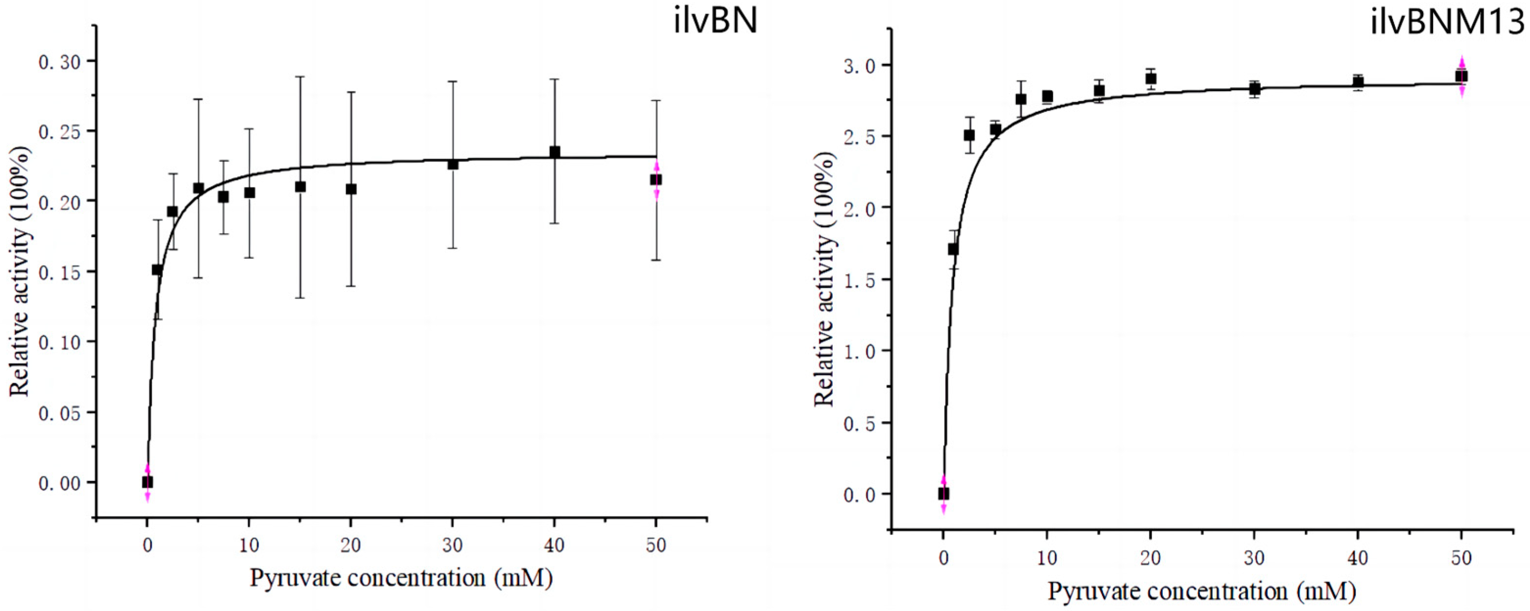
3.3. Enzyme Kinetic Analysis

Enzyme kinetic analysis provided a clearer picture of the inhibition of wild-type and mutant AHAS by branched-chain amino acids and its effect on enzyme activity. Four purified AHASs were subjected to in vitro enzyme activation experiments and added to the enzyme activation reaction system according to the concentrations and combinations of branched-chain amino acids mentioned in Section 2. Wild-type AHAS was inhibited by all concentrations and combinations of branched-chain amino acids (Figure 3). When the three branched-chain amino acids acted alone, 40 mM was the maximum inhibitory concentration; beyond 40 mM, the activity of wild-type AHAS no longer decreased with increasing concentrations of branched-chain amino acids, and L-valine inhibited wild-type AHAS to the greatest extent. The enzymatic activity of wild-type AHAS in the presence of 40 mM L-valine was only 47% of that in the absence of branched-chain amino acids. The enzyme activity of wild-type AHAS in the presence of 40 mM L-leucine was 62% in the presence of 40 mM L-isoleucine and 50% in the presence of 40 mM L-isoleucine. When two or three amino acids acted together, the 35 mM inhibition was maximal, but the inhibition was relatively weak when leucine and isoleucine acted together, reaching 50% without feedback inhibition. The inhibition was relatively strong when valine and leucine acted together, with an enzyme activity of 41%. The mutant AHAS was tested for resistance to feedback inhibition and was found to be completely uninhibited by L-valine, with 92% enzyme activity in the presence of 40 mM L-leucine and 83% enzyme activity in the presence of 40 mM L-isoleucine. Tolerance to any combination of branched-chain amino acids also significantly improved. Comparison of the resistance to feedback inhibition between wild-type AHAS and M13 mutant AHAS showed that the mutant AHAS had increased tolerance to all L-valine-containing combinations.

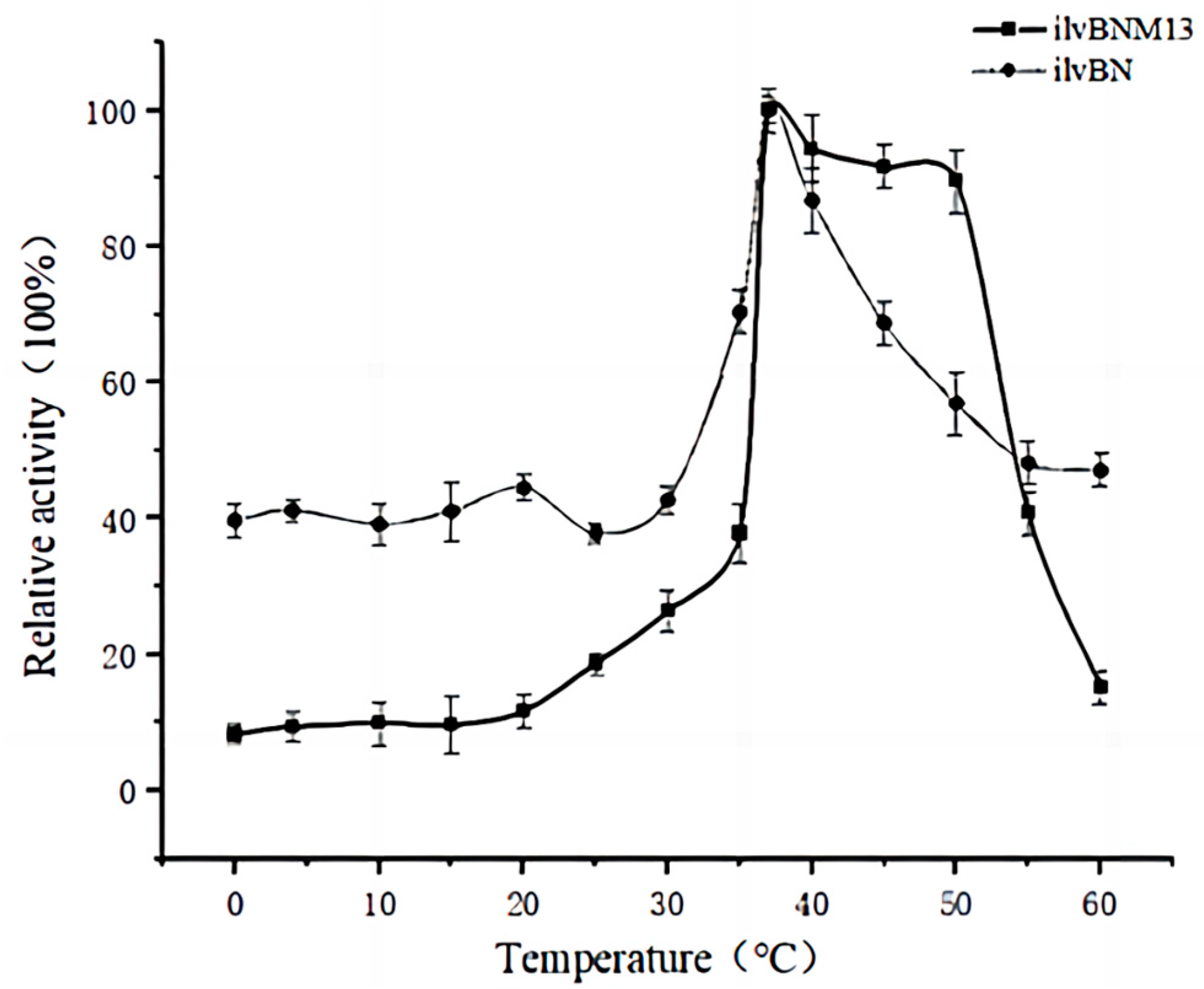
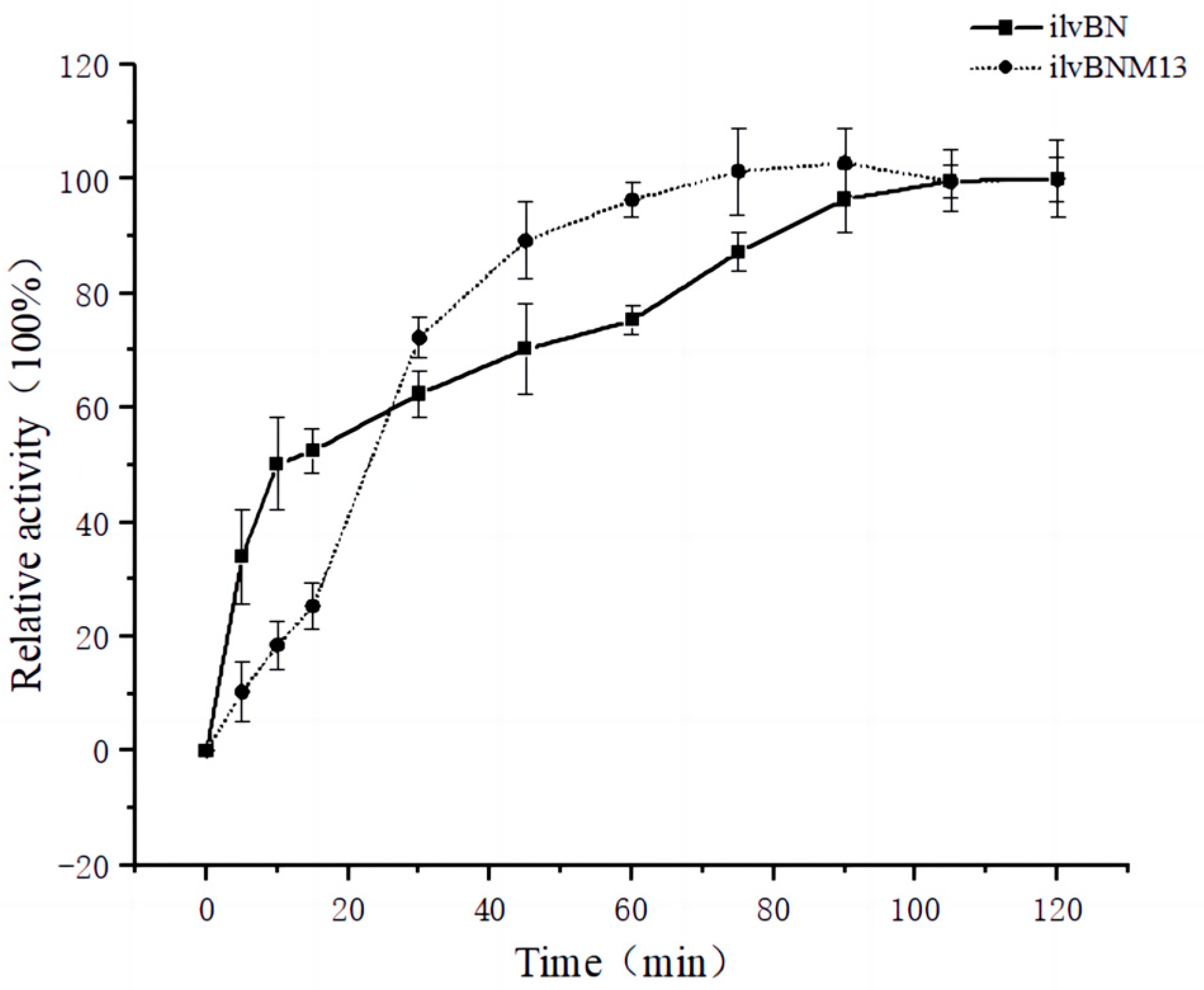
3.4. Effect of Mutations on the Enzymatic Properties of AHAS

Mutations usually cause changes in enzymatic properties, and after comparing the resistance to feedback inhibition of wild-type and M13 mutant AHAS, we characterized the enzymatic properties of AHAS and discussed the differences between the two AHASs in terms of catalytic properties, optimal reaction conditions, and Mie constants.
Figure 4 shows the Michaelis–Menten equation fitted by origin 2022, with the enzyme activity at maximum substrate concentration set to 100%, plotted to plot the fit. As shown in Figure 4, the wild-type AHAS reached 10% of the maximum conversions at 5 min into the reaction, and the Km value of wild-type AHAS with pyruvate as the substrate was 4.5253 ± 0.07631 mM. Similarly, the M13 mutant AHAS reached 10% of the maximum conversions 2.5 min into the reaction, with a Km value of 6.3877 ± 0.06232 mM. The time for wild-type and M13 mutant AHAS to catalyze pyruvate to a maximum conversion of 10% was calculated by GraphPad equation Y = Et × cat × X/(Km + X), with Y being the enzyme activity, Et being the enzyme concentration, and kcat being the number of conversions.
The enzyme activity when Mg2+ was used as a metal ion was defined as 100%, and the data were fitted to origin 2022 and plotted as a bar graph to obtain Figure 5. Figure 5 shows the most suitable metal ions for wild-type and M13 mutant AHAS. The relative enzyme activity of Mg2+ was 100% when Mg2+ was used as a control for comparison with other ions. It can be observed that Mg2+ was the most suitable metal ion for wild-type AHAS, whereas Cu2+ and Fe3+ strongly inhibited the enzyme activity of wild-type AHAS, which was reduced by 80% and 70%, respectively, in the presence of the two ions. For the M13 mutant AHAS, Fe2+ was the most suitable metal ion, increasing the enzyme activity 1.5 times relative to the Mg2+ ion. Among them, Cu2+ most strongly inhibited the enzymatic activity of the M13 mutant AHAS, followed by Co2+, Ca2+, and Na+. None of the other metal ions had a significant inhibitory effect on the enzymatic activity of the M13 mutant AHAS.
To explore the optimal pH for the wild-type and M13 mutant AHASs, we chose four buffers with different pH values. Figure 6 shows the effect of pH on the enzyme activity of wild-type and M13 mutant AHASs, with potassium phosphate buffer at pH 7.5 as a control for comparison. The relative enzyme activity of potassium phosphate buffer at pH 7.5 was 100%. The optimal pH for both wild-type and M13 mutant AHASs was 7.5. Compared with wild-type AHAS, M13 mutant AHAS was more tolerant to acidity; in citrate buffer at pH 3, the relative enzyme activity of wild-type AHAS was only 15% of that in potassium phosphate buffer at pH 7.5, whereas the M13 mutant AHAS reached 53% of that in potassium phosphate buffer at pH 7.5.
The enzyme activity under optimal temperature conditions was set to 100% and the data were fitted to origin 2022 and plotted to plot a line graph to obtain Figure 7. In terms of the optimal temperature, both wild-type and M13 mutant AHAS had an optimal temperature of 37 °C. However, wild-type AHAS had higher tolerance for both low and high temperatures than M13 mutant AHAS (Figure 7). Below 20 °C, the enzyme activity of wild-type AHAS was 40% of the enzyme activity at the optimal temperature, whereas that of M13 mutant AHAS was only 15% of that at 37 °C. However, in the range of 40–50 °C, M13 mutant AHAS was more tolerant than the wild type, and in this 10 °C temperature interval, the enzyme activity of M13 mutant AHAS changed very little and was above 90 °C of the optimal temperature. Above 50 °C, the activity of M13 mutant AHAS decreased rapidly, and at 60 °C, it was only 15% of that at the optimal temperature of 37 °C.
Figure 8 shows the relative enzyme activity versus time for wild-type and M13 mutant AHAS at 37 °C and pH 7.5 potassium phosphate buffer. The enzyme activity of wild-type and M13 mutant AHAS at 120 min was taken as 100% and a line graph was created using origin 2022. Analysis of the effect of time on wild-type and M13 mutant AHAS showed that, based on the original enzyme activity system, the reaction was complete after 90 min of reaction for wild-type AHAS and after 75 min for M13 mutant AHAS. Within 7.5 min, the wild-type AHAS enzymatic reaction showed zero-level reaction kinetics, and the reaction was carried out within 7.5 min as a suitable node for the determination of enzyme activity. Within 30 min, the enzymatic reaction of the M13 mutant AHAS showed zero-stage kinetics, and 30 min was a suitable time point for determining enzyme activity (Figure 8).

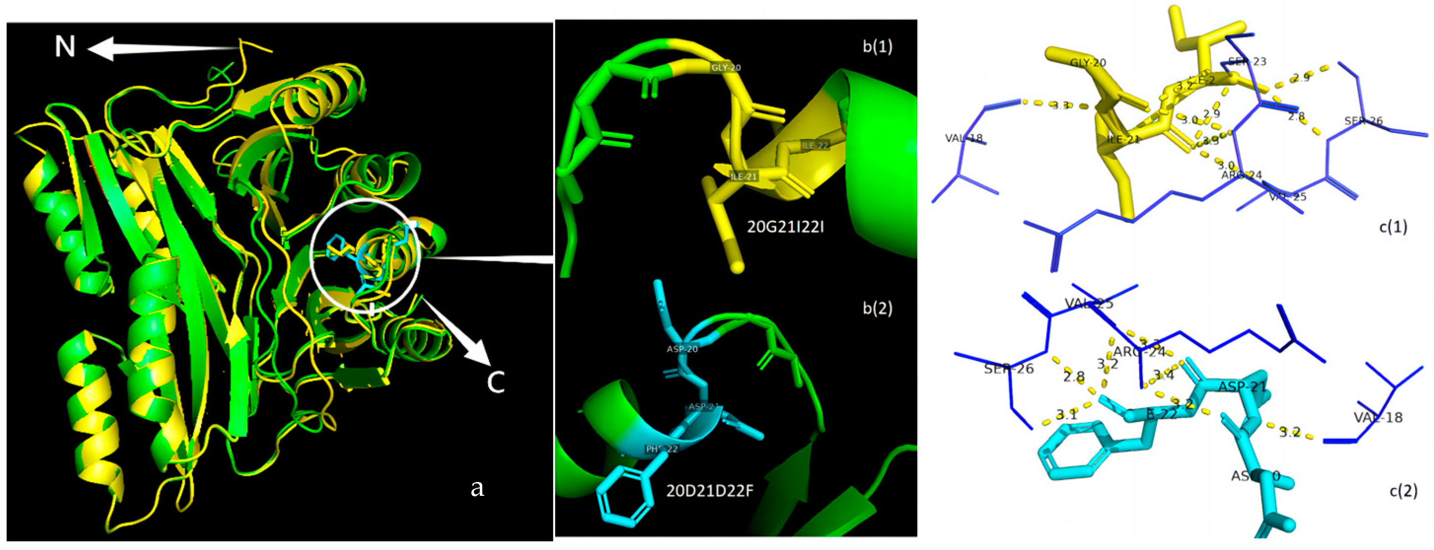
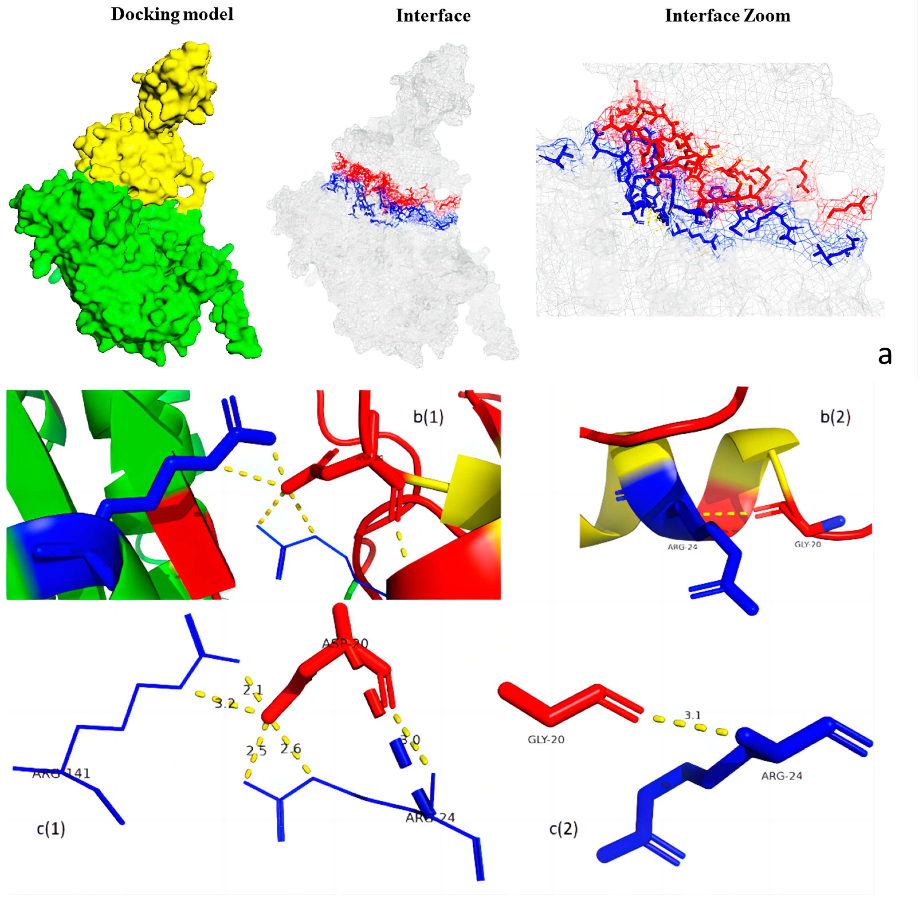
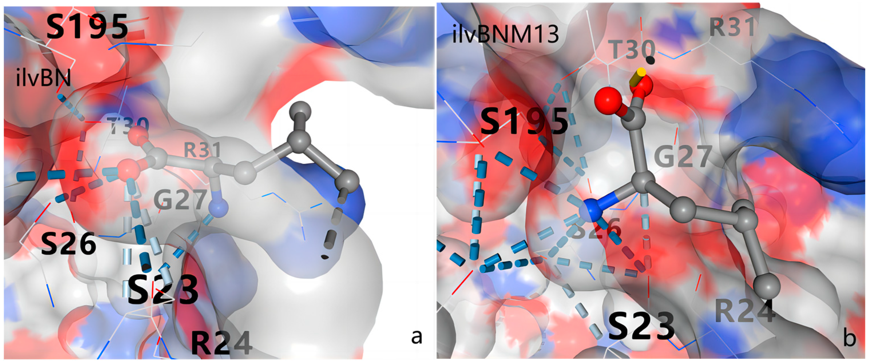
3.5. Protein Structure Analysis

The crystal structures of the CSUs of AHAS from S. cerevisiae, A. thaliana [16,17], and Pseudomonas aeruginosa protease [18] have been reported. In the investigation of the role of residues 20–22 of the RSU in AHAS function, crystal structure simulations of the C. glutamicum RSU revealed that the three consecutive amino acid residues were located exactly at the turn of the N-terminal first α-structural domain initiation (Figure 9). Simulations of its mutated structure revealed that the three amino acid changes did not alter its structure (Figure 10a,b); however, the number and length of its intrachain hydrogen bonds were altered (Figure 10c). Similarly, simulation of the AHAS complex structure and calculation of the molecular docking results of CSU and RSU using PyMOL 2.4 (Figure 11) revealed that of the three consecutively mutated amino acids at positions 20–22 in the RSU, only the amino acid at position 20 was involved in the interaction with CSU (Figure 11b). Before the mutation, the glycine at position 20 was only connected to the arginine at position 24 of the RSU by a polar bond and did not interact with CSU. However, when the glycine at position 20 was mutated to aspartic acid, the aspartic acid at position 20 of the RSU and the arginine residue at position 141 in CSU were connected by two polar bonds, and a relatively strong interaction occurred (Figure 11c).
The overall structure of AHAS is dimeric, with two RSUs and two CSUs. The RSU monomer and dimer are similar to those of other RSU structures, such as those reported previously [8,19]. The CSU monomer is further separated into three substructural domains. The entire AHAS dimer has a two-fold amorphous symmetry. The RSU dimer is located at the top of the CSU dimer. Interactions between the RSU and CSU monomers were also observed within the binding interface. The M13 mutation mutates glycine at position 20 of the RSU to aspartic acid, which can interact with arginine at position 141 of CSU, leading to a significant increase in AHAS activity.
Previous studies have shown that the interaction interface is highly conserved in three-dimensional structures despite wide variation in amino acid sequences across species, which explains why different RSU isoforms can activate the same CSUs [6]. Our simulated interaction interface between RSU and CSU was consistent with this conserved interaction interface. When RSU bound to CSU, the region of the linker between the α- and β-structural domains changed. This affected the folding of CSU, which in turn led to changes in the conformation of the active site and the position of ThDP. The M13 mutation we induced altered the interaction site between RSU and CSU, which led to altered RSU and CSU binding, affecting the position of ThDP and causing an alteration in the metal ion preference of the M13 mutant AHAS.
In addition to this, we simulated the docking of branched-chain amino acids and AHAS using AutoDock. In the study of Arabidopsis structure, the binding site of L-valine was located at the beginning of the first α-helix of the RSU, which was used as a docking template for docking of L-valine and AHAS using AutoDock, and the results are shown in Figure 12, Figure 13 and Figure 14. Three consecutively mutated amino acids at positions 20–22 of the RSU changed the docking sites of L-valine and AHAS, and attenuated the binding between L-valine and AHAS, which may why M13 mutant AHAS abolished the feedback inhibition of L-valine on it. In the docking simulations of L-leucine and L-isoleucine with wild-type and M13 mutant AHAS, although no such feature was found, the docking sites of the three branched-chain amino acids and AHAS overlapped, which may have been because of the change in the binding mode of L-valine and AHAS affecting the binding of L-leucine and L-isoleucine, thus changing the feedback inhibition of L-leucine and L-isoleucine on AHAS. In the analysis of the feedback inhibition of wild-type and M13 mutant AHAS by the three branched-chain amino acids, we found that the feedback inhibition of AHAS by the simultaneous action of the two amino acids L-leucine and L-isoleucine was weaker than that by the simultaneous action of the three branched-chain amino acids, probably because of the fact that there was a partial overlap in the binding sites of L-leucine and L-isoleucine and AHAS. In the presence of these two amino acids, L-leucine and L-isoleucine competed for the binding site of AHAS, and the feedback inhibition was thus weakened. Our simulation of the docking site by AutoDock supports this.

4. Conclusions

In the present study, we determined the enzymatic activity and feedback inhibition of wild-type and M13 mutant AHAS in Corynebacterium glutamicum. Previous studies by Eliáková, V. et al. revealed feedback inhibition of wild-type AHAS in the presence of a single branched-chain amino acid at a final concentration of 5 mM and in the presence of three branched-chain amino acids, as well as feedback inhibition of M13 mutant AHAS in the presence of valine at a final concentration of 10 mM. Our study complemented this by further increasing the concentration of branched-chain amino acids to 100 mM and by adopting a more comprehensive combination of branched-chain amino acids to examine the feedback inhibition of branched-chain amino acids in wild-type and M13 mutant AHAS, respectively. We found that the feedback inhibition of acetohydroxyacid synthase by any combination of branched-chain amino acids had a certain limit, with 40 mM as the maximum inhibitory concentration in the presence of a single branched-chain amino acid, and the feedback inhibition did not increase after the concentration of branched-chain amino acids exceeded 40 mM. This maximum inhibitory concentration was even lower for two and three branched-chain amino acid combinations: 30 mM and 25 mM. Moreover, the M13 mutation not only completely relieved feedback inhibition by L-valine but also greatly reduced feedback inhibition by L-leucine, L-isoleucine, and any combination of the three amino acids. In addition, it significantly increased the enzymatic activity of AHAS.
The study revealed that the optimal pH of both wild-type and M13 mutant AHAS was 7.5 and that the optimal temperature was 37 °C. In the assay of optimal metal ions, Fe2+ significantly increased the activity of the M13 mutant AHAS; Mn2+, K+, Zn2+, and Fe3+ had the same activation ability as Mg2+, whereas the activation ability of all other metal ions was less than that of magnesium ions. However, in wild-type AHAS, Mg2+ was the most suitable metal ion, and all other ions had inhibitory effects.
We simulated the CSU and RSU structures of AHAS using AlphaFold2. The amino acid mutation at positions 20–22 in the RSU did not change the three-dimensional structure of the subunit, but it did alter the number and length of intrachain hydrogen bonds. Among them, the amino acid at position 20 was also related to RSU interaction, and the mutation of the amino acid at position 20 of the RSU enhanced the interaction between RSU and CSU, which might be related to the increase in enzyme activity after the mutation. In docking site simulations of branched-chain amino acids and AHAS, the M13 mutation did not change the binding sites of ligands and substrates to AHAS but did have effects on the binding of branched-chain amino acids to AHAS. The M13 mutation occurred in the binding site of L-valine and AHAS, and relieved the feedback inhibition of AHAS by L-valine. The M13 mutation indirectly weakened the binding of L-leucine and L-isoleucine to AHAS, and weakened the feedback inhibition of AHAS by L-leucine and L-isoleucine.
Overall, our study gives the optimal reaction conditions of M13 mutant AHAS and compares the differences between the optimal conditions of wild-type AHAS and M13 mutant AHAS, which creates conditions for better application of M13 mutant AHAS in the production of branched-chain amino acids. Beyond that, our results reveal possible reasons for the M13 mutation in C. glutamicum AHAS to relieve L-valine inhibition, providing a plausible explanation for the increase in enzyme activity after the M13 mutation. Through structural modeling of RSU and CSU, the interaction sites between CSU and RSU and the docking sites of branched-chain amino acids with RSU were identified, which provided directions for the subsequent enhancement of the enzyme activity and anti-feedback inhibition ability of AHAS.

Author Contributions

Conceptualization, Y.T. and H.Z.; methodology, Y.T. and H.Z.; software, N.W.; validation, Z.A.; formal analysis, X.G. and Z.A; investigation, Y.T. and H.Z.; resources, Z.A.; data curation, Y.M.; writing—original draft preparation, Y.T. and H.Z.; writing—review and editing, H.Z.; visualization, X.G.; supervision, H.Z.; project administration, Z.A. and Y.M.; funding acquisition, H.Z. All authors have read and agreed to the published version of the manuscript.

Funding

This study was supported by the National Natural Science Foundation of China (No. 32001836), the Natural Science Foundation of Shandong Province (ZR201911180224), the Natural Science Foundation of Shandong Province (ZR2020MC043), and the Visiting Study and Research Funds for Teachers of Ordinary Undergraduate Colleges and Universities in Shandong Province.

Institutional Review Board Statement

Not applicable.

Informed Consent Statement

Not applicable.

Data Availability Statement

Data are contained within the article.

Conflicts of Interest

Author Zhiqiang An was employed by the company MabPlex International. The remaining authors declare that the research was conducted in the absence of any commercial or financial relationships that could be construed as a potential conflict of interest.

References

  1. Duggleby, R.G. Domain relationships in thiamine diphosphate-dependent enzymes. Acc. Chem. Res. 2006, 39, 550–557. [Google Scholar] [CrossRef] [PubMed]
  2. Schloss, J.V.; Ciskanik, L.M.; Dyk, D.E.V. Origin of the herbicide binding site of acetolactate synthase. Nature 1988, 331, 360–362. [Google Scholar] [CrossRef]
  3. Lonhienne, T.; Garcia, M.D.; Guddat, L.W. The Role of a FAD Cofactor in the Regulation of Acetohydroxyacid Synthase by Redox Signaling Molecules. J. Biol. Chem. 2017, 292, 5101–5109. [Google Scholar] [CrossRef] [PubMed]
  4. Duggleby, R.; Pang, S.S. Acetohydroxyacid Synthase. J. Biochem. Mol. Biol. 2000, 33, 1–36. [Google Scholar]
  5. Niu, X.; Liu, X.; Zhou, Y.; Niu, C.; Xi, Z.; Su, X.D. Preliminary X-ray crystallographic studies of the catalytic subunit of Escherichia coli AHAS II with its cofactors. Acta Crystallogr. Sect. F Struct. Biol. Cryst. Commun. 2011, 67, 659–661. [Google Scholar] [CrossRef] [PubMed]
  6. Zhang, Y.; Li, Y.; Liu, X.; Sun, J.; Li, X.; Lin, J.; Yang, X.; Xi, Z.; Shen, Y. Molecular architecture of the acetohydroxyacid synthase holoenzyme. Biochem. J. 2020, 477, 2439–2449. [Google Scholar] [CrossRef] [PubMed]
  7. Liu, Y.; Li, Y.; Wang, X. Molecular evolution of acetohydroxyacid synthase in bacteria. MicrobiologyOpen 2017, 6, e00524. [Google Scholar] [CrossRef]
  8. Kaplun, A.; Vyazmensky, M.; Zherdev, Y.; Belenky, I.; Slutzker, A.; Mendel, S.; Barak, Z.; Chipman, D.M.; Shaanan, B. Structure of the regulatory subunit of acetohydroxyacid synthase isozyme III from Escherichia coli. J. Mol. Biol. 2006, 357, 951–963. [Google Scholar] [CrossRef] [PubMed]
  9. Vinogradov, V.; Vyazmensky, M.; Engel, S.; Belenky, I.; Kaplun, A.; Kryukov, O.; Barak, Z.; Chipman, D.M. Acetohydroxyacid synthase isozyme I from Escherichia coli has unique catalytic and regulatory properties. Biochim. Biophys. Acta 2006, 1760, 356–363. [Google Scholar] [CrossRef] [PubMed]
  10. Barak, Z.; Chipman, D.M. Allosteric regulation in Acetohydroxyacid Synthases (AHASs)—Different structures and kinetic behavior in isozymes in the same organisms. Arch. Biochem. Biophys. 2012, 519, 167–174. [Google Scholar] [CrossRef] [PubMed]
  11. Lonhienne, T.; Low, Y.S.; Garcia, M.D.; Croll, T.; Gao, Y.; Wang, Q.; Brillault, L.; Williams, C.M.; Fraser, J.A.; McGeary, R.P.; et al. Structures of fungal and plant acetohydroxyacid synthases. Nature 2020, 586, 317–321. [Google Scholar] [CrossRef] [PubMed]
  12. Westerfeld, W.W. A colorimetric determination of blood acetoin. J. Biol. Chem. 1945, 161, 495–502. [Google Scholar] [CrossRef]
  13. Eliáková, V.; Pátek, M.; Holátko, J.; Nevera, J.; Leyval, D.; Goergen, J.L.; Delaunay, S. Feedback-Resistant Acetohydroxy Acid Synthase Increases Valine Production in Corynebacterium glutamicum. Am. Soc. Microbiol. (ASM) 2005, 71, 207–213. [Google Scholar] [CrossRef] [PubMed]
  14. Lawther, R.P.; Calhoun, D.H.; Adams, C.W.; Hauser, C.A.; Gray, J.; Hatfield, G.W. Molecular basis of valine resistance in Escherichia coli K-12. Proc. Natl. Acad. Sci. USA 1981, 78, 922–925. [Google Scholar] [CrossRef] [PubMed]
  15. Eoyang, L.; Silverman, P.M. Role of small subunit (IlvN polypeptide) of acetohydroxyacid synthase I from Escherichia coli K-12 in sensitivity of the enzyme to valine inhibition. J. Bacteriol. 1986, 166, 901–904. [Google Scholar] [CrossRef] [PubMed]
  16. Bar-Ilan, A.; Balan, V.; Tittmann, K.; Golbik, R.; Vyazmensky, M.; Hübner, G.; Barak, Z.; Chipman, D.M. Binding and activation of thiamin diphosphate in acetohydroxyacid synthase. Biochemistry 2001, 40, 11946–11954. [Google Scholar] [CrossRef] [PubMed]
  17. Lonhienne, T.; Garcia, M.D.; Pierens, G.; Mobli, M.; Nouwens, A.; Guddat, L.W. Structural insights into the mechanism of inhibition of AHAS by herbicides. Proc. Natl. Acad. Sci. USA 2018, 115, E1945–E1954. [Google Scholar] [CrossRef] [PubMed]
  18. Mccourt, J.A.; Pang, S.S.; King-Scott, J.; Guddat, L.W.; Duggleby, R.G. Herbicide-binding sites revealed in the structure of plant acetohydroxyacid synthase. Proc. Natl. Acad. Sci. USA 2006, 103, 569–573. [Google Scholar] [CrossRef]
  19. Karanth, N.M.; Sarma, S.P. The coiL-to-helix transition in IlvN regulates the allosteric control of Escherichia coli acetohydroxyacid synthase I. Biochemistry 2013, 52, 70–83. [Google Scholar] [CrossRef] [PubMed]
Figure 1. Sequence comparison of wild-type and three AHAS proteins with different tag positions.
Fermentation 10 00311 g001
Figure 2. Comparison of enzyme activities of wild-type and three M13 mutant AHAS with different labeling positions. Horizontal coordinates are wild-type and AHAS with different labeling positions, and vertical coordinates are relative enzyme activities.
Fermentation 10 00311 g002
Figure 3. Feedback inhibition of wild-type and M13 mutant AHAS by different combinations of branched-chain amino acids. The x-axis is the final concentration of three different combinations of branched-chain amino acids in the enzyme activity assay system in mM, and the y-axis is the enzyme activity in the presence of different final concentrations of branched-chain amino acids in U/mg. (a) The inhibition of wild-type AHAS by different concentrations and combinations of branched-chain amino acids; (b) the inhibition of M13 mutant AHAS by different concentrations and combinations of branched-chain amino acids; (c) the inhibition of C-terminal M13 mutant AHAS containing a His tag and an EKase site by different concentrations and combinations of branched-chain amino acids; (d) the inhibition of N-terminal M13 mutant AHAS containing a His tag and an EKase site, and the inhibition of M13 mutant AHAS containing a His tag and an EKase site at the N-terminus by different concentrations and combinations of branched-chain amino acids.
Fermentation 10 00311 g003aFermentation 10 00311 g003b
Figure 4. Wild-type and M13 mutant AHAS steady-state kinetic results.
Fermentation 10 00311 g004
Figure 5. Optimal metal ion detection results for wild-type and M13 mutant AHAS at 37 °C.
Fermentation 10 00311 g005
Figure 6. Wild-type (left panel) and M13 mutant (right panel) AHAS optimal pH determination.
Fermentation 10 00311 g006
Figure 7. Optimal reaction temperature results for wild-type and M13 mutant AHAS in phosphate buffer at pH 7.5.
Fermentation 10 00311 g007
Figure 8. Wild-type and M13 mutant AHAS optimal reaction times were determined.
Fermentation 10 00311 g008
Figure 9. Structure of ilvN. The original ilvN structure is shown in blue, and the positions of the mutated amino acids, glycine at position 20, isoleucine at position 21, and isoleucine at position 22, are shown in yellow.
Fermentation 10 00311 g009
Figure 10. Structural changes in ilvN before and after mutation. (a) A comparison of the structures of ilvN and ilvNM13. The green part is the structure of ilvN, and the yellow part is the structure of ilvNM13. Comparison of the structures of ilvN and ilvNM13 shows no change in the overall structure of the two.; (b1,b2) the change in amino acids at positions 20–22 before and after the fixed-point mutation, (b1) is the amino acid structure before the mutation, and (b2) is the amino acid structure after the mutation; (c1,c2) the change in the number and length of the polar bonds before and after the mutation, (c1) is the position and length of the polar bond before the mutation, and (c2) is the polar bond after the mutation length and position.
Fermentation 10 00311 g010
Figure 11. IlvN and ilvB interaction sites. (a) The composite structure of ilvB and ilvN, where the yellow part is ilvN, the green part is ilvB, and the red and blue part is the region of interaction between ilvB and ilvN; (b1,b2) the position of the amino acid structure involving interactions involving ilvB and ilvN as well as the polar bonds in the three consecutive amino acid mutations 20–22, where the yellow dashed line is the hydrogen bond, the blue portion is ilvB, and the red portion is ilvN. (b1) is after the fixed-point mutation, and (b2) is before the fixed-point mutation; (c1,c2) the change in the position and length of the polar bond before and after the mutation. The red portion is ilvN and the blue portion is ilvB. (c1) is after the fixed-point mutation, and (c2) is before the fixed-point mutation.
Fermentation 10 00311 g011
Figure 12. Action sites of L-valine in AHAS. (a) Action site of L-valine in M13 mutant AHAS; (b) action site of L-valine in wild-type AHAS.
Fermentation 10 00311 g012
Figure 13. Action sites of L-leucine in AHAS. (a) Action site of L-leucine in M13 mutant AHAS; (b) action site of L-leucine in wild-type AHAS.
Fermentation 10 00311 g013
Figure 14. Action sites of L-isoleucine in AHAS. (a) Action site of L-isoleucine in M13 mutant AHAS; (b) action site of L-leucine in wild-type AHAS.
Fermentation 10 00311 g014
Table 1. Strains and plasmids.
Strain or PlasmidRelevant CharacteristicsReference or Source
C. glutamicum strains ATCC 13032Wild typeLaboratory preservation
E. coli strains DH5αWild typeLaboratory preservation
E. coli strains BL21Wild typeLaboratory preservation
DH5α/pET-28a-ilvBNDH5αharboring pET-28a-ilvBNThis work
DH5α/pET-28a-ilvBNCNM13DH5αharboring pET-28a-ilvBNCNM13This work
DH5α/pET-28a-ilvBNCM13DH5αharboring pET-28a-ilvBNCM13This work
DH5α/pET-28a-ilvBNNM13DH5αharboring pET-28a-ilvBNNM13This work
BL 21/pET-28a-ilvBNBL 21 harboring pET-28a-ilvBNThis work
BL 21/pET-28a-ilvBNCNM13BL 21 harboring pET-28a-ilvBNCNM13This work
BL 21/pET-28a-ilvBNCM13BL 21 harboring pET-28a-ilvBNCM13This work
BL 21/pET-28a-ilvBNNM13BL 21 harboring pET-28a-ilvBNNM13This work
plasmids
pET-28aE. coli vector, KmrLaboratory preservation
pET-28a-ilvBNpET-28a with the insertion of ilvBNThis work
pET-28a-ilvBNCNM13pET-28a with the insertion of ilvBNCNM13This work
pET-28a-ilvBNCM13pET-28a with the insertion of ilvBNCM13This work
pET-28a-ilvBNNM13pET-28a with the insertion of ilvBNNM13This work
Table 2. Oligonucleotide primers.
Primer5′-3′ Sequence a
ilvBN-FCGCGGATCCATGAATGTGGCAGCTTCTCAACAG
ilvBN-RGGGAAGCTTGATCTTGGCCGGAGCCATG
ilvBNM13-FGATGACTTTTCCCGCGTATCAGGTATGTTCACC
ilvBNM13-RAAAGTCATCGTCTACGTCCTGAACGAGTACGGAC
Pet-CFAAAGCCCGAAAGGAAGCTGAGT
Pet-CRGCCCATGGTATATCTCCTTCTTAAAGT
Pet-NFGCTGAGTTGGCTGCTGCCACCG
Pet-NRGGTATATCTCCTTCTTAAAGTTAAACAAAATTATTTCTAGAGGGG
ilvBNM13C1CGCGACGACGACGACAAGGGATCCATGAATGTGGCAGCTTCTCAACAGCCC
ilvBNM13N1GGGAAGCTTGTCATCGTCATCCTTGATCTTGGCCGGAGCCATGGTCTTC
ilvBNCM13-F1CATCATCATCATCATCACAGCAGCGGCC
ilvBNCM13-R1TCAGATCTTGGCCGGAGCCATGGTC
ilvBNNM13-F1ATGAATGTGGCAGCTTCTCAACAGC
ilvBNNM13-R1TTTGTTAGCAGCCGGATCAAGCTTT
ilvBNCM13-F2gaaggagatataccatgggcCATCATCATCATCATCACAGCAGC
ilvBNCM13-R2tcagcttcctttcgggctttTCAGATCTTGGCCGGAGCC
ilvBNNM13-F2ctttaagaaggagatataccATGAATGTGGCAGCTTCTCAACA
ilvBNNM13-R2gtggcagcagccaactcagcTTTGTTAGCAGCCGGATCAAG
a Homologous arms are indicated in lower case.
Disclaimer/Publisher’s Note: The statements, opinions and data contained in all publications are solely those of the individual author(s) and contributor(s) and not of MDPI and/or the editor(s). MDPI and/or the editor(s) disclaim responsibility for any injury to people or property resulting from any ideas, methods, instructions or products referred to in the content.

