Article

Modifications of Constitutive Promoter to Large-Scale Synthesize Porcine Myoglobin in Komagataella phaffii

by
Danni Sun
1,2,3,4,
Yunpeng Wang
1,2,3,4,
Jingwen Zhou
1,2,3,4,
Jianghua Li
1,2,3,4,
Jian Chen
1,2,3,4,
Guocheng Du
1,2,3,4,5,* and
Xinrui Zhao
1,2,3,4,*
1
Science Center for Future Foods, Jiangnan University, 1800 Lihu Road, Wuxi 214122, China
2
Key Laboratory of Industrial Biotechnology, Ministry of Education, School of Biotechnology, Jiangnan University, 1800 Lihu Road, Wuxi 214122, China
3
Jiangsu Province Engineering Research Center of Food Synthetic Biotechnology, Jiangnan University, 1800 Lihu Road, Wuxi 214122, China
4
Engineering Research Center of Ministry of Education on Food Synthetic Biotechnology, Jiangnan University, 1800 Lihu Road, Wuxi 214122, China
5
Key Laboratory of Carbohydrate Chemistry and Biotechnology, Ministry of Education, Jiangnan University, 1800 Lihu Road, Wuxi 214122, China
*
Authors to whom correspondence should be addressed.
Fermentation 2025, 11(2), 49; https://doi.org/10.3390/fermentation11020049
Submission received: 13 January 2025 / Revised: 18 January 2025 / Accepted: 20 January 2025 / Published: 22 January 2025
(This article belongs to the Section Fermentation for Food and Beverages)

Abstract

:
Myoglobin (MG) is a heme-binding protein and can be used as a color and flavor additive for artificial meat. After the selection of stable constitutive expression, although the synthesis of porcine myoglobin (pMG) was achieved through the application of a modified GAP promoter (G1 promoter) in Komagataella phaffii, the lower titer of pMG cannot meet the requirements of commercial production. Herein, another powerful constitutive promoter (GCW14 promoter) was chosen and modified through randomizing its core region for the first time, leading to an increase of 1.18 to 6.01 times in strength. In addition, under the control of a mutated PGCW14 promoter (PGCWm-121), the titer of pMG was further enhanced by optimizing the integrated copy numbers of the pMG gene and knocking out the Yps1-1 protease. Applying the best engineered strain and suitable fermentation conditions, the highest titer of pMG (547.59 mg/L) was achieved in fed-batch fermentation using a cheap and chemically synthesized medium. Furthermore, the obtained pMG had similar peroxide-specific activity (427.50 U/mg) with the extracted natural product after the food-grade purification. The applied strategy can be utilized to synthesize other high value-added hemoproteins, enriching the applications of functional components in the field of artificial meat.

1. Introduction

Myoglobin (MG) is an oxygen-binding monomeric protein containing heme [1]. The primary functions of MG include the storage of oxygen and the facilitation of oxygen diffusion. In addition, MG can also be used as a biocatalyst [2], an iron supplement [3], and a diagnostic tool for diseases [4]. Among different kinds of MG, porcine MG (pMG), consisting of 154 amino acids with a molecular weight of approximately 16.50 kDa [5], is closely related to the color, texture, and flavor of meat [6,7]. Therefore, pMG has great potential in the production of artificial meat.
Since the production of pMG requires appropriate folding or processing to acquire biological activity, although the biosynthesis of pMG has been achieved in various microorganisms, the expression of pMG is maintained at a lower level [8]. For example, most of the recombinant pMG exists in the form of inclusion bodies in Escherichia coli [9,10]. Thus, it is important to select a suitable microbial host for the synthesis of pMG. Komagataella phaffii is one of the most efficient systems for the expression of eukaryotic proteins. Compared with other expressional hosts, the advantages of the K. phaffii expression system include the stable integrated expression into the genome, the efficient secretory expression for the easy downstream purification, the low-cost high-cell-density fermentation, and the superior capability in protein folding and post-translational modifications [11].
In previous research, the synthesis of pMG has been achieved in K. phaffii under the control of the commonly used and powerful AOX1 promoter (PAOX1) [12]. However, PAOX1 is a methanol-inducible promoter, which restricts its applications in food and medicine [13]. To solve this problem, the constitutive expression system simplifies the fermentation process and eliminates the methanol induction stage. Thus, we constructed a new X33 strain that can secrete pMG by an α-factor secretory signal peptide under the control of a modified constitutive GAP promoter (G1 promoter) [5]. Based on this engineered strain, the titer of pMG reached 285.42 mg/L in fed-batch fermentation using the modified BMGY-rich medium. However, in general, the titer of pMG was still lower and the cost of fermentation cannot meet the requirements of industrial production.
To enhance the constitutive expression of pMG in K. phaffii, other reported constitutive promoters can be alternatively applied, including GCW14, G1, G6, TEF1, and PGK1 promoters. Among these promoters, the constitutive PGCW14 is a potentially highly active promoter [14]. Using enhanced green fluorescent protein (EGFP) as a reporter, its fluorescence controlled by PGCW14 was about 10 times higher than the fluorescent value controlled by PGAP [15]. Thus, it is necessary to select and modify other constitutive promoters to increase the synthesis of pMG. Besides the promoter, the integrated copy number of genes is another factor that can influence the expressional level of exogenous proteins [16]. The expression level of thermophilic endoglucanase (TtCel45) in K. phaffii was directly proportional to the gene copy number, within a range of one to three copies. The endoglucanase activity produced by the three copies’ strain was measured to be 51.68% higher than that of the single-copy strain [17]. In addition, the CRISPR-Cas9 systems are an efficient, label-free, multi-site gene knock-in method. Five efficient gRNA targets, PAOX1UP-g2, PTEF1UP-g1, PFLD1UP-g1, PGAPUP-g2, and AOXTTDOWN, were described by Liu et al. [18]. This power technique can conveniently manipulate and regulate the copy number of exogenous genes.
In this study, the optimal promoter and copy number for pMG expression were selected. Next, to enhance the expressional strength of PGCW14, the core area in PGCW14 was randomly mutated and the mutant library was established. After using the ratio of fluorescence between green and red fluorescent protein, the optimal mutated PGCW14 promoter was screened by flow cytometry. The synthesis of pMG was significantly improved in the engineered K. phaffii X33 strain with two copies of pMG genes under the control of the mutated PGCW14 promoter. In addition, the expression of pMG was further enhanced by overexpressing proteins involved in the anti-stress response and inhibition of its degradation by knocking out proteases. Finally, the highest titer of pMG was performed at the fermenter level to meet the demand of future commercial applications.

2. Materials and Methods

2.1. Reagents, Strains, Plasmids, and Culture Conditions

Chemicals were purchased from Sinopharm Chemical Reagent (Shanghai, China). A yeast nitrogen base and primers were purchased from Sangon Biotech (Shanghai, China). GeneJET PCR Purification Kit, 10% bis-Tris protein gel, and quick-cut enzymes Eco91 I (BstE II) and Bln I (Avr II) were obtained from Thermo Scientific (Shanghai, China). A Plasmid MiniPrep Kit and FastPfu Fly DNA Polymerase were acquired from TransGen Biotech (Beijing, China). Rapid Taq Master Mix-P222 × 2 was provided by Vazyme (Nanjing, China). A 5 L bioreactor was acquired from T&J Bioengineering (Shanghai, China). The anion-exchange chromatography column (Q Sepharose column) was obtained from Smart-Life Sciences (Changzhou, China).
The K. phaffii strains and constructed plasmids used in this study are listed in Tables S1 and S2. Plasmids were constructed using the Gibson assembly method. The plasmids pGAPZαA and pMD18-T (Invitrogen, Shanghai, China) were used to generate recombinant vectors for pMG expression (accession number NP_999401 in NCBI GenBank database). The K. phaffii X33 strain (Invitrogen) was used to express pMG. In an LB medium, 100 μg/mL of Ampicillin was used for the screening of recombinant E. coli DH5α cells carrying the recombinant plasmids. In a low-salt LB (LLB) medium with 50 μg/mL of Zeocin, E. coli DH5α strains containing the pGAPZαA-related modified plasmids were selected. A YPD medium containing 100 μg/mL Zeocin was used to screen for the recombinant K. phaffii strains.

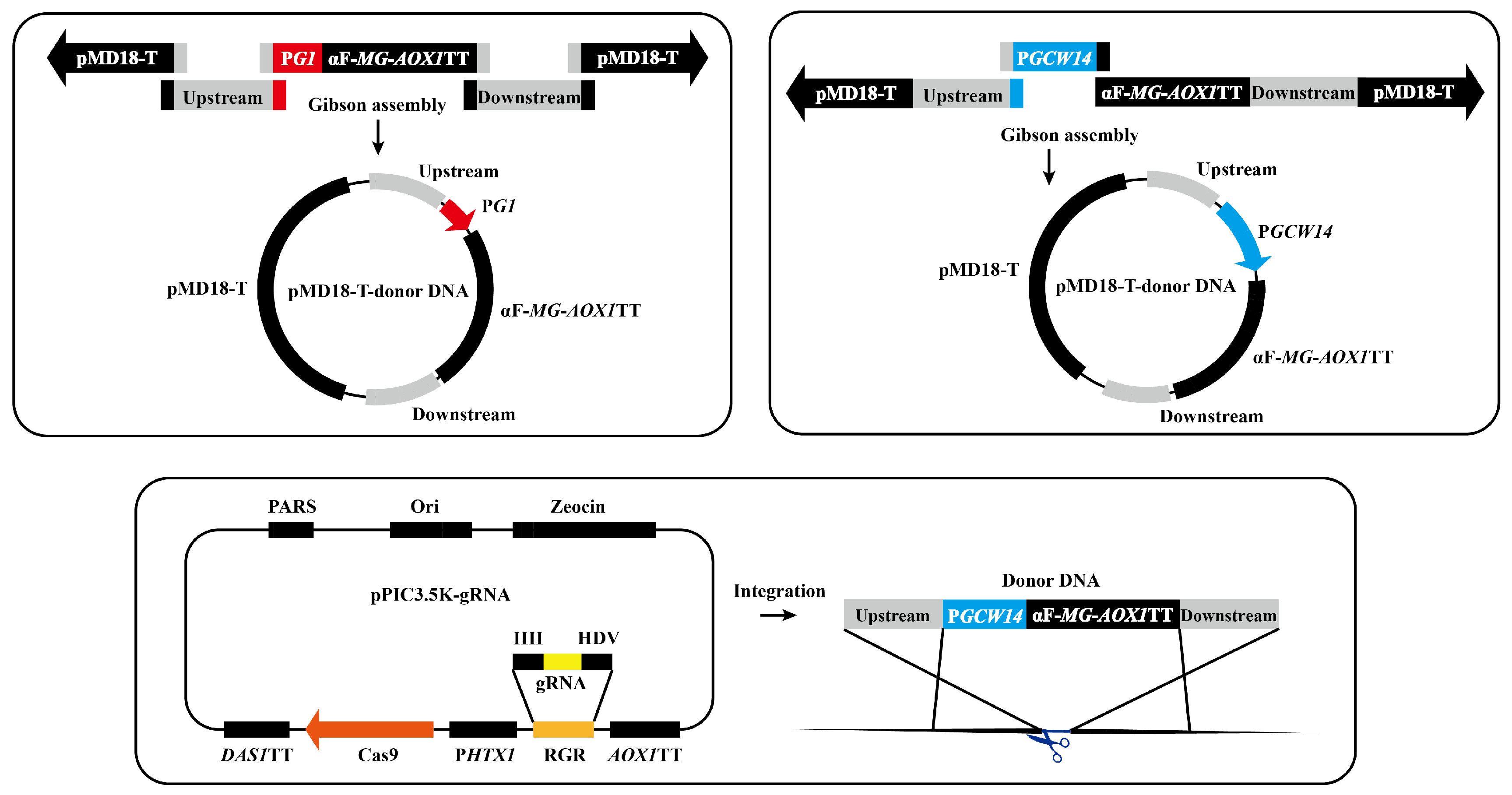
2.2. Optimization of Promoter and Copy Number for pMG Expression

The plasmid pG1ZαA-pMG was used as a template, and the upstream primer 1.6kb-G1-F and the downstream primer 1.6kb-AOXTT-R were used to obtain a fragment of an expression cassette. Primer pairs 2.7kb-18T-F/R were used to amplify the fragment as a plasmid skeleton from pMD18-T. The upstream and downstream homologous arms (~1 kb) of PAOX1UP-g2(Ag2), PTEF1UP-g1(Tg1), and PFLD1UP-g1(Fg1) were acquired by primers listed in Table S3. Taking the plasmid pMD18-T-Ag2-G1-pMG as a template, the four obtained fragments were assembled using the Gibson system and the assembly product was transformed into E. coli DH5α. Primer pairs 0.8kb-GCW14-F/R were used to acquire the GCW14 promoter from the K. phaffii genome. Then, the PG1 in the plasmid pMD18-T-Ag2/Tg1/Fg1-G1-pMG were replaced with the PGCW14. After DNA sequencing, the donor DNA was acquired by PCR amplification (amplified by primer pairs of donor-Ag2-F/R, donor-Tg1-F/R, or donor-Fg1-F/R).
For single-locus integration [19], K. phaffii X33-Δku70 was transformed with 400 ng gRNA-Cas9 plasmid and 4 μg donor DNA by electroporation (Figure 1). The steps of electroporation had been detailed in a previous report [18]. Transformants were selected on YPD plates with Zeocin and confirmed through DNA sequencing. Then, the strain discarded the gRNA-Cas9 plasmid until it only could grow on the YPD plate [20]. Next, the engineered strain was used to integrate the pMG gene at a new locus. At least three single bacterial colonies were selected for shaking-flask experiments in triplicate.

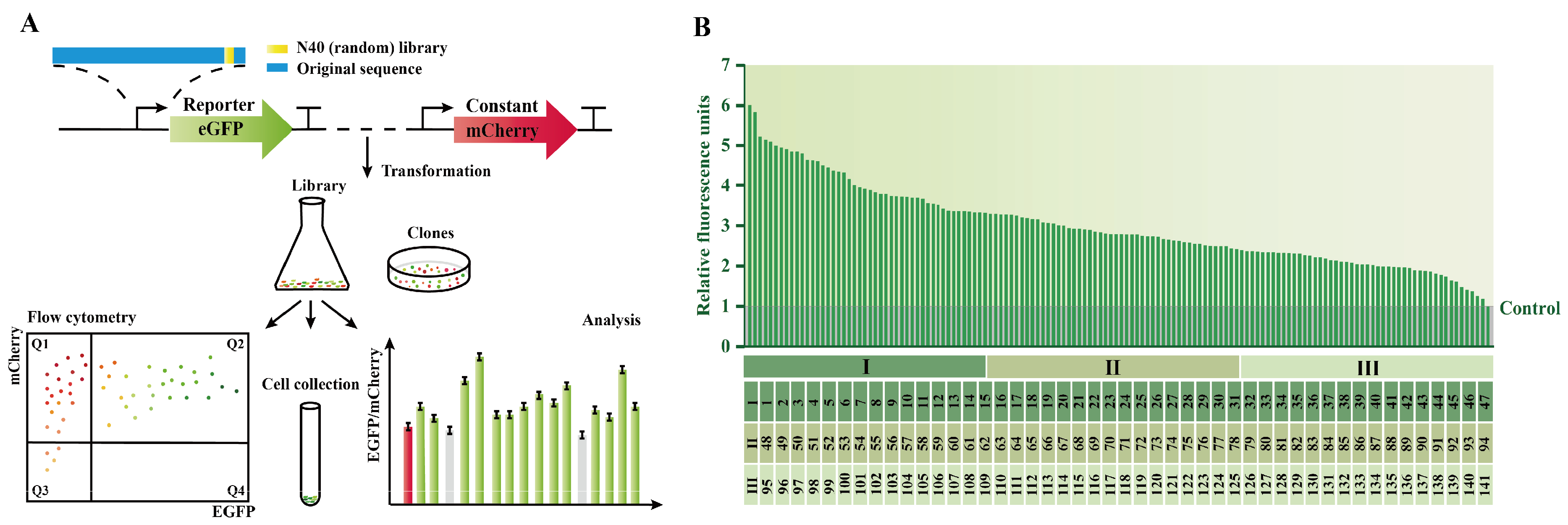
2.3. Construction of PGCW14 Mutant Library

The signal peptide in plasmid pMD18-T-Ag2-GCW14-pMG was removed and the pMG gene was replaced with the mCherry gene. The new constructed plasmid was transformed into K. phaffii X33-Δku70 by electroporation. After a DNA sequence analysis, the acquired K. phaffii X33-Δku70-mCherry strain that stably expressed red fluorescent protein was used to eliminate the environmental color interference in the following detection of green fluorescence [21].
To construct a plasmid containing the EGFP gene, the pGCW14ZαA-pMG plasmid was used as a template and the primer pairs 3.2kb-pZA-EGFP-F/R were used to obtain a fragment without the α-factor and pMG gene and the EGFP gene fragment was amplified through the primer pairs 0.7kb-EGFP-F/R. These two fragments were assembled through the Gibson method, and the obtained plasmid was then transformed into E. coli DH5α. To construct the mutations in the core region of PGCW14, the extracted plasmid pGCW14ZA-EGFP was used for PCR by the upstream primer of saturated random mutation 3.9k-GCW14-N40-F and the downstream primer 3.9k-GCW14-N40-R. Then, the fragment was transformed into E. coli DH5α and was cultured in the LLB medium with 50 μg/mL of Zeocin for 16–18 h. The mixed plasmids were extracted from the culture medium. The mixture of PGCW14 mutant plasmids was linearized by restriction digestion with Eco91 I (BstE II) and transformed into K. phaffii X33-Δku70-GCW14-mCherry by electroporation. After 48 h of growth in the YPD medium with 100 μg/mL of Zeocin, the cells were washed three times with 1× PBS buffer for subsequent screening.

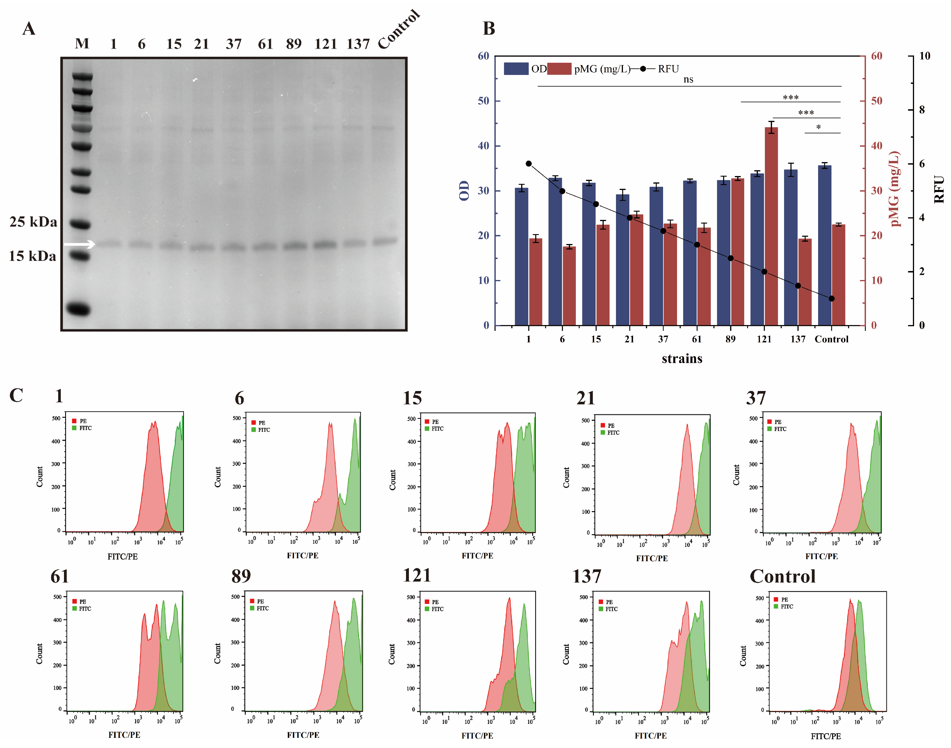
2.4. Screening of the PGCW14 Mutant with Enhanced Strength

The cells at the exponential growth phase were harvested and diluted with 1× PBS to a final OD600 of around 0.3. Cells were loaded into germ-free test tubes and sent to the testing platform for flow cytometry. Flow cytometry was performed on a BD FACSAria Ⅲ flow cytometer using BD FACSDiva version 7.0 Software. The ratio of EGFP/mCherry was used to measure the strength of mutated promoters [22]. The obtained cells were collected into tubes based on an EGFP/mCherry ratio higher than the ratio in the control (fluorescent proteins expressed by initial PGCW14). To specifically detect the expressional intensity, the sorted cells were spread onto plates (supplemented with 100 μg/mL Zeocin) for 2–3 days, and single colonies were used for the same pretreatment method for a flow cytometry analysis.
The cx-GCW14-N40-F and chr10587-r primers were utilized to amplify fragments containing mutated promoters from the selected cells. The sequences of the core region for each mutated promoter were confirmed by DNA sequencing. Then, the first round of PCR products was used as a template and the 0.8kb-GCW14-F and 0.8kb-GCW14-R primers were used for the second round of amplification. The correct PGCW14 mutants obtained were confirmed by DNA sequencing and were ligated to the pGCW14ZαA-pMG plasmid without PGCW14 by the Gibson method. The positive transformants were selected on a YPD plate with Zeocin.

2.5. The Enhancement of pMG Expression by the Inhibition of Protein Degradation

The fragment without PG1 was obtained from the pG1ZαA-pMG plasmid and the plasmid skeleton and the fragment PGCW14 were ligated by the Gibson method. The pGCW14ZαA-pMG plasmid was confirmed by DNA sequencing and transformed into K. phaffii strains’ knockout of proteases, including K. phaffii X33-Δku70pep4, X33-Δku70yps1, X33-Δku70prb1, X33-Δku70pep4yps1, and X33-Δku70pep4yps1prb1 [12]. In addition, K. phaffii X33-Δku70-GCW14-pMG was used as a control.

2.6. The Enhancement of pMG Expression by the Co-Expression of Proteins of Anti-Stress Response Systems

Nine proteins involved in the anti-stress response were selected to construct different vectors [23,24], including ERO1, PDI, HAC1, KAR2, IRE1, AHA1, PRX1, YPT6, and YAP1. The details of these proteins are contained in Table 1. Taking the construction of plasmid pGAPZαA-ERO1 as an example, primer pairs 1.6kb-ERO1-F/R were used to obtain a gene encoding ERO1 protein from the K. phaffii genome. Primers 2.8kb-pGAPZαA-F/R were used to amplify the fragment as a plasmid skeleton from the pGAPZαA plasmid. The two fragments were assembled by the Gibson method. The reaction system containing the circular plasmid was transformed into E. coli DH5α. After DNA sequencing, the linearized gene fragment was transformed into K. phaffii by electroporation.

2.7. Cultural Conditions for pMG Expression at Shaking-Flask Level

After growing on a YPD plate for 2–3 days, single colonies were inoculated into a 12 mL test tube containing 2 mL of the YPD medium and were incubated at 30 °C and 220 rpm for 16–18 h to acquire the primary seed culture. The primary seed culture was transferred to a medium containing a 2% (v/v) inoculum. All shaking-flask experiments were carried out in triplicate for 60 h. The expression of pMG was verified by sodium dodecyl sulfate polyacrylamide gel electrophoresis (SDS-PAGE), and the titer of pMG was detected by the Bradford method (Beyotime Biotech, Shanghai, China) combined with Image Lab version 5.2.1 grayscale analysis software (Bio-Rad, Shanghai, China).

2.8. Conditions for pMG Expression at Fermenter Level

The acquisition of the original seed culture was consistent with the mentioned method for the shaking-flask experiment. Then, the primary seed culture (1 mL) was inoculated into the 250 mL shaking flasks supplied with 50 mL YPD medium and grown at 30 °C and 220 rpm for 24 h. The secondary seed solution (200 mL) was transferred into a 5 L bioreactor containing 1.8 L BSM medium. The temperature was maintained at 30 °C and the pH value of the culture medium was adjusted to 5.50 ± 0.10 using 50% NH4OH. The air flow rate was set at 3 L/min and the agitation speed ranged from 200 to 800 rpm. After 12 h of fermentation, hemin chloride with a final concentration of 40 mg/L was added to the culture medium and the final concentration of fed hemin chloride was set at 150 mg/L. Then, the DO-stat strategy was used for feeding, maintaining the dissolved oxygen (DO) level at 30% of the air saturation value.

2.9. Purification of Food-Grade pMG

The strategy for the food-grade purification of pMG was performed as previously mentioned [5]. The pMG secreted in 1 L of a fermentation supernatant was concentrated to 200 mL through a Vivaflow 200R (Sartorius, Germany) ultrafiltration membrane package. We collected the eluent by 20% NaCl to obtain the purified sample of pMG. Purified hemoproteins were desalted using Amicon Ultra 3 K (Millipore, Darmstadt, Germany).

2.10. The Peroxidase-Specific Activity of Synthesized pMG

The peroxidase (POD)-specific activity of purified pMG was determined using the 3,3′,5,5′-Tetramethylbenzidine (TMB) Chromogen Solution reagent kit (P0209, Beyotime Biotech, Shanghai, China) [25]. POD-specific activity (U/mg) is defined as the ratio of total POD activity (U/mL) to the concentration of pMG (mg/mL).

3. Results and Discussion

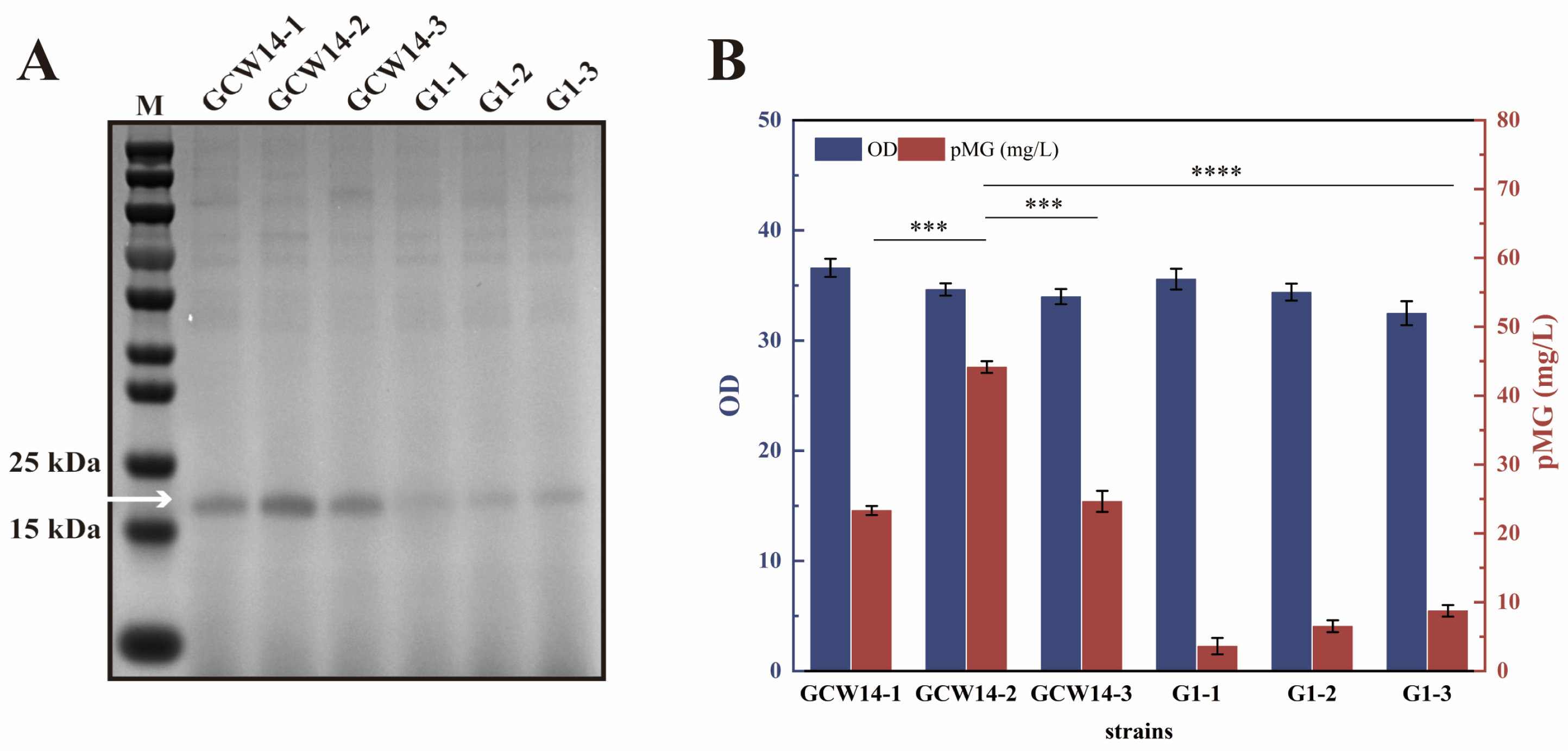
3.1. Selection of Proper Promoter and Integrated Copy Number for pMG Expression

In previous research, the expression of pMG was relatively weak in K. phaffii [26]. The main reason was the lower strength of expression elements, including the application of the GAP promoter (PGAP). PG1 [27] and PGCW14 [28] are also constitutive promoters with significantly higher strength than PGAP in K. phaffii. Thus, these two promoters were selected to enhance the expression of pMG. In addition, three efficient gRNA targets (PAOX1UP-g2, PTEF1UP-g1, and PFLD1UP-g1) were used to sequentially integrate the recombinant pMG gene to achieve the multi-copy expression of pMG [29]. The non-homologous-end-joining defective strain (K. phaffii X33-Δku70) enhances the homology-directed repair (HDR) efficiency of the target gene [30,31]. The engineered strains (based on K. phaffii X33-Δku70 [12]) expressing one, two, and three copies of pMG, driven by the PG1 and PGCW14, respectively, were fermented in the BMGY medium for 60 h.
Based on the band of pMG (17 kDa), the result showed that the titers of pMG expressed by the strains under the control of the GCW14 promoter initially increased and then decreased with the increasing copy number of the pMG gene. This result may be caused by the high metabolic burden and protein-folding stress in the endoplasmic reticulum [32]. The highest titer of pMG (44.16 mg/L) was obtained by the strain GCW14-2, representing an 89.37% increase compared to the pMG titer (23.32 mg/L) obtained by the strain GCW14-1. The titers of pMG expressed under the control of the G1 promoter by the strains G1-1 to G1-3 were gradually enhanced with the increasing copy number of the pMG gene, but the highest titer of 8.76 mg/L remained significantly lower than the titers obtained by the strain GCW14-1 (Figure 2). Therefore, the PGCW14 was selected as the most suitable promoter in the following experiments.

3.2. The Construction of PGCW14 Mutant Libraries to Enhance Its Strength

The core region of a classic eukaryotic promoter includes a spacer sequence between the transcription start site and upstream TATA box [22,33], which is highly correlated with the strength of the promoter. As for PGCW14, there are 40 bases from −1 to −40 positions. Thus, these 40 bases were randomly mutated to enhance the strength of PGCW14. To evaluate the effect of mutations, the original PGCW14 was used to control the expression of red fluorescent protein (mCherry) and the mutants of PGCW14 were used to express green fluorescent protein (EGFP). In the control group, the expressions of EGFP and mCherry were controlled by the original PGCW14 promoter. The relative fluorescence units (RFUs) were defined as the value obtained by comparing the fluorescence ratios between the mutants and the control group. The intensity of the mutated promoter was, respectively, normalized by the RFU, and nearly 20,000 recombinant cells were sorted using high-throughput flow cytometry. As a result, there were 141 mutants of PGCW14 with higher EGFP/mCherry fluorescent ratios than the value in the control (Figure 3). After DNA sequencing, the mutated sequences were obtained and their detailed information was summarized in Table S4. The highest relative fluorescence unit of mutant PGCW14 (PGCWm-1) increased to 6.01 times, which suggests a great improvement of PGCW14 strength.

3.3. The Application of PGCW14 Mutants to Increase pMG Expression

Compared to the utilization of the inducible promoter in the K. phaffii strain, the titer of pMG expressed by the constitutive PGCW14 promoter remained at a lower level. To enhance promoter strength and ensure better adaptation to pMG expression, nine mutants of PGCW14 with different strengths (PGCWm-1, PGCWm-6, PGCWm-15, PGCWm-21, PGCWm-37, PGCWm-61, PGCWm-89, PGCWm-121, and PGCWm-137, fluorescent units ranging from 6.01 to 1.48 times) were selected from the positive mutated library for the expression of pMG. The results showed that the best expressional effect for pMG was achieved by the PGCWm-121 with a moderate strength (2.00 times fluorescent unit) and the titer of pMG (44.14 mg/L) increased by 96.44% compared to the titer obtained by the original PGCW14 (22.47 mg/L) (Figure 4). There was a nonlinear correlation between the promoter strength and expressional level, which might be caused by the excessive metabolic burden, and other factors inhibited the cell growth or the expression of pMG [20]. Based on the effect on pMG expression, the mutants of the PGCW14 promoter with a broader expressional range could be further used for the constitutive production of other useful proteins and the regulation of metabolic engineering in K. phaffii.

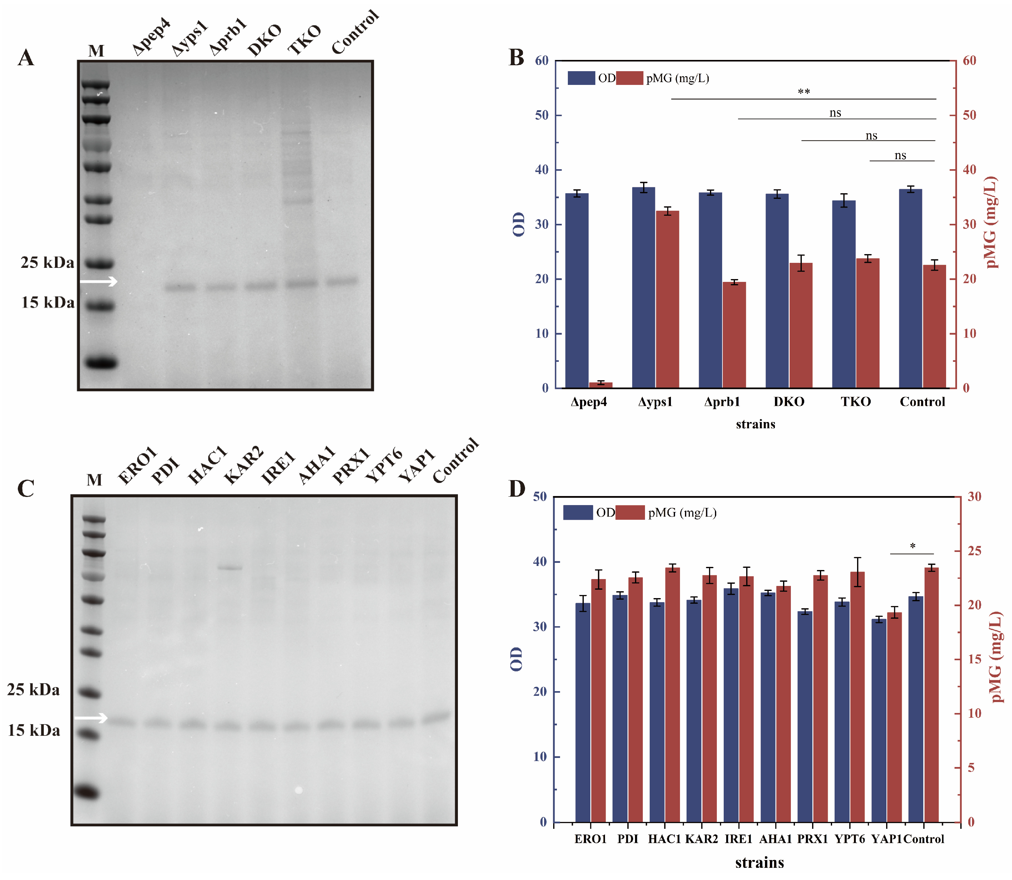
3.4. The Construction of a Proper K. phaffii Host for the Efficient pMG Expression

Based on the proper PGCWm-121 promoter, the engineered K. phaffii strain X33-GCWm-121-pMG*2 was constructed. However, applying this engineered strain, the titer of pMG presented earlier has an increase and later decrease trend from 24 to 84 h. It suggests that there was an obvious degradation of pMG during the longer fermentation period [25]. According to the previous reports, during the fed-batch fermentation of K. phaffii, vacuolar aspartic protease A (Pep4) and the GPI-anchored aspartic protease (Yps1-1) can be found in the secreted proteome [34]. In addition, the recombinant K. phaffii strain lacking the prb1 gene (encoding vacuolar serine protease B) can also reduce the proteolytic activity [35]. Therefore, these three proteases were knocked out in the K. phaffii strain X33-Δku70 to investigate their effects on pMG expression. Among the five protease-deficient strains, the K. phaffii X33-Δku70yps1-GCW14-pMG achieved higher pMG expression (32.47 mg/L), which was 43.86% higher than the titer obtained by control strain X33-Δku70-GCW14-pMG (Figure 5A,B). This result was consistent with the previous report that the utilization of Yps1 protease-deficient strains can effectively inhibit the degradation of pMG expressed under the control of the inducible PAOX1 system [12].
The overexpression of proteins of anti-stress response systems was another approach to overcome the challenges during the secretion of heterogenous proteins in K. phaffii [36]. Hence, nine endogenous proteins were selected to co-express with pMG, respectively. However, the results showed that the overexpression of these proteins could not significantly enhance the pMG-secreted expression level (Figure 5C, D). Thus, the co-expression of proteins involved in the anti-stress response was not applied in the final engineered strain.
To further enhance the efficiency of pMG expression, the key gene KU70 that was knocked out to facilitate the gene integration into the genome was reintroduced to minimize the potential impacts on cells [37]. Based on all the effective strategies, the final engineered K. phaffii strain X33-Δyps1-GCWm-121-pMG*2 was constructed and was fermented in the BMGY medium for 60 h. The final titer of pMG reached 59.12 mg/L, representing a 153.52% increase compared to the original strain GCW14-1 (23.32 mg/L) (Figure 6).

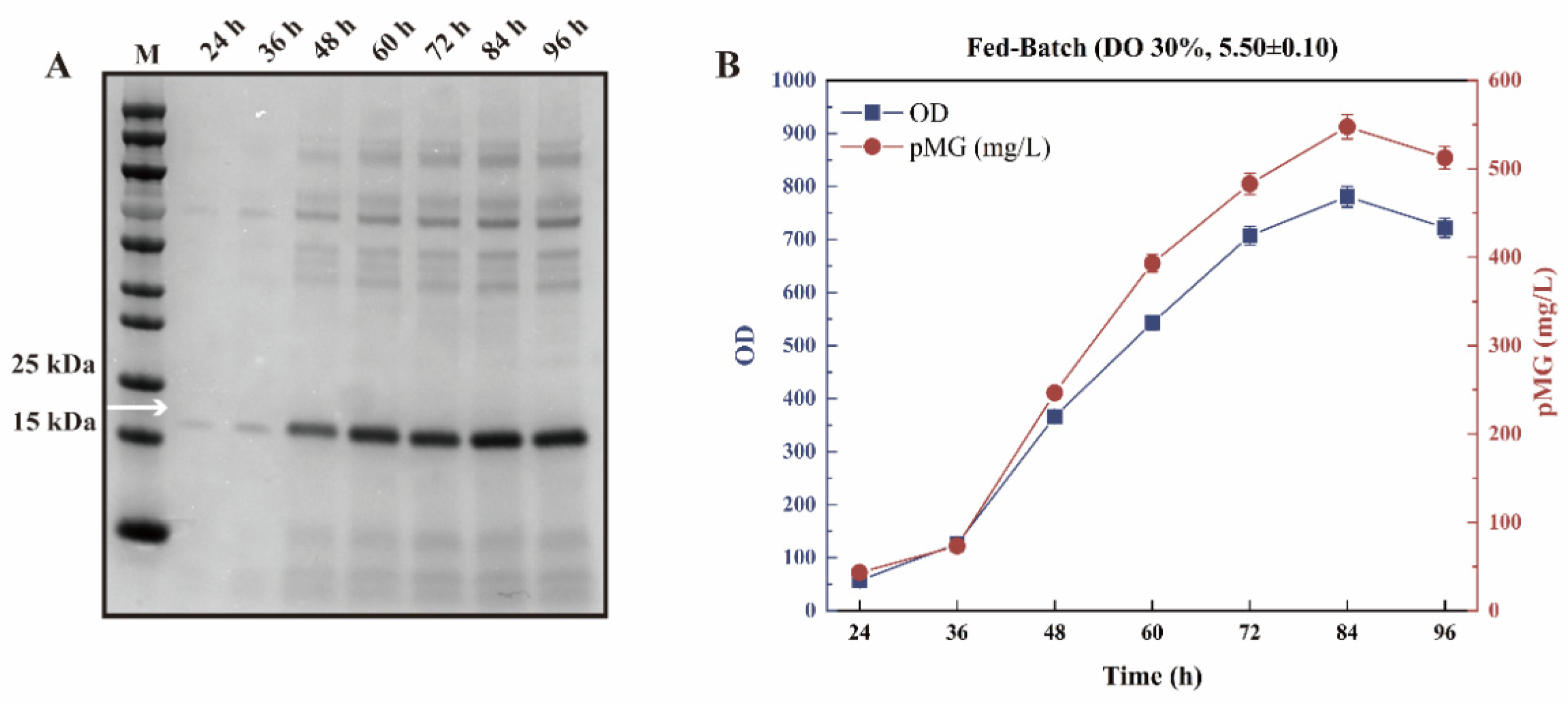
3.5. The Biochemical Properties of pMG Efficiently Expressed by Fed-Batch Fermentation Using an Economical Medium

The shaking-flask fermentation is hard to control and is not suitable for industrial production, especially for a limited oxygen supply [38]. Thus, a large-scale fermentation system should be established for pMG expression [39] and an economical industrial medium has to be determined at first. Compared to the rich BMGY medium (USD 10.01 per liter), the BSM medium (USD 1.15 per liter) was composed of basic salts, a trace metal solution (PTM1), and glycerol [40], reducing the cost of production by 88.5%. Thus, the BSM medium was chosen for the industrial expression of pMG. Besides the medium, the fermentation conditions are also important to produce heterogenous proteins in K. phaffii [41]. Dependent on the previous optimized fermentation conditions for the constitutive expression, the fermentation temperature was set at 30 °C, and 30% dissolved oxygen (DO) stat was applied for the fed-batch fermentation. In addition, the industrial-grade hemin was supplemented at a final concentration of 150 mg/L because heme is the essential co-factor for the activity of pMG [42]. During the period of fed-batch fermentation, pMG was secreted after 24 h and the expressional level of pMG continued to increase until 84 h. The highest titer of pMG reached 547.59 mg/L in a 5 L fermenter (Figure 7), which was increased by 191.85% compared to previous studies (BMGY medium) [5].
As the affinity tag (His tag) can be converted into histamine that has potential in triggering allergic reactions in humans [43], the His tag was not included in the recombinant pMG gene. The ultrafiltration concentration–anion exchange (gravity chromatography column) with the advantages of a simple operation, low cost, and high recovery rate [5] was used to prepare the purified pMG in large-scale applications in food processing. The results showed that the obtained pMG had a recovery rate of 54.25% and a purity of 93.41% (Figure 8A,B).
In the following, the POD-specific activity of synthesized pMG by K. phaffii was evaluated. To eliminate the influence of endogenous secretory proteins in K. phaffii on the detection of POD activity, we tested the fermentation supernatant and related purified sample obtained from the control strain K. phaffii X33-Δyps1 without the pMG gene. The results showed that neither the fermentation supernatant nor the purified sample from the control strain exhibited POD activity. The POD activity of purified pMG after desalination was 123.53 U/mL. Based on the titer of pMG (288.97 mg/L), the calculated POD-specific activity was 427.50 U/mg (Figure 8C).
In summary, a mutant promoter library was created through random mutation in the core region of PGCW14. Among numerous mutants, the strengths of 141 positive mutants increased by 1.18 and 6.01 times, respectively. The efficient synthesis of pMG was achieved by selecting the mutated PGCWm-121 with moderate strength, and double integrated copies is the most suitable copy number for pMG expression. In addition, the degradation of pMG was inhibited by knocking out the protease (Yps1-1). The highest titer of pMG reached 547.59 mg/L at the fermenter level using the economical BSM medium with feeding 150 mg/L of hemin (30% DO and 30 °C). After the food-grade purification, the obtained pMG had similar peroxide-specific activity (427.50 U/mg) with the extracted natural standard.

Supplementary Materials

The following supporting information can be downloaded at: https://www.mdpi.com/article/10.3390/fermentation11020049/s1, Table S1. The strains used in this study. Table S2. The plasmids used in this study. Table S3. The primers used in this study. Table S4. PGCW14 mutants (PGCWm) in this study.

Author Contributions

X.Z. and D.S. conceived the project. D.S. and Y.W. performed the experiments and data analysis. J.Z., J.L., J.C. and G.D. supervised the project. D.S. wrote the manuscript and X.Z. revised the manuscript. All authors have read and agreed to the published version of the manuscript.

Funding

This article was supported by the Foundation for Innovative Research Groups of the National Natural Science Foundation of China (32021005), the Jiangsu Basic Research Center for Synthetic Biology (BK20233003), and the National First-class Discipline Program of Light Industry Technology and Engineering (LITE2018-08).

Institutional Review Board Statement

Not applicable.

Informed Consent Statement

Not applicable.

Data Availability Statement

The original contributions presented in this study are included in the article/Supplementary Material. Further inquiries can be directed to the corresponding authors.

Conflicts of Interest

The authors declare no conflicts of interest.

References

  1. Ordway, G.A.; Garry, D.J. Myoglobin: An essential hemoprotein in striated muscle. J. Exp. Biol. 2004, 207, 3441–3446. [Google Scholar] [CrossRef]
  2. Tan, B.; Sun, B.; Sun, N.; Li, C.; Zhang, J.; Yang, W. Structure, functional properties and iron bioavailability of Pneumatophorus japonicus myoglobin and its glycosylation products. Int. J. Biol. Macromol. 2021, 173, 524–531. [Google Scholar] [CrossRef]
  3. Sun, B.; Zhang, P.; Zhang, J.; Huang, T.; Li, C.; Yang, W. Preparation, characterization and bioavailability studies of Tegillarca granosa hemoglobin and its glycosylated products. Int. J. Biol. Macromol. 2022, 219, 11–20. [Google Scholar] [CrossRef]
  4. Chen, J.; Ran, F.; Chen, Q.; Luo, D.; Ma, W.; Han, T.; Wang, C.; Wang, C. A fluorescent biosensor for cardiac biomarker myoglobin detection based on carbon dots and deoxyribonuclease I-aided target recycling signal amplification. RSC Adv. 2019, 9, 4463–4468. [Google Scholar] [CrossRef]
  5. Zhang, B.; Zhao, X.; Wang, Z.; Wang, H.; Zhou, J.; Du, G.; Chen, J.; Li, J. Efficient secretory expression and purification of food-grade porcine myoglobin in Komagataella phaffii. J. Agric. Food Chem. 2021, 69, 10235–10245. [Google Scholar] [CrossRef]
  6. Liu, Q.; Long, Y.; Zhang, Y.F.; Zhang, Z.Y.; Yang, B.; Chen, C.Y.; Huang, L.S.; Su, Y. Phenotypic and genetic correlations of pork myoglobin content with meat colour and other traits in an eight breed-crossed heterogeneous population. Animal 2021, 15, 100364. [Google Scholar] [CrossRef] [PubMed]
  7. Lee, H.; Yong, H.; Kim, M.; Choi, Y.; Jo, C. Status of meat alternatives and their potential role in the future meat market—A review. Asian-Australas. J. Anim. Sci. 2020, 33, 1533–1543. [Google Scholar] [CrossRef] [PubMed]
  8. Karbalaei, M.; Rezaee, S.A.; Farsiani, H. Pichia pastoris: A highly successful expression system for optimal synthesis of heterologous proteins. J. Cell. Physiol. 2020, 235, 5867–5881. [Google Scholar] [CrossRef]
  9. Hasan, M.M.; Ushio, H.; Ochiai, Y. Expression and characterization of rainbow trout Oncorhynchus mykiss recombinant myoglobin. Fish Physiol. Biochem. 2021, 47, 1477–1488. [Google Scholar] [CrossRef]
  10. Meng, Y.; Xie, L.; You, K.; Chen, W. High-level secretory production of leghemoglobin and myoglobin in Escherichia coli through inserting signal peptides. Food Biosci. 2023, 56, 103356. [Google Scholar] [CrossRef]
  11. Ye, Y.; Sun, J.; Xu, J.; Li, P.; Sheng, L.; Qian, Y.; Ji, J.; Han, X.; Zhao, X.; Zhou, J.; et al. Allergenic risk assessment of porcine myoglobin expressed by engineered Komagataella phaffii. Fundam. Res. 2024, 4, 1339–1348. [Google Scholar] [CrossRef] [PubMed]
  12. Yu, F.; Zhao, X.; Zhou, J.; Lu, W.; Li, J.; Chen, J.; Du, G. Biosynthesis of high-active hemoproteins by the efficient heme-supply Pichia Pastoris chassis. Adv. Sci. 2023, 10, 2302826. [Google Scholar] [CrossRef] [PubMed]
  13. Unver, Y.; Ari, B.; Acar, M.; Arslan, S.Y. A self-inducible heterologous protein expression system in Komagataella phaffii (Pichia pastoris). 3 Biotech 2024, 14, 193. [Google Scholar] [CrossRef]
  14. Zhang, X.; Zhang, X.; Liang, S.; Ye, Y.; Lin, Y. Key regulatory elements of a strong constitutive promoter, PGCW14, from Pichia pastoris. Biotechnol. Lett. 2013, 35, 2113–2119. [Google Scholar] [CrossRef] [PubMed]
  15. Liang, S.; Zou, C.; Lin, Y.; Zhang, X.; Ye, Y. Identification and characterization of PGCW14: A novel, strong constitutive promoter of Pichia pastoris. Biotechnol. Lett. 2013, 35, 1865–1871. [Google Scholar] [CrossRef]
  16. Shen, Q.; Cui, J.; Wang, Y.; Hu, Z.; Xue, Y.; Zheng, Y. Identification of a novel growth-associated promoter for biphasic expression of heterogenous proteins in Pichia pastoris. Appl. Ind. Microbiol. 2024, 90, e0174023. [Google Scholar] [CrossRef] [PubMed]
  17. Dai, W.; Dong, H.; Zhang, Z.; Wu, X.; Bao, T.; Gao, L.; Chen, X. Enhancing the heterologous expression of a thermophilic endoglucanase and its cost-Effective production in Pichia pastoris using multiple strategies. Int. J. Mol. Sci. 2023, 24, 15017. [Google Scholar] [CrossRef]
  18. Liu, Q.; Shi, X.; Song, L.; Liu, H.; Zhou, X.; Wang, Q.; Zhang, Y.; Cai, M. CRISPR–Cas9-mediated genomic multiloci integration in Pichia pastoris. Microb. Cell Fact. 2019, 18, 144. [Google Scholar] [CrossRef] [PubMed]
  19. Schusterbauer, V.; Fischer, J.E.; Gangl, S.; Schenzle, L.; Rinnofner, C.; Geier, M.; Sailer, C.; Glieder, A.; Thallinger, G.G. Whole genome sequencing analysis of effects of CRISPR/Cas9 in Komagataella phaffii: A budding yeast in distress. J. Fungi 2022, 8, 992. [Google Scholar] [CrossRef]
  20. Shen, Q.; Yu, Z.; Zhou, X.; Zhang, S.; Zou, S.; Xiong, N.; Xue, Y.P.; Liu, Z.Q.; Zheng, Y. Identification of a novel promoter for driving antibiotic-resistant genes to reduce the metabolic burden during protein expression and effectively select multiple integrations in Pichia pastoris. Appl. Microbiol. Biotechnol. 2021, 105, 3211–3223. [Google Scholar] [CrossRef]
  21. de Boer, C.G.; Vaishnav, E.D.; Sadeh, R.; Abeyta, E.L.; Friedman, N.; Regev, A. Deciphering eukaryotic gene-regulatory logic with 100 million random promoters. Nat. Biotechnol. 2019, 38, 56–65. [Google Scholar] [CrossRef] [PubMed]
  22. Lubliner, S.; Regev, I.; Lotan-Pompan, M.; Edelheit, S.; Weinberger, A.; Segal, E. Core promoter sequence in yeast is a major determinant of expression level. Genome Res. 2015, 25, 1008–1017. [Google Scholar] [CrossRef] [PubMed]
  23. Yu, Y.; Liu, Z.; Chen, M.; Yang, M.; Li, L.; Mou, H. Enhancing the expression of recombinant κ-carrageenase in Pichia pastoris using dual promoters, co-expressing chaperones and transcription factors. Biocatal. Biotransform. 2019, 38, 104–113. [Google Scholar] [CrossRef]
  24. Huang, M.; Gao, Y.; Zhou, X.; Zhang, Y.; Cai, M. Regulating unfolded protein response activator HAC1p for production of thermostable raw-starch hydrolyzing α-amylase in Pichia pastoris. Bioprocess Biosyst. Eng. 2016, 40, 341–350. [Google Scholar] [CrossRef]
  25. Shao, Y.; Xue, C.; Liu, W.; Zuo, S.; Wei, P.; Huang, L.; Lian, J.; Xu, Z. High-level secretory production of leghemoglobin in Pichia pastoris through enhanced globin expression and heme biosynthesis. Bioresour. Technol. 2022, 363, 127884. [Google Scholar] [CrossRef] [PubMed]
  26. Nguyen, L.; Schmelzer, B.; Wilkinson, S.; Mattanovich, D. From natural to synthetic: Promoter engineering in yeast expression systems. Biotechnol. Adv. 2024, 77, 108446. [Google Scholar] [CrossRef] [PubMed]
  27. Qin, X.; Qian, J.; Yao, G.; Zhuang, Y.; Zhang, S.; Chu, J. GAP promoter library for fine-tuning of gene expression in Pichia pastoris. Appl. Environ. Microbiol. 2011, 77, 3600–3608. [Google Scholar] [CrossRef] [PubMed]
  28. Yan, C.; Yu, W.; Zhai, X.; Yao, L.; Guo, X.; Gao, J.; Zhou, Y.J. Characterizing and engineering promoters for metabolic engineering of Ogataea polymorpha. Synth. Syst. Biotechnol. 2022, 7, 498–505. [Google Scholar] [CrossRef] [PubMed]
  29. Wu, L.; Xu, Y.; Yu, X. Efficient CRISPR-mediated C-to-T base editing in Komagataella phaffii. Biotechnol. J. 2024, 19, 2400115. [Google Scholar] [CrossRef]
  30. Ghosh, D.; Raghavan, S.C. Nonhomologous end joining: New accessory factors fine tune the machinery. Trends Genet. 2021, 37, 582–599. [Google Scholar] [CrossRef]
  31. Weninger, A.; Fischer, J.E.; Raschmanová, H.; Kniely, C.; Vogl, T.; Glieder, A. Expanding the CRISPR/Cas9 toolkit for Pichia pastoris with efficient donor integration and alternative resistance markers. J. Cell. Biochem. 2017, 119, 3183–3198. [Google Scholar] [CrossRef] [PubMed]
  32. Yu, S.; Miao, L.; Huang, H.; Li, Y.; Zhu, T. High-level production of glucose oxidase in Pichia pastoris: Effects of Hac1p overexpression on cell physiology and enzyme expression. Enzyme Microb. Technol. 2020, 141, 109671. [Google Scholar] [CrossRef]
  33. Tang, H.; Wu, Y.; Deng, J.; Chen, N.; Zheng, Z.; Wei, Y.; Luo, X.; Keasling, J. Promoter architecture and promoter engineering in Saccharomyces cerevisiae. Metabolites 2020, 10, 320. [Google Scholar] [CrossRef] [PubMed]
  34. Burgard, J.; Grünwald-Gruber, C.; Altmann, F.; Zanghellini, J.; Valli, M.; Mattanovich, D.; Gasser, B. The secretome of Pichia pastoris in fed-batch cultivations is largely independent of the carbon source but changes quantitatively over cultivation time. Microb. Biotechnol. 2019, 13, 479–494. [Google Scholar] [CrossRef]
  35. Marsalek, L.; Gruber, C.; Altmann, F.; Aleschko, M.; Mattanovich, D.; Gasser, B.; Puxbaum, V. Disruption of genes involved in CORVET complex leads to enhanced secretion of heterologous carboxylesterase only in protease deficient Pichia pastoris. Biotechnol. J. 2017, 12, 1600584. [Google Scholar] [CrossRef]
  36. Xiao, X.; Liu, Z.; Chen, Y.; Wang, G.; Li, X.; Fang, Z.; Huang, S.; Liu, Z.; Yan, Y.; Xu, L. Over-expression of active Candida rugosa lip1 in Pichia pastoris via high cell-density fermentation and its application to resolve racemic ibuprofen. Biocatal. Biotransform. 2016, 33, 260–269. [Google Scholar] [CrossRef]
  37. Wang, X.; Li, Y.; Jin, Z.; Liu, X.; Gao, X.; Guo, S.; Yu, T. A novel CRISPR/Cas9 system with high genomic editing efficiency and recyclable auxotrophic selective marker for multiple-step metabolic rewriting in Pichia pastoris. Synth. Syst. Biotechnol. 2023, 8, 445–451. [Google Scholar] [CrossRef] [PubMed]
  38. Zhu, W.; Xu, R.; Gong, G.; Xu, L.; Hu, Y.; Xie, L. Medium optimization for high yield production of human serum albumin in Pichia pastoris and its efficient purification. Protein Expr. Purif. 2021, 181, 105831. [Google Scholar] [CrossRef]
  39. Guo, Y.; Liao, Y.; Wang, J.; Ma, C.; Qin, J.; Feng, J.; Li, Y.; Wang, X.; Chen, K. Methylotrophy of Pichia pastoris: Current advances, applications, and future perspectives for methanol-based biomanufacturing. Acs Sustain Chem Eng. 2022, 10, 1741–1752. [Google Scholar] [CrossRef]
  40. Bolmanis, E.; Bogans, J.; Akopjana, I.; Suleiko, A.; Kazaka, T.; Kazaks, A. Production and purification of soy leghemoglobin from Pichia pastoris cultivated in different expression media. Processes 2023, 11, 3215. [Google Scholar] [CrossRef]
  41. Hu, Y.; Bai, R.; Dou, S.; Wu, Z.m.; Abdulkhani, A.; Asadollahi, M.A.; Abomohra, A.E.-F.; Sun, F. Constitutive expression of codon optimized Trichoderma reesei TrCel5A in Pichia pastoris using GAP promoter. Syst. Microbiol. Biomanuf. 2022, 2, 498–506. [Google Scholar] [CrossRef]
  42. Yu, F.; Li, C.; Zhang, T.; Zhou, J.; Li, J.; Chen, J.; Du, G.; Zhao, X. Developing a novel heme biosensor to produce high-active hemoproteins in Pichia pastoris through comparative transcriptomics. Metab. Eng. 2024, 84, 59–68. [Google Scholar] [CrossRef] [PubMed]
  43. Liu, Y.; Li, H.; Wang, C.; Chen, S.; Lian, R.; Wang, W.; Fu, L.; Wang, Y. Immunological disturbance effect of exogenous histamine towards key immune cells. Food Sci. Hum. Wellness. 2024, 13, 1856–1863. [Google Scholar] [CrossRef]
Figure 1. The schematic diagram of constructing recombinant K. phaffii strains by CRISPR/Cas9 technology.
Fermentation 11 00049 g001
Figure 2. Comparison of pMG expression in recombinant strains for different promoters and copy numbers. (A) Results of SDS-PAGE electrophoresis. The molecular weight of band marked with an arrow is 17kDa. (B) GCW14-1: K. phaffii X33-Δku70-GCW14-pMG. GCW14-2: K. phaffii X33-Δku70-GCW14-pMG*2. GCW14-3: K. phaffii X33-Δku70-GCW14-pMG*3. G1-1: K. phaffii X33-Δku70-G1-pMG. G1-2: K. phaffii X33-Δku70-G1-pMG*2. G1-3: K. phaffii X33-Δku70-G1-pMG*3. “****” p < 0.0001 vs. GCW14-2, “***” p < 0.001 vs. GCW14-2.
Fermentation 11 00049 g002
Figure 3. The construction of the PGCW14 mutant library. (A) The schematic diagram of mutation. In the step of flow cytometry detection, color dots were used to represent the relative fluorescence ratios (EGFP/mCherry) of single cells. High ratios of single cells were represented by green dots, while low ratios of single cells are represented by red dots. In the step of flow analysis, single cells with higher ratios (green columns) than the control group (red column) were collected, and other cells (grey columns) were discarded. (B) The relative strengths of 141 mutant promoters were normalized to the strength obtained by the wild-type PGCW14. The serial numbers of each column are sequentially labeled below the figure, corresponding to regions I, II, and III on the horizontal axis.
Fermentation 11 00049 g003
Figure 4. The application of the PGCW14 mutant libraries. (A) The results of SDS-PAGE electrophoresis. The molecular weight of band marked with an arrow is 17 kDa. (B) The application of PGCW14 mutants for pMG expression at the shaking-flask scale. “***” p < 0.001 vs. control, “*” p < 0.05 vs. control, and “ns” stands for “not significant” (p ≥ 0.05). (C) Representative flow cytometry histograms for the nine mutant promoters and the wild-type PGCW14.
Fermentation 11 00049 g004
Figure 5. The construction of a proper K. phaffii host for the efficient pMG expression at the shaking-flask scale. (A) The results of SDS-PAGE electrophoresis for deleting proteases. The molecular weight of band marked with an arrow is 17kDa. (B) The comparison of pMG expression in recombinant strains for knocking out proteases. Δyps1: K. phaffii X33-Δku70yps1-GCW14-pMG. Δpep4: K. phaffii X33-Δku70pep4-GCW14-pMG. Δprb1: K. phaffii X33-Δku70prb1-GCW14-pMG. DKO: K. phaffii X33-Δku70pep4yps1-GCW14-pMG. TKO: K. phaffii X33-Δku70pep4yps1prb1-GCW14-pMG. Control: K. phaffii X33-Δku70-GCW14-pMG. (C) The results of SDS-PAGE electrophoresis for different proteins of anti-stress response systems. The molecular weight of band marked with an arrow is 17 kDa. (D) The comparison of pMG expression in recombinant strains for proteins of anti-stress response systems. ERO1: K. phaffii X33-Δku70-GCW14-pMG-GAP-ERO1. PDI: K. phaffii X33-Δku70-GCW14-pMG-GAP-PDI. HAC1: K. phaffii X33-Δku70-GCW14-pMG-GAP-HAC1. KAR2: K. phaffii X33-Δku70-GCW14-pMG-GAP-KAR2. IRE1: K. phaffii X33-Δku70-GCW14-pMG-GAP-IRE1. AHA1: K. phaffii X33-Δku70-GCW14-pMG-GAP-AHA1. PRX1: K. phaffii X33-Δku70-GCW14-pMG-GAP-PRX1. YPT6: K. phaffii X33-Δku70-GCW14-pMG-GAP-YPT6. YAP1: K. phaffii X33-Δku70-GCW14-pMG-GAP-YAP1. Control: K. phaffii X33-Δku70-GCW14-pMG. “**” p < 0.01 vs. control, “*” p < 0.05 vs. control, and “ns” stands for “not significant” (p ≥ 0.05).
Fermentation 11 00049 g005
Figure 6. The fermentation analysis of pMG from the final strain K. phaffii X33-Δyps1-GCWm-121-pMG*2 at the shaking-flask scale. (A) The results of SDS-PAGE electrophoresis. The molecular weight of band marked with an arrow is 17 kDa. (B) The comparison of optimal strains for various strategies. “***” p < 0.001 vs. GCW14-1; “ns” stands for “not significant” (p ≥ 0.05).
Fermentation 11 00049 g006
Figure 7. The fermentation analysis of pMG from the final strain, K. phaffii X33-Δyps1-GCWm-121-pMG*2, in 5 L of the fermenter. (A) The results of SDS-PAGE electrophoresis. The molecular weight of band marked with an arrow is 17 kDa. (B) The expression of pMG from the final strain by fed-batch fermentation.
Fermentation 11 00049 g007
Figure 8. (A) The results of SDS-PAGE electrophoresis for purified pMG by a Q anion exchanger. 1: Fermentation supernatant. 2: Desalinated and concentrated sample. 3: Purified protein sample (desalination). The molecular weight of band marked with an arrow is 17 kDa. (B) The analysis of the purification effect. 1: Fermentation supernatant. 2: Desalinated and concentrated sample. 3: Purified protein sample (no desalination). “****” p < 0.0001, “***” p < 0.001. (C) The titer of purified pMG (desalination) and the biochemical properties including POD activity, and POD-specific activity.
Fermentation 11 00049 g008
Table 1. Proteins of anti-stress response systems used in this study.
Gene NameGene/Protein IDAnnotation
ERO1PAS_chr1_0011Endoplasmic reticulum oxidoreductase
PDIPAS_chr1_0160Protein disulfide isomerase
HAC1PAS_chr1_0381Suppressor homologous to ATF/CREB1
KAR2PAS_chr2_0140Immunoglobulin-binding protein
IRE1PAS_chr2_0202Endoplasmic reticulum stress transducer
AHA1PAS_chr3_0170Activator of Hsp90 ATPase
PRX1PAS_chr3_0906Thioredoxin-linked peroxidase
YPT6PAS_chr4_0165GTPase
YAP1PAS_chr4_0601Transcription factor response to oxidative stress
Disclaimer/Publisher’s Note: The statements, opinions and data contained in all publications are solely those of the individual author(s) and contributor(s) and not of MDPI and/or the editor(s). MDPI and/or the editor(s) disclaim responsibility for any injury to people or property resulting from any ideas, methods, instructions or products referred to in the content.

