Article

New Halophilic Community Degrades Plastics: A Metagenomic Study

by
Nikolay Krumov
1,
Nikolina Atanasova
1,2,
Ivanka Boyadzhieva
1,2,
Tsvetelina Paunova-Krasteva
1,
Kaloyan Berberov
1,
Kaloyan Petrov
3 and
Penka Petrova
1,2,*
1
Institute of Microbiology, Bulgarian Academy of Sciences, 1113 Sofia, Bulgaria
2
Center of Competence “Clean Technologies for a Sustainable Environment—Water, Waste, Energy for a Circular Economy” (Clean & Circle), 1164 Sofia, Bulgaria
3
Institute of Chemical Engineering, Bulgarian Academy of Sciences, 1113 Sofia, Bulgaria
*
Author to whom correspondence should be addressed.
Fermentation 2025, 11(4), 227; https://doi.org/10.3390/fermentation11040227 (registering DOI)
Submission received: 28 February 2025 / Revised: 10 April 2025 / Accepted: 18 April 2025 / Published: 18 April 2025
(This article belongs to the Special Issue Microbial and Enzymatic Degradation of Plastics)

Abstract

:
Biodegradation is an advanced method for reducing plastic waste in the environment, involving the participation of microbial communities with plastic-degrading properties. Our study presents a novel halophilic community isolated from the plastic-contaminated region in Burgas Lake, Bulgaria. In a medium containing 15% sodium chloride, the community can degrade a significant amount of polycaprolactone (PCL) as a sole carbon source, as well as the plastics polystyrene (PS) and polypropylene (PP), albeit to a lesser extent. The community showed high hydrophobicity and the ability to form a biofilm on PCL beads, as well as high esterase activity and significant biodegradation capacity, as demonstrated by measuring the weight of the PCL material after cultivation for 4 and 8 weeks. Moreover, a scanning electron microscopy (SEM) analysis revealed visible cracks, craters, and holes in the surface of the polymer particles. The metagenomic study revealed that Halomonas profundus dominated the community with a proportion of 95.13%, followed by Alloalcanivorax venustensis (2.73%), Chromohalobacter marismortui (0.72%), and Halomonas caseinilytica (0.78%). However, most of the species in the community were not previously known as PCL-degrading. Thus, studying the diversity of the halophile community can significantly improve our fundamental understanding and clarify their potential applications for environmental and water–plastic remediation.

1. Introduction

Plastics became some of the most widely used materials shortly after their invention. Their mass production began in the first half of the 20th century, with plastic consumption increasing about 180-fold from 1950 to 2018 [1]. Today, China is the world’s largest producer of plastic materials, with a share of 33% in 2023; other Asian countries contribute 19% of global plastics manufacturing, and North America holds a share of 17%. Total global plastic production exceeded 413 million metric tons in 2023, with 4% annual growth and an estimated value of over 1050 billion US dollars until 2033 [2].
The fact that plastics threaten the environment was realized by 1970 [3], when the increased presence of plastic pellets in the North Atlantic Ocean was observed. Since then, reports of their harmful effects on wildlife and natural habitats have increased dramatically, making plastic pollution a major concern [4,5]. The environmentally hazardous nature of these materials is due to their extremely slow rate of decomposition, which results in the accumulation of damaging levels of plastic debris in both marine [6,7,8] and terrestrial ecosystems [9,10]. Therefore, searching for novel degradation methods to reduce global plastic waste is essential for solving this pressing environmental issue.
A green approach to naturally decomposing plastics is biodegradation, which can be carried out by a diverse array of microorganisms. Several bacterial [11,12] and fungal [13] phyla are capable of plastic degradation. Many authors discuss the biodegradation of polystyrene [13], polypropylene [14], polycaprolactone [15], and polyethylene [16]. The biochemical basis of biodegradation can be attributed to the ability of microorganisms to form biofilms and produce plastic-active enzymes (PAZymes), including lipases, esterases, and cutinases, which can break down polymers into monomeric units for metabolism [17,18]. Lipases and esterases can target the bonds in the polymeric structures of these compounds because plastics share important chemical similarities with their natural substrates, which contain an aliphatic hydrocarbon chain—a distinct feature of naturally occurring lipids and their related biological molecules.
Synthetic plastic polymers possess a complex structure vulnerable to microbial consortia with complementary metabolic capabilities, collectively driving the process of plastic degradation [19]. A common strategy for isolating plastic-active microbial communities involves examining natural ecosystems exposed to high levels of plastic pollution, such as aquatic environments [20] and plastic-contaminated landfills [21].
Investigating the phylogenetic diversity of extremophilic communities, which demonstrate a range of unique biochemical processes and the capacity to express PAZymes, may provide crucial tools for plastic degradation [22]. Our recent studies revealed the potential of a thermophilic community isolated from a Bulgarian hot spring contaminated with plastic debris to degrade polycaprolactone [23,24]. In this study, we explore the capabilities of a distinct type of extremophilic bacterial community—halophiles—by presenting the selection and characterization of a halophilic community that can degrade plastics through surface adhesion as a biofilm. Below, we outline the significant potential of natural halophiles to degrade plastics, their species diversity, and the remarkable esterase activity demonstrated by the community.

2. Materials and Methods

2.1. Sample

The sample was collected from plastic-contaminated lye in Burgas Lake, Bulgaria, which is the largest natural lake in the country (42°30′ N 27°24′ E), covering an area of 27.60 km2 and reaching a depth of 34 m. The sample was then transported and stored at a temperature of 4 °C.

2.2. Media and Plastic Degradation Conditions

The collected sample was inoculated in a mineral medium containing one of three different types of plastics—polycaprolactone (PCL), polystyrene (PS), and polypropylene (PP)—as the sole carbon source. The basal medium comprised the following components (g/L): NH4NO3, 0.1; KH2PO4, 0.27; K2HPO4, 1.4; MgSO4 × 7H2O, 0.102; FeSO4 × 7H20, 0.02; Na2MoO4 × 2H2O, 0.005; NaCl, 150; and yeast extract, 1; with the pH adjusted to 7.1.
To avoid contamination, we sterilized the plastics by soaking them in 70% ethanol and washing them twice with distilled H2O before their addition to the basal medium, which was then inoculated with the seed culture containing the halophilic community. The final concentration of each plastic was 0.3% (w/v). Cultivation was performed on a rotary shaker at 35 °C and 80 rpm for two weeks. After this period, the medium was replaced with a fresh one, and cultivation continued for two to four weeks. The growth of the bacterial community was assessed by measuring the optical density at a wavelength of 600 nm using a Helios Omega UV-VIS spectrophotometer (Thermo Scientific, Waltham, MA, USA), with the mineral medium used as a blank.

2.3. Estimation of the Gravimetric Weight of the Plastics

Plastic biodegradation was evaluated by measuring the residual weight of the beads after four and eight weeks of incubation with the microbial community. The bacterial biofilm was removed from the plastic surface after treatment with 2% SDS for four hours at room temperature. The beads were then rinsed with water and dried until a constant weight was achieved. The degradation rate of the beads was calculated based on their gravimetric weight loss (WC) using the formula (W0 − W1)/W0 × 100 = WC, where W0 represents the initial weight of the beads, and W1 denotes the weight of the beads after cultivation.

2.4. Esterase Activity Determination

The esterase activity was measured in the supernatant after centrifuging the culture liquid at 4000× g for 20 min. Hydrolysis of p-nitrophenyl palmitate (p-NPP) as a substrate was determined spectrophotometrically at 35 °C in a 0.05 M sodium phosphate buffer, pH 7.5, at a wavelength of 420 nm [25]. One unit of esterase activity was defined as the amount of enzyme required to liberate 1 µM of p-nitrophenol per minute under the specified conditions. The molar extinction coefficient for p-nitrophenol was found to be 3.39 × 10³/M.

2.5. Polycaprolactone Biodegradation

To assess the novel community’s ability to utilize PCL as the sole carbon source, the modified method of Almeida et al. [26] was employed. Agar mineral nutrient medium containing 0.3% PCL was inoculated with a 50 µL cell suspension of the bacterial community. Biodegradation activity was evaluated by observing the formation of clear halos after incubation at 35 °C for 14 days. The agar surface was stained with Coomassie brilliant blue solution for contrast.

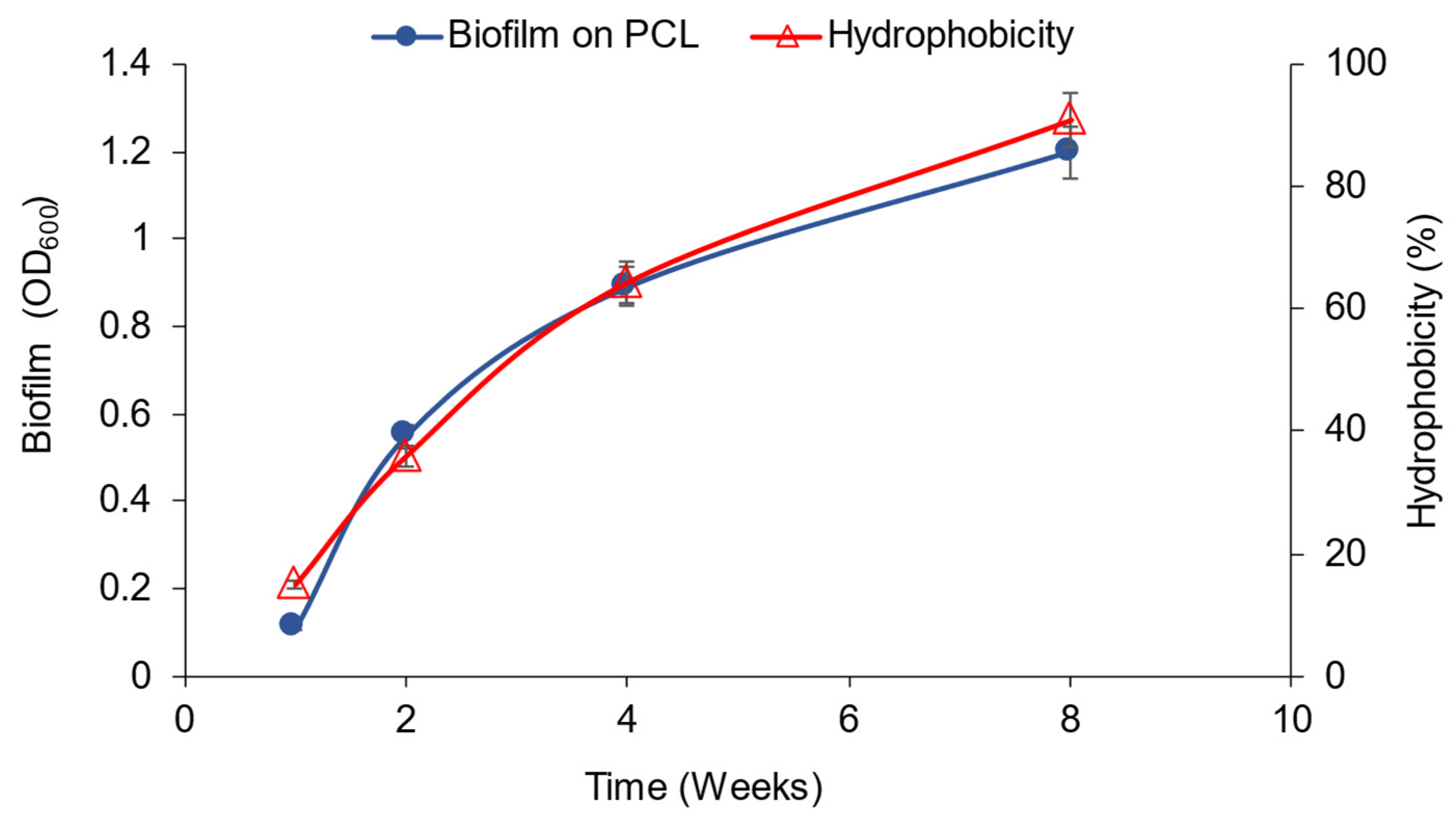
2.6. Biofilm Formation Estimation

To evaluate the biofilm formation, the plastic beads were washed with 96% ethanol for 20 min and were centrifuged at 4000× g. The degree of biofilm formation was determined by measuring the optical density of the supernatant at a wavelength of 600 nm.

2.7. Bacterial Hydrophobicity Assay

Bacterial hydrophobicity was assessed using the BATH method [27]. The PCL beads were soaked in 1 mL of phosphate urea magnesium (PUM) buffer for 10 min and shaken at 150 rpm. The PUM buffer contained (g/L): K2HPO4, 17; KH2PO4, 7.26; urea, 1.8; MgSO4 × 7H2O, 0.2. The plastic beads and supernatant were separated. The released cells’ optical density (OD) in the buffer was measured as the initial OD (H0). The beads were rewashed with 1 mL of PUM containing 0.2 mL of n-hexadecane for 10 min until phase separation occurred. The PUM supernatant phase was taken, and the OD was measured as the final OD (H1). The PUM buffer without cells served as the blank. The hydrophobicity of the bacterial isolates was determined using the following formula:
Hydrophobicity (%) = (H0 − H1)/(H0) × 100.

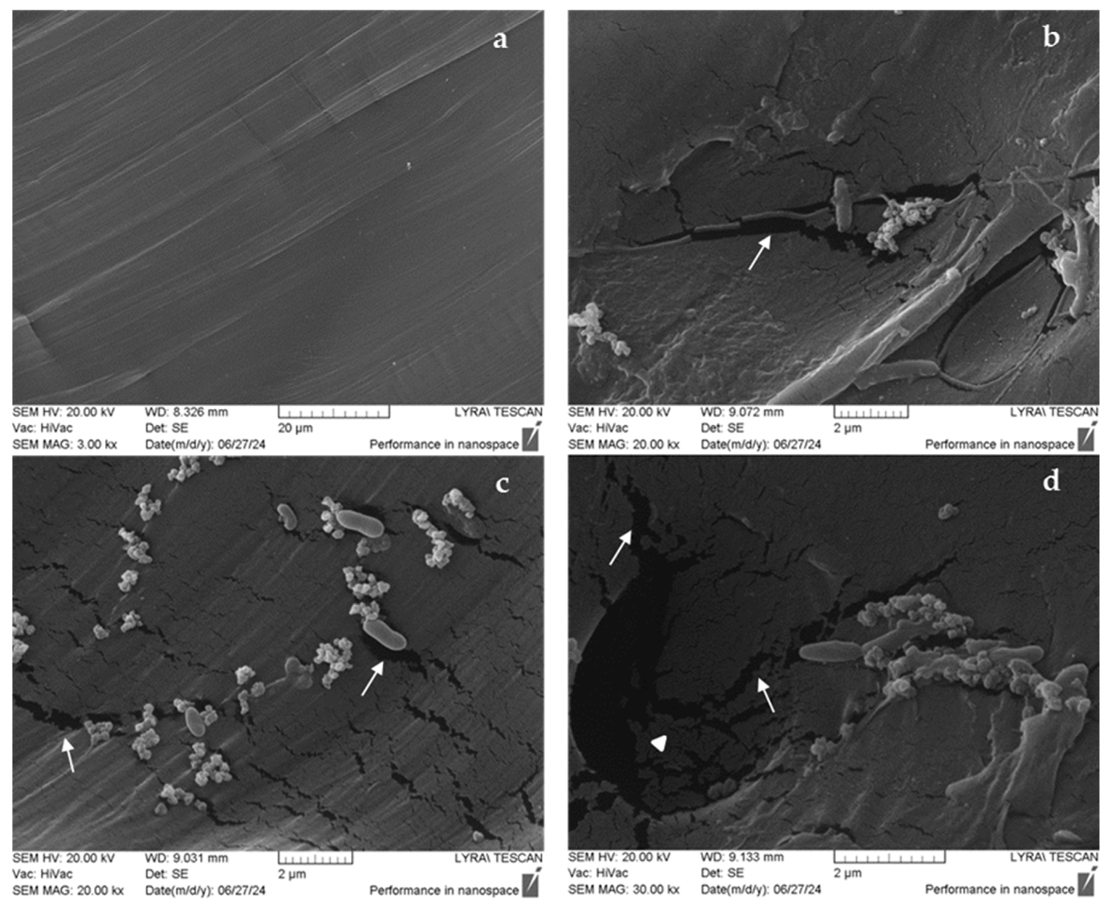
2.8. Scanning Electron Microscopy (SEM)

Each sample was processed in three replicates. The plastics were fixed in 4% glutaraldehyde in 0.1 M Na cacodylate buffer (pH 7.2) for two hours, then washed and post-fixed in 1% OsO4 for one hour at 4 °C. A dehydration process was carried out using a graded ethanol series at 15 min intervals. The samples were mounted on scanning electron microscopy holders and gold-coated using a vacuum evaporator (Edwards, Irvine, CA, USA). Observations were performed using a Lyra/Tescan scanning electron microscope (TESCAN GROUP a.s., Brno, Czech Republic) with an accelerating voltage of 20 kV.

2.9. DNA Isolation, Metagenome Sequencing, and Bioinformatics Analysis

DNA for metagenomic analysis (1.2 μg) was isolated from the enriched community after 4 weeks of cultivation in a mineral medium with PCL using the GeneMATRIX Bacterial & Yeast Genomic DNA Purification Kit (EURx, Gdansk, Poland).
The Metagenome 16S V3-V4 amplicon library was prepared by Macrogen Inc. (Seoul, Republic of Korea) using a TruSeq DNA PCR-Free Kit. The sequencing was performed with Illumina HiSeq 2000 Sequencing System, which generated 92,827 reads (read length 300 bp). After sequencing, Cutadapt (v3.2) [28] was used to remove the adapter and primer sequences from the raw data. Forward and reverse reads were trimmed to 250 bp and 200 bp, respectively. To generate amplicon sequence variants (ASVs), the reads underwent error correction, merging, and denoising processes using DADA2 (v1.18.0) [29]. Sequences with an expected error of 2 or more were excluded. Erroneous reads were denoised based on an established error model. Following error correction, paired-end reads were merged by overlapping. Chimeric sequences were eliminated using the consensus method with the removeBimeraDenovo function in DADA2. ASVs shorter than 350 bp were filtered out. Normalization was conducted using QIIME (v1.9) for microbial community comparison analysis [30]. During this process, subsampling was performed based on the sample with the lowest read count to ensure comparability. Each ASV was aligned to the organism with the highest similarity in the corresponding reference database, NCBI_16S, utilizing algorithms such as BLAST+ (v2.9.0, with query coverage > 85% and identity > 85%) [31]. Alpha diversity metrics, including the Shannon index, Gini-Simpson index, and PD whole tree, were calculated to represent species complexity within individual samples.
The SRA metagenome data are available in NCBI GenBank with the following accession numbers: Biosample SAMN46967729, BioProject PRJNA1226943.

3. Results

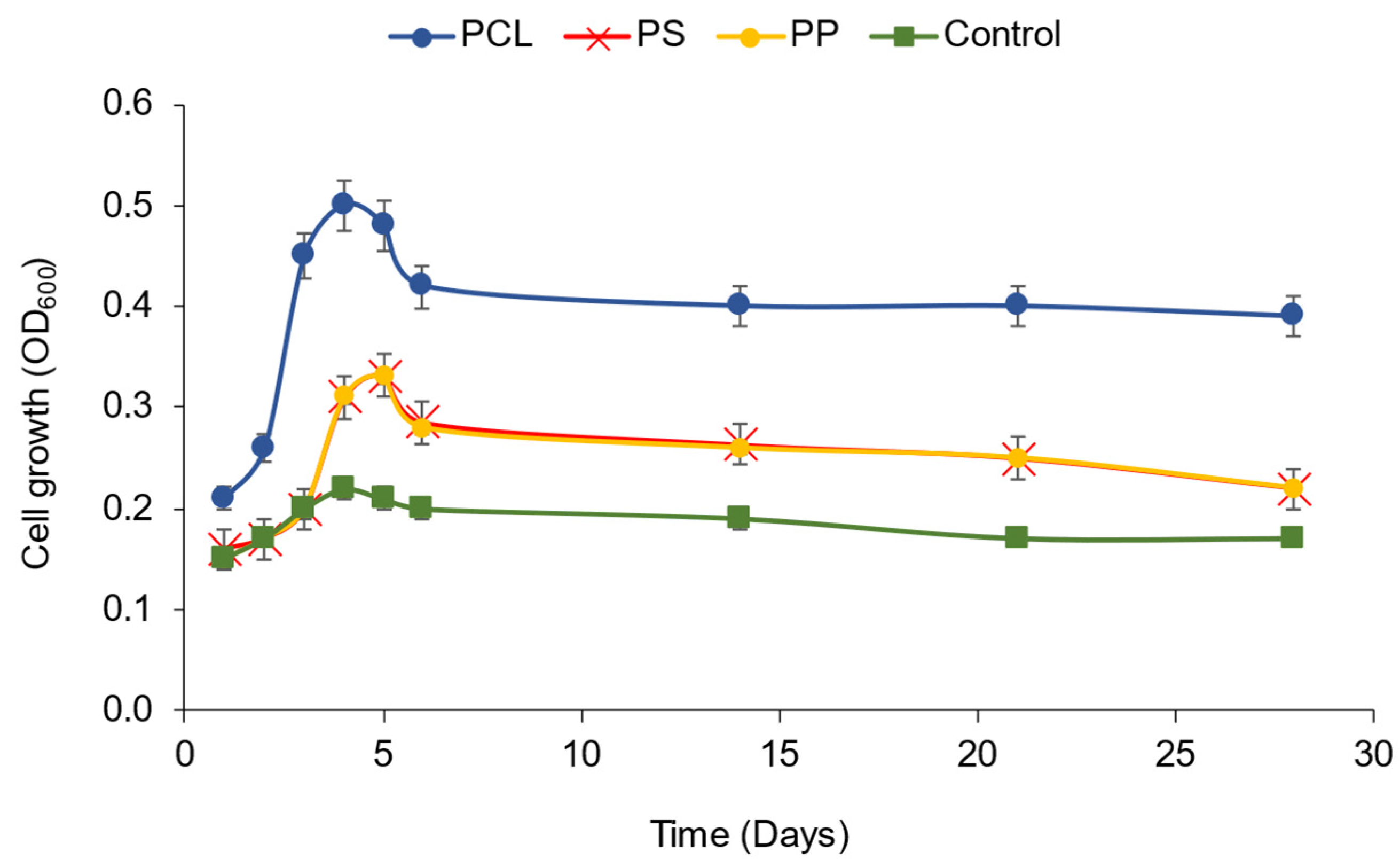
3.1. Isolation of the Plastic-Degrading Halophilic Community

The initial selection of the halophilic community capable of degrading plastic was based on bacterial growth monitoring by measuring the optical density (OD600). The cultures were cultivated in a mineral medium containing 15% NaCl and plastic (0.3% PCL, PS, or PP) as the sole carbon source (Figure 1).
During the first week of community cultivation, the optical density (OD) increased across all three types of plastic substrates, peaking on the fourth day of fermentation with OD600 = 0.5 for polycaprolactone (PCL) and 0.3 for polystyrene (PS) and polypropylene (PP). By the seventh day, the OD decreased slightly and remained consistent over the following two weeks. The reduction in planktonic growth corresponded with the formation of biofilms, which were most pronounced on the surfaces of PCL beads, particularly strong after the fifth day of cultivation. The biofilm formed on PP was considerably thinner, and no biofilm formation was observed on PS.
The halophilic community displayed significant esterase activity when grown at PCL and relatively lower in the presence of PS and PP as the sole carbon source (Figure 2).
The highest values of esterase activity were recorded on the PCL substrate on the fourth day of cultivation—46 U/mL—followed by a slight decrease to 31 U/mL after 4 weeks. With PS and PP substrates on the fourth day, the esterase activity was 9 U/mL, dropped threefold after 2 weeks, and disappeared after 4 weeks of cultivation. In contrast, the control maintained esterase activity of about 5 U/mL throughout fermentation. Dense growth and formation of a significant biofilm were observed on the surface of PCL beads after the fifth day of cultivation, while the biofilm formed on PP was much smaller. No biofilm formation was observed on PS.

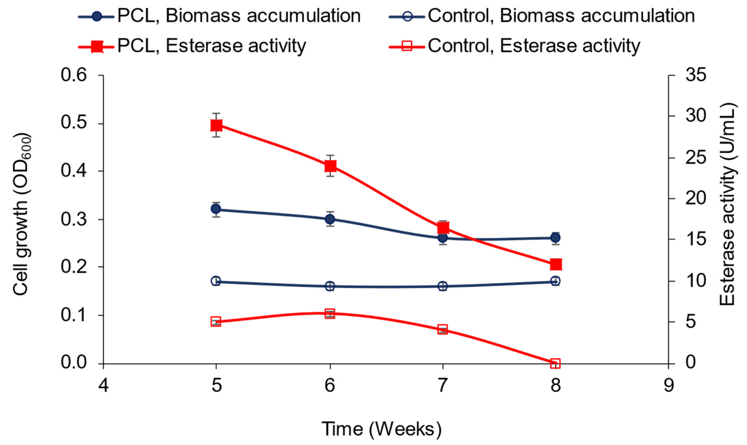
3.2. Polycaprolactone (PCL) Degradation by the Novel Halophilic Community

A qualitative test for PCL degradation on agar mineral medium containing it as the sole carbon source showed that a visible blue halo appeared around the wells containing the community (Figure 3). After staining the agar with Coomassie blue, the appearance of a halo indicated its ability to utilize PCL as a source of carbon and energy.
Given the highest esterase activity noted in the presence of PCL and the formation of a biofilm on its surface by the community, the study of the halophilic community’s ability to degrade PCL continued for an additional four weeks under the same conditions. The optical density, esterase activity (Figure 4), and the biofilm formed (Figure 5) were measured weekly.
During the community’s fermentation of PCL from the fourth to the eighth week, a decrease in esterase activity (to 12 U/mL) and relatively constant biomass was observed. No esterase activity was visible in the control group by the eighth week. At the same time, a significant increase in biofilm on the plastic beads occurred. Biofilm formation was quantified during the first, fourth, and eighth weeks by measuring the optical density of the bacteria after washing the plastic beads with ethanol (Figure 5).
Visible biofilm on the surface of the plastic beads was observed after the fifth day of cultivation and increased during the cultivation, with the highest value at the end. The biofilm increased from OD600 = 0.11 (after the first week) to OD600 = 1.20 (after the eighth week), coinciding with the culture’s hydrophobicity rise.
Another evidence of PCL biodegradation was the decrease in gravimetric weight of PCL beads estimated after four and eight weeks of cultivation. The total weight of PCL at the beginning was 597.8 mg, after 4 weeks—560.2 mg (6.3% decrease), and after 8 weeks—541.3 mg, comprising a reduction of 9.5%. The weight of the control (mineral medium without the community) remained unchanged at 589.8 mg, indicating that the plastic did not self-degrade.
The surface changes of the plastic, cultivated for four weeks in the presence of the bacterial community, were examined using scanning electron microscopy (SEM). Figure 6 presents changes in the surface architecture of the plastic beads after incubation for four weeks in the presence of the bacterial community.
Clearly defined surface modifications were observed in the treated PCL compared to the untreated control. The electron microscopy micrograph of the control PLA sample illustrated a smooth plastic surface free of indentations, cracks, or any damage (Figure 6a). In contrast, the treated samples, incubated with the microbial community, displayed various defects (Figure 6b–d). These surface defects were identified as cracks, craters (marked with white arrows), indentations, and holes (marked with a white triangle) in the architecture of the underlying plastic. Since these surface injuries were in areas of microbial adhesion or in close proximity, they are most likely attributed to the enzyme activity of the respective microbial cells.

3.3. Bacterial Biodiversity of the Halophilic Community

The metagenomic analysis of the halophilic community forming a biofilm on the surface of PCL beads revealed a dominance of the class Gammaproteobacteria (99.58%), along with a significantly weaker presence of Alphaproteobacteria (0.26%) and Betaproteobacteria (0.17%). Figure 7 shows the most abundant bacterial families in the community.
With the highest representation was the order Oceanospirillales (99.57%), which included three genera: Halomonas (95.92%), Alloalcanivorax (2.73%), and Chromohalobacter (0.93%). The main bacterial species in the community was Halomonas profundus, which comprised 95.13% of all species present. Alloalcanivorax venustensis (2.73%) and Chromohalobacter marismortui (0.72%) were present in much smaller percentages (Figure 8).
The dominance of Halomonas in the bacterial community indicated its active role in the PCL degradation process.

4. Discussion

Marine ecosystems are vulnerable to pollution from all types of plastics. The flow of rivers transports approximately 6 million tons of plastic waste; of this amount, around 1.7 million tons enter the oceans each year. Several types of plastic rank at the top of the list of pollutants, including polystyrene, polycarbonates, polyester, polyethylene, polypropylene, and biodegradable plastic polycaprolactone [32]. However, due to the harsh sea conditions, such as salinity and cold, not every biodegradation approach is possible, and only specific communities of extremophile microorganisms are suitable. Fortunately, several extremophile species have already been discovered that can degrade polymers such as PE, PP, PCL, and polyethylene terephthalate (PET), using them as a primary carbon source for energy [22,23,33,34,35,36,37,38]. Therefore, studying plastic biodegradation by halophilic and psychrophilic bacteria has gained popularity and relevance [22,39].
Here, we report the isolation and characterization of a novel halophilic bacterial community capable of partially degrading the polymers PS and PP and exhibiting high efficiency with PCL. Several key properties ensure the biodegradability of the community: (i) the capacity to form a biofilm due to enhanced hydrophobicity of the cell surface within the consortium, (ii) demonstration of high extracellular esterase activity, and (iii) a specific consortium of halophilic species that may promote the synergistic action of hydrolytic enzymes. Thus, in addition to the SEM images that reveal the PCL surface damaged by the bacterial community, evidence of the degradation processes is the undeniable weight reduction of the PCL beads.
The biodegradation of plastics begins with the attachment of microorganisms and the formation of a biofilm on their surfaces, which is referred to as a plastisphere [40,41]. In this ‘shell’ around the plastic particles, communities of microorganisms, mainly bacteria, are capable of causing changes in the physicochemical structure of the plastic, such as alterations in functional groups, loss of crystallinity, injuries in surface morphology, and weight loss [42].
Since plastic polymers are hydrophobic, bacteria must initiate hydrophobic interactions with the plastic surface. The new halophilic community showed high hydrophobicity (over 90% by the eighth week). This property provoked its considerable ability to form a biofilm on PCL beads with attached cells reaching an OD of 1.2 (at week eight). Bardají et al. [43] reported the correlation between hydrophobicity and biofilm formation regarding the biodegradation of polyethylene; here, we also highlight it for PCL. Bacterial cell walls and membranes usually influence their hydrophobicity. Generally, Gram-negative bacteria are more hydrophobic than Gram-positive ones; moreover, they can release outer membrane vesicles, significantly enhancing bacterial adhesion to hydrophobic materials [44]. Thus, polyethylene destruction by the Gram(-) Alcanivorax borkumensis was reported [45], whereas Pseudomonas nitroreducens S8 and P. monteilii S17 were known to colonize the surface of PET. After forming a thin biofilm layer with a consistent population density, the last species utilize PET as a carbon source, synergistically degrading the polymer surface [46]. In a similar biofilm on PET and PE in marine waste, the presence of Halomonas spp. was detected (by 16S rRNA analysis), indicating its participation in the possible degradation of these plastics [47].
An essential role of biofilm formation is to increase the concentration of enzymes around the substrate and allow them to persist longer by becoming incorporated into the biofilm matrix, thus prolonging the overall rate of plastic degradation [48]. The enzymes primarily involved in the degradation of plastics fall into two main categories, hydrolases, and oxidases, and are further classified as esterases, proteases, cutinases, dehydrogenases, or laccases. Esterases (EC 3.1.1.-) hydrolyze plastics by cleaving an ester bond in the carbon chain, primarily targeting aliphatic polyesters. PCL degradation is carried out by esterases and/or depolymerases secreted extracellularly [49]. Among these, the most studied enzymes belong to the classes of carboxyl ester hydrolases (EC 3.1.1) and, in particular, lipases (EC 3.1.1.3) and cutinases (EC 3.1.1.74). It is difficult to classify these enzymes based on substrate specificity alone [37]. This activity is usually detected by hydrolysis assay of p-nitrophenyl esters with different chain lengths (C2–C18), as in the present study, p-nitrophenyl palmitate was used [25]. Inglis et al. [50] showed that although PCL degradation does not necessarily correlate with high esterase activity, isolates unable to degrade PCL failed to produce measurable esterase activities. Esterase activity can indicate the presence of strains responsible for PCL degradation in microbial communities; for example, Shin et al. selected Bacillus sp. NR4 for this purpose due to its high esterase activity, and the strain showed an 85.6% yield of PCL degradation after 10 days [51]. Therefore, the high esterase activity reported for the new halophilic community (five-fold higher with PCL than with PS and PP) is the most probable reason for the visible damage observed on PCL particles. The post-hydrolysis steps of PCL biodegradation generate 6-hydroxyhexanoic acid (6-HCA), which, in aerobic microorganisms found in the halophilic community, is most likely metabolized via the beta-oxidation catabolic pathway, with the end products being carbon dioxide and water [37,52].
Similar enzymatic activities have been reported in other bacteria. The high efficiency of an aromatic polyesterase produced by Ideonella sakaiensis 201-F6 in degrading PET has been demonstrated [53]. Bacterial esterases that can degrade polyurethanes have also been reported [35], and cutinase, initially noted for hydrolyzing ester bonds in the plant polymer cutin, has also been recognized as a plastic-degrading enzyme [22]. Several esterases and lipases from P. cepacia, Humicola insolens, and Candida antarctica have been shown to destroy the ε-caprolactone ring [54]. However, the only enzyme identified so far to attack PCL is lipase from Alcaligenes faecalis [55].
An important merit of the present work is clarifying the species composition of the halophilic community that exhibits esterase activity. Marine bacterial communities that can degrade plastics have been reported to include Cobetia sp., Halomonas sp., Exigobacterium sp., and Alcanivorax sp., as they use low-density polyethylene (LDPE) as a carbon source [56]. So far, microorganisms that can degrade PCL have been isolated relatively rarely; they have been assigned to the genera Pseudomonas, Alcanivorax, and Tenacibaculum and have been found in deep-sea sediments [57]. The reported new halophilic community contains a limited number of species–11, from seven families of bacteria, most unreported for plastic degrading ability. The primary bacterium in the halophilic community, Halomonas profundus, was shown to participate in the PCL-degrading community for the first time. It is a halophilic species initially isolated from deep-sea hydrothermal vent shrimp. It has garnered interest due to its ability to produce polyhydroxyalkanoates (PHAs), biodegradable polyesters considered potential alternatives to conventional plastics. Under optimal conditions, H. profundus synthesizes PHAs such as poly(3-hydroxybutyrate) (PHB) and poly(3-hydroxybutyrate-co-3-hydroxyvalerate) (PHBV) from various carbon sources, including glucose, glycerol, acetate, octanoate, and pyruvate. PHAs possess properties suitable for diverse applications, positioning H. profundus as a promising candidate for biotechnological processes aimed at sustainable plastic production [58].
H. caseinilytica, a related halophilic species, is remarkable for its ability to biodegrade the high molecular weight polycyclic aromatic hydrocarbon coronene and some similar molecules with lower molecular weights, such as benzo[a]pyrene, pyrene, and phenanthrene [59]. Promising applications of these compounds in organic electronics are forthcoming [60]; studying their chemical properties and potential biodegradation is essential for future exploitation.
Chromohalobacter marismortui is a key member of the Halomonadaceae family. This halophilic microorganism has been shown to effectively precipitate phosphate and carbonate minerals [61] in a complex process occurring on the surface of the bacterial cells, as evidenced by SEM studies [62]. Plastic precipitation is crucial for the fate of plastics in natural environments, as it can significantly enhance biofilm formation, which plays a fundamental role in the biodegradation of these materials and contributes to abiotic degradation through metal-mediated catalysis [63,64]. In addition to withstanding high salt concentrations, another member of the Chromohalobacter genus, C. beijerinckii, has been identified in the microbial community and shown to be psychrophilic, evidenced by its successful growth at a temperature of 5 °C. This microorganism appears capable of synthesizing biogenic amines, particularly histamine and tyramine [65], suggesting it may have potential as a producer of substances that can be utilized in specialized materials, such as tyramine-based hydrogels.
There is limited information regarding the role of Alloalcanivorax in plastic biodegradation. Alloalcanivorax spp. has been identified in biofilms formed on PE and, to a lesser extent, on PHA during their biodegradation in seawater, but without further analysis [66].
Pseudomonas juntendii, an unrelated species of Gammaproteobacteria discovered in the Burgas Lake sample, stands out due to its remarkable ability to thrive in a medium containing PET [67]. This polymeric compound, commonly used in the production of textiles and packaging, represents a significant pollutant, as it not only disrupts water and soil habitats but also poses serious risks to human health [68]. The species has been successfully isolated from waste deposits in the Mediterranean basin and exhibits increased growth in a nutrient medium supplemented with PET. SEM analysis of samples from the collected waste shows that the community from which P. juntendii was isolated forms distinct biofilms on the surface of the plastic, with bacteria firmly attached, indicating a strong affinity of the microorganisms for the material.
Interestingly, Afipia felis, initially thought to be the primary causative agent of cat-scratch disease, also exhibits properties associated with the biodegradation of toxic compounds. A methylotrophic strain isolated from Signy Island in Antarctica was found to biodegrade and utilize methanesulfonate, an extremely carcinogenic substance, as a carbon source [69]. Furthermore, an unidentified strain of Afipia sp. has also been shown to biodegrade haloacetic acid, representing an undesirable health-concerning byproduct appearing in drinking water due to chlorination [70]. Rhodopseudomonas boonkerdii, a member of Afipia’s family (Nitrobacteraceae) found in the halophilic community’s content, has another notable property. This species is capable of developing in soils rich in heavy metal ions such as molybdenum, zinc, cobalt, and copper and appears resistant to many antibiotics, including streptomycin, neomycin, spectinomycin, and tetracycline [71]. Marine water contaminated with plastic waste is often linked to higher concentrations of heavy metal ions [72], which may help explain the presence of such microorganisms in the community. This might also account for the recording of Bosea lather; this typical plant nodule-associated microorganism has shown the ability to resist heavy metals [73].

5. Conclusions

Contaminated natural habitats are excellent sources for isolating plastic-degrading microorganisms. Halophilic communities exhibit lower biodiversity but possess strong degradation capabilities. The new halophilic community demonstrated high esterase activity, which is probably responsible for PCL biodegradation, as shown by SEM, increased hydrophobicity, and the ability to form biofilms. The degradation efficiency of PCL was significant, while that of PP and PS was relatively poor. However, compared to other microorganisms that degrade PCL, the described community destroys the polymer relatively slowly. An advantage of the studied community is that most of its species are halophilic and can depurate plastic-polluted marine habitats and other saline environments. Additional studies are needed to intensify the process by establishing the optimal composition of the medium and optimal process parameters for purification from PCL on a large scale. As detected for the first time, H. profundus was identified as a dominant species within a plastic-degrading halophilic community. Since this bacterium synthesizes the bioplastic polyhydroxybutyrate (PHB) and participates in PCL degradation, future research may clarify halophiles’ role in the carbon cycle of biodegradable plastics in terrestrial and marine ecosystems.

Author Contributions

Conceptualization, N.A. and I.B.; methodology, N.A. and T.P.-K.; investigation, N.K., N.A., T.P.-K., K.B., P.P. and I.B.; writing—original draft preparation, N.K. and N.A.; writing—review and editing, P.P. and K.P. All authors have read and agreed to the published version of the manuscript.

Funding

Center of Competence “Clean Technologies for a Sustainable Environment—Water, Waste, Energy for a Circular Economy” (Clean and Circle), financed through the Research, Innovation and Digitalization for Smart Transformation Program 2021–2027 (PNIIDIT), Grant no. BG16RFPR002-1.014.

Institutional Review Board Statement

Not applicable.

Informed Consent Statement

Not applicable.

Data Availability Statement

The metagenome data can be found in the NCBI GenBank under accession number Biosample SAMN46967729, BioProject PRJNA1226943.

Conflicts of Interest

The authors declare no conflicts of interest. The funders had no role in the study’s design; in the collection, analyses, or interpretation of data; in the writing of the manuscript; or in the decision to publish the results.

References

  1. Pilapitiya, P.G.C.N.T.; Ratnayake, A.S. The world of plastic waste: A review. Clean. Mater. 2024, 11, 100220. [Google Scholar] [CrossRef]
  2. The Market Value of Plastics Worldwide Will Be in 2023, with a Forecast for 2033. Available online: https://www.statista.com/statistics/1060583/global-market-value-of-plastic/ (accessed on 28 January 2025).
  3. Carpenter, E.J.; Smith, K.L. Plastics on the Sargasso sea surface. Science 1972, 175, 1240–1241. [Google Scholar] [CrossRef]
  4. Williams, A.T.; Rangel-Buitrago, N. The past, present, and future of plastic pollution. Mar. Pollut. Bull. 2022, 176, 113429. [Google Scholar] [CrossRef]
  5. Terrazas-López, R.; Guadarrama-Guzman, P.; Sujitha, S.B.; Arreola-Mendoza, L.; Ponniah, J.M. The Occurrence of Microplastics in the Marine Food Web in Latin America: Insights on the Current State of Knowledge and Future Perspectives. Sustainability 2024, 16, 5905. [Google Scholar] [CrossRef]
  6. Thushari, G.G.N.; Senevirathna, J.D.M. Plastic pollution in the marine environment. Heliyon 2020, 6, e04709. [Google Scholar] [CrossRef]
  7. Viel, T.; Manfra, L.; Zupo, V.; Libralato, G.; Cocca, M.; Costantini, M. Biodegradation of Plastics Induced by Marine Organisms: Future Perspectives for Bioremediation Approaches. Polymers 2023, 15, 2673. [Google Scholar] [CrossRef]
  8. Boldrini, A.; Gaggelli, N.; Falcai, F.; Polvani, A.; Talarico, L.; Galgani, L.; Cirrone, R.; Liu, X.; Loiselle, S. Emerging Contaminants from Bioplastic Pollution in Marine Waters. Water 2024, 16, 3676. [Google Scholar] [CrossRef]
  9. Hurley, R.; Horton, A.; Lusher, A.; Nizzetto, L. Chapter 7—Plastic waste in the terrestrial environment. In Plastic Waste and Recycling; Letcher, T.M., Ed.; Academic Press: Cambridge, MA, USA, 2020; pp. 163–193. [Google Scholar] [CrossRef]
  10. Rai, M.; Pant, G.; Pant, K.; Aloo, B.N.; Kumar, G.; Singh, H.B.; Tripathi, V. Microplastic Pollution in Terrestrial Ecosystems and Its Interaction with Other Soil Pollutants: A Potential Threat to Soil Ecosystem Sustainability. Resources 2023, 12, 67. [Google Scholar] [CrossRef]
  11. Heris, Y.S. Bacterial biodegradation of synthetic plastics: A review. Bull. Natl. Res. Cent. 2024, 48, 87. [Google Scholar] [CrossRef]
  12. Tanasupawat, S.; Takehana, T.; Yoshida, S.; Hiraga, K.; Oda, K. Ideonella sakaiensis sp. nov., isolated from a microbial consortium that degrades poly(ethylene terephthalate). Int. J. Syst. Evol. Microbiol. 2016, 66, 2813–2818. [Google Scholar] [CrossRef]
  13. Ekanayaka, A.H.; Tibpromma, S.; Dai, D.; Xu, R.; Suwannarach, N.; Stephenson, S.L.; Dao, C.; Karunarathna, S.C. A review of the fungi that degrade plastic. J. Fungi 2022, 8, 772. [Google Scholar] [CrossRef] [PubMed]
  14. Ho, B.T.; Roberts, T.K.; Lucas, S. An overview on biodegradation of polystyrene and modified polystyrene: The microbial approach. Crit. Rev. Biotechnol. 2017, 38, 308–320. [Google Scholar] [CrossRef] [PubMed]
  15. Longo, C.; Savaris, M.; Zeni, M.; Brandalise, R.N.; Grisa, A.M.C. Degradation study of polypropylene (PP) and bioriented polypropylene (BOPP) in the environment. Mater. Res. 2011, 14, 442–448. [Google Scholar] [CrossRef]
  16. Lefèvre, C.; Tidjani, A.; Wauven, C.V.; David, C. The interaction mechanism between microorganisms and substrate in the biodegradation of polycaprolactone. J. Appl. Pol. Sci. 2001, 83, 1334–1340. [Google Scholar] [CrossRef]
  17. Ghatge, S.; Yang, Y.; Ahn, J.-H.; Hur, H.-G. Biodegradation of polyethylene: A brief review. Appl. Biol. Chem. 2020, 63, 27. [Google Scholar] [CrossRef]
  18. Pometto, A.L.; Lee, B.T.; Johnson, K.E. Production of an extracellular polyethylene-degrading enzyme(s) by Streptomyces species. Appl. Environ. Microbiol. 1992, 58, 731–733. [Google Scholar] [CrossRef]
  19. Roman, E.K.B.; Ramos, M.A.; Tomazetto, G.; Foltran, B.B.; Galvão, M.H.; Ciancaglini, I.; Tramontina, R.; de Almeida Rodrigues, F.; da Silva, L.S.; Sandano, A.L.H.; et al. Plastic-degrading microbial communities reveal novel microorganisms, pathways, and biocatalysts for polymer degradation and bioplastic production. Sci. Total Environ. 2024, 949, 174876. [Google Scholar] [CrossRef]
  20. Oberbeckmann, S.; Labrenz, M. Marine microbial assemblages on microplastics: Diversity, adaptation, and role in degradation. Ann. Rev.Mar. Sci. 2020, 12, 209–232. [Google Scholar] [CrossRef]
  21. Skariyachan, S.; Patil, A.A.; Shankar, A.; Manjunath, M.; Bachappanavar, N.; Kiran, S. Enhanced polymer degradation of polyethylene and polypropylene by novel thermophilic consortia of Brevibacillus sps. and Aneurinibacillus sp. screened from waste management landfills and sewage treatment plants. Polym. Deg. Stab. 2018, 149, 52–68. [Google Scholar] [CrossRef]
  22. Atanasova, N.; Stoitsova, S.; Paunova-Krasteva, T.; Kambourova, M. Plastic Degradation by Extremophilic Bacteria. Int. J. Mol. Sci. 2021, 22, 5610. [Google Scholar] [CrossRef]
  23. Atanasova, N.; Paunova-Krasteva, T.; Stoitsova, S.; Radchenkova, N.; Boyadzhieva, I.; Petrov, K.; Kambourova, M. Degradation of Poly(ε-caprolactone) by a Thermophilic Community and Brevibacillus thermoruber Strain 7 Isolated from Bulgarian Hot Spring. Biomolecules 2021, 11, 1488. [Google Scholar] [CrossRef] [PubMed]
  24. Atanasova, N.; Paunova-Krasteva, T.; Kambourova, M.; Boyadzhieva, I. A Thermostable Lipase Isolated from Brevibacillus thermoruber Strain 7 Degrades Ɛ-Polycaprolactone. BioTech 2023, 12, 23. [Google Scholar] [CrossRef] [PubMed]
  25. Gupta, N.; Rathi, P.; Gupta, R. Simplified para-nitrophenyl palmitate assay for lipases and esterases. Anal. Biochem. 2002, 311, 98–99. [Google Scholar] [CrossRef]
  26. Almeida, E.L.; Carrillo Rincón, A.F.; Jackson, S.A.; Dobson, A.D. In silico screening and heterologous expression of a polyethylene terephthalate hydrolase (PETase)-like enzyme (SM14est) with polycaprolactone (PCL)-degrading activity from the marine sponge-derived strain Streptomyces sp. SM14. Front. Microbiol. 2019, 10, 2187. [Google Scholar] [CrossRef]
  27. Rosenberg, M. Bacterial adherence to hydrocarbons: A useful technique for studying cell surface hydrophobicity. FEMS Microbiol. Lett. 1984, 22, 289–295. [Google Scholar] [CrossRef]
  28. Martin, M. Cutadapt removes adapter sequences from high-throughput sequencing reads. EMBnet. J. 2011, 17, 10–12. [Google Scholar] [CrossRef]
  29. Callahan, B.J.; McMurdie, P.J.; Rosen, M.J.; Han, A.W.; Johnson, A.J.A.; Holmes, S.P. DADA2: High-resolution sample inference from Illumina amplicon data. Nat. Methods 2016, 13, 581–583. [Google Scholar] [CrossRef]
  30. Caporaso, J.G.; Kuczynski, J.; Stombaugh, J.; Bittinger, K.; Bushman, F.D.; Costello, E.K.; Fierer, N.; Peña, A.G.; Goodrich, J.K.; Gordon, J.I.; et al. QIIME allows analysis of high-throughput community sequencing data. Nat. Methods 2010, 7, 335–336. [Google Scholar] [CrossRef]
  31. Camacho, C.; Coulouris, G.; Avagyan, V.; Ma, N.; Papadopoulos, J.; Bealer, K.; Madden, T.L. BLAST+: Architecture and applications. BMC Bioinform. 2009, 10, 421. [Google Scholar] [CrossRef]
  32. Carney Almroth, B.; Eggert, H. Marine Plastic Pollution: Sources, Impacts, and Policy Issues. Rev. Environ. Econ. Policy 2019, 13, 317–326. [Google Scholar] [CrossRef]
  33. Zandieh, M.; Griffiths, E.; Waldie, A.; Li, S.; Honek, J.; Rezanezhad, F.; Van Cappellen, P.; Liu, J. Catalytic and biocatalytic degradation of microplastics. Exploration 2023, 4, 20230018. [Google Scholar] [CrossRef] [PubMed]
  34. Taniguchi, I.; Yoshida, S.; Hiraga, K.; Miyamoto, K.; Kimura, Y.; Oda, K. Biodegradation of PET: Current status and application aspects. ACS Catal. 2019, 9, 4089–4105. [Google Scholar] [CrossRef]
  35. Magnin, A.; Pollet, E.; Perrin, R.; Ullmann, C.; Persillon, C.; Phalip, V.; Avérous, L. Enzymatic recycling of thermoplastic polyurethanes: Synergistic effect of an esterase and an amidase and recovery of building blocks. Waste Manag. 2019, 85, 141–150. [Google Scholar] [CrossRef]
  36. Atanasova, N.; Paunova-Krasteva, T.; Stoitsova, S.; Kambourova, M.; Shapagin, A.; Matveev, V.; Provotorova, E.; Elcheninov, A.; Sokolov, T.; Bonch-Osmolovskaya, E. Plastic Degradation by Extremophilic Microbial Communities Isolated from Bulgaria and Russia. Ecol. Balk. 2021, 13, 211–222. [Google Scholar]
  37. Suzuki, M.; Tachibana, Y.; Kasuya, K. Biodegradability of poly(3-hydroxyalkanoate) and poly(ε-caprolactone) via biological carbon cycles in marine environments. Polym. J. 2021, 53, 47–66. [Google Scholar] [CrossRef]
  38. Yoon, Y.; Park, H.; An, S.; Ahn, J.H.; Kim, B.; Shin, J.; Kim, Y.E.; Yeon, J.; Chung, J.H.; Kim, D.; et al. Bacterial degradation kinetics of poly(Ɛ-caprolactone) (PCL) film by Aquabacterium sp. CY2-9 isolated from plastic-contaminated landfill. J. Environ. Manag. 2023, 335, 117493. [Google Scholar] [CrossRef]
  39. Kotova, I.B.; Taktarova, Y.V.; Tsavkelova, E.A.; Egorova, M.A.; Bubnov, I.A.; Malakhova, D.V.; Shirinkina, L.I.; Sokolova, T.G.; Bonch-Osmolovskaya, E.A. Microbial Degradation of Plastics and Approaches to Make it More Efficient. Microbiology 2021, 90, 671–701. [Google Scholar] [CrossRef]
  40. Tiwari, N.; Bansal, M.; Santhiya, D.; Sharma, J.G. Insights into microbial diversity on plastisphere by multi-omics. Arch. Microbiol. 2022, 204, 216. [Google Scholar] [CrossRef]
  41. Amaral-Zettler, L.A.; Zettler, E.R.; Mincer, T.J. Ecology of the plastisphere. Nat. Rev. Microbiol. 2020, 18, 139–151. [Google Scholar] [CrossRef]
  42. Ghosh, S.; Qureshi, A.; Purohit, H.J. Microbial degradation of plastics: Biofilms and degradation pathways. In Contaminants in Agriculture and Environment: Health Risks and Remediation; Kumar, V., Kumar, R., Singh, J., Kumar, P., Eds.; Agro Environ Media: Haridwar, India, 2019; Volume 1, pp. 184–199. [Google Scholar] [CrossRef]
  43. Bardají, D.K.R.; Moretto, J.A.S.; Furlan, J.P.R.; Stehling, E.G. A mini-review: Current advances in polyethylene biodegradation. World J. Microbiol. Biotechnol. 2020, 36, 32. [Google Scholar] [CrossRef]
  44. Baumgarten, T.; Sperling, S.; Seifert, J.; Von Bergen, M.; Steiniger, F.; Wick, L.Y.; Heipieper, H.J. Membrane vesicle formation as a multiple-stress response mechanism enhances Pseudomonas putida DOT-T1E cell surface hydrophobicity and biofilm formation. Appl. Environ. Microbiol. 2012, 78, 6217–6224. [Google Scholar] [CrossRef] [PubMed]
  45. Delacuvellerie, A.; Cyriaque, V.; Gobert, S.; Benali, S.; Wattiez, R. The plastisphere in marine ecosystem hosts potential specific microbial degraders including Alcanivorax borkumensis as a key player for the degradation. J. Hazard. Mater. 2019, 380, 120899. [Google Scholar] [CrossRef] [PubMed]
  46. Yan, Z.F.; Xu, K.W.; Wu, J. Synergism Between Multi-Pseudomonas and Cutinase for Biodegradation of Crude Oil-Based Derivatives. Curr. Microbiol. 2022, 80, 30. [Google Scholar] [CrossRef]
  47. Okshevsky, M.; Gautier, E.; Farner, J.M.; Schreiber, L.; Tufenkji, N. Biofilm formation by marine bacteria is impacted by concentration and surface functionalization of polystyrene nanoparticles in a species-specific manner. Environ. Microbiol. Rep. 2020, 12, 203–213. [Google Scholar] [CrossRef]
  48. Howard, S.A.; McCarthy, R.R. Modulating biofilm can potentiate activity of novel plastic-degrading enzymes. npj Biofilms Microbiomes 2023, 9, 72. [Google Scholar] [CrossRef]
  49. Zampolli, J.; Vezzini, D.; Brocca, S.; Di Gennaro, P. Insights into the biodegradation of polycaprolactone through genomic analysis of two plastic-degrading Rhodococcus bacteria. Front. Microbiol. 2024, 14, 1284956. [Google Scholar] [CrossRef]
  50. Inglis, G.D.; Yanke, L.J.; Selinger, L.B. Cutinolytic esterase activity of bacteria isolated from mixed-plant compost and characterization of a cutinase gene from Pseudomonas pseudoalcaligenes. Can. J. Microbiol. 2011, 57, 902–913. [Google Scholar] [CrossRef]
  51. Shin, N.; Kim, S.H.; Cho, J.Y.; Hwang, J.H.; Kim, H.J.; Oh, S.J.; Park, S.-H.; Park, K.; Bhatia, S.K.; Yanget, Y.-H. Fast Degradation of Polycaprolactone/Poly(butylene adipate-co-terephthalate) Blends by Novel Bacillus Strain NR4 with Broad Degrading Activity. J. Polym. Environ. 2024, 32, 898–912. [Google Scholar] [CrossRef]
  52. Wang, Z.; Hou, X.; Shang, G.; Deng, G.; Luo, K.; Peng, M. Exploring Fatty Acid β-Oxidation Pathways in Bacteria: From General Mechanisms to DSF Signaling and Pathogenicity in Xanthomonas. Curr. Microbiol. 2024, 81, 336. [Google Scholar] [CrossRef]
  53. Yoshida, S.; Hiraga, K.; Takehana, T.; Taniguchi, I.; Yamaji, H.; Maeda, Y.; Toyohara, K.; Miyamoto, K.; Kimura, Y.; Oda, K. A bacterium that degrades and assimilates poly(ethylene terephthalate). Science 2016, 351, 1196–1199. [Google Scholar] [CrossRef]
  54. Sharma, R.; Thakur, V.; Sharma, M.; Birkeland, N.K. Biocatalysis through thermostable lipases: Adding flavor to chemistry. In Thermophilic Microbes in Environmental and Industrial Biotechnology; Springer: Dordrecht, The Netherlands, 2013; pp. 905–927. [Google Scholar] [CrossRef]
  55. Oda, Y.; Oida, N.; Urakami, T.; Tonomura, K. Polycaprolactone depolymerase produced by the bacterium Alcaligenes faecalis. FEMS Microbiol. Lett. 1997, 152, 339–343. [Google Scholar] [CrossRef] [PubMed]
  56. Khandare, S.D.; Chaudhary, D.R.; Jha, B. Marine bacterial biodegradation of low-density polyethylene (LDPE) plastic. Biodegradation 2021, 32, 127–143. [Google Scholar] [CrossRef] [PubMed]
  57. Feng, S.; Yue, Y.; Chen, J.; Zhou, J.; Li, Y.; Zhang, Q. Biodegradation mechanism of polycaprolactone by a novel esterase MGS0156: A QM/MM approach. Environ. Sci. Proc. Impacts 2020, 22, 2332–2344. [Google Scholar] [CrossRef] [PubMed]
  58. Simon-Colin, C.; Raguénès, G.; Cozien, J.; Guezennec, J.G. Halomonas profundus sp. nov., a new PHA-producing bacterium isolated from a deep-sea hydrothermal vent shrimp. J. Appl. Microbiol. 2008, 104, 1425–1432. [Google Scholar] [CrossRef]
  59. Okeyode, A.H.; Al-Thukair, A.; Chanbasha, B.; Nazal, M.K.; Afuecheta, E.; Musa, M.M.; Algarni, S.; Nzila, A. Degradation of the highly complex polycyclic aromatic hydrocarbon coronene by the halophilic bacterial strain Halomonas caseinilytica, 10SCRN4D. Arch. Environ. Prot. 2023, 49, 78–86. [Google Scholar] [CrossRef]
  60. Kumar, S.; Tao, Y. Coronenes, Benzocoronenes and Beyond: Modern aspects of their syntheses, properties, and applications. Chem. Asian J. 2021, 16, 621–647. [Google Scholar] [CrossRef]
  61. Rivadeneyra, M.A.; Martín-Algarra, A.; Sánchez-Navas, A.; Martín-Ramos, D. Carbonate and Phosphate Precipitation by Chromohalobacter marismortui. Geomicrobiol. J. 2005, 23, 1–13. [Google Scholar] [CrossRef]
  62. Rivadeneyra, M.A.; Martín-Algarra, A.; Sánchez-Román, M.; Sánchez-Navas, A.; Martín-Ramos, J.D. Amorphous Ca-phosphate precursors for Ca-carbonate biominerals mediated by Chromohalobacter marismortui. ISME J. 2010, 4, 922–932. [Google Scholar] [CrossRef]
  63. Rogers, K.L.; Carreres Calabuig, J.A.; Nynke, K.; Shashoua, Y.; Posth, N. Potential Role of Metal Precipitation on Plastic Fate: Transport and Degradation. Abstract from Micro 2020 International Conference. Available online: https://pure.kb.dk/en/publications/potential-role-of-metal-precipitation-on-plastic-fate-transport-a (accessed on 12 February 2025).
  64. Dodhia, M.S.; Rogers, K.L.; Fernández-Juárez, V.; Carreres-Calabuig, J.A.; Löscher, C.R.; Tisserand, A.A.; Keulen, N.; Riemann, L.; Shashoua, Y.; Posth, N.R. Microbe-mineral interactions in the Plastisphere: Coastal biogeochemistry and consequences for degradation of plastics. Front. Mar. Sci. 2023, 10, 1134815. [Google Scholar] [CrossRef]
  65. Beutling, D.M.; Peçonek, J.; Stan-Lotter, H. Chromohalobacter beijerinckii: A psychrophilic, extremely halotolerant and enzymatically active microbe from salted food with the capacity for biogenic amine production. Europ. Food Res. Technol. 2009, 229, 725–730. [Google Scholar] [CrossRef]
  66. Derippe, G.; Philip, L.; Lemechko, P.; Eyheraguibel, B.; Meistertzheim, A.L.; Pujo-Pay, M.; Conan, P.; Barbe, V.; Bruzaud, S.; Ghiglione, J.F. Marine biodegradation of tailor-made polyhydroxyalkanoates (PHA) influenced by the chemical structure and associated bacterial communities. J. Hazard. Mater. 2024, 462, 132782. [Google Scholar] [CrossRef] [PubMed]
  67. Vidal-Verdú, À.; Latorre-Pérez, A.; Molina-Menor, E.; Baixeras, J.; Peretó, J.; Porcar, M. Living in a bottle: Bacteria from sediment-associated Mediterranean waste and potential growth on polyethylene terephthalate. Microbiol. Open 2022, 11, e1259. [Google Scholar] [CrossRef] [PubMed]
  68. Dhaka, V.; Singh, S.; Anil, A.G.; Naik, T.S.S.K.; Garg, S.; Samuel, J.; Kumar, M.; Ramamurthy, P.C.; Singh, J. Occurrence, toxicity and remediation of polyethylene terephthalate plastics. A review. Environ. Chem. Lett. 2022, 20, 1777–1800. [Google Scholar] [CrossRef] [PubMed]
  69. Moosvi, S.A.; Pacheco, C.C.; McDonald, I.R.; De Marco, P.; Pearce, D.A.; Kelly, D.P.; Wood, A.P. Isolation and properties of methanesulfonate-degrading Afipia felis from Antarctica and comparison with other strains of A. felis. Environ. Microbiol. 2004, 7, 22–33. [Google Scholar] [CrossRef]
  70. Zhang, P.; Hozalski, R.M.; Leach, L.H.; Camper, A.K.; Goslan, E.H.; Parsons, S.A.; Xie, Y.F.; LaPara, T.M. Isolation and characterization of haloacetic acid-degrading Afipia spp. from drinking water. FEMS Microbiol. Lett. 2009, 297, 203–208. [Google Scholar] [CrossRef]
  71. Bao, Z.; Wang, C.; Cao, J.; Zhang, T.; Guo, Y.; Sato, Y.; Nishizawa, T.; Ohta, H. Tardiphaga alba sp. nov., a heavy-metal-tolerant bacterium isolated from garden soil. Int. J. Syst. Evol. Microbiol. 2024, 74, 006238. [Google Scholar] [CrossRef]
  72. Attaelmanan, A.G.; Aslam, H.; Ali, T.; Dronjak, L. Mapping of heavy metal contamination associated with microplastics marine debris—A case study: Dubai, UAE. Sci. Total Environ. 2023, 891, 164370. [Google Scholar] [CrossRef]
  73. Lemboub, C.; Chekireb, D. Rock Phosphate Solubilization and Heavy Metals Resistance of Rhizobacteria Isolated from Nodules of Lathyrus occurs. J. Pure Appl. Microbiol. 2018, 12, 473–482. [Google Scholar] [CrossRef]
Figure 1. Time profiles of biomass accumulation of halophilic community with plastic as a sole carbon source: PCL, polycaprolactone; PS, polystyrene; PP, polypropylene. The community was cultivated in a mineral medium without plastic in the control.
Fermentation 11 00227 g001
Figure 2. Time profiles of biomass accumulation in a halophilic community with plastic as a sole carbon source. PCL, polycaprolactone; PS, polystyrene; PP, polypropylene.
Fermentation 11 00227 g002
Figure 3. PCL degradation in mineral medium agar. (a) BL, Burgas Lake halophilic community; (b) C, control mineral broth.
Fermentation 11 00227 g003
Figure 4. Time profiles of biomass accumulation (circles) and esterase activity (squares) in the halophilic community with PCL as a sole carbon source (filled symbols) and control without PCL (open symbols).
Fermentation 11 00227 g004
Figure 5. Biofilm formation by the halophilic community of PCL beads and hydrophobicity measurement.
Fermentation 11 00227 g005
Figure 6. The biodegradation activity of a bacterial community isolated from Burgas Lake. Scanning electron microscopy (SEM) micrographs illustrate the surface architecture of PCL plastic. (a) Control sample; (bd) PCL samples cultivated with the bacterial community. The images show cracks, craters (white arrow), and holes (white triangle). Bars = 2–20 µm.
Fermentation 11 00227 g006
Figure 7. Bacterial families present in the new halophilic community degrading PCL.
Fermentation 11 00227 g007
Figure 8. Bacterial species present in the new halophilic community degrading PCL.
Fermentation 11 00227 g008
Disclaimer/Publisher’s Note: The statements, opinions and data contained in all publications are solely those of the individual author(s) and contributor(s) and not of MDPI and/or the editor(s). MDPI and/or the editor(s) disclaim responsibility for any injury to people or property resulting from any ideas, methods, instructions or products referred to in the content.

