Article

Temperature and pH Profiling of Extracellular Amylase from Antarctic and Arctic Soil Microfungi

1
National Antarctic Research Centre, Universiti Malaya, Kuala Lumpur 50603, Malaysia
2
Institute of Biological Science, Universiti Malaya, Kuala Lumpur 50603, Malaysia
3
British Antarctic Survey, Cambridge CB3 0ET, UK
4
Department of Zoology, University of Johannesburg, Auckland Park, P.O. Box 524, Johannesburg 2006, South Africa
5
Instituto Antártico Chileno, Plaza Muñoz Gamero, Punta Arenas 1055, Chile
6
Institute of Nature Conservation, Polish Academy of Sciences, Mickiewicza 33, 31-120 Kraków, Poland
7
Institute of Ocean and Earth Science, Universiti Malaya, Kuala Lumpur 50603, Malaysia
*
Author to whom correspondence should be addressed.
Fermentation 2022, 8(11), 601; https://doi.org/10.3390/fermentation8110601
Submission received: 25 August 2022 / Revised: 20 October 2022 / Accepted: 29 October 2022 / Published: 3 November 2022
(This article belongs to the Special Issue Extremophiles—Source for Novel Biomolecules with Applied Potential)

Abstract

:
While diversity studies and screening for enzyme activities are important elements of understanding fungal roles in the soil ecosystem, extracting and purifying the target enzyme from the fungal cellular system is also required to characterize the enzyme. This is, in particular, necessary before developing the enzyme for industrial-scale production. In the present study, partially purified α-amylase was obtained from strains of Pseudogymnoascus sp. obtained from Antarctic and Arctic locations. Partially purified α-amylases from these polar fungi exhibited very similar characteristics, including being active at 15 °C, although having a small difference in optimum pH. Both fungal taxa are good candidates for the potential application of cold-active enzymes in biotechnological industries, and further purification and characterization steps are now required. The α-amylases from polar fungi are attractive in terms of industrial development because they are active at lower temperatures and acidic pH, thus potentially creating energy and cost savings. Furthermore, they prevent the production of maltulose, which is an undesirable by-product often formed under alkaline conditions. Psychrophilic amylases from the polar Pseudogymnoascus sp. investigated in the present study could provide a valuable future contribution to biotechnological applications.

1. Introduction

Antarctica is the least disturbed continent in the world owing to its harsh climate and geographical isolation. It is a fertile ground for scientific exploration to identify untapped resources from its extremophilic biota. Microbes in Antarctica are exposed to very low temperatures, wide temperature fluctuations, desiccation, and strong winds, which require them to produce unique enzymes and secondary metabolites as an adaptation strategy to survive [1,2]. Likewise, much of the Arctic experiences comparable climatic and environmental stresses, providing a foundation for studies of polar microbial diversity and biochemical adaptations [3,4]. These regions may serve as reservoirs of unexplored secondary metabolites and novel enzymes [5,6].
Microfungi in the polar regions play a dominant role in decomposition processes [7,8,9] by secreting extracellular enzymes and other secondary metabolites [10,11]. However, understanding of the roles of soil microfungi and their extracellular products in the general soil ecosystem remains poor [10,12]. Even small changes in the decomposition achieved by soil microfungi can affect the availability of carbon for heterotrophs, with these effects then cascading through the entire trophic web. Understanding the ecophysiological responses of soil microfungi to environmental stress and changes therein is also an important element in understanding how they have responded to past climates and in predicting their responses toward future climate change. The impacts of warming caused by global climate change are predicted to be most apparent at high latitudes [13].
α-amylases are enzymes with great importance in biotechnology, for instance, in the food manufacturing, pharmaceutical, and textile industries [14,15]. α-amylases act on the α-D-(1-4) glycosidic bonds in starch polymers randomly to produce the monomer glucose, maltose (dimer of glucose), maltotriose (trimer of glucose), and other oligosaccharides [16]. α-amylases, secreted intracellularly or extracellularly, have been widely reported from various microorganisms, especially bacteria [17,18]. Microbial α-amylases are the major contributors amongst commercially produced α-amylases through their ‘Generally Recognized As Safe’ (GRAS) status. Fungal sources of α-amylases, such as Aspergillus oryzae (Ahlb.) Cohn 1884, A. niger Tiegh. 1867 and A. awamori Nakaz. 1907, make a very large proportional contribution to commercial α-amylase production [19], while A. fumigatus Fresen. 1863 and Penicillium chrysogenum Thom. 1910 are used for the same purpose in solid-state fermentation techniques [19,20,21,22]. The majority of these enzymes have, to date, been obtained from mesophilic or thermophilic fungi [23,24,25]. It is predicted that the reliance on chemical catalysts in industrial processes that may negatively impact the environment will be reduced by 40% and replaced by biologically derived enzymes by 2030 [26].
Extracellular α-amylases have been reported from various microorganisms obtained from different geographical locations. However, there are few reports of psychrophilic α-amylases from the polar regions. A small number of studies have addressed cold-active amylases and their role in biological processes in Antarctica from King George Island [14,27,28,29], Victoria Land [30], and Wilkes Land [31], with the majority of available reports being from the continental Antarctic [32]. To date, most enzyme studies from the Antarctic region have focused on bacteria or, in some fungal studies, specifically on yeasts [31,33,34]. There are, similarly, very few published Arctic fungal enzyme studies. Some studies have used soil itself as a simple proxy to evaluate the presence of microbial enzymes, with no attempt to identify the source organisms e.g., [35]. Other studies have focused on either enzyme screening or the evaluation of crude enzyme activity, such as those of lipase at Bellsund, Svalbard [36], and amylase, cellulase, pectinase, phosphatase, esterase, protease, and urease at Ny-Ålesund, Svalbard [10,37].
Two key environmental factors to be considered in studies of enzymes are temperature and pH [38]. The temperature has a strong influence on the physiology of soil microfungi, as well as directly impacting the kinetics of enzymes. pH has an influence on the conformation of the enzyme molecule and hence its activity [39]. Increasing pH can decrease fungal growth [40], which can then affect enzyme production [41,42] and, subsequently, the decomposition rate of soil organic matter (SOM). The two α-amylases considered in the present study originated from Arctic and Antarctic polar regions characterized by different environmental conditions. We set out to assess whether their responses to temperature and pH changes differed. This report provides an important contribution to the knowledge of psychrophilic α-amylases of fungal origin from the polar regions.

2. Materials and Methods

2.1. Culture Conditions

Strains of the fungal genus Pseudogymnoascus from Antarctic (GenBank accession no: MF692996) [43] and Arctic (GenBank accession no: MK448240) [44] locations were retrieved from the National Antarctic Research Centre (NARC) culture collection (held at Universiti Malaya, Kuala Lumpur). The Antarctic culture was originally isolated from Fildes Peninsula (GPS: 62°11′37″ S 58°59′35″ W), King George Island during the austral summer of 2006/07, while the Arctic culture was isolated in the boreal summer of 2010 from Hornsund (GPS: 77°00′15″ N, 15°25′02″ E), Svalbard. These cultures were maintained at 4 °C to match their typical summer environmental temperature.
Both fungi were revived and cultivated separately in 1 L of minimal medium containing 1.4 g/L KH2PO4, 10 g/L NH4NO3, 0.5 g/L KCl, 0.1 g/L MgSO4, 0.01g/L FeSO4, and 20 g/L soluble starch as the sole carbon source in 2 L flasks. The pH was adjusted to 6.0 prior to sterilization and incubated at 15 °C. The culture condition described here is an optimized condition determined using the one factor at a time (OFAT) method [45].

2.2. Crude Enzyme Production

After incubation for 10 d, individual flask contents were poured into 50 mL tubes and centrifuged at 4000 rpm for 30 min at 4 °C to obtain a cell-free solution. To ensure the supernatant was completely cell-free, the contents of the tubes were filtered through a PES membrane syringe filter (0.45 µm). The filtrate contained the crude enzyme (amylase). Each flask was filtered and processed separately, with the activity values obtained and then averaged. The 10 mL of supernatant was prepared in 1 mL aliquots and stored at −20 °C until required in order to avoid frequent freeze–thaw cycles that may reduce enzyme activity.

2.3. Enzyme Activity Assessment

The total protein content and enzyme activity of amylase in the crude filtrates were assayed using the Bradford Protein Assay [46] and the modified reducing sugar method [44], respectively. For this purpose, 1 mL aliquots in 1.5 mL vials were used. Enzyme activity (U/mL) was calculated based on the reducing sugar assay of Miller [47], using a glucose standard curve, while protein content was determined using a BSA standard curve. The specific activity of the enzyme was calculated by dividing enzyme activity (U/mL) by protein content (mg/mL) and expressing the resulting value in U/mg. One unit of enzyme is defined as the amount of enzyme required to liberate 1 µmol of reducing sugar (glucose). The means of the specific activity of the two species were compared using the independent samples T Test.

2.4. Partial Purification

α-amylases were purified from the crude filtrates stored at −20 °C. Crude filtrates from the Antarctic and Arctic cultures were subjected to freeze-drying. Freeze-dried crude amylase samples were diluted in approximately 20 mL of sodium phosphate buffer (50 mM, pH 6.6) and injected into AKTA Fast Protein Liquid Chromatography (FPLC) (GE Healthcare, UK) equipped with UNICORN software version 5.1.
A column matrix containing starch linked by epichlorohydrin was prepared following Kobayashi et al. [48] and stored at 4 °C in phosphate buffer at pH 6.6 containing 20% 1 M ammonium sulfate (AMS). The starch gel matrix was packed into a column (10 cm × 1 cm) and equilibrated with a phosphate buffer with AMS at pH 6.6. The crude enzyme was loaded onto the column and run at a flow rate of 0.5 mL/min. Unbound proteins were discarded, and bound protein was eluted with the same buffer without AMS (phosphate buffer pH 6.6). The column was adapted to an ice bath prepared with crushed ice to keep the temperature low in order to reduce the risk of losing activity.

2.5. Enzyme Activity and Temperature and pH Optimization

The total protein content of the partially purified α-amylase was calculated using the Bradford Protein assay [46], and the enzyme activity was calculated following Miller [47] and Xiao et al. [49]. The assay was conducted in micro volumes in microplates. The means of the specific activity of the enzymes were compared using the independent T Test.
Temperatures of 4, 10, 15, 20, 25, and 30 °C were chosen for both polar α-amylases for optimization. Samples were incubated for 30 min. A range of pH (4, 5, 6, 7, 8, and 9) was chosen for pH optimization, again with 30 min incubation. For pH 4.0 and 5.0, sodium acetate buffer was used, while phosphate buffer was used for pH 6.0, 7.0, and 8.0, and Tris HCl buffer was used for pH 9.0. Enzyme activities at each testing point were assayed following Xiao et al. [49].

2.6. Visualization of Partially Purified α-Amylases

A large batch culture was prepared in order to generate sufficient enzymes for visualization on Sodium Dodecyl Sulfate Polyacrylamide Gel (SDS-PAGE). For this purpose, 1 L medium of the same composition as described above was prepared and inoculated with 5 mycelial disks of 10 mm diameter. The crude extract was obtained and partially purified as described above. The partially purified α-amylases were concentrated using a Sartorius Vivaspin 6 MWCO 10 kDa ultracentrifuge to give a final volume of 30–100 μL. The samples were subjected to SDS-PAGE using the Bio-Rad system following Laemmli [50]. Invitrogen BenchMark Protein marker, which includes a wide range of proteins with molecular weight (MW) from 15 to 220 kDa, was run together with the samples to allow estimation of the MW of the unknown protein. Electrophoresis was stopped when the samples reached the bottom of the resolving gel. The gel was then carefully removed from the glass plates and placed in Coomassie Blue staining solution. Molecular weights of α-amylases were determined using the Invitrogen BenchMark Protein Ladder with known molecular weight as reference.

3. Results

3.1. Specific Activity of Crude Amylases

Specific enzyme activity of crude amylase obtained from Antarctic Pseudogymnoascus sp. was greater than that of the Arctic strain. The former showed a specific activity of 2.57 U/mg, while the latter showed 0.82 U/mg (Table 1). Homogeneity of variance was confirmed using Levene’s F test (F(4) = 1.429, p = 0.298). The independent t test showed a significant difference between the crude amylase from Antarctic and Arctic Pseudogymnoascus sp. (t = 14.03, p = 0.0001).

3.2. Specific Activity of the Partially Purified Enzymes

Partial purification of the α-amylases was achieved by running the crude samples through a starch gel matrix fitted to an FPLC. Specific activities of the partially purified α-amylases from Antarctic and Arctic strains were greater than those of the crude amylases, as expected. The partially purified sample of Antarctic Pseudogymnoascus sp. was purified to 1.95-fold and exhibited a specific activity of 5.01 U/mg. The partially purified Arctic sample was purified to 29.09-fold and showed 23.85 U/mg specific activity (Table 1). Variances were tested for homogeneity using Levene’s F test (F(4) = 2.864, p = 0.166). The independent T test showed that the α-amylase obtained from Arctic Pseudogymnoascus sp. showed significantly greater activity compared to that of the Antarctic strain (t = −17.33, p = 0.0001).

3.3. Temperature and pH Optimization

Antarctic α-amylase produced the highest activity at a temperature of 15 °C. The activity of this enzyme was also higher at 10 °C in comparison with measurements at the higher temperatures of 20, 25, and 30 °C, although not significantly different (Figure 1). The optimum pH for α-amylase from this isolate was 6.0. One-way ANOVA was conducted as the data were distributed normally, which indicated that there were significant differences across the pH range tested (F(5, 12) = 265.01, p < 0.01). Tukey pairwise post hoc tests indicated that enzyme activities observed at pH 6.0 and 7.0 were significantly higher than at the other pH levels tested (Figure 2). α-amylase obtained from Arctic Pseudogymnoascus sp. also showed optimum performance at a temperature of 15 °C. As the data were not normally distributed, Kruskal–Wallis ANOVA was conducted. This showed that there was a significant difference across temperatures (H(5) = 14.60, p < 0.05) in enzyme activity. Further post hoc pairwise comparisons showed a pairwise significant difference between 15 °C and 30 °C (Figure 1). The optimum pH of the Arctic α-amylase, 5.0, was slightly more acidic than that of its Antarctic counterpart. Kruskal–Wallis ANOVA was conducted as the data were not normally distributed, which showed there was a significant difference (H(4) = 13.28, p < 0.01) between enzyme activity tested across the range of pH. Further non-parametric post hoc pairwise comparisons identified a significant difference between pH 4.0 and pH 5.0 (Figure 2).

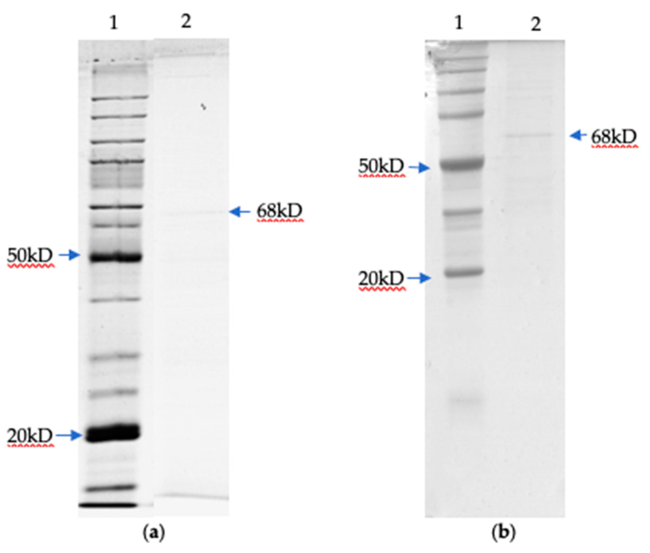
3.4. Determination of Molecular Weight by SDS PAGE

The molecular weights of the two α-amylases were estimated with reference to a standard curve built using the BenchMark protein marker. The band obtained in SDS PAGE indicated the presence of partially purified α-amylase of each strain. The Antarctic and Arctic α-amylases were both estimated to have a molecular weight of 68 kDa (Figure 3).

4. Discussion

The optimum temperature of α-amylases obtained from both polar strains in the present study was 15 °C, confirming that both strains produce cold-active enzymes, which could be an adaptation strategy [51,52]. Cold-active enzymes are reported to have low conformational stability [53], which may be a result of random genetic drift [54]. However, the increased activity at lower temperatures does not affect the overall protein stability [55]. In the present study, the reduction in activity as temperature increased indicates the tertiary structure of the enzyme loses its stability [56]. The underlying molecular mechanism for this cold adaptability could be due to (a) core hydrophobicity reduction, surface hydrophobicity increment, a low arginine/lysine ratio, weak interaction between inter-domain and inter-subunits, more and extended loops; (b) alpha helices in the secondary structure; (c) reduction in content of secondary structure, reduced and weak metal binding sites, fewer disulfide bridges, and electrostatic interactions; and, finally, (d) reduced oligomerization and an increase in the non-conformational entropy of the unfolded state [57,58,59]. As enzyme production at industrial scales is commonly performed at low to moderate temperatures [60], these two candidate Pseudogymnoascus strains from the Arctic and Antarctic have good potential for use in biotechnological applications.
The Antarctic α-amylase examined here had an optimum pH of 6.0, similar to that reported by Iefuji et al. [61]. The Arctic α-amylase had a slightly more acidic optimum pH of 5.0. The differences in optimum pH could be due to environmental pH, which has a direct influence on extracellular enzymes. When there are changes in [H+] in the soil environment, the ionization state and 3D structure of the active sites of the enzymes will be modified [62], thereby affecting their activity [63,64,65]. The Arctic α-amylase, in particular, may be a good candidate for examination for biotechnological application because the amount of maltulose, a by-product formed at higher pH during industrial enzymatic reactions, is reduced considerably when acidophilic enzymes are used [66]. The acidic pH optima of both Antarctic and Arctic α-amylases are consistent with previous reports of bacterial and fungal α-amylases, which frequently have slightly acidic to neutral pH optima [67].
The calculated molecular weights of the α-amylases obtained in the present study were 68 kDa for both Antarctic and Arctic enzymes. This is consistent with α-amylase obtained from Aspergillus sp. (68 kDa) [68]. A recent study has reported α-amylase of bacterial origin (Pseudoalteromonas sp.) from Antarctic sea ice is 61 kDa [69]. Molecular weights of α-amylases of microbes have previously been assessed as 50–60 kDa [70,71,72], although other reports are highly variable, ranging from 10 to 210 kDa. The smallest molecule (10 kDa) was reported from Bacillus caldolyticus [73] and the largest (210 kDa) from Chloroflexus aurantiacus [74], both being bacteria.
Our data also showed that both polar α-amylases declined in activity as temperature increased above 15 °C. Although not a primary objective of this study, this activity reduction can be related to a scenario of environmental temperature increase. It is predicted that, by the end of this century, global mean air temperature will increase by 1.8 to 3.6 °C, with this being magnified at higher latitudes [75]. Representatives of Pseudogymnoascus are frequently reported from the polar regions e.g., [27,43,76,77], where they are believed to play an important role in decomposition processes [78]. As polar soils typically experience temperatures well below the fungal optimum of 15 °C at present [79], increasing temperatures are likely to increase enzyme activity, generating positive feedback on decomposition rates and, consequently, accelerating climate change by releasing CO2 into the atmosphere. On the contrary, Misiak et al. [80] who conducted a study at Mars Oasis, Antarctic Peninsula using P. roseus suggested that as long as there is high water availability, increasing temperature (i.e., >20 °C) will inhibit the growth of the fungal hyphae and eventually its ability to decompose organic matter. Therefore, we infer that the enzyme activity increases following temperature rise until the enzyme activity reaches a maximum point, by which further temperature increase begins to inhibit and reduce enzyme activity.
The Antarctic and Arctic α-amylases studied here showed very similar characteristics, with MW of 68 kDa and similar optimum temperature and acidic pH values, but neither showed close matches to characterized enzymes in available databases. Further analysis is needed to confirm if they are novel enzymes. In terms of purification fold, Gao et al. [29] reported 5.72-fold after the first purification step, while the present study recovered 1.95-fold amylase from Antarctic Pseudogymnoascus sp. and 29.09-fold from Arctic Pseudogymnoascus sp. The differences between the studies are likely due to variability in the methods or column matrices used. Both microfungi investigated in this study are good candidates for further examination in the biotechnology industry, as they show the greatest activity at acidic pH, which is desirable to reduce problems related to by-product formation, and an optimum temperature of 15 °C, which could help to reduce energy use and costs.
Studies focused on amylases of fungal origin are extremely limited. Those studies of cold-active enzymes that are available refer to fungi that were obtained from other parts of the world [60,81]. As the fungi of polar regions are generally understudied, it is clear that they could contain currently unknown enzymatic resources, such as the α-amylases identified in the present study. Available studies involving psychrophilic or cold active α-amylases from fungi and bacteria are summarized in Table 2. It is worth mentioning the different enzyme characteristics between the α-amylase of Pseudogymnoascus sp. (in the present study) and α-amylase from Geomyces pannorum R1-2 [29], which the latter showed a fourfold increase (from 15 to 70 °C) despite a 5 °C difference culture temperature. However, this may be due to the recombinant nature of the enzyme used in that study.
Pseudogymnoascus sp. investigated in the present study have potential applications in biotechnology. Furthermore, this fungal species could provide the key to answering questions about fungal-related decomposition processes in the polar regions. However, fundamental investigations are still required, including (a) the use of genomic and transcriptomic approaches to resolving taxonomic issues and (b) obtaining the DNA sequences to be cloned in heterologous expression systems. The recombinant protein production could be optimized and analyzed for substrate specificity, chiral selectivity, and confirmation of the 3D structure of the enzyme.

5. Conclusions

α-amylase obtained from Pseudogymnoascus strains of Antarctic and Arctic origin exhibited similar characteristics in terms of optimum temperature (15 °C) but differed slightly in optimum pH, although both optima were slightly acidic. However, in the natural habitat of these microbes, temperature and pH are not the only influences on fungal enzyme production, and other factors will be involved, such as C and N availability and interactions with other microbes. More detailed multivariate studies are required to enable robust conclusions to be made about microfungal function in decomposition and its relationship with environmental variables and climate change.

Author Contributions

Conceptualization, A.K., S.A.A. and P.C.; methodology, A.K., S.A.A. and Z.A.; validation, P.C., S.A.A. and Z.A.; formal analysis, A.K.; investigation, A.K.; resources, A.K., S.A.A., J.S. and M.G.-A.; data curation, A.K.; writing—original draft preparation, A.K.; writing—review and editing, P.C., S.A.A. and M.R.-I.; visualization, A.K.; supervision, P.C., S.A.A. and Z.A.; project administration, S.A.A.; funding acquisition, A.K., S.A.A. and M.R.-I. All authors have read and agreed to the published version of the manuscript.

Funding

This research was funded by Yayasan Penyelidikan Antartika Sultan Mizan (YPASM), University Malaya, grant numbers PG041-2013A & RP007-2012A and Ministry of Science Technology and Innovation (MOSTI) grant number GA006-2014FL. PC is supported by NERC core funding to the BAS ‘Biodiversity, Evolution and Adaptation’ Team.

Institutional Review Board Statement

Not applicable.

Informed Consent Statement

Not applicable.

Data Availability Statement

Not applicable.

Acknowledgments

Our sincere gratitude goes to Instituto Antartico Chileno (INACH) for providing logistic support during sample collection in Antarctica.

Conflicts of Interest

The authors declare no conflict of interest. The funders had no role in the design of the study; in the collection, analyses, or interpretation of data; in the writing of the manuscript; or in the decision to publish the results.

References

  1. Ruisi, S.; Barreca, D.; Selbmann, L.; Zucconi, L.; Onofri, S. Fungi in Antarctica. Rev. Environ. Sci. Biotechnol. 2007, 6, 127–141. [Google Scholar] [CrossRef]
  2. Tian, Y.; Li, Y.L.; Zhao, F.C. Secondary Metabolites from Polar Organisms. Mar. Drugs 2017, 15, 28. [Google Scholar] [CrossRef] [PubMed] [Green Version]
  3. Zhang, Z.; Li, S.; Gu, X.; Li, J.; Lin, X. Biosynthesis, characterization and antibacterial activity of silver nanoparticles by the Arctic anti-oxidative bacterium Paracoccus sp. Artif. Cells Nanomed. Biotechnol. 2019, 47, 1488–1495. [Google Scholar] [CrossRef] [PubMed] [Green Version]
  4. Colby, G.A.; Ruuskanen, M.O.; St. Pierre, K.A.; St. Louis, V.L.; Poulain, A.J.; Aris-Brosou, S. Warming Climate Is Reducing the Diversity of Dominant Microbes in the Largest High Arctic Lake. Front. Microbiol. 2020, 11, 1–12. [Google Scholar] [CrossRef] [PubMed]
  5. Pereira, F. Metagenomics: A gateway to drug discovery. In Advances in Biological Science Research; Meena, S.N., Naik, M.M., Eds.; Academic Press: Goa, India, 2019; pp. 453–468. [Google Scholar]
  6. Zucconi, L.; Canini, F.; Temporiti, M.E.; Tosi, S. Extracellular Enzymes and Bioactive Compounds from Antarctic Terrestrial Fungi for Bioprospecting. Int. J. Environ. Res. Public Health 2020, 17, 6459. [Google Scholar] [CrossRef] [PubMed]
  7. Arenz, B.E.; Blanchette, R.A.; Farrell, R.L. Fungal Diversity in Antarctic Soils. In Antarctic Terrestrial Microbiology; Cowan, D., Ed.; Springer: Berlin/Heidelberg, Germany, 2014; pp. 35–53. [Google Scholar]
  8. Walton, D.W.H. Cellulose decomposition and its relationship to nutrient cycling at South Georgia. In Antarctic Nutrient Cycling and Food Webs, Proceedings of the 4th SCAR Symposium on Antarctic Biology, Wilderness, South Africa, 12–16 September 1983; Siegfried, W.R., Condy, P.R., Laws, R.M., Eds.; Springer: Berlin/Heidelberg, Germany, 1985; pp. 192–199. [Google Scholar]
  9. Crowther, T.W.; Van Den Hoogen, J.; Wan, J.; Mayes, M.A.; Keiser, A.D.; Mo, L.; Averill, C.; Maynard, D.S. The global soil community and its influence on biogeochemistry. Science 2019, 365, eaav0550. [Google Scholar] [CrossRef]
  10. Singh, S.M.; Singh, S.K.; Yadav, L.S.; Singh, P.N.; Ravindra, R. Filamentous soil fungi from Ny-Ålesund, Spitsbergen, and screening for extracellular enzymes. Polar Biol. 2012, 65, 45–55. [Google Scholar] [CrossRef]
  11. Abneuf, M.A.; Krishnan, A.; Gonzalez-Aravena, M.; Pang, K.L.; Convey, P.; Mohamad-Fauzi, N.; Rizman-Idid, M.; Alias, S.A. Antimicrobial activity of microfungi from maritime Antarctic soil. Czech Polar Rep. 2016, 6, 141–154. [Google Scholar] [CrossRef]
  12. Irawan, B.; Afandi, A.; Hadi, S. Effects of saprophytic microfungi application on soil fertility based on their decomposition properties. J. Appl. Biol. Sci. 2017, 11, 15–19. [Google Scholar]
  13. IPCC. Climate Change: Synthesis Report. Contribution of Working Groups I, II and III to the Fifth Assessment Report of the Intergovernmental Panel on Climate Change; Core Writing Team, Rajendra, K., Meyer, L.A., Eds.; IPCC: Geneva, Switzerland, 2014; 151p. [Google Scholar]
  14. Carrasco, M.; Villarreal, P.; Barahona, S.; Alcaíno, J.; Cifuentes, V.; Baeza, M. Screening and characterization of amylase and cellulase activities in psychrotolerant yeasts. BMC Microbiol. 2016, 16, 21–29. [Google Scholar] [CrossRef] [Green Version]
  15. Duarte, A.F.; Dayo-Owoyemi Nobre, F.S.; Pagnocca, F.C.; Chaud, L.S.; Pessoa, A.; Felipe, M.D.G.A.; Sette, L.D. Taxonomic assessment and enzymes production by yeasts isolated from marine and terrestrial Antarctic samples. Extremophiles 2013, 17, 1023–1035. [Google Scholar] [CrossRef] [PubMed]
  16. Ryan, S.M.; Fitzgerald, G.F.; Sinderen, D.V. Screening for and identification of starch-, amylopectin-, and pullulan-degrading activities in bifidobacterial strains. Appl. Environ. Microbiol. 2006, 72, 5289–5296. [Google Scholar] [CrossRef] [Green Version]
  17. Offen, W.A.; Viksoe-Nielsen, A.; Borchert, T.V.; Wilson, K.S.; Davies, G. Three-dimensional structure of a variant ‘Termamyl-like’ Geobacillus stearothermophilus α-amylase at 1.9 Å resolution. Acta Crystallogr. F Struct. Biol. Commun. 2015, 71, 66–70. [Google Scholar] [CrossRef] [PubMed] [Green Version]
  18. Liu, Y.; Yu, J.; Li, F.; Peng, H.; Zhang, X.; Xiao, Y.; He, C. Crystal structure of a raw-starch-degrading bacterial α-amylase belonging to subfamily 37 of the glycoside hydrolase family GH13. Sci. Rep. 2017, 7, 44067. [Google Scholar] [CrossRef] [PubMed]
  19. Goto, C.E.; Barbosa, E.P.; Kistner, L.C.; Moreira, F.G.; Lenartovicz, V.; Peralta, R.M. Production of amylase by Aspergillus fumigatus utilizing alpha-methyl-D-glycoside, a synthetic analogue of maltose, as substrate. FEMS Microbiol. Lett. 1998, 167, 139–143. [Google Scholar] [PubMed] [Green Version]
  20. Bin, G.; Laisu, X.; Youfang, D.; Yanquan, L. Screening of alpha amylase high-producing strains from Bacillus subtilis. J. Zhejiang 1999, 23, 88–92. [Google Scholar]
  21. Singh, S.; Singh, S.; Bali, V.; Sharma, L.; Mangla, J. Production of Fungal Amylases Using Cheap, Readily Available Agriresidues, for Potential Application in Textile Industry. Biomed. Res. Int. 2014, 2014, 215748. [Google Scholar] [CrossRef] [Green Version]
  22. Singh, S.; Mangla, J.; Singh, S. Evaluation of Aspergillus fumigatus NTCC1222 as a source of enzymes for detergent industry. Res. Environ. Sustain. 2021, 5, 1–5. [Google Scholar] [CrossRef]
  23. Naili, B.; Sahnoun, M.; Bejar, S.; Kammoun, R. Optimization of Submerged Aspergillus oryzae S2 α-Amylase Production. Food Sci. Biotechnol. 2016, 25, 185–192. [Google Scholar] [CrossRef]
  24. Abdullah, R.; ul-Haq, I. Purification and characterisation of a-amylase produced by mutant strain of Aspergillus oryzae EMS-18. Nat. Prod. Res. 2014, 29, 710–716. [Google Scholar] [CrossRef]
  25. Kamaraj, M.; Subramaniam, D. Optimization of Experimental Variables for the Production of α- Amylase by Aspergillus Oryzae Using Rice Bran. Int. J. Adv. Res. Eng. Technol. 2020, 11, 113–126. [Google Scholar]
  26. Martinez-Martinez, M.; Coscolin, C.; Santiago, G.; Chow, J.; Stogios, P.J.; Bargiela, R.; Gertler, C.; Navarro-Fernández, J.; Bollinger, A.; Thies, S.; et al. Determinants and Prediction of Esterase Substrate Promiscuity Patterns. ACS Chem. Biol. 2018, 13, 225–234. [Google Scholar] [CrossRef] [PubMed] [Green Version]
  27. Krishnan, A.; Alias, S.A.; Wong, C.V.; Pang, K.L.; Convey, P. Extracellular hydrolase enzyme production by soil fungi from King George Island, Antarctica. Polar Biol. 2011, 34, 1535–1542. [Google Scholar] [CrossRef]
  28. Krishnan, A.; Convey, P.; Gonzalez-Rocha, G.; Alias, S.A. Production of extracellular hydrolase enzymes by fungi from King George Island. Polar Biol. 2016, 39, 65–76. [Google Scholar] [CrossRef]
  29. Gao, B.; Mao, Y.; Zhang, L.; He, L.; Wei, d. A novel saccharifying a-amylase of Antarctic psychrotolerant fungi Geomyces pannorum: Gene cloning, functional expression, and characterization. Starch/Stärke 2016, 68, 20–28. [Google Scholar] [CrossRef]
  30. Gesheva, V. Distribution of psychrophilic microorganisms in soils of Terra Nova Bay and Edmonson Point, Victoria Land and their biosynthetic capabilities. Polar Biol. 2009, 32, 1287–1291. [Google Scholar] [CrossRef]
  31. Gesheva, V.; Vasileva-Tonkova, E. Production of enzymes and antimicrobial compounds by halophilic Antarctic Nocardioides sp. grown on different carbon sources. World J. Microbiol. Biotechnol. 2012, 28, 2069–2076. [Google Scholar] [CrossRef] [Green Version]
  32. Margesin, R.; Neuner, G.; Storey, K.B. Cold-loving microbes, plants, and animals- fundamental and applied aspects. Naturwissenschaften 2007, 94, 77–99. [Google Scholar] [CrossRef]
  33. Birgisson, H.; Delgado, O.; Garcia Arroyo, L.; Hatti-Kaul, R.; Mattiasson, B. Cold-adapted yeasts as producers of cold-active polygalacturonases. Extremophiles 2003, 7, 185–193. [Google Scholar] [CrossRef]
  34. Carrasco, M.; Rozas, J.M.; Barahona, S.; Alcaíno, J.; Cifuentes, V.; Baeza, M. Diversity and extracellular enzymatic activities of yeasts isolated from King George Island, the sub-Antarctic region. BMC Microbiol. 2012, 12, 251–259. [Google Scholar] [CrossRef] [Green Version]
  35. Zak, D.R.; Kling, G.W. Microbial community composition and function across an Arctic tundra landscape. Ecology 2006, 87, 1659–1670. [Google Scholar] [CrossRef]
  36. Bancerz, R.; Ginalska, G.; Fiedurek, J.; Gromada, A. Cultivation conditions and properties of extracellular crude lipase from the psychrotrophic fungus Penicillium chrysogenum 9’. J. Ind. Microbiol. Biotechnol. 2005, 32, 253–260. [Google Scholar] [CrossRef] [PubMed]
  37. Gawas-Sakhalkar, P.; Singh, S.M. Fungal community associated with Arctic moss, Tetraplodon mimoides and its rhizosphere: Bioprospecting for production of industrially useful enzymes. Curr. Sci. 2011, 100, 1701–1705. [Google Scholar]
  38. Zaferanloo, B.; Bhattacharjee, S.; Ghorbani, M.M.; Mahon, P.J.; Palombo, E.A. Amylase production by Preussia minima, a fungus of endophytic origin: Optimization of fermentation conditions and analysis of fungal secretome by LC-MS. BMC Mcrobiol. 2014, 14, 55–66. [Google Scholar] [CrossRef]
  39. Punekar, N.S. Exploiting Enzymes: Technology and Applications. In ENZYMES: Catalysis, Kinetics and Mechanisms; Springer: Singapore, 2018; pp. 14–31. [Google Scholar]
  40. Rousk, J.; Brookes, P.C.; Bååth, E. Contrasting Soil pH Effects on Fungal and Bacterial Growth Suggest Functional Redundancy in Carbon Mineralization. Appl. Environ. Microbiol. 2009, 75, 1589–1596. [Google Scholar] [CrossRef] [Green Version]
  41. Rajoka, M.I.; Akhtar, M.W.; Hanif, A.; Khalid, A.M. Production and characterization of a highly active cellobiase from Aspergillus niger grown in solid state fermentation. World J. Microbiol. Biotechnol. 2006, 22, 991–998. [Google Scholar] [CrossRef]
  42. Dojnov, B.; Božić, N.; Nenadović, V.; Ivanović, J.; Vujčić, Z. Purification and properties of midgut α-amylase isolated from Morimus funereus (Coleoptera: Cerambycidae) larvae. Comp. Biochem. Physiol. 2008, 149, 153–160. [Google Scholar] [CrossRef]
  43. Krishnan, A.; Convey, P.; Gonzalez, M.; Smykla, J.; Alias, S.A. Effects of temperature on extracellular hydrolase enzymes from soil microfungi. Polar Biol. 2018, 41, 537–551. [Google Scholar] [CrossRef]
  44. Wong, H.J.; Mohamad-Fauzi, N.; Rizman-Idid, M.; Convey, P.; Alias, S.A. Protective mechanisms and responses of micro-fungi towards ultraviolet-induced cellular damage. Polar Sci. 2018, 20, 19–34. [Google Scholar] [CrossRef]
  45. Bhardwaj, N.; Verma, V.K.; Chaturvedi, V.; Verma, P. GH10 XynF1 and Xyn11A: The predominant xylanase identified in the profiling of extracellular proteome of Aspergillus oryzae LC1. Ann. Microbiol. 2018, 68, 731–742. [Google Scholar] [CrossRef]
  46. Bradford, M.M. A rapid and sensitive method for the quantitation of microgram quantities of protein utilizing the principle of protein dye binding. Anal. Biochem. 1976, 72, 248–254. [Google Scholar] [CrossRef]
  47. Miller, G.L. Use of dinitrosalicylic acid reagent for determination of reducing sugar. Anal. Chem. 1959, 31, 426–428. [Google Scholar] [CrossRef]
  48. Kobayashi, M.; Sasaki, Y.; Kobayashi, S. Purification of amylases and other enzymes by a forced-affinity chromatography method. Biosci. Biotechnol. Biochem. 1997, 61, 813–816. [Google Scholar] [CrossRef] [PubMed]
  49. Xiao, Z.; Storms, R.; Tsang, A. Microplate-based carboxymethylcellulose assay for endoglucanase activity. Anal. Biochem. 2006, 342, 176–178. [Google Scholar] [CrossRef]
  50. Laemmli, U.K. Cleavage of structural proteins during the assembly of the head of bacteriophage T4. Nature 1970, 227, 680–685. [Google Scholar] [CrossRef]
  51. Hassan, N.; Rafiq, M.; Hayat, M.; Shah, A.A.; Hasan, F. Psychrophilic and psychrotrophic fungi: A comprehensive review. Rev Environ. Sci. Biotechnol. 2016, 15, 147–172. [Google Scholar] [CrossRef]
  52. Robinson, C.H. Cold adaptation in Arctic and Antarctic fungi. New Phytol. 2001, 151, 341–353. [Google Scholar] [CrossRef]
  53. D’Amico, S.; Claverie, P.; Collins, T.; Georlette, D.; Gratia, E.; Hoyoux, A.; Meuwis, M.-A.; Feller, G.; Gerday, C. Molecular basis of cold adaptation. Philos. Trans. R. Soc. B 2002, 357, 917–925. [Google Scholar] [CrossRef] [Green Version]
  54. Miyazaki, K.; Wintrode, P.L.; Grayling, R.A.; Rubingh, D.N.; Arnold, F.H. Directed evolution study of temperature adaptation in a psychrophilic enzyme. J. Mol. Biol. 2000, 297, 1015–1026. [Google Scholar] [CrossRef] [Green Version]
  55. Gault, S.; Higgins, P.M.; Cockell, C.S.; Gillies, K. A meta-analysis of the activity, stability, and mutational characteristics of temperature-adapted enzymes. Biosci. Rep. 2021, 41, 1–10. [Google Scholar] [CrossRef]
  56. Bisswanger, H. Enzyme assays. Perspect. Sci. 2014, 1, 41–55. [Google Scholar] [CrossRef] [Green Version]
  57. Cavicchioli, R.; Siddiqui, K.S. Cold-adapted enzymes. In Enzyme Technology; Pandey, A., Webb, C., Soccol, C.R., Larroche, C., Eds.; Springer Science: New York, NY, USA, 2006; pp. 615–638. [Google Scholar]
  58. Ramli, A.M.; Azhar, M.A.; Shamsir, M.S.; Rabu, A.; Abdul Murad, A.M.; Mahadi, N.M.; Illias, R.M. Sequence and structural investigation of a novel psychrophilic a-amylase from Glaciozyma antarctica PI12 for cold adaptation analysis. J. Mol. Model. 2013, 19, 3369–3383. [Google Scholar] [CrossRef] [PubMed]
  59. Ramli, A.M.; Mahadi, N.M.; Shamsir, M.S.; Rabu, A.; Joyce-Tan, K.H.; Abdul Murad, A.M.; Illias, R.M. Structural prediction of a novel chitinase from the psychrophilic Glaciozyma antarctica PI12 and an analysis of its structural properties and function. J. Comput. Aided Mol. Des. 2012, 26, 947–961. [Google Scholar] [CrossRef] [PubMed]
  60. Duarte, A.F.; dos Santos, J.A.; Vianna, M.V.; Vieira, J.F.; Mallagutti, V.H.; Inforsato, F.J.; Wentzel, L.C.P.; Lario, L.D.; Rodrigues, A.; Pagnocca, F.C.; et al. Cold-adapted enzymes produced by fungi from terrestrial and marine Antarctic environments. Crit. Rev. Biotechnol. 2018, 38, 600–619. [Google Scholar] [CrossRef] [PubMed]
  61. Iefuji, H.; Chino, M.; Kato, M.; Iimura, Y. Raw-starch-digesting and thermostable α-amylase from the yeast Cryptococcus sp. S-2: Purification, characterization, cloning and sequencing. Biochem. J. 1996, 318, 989–996. [Google Scholar] [CrossRef] [PubMed] [Green Version]
  62. Tipton, K.F.; Dixon, H.F. Effects of pH on enzymes. In Methods in Enzymology; Daniel, L.P., Ed.; Academic Press: Cambridge, MA, USA, 1979; Volume 63, pp. 183–234. [Google Scholar]
  63. King, G.M. Characterization of β-Glucosidase Activity in Intertidal Marine Sediments. Appl. Environ. Microbiol. 1986, 51, 373–380. [Google Scholar] [CrossRef] [Green Version]
  64. Chróst, R.J. Environmental control of the synthesis and activity of aquatic microbial ectoenzymes. In Microbial Enzymes in Aquatic Environments; Chróst, R.J., Ed.; Springer: New York, NY, USA, 1991; pp. 29–59. [Google Scholar]
  65. Münster, U. Extracellular enzyme activity in eutrophic and polyhumic lakes. In Microbial Enzymes in Aquatic Environments; Chróst, R.J., Ed.; Springer: New York, NY, USA, 1991; pp. 96–122. [Google Scholar]
  66. Liu, X.D.; Xu, Y. A novel raw starch digesting α-amylase from a newly isolated Bacillus sp. YX-1: Purification and characterization. Bioresour. Technol. 2008, 99, 4315–4320. [Google Scholar] [CrossRef]
  67. Pandey, A.; Nigam, P.; Soccol, C.R.; Soccol, V.T.; Singh, D.; Mohan, R. Advances in microbial amylases. Appl. Biochem. Biotechnol. 2000, 31, 135–152. [Google Scholar] [CrossRef]
  68. Abe, J.I.; Nakajima, K.; Nagano, H.; Hizukuri, S.; Obata, K. Properties of the raw-starch digesting amylase of Aspergillus sp. K-27: A synergistic action of glucoamylase and alpha-amylase. Carbohydr. Res. 1988, 175, 85–92. [Google Scholar] [CrossRef]
  69. Wang, X.; Kan, G.; Shi, C.; Xie, Q.; Ju, Y.; Wang, R.; Qiao, Y.; Ren, X. Purification and characterization of a novel wild-type α-amylase from Antarctic sea ice bacterium Pseudoalteromonas sp. M175. Protein Expr. Purif. 2019, 164, 105444. [Google Scholar] [CrossRef]
  70. Vihinen, M.; Mantsala, P. Microbial amylolytic enzymes. Crit. Rev. Biochem. Mol. 1989, 24, 329–418. [Google Scholar] [CrossRef] [PubMed]
  71. Xian, L.; Wang, F.; Luo, X.; Feng, Y.L.; Feng, J.X. Purification and Characterization of a Highly Efficient Calcium-Independent α-Amylase from Talaromyces pinophilus 1-95. PLoS ONE 2015, 10, e0121531. [Google Scholar] [CrossRef] [PubMed] [Green Version]
  72. Keskin, S.; Ertunga, S. Purification, immobilization and characterization of thermostable α-amylase from a thermophilic bacterium Geobacillus sp. TF14. Turk. J. Biochem. 2017, 42, 633–642. [Google Scholar] [CrossRef]
  73. Grootegoed, J.A.; Lauwers, A.M.; Heinen, W. Separation and partial purification of extracellular amylase and protease from Bacillus caldolyticus. Arch. Microbiol. 1973, 90, 223. [Google Scholar] [CrossRef]
  74. Ratanakhanokchai, K.; Kaneko, J.; Kamio, Y.; Izaki, K. Purification and properties of a maltotetraose and maltotriose producing amylase from Chloroflexus aurantiacus. Appl. Environ. Microbiol. 1992, 58, 2490–2494. [Google Scholar] [CrossRef] [Green Version]
  75. IPCC. Summary for Policymakers. In Climate Change 2013: The Physical Science Basis; Cambridge University Press: Cambridge, UK, 2013. [Google Scholar]
  76. Marshall, W.A. Aerial Transport of Keratinaceous Substrate and Distribution of the Fungus Geomyces pannorum in Antarctic Soils. Microb. Ecol. 1998, 36, 212–219. [Google Scholar] [CrossRef]
  77. Bridge, P.L.; Spooner, B.M.; Roberts, P.J. Non-lichenized fungi from the Antarctic region. Mycotaxon 2008, 106, 485–490. [Google Scholar]
  78. Arenz, B.E.; Blanchette, R.A. Distribution and abundance of soil fungi in Antarctica at sites on the Peninsula, Ross Sea Region and McMurdo Dry Valleys. Soil Biol. Biochem. 2011, 43, 308–315. [Google Scholar] [CrossRef]
  79. Convey, P.; Coulson, S.J.; Worland, M.R.; Sjöblom, A. The importance of understanding annual and shorter term temperature patterns and variation in the upper layers of polar soils for terrestrial biota. Polar Biol. 2018, 41, 1587–1605. [Google Scholar] [CrossRef] [Green Version]
  80. Misiak, M.; Goodall-Copestake, W.P.; Sparks, T.H.; Worland, M.R.; Boddy, L.; Magan, N.; Convey, P.; Hopkins, D.W.; Newsham, K.K. Inhibitory effects of climate change on the growth and extracellular enzyme activities of a widespread Antarctic soil fungus. Glob. Chang. Biol. 2021, 27, 1111–1125. [Google Scholar] [CrossRef]
  81. Cavicchioli, R.; Siddiqui, K.S.; Andrews, D.; Sowers, K.R. Low-temperature extremophiles and their applications. Curr. Opin. Biotechnol. 2002, 13, 253–261. [Google Scholar] [CrossRef]
  82. De Mot, R.; Verachtert, H. Purification and characterization of extracellular a-amylase and glucoamylase from the yeast Candida antarctica CBS 6678. Eur. J. Biochem. 1987, 164, 643–654. [Google Scholar] [CrossRef] [PubMed]
  83. He, L.; Mao, Y.; Zhang, L.; Wang, H.; Alias, S.A.; Gao, B.; Wei, D. Functional expression of a novel α-amylase from Antarctic psychrotolerant fungus for baking industry and its magnetic immobilization. BMC Biotechnol. 2017, 17, 17–22. [Google Scholar] [CrossRef] [PubMed] [Green Version]
  84. Samie, N.; Noghabi, K.A.; Gharegozloo, Z.; Zahiri, H.S.; Ahmadian, G.; Sharafi, H.; Behrozi, R.; Vali, H. Psychrophilic a-amylase from Aeromonas veronii NS07 isolated from farm soils. Process Biochem. 2012, 47, 1381–1387. [Google Scholar] [CrossRef]
  85. Roohi; Kuddus, M.; Saima. Cold-active detergent-stable extracellular α-amylase from Bacillus cereus GA6: Biochemical characteristics and its perspectives in laundry detergent formulation. J. Biochem. Technol. 2013, 4, 636–644. [Google Scholar]
  86. Kuddus, M.; Arif, J.M.; Ramteke, P.W. An overview of coldactive microbial a-amylase: Adaptation strategies and biotechnological potentials. Biotechnology 2011, 3, 246–258. [Google Scholar] [CrossRef]
  87. Chessa, J.P.; Feller, G.; Gerday, C. Purification and characterization of the heat-labile a-amylase secreted by the psychrophilic bacterium TAC 240B. Can. J. Microbiol. 1999, 45, 452–457. [Google Scholar] [CrossRef]
  88. Smith, M.R.; Zahnley, J.C. Production of amylase by Arthrobacter psychrolactophilus. J. Ind. Microbiol. Biotechnol. 2005, 32, 277–283. [Google Scholar] [CrossRef] [Green Version]
  89. Rathour, R.; Gupta, J.; Tyagi, B.; Thakur, I.S. Production and characterization of psychrophilic α-amylase from a psychrophilic bacterium, Shewanella sp. ISTPL2. Amylase 2020, 4, 1–10. [Google Scholar] [CrossRef]
Figure 1. Temperature optimization of α-amylase obtained from Antarctic Pseudogymnoascus sp. and Arctic Pseudogymnoascus sp. (* p < 0.05).
Fermentation 08 00601 g001
Figure 2. pH optimization using different buffers for α-amylase obtained from Antarctic Pseudogymnoascus sp. and Arctic Pseudogymnoascus sp. (* p < 0.05).
Fermentation 08 00601 g002
Figure 3. This figure shows 12% Sodium Dodecyl Sulfate Polyacrylamide Gel Electrophoresis of (a) Antarctic α-amylase from Pseudogymnoascus sp. stained in Coomassie Blue. (Lane 1: Invitrogen BenchMark Protein marker, Lane 2: Partially purified α-amylase from Pseudogymnoascus sp. from the Antarctic); (b) Arctic α-amylase from Pseudogymnoascus sp. stained in Coomassie Blue. (Lane 1: Invitrogen BenchMark Protein marker, Lane 2: Partially purified α-amylase from Pseudogymnoascus sp. from the Arctic).
Fermentation 08 00601 g003
Table 1. Purification steps for Antarctic and Arctic Pseudogymnoascus sp.
StrainSampleEnzyme Activity (µmo/min/mL)Total Protein Content (mg/mL)Specific Activity (U/mg)Purification FoldRecovery %
Antarctic Pseudogymnoascus sp.Crude40.6415.802.571100
Partially purified enzyme3.710.745.011.959.13
Arctic Pseudogymnoascus sp.Crude enzyme18.6722.670.821100
Partially purified enzyme3.10.1323.8529.0916.60
Table 2. Characteristics of psychrophilic α-amylases from fungal and bacterial sources.
OriginGrowth ConditionEnzyme CharacteristicsSpecific Activity
(U/mg)
MW (kDa)Recombinant/Wild Type EnzymeReference
TemperaturepHOptimum TemperatureOptimum pH
Fungi
Pseudogymnoascus sp.156155–65.01 & 23.8568Wild typePresent study
Moesziomyces antarcticus29NR624.2NR50Wild type[82]
Geomyces pannorum R1-2
(currently known as Pseudogymnoascus pannorum)
20NR706.09.78 × 103NRRecombinant[29]
Geomyces pannorum R1-2
(currently known as Pseudogymnoascus pannorum)
NRNR40512.8 × 10352Recombinant[83]
Bacteria
Aeromonas veronii (Bacteria)10NR10443063Wild type[84]
Bacillus cereusNRNR2010175.9255Wild type[85]
Microbacterium foliorum GA2 and Bacillus cereus GA620NR209 & 10NRNRWild type[86]
strain TAC 240BNRNR 7.57150Wild type[87]
Arthrobacter psychrolactophilus22720NR 105 & 26Wild type[88]
Shewanella sp. ISTPL2 (bacteria)106.94836,690.4745Wild type[89]
Pseudoalteromonas sp. M175158307.5289.7961Wild type[69]
Publisher’s Note: MDPI stays neutral with regard to jurisdictional claims in published maps and institutional affiliations.

