Article

Preparation and Antioxidant Activity In Vitro of Fermented Tremella fuciformis Extracellular Polysaccharides

1
Guangdong Province Key Laboratory for Biotechnology Drug Candidates, School of Biosciences and Biopharmaceutics, Guangdong Pharmaceutical University, Guangzhou 510006, China
2
School of Food Science, Guangdong Pharmaceutical University, Guangzhou 510006, China
*
Author to whom correspondence should be addressed.
Fermentation 2022, 8(11), 616; https://doi.org/10.3390/fermentation8110616
Submission received: 27 September 2022 / Revised: 3 November 2022 / Accepted: 5 November 2022 / Published: 9 November 2022
(This article belongs to the Section Fermentation for Food and Beverages)

Abstract

:
This study was aimed at increasing the capacity of fermented Tremella fuciformis extracellular polysaccharides (TEPS) for possible functional food applications. Thus, strain varieties, fermentation parameters and purification conditions, and the in vitro antioxidant activities of purified EPS fractions were investigated. An EPS high-yield strain Tf526 was selected, and the effects of seven independent fermentation factors (time, temperature, initial pH, inoculum size, shaking speed, carbon, and nitrogen source) on the EPS yield were evaluated. By single factor optimization test, yeast extract and glucose were chosen as nitrogen sources and carbon sources, respectively, and with initial pH of 6.0, inoculum size of 8%, shaking speed of 150 rpm, and culture at 25 °C for 72 h, the optimal yield of TEPS reached 0.76 ± 0.03 mg/mL. Additionally, A-722MP resin showed the most efficient decoloration ratio compared to six other tested resins. Furthermore, optimal decoloration parameters of A-722MP resin were obtained as follows: decoloration time of 2 h, resins dosage of 2 g, and temperature of 30 °C. Decoloration ratio, deproteinization ratio, and polysaccharide retention ratio were 62.14 ± 2.3%, 81.21 ± 2.13%, and 73.42 ± 1.96%, respectively. Furthermore, the crude TEPS was extracted and four polysaccharide fractions were isolated and purified as Tf1-a, Tf1-b, Tf2, and Tf3 by the DEAE-Sepharose FF column and the Sephasryl S100 column. In general, the antioxidant activities of the Lf1-a and Lf1-b were lower compared with Vc at the concentration of 0.1 to 3 mg/mL, but the FRAP assay, DPPH scavenging activity, and hydroxyl radical scavenging activity analysis still revealed that Tf1-a and Tf1-b possess significant antioxidant activities in vitro. At the concentration of 3 mg/mL, the reducing power of Lf1-a and Lf1-b reached 0.86 and 0.70, the maximum DPPH radical were 54.23 ± 1.68% and 61.62 ± 2.73%, and the maximum hydroxyl radicals scavenging rates were 58.76 ± 2.58% and 45.81 ± 1.79%, respectively. Moreover, there were significant correlations (r > 0.8) among the selected concentrations and antioxidant activities of TEPS major fractions Tf1-a and Tf1-b. Therefore, it is expected that Tf1-a and Tf1-b polysaccharide fractions from fermented TEPS may serve as active ingredients in functional foods.

1. Introduction

Polysaccharides are the principal bioactive components of medicinal and edible mushrooms, which have attracted attention in the fields of medicine and food [1]. They are widely distributed in nature and are found primarily in the tissue cells of plants, animals, and edible mushrooms. Microbial extracellular polysaccharides (EPS) and edible fungus polysaccharides are valuable biopolymers used in the fermentation industry, food, and health products, and have thus become one of the research hotspots in medicine, molecular biology, and food science [2]. Modern pharmacological research has shown that edible fungal polysaccharides have multiple biological functions such as antioxidant [3], anticoagulating [4], antiviral [5], antitumor [6], and improvement in cognitive function [7].
Mushrooms have been used as a therapeutic food in China for hundreds of years due to their wide spectrum of antioxidant, immunomodulatory, and therapeutic properties. Particularly, taking mycelia as a substitute for mushrooms used in food or pharmaceutical preparations has attracted more attention recently [8,9,10]. Polysaccharide fractions of mycelia are generally considered to have antioxidant activities [11] and antitumor or immunomodulatory properties through the regulation of cytokine production [12]. Tremella fuciformis (T. fuciformis) belonging to the order of the Tremellales, and the family of the Tremellaceae is a known edible macro fungus that has medicinal value. T. fuciformis is very popular in China as a medicinal remedy with nutritive and tonic actions to ameliorate debility and exhaustion [13]. Therefore, T. fuciformis is widely cultivated in China, and the products are distributed worldwide [14]. T. fuciformis, commonly known as snow ear, silver ear fungus, and white jelly mushroom, grows in the form of a yeast-like monocyte that reproduces when budding, where a single monocyte can rapidly form a colony and grow by vegetative propagation [15]. It is also rich in polysaccharides, triterpenoids, protein, dietary fiber, vitamins, and chitin [16]. The various physiological activities, such as improving the surface of the immune membrane and preventing the aging and degradation of microtubules, have also been used for medical purposes [17]. Recently, T. fuciformis extracellular polysaccharides (TEPS), as one of the principal biologically active components of transferrin [18], has been used for cancer prevention and immune system boost [19]. Additionally, TEPS showed a variety of pharmacological activities, including antidiabetic [20], anti-inflammatory [21], antiviral [22], radiation-resistant [23], neuroprotective effects [24], and antioxidant activity [25].
It is well known that the biologically active substances of edible fungus are affected by the cultivation substrate and geographical environment, which leads to problems such as difficulty in extracting active substances from the fruit bodies of edible fungi, a long production cycle, and high cost [26]. Submerged fermentation is carried out in a bioreactor, which can automatically adjust the dissolved oxygen, pH value, shaking speed, temperature, and nutrient supply during the culture, providing an optimal growth and metabolism environment for the fungus mycelium. The cultivation period of edible fungi is shortened, and the deep fermentation of medicinal fungi can not only obtain a large number of mycelium but also extract active metabolites from the fermentation broth. With the development of the industrialized production of fermentation of edible fungi, the optimization of fermentation conditions becomes the key factor to obtain high-yield microbial products. The liquid culture of edible fungi mainly includes three aspects: selection of the strains, composition of nutrient substances in the medium, and the optimization of culture parameters. In other studies, the content of polysaccharides reached 3.2 mg/mL of TEPS [27], 5.69 ± 0.02 mg/mL of TEPS [28], or 8.18 ± 0.10 mg/mL of Schizophyllan [29]. Moreover, many studies have analyzed the similar polysaccharides extracted from its mycelium as well as the raw extract of the fermentation broth [2,30,31].
Mushroom mycelium is an excellent source of polysaccharides for the development of natural antioxidants. However, the relationship between different polysaccharide fractions and antioxidant activities of T. fuciformis is not clear [32]. Therefore, the content of this study is to improve the yield of TEPS and mycelium through the optimization of liquid submerged fermentation, as well as to study the purification process of TEPS and analyze the antioxidant activities of different polysaccharide fractions. It is supposed to provide valuable information for the industrial production of tremella mycelium and TEPS utilization.

2. Materials and Methods

2.1. Mushroom Strains and Culture Media

Strains of T. fuciformis (Tf9902, Tf9903) were obtained from the Yunnan Institute of Microbiology, and strains of T. fuciformis (Tf526) were purchased from the Microbial Treasure Center of Guangdong Province. All other chemicals and reagents were of analytical grade and purchased from local suppliers.
The solid medium for T. fuciformis consisted of 20 g glucose, 5 g yeast extract, 0.5 g KH2PO4, 1 g MgSO4·7H2O, and 18 g agar dissolved in 1 L purified water, with a natural pH. The medium was then sterilized at 115 °C for 30 min.
The liquid medium for T. fuciformis consisted of 20 g glucose, 5 g yeast extract, 0.5 g KH2PO4, and 1 g MgSO4.7H2O, dissolved in 1 L purified water, with a natural pH. The medium was then sterilized at 115 °C for 30 min.

2.2. Screening for High Yield Strain of Tremella Mycelium and Exopolysaccharide

T. fuciformis was initially grown on the solid medium at 25 °C for 3 days, and the single colony was transferred to a liquid medium at 25 °C, 120 rpm for 3 days. The resulting seed liquid was inoculated with a 10% inoculum size (v/v) in the fermentation medium, on a rotary shaker (THZ-82A, PUTIAN Instrument Equipment Co. Ltd., Changzhou, China) at 120 rpm and 25 °C. After 3 days culture, the supernatants and mycelium were isolated at 16,099 g for 10 min by centrifugation (Allegra X-15R, Beckman Coulter, Pasadena, CA, USA), with the supernatant solution finally being stored at −20 °C until analysis, and then the precipitate dried at 70 °C to a constant weight. Finally, the dry cell weight (DCW) was measured using an electronic scale (JJ-2000B, Biuged, Shanghai, China).
The crude exopolysaccharide mixture obtained was separated using the ethanol precipitation method. The total sugar content was estimated using the phenol-sulfuric acid method, with slight modifications [33]. At a concentration of 6% (v/v), 1 mL of a phenol solution was added to 1 mL of fermentation supernatant. After vortexing, 5 mL of concentrated sulfuric acid was added to the mixture, which was then vortexed again, and then left to stand at room temperature for 30 min. The absorbance at 490 nm in each tube was then determined by UV-visible spectrophotometer (UV-5500PC, Shenhua, Guangdong, China). Glucose (50–500 μg/mL) was used as a standard to calculate total sugar content. The reducing sugar content was determined by the DNS method [34]. For the measurement, 2 mL of DNSA reagent was pipetted into a test tube containing 1 mL of fermentation supernatant and kept at 95 °C for 5 min. After cooling, 7 mL of distilled water was added to the solution, and the absorbance of the resulting solution was measured at 540 nm using a UV-visible spectrophotometer. The reducing sugar content was calculated from the calibration curve of standard D-glucose (200–1000 mg/L). The EPS yield was equivalent to the total sugar content minus the reducing sugar content.

2.3. Optimization of Fermentation Process

Different fermentation times (24, 48, 72, 96 and 120 h), temperatures (22, 25, 28, 30 and 33 °C), initial pH (5, 5.5, 6.0, 6.5, 7.0, and 7.5), inoculum size (2, 5, 8, 10, and 13%, v/v), and shaking speed (120, 150, 180, 210, and 240 rpm) were investigated. The various carbon sources (glucose, xylose, sucrose, sorbitol, mannitol, lactose, and maltose) and nitrogen sources (yeast extract powder, beef extract, tryptone, ammonium sulfate, and sodium nitrate) were selected by the single factor experiment [27]. The yield of EPS was measured and calculated using the method described above.

2.4. Isolation and Purification of EPS from T. fuciformis

2.4.1. Preliminary Screening of Resins

It is well known that macroporous adsorption resins (MARs) are efficient matrices for the enrichment of bioactive substances from plant resources due to their high selectivity and adsorption capacity, as well as stability and resistance to degradation by osmotic shock and oxidation. They are a group of polymers containing a permanent network of pores independent of the state of swelling of the resin thus display much better solvent tolerance than gel-type resins. The adsorption performance of MARs is closely related to its polarity. Non-polar resins with a strong hydrophobic pore surface, without any functional groups, are suitable for adsorbing non-polar substances; medium-polar resins with both hydrophilic and hydrophobic surface properties, containing an ester group, are suitable for adsorbing both non-polar and polar substances; and polar resins adsorb polar substance mainly through electrostatic interactions. Static adsorption tests of polysaccharides were performed using different resins (DM130, HPD600, DA201, HPD100, AB-8, XAD1180N, and A-722MP, Tulsimer, Beijing, China), which had different polarity and particle sizes, and their chemical and physical properties, including polarity, particle size, surface area, and pore diameter are shown in Table 1 [35,36,37,38].
At each time, pretreated resins of 2 g and 20 mL of crude polysaccharide solution (10 mg/mL) were added to an Erlenmeyer flask. The flask was then shaken in a constant temperature shaker at 120 rpm, 30 °C, for 3 h. After reaching adsorption equilibrium, the polysaccharides were obtained by filtering the resins. Decoloration ratio was measured by detecting the absorbance of the solutions at 370 nm on a microplate reader (MR-96A, BEIDENG Instrument Equipment Co. Ltd., Nanjing, China). The concentration of proteins was calculated by the Coomassie brilliant blue micro assay procedure at 595 nm on a microplate reader. The determination of exopolysaccharide content was measured by the phenol-sulfuric acid method at 490 nm on a microplate reader. The optimum resin for polysaccharides was determined according to the decolorization rate (a), protein removal rate (b), and polysaccharides retention rate (c).
Decolorization ratio (%) = (OD0 − ODe)/OD0 × 100
Deproteinization ratio (%) = (W0 − We)/W0 × 100
Polysaccharide recovery ratio (%) = Me/M0 × 100
Selectivity coefficient (ζ) = 0.2a + 0.3b + 0.5c
where OD0 and ODe are the initial and equilibrium absorbance of polysaccharides, W0 and We are the initial and equilibrium protein content, and M0 and Me are the initial and equilibrium polysaccharides content.
The adsorption time (0.5, 1, 1.5, 2, 2.5, and 3 h), resins dosage (1, 2, 3, 4, and 5 g), and adsorption temperature (20, 30, 40, and 50 °C) were selected by the single factor experiment.

2.4.2. Separation of Polysaccharide Fractions

Diethylaminoethanol (DEAE)-Sepharose, which is composed of a polysaccharide Sepharose covalent linked with an ionic group diethylaminoethanol, has been widely used in polysaccharides separation and purification [39,40,41]. Size-exclusion chromatography was developed for the separation of large molecules, such as proteins, polymers, peptides, nucleic acids, and polysaccharides, according to their molecular size when a solution is used to transport the sample through the pore size of a stationary phase in a column [42]. Polymeric packing materials develop porosity between the polymer chains or between clusters of polymer chains in the swollen state. The polymeric packing materials have complex molecular structures that control the hydrodynamic size because of short and long chain branching [43]. Sephacryl High Resolution chromatography resins are highly versatile size exclusion chromatography resins that offer a wide range of fractionation capabilities. The matrix of Sephacryl High Resolution resins is a cross-linked copolymer of allyl dextran and N,N’-methylene bisacrylamide. According to the product description, this crosslinking gives good rigidity and chemical stability. Crude EPS obtained by ethanol precipitation were dissolved in distilled water and then fractionated by anion exchange chromatography on a DEAE-Sepharose column (20 mm × 100 cm). After loading the sample, the column was eluted with distilled water and NaCl gradient solution (0.1, 0.2, 0.3, 0.4, 0.5, and 0.6 mol/L) at a flow rate of 1.25 mL/min [44,45], and different fractions (15 mL/tube) were collected. The fraction solution was then purified by size-exclusion chromatography on a Sephacryl S100 column (10 mm × 60 cm) with distilled water at a flow rate of 0.05 mL/min [46].

2.5. Antioxidant Activity Assays

2.5.1. Ferric Ion Reducing/Antioxidant Power (FRAP) Assay

The FRAP assay was conducted according to Chattopadhyay et al. [47], with minor modifications. The oxidant was prepared by mixing 0.5 mL of a 0.02 M PBS (pH 6.6) and 0.5 mL of 0.03 M FeCl3.6H2O. Then, 0.5 mL polysaccharide solution was added and warmed to 50 °C for 20 min. After 0.5 mL of 0.8 M ferrous chloride was added, the mixture was centrifugated at 16,099 g for 15 min (TUMENTL-15, TUMEN Instrument Equipment Co. Ltd., Jilin, China), and then 0.1 mL FeCl3.6H2O and 0.5 mL distilled water was added to the supernatant. Absorbance readings were taken after 15 min.

2.5.2. DPPH Scavenging Activity

The DPPH scavenging activity method was used, as described by Chen et al. [48], with slight modifications. The polysaccharide sample solution (1.5 mL) and DPPH solution (0.5 mL) were added to the test tube, respectively. The absorbance, A1, was measured at 517 nm after 60 min reaction at 25 °C. In the second group, 1.5 mL polysaccharide sample solution and 0.5 mL ethanol were added as a control to measure the absorbance, A2. The absorbance, A0, was measured as a blank control by adding 1.5 mL distilled water and 0.5 mL DPPH solution.
DPPH scavenging activity (%) = [1 − (A1A2)/A0] × 100

2.5.3. Hydroxyl Radical Scavenging Activity

The antioxidant activity of EPS was assayed based on the hydroxyl radical scavenging activity using the Fenton system, with slight modification, as reported by Deng et al. [32]. An FeSO4 solution (0.5 mL, 2 mM), salicylic acid-ethanol solution (0.25 mL, 2 mm), sample (0.2 mL), and H2O2 (0.25 mL, 0.1% v/v) were mixed and reacted as the Fenton system. After incubation at 37 °C for 60 min, the absorbance of the resulting solution was measured at 510 nm, and distilled water was used as the blank control.
Hydroxyl radical scavenging ability (%) = [1 − (A1A2)/A0] × 100
where A1 is the absorbance of the polysaccharide solution, A2 is the absorbance of distilled water instead of a salicylic acid-ethanol solution, and A0 is the absorbance of distilled water instead of sample solution.

2.6. Statistical Analysis

All experiments were performed in triplicate, and the data were analyzed by using GraphPad Prism 8.0 for Windows (San Diego, CA, USA) and then presented as the mean value ± standard deviation (SD). Data were evaluated by one-way analysis of variance to detect statistical significance, followed by post hoc multiple comparisons (Dunn’s test). p < 0.05 was considered to indicate a statistically significant difference.

3. Results and Discussion

3.1. Screening of Strains and Effect of Fermentation Conditions

3.1.1. Preliminary Screening for High-Yield Strain

All strains (Tf526, Tf9902, and Tf9903) presented a mycelial morphology of thick radially with differences in density, growth, and pigmentation when growing in a petri dish. Among the three tested strains, the Tf526 strain presented the whitest mycelium with the least pigment, but the outermost mycelium of Tf9902 and Tf9903 presented yellowing (Figure 1). Meanwhile, the Tf526 mycelium is thick and dense with fewer branches under a microscope. Cho et al. Reported the T. fuciformis DG-02 under the shaking flask fermentation conditions, the highest and lowest TEPS of 1.50 ± 0.32 mg/mL and 0.32 ± 0.12 mg/mL, respectively, and the highest and lowest DCW of 6.79 ± 0.04 mg/mL and 0.74 ± 0.01 mg/mL, respectively [2]. In this study, the Tf526 strain produced the maximal DCW and TEPS of 6.30 ± 0.45 mg/mL and 0.55 ± 0.70 mg/mL, respectively (Figure 2). Therefore, the Tf526 strain was selected as the target strain for the following experiments.

3.1.2. Effect of Fermentation Time

The effects of fermentation time (24–120 h) on the yield of the Tf526 strain were studied under the condition of an inoculum size of 5.0% (v/v) and shaking speed of 120 rpm at 25 °C. The results are depicted in Figure 3a. The yield of DCW and TEPS increased with the culture time in the range of 24 to 72 h. Under the 72 h culture, the yield of DCW and TEPS increased to 6.38 ± 0.15 mg/mL and 0.55 ± 0.03 mg/mL, respectively. However, as culture time continue increasing (72–120 h), the yield of DCW began to decrease, and there was an insignificance (p > 0.05) on the yield of TEPS. Therefore, we selected 72 h as the optimal fermentation time and carried out the following experiments.

3.1.3. Effect of Fermentation Temperature

To ensure the optimal temperature for the growth and TEPS production of T. fuciformis, the Tf526 strain was cultivated in shaken flasks at various temperatures (22, 25, 28, 30, and 33 °C). As shown in Figure 3b, the yield of DCW and TEPS increased to 6.80 ± 0.28 mg/mL and 0.63 ± 0.05 mg/mL at 25 °C, respectively. The yield of DCW then began to decrease in the range of 25 to 33 °C and had a significant effect (p < 0.05). Simultaneously, there was an insignificance (p > 0.05) in the yield of TEPS between the temperatures of 25 and 28 °C. Lee et al. showed that maximum cell growth and exopolysaccharide content formation were achieved at a temperature of 25 °C in a submerged culture of Grifola frondosa [49]. Therefore, we carried out the following experiments with a temperature of 25 °C.

3.1.4. Effect of Initial pH

The effect of the initial pH for the fermentation was studied by varying the pH from 5 to 7.5. The yield of DCW and TEPS increased with the initial pH in the range of 5 to 6.0 (Figure 3c). The highest yield of DCW was observed at pH 6.0 (6.86 ± 0.32 mg/mL), and the yield of TEPS reached 0.63 ± 0.02 mg/mL. The yield of DCW then began to decrease during the pH period of 6.0–7.5 and had a significant difference (p < 0.05). Cho et al. [2] showed that maximum cell growth and exopolysaccharide content formation were achieved at an alkaline pH of 8.0 to 9.0 in a submerged culture of T. fuciformis. This pH optimum is comparable, in that many other mushrooms have neutral pH optima for both cell growth and EPS production in submerged culture [49]. The results showed that the growth of the Tf526 strain is closely related to the secretion of TEPS in a weakly acidic environment. Hence, we set the initial pH as 6.0 for the following experiments.

3.1.5. Effect of Inoculum Size

As shown in Figure 3d, between 2% and 8% of inoculum size, a rapid increase in TEPS was observed from 0.52 ± 0.04 mg/mL to 0.73 ± 0.02 mg/mL. When the inoculum size increased from 8% to 13%, TEPS declined from 0.73 ± 0.02 mg/mL to 0.55 ± 0.03 mg/mL. In addition, we also found that the DCW of the Tf526 strain had the same tendency as TEPS. Deng et al. found that the yield of Schizophyllan increased with an inoculum size in the range of 2.5% to 10%. The yield of Schizophyllan reached 8.18 ± 0.10 mg/mL at the inoculum size of 10.0% [29]. As it is important to achieve a balance between inoculum size and available nutrients, an inoculum size that is too high might result in the nutrient competition of strains on limited substrates. Hence, the inoculum size was selected as 8% in the fermentation system.

3.1.6. Effect of Shaking Speed

Oxygen supply is important for both DCW growth and EPS production, and several studies have reported that the yield of EPS was obtained at a high shaking speed level [2]. The TEPS yield increased with a shaking speed in the range of 120 to 180 rpm, and the DCW yield had the same tendency as TEPS (Figure 3e). The DCW and TEPS yield reached 7.13 ± 0.15 mg/mL and 0.68 ± 0.03 mg/mL at the shaking speed of 180 rpm, respectively. Jo et al. shown that when the shaking speed was increased to 300 rpm, 3.2 mg/mL of TEPS was production [27]. However, with the increased shaking speeds (180–240 rpm) in this study, there was a reduction in the yield of DCW and TEPS. This phenomenon may be due to the excessive shaking speed decreasing mycelium growth and EPS yield because of the shearing effect. Thus, final shaking speed was selected as 180 rpm to improve the production of TEPS and DCW as much as possible.

3.1.7. Effect of Carbon Source and Nitrogen Source

Specific carbon and nitrogen sources are key nutrients for microbial growth or metabolite production. Under the above optimization experiments, various carbon sources were provided, and the highest yield of TEPS (7.53 ± 0.17 mg/mL) and DCW (0.76 ± 0.03 mg/mL) was obtained in the conditions of glucose as a carbon source. Compared with xylose (7.09 ± 0.19 mg/mL) and maltose (6.18 ± 0.129 mg/mL), glucose is the preferred carbon source for Tf526. (Figure 3f). Simultaneously, DCW and TEPS yields reached 7.40 ± 0.14 mg/mL and 0.72 ± 0.03 mg/mL with yeast extract, respectively (Figure 3g). The differences of TEPS between the five nitrogen sources were not significant. In comparison with organic nitrogen sources, inorganic nitrogen sources give rise to relatively lower DCW and TEPS. Jo Min-Ho et al.’s study showed that the T. fuciformis TFCUV5 showed the highest EPS yield and the highest cell growth with glucose and yeast extract; meanwhile, the EPS yield was improved upon fermentation by the mutant strain through optimization of the medium composition and culture conditions to a maximum of 1.90 mg/mL·day at fermentor level [27]. Ge et al. found that the greatest yield of T. fuciformis polysaccharide (5.69 ± 0.02 mg/mL) was obtained in the conditions of glucose as a carbon source at fermenter level [28]. Therefore, the yield of extracellular polysaccharides did not increase significantly in this study compared to Jo Min-Ho et al.’s and Ge et al.’s works. The possible reason for this is the activity of the T. fuciformis strain and fermentation mode, and further statistical optimization of fermentor level are needed to optimize the TEPS expressing level.

3.2. Selection of Optimal Decoloration Resin

The adsorption functions of resins are determined by sieve classification, surface adsorption, surface electrical property, and hydrogen bonding interactions. Hence, the adsorption characteristics of resins are in close relation to their pore diameters, pore volumes, surface areas, and functional groups [50]. The preferred decoloration resin should have good pigment adsorptivity and recovery capability for polysaccharides. As shown in Table 2, the decolorization ratio and deproteinization ratio of the three polar resins (DA201, HPD600, and A-722MP) were higher than those of the other weakly polar or non-polar resins (DM130, AB-8, HPD-100, and XAD-1180N), but the polysaccharide recovery ratio of the weakly polar or non-polar resins was relatively high. A-722MP resin exhibited optimal decoloration ratio and deproteinization ratio of 62.73 ± 1.18 mg/mL and 81.72 ± 1.21 mg/mL, respectively, and the polysaccharide recovery ratio increased to 72.73 ± 2.63 mg/mL. The polysaccharide recovery ratio of HPD100 reached the highest at 75.52 ± 1.96 mg/mL, but the decoloration ratio and deproteinization ratio were both significantly lower than A-722MP. Therefore, the resin of A-722MP was selected to perform the following experiments.
Macroporous resin is a polymeric adsorbent with large internal surface areas and a rigid network. The adsorption mechanism of macroporous resin is primarily based on the van der Waals forces, hydrogen bonds, molecular sieving effect, dipole–dipole forces (“polar” interactions), and cation-anion interactions (“ionic” interactions) [51]. A-722MP resin is a strong basic anion exchange resin and exhibits a higher adsorption ratio to pigments of TEPS than weakly polar or non-polar resins. Thus, it can be speculated that the polysaccharide pigments may be mainly polar substances with negative charges. Additionally, owing to its higher surface area and bigger average pore diameter, A-722MP exhibited the highest decoloration ratio. Therefore, A-722MP resin could adsorb pigments and be used for decolorization of polysaccharides.
Decoloration time is an important factor that affects the decoloration and deproteinization ratio of resins. A longer time is favored for the adsorption of pigment impurities. However, excessive extension of time can also cause a massive loss of polysaccharides. As shown in Table 3, the decoloration and deproteinization ratio of A-722MP resin increased with the extension of time, but the polysaccharide recovery ratio decreased with time. The selectivity coefficient value of A-722MP resin showed an overall trend of first increasing and then decreasing, reaching a maximum of 74.12% at 2 h. The results suggest that the decoloration efficiency has reached an optimal state at 2 h. Thus, a decoloration time of 2 h was selected as the optimization value.
A suitable amount of added resin is important in purification so that optimal efficiency can be achieved with the lowest cost [52]. As shown in Table 4, the decoloration and deproteinization ratio of A-722MP resin increased with the extension of resin dosage from 1 g to 5 g; simultaneously, the polysaccharide recovery ratio decreased with the resin dosage. The selectivity coefficient value of A-722MP resin then showed an overall trend of first increasing and then decreasing, reaching a maximum at 2 g of 73.21%.
Investigation of the effect of temperature on the decoloration ratio, deproteinization ratio, and polysaccharide recovery ratio of the A-722MP resin was carried out using different decoloration temperatures of 20, 30, 40, or 50 °C. The decoloration ratio had a significant increase trend as the temperature increased from 20 to 40 °C, and the deproteinization ratio had the same tendency (Table 5). However, adsorption amounts of polysaccharides were also increased with the increase in temperature. The polysaccharide recovery ratio had a decreased trend with the increased temperature. When the temperature was 30 °C, the selectivity coefficient reached a maximum value, and the polysaccharide recovery ratio reached 73.42 ± 1.96 mg/mL. Therefore, 30 °C was adopted in the following experiments considering the effect on pigment removal and polysaccharide retention.

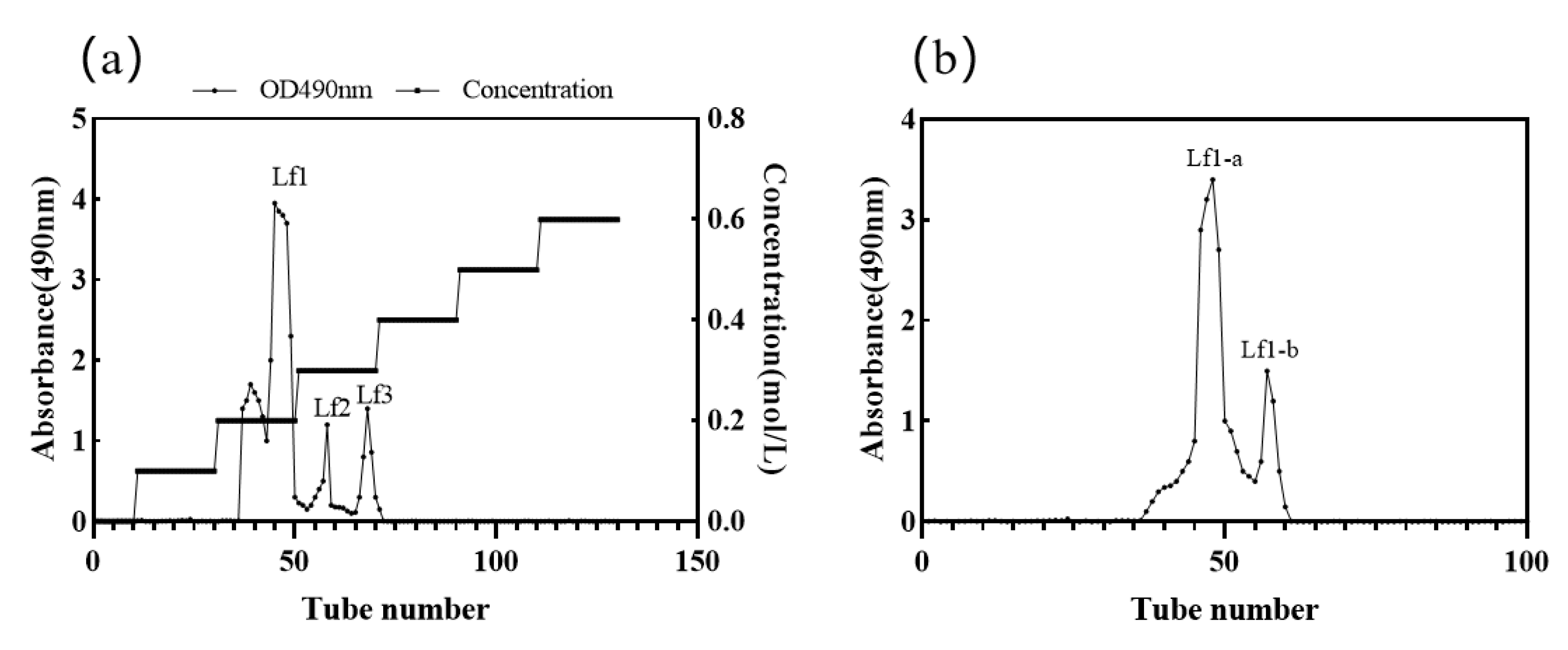
3.3. Isolation and Purification of Exopolysaccharides from T. fuciformis

DEAE-Sepharose is a material with both ionic functional groups and a sugar supporting part, which will interact with glycans through ionic interaction and hydrogen bonding [53]. Studies have shown that the molecular weights of the two main types of TEPS in China are 45,461 kDa and 25,981 kDa, respectively [54]. The molecular weight of TEPS purified by the DEAE column is 1140 kDa [28]. Three major components, namely Lf1, Lf2, and Lf3, were washed with 0.2% NaCl and 0.3% NaCl by the DEAE-Sepharose FF column, respectively (Figure 4a). Zheng et al. also isolated 3 high molecular weight polysaccharide components (TEPS-1, TEPS-2, and TEPS-3) from the extracellular polysaccharides of T. fuciformis. The peak time and intensity were roughly the same as the results of this experiment, which also demonstrated that TEPS-1 ultraviolet absorption intensity was the largest. TEPS-1 is mainly composed of glucose, xylose, mannose, and fucose, while both TEPS-2 and TEPS-3 are mainly composed of rhamnose, arabinose, mannose, galactose, and glucose. FT-IR analysis revealed that TEPS-1, TEPS-2, and TEPS-3 have typical polysaccharide structures [55]. The isolated components of Lf1 were further purified by a Sephacryl S100 column, and two single peaks, namely Lf1-a and Lf1-b, were obtained (Figure 4b). The separation mechanism of size-exclusion chromatography is dependent on the molecular size of the analytes and the pore size of the size exclusion chromatography stationary phase. On filtration through a bed of starch particles, high and low molecular weight compounds also behave differently [56]. Larger molecules excluded from the pores of the packing material are eluted more rapidly than smaller molecules, whereas the smaller molecules with greater access to the pores elute more slowly. The smallest molecular weight dextran had the longest retention time; the dextran with the largest molecular weight eluted first. Thus, the molecular weight of Lf1-a is greater than that of Lf1-b (Figure 4b). A previous study showed that during the elution stage of 0.3–0.4 mol/L sodium chloride, an elution peak was collected, and the absorbance was between 1.2 and 1.5. The Lf1-a in this study may be the same as the above components; its molecular weight is approximately 2033 kDa, and it is mainly composed of rhamnose, mannose, and glucose [24].

3.4. Determination of Antioxidant Activity

3.4.1. FRAP Scavenging Efficiency

The FRAP assay was used to measure the reducing power of the antioxidants. DPPH radical and hydroxyl radical scavenging rates are widely used to measure the free radical scavenging ability of polysaccharides, and they are also indicators of the potential antioxidant capacity of polysaccharides. All of the above are excellent methods for measuring antioxidant activity in vitro, but of course each method has certain limitations due to its different mechanism, and one isolated method cannot completely evaluate the antioxidant capacity of samples. In this research, several methods for the determination of antioxidant activity were used to reflect the antioxidant capacity of Tf1-a and Tf1-b.
The reducing power is considered to be an important indicator of antioxidant activity in natural compounds. The FRAP assay treats the antioxidants contained in the samples as reductants in a redox-linked colorimetric reaction, and the value reflects the reducing power of antioxidants. The procedure is relatively simple and easy to standardize [57]. The antioxidant potentials of different samples were estimated by their ability to reduce the TPTZ-Fe (III) complex to the TPTZ-Fe (II) complex, with maximum absorption at 593 nm. The reduction of absorbance is proportional to the antioxidant content. The reducing powers of Lf1-a and Lf1-b are shown in Figure 5a. The antioxidant capacities of Lf1-a and Lf1-b correlated well with their increasing concentration. The antioxidant capacity of Lf1-a was higher than Lf1-b, but both were significantly lower than the Vc reduction force. Tang Z et al.’s study showed that the Fe3+ reduction ability of amaranth polysaccharide (2 mg/mL) is approximately 0.90 [58]. In this study, at the concentration of 3 mg/mL, the reducing power of Lf1-a and Lf1-b reached 0.86 and 0.70, respectively.
It has been previously reported that the reducing properties are generally associated with the presence of reductones, which can break free radical chain by donating a hydrogen atom. Reductones are also reported to react with certain precursors of peroxide, thus preventing peroxide formation. Tseng et al. found that the introduction of the sulfate group might lead to the diminution of hydroxyl groups and the steric conformation of polysaccharides, which decreased the electron cloud density of active hydroxyl groups and prevented some active sulfate groups from binding to the metal ion, and it resulted in the decrease in the reducing power [59]. In this assay, TEPS with high donating hydrogen abilities showed excellent reducing power, and Lf1-a might donate more electrons or act as a more efficient electron donor in our FRAP assay. Yang et al. found the sugar content of polysaccharides was reduced after phosphorylation modification, so it was speculated that the reducing ability might be related to the phosphorylated structure and sugar content of polysaccharide [60]. The higher the sugar content the polysaccharide would contain, the stronger reducing ability it would show [61]. From the perspective of the sugar content of TEPS, it was speculated that the excellent reducing ability might be related to the sugar content of polysaccharide. Additionally, the reduction ability of polysaccharides may be related to the carboxymethyl structure, and some studies have shown that carboxymethylated Daucus carota polysaccharide had lower reducing ability than unmodified ones [62].

3.4.2. DPPH Scavenging Efficiency

The DPPH radical is a stable lipid free radical with an absorption peak at 517 nm. The DPPH can capture either electrons or hydrogen atoms from antioxidants and pair with its free radicals. Therefore, the DPPH scavenging activity assay is a useful method for determining the free radical scavenging activities of antioxidant materials. The greater scavenging rate on DPPH radicals shows that the antioxidant ability of the materials is stronger.
Figure 5b shows the DPPH assay results, with Vc as the positive control. For Vc, the DPPH scavenging efficiency remained above 40% and increased with increasing concentration. The DPPH scavenging efficiency increased to 90% with the highest level of Vc concentration. The DPPH scavenging efficiency of TEPS increased with the concentration, but none of the two polysaccharides tested reached the level of Vc. At the same time, it was found that the antioxidant activity of Lf1-a and Lf1-b increased significantly under high concentrations. For the concentration of 3 mg/mL, the maximum DPPH radical were 54.23 ± 1.68% and 61.62 ± 2.73% for Lf1-a and Lf1-b, respectively. TEPS had higher DPPH scavenging ability compared to the 42.76% of Phellinus igniarius, a well-known edible and medicinal mushrooms [63], and TEPS had a comparable antioxidant property with the 65.74% of notoginseng polysaccharide [64] and the 64.85% of probiotic exopolysaccharides [65]. However, the DPPH values of TEPS were lower compared with other reported mushrooms of 85% [66], which might be greatly affected by many factors.
It was reported that the effect of antioxidants on DPPH radical scavenging was due to their hydrogen donating ability [67]. The sulfated derivatives showed excellent scavenging activity on DPPH radicals, which might be attributable to its strong hydrogen donating ability by activating the hydrogen atom of the anomeric carbon [68]. Chen et al. used the chlorosulfonic acid-pyridine method to chemically modify Codonopsis polysaccharides. Through in vivo antioxidant experiments, they found that the 2,2-diphenyl-1- picrylhydrazyl (DPPH)-free radical scavenging activity of sulfated Codonopsis polysaccharides could reach 85.71%, which is higher than the free radical scavenging activity of Codonopsis polysaccharide without sulfate modification [69]. The results demonstrated that Lf1-a is more capable of donating hydrogen than Lf1-b.

3.4.3. Hydroxyl Radical Scavenging Assay

The hydroxyl radical is considered to be a highly potent oxidant that can react with most biomacromolecules functioning in living cells and induce severe damage to the adjacent biomolecules. Thus, removing hydroxyl radicals is important for antioxidant defense in cell or food systems.
The results of the hydroxyl radical scavenging activities of Lf1-a and Lf1-b are given in Figure 5c. The scavenging activities of Lf1-a and Lf1-b increased with the concentration of polysaccharides. However, the scavenging abilities of Lf1-a and Lf1-b for hydroxyl radicals were significantly lower than that of Vc at the same concentration. At a concentration of 3 mg/mL, the maximum hydroxyl radical scavenging rates were 92.23 ± 2.69%, 58.76 ± 2.58%, and 45.81 ± 1.79% for Vc, Lf1-a, and Lf1-b, respectively. This result indicated that the scavenging hydroxyl radical activity of Lf1-a was higher than that of Lf1-b and showed the same tendency as the FRAP values and DPPH values.
Zhang et al. showed that the scavenging rate of 1.52 mg/mL TEPS was 64.2% [70], and Zheng et al. showed that the hydroxyl radical scavenging rate of 2 mg/mL TEPS reached approximately 90%. From the test data, the Fenton values in this experiment showed a certain gap compared to other studies, with the antioxidant activity of TEPS being less than 60%. It can be shown that, on the whole, TEPS has a poor scavenging ability of the hydroxyl radical scavenging assay. The hydroxyl radical scavenging ability of polysaccharides may be related to the glycosidic bond structure. Ge et al. Reported that there are 1,4 and 1,3,4 glycoside bonds in T. fuciformis polysaccharide’s structure, and it showed good antioxidative and hydroxyl radical scavenging abilities [28].
As shown in Table 6, there were significant correlations between FRAP values and Lf1-a (r = 0.9199, p = 0.026) and Lf1-b (r = 0.9441, p = 0.015), respectively. The DPPH scavenging efficiency showed a significant correlation with Lf1-a (r = 0.8984, p = 0.038) and Lf1-b (r = 0.9521, p = 0.012), respectively. Furthermore, the hydroxyl radical scavenging activity had a clear correlation with Lf1-a (r = 0.9716, p = 0.0057) and Lf1-b (r = 0.9716, p = 0.0057), respectively. Thus, the antioxidant activity determined by the FRAP assay, DPPH scavenging activity, and hydroxyl radical scavenging activity shows a positive correlation with the component concentration of TEPS.
Molecular weight plays an important role in antioxidant activity; within a certain range of molecular weight, the smaller the molecular weight, the better the antioxidant activity of polysaccharides [71]. Meanwhile, studies have shown that the higher molecular weight component of laminaria japonica polysaccharide, the stronger the antioxidant activity [72]. In other studies on T. fuciformis polysaccharides (TFP), the results showed that the larger molecular weight TFP-1 component had stronger reducing power than the smaller molecular weight TFP-2 and TFP-3 components, which was consistent with the experimental results in this study [55]. The structure of T. fuciformis polysaccharides is different from other fungal polysaccharides in that it is composed of a mannan α-glycosidic bond as the main chain and contains side chains of xylose and glucuronic acid. The polysaccharides with β-(1, 3)-glucan as the main chain and β-(1, 6) branched chains play an important role in exerting biological activity. Additionally, from many studies, it can be seen that polysaccharides containing glucuronic acid residues or glucuronic acid are more biologically active, and the antioxidant activity of silver fungus polysaccharides was positively correlated with the glucuronide content [73,74]. The antioxidant activity of TEPS may be related to these factors, including the composition of monosaccharides, the type of glycosidic bond, the groups on the α-D-mannose backbone, the molecular weight, and the glucuronide content. Certainly, accurate characterization of high molecular weight branched polysaccharides are necessary to better investigate conformational relationships as polysaccharides’ chemical structure and chain conformation have a profound effect on their activity. The detailed chemical structure and in vivo bioactivity of TEPS fractions are still under investigation.

4. Conclusions

In the present study, the Tf526 strain of T. fuciformis, which, with a high yield of DCW and TEPS, was screened, and its major fraction of Tf1-a and Tf1-b extracellular polysaccharides showed significant in vitro antioxidant activity. The yield of TEPS was significantly improved from 0.55 ± 0.70 mg/mL to 0.76 ± 0.03 mg/mL through a submerged culture under the optimal conditions (fermentation time: 72 h, fermentation temperature: 25 °C, initial pH: 6.0, inoculum size: 8%, shaking speed: 150 rpm, nitrogen source: yeast extract, and carbon source: glucose). In addition, A-722MP resin was chosen from seven resins for decoloration of TEPS, owing to its higher decoloration ratio, deproteinization ratio, and polysaccharide recovery ratio. The optimal decoloration parameters of TEPS were obtained by single-factor experiments as follows: decoloration time of 2 h, resins dosage of 2 g, and temperature of 30 °C. Four fractions of single component polysaccharides were isolated and named Tf1-a, Tf1-b, Tf2, and Tf3. The EPS from the Tf526 strain showed a strong capability of free radical scavenging, DPPH scavenging, and hydroxyl radical scavenging. These results demonstrated the optimized fermentation and purification parameters for improved TEPS yield and strong antioxidant activity. Thus, the major polysaccharide fraction Tf1-a and Tf1-b has profound potential in the development of functional foods.

Author Contributions

Conceptualization, L.Z.; Data curation, Y.L.; Investigation, X.J.; Methodology, B.Y.; Resources, L.Z.; Software, Y.D.; Supervision, R.G.; Validation, B.Y. and R.G.; Visualization, Y.D.; Writing—original draft, Q.H.; Writing—review & editing, L.Z. All authors have read and agreed to the published version of the manuscript.

Funding

This research was funded by Guangdong Pharmaceutical University Research Project (43255098).

Institutional Review Board Statement

Not applicable.

Informed Consent Statement

Not applicable.

Data Availability Statement

The data presented in this study are available on request from the corresponding author.

Conflicts of Interest

The authors declare no conflict of interest.

Abbreviations

EPS, extracellular polysaccharides; TEPS, Tremella fuciformis polysaccharides; DCW, dry cell weight; MARs, macroporous adsorption resins; DPPH, 1,1-diphenyl-2-picrylhydrazyl radical; KDa, kilodalton; FRAP, ferric reducing antioxidant assay; SD, standard deviation.

References

  1. Wu, D.T.; Deng, Y.; Zhao, J.; Li, S.P. Molecular characterization of branched polysaccharides from Tremella fuciformis by asymmetrical flow field-flow fractionation and size exclusion chromatography. J. Sep. Sci. 2017, 40, 4272–4280. [Google Scholar] [CrossRef] [PubMed]
  2. Cho, E.J.; Oh, J.Y.; Chang, H.Y.; Yun, J.W. Production of exopolysaccharides by submerged mycelial culture of a mushroom Tremella fuciformis. J. Biotechnol. 2006, 127, 129–140. [Google Scholar] [CrossRef] [PubMed]
  3. Park, K.M.; Kwon, K.M.; Lee, S.H. Evaluation of the antioxidant activities and tyrosinase inhibitory property from mycelium culture extracts. Evid. Based Compl. Alt. 2015, 2015, 1–7. [Google Scholar] [CrossRef] [PubMed] [Green Version]
  4. Wen, Y.; Jia, B.; Peng, T. Research on chemical constituents and pharmacological action of polysaccharide in Poria cocos. Medicinal Plant 2014, 5, 51–54. [Google Scholar]
  5. Mazumder, S.; Ghosal, P.K.; Pujol, C.A.; Carlucci, M.J.; Damonte, E.B.; Ray, B. Isolation, chemical investigation and antiviral activity of polysaccharides from Gracilaria corticata (Gracilariaceae, Rhodophyta). Int. J. Biol. Macromol. 2002, 31, 87–95. [Google Scholar] [CrossRef]
  6. Kim, S.B.; Lee, G.W.; Lee, U.-Y.; Lee, T.S. The immuno-modulatory and antitumor effects of crude polysaccharides extracted from Tremella fuciformis. Korea J. Mycol. 2006, 34, 105–111. [Google Scholar]
  7. Kim, J.H.; Ha, H.C.; Lee, M.S.; Kang, J.I.; Kim, H.S.; Lee, S.Y.; Pyun, K.H.; Shim, I. Effect of Tremella fuciformis on the neurite outgrowth of PC12h cells and the improvement of memory in rats. Biol. Pharm. Bull. 2007, 30, 708–714. [Google Scholar] [CrossRef] [Green Version]
  8. Mustafin, K.; Bisko, N.; Blieva, R.; Al-Maali, G.; Krupodorova, T.; Narmuratova, Z.; Saduyeva, Z.; Zhakipbekova, A. Antioxidant and antimicrobial potential of Ganoderma lucidum and Trametes versicolor. Turk. J. Biochem. 2022, 47, 487–489. [Google Scholar] [CrossRef]
  9. Aranha, G.M.; Contato, A.G.; dos Santos Salgado, J.C.; de Oliveira, T.B.; Retamiro, K.M.; Ortolan, G.G.; Crevelin, E.J.; Nakamura, C.V.; Beraldo de Moraes, L.A.; Peralta, R.M.; et al. Biochemical characterization and biological properties of mycelium extracts from Lepista sordida GMA-05 and Trametes hirsuta GMA-01: New mushroom strains isolated in Brazil. Braz J. Microbiol. 2022, 58, 349–358. [Google Scholar] [CrossRef]
  10. Li, T.J.; Lee, T.Y.; Lo, Y.; Lee, L.Y.; Li, I.C.; Chen, C.C.; Chang, F.C. Hericium erinaceus mycelium ameliorate anxiety induced by continuous sleep disturbance in vivo. BMC Complement. Med. 2021, 21, 1–10. [Google Scholar] [CrossRef]
  11. Kozarski, M.; Klaus, A.; Niksic, M.; Jakovljevic, D.; Helsper, J.P.F.G.; Van Griensven, L.J.L.D. Antioxidative and immunomodulating activities of polysaccharide extracts of the medicinal mushrooms Agaricus bisporus, Agaricus brasiliensis, Ganoderma lucidum and Phellinus linteus. Food Chem. 2011, 129, 1667–1675. [Google Scholar] [CrossRef]
  12. Jeong, S.C.; Koyyalamudi, S.R.; Hughes, J.M.; Khoo, C.; Bailey, T.; Marripudi, K.; Park, J.P.; Kim, J.H.; Song, C.H. Antioxidant and immunomodulating activities of exo-and endopolysaccharide fractions from submerged mycelia cultures of culinary-medicinal mushrooms. Int. J. Med. Mushrooms 2013, 15, 251–266. [Google Scholar] [CrossRef] [PubMed]
  13. Park, H.J.; Shim, H.S.; Ahn, Y.H.; Kim, K.S.; Park, K.J.; Choi, W.K.; Ha, H.C.; Kang, J.I.; Kim, T.S.; Yeo, I.H.; et al. Tremella fuciformis enhances the neurite outgrowth of PC12 cells and restores trimethyltin-induced impairment of memory in rats via activation of CREB transcription and cholinergic systems. Behav. Brain Res. 2012, 229, 82–90. [Google Scholar] [CrossRef] [PubMed]
  14. Kang, L.; Li, Q.; Lin, J.; Guo, L. Biosynthesis of Resveratrol in Blastospore of the Macrofungus Tremella fuciformis. Mol. Biotechnol. 2015, 57, 675–684. [Google Scholar] [CrossRef] [PubMed]
  15. Zhu, H.; Tian, B.; Liu, W.; Zhang, S.; Cao, C.; Zhang, Y.; Zou, W. A three-stage culture process for improved exopolysaccharide production by Tremella fuciformis. Bioresour. Technol. 2012, 116, 526–528. [Google Scholar] [CrossRef]
  16. Han, C.K.; Chiang, H.C.; Lin, C.Y.; Tang, C.H.; Lee, H.; Huang, D.D.; Zeng, Y.R.; Chuang, T.N.; Huang, Y.L. Comparison of immunomodulatory and anticancer activities in different strains of Tremella fuciformis Berk. Am. J. Chin. Med. 2015, 43, 1637–1655. [Google Scholar] [CrossRef]
  17. Gao, Q.; Seljelid, R.; Chen, H.; Jiang, R. Characterisation of acidic heteroglycans from Tremella fuciformis Berk with cytokine stimulating activity. Carbohydr. Res. 1996, 288, 135–142. [Google Scholar] [CrossRef]
  18. Zhang, Y.; Pei, L.; Gao, L.; Huang, Q.; Qi, J. A neuritogenic compound from Tremella fuciformis. China J. Chin. Mat. Med. 2011, 36, 2358–2360. [Google Scholar]
  19. Du, X.J.; Zhang, J.S.; Yang, Y.; Tang, Q.J.; Jia, W.; Pan, Y.J. Purification, chemical modification and immunostimulating activity of polysaccharides from Tremella aurantialba fruit bodies. J. Zhejiang Univ. Sci. B 2010, 11, 437–442. [Google Scholar] [CrossRef]
  20. Kiho, T.; Kochi, M.; Usui, S.; Hirano, K.; Aizawa, K.; Inakuma, T. Antidiabetic effect of an acidic polysaccharide (TAP) from Tremella aurantia and its degradation product (TAP-H). Biol. Pharm. Bull. 2001, 24, 1400–1403. [Google Scholar] [CrossRef] [Green Version]
  21. Jiang, R.Z.; Wang, Y.; Luo, H.M.; Cheng, Y.Q.; Chen, Y.H.; Gao, Y.; Gao, Q.P. Effect of the molecular mass of Tremella polysaccharides on accelerated recovery from cyclophosphamide-induced leucopenia in rats. Molecules 2012, 17, 3609–3617. [Google Scholar] [CrossRef] [PubMed] [Green Version]
  22. Zhao, X.N.; Hu, Y.L.; Wang, D.Y.; Guo, L.W.; Yang, S.J.; Fan, Y.P.; Zhao, B.K.; Wang, Y.L.; Abula, S. Optimization of sulfated modification conditions of Tremella polysaccharide and effects of modifiers on cellular infectivity of NDV. Int. J. Biol. Macromol. 2011, 49, 44–49. [Google Scholar] [CrossRef] [PubMed]
  23. Xu, W.; Shen, X.; Yang, F.; Han, Y.; Li, R.; Xue, D.; Jiang, C. Protective effect of polysaccharides isolated from Tremella fuciformis against radiation-induced damage in mice. J. Radiat. Res. 2012, 53, 353–360. [Google Scholar] [CrossRef] [PubMed]
  24. Jin, Y.; Hu, X.; Zhang, Y.; Liu, T. Studies on the purification of polysaccharides separated from Tremella fuciformis and their neuroprotective effect. Mol. Med. Rep. 2016, 13, 3985–3992. [Google Scholar] [CrossRef] [Green Version]
  25. Wang, Z.; Zeng, Y.; Luo, D. Structure elucidation of a non-branched and entangled heteropolysaccharide from Tremella sanguinea Peng and its antioxidant activity. Carbohydr. Polym. 2016, 152, 33–40. [Google Scholar] [CrossRef]
  26. Kala, K.; Kryczyk-Poprawa, A.; Rzewinska, A.; Muszynska, B. Fruiting bodies of selected edible mushrooms as a potential source of lovastatin. Eur. Food Res. Technol. 2020, 246, 713–722. [Google Scholar] [CrossRef] [Green Version]
  27. Jo, M.H.; Kim, B.; Ju, J.H.; Heo, S.Y.; Ahn, K.H.; Lee, H.J.; Yeom, H.-S.; Jang, H.; Kim, M.-S.; Kim, C.-H.; et al. Tremella fuciformis TFCUV5 mycelial culture-derived exopolysaccharide production and its anti-aging effects on skin cells. Biotechnol. Bioproc. E 2021, 26, 738–748. [Google Scholar] [CrossRef]
  28. Ge, X.Y.; Huang, W.W.; Xu, X.Q.; Lei, P.; Sun, D.F.; Xu, H.; Li, S. Production, structure, and bioactivity of polysaccharide isolated from Tremella fuciformis XY. Int. J. Biol. Macromol. 2020, 148, 173–181. [Google Scholar] [CrossRef]
  29. Deng, Y.F.; Huang, Q.; Hu, L.; Liu, T.; Zheng, B.S.; Lu, D.J.; Guo, C.W.; Zhou, L. Enhanced exopolysaccharide yield and antioxidant activities of Schizophyllum commune fermented products by the addition of Radix Puerariae. Rsc Adv. 2021, 11, 38219–38234. [Google Scholar] [CrossRef]
  30. Gusman, J.K.; Lin, C.Y.; Shih, Y.C. The Optimum submerged culture condition of the culinary-medicinal white Jelly mushroom Tremella fuciformis (Tremellomycetes) and its antioxidant properties. Int. J. Med. Mushrooms 2014, 16, 293–302. [Google Scholar] [CrossRef]
  31. Cho, E.J.; Hwang, H.J.; Kim, S.W.; Oh, J.Y.; Baek, Y.M.; Choi, J.W.; Bae, S.H.; Yun, J.W. Hypoglycemic effects of exopolysaccharides produced by mycelial cultures of two different mushrooms Tremella fuciformis and Phellinus baumii in ob/ob mice. Appl. Microbiol. Biotechnol. 2007, 75, 1257–1265. [Google Scholar] [CrossRef] [PubMed]
  32. Deng, C.; Sun, Y.; Fu, H.; Zhang, S.; Chen, J.; Xu, X. Antioxidant and immunostimulatory activities of polysaccharides extracted from Tremella aurantialba mycelia. Mol. Med. Rep. 2016, 14, 4857–4864. [Google Scholar] [CrossRef] [PubMed] [Green Version]
  33. Chen, S.; Huang, H.; Huang, G. Extraction, derivatization and antioxidant activity of cucumber polysaccharide. Int. J. Biol. Macromol. 2019, 140, 1047–1053. [Google Scholar] [CrossRef] [PubMed]
  34. Huang, F.; Hong, R.; Yi, Y.; Bai, Y.; Dong, L.; Jia, X.; Zhang, R.; Wang, G.; Zhang, M.; Wu, J. In vitro digestion and human gut microbiota fermentation of longan pulp polysaccharides as affected by Lactobacillus fermentum fermentation. Int. J. Biol. Macromol. 2020, 147, 363–368. [Google Scholar] [CrossRef] [PubMed]
  35. Hu, Z.; Zhou, H.; Li, Y.; Wu, M.; Yu, M.; Sun, X. Optimized purification process of polysaccharides from Carex meyeriana Kunth by macroporous resin, its characterization and immunomodulatory activity. Int. J. Biol. Macromol. 2019, 132, 76–86. [Google Scholar] [CrossRef] [PubMed]
  36. Hou, M.; Hu, W.; Xiu, Z.; Shi, Y.; Hao, K.; Cao, D.; Guan, Y.; Yin, H. Efficient enrichment of total flavonoids from Pteris ensiformis Burm. extracts by macroporous adsorption resins and in vitro evaluation of antioxidant and antiproliferative activities. J. Chromatogr. B 2020, 1138, 121960. [Google Scholar] [CrossRef]
  37. Mou, Y.; Li, J.; Zhou, K.; Yu, R.; Xu, D.; Luo, H.; Lai, D.; Zhou, L. Enhanced production of palmarumycins c12 and C13 in mycelial liquid culture of the endophytic fungus berkleasmium sp Dzf12 with in situ macroporous resin adsorption. Trop J. Pharm. Res. 2015, 14, 407–414. [Google Scholar] [CrossRef] [Green Version]
  38. Rajesh, N.; Kumar, A.S.K.; Kalidhasan, S.; Rajesh, V. Trialkylamine impregnated macroporous polymeric sorbent for the effective removal of chromium from industrial wastewater. J. Chem. Eng. Data 2011, 56, 2295–2304. [Google Scholar] [CrossRef]
  39. Liu, G.; Xu, S.; Chen, L. Chemical composition and bioactivities of a water-soluble polysaccharide from the endodermis of shaddock. Int. J. Biol. Macromol. 2012, 51, 763–766. [Google Scholar] [CrossRef]
  40. Wu, Y.; Zhou, H.; Wei, K.; Zhang, T.; Che, Y.; Nguyen, A.D.; Pandita, S.; Wan, X.; Cui, X.; Zhou, B.; et al. Structure of a new glycyrrhiza polysaccharide and its immunomodulatory activity. Front. Immunol. 2022, 13, 1007186. [Google Scholar] [CrossRef]
  41. Shi, K.; Yang, G.; He, L.; Yang, B.; Li, Q.; Yi, S. Purification, characterization, antioxidant, and antitumor activity of polysaccharides isolated from silkworm cordyceps. J. Food Biochem. 2020, 44, e13482. [Google Scholar] [CrossRef] [PubMed]
  42. Lee, Y.R.; Li, X.; Ma, W.; Row, K.H. Retention of large biological molecules by size-exclusion chromatography. Anal. Lett. 2017, 50, 905–915. [Google Scholar] [CrossRef]
  43. Tackx, P.; Tacx, J. Chain architecture of LDPE as a function of molar mass using size exclusion chromatography and multi-angle laser light scattering (SEC-MALLS). Polymer 1998, 39, 3109–3113. [Google Scholar] [CrossRef]
  44. Wu, Y.J.; Wei, Z.X.; Zhang, F.M.; Linhardt, R.J.; Sun, P.L.; Zhang, A.Q. Structure, bioactivities and applications of the polysaccharides from Tremella fuciformis mushroom: A review. Int. J. Biol. Macromol. 2019, 121, 1005–1010. [Google Scholar] [CrossRef]
  45. Liu, J.; Meng, C.G.; Yan, Y.H.; Shan, Y.N.; Kan, J.; Jin, C.H. Structure, physical property and antioxidant activity of catechin grafted Tremella fuciformis polysaccharide. Int. J. Biol. Macromol. 2016, 82, 719–724. [Google Scholar] [CrossRef]
  46. Zou, Y.; Hou, X. Extraction optimization, composition analysis, and antioxidation evaluation of polysaccharides from white Jelly mushroom, Tremella fuciformis (Tremellomycetes). Int. J. Med. Mushrooms 2017, 19, 1113–1121. [Google Scholar] [CrossRef]
  47. Chattopadhyay, N.; Ghosh, T.; Sinha, S.; Chattopadhyay, K.; Karmakar, P.; Ray, B. Polysaccharides from Turbinaria conoides: Structural features and antioxidant capacity. Food Chem. 2010, 118, 823–829. [Google Scholar] [CrossRef]
  48. Chen, F.; Huang, G. Antioxidant activity of polysaccharides from different sources of ginseng. Int. J. Biol. Macromol. 2019, 125, 906–908. [Google Scholar] [CrossRef]
  49. Lee, B.C.; Bae, J.T.; Pyo, H.B.; Choe, T.B.; Kim, S.W.; Hwang, H.J.; Yun, J.W. Submerged culture conditions for the production of mycelial biomass and exopolysaccharides by the edible Basidiomycete Grifola frondosa. Enzyme Microb. Technol. 2004, 35, 369–376. [Google Scholar] [CrossRef]
  50. Shi, Y.; Liu, T.; Han, Y.; Zhu, X.; Zhao, X.; Ma, X.; Jiang, D.; Zhang, Q. An efficient method for decoloration of polysaccharides from the sprouts of Toona sinensis (A. Juss.) Roem by anion exchange macroporous resins. Food Chem. 2017, 217, 461–468. [Google Scholar] [CrossRef]
  51. Kammerer, J.; Carle, R.; Kammerer, D.R. Adsorption and Ion Exchange: Basic principles and their application in food processing. J. Agric. Food Chem. 2011, 59, 22–42. [Google Scholar] [CrossRef] [PubMed]
  52. Wang, X.Y.; Wang, S.S.; Huang, S.S.; Zhang, L.H.; Ge, Z.Z.; Sun, L.P.; Zong, W. Purification of polyphenols from Distiller’s grains by macroporous resin and analysis of the polyphenolic components. Molecules 2019, 24, 1284. [Google Scholar] [CrossRef] [PubMed] [Green Version]
  53. Zhu, H.; Li, X.; Qu, J.; Xiao, C.; Jiang, K.; Gashash, E.; Liu, D.; Song, J.; Cheng, J.; Ma, C.; et al. Diethylaminoethyl Sepharose (DEAE-Sepharose) microcolumn for enrichment of glycopeptides. Anal. Bioanal. Chem. 2017, 409, 511–518. [Google Scholar] [CrossRef] [PubMed]
  54. Li, P.; Jiang, Z.; Sun, T.; Wang, C.; Chen, Y.Y.; Yang, Z.Y.; Du, B.; Liu, C.Y. Comparison of structural, antioxidant and immuno-stimulating activities of polysaccharides from Tremella fuciformis in two different regions of China. Int. J. Food Sci. Technol. 2018, 53, 1942–1953. [Google Scholar] [CrossRef]
  55. Zheng, Q.W.; He, B.L.; Wang, J.Y.; Huang, S.S.; Zou, Y.; Wei, T.; Ye, Z.W.; Guo, L.Q.; Lin, J.F. Structural analysis and antioxidant activity of extracellular polysaccharides extracted from culinary-medicinal white Jelly mushroom Tremella fuciformis (Tremellomycetes) conidium cells. Int. J. Med. Mushrooms 2020, 22, 489–500. [Google Scholar] [CrossRef]
  56. Porath, J.; Flodin, P. Gel filtration: A method for desalting and group separation. Nature 1959, 183, 1657–1659. [Google Scholar] [CrossRef]
  57. Xu, W.; Zhang, F.; Luo, Y.; Ma, L.; Kou, X.; Huang, K. Antioxidant activity of a water-soluble polysaccharide purified from Pteridium aquilinum. Carbohydr. Res. 2009, 344, 217–222. [Google Scholar] [CrossRef]
  58. Tang, Z.; Zhou, C.; Cai, Y.; Tang, Y.; Sun, W.; Yao, H.; Zheng, T.; Chen, H.; Xiao, Y.; Shan, Z.; et al. Purification, characterization and antioxidant activities in vitro of polysaccharides from Amaranthus hybridus L. PeerJ 2020, 8, 9077. [Google Scholar] [CrossRef]
  59. Tseng, Y.H.; Yang, J.H.; Mau, J.L. Antioxidant properties of polysaccharides from Ganoderma tsugae. Food Chem. 2008, 107, 732–738. [Google Scholar] [CrossRef]
  60. Yang, W.J.; Zhang, Y.Q.; Tang, A.; Ruan, Q.Q.; Huang, G.L. Preparation and antioxidant activity of phosphorylated polysaccharide from purple sweet potato. Chem. Biol. Drug Des. 2021, 98, 828–834. [Google Scholar] [CrossRef]
  61. Chen, L.; Huang, G. Antioxidant activities of phosphorylated pumpkin polysaccharide. Int. J. Biol. Macromol. 2019, 125, 256–261. [Google Scholar] [CrossRef] [PubMed]
  62. Qi, K.; Xia, G.D.; Huang, G.L.; Huang, H.L. Extraction, chemical modification, and antioxidant activities of Daucus carota polysaccharide. Chem. Biol. Drug Des. 2021, 98, 1098–1103. [Google Scholar] [CrossRef] [PubMed]
  63. Yuan, Q.; Zhao, L.; Li, Z.; Harqin, C.; Peng, Y.; Liu, J. Physicochemical analysis, structural elucidation and bioactivities of a high-molecular-weight polysaccharide from Phellinus igniarius mycelia. Int. J. Biol. Macromol. 2018, 120, 1855–1864. [Google Scholar] [CrossRef]
  64. Shen, S.; Zhou, C.; Zeng, Y.; Zhang, H.; Hossen, M.A.; Dai, J.; Li, S.; Qin, W.; Liu, Y. Structures, physicochemical and bioactive properties of polysaccharides extracted from Panax notoginseng using ultrasonic/microwave-assisted extraction. Lwt-Food Sci. Technol. 2022, 154, 112446. [Google Scholar] [CrossRef]
  65. Mandhi, A.; Leban, N.; Chakroun, I.; Chaouch, M.A.; Hafsa, J.; Fdhila, K.; Mandouani, K.; Majdoub, H. Extracellular polysaccharide derived from potential probiotic strain with antioxidant and antibacterial activities as a prebiotic agent to control pathogenic bacterial biofilm formation. Microb. Pathog. 2017, 109, 214–220. [Google Scholar]
  66. Wang, Y.; Jia, J.; Ren, X.; Li, B.; Zhang, Q. Extraction, preliminary characterization and in vitro antioxidant activity of polysaccharides from Oudemansiella radicata mushroom. Int. J. Biol. Macromol. 2018, 120, 1760–1769. [Google Scholar] [CrossRef] [PubMed]
  67. Chen, Y.; Xie, M.Y.; Nie, S.P.; Li, C.; Wang, Y.X. Purification, composition analysis and antioxidant activity of a polysaccharide from the fruiting bodies of Ganoderma atrum. Food Chem. 2008, 107, 231–241. [Google Scholar] [CrossRef]
  68. Chen, Y.; Zhang, H.; Wang, Y.X.; Nie, S.P.; Li, C.; Xie, M.Y. Sulfated modification of the polysaccharides from Ganoderma atrum and their antioxidant and immunomodulating activities. Food Chem. 2015, 186, 231–238. [Google Scholar] [CrossRef]
  69. Liu, C.; Chen, J.; Li, E.T.; Fan, Q.; Wang, D.Y.; Li, P.; Li, X.P.; Chen, X.Y.; Qiu, S.L.; Gao, Z.Z.; et al. The comparison of antioxidative and hepatoprotective activities of Codonopsis pilosula polysaccharide (CP) and sulfated CP. Int. Immunopharmacol. 2015, 24, 299–305. [Google Scholar] [CrossRef]
  70. Zhang, Z.; Wang, X.; Zhao, M.; Qi, H. Free-radical degradation by Fe2+/Vc/H2O2 and antioxidant activity of polysaccharide from Tremella fuciformis. Carbohydr. Polym. 2014, 112, 578–582. [Google Scholar] [CrossRef]
  71. Li, M.; Ma, F.; Li, R.; Ren, G.; Yan, D.; Zhang, H.; Zhu, X.; Wu, R.; Wu, J. Degradation of Tremella fuciformis polysaccharide by a combined ultrasound and hydrogen peroxide treatment: Process parameters, structural characteristics, and antioxidant activities. Int. J. Biol. Macromol. 2020, 160, 979–990. [Google Scholar] [CrossRef] [PubMed]
  72. Zha, S.; Zhao, Q.; Zhao, B.; Ouyang, J.; Mo, J.; Chen, J.; Cao, L.; Zhang, H. Molecular weight controllable degradation of Laminaria japonica polysaccharides and its antioxidant properties. J. Ocean. Univ. China 2016, 15, 637–642. [Google Scholar] [CrossRef]
  73. Camesano, T.A.; Wilkinson, K.J. Single molecule study of xanthan conformation using atomic force microscopy. Biomacromolecules 2001, 2, 1184–1191. [Google Scholar] [CrossRef] [PubMed]
  74. Chen, H.X.; Zhang, M.; Xie, B.J. Quantification of uronic acids in tea polysaccharide conjugates and their antioxidant properties. J. Agric. Food Chem. 2004, 52, 3333–3336. [Google Scholar] [CrossRef] [PubMed]
Figure 1. Microscopic observation of three strains of Tremella fuciformis (Tf526, Tf9902, Tf9903) on the solid culture plate. (a) Front view of Tremella fuciformis (Tf526, Tf9902, Tf9903) on a culture plate. (b) Back view of Tremella fuciformis (Tf526, Tf9902, Tf9903) on a culture plate. (c) Mycelium micro examination (200×) of Tremella fuciformis (Tf526, Tf9902, Tf9903) obtained by liquid culture.
Fermentation 08 00616 g001
Figure 2. Strain selection for the high yield of EPS and DCW production. Data are expressed as mean ± SD, n = 3. Values with no letters in common are significantly different (p < 0.05).
Fermentation 08 00616 g002
Figure 3. Effects of each variable factor on the yield of EPS and DCW from Tf526 strain: (a) culture time; (b) culture temperature; (c) Initial pH; (d) Inoculum size; (e) shaking speed; (f) Carbon source; (g) Nitrogen source. Data are expressed as mean ± SD, n = 3. Values with no letters in common are significantly different (p < 0.05).
Fermentation 08 00616 g003
Figure 4. Chromatography purification of TEPS: (a) TEPS fraction of Lf1, Lf2, and Lf3 using stepwise elution on DEAE-Sepharose FF column (20 mm × 100 cm, 1.25 mL/min); (b) Lf1-a and Lf1-b TEPS from Lf1 subsequently obtained on Sephacryl S100 column (10 mm × 60 cm, 0.5 mL/min).
Fermentation 08 00616 g004
Figure 5. In vitro antioxidant capacity of Lf1-a and Lf1-b: (a) Reduction capacity; (b) DPPH scavenging activities; (c) Hydroxyl radical scavenging activities. Data are expressed as mean ± SD, n = 3. Values with no letters in common are significantly different (p < 0.05).
Fermentation 08 00616 g005
Table 1. Chemical and physical properties of the macroporous resins employed.
ResinsParticle Size (mm)Surface Area (m2/g)Average Pore Diameter (nm)Polarity
A-722MP0.30–1.22650–70020–50Polar
DA-2010.30–1.25≥20010–13Polar
HPD-6000.30–1.25550–6008–9polar
AB-80.30–1.25450–53013–14Weak-polar
DM1300.30–1.25500–5509–10Weak-polar
HPD-1000.30–1.25650–7008.5–9Non-polar
XAD-1180N0.35–0.60150–9004–9Non-polar
Table 2. Effect of seven different varieties of resin on deproteinization ratio, decoloration ratio, and polysaccharide recovery ratio.
Resin Deproteinization Ratio (%)Decolorization Ratio (%)Polysaccharide Recovery Ratio (%)ζ Value
(%)
A-722MP81.72 ± 1.21 a62.73 ± 1.18 a72.73 ± 2.63 ab73.43
AB-858.46 ± 1.83 c38.33 ± 1.48 cd69.71 ± 1.88 b60.06
DA20168.72 ± 2.21 b45.77 ± 2.13 bc58.72 ± 2.03 c59.13
DM13060.23 ± 2.13 c42.36 ± 2.18 c72.42 ± 2.98 ab62.75
HPD10033.65 ± 1.23 e24.86 ± 1.56 e75.52 ± 1.96 a52.83
HPD60065.32 ± 2.43 b49.21 ± 1.33 b53.42 ± 1.68 d56.15
XAD1180N32.86 ± 2.43 e34.53 ± 2.14 d70.43 ± 1.78 b51.98
Values with no letters in common are significantly different (p < 0.05).
Table 3. Effect of time on decoloration ratio, deproteinization ratio, polysaccharide recovery ratio, and selectivity coefficients of A-722MP resin.
Time/hDeproteinization Ratio (%)Decolorization Ratio (%)Polysaccharide Recovery Ratio (%)ζ Value (%)
0.542.13 ± 2.13 d24.69 ± 1.43 d88.34 ± 2.18 a61.75
162.33 ± 1.23 c45.22 ± 1.68 c80.52 ± 1.78 b67.56
1.573.2 ± 2.52 b56.53 ± 2.03 b75.65 ± 2.32 c69.32
280.91 ± 1.79 a62.27 ± 1.89 a72.86 ± 2.14 c74.12
2.583.37 ± 1.56 a63.12 ± 1.32 a69.23 ± 1.16 d70.58
383.16 ± 2.23 a63.86 ± 2.42 a64.19 ± 1.45 e67.53
Values with no letters in common are significantly different (p < 0.05).
Table 4. Effect of resin dosage on decoloration ratio, deproteinization ratio, polysaccharide recovery ratio, and selectivity coefficients of A-722MP resin.
Resin Dosage/g Deproteinization Ratio %Decolorization Ratio %Polysaccharide Recovery Ratio %ζ Value %
162.33 ± 1.53 c42.12 ± 2.41 d89.30 ± 1.15 a71.76
280.61 ± 1.59 b63.17 ± 1.36 c72.86 ± 1.54 b73.21
386.2 ± 2.41 a69.55 ± 1.08 b60.22 ± 1.24 c69.71
488.93 ± 2.74 a72.25 ± 2.76 ab56.23 ± 1.13 cd69.24
590.23 ± 1.36 a75.52 ± 2.82 a51.6 ± 2.56 d67.97
Values with no letters in common are significantly different (p < 0.05).
Table 5. Effect of temperature on decoloration ratio, deproteinization ratio, polysaccharide recovery ratio, and selectivity coefficients of A-722MP resin.
Temperature/°C Deproteinization Ratio %Decolorization Ratio %Polysaccharide Recovery Ratio %ζ Value %
2032.46 ± 1.26 c34.18 ± 1.36 c85.32 ± 1.65 a59.23
3081.21 ± 2.13 b62.14 ± 2.3 a73.42 ± 1.96 b73.5
4084.23 ± 2.61 b64.55 ± 1.73 a60.22 ± 2.32 c 68.29
5090.93 ± 2.74 a55.36 ± 2.41 b52.28 ± 2.18 d64.49
Values with no letters in common are significantly different (p < 0.05).
Table 6. Correlation of Lf1-a, Lf1-b, and antioxidant activity.
TEPS Fractions (3 mg/mL) FRAP Values DPPH Values Fenton System
Lf1-a 0.9199 0.8984 0.9716
Lf1-b 0.9441 0.9521 0.9716
Publisher’s Note: MDPI stays neutral with regard to jurisdictional claims in published maps and institutional affiliations.

