Article

Protection against Osteoporosis by Fermented Mulberry Vinegar Supplementation via Inhibiting Osteoclastic Activity in Ovariectomized Rats and Osteoclastic Cells

1
Department of R&D, Microbial Institute for Fermentation Industry, Sunchang-gun 56048, Korea
2
Department of Food and Nutrition, Obesity/Diabetes Research Center, Hoseo University, Asan 31499, Korea
*
Authors to whom correspondence should be addressed.
Fermentation 2022, 8(5), 211; https://doi.org/10.3390/fermentation8050211
Submission received: 6 April 2022 / Revised: 2 May 2022 / Accepted: 3 May 2022 / Published: 5 May 2022
(This article belongs to the Special Issue Nutrition and Health of Fermented Foods)

Abstract

:
Menopause increases the osteoporosis risk, to which phytoestrogen intake can be beneficial. This study hypothesized that mulberry vinegar had a preventive effect on osteoporosis by decreasing osteoclastic activity. The hypothesis was tested in ovariectomized (OVX) rats and RANKL-differentiated osteoclast cells. OVX rats were given 0(OVX-CON), 0.5(OVX-MVL), 1(OVX-MVM), and 2(OVX-MVH) fermented mulberry vinegar (MV) mL/kg body weight (BW) daily for 12 weeks. Sham-operated rats had no MV supplementation (Normal-CON). The osteoporosis-related biomarkers were measured, and Micro-CT determined the bone mass of the femur. RANKL-differentiated Raw 264.7 cells were treated with MV (0–100 μg/mL). The cell viability, osteoporosis-related mRNA expression, and protein contents were measured. MV contained Acetobacter pasteurianus (7.31 log CFU/mL), citric acid (106 mg/mL), lactic acid (19.2 mg/mL), acetic acid (15.0 mg/mL), and rutin (0.36 mg/mL). OVX-MVM elevated the serum 17β-estradiol concentration similar to the Normal-CON group, but it did not prevent the decrease in uterine weight. OVX-MVM prevented the increase in osteoclastic-related parameters, including cathepsin K(CtsK), receptor activator of NF-κB ligand (RANKL), and tartrate-resistant acid phosphatase (TRAP) in the circulation. OVX-MVH also lowered C-telopeptide of type Ⅰ collagen as much as the Normal-CON group (p < 0.05). By contrast, OVX-MVH increased the serum osteoprotegerin concentration, an inhibitor of osteoclasts, better than the Normal-CON group (p < 0.05). These changes were integrated to alter the bone mineral density (BMD) in Micro-CT analysis: OVX-MVM and OVX-MVH prevented BMD decrease after OVX as much as the Normal-CON. In RANKL-differentiated osteoclast cells, the MV treatment for 24 and 48 h decreased RANKL-induced differentiation in osteoclast cells dose-dependently up to 100 µg/mL. Its decrease was related to inhibiting the TRAP activity and reducing TRAP-positive multinucleated cells during the five-day administration of RANKL. MV treatments also decreased mRNA expression of osteoclast-related genes (TRAP, Ctsk, OSCAR, and NFATc1). MV suppressed the protein contents of NFATc1 and c-FOS-related osteoclast. In conclusion, MV intake (1 mg/kg bw) protected against BMD loss mainly by inhibiting the osteoclastic activity (RANKL/RANK/TRAP) in OVX rats. MV may develop as a functional food for anti-osteoporosis in menopausal women.

1. Introduction

Osteoporosis is a progressive skeletal disease characterized by micro-architectural bone destruction and low bone mineral density, resulting in bone fragility and an elevated bone fracture risk. It is a severe health problem, particularly for menopausal women, and is expected to increase mortality worldwide due to the expanding aged population [1]. Osteoporosis is more prevalent in women than men in the elderly because women have a lower peak bone mass than men, and bone mineral density loss in women is accelerated after menopause [1]. Although osteoblasts and osteoclasts contain estrogen receptors, estrogen deficiency increases osteoclastic bone resorption by promoting osteoclast formation and activity and decreasing apoptosis [2]. Therefore, estrogen prevents bone cell loss by regulating the osteoclast numbers and activity.
Bone loss by an estrogen deficiency is involved in impaired immune response and increased inflammation and oxidative stress [2]. On the other hand, the inflammatory response mediated by macrophages is complex in bone metabolism [3]. Estrogen deficiency contributes to the increased expression of proinflammatory cytokines, including interleukin (IL)-1, IL-6, IL-7, and tumor necrosis factor-alpha (TNF-α). The cytokines elevate the synthesis of macrophage colony-stimulating factor (MCSF) and receptor activator of NF-κB (RANK) ligand (RANKL) in osteoblasts to suppress their proliferation [3]. An estrogen deficiency fails to increase the osteoprotegerin (OPG) secreted from osteoblast cells to neutralize RANKL and decrease MCSF [4]. Meanwhile, OPG is an osteogenic factor in callus formation and remodeling during bone fracture healing [5]. Therefore, the OPG/RANKL/RANK system may play a critical role in osteoporosis after menopause. On the other hand, people and experimental animals in an inflammation dysregulation state, such as diabetes, obesity, and menopause, develop systemic chronic inflammation responses to decreased bone callus formation [6]. Furthermore, aberrant or prolonged immune responses during menopause result in low-grade inflammation involved in the pathogenesis of osteoporosis [7]. Therefore, proinflammatory cytokines act as triggers of bone resorption to break the balance of bone formation and resorption in menopausal women.
Genetic and environmental factors influence postmenopausal osteoporosis risk: metabolic diseases, including obesity and diabetes, and nutritional statuses, such as calcium and vitamin D, modulate the osteoporosis prevalence [1,8,9]. On the other hand, postmenopausal osteoporosis can be prevented and alleviated with hormone-replacement therapy (17β-estradiol and progesterone) because of an estrogen deficiency [10]. On the other hand, a randomized clinical trial showed that hormone replacement therapy has some adverse effects, such as increasing incidence of coronary artery disease (29%), stroke (41%), thromboembolism (111%), and breast cancer (26%) [11]. Therefore, alternative therapeutic agents have been studied to prevent or delay the loss of bone mineral density (BMD) after menopause. Isoflavonoids in soybeans and black cohosh are effective agents for reducing osteoporosis risk [12,13]. Hence, there are demands for better agents to prevent and relieve osteoporosis.
Mulberry (Morus alba L.) fruit is used as a food and traditional medicine. Its bioactive compounds are anthocyanins, quercetin, rutin, chlorogenic acid, and polysaccharides [14,15]. Mulberry fruit has been reported to have various biological functions, mainly through antioxidant, anti-inflammatory, and insulin-sensitive activities [16,17]. It protects against hyperglycemia, non-alcoholic fatty liver, dyslipidemia, tumors, and neurodegenerative diseases [14,15,16]. It also has a beneficial effect on osteoporosis by increasing Runt-related transcription factor 2 and decreasing RANK and RANKL in the femur of ovariectomized rats [17]. When mulberry is fermented with yeast and bacteria to promote functionality, mulberry vinegar (MV) contains acetate, citric acid, and succinic acid produced from sugars and modified mulberry bioactive compounds [18]. MV is demonstrated to have higher superoxide scavenging activity than mulberry juice, by approximately five times the half-maximal inhibitory concentration (IC50) [19]. Thus, MV may have a beneficial effect on osteoporosis in menopausal women. This study hypothesized that MV had a preventive impact on osteoporosis by decreasing osteoclastic activity. The hypothesis was tested in ovariectomized rats and RANKL-differentiated osteoclast in vivo and in vitro studies.

2. Materials and Methods

2.1. MV Production by Fermentation with Microorganisms

Mulberry (Sunchan-gun, Korea) was processed into juice and made into 22 Brix by adding sugar and was sterilized at 100 °C for 10 min. It was inoculated with 1% Saccharomyces cerevisiae SRCM101756 and cultured in a shaking chamber at 20 °C at 150 rpm for 48 h to produce the fermented mulberry wine. After heating at 100 °C for 10 min to eliminate the yeast, the fermented mulberry wine was inoculated with 10% Acetobacter pasteurianus SRCM102419 and cultured in a shaking chamber at 30 °C for 150 rpm for nine days. The final product is MV, and it has been used for cell culture and animal studies. The final MV product showed 6 Brix.

2.2. MV Quality

The MV was diluted with sterilized water (X10) to measure MV quality, and it was inoculated into yeast malt (YM) and glucose yeast extract (GYE) agar plates, respectively, and cultured at 30 °C for nine days. We counted the number of colonies in the plate for live Acetobacter pasteurianus counts. The pH and total acidity were determined using a pH meter (Seven Multi, Mettler Toledo GmBH, Germany) and titration with 0.1N NaOH (Sigma Co., St. Loise, MO, USA), respectively. The total phenolic compounds were determined using the Folin–Ciocalteu method [20,21]. After 3 min, 10% (w/v) Na2CO3 was added to MV and conducted its reaction in the dark for 60 min, and the optical density was measured at 725 nm in a UV spectrophotometer (JASCO, Tokyo, Japan). The total flavonoid contents were measured using the modified Davis method [21]. MV was mixed with 5% sodium nitrite, and after 5 min, 10% aluminum chloride (3:1:1, v/v/v; Sigma Co.) was added. Each mixture was neutralized with 1 N NaOH after six minutes of incubation at room temperature, and its optical density was measured at 510 nm using a UV spectrophotometer (Perkin Elmer, Boston, MA, USA). Total phenol and flavonoid contents are expressed as mg gallic acid (GAE) and quercetin equivalents (QE) per mL, respectively.
The MV was diluted tenfold with sterilized water and filtered through a 0.45 μm membrane filter. The organic acid contents (oxalic acid, citric acid, succinic acid, lactic acid, and acetic acid) were measured with high-performance liquid chromatography (HPLC, Agilent) under the analysis conditions (Supplementary Materials Table S1). The rutin contents in MV were assessed using an Agilent 1200 series HPLC system with an HPLC-DAD (Diode Array Detector, Palo Alto, CA, USA) under the analysis conditions (Supplementary Materials Table S2). Rutin (1 mg/mL; Sigma Co.) was used as a standard.

2.3. Ovariectomy (OVX) Operation for Rats

Female Sprague–Dawley rats aged approximately 12 weeks (weighing 265 ± 18 g) were housed in polycarbonate cages (three rats per case) in a controlled environment (22 ± 2 °C temperature, 50 ± 10% humidity with a 12-h light/dark cycle). This study followed the NIH Guide for the Care and Use of Laboratory Animals guidelines and was approved by the Animal Care and Use Committee of Invivo company, Korea (IV-RB-02-2106-18). The Sprague-Dawley rats purchased from Samtako Inc. (Osan-si, Korea) were acclimated for one week in the animal facility. After anesthesia with subcutaneous injection of a mixture of ketamine and xylazine (100 and 10 mg/kg body weight, respectively), they underwent OVX. Each ovary was isolated by ligating the most proximal portion of each oviduct and removed with scissors. Female Sprague–Dawley rats (n = 10) had a sham operation (Sham) without removing ovaries instead of OVX.

2.4. Experimental Design for Animal Study

Previous studies have demonstrated that vinegar intake (1–2 mL/kg body weight) has beneficial functionality [22]. Moreover, the present cell study demonstrated that the dosage of 50–100 μg/mL MV prevented the proliferation of RAW 264.7 cells induced by RANKL, and over 300 μg/mL MV induced cytotoxicity. We decided that the animals had oral consumption of 0.5–2 mL undiluted MV/kg body weight. After the seven-day recovery from the OVX surgery, forty OVX rats were assigned randomly to four different groups: (1) water (2 mL/kg body weight; control), (2) MV (0.5 mL/kg body weight; MVL), (3) MV (1 mL/kg body weight; MVM), and MV (2 mL/kg body weight; MVH). Each group was given water or undiluted MV orally by feeding a needle five times a week for 12 weeks because the BMD changes needed a long period after OVX [22]. The sham-operated rats had water (2 mL/kg body weight; normal-control) after 1 week from the surgery. The rats were given free access to water and diets during the experimental periods. The body weight was measured every week. The rats had a standard diet containing 15 energy% fat, 23 En% protein, and 62 En% carbohydrates (Samtako, Osan, Korea). Under anesthesia with a ketamine and xylazine mixture, blood was collected from the inferior vena cava and tissues in rats during the 12-week treatment. After scarifying the rats, the femur was dissected.

2.5. Micro-Computed Tomography (Micro-CT)

The femur was fixed with 4% paraformaldehyde, and its bone status was measured. Micro-CT imaging was performed using a Quantum GX Micro-CT imaging system (PerkinElmer, Hopkinton, MA, USA), located at the Korea Basic Science Institute (Gwangju, Korea). The X-ray source was set to 90 kV and 88 mA levels with a field of view of 10 mm (voxel size, 20 μm; scanning time, 4 min). The 3D images were visualized with a 3D Viewer consisting of software within the Quantum GX, and its resolution was set at 4.5 μm. The structural parameters for trabecular bone were analyzed following scanning using the Analyze 12.0 software (Analyze Direct, Overland Park, KS, USA). The BMD for the femur was estimated using a hydroxyapatite phantom (QRM-MicroCT-HA, QRM GmbH, Möhrendorf, Germany) with scanned the femur using the same parameters. We calculated BMD, total volume (TV), bone volume (BV), bone surface (BS), bone surface density (BS/TV), trabecular thickness (Tb.Th), trabecular separation (Tb.Sp), and trabecular number (Tb.N) of the femur.

2.6. Biochemical Assays for Blood

The blood was centrifuged at 3000 rpm for 10 min, and the serum was separated. Serum bone-related biomarkers, including OPG (Abcam, Cambridge, UK), C-telopeptide of type Ⅰ collagen (CTX-1; MyBioSource, San Diego, CA, USA), tartrate-resistant acid phosphatase (TRAP; MyBioSource, San Diego, CA, USA), and RANKL (MyBioSource, San Diego, CA, USA), were determined using an ELISA kit. The optical density was measured with a Multi Detection Reader (Infinite 200; TECAN Group Ltd., Männedorf, Switzerland).

2.7. Cell Culture and Cytotoxicity Assay

Murine macrophage RAW 264.7 cells provided by the Korea Cell Line Bank (Seoul, Korea) were cultured in the DMEM media (Invitrogen, Carlsbad, CA, USA) at 37 °C in a humidified atmosphere containing 5% CO2 in the air. The DMEM media contained 10% heat-inactivated fetal bovine serum (FBS; Thermofisher, Waltham, MA, USA) and 1% penicillin/ streptomycin (Thermofisher, Waltham, MA, USA).
Dulbecco phosphate-buffered saline (1X PBS; Invitrogen, Carlsbad, CA, USA) was used for dissolving the RANKL (10 μg/mL), and dried MV was solubilized with distilled water (10 mg/mL). Raw 264.7 cells were seeded in a 96-well plate at 5 × 10³ cells/well in the alpha-minimal essential medium (α-MEM; Gibco, Grand Island, NY, USA) containing 10% heat-inactivated FBS, 100-U/mL penicillin, and 100-µg/mL streptomycin. 100 ng/mL RANKL (Peprotech, Rocky Hill, CT, USA) was administered. After 1 h, different dosages of MV (20–500 µg/mL) were added. After 24 h and 48 h, the cell viability was measured using a WST-1 assay kit (ITBio, Seoul, Korea) at 562 nm using a Multi Detection Reader (Infinite 200, TECAN Group Ltd., Switzerland). RANKL and MV solvents without RANKL and MV were treated in the Control group.
The intracellular TRAP was stained after a five-day treatment with 100 ng/mL RANKL into Raw 267.4 cells for their differentiation. The mRNA expression of TARP (tartrate-resistant acid phosphatase 5, Acp5), osteoclast-associated immunoglobulin-like receptor (Oscar), cathepsin K (CtsK), and glyceraldehyde-3-phosphate dehydrogenase (Gapdh) was also analyzed with real-time PCR analysis. The primers of the genes were made in Bioneer Inc. (Daejeon, Korea). The nuclear factor of the activated T cells 1 (NFATc1) and the c-Fos protein contents in the cell lysates harvested with Protein Extraction Solution (iNtRON, Cat. 17081; Sungnam-si, Korea) were estimated using a Western blot assay.

2.8. TRAP Staining

The RAW 264.7 cells were differentiated with RANKL (100 ng/mL) for five days when the medium was changed every two days. Mature osteoclast cells were washed with Dulbecco PBS and fixed in 10% formaldehyde for 10 min. The fixed cells were stained using a TRAP staining kit (Sigma-Aldrich, St. Louise, MO, USA), incubated at 37 °C for 3 h, and the optical density was measured at 405 nm in a Multi Detection Reader (Infinite 200; TECAN Group Ltd., Männedorf, Switzerland).
The differentiated cells were washed with PBS and fixed with a methanol and acetone mixture (1:1, v:v). After removing the solvent, a TRAP solution was added, incubated at 37 °C for 30 min, and the cells were washed three times with deionized water. TRAP-positive multinucleated (>3 nuclei) cells were counted under an inverted microscope (Olympus, Tokyo, Japan).

2.9. Real-Time PCR Analysis

After treating the differentiated Raw 264.7 cells with 100 ng/mL RANKL administration for 5 days, different dosages of MV were treated in the differentiated cells. After washing the cells with PBS, they were mixed with a monophasic solution of phenol and guanidine isothiocyanate (TRIzol reagent; Gibco-BRL, Rockville, MD, USA) to extract total RNA from the cells. RNase inhibitor (BioFACTTM, Cat RI152-20h, Korea) was used during total RNA synthesis. The cDNAs were equally generated from the total RNA using Superscript III reverse transcriptase and high-fidelity Taq DNA polymerase (Invitrogen, Carlsbad, CA, USA) using a polymerase chain reaction (PCR). The cDNAs were mixed with a real-time PCR master mix (BioFACTTM, Daejeon, Korea) and the primers for the specific genes (Supplementary Materials Table S3). The mixtures were amplified by real-time PCR (TP600; TaKaRa Shiga, Japan) using the specific genes with an optimal thermal cycling condition. The expression of NFATc1, Acp5, Oscar, CtsK, and Gapdh was measured with corresponding primers in RANKL-induced osteoclast cells, as described previously [23]. The cycle of threshold (CT) for each sample was measured by real-time PCR. The gene expression levels in the cells were calculated using the ratio of interest gene expression and the housekeeping gene (Gapdh).

2.10. Immunoblot Analysis

The differentiated Raw264.7 cells with 100 ng/mL RANKL administration for 5 days were treated with MV and lysed with Protein Extraction Solution (iNtRON, Cat. 17081; Sungnam-si, Korea), adding protease inhibitors (EDTA free Complete mini; Merck, Darmstadt, Germany). The supernatants (lysates) were separated by centrifuging at 20,000× g 4 °C for 10 min. Lysates containing equal amounts of protein (30–50 μg) ran in 7.5% SDS-PAGE, and the proteins on the gel were transferred into the nitrocellulose membrane (Millipore, Burlington, MA, USA). The membranes were used for immunoblotting with the specific primary antibodies (1:1000 dilution) against NFATc1, c-Fos, and β-actin (Santa Cruz, Santa Cruz, CA, USA) and the secondary antibody (m-IgGk, Santa Cruz; 1:3000 dilution) was used for visualization, as described elsewhere [24]. The intensity of protein expression was determined using the Image program.

2.11. Statistical Analysis

Statistical analysis was performed using SPSS software version 23 (IBM SPSS, Armonk, NY, USA). The sample size was calculated using a G power program (power = 0.90 and effect size = 0.5), and the sample size of each group was 10 for the animal study. The results are expressed as the means ± standard deviation (SD) when the normal distribution was checked using a Proc univariate. The statistical differences were assessed with one-way ANOVA in the metabolic variables among the OVX-CON, OVX-MVL, OVX-MVM, OVX-MVH, and Normal-CON groups. The Duncan test as a posthoc test was conducted to determine multiple comparisons of the groups when one-way ANOVA showed a significant difference. One-way ANOVA and Duncan tests were also applied to cell studies. Significant differences in the main effects among the groups were identified at p < 0.05.

3. Results

3.1. Characteristics of MV and Its Rutin Contents

The pH of the MV was 2.26, and the acidity was 4.07%, whereas its live bacteria count (mainly Acetobacter pasteurianus) was 7.31 log CFU/mL (Table 1). Oxalic acid and succinic acid were not detected, while the MV contained citric acid (106.0 ± 6.06 mg/mL), lactic acid (19.2 ± 4.43 mg/mL), and acetic acid (15.0 ± 9.32 mg/mL) (Table 1). The total phenol and total flavonoid contents were 570 ± 0.18 µg GAE/mL and 80.0 ± 3.03 µg QE/mL, respectively. The rutin contents were 0.36 ± 0.04 mg/mL (Table 1) when the standard curve showed R2 = 99.97% (Supplementary Materials Figure S1).

3.2. Weight Gain and Visceral Fat Mass

The body weights at 12 weeks and weight gains during 12 weeks were much higher in the OVX rats than in the Normal-control (p < 0.05), but they were not different among the different dosages of MV groups in OVX rats (Table 2). The uterine weight was lower in the OVX rats than in the Normal-CON (p < 0.05), and different dosages of MV intake tended to increase it compared to the OVX-CON but not significantly (Table 2). Serum 17β-estradiol concentrations decreased in the OVX rats compared to the Normal-CON (p < 0.05) while they were higher in the OVX-MVM group, but not in OVX-MVL and OVX-MVH, than in the OVX-CON group. The OVX-MVM group elevated them as much as the Normal-CON group (p < 0.05; Table 2). The serum 17β-estradiol concentrations were consistent with uterine weight (Table 3). Serum CTX-1 concentrations were higher in the OVX-CON than in the Normal-COX, while OVX-MVH decreased them compared to OVX-CON (p < 0.05; Table 2). The serum CTX-1 concentrations in the OVX-MVH group were similar to those in the Normal-CON group (p > 0.05; Table 2). Serum OPG concentrations were reduced in the OVX-CON group compared to the Normal-con group, and OVX-MVL, OVX-MVM, and OVX-MVH groups inhibited the reduction of the serum OPG concentration (p < 0.05). OVX-MVM and OVX-MVH increased the serum OPG concentrations, higher than the Normal-CON (p < 0.05). Serum RANKL concentrations increased in the OVX rats compared to the Normal-con group, and MV at all dosages inhibited the increase compared to the OVX-CON (p < 0.05; Table 2). TRAP is another indicator of osteoclast differentiation, and TRAP activation means osteoclast activation. The OVX-CON increased serum TRAP concentrations compared to the Normal-CON group, while all MV intake groups reduced them compared to the OVX-CON but not as much as the Normal-CON (p < 0.05; Table 2).

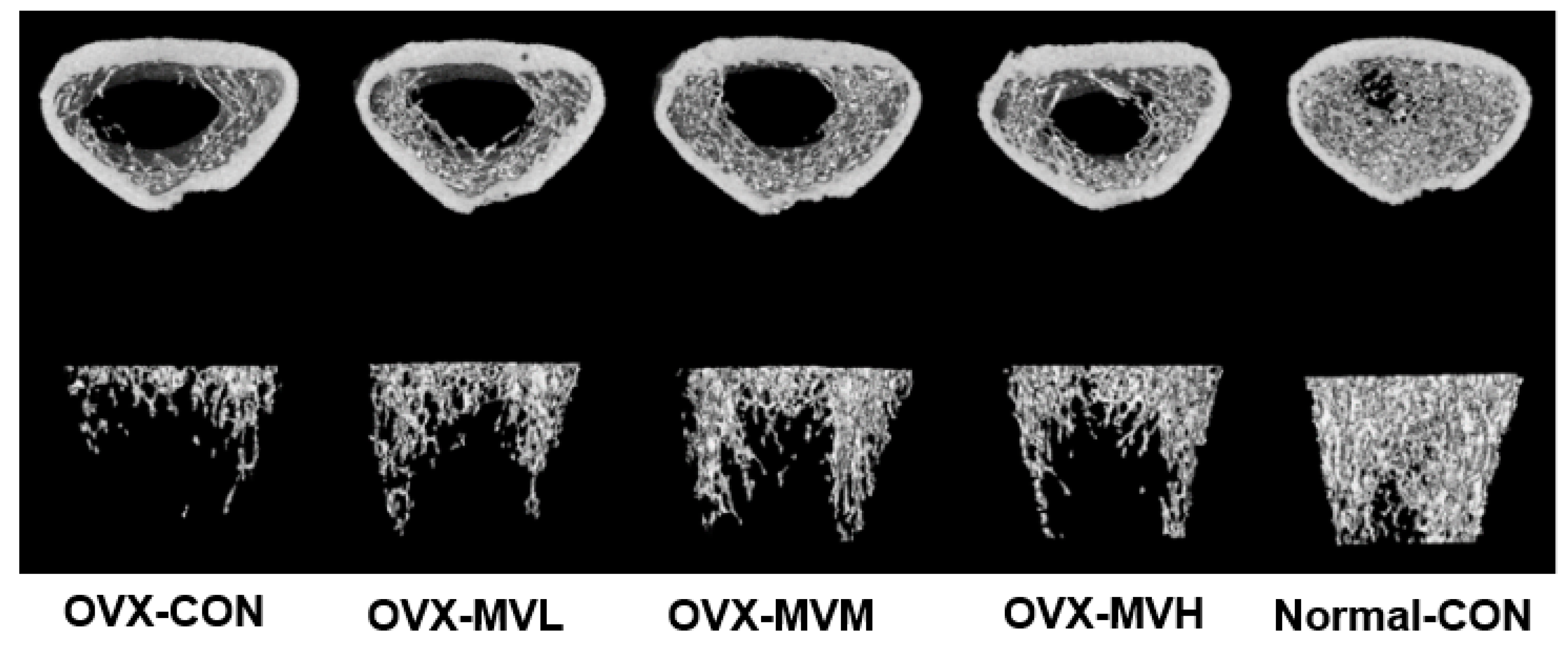
3.3. BMD by Micro-CT

The ratio of the segmented bone volume to the total volume of the bone region (BV/TV) was similar among the groups, even though the Normal-CON group was slightly higher (57.75 ± 1.07%) than the OVX-CON group (54.50 ± 1.22%). The MV showed a similar value to the OVX-CON group (Table 3; Figure 1). The bone surface and bone volume ratio (BS/BV) was similar in all groups, but it tended to be lower in the OVX-con group than in the Normal-CON group, and MV treatment tended to prevent its decrease. The mean Tb.Th was similar in all groups. The average Tb.N tended to be lower in the OVX-CON group than in the Normal-con group, while MV treatment produced similar results to the OVX-CON group (Table 3; Figure 1). The mean Tb.Sp showed an opposite trend of Tb.N. On the other hand, the BMD was much lower in the OVX-CON group than in the Normal-CON group (p < 0.05). At the same time, it was significantly higher in the OVX-MVM group than in the OVX-CON group (p < 0.05), but it was much lower in the OVX-MVM group than in the Normal-CON group (p < 0.05; Table 3, Figure 1). Therefore, MV treatment slightly prevented the decrease in BMD in OVX rats.

3.4. Cell Viability RANKL-Induced Osteoclasts from RAW 264.7 Cells

RANKL-induced osteoclasts increased cell proliferation during 24 and 48 h. Dried MV treatment (at 50–100 µg/mL) prevented the cell proliferation compared to the 0 MV μg/mL treatment in 24 h and 48 h incubation (p < 0.05; Figure 2). The MV treatment with 100 µg/mL suppressed the proliferation as much as the Control (no RANKL administration) in 48 h incubation. The ≥300 µg/mL MV treatment reduced cell proliferation more than the Control in the 24 h and 48 h incubation (p < 0.05), indicating a high dosage (300 μg/mL) of MV-induced cell apoptosis, potentially due to increased acidity. Thus, further study was conducted at MV concentrations up to 100 µg/mL.

3.5. TRAP-Positive Cells and TRAP Activity

TRAP-positive multinucleated cells (MNCs) appeared in the RANKL-administered groups. However, they were not detected in the Control group (no RANKL administration), suggesting that RANKL differentiated RAW264.7 cells into osteoclasts, as shown in Figure 3A,B. The MV treatment with RANKL significantly decreased the number of TRAP + MNCs dose-dependently (0 μg/mL: 11.0 ± 1.8; 10 μg/mL: 6.7 ± 2.2; 50 μg/mL: 5.3 ± 2.0; and 100 µg/mL: 3.5 ± 1.0 number of TRAP-positive cells/visible field; p < 0.05). However, the TRAP+MNCs number in the highest dosage of MV was higher than the Control (p < 0.05).
TRAP activity is a biomarker of osteoclasts to confirm osteoclast differentiation in RANKL-induced osteoclast differentiation. RANKL increased the TRAP activity by 205.8% for a five-day administration compared to the Control (no RANKL administration; p < 0.05). The MV treatment decreased the TRAP activity in a dose-dependent manner from the no mulberry-treatment group (p < 0.05): 10, 50, and 100 μg/mL MV decreased the TRAP activity by 174.1 ± 15.3%, 161.6 ± 15.3%, and 156.1 ± 2.0%, respectively (Figure 3C). Similar to the TRAP + MNCs number, the 100 μg/mL MV treatment decreased TRAP activity compared to the 0 μg/mL MV treatment (p < 0.05), but it did not reach TRAP activity in the Control.

3.6. mRNA Expression of Osteoclast-Related Genes

Osteoclast differentiation by RANKL increased Acp5, Ctsk, Oscar, and NFATc1 expression to elevate bone resorption [25]. The relative Acp5 mRNA expression increased in the RANKL administration group by approximately 13-fold compared to no RANKL administration (Control; p < 0.05; Figure 4A). The MV treatment (10, 50, and 100 µg/mL) decreased relative Acp5 mRNA expression compared to the no MV treatment in the RANKL administration groups. The 100 μg/mL MV treatment reduced it as much as the Control (Figure 4A). The relative CtsK mRNA expression increased with RANKL administration compared to the Control (p < 0.05), while the MV treatments inhibited its expression dose-dependently (p < 0.05). However, a high dosage did not suppress it as much as the no RNAKL treatment (p < 0.05; Figure 4B). The relative mRNA expression of Oscar and NFATc1 was elevated by RNAKL administration compared to the Control (p < 0.05), and the MV treatments (10–100 µg/mL) suppressed its upregulation as much as no RNAKL administration (Figure 4C,D).

3.7. Osteoclast-Related Protein Contents

RANK activation by RANKL activates TRAF6 to stimulate IkappaB, extracellular signal-regulated kinase (Erk), JNK-1, and P38, and NFATc1 binds with NF-κB and c-Fos. The NFATc1 complex elevates the osteoclast-related genes, and the NFATc1 and c-Fos protein levels indicate osteoclast activation by RANKL. The c-Fos protein contents were much higher in the RNAKL treated group than in the Control group (p < 0.05), and they decreased by 50% with MV treatment (10–100 μg/mL) compared to the no MV treatment (p < 0.05; Figure 5A,B). The protein contents of NFATc1 in the RANKL-administered osteoclast cells were much higher than those in the Control. However, they were reduced by approximately 70% in the MV treatment (10–100 μg/mL) compared to the no MV treatment, regardless of dosage (p < 0.05; Figure 5A,C).

4. Discussion

Menopause inhibits ovarian hormone production, particularly 17β-estradiol, and it increases insulin resistance, visceral fat mass, and dyslipidemia, leading to cardiovascular disease risk. It also accelerates bone loss by up to 20% and increases the osteoporosis risk. On the other hand, hormone replacement therapy (HRT) prevents bone loss, despite its inconsistent effects on cardiovascular diseases. HRT has been prescribed for menopausal women having a high osteoporosis risk, but it has some adverse effects, such as deep vein thrombosis, uterine cancer, breast cancer, and stroke. Menopause is extended for long periods, and functional foods need to be developed for HRT substitution without adverse effects. The present study showed that daily MVM intake (1 mL/kg body weight) inhibited BMD decrement in estrogen-deficient rats. The inhibition of BMD loss by MVM was associated with the partial prevention of decreased serum 17β-estradiol and OPG concentration and increased serum osteoclastic biomarkers, including RANKL, CTX-1, and TRAP, in OVX rats. The changes were confirmed in RANKL-differentiated osteoclast cells. The MV treatment (10–100 µg/mL) decreased the TRAP-positive cells; their activity and expression decreased dose-dependently. MV administration reduced the mRNA expression downstream of the RANK signaling genes, such as Oscar and Ngatc1, and the protein contents of c-Fos and Nfatc1. Therefore, MV intake (1–2 mL/kg bw/day) partially reduced the estradiol-related osteoclast activity to protect against osteoporosis risk caused by an estrogen deficiency. It may be considered an alternative therapy for menopausal-induced osteoporosis.
An ovariectomy in experimental animals resulted in similar characteristics to menopausal women [26,27]. Ovariectomized rats induced weight gain, insulin resistance, hot flushes, dyslipidemia, and osteoarthritis [26,27], and a herbal treatment prevented the menopausal symptoms [28,29]. In particular, the intake of puerarin, mulberry, and Solanum nigrum Line water extract inhibited the decrease in BMD by suppressing osteoclast differentiation in OVX-induced animal models [17,30,31]. Bone mineral loss in postmenopausal women is related to promoting the RANKL/RANK system in osteoclast cells and suppressing the bone morphogenetic protein and wingless-type and integrase 1 (Wnt) signaling pathways in osteoblast cells [32]. The RANKL/RANK system in osteoclast cells is counterbalanced with OPG in osteoblast cells to modulate bone mass. An estrogen deficiency breaks the balance to increase RANKL/RANK and decrease OPG, contributing to bone loss [33]. RANKL binds to RANK on the surfaces of the osteoclast precursor cells to stimulate osteoclast precursor cells and differentiate into mature osteoclasts, leading to BMD loss [34]. Estrogen modifies RANKL expression, an essential cytokine of bone resorption by osteoclasts, and estrogen deficiency fails to suppress RANKL expression in the osteoclast cells [4]. The present study revealed consistent results in increasing the serum RANKL concentration and decreasing the serum OPG concentration in OVX rats. MV intake protected against increasing serum RANKL concentrations and decreasing serum OPG concentrations.
Estrogen deficiency potentially increases bone resorption by the activation of RANKL → NF-κB → TARP and CTX-1 increment → elevation of TNF-α and IL-1β. It decreases bone formation by activating osteoblast apoptosis (Figure 6) [35]. As a result, estrogen deficiency promotes bone loss, leading to osteoporosis (Figure 6). The present study showed that MV intake mainly protected bone loss by suppressing the osteoclastic pathway by partially preventing estrogen reduction and reducing RANKL production and its signal pathway (Figure 6). Therefore, MV can delay the acceleration of bone loss by an estrogen deficiency in menopausal women. A large-scale randomized clinical trial must be conducted to confirm the MV effect in menopausal women.
Moreover, RANKL/RANK activates Erk, cFos, and NFATc1, which stimulate the mRNA expression of Ctsk, TRAP, and MMP-9 during osteoclast formation. CTX-1 is an osteoclast metabolite by bone resorption mediated by Ctsk, type 1 collagen-degrading enzymes during bone resorption. These processes elevate the production of reactive oxygen species to accelerate bone resorption [36]. As a result, RANKL/RANK stimulates the bone resorption activity of osteoclasts. The present study also showed that the serum CTX-1 and TRAP concentrations were higher in OVX-CON than the Normal-CON and reduced with MV treatment [36]. Hence, the MV treatment decreased RANKL to inhibit the initiation of osteoclast differentiation from osteoclast precursor cells to form osteoclasts and the bone resorption activity by osteoclasts in OVX rats that activated the RANKL/RANK due to estrogen deficiency.
There are some potential limitations of the present study. First, the OVX rat is a suitable animal model for studying human menopausal women [23,26,27], but it may have a different pathway to induce osteoporosis. Second, BMD loss resulted from the imbalance of osteoblastic and osteoclastic processes in OVX rats. However, the procedures were not directly checked in an animal study but studied in cell culture. Therefore, a human study needs to confirm the ability to translate rodent and cell-based results to humans.

5. Conclusions

A 12-week MV intake showed a similar preventive osteoporosis activity in OVX rats by suppressing the RANKL/RANK/TRAP pathway. MVM and MVH intake inhibited the decrease in BMD decrease in OVX rats as much as the Normal-con in Micro-CT analysis. The prevention of BMD loss by MVM and MVH was associated with inhibiting RANKL/RANK/TRAP induced by OVX. Fermented MV containing live Acetobacter pasteurianus acts as an inhibitor of bone resorption. Therefore, MV intake (1–2 mL/kg body weight/day for the rat; equivalent to 9–18 mL per day for human) had anti-osteoporosis activity by inhibiting the osteoclastic activity induced by OVX. These results suggested that MV may be developed as a functional food for anti-osteoporosis in menopausal women. A randomized clinical trial is needed to confirm the preventive efficacy for osteoporosis.

Supplementary Materials

The following supporting information can be downloaded at: https://www.mdpi.com/article/10.3390/fermentation8050211/s1, Figure S1: A. Standard curve for rutin, B. HPLC chromatogram of mulberry vinegar; Table S1: HPLC analysis conditions for organic acids and free sugars in mulberry vinegar; Table S2: HPLC-DAD analysis conditions for rutin contents in mulberry vinegar; Table S3: Primer sequences for real-time PCR.

Author Contributions

Conceptualization, E.J.Y. and D.-Y.J.; methodology, S.P., E.J.Y. and S.W.J.; resources, S.W.J. and H.J.K.; data collection and analysis, E.J.Y., S.K.P. and K.Y.Y.; writing—original draft preparation, S.P.; writing—review and editing, E.J.Y., S.W.J. and D.-Y.J.; supervision, D.-Y.J. and S.P. All authors have read and agreed to the published version of the manuscript.

Funding

This research was supported by Local Strategic Food Industry Promote Program, Ministry of Agriculture, Food and Rural Affairs, Republic of Korea (MIFI 2021-01).

Institutional Review Board Statement

The animal study was proceeded according to the NIH Guide for the Care and Use of Laboratory Animals guidelines and was approved by the Animal Care and Use Committee of Invivo company, Korea (IV-RB-02-2106-18).

Informed Consent Statement

Not applicable.

Data Availability Statement

Data are available from the corresponding author on reasonable request with a reasonable reason.

Conflicts of Interest

The authors declare no conflict of interest.

References

  1. Park, S.; Daily, J.W.; Song, M.Y.; Kwon, H.K. Gene-gene and gene-lifestyle interactions of AKAP11, KCNMA1, PUM1, SPTBN1, and EPDR1 on osteoporosis risk in middle-aged adults. Nutrition 2020, 79–80, 110859. [Google Scholar] [CrossRef] [PubMed]
  2. Riggs, B.L. The mechanisms of estrogen regulation of bone resorption. J. Clin. Investig. 2000, 106, 1203–1204. [Google Scholar] [CrossRef] [PubMed] [Green Version]
  3. Fischer, V.; Haffner-Luntzer, M. Interaction between bone and immune cells: Implications for postmenopausal osteoporosis. Semin. Cell Dev. Biol. 2022, 123, 14–21. [Google Scholar] [CrossRef] [PubMed]
  4. Streicher, C.; Heyny, A.; Andrukhova, O.; Haigl, B.; Slavic, S.; Schüler, C.; Kollmann, K.; Kantner, I.; Sexl, V.; Kleiter, M.; et al. Estrogen Regulates Bone Turnover by Targeting RANKL Expression in Bone Lining Cells. Sci. Rep. 2017, 7, 6460. [Google Scholar] [CrossRef] [Green Version]
  5. Kon, T.; Cho, T.J.; Aizawa, T.; Yamazaki, M.; Nooh, N.; Graves, D.; Gerstenfeld, L.C.; Einhorn, T.A. Expression of osteoprotegerin, receptor activator of NF-kappaB ligand (osteoprotegerin ligand) and related proinflammatory cytokines during fracture healing. J. Bone Miner. Res. 2001, 16, 1004–1014. [Google Scholar] [CrossRef]
  6. Chen, L.; Cheng, S.; Sun, K.; Wang, J.; Liu, X.; Zhao, Y.; Yang, J.; Zhao, D.; Xue, C.; Tao, Y.; et al. Changes in macrophage and inflammatory cytokine expressions during fracture healing in an ovariectomized mice model. BMC Musculoskelet. Disord. 2021, 22, 494. [Google Scholar] [CrossRef]
  7. Ilesanmi-Oyelere, B.L.; Schollum, L.; Kuhn-Sherlock, B.; McConnell, M.; Mros, S.; Coad, J.; Roy, N.C.; Kruger, M.C. Inflammatory markers and bone health in postmenopausal women: A cross-sectional overview. Immun. Ageing 2019, 16, 15. [Google Scholar] [CrossRef]
  8. Paschalis, E.P.; Gamsjaeger, S.; Hassler, N.; Fahrleitner-Pammer, A.; Dobnig, H.; Stepan, J.J.; Pavo, I.; Eriksen, E.F.; Klaushofer, K. Vitamin D and calcium supplementation for three years in postmenopausal osteoporosis significantly alters bone mineral and organic matrix quality. Bone 2017, 95, 41–46. [Google Scholar] [CrossRef]
  9. Park, S.; Park, C.Y.; Ham, J.O.; Lee, B.K. Familial interactions and physical, lifestyle, and dietary factors to affect bone mineral density of children in the KNHANES 2009–2010. J. Bone Miner. Metab. 2014, 32, 455–467. [Google Scholar] [CrossRef]
  10. Gambacciani, M.; Cagnacci, A.; Lello, S. Hormone replacement therapy and prevention of chronic conditions. Climacteric 2019, 22, 303–306. [Google Scholar] [CrossRef]
  11. Rossouw, J.E.; Anderson, G.L.; Prentice, R.L.; LaCroix, A.Z.; Kooperberg, C.; Stefanick, M.L.; Jackson, R.D.; Beresford, S.A.; Howard, B.V.; Johnson, K.C.; et al. Risks and benefits of estrogen plus progestin in healthy postmenopausal women: Principal results From the Women’s Health Initiative randomized controlled trial. JAMA 2002, 288, 321–333. [Google Scholar] [PubMed] [Green Version]
  12. Picherit, C.; Bennetau-Pelissero, C.; Chanteranne, B.; Lebecque, P.; Davicco, M.-J.; Barlet, J.-P.; Coxam, V. Soybean Isoflavones Dose-Dependently Reduce Bone Turnover but Do Not Reverse Established Osteopenia in Adult Ovariectomized Rats. J. Nutr. 2001, 131, 723–728. [Google Scholar] [CrossRef] [PubMed] [Green Version]
  13. Ji-Sun, K.; Hyunjung, L.; Farida Sukma, N.; Chang Hwa, J.; Young-Jin, J.; Tae-Youl, H.; Jiyun, A. Dry-Fermented Soybean Food (Cheonggukjang) Ameliorates Senile Osteoporosis in the Senescence-Accelerated Mouse Prone 6 Model. J. Med. Food 2019, 22, 1047–1057. [Google Scholar]
  14. Park, S.; Zhang, T.; Qiu, J.Y.; Wu, X. The Combination of Mulberry Extracts and Silk Amino Acids Alleviated High Fat Diet-Induced Nonalcoholic Hepatic Steatosis by Improving Hepatic Insulin Signaling and Normalizing Gut Microbiome Dysbiosis in Rats. Evid. Based Complement. Alternat. Med. 2019, 2019, 8063121. [Google Scholar] [CrossRef]
  15. Park, S.; Kim, D.S.; Wu, X.; Qiu, J.Y. Mulberry and dandelion water extracts prevent alcohol-induced steatosis with alleviating gut microbiome dysbiosis. Exp. Biol. Med. 2018, 243, 882–894. [Google Scholar] [CrossRef] [PubMed]
  16. Yuan, Q.; Zhao, L. The Mulberry (Morus alba L.) Fruit—A Review of Characteristic Components and Health Benefits. J. Agric. Food Chem. 2017, 65, 10383–10394. [Google Scholar] [CrossRef]
  17. Jao, H.Y.; Hsu, J.D.; Lee, Y.R.; Lo, C.S.; Lee, H.J. Mulberry water extract regulates the osteoblast/osteoclast balance in an ovariectomized rat model. Food Funct. 2016, 7, 4753–4763. [Google Scholar] [CrossRef]
  18. Xia, T.; Zhang, B.; Duan, W.; Zhang, J.; Wang, M. Nutrients and bioactive components from vinegar: A fermented and functional food. J. Func. Foods 2020, 64, 103681. [Google Scholar] [CrossRef]
  19. Se-In, B.; Geon Hyeong, G.; Eun Ju, C.; Ah Young, L.; Weon Taek, S. Characteristics of fermented vinegar using mulberry and its antioxidant activity. Korean Soc. Food Preserv. 2020, 27, 651–662. [Google Scholar]
  20. Ko, B.S.; Lee, H.W.; Kim, D.S.; Kang, S.; Ryuk, J.A.; Park, S. Supplementing with Opuntia ficus-indica Mill and Dioscorea nipponica Makino extracts synergistically attenuates menopausal symptoms in estrogen-deficient rats. J. Ethnopharmacol. 2014, 155, 267–276. [Google Scholar] [CrossRef]
  21. AOAC. Official Methods of Analysis. Method Association of Official Analytical Communities, 19th ed.; AOAC International: Arlington, VA, USA, 2012. [Google Scholar]
  22. Yang, H.J.; Kim, M.J.; Qiu, J.Y.; Zhang, T.; Wu, X.; Jang, D.J.; Park, S. Rice Porridge Containing Welsh Onion Root Water Extract Alleviates Osteoarthritis-Related Pain Behaviors, Glucose Levels, and Bone Metabolism in Osteoarthritis-Induced Ovariectomized Rats. Nutrients 2019, 11, 1503. [Google Scholar] [CrossRef] [PubMed] [Green Version]
  23. Park, S.; Kim, D.S.; Kang, S.; Moon, B.R. Fermented soybeans, Chungkookjang, prevent hippocampal cell death and beta-cell apoptosis by decreasing proinflammatory cytokines in gerbils with transient artery occlusion. Exp. Biol. Med. 2015, 241, 296–307. [Google Scholar] [CrossRef] [PubMed] [Green Version]
  24. Lee, Y.J.; Park, S.Y.; Lee, S.J.; Boo, Y.C.; Choi, J.Y.; Kim, J.E. Ucma, a direct transcriptional target of Runx2 and Osterix, promotes osteoblast differentiation and nodule formation. Osteoarthr. Cartil. 2015, 23, 1421–1431. [Google Scholar] [CrossRef] [PubMed] [Green Version]
  25. Yamashita, T.; Uehara, S.; Udagawa, N.; Li, F.; Kadota, S.; Esumi, H.; Kobayashi, Y.; Takahashi, N. Arctigenin Inhibits Osteoclast Differentiation and Function by Suppressing Both Calcineurin-Dependent and Osteoblastic Cell-Dependent NFATc1 Pathways. PLoS ONE 2014, 9, e85878. [Google Scholar] [CrossRef] [PubMed] [Green Version]
  26. Park, S.; Kim, D.S.; Kang, E.S.; Kim, D.B.; Kang, S. Low-dose brain estrogen prevents menopausal syndrome while maintaining the diversity of the gut microbiomes in estrogen-deficient rats. Am. J. Physiol. Endocrinol. Metab. 2018, 315, E99–E109. [Google Scholar] [CrossRef]
  27. Park, S.; Hong, S.M.; Ahn, I.L.S.; Kim, D.S.; Kim, S.H. Estrogen Replacement Reverses Olanzapine-Induced Weight Gain and Hepatic Insulin Resistance in Ovariectomized Diabetic Rats. Neuropsychobiology 2010, 61, 148–161. [Google Scholar] [CrossRef]
  28. Yang, H.J.; Ko, B.S.; Kwon, D.Y.; Lee, H.W.; Kim, M.J.; Ryuk, J.; Kang, S.; Kim, D.S.; Park, S. Asian Elm tree inner bark prevents articular cartilage deterioration in ovariectomized obese rats with monoiodoacetate-induced osteoarthritis. Menopause 2016, 23, 197–208. [Google Scholar] [CrossRef]
  29. Lee, H.W.; Ko, B.S.; Kang, S.; Ryuk, J.A.; Kim, M.J.; Park, S. Dangguijihwang-tang and Dangguijakyak-san prevent menopausal symptoms, and Dangguijihwang-tang prevents articular cartilage deterioration in ovariectomized obese rats with monoiodoacetate-induced osteoarthritis. Evid. Based Complement. Alternat. Med. 2017, 2017, 5658681. [Google Scholar] [CrossRef] [Green Version]
  30. Cheng, J.; Zhou, L.; Liu, Q.; Tickner, J.; Tan, Z.; Li, X.; Liu, M.; Lin, X.; Wang, T.; Pavlos, N.J.; et al. Cyanidin Chloride inhibits ovariectomy-induced osteoporosis by suppressing RANKL-mediated osteoclastogenesis and associated signaling pathways. J. Cell Physiol. 2018, 233, 2502–2512. [Google Scholar] [CrossRef]
  31. Kim, J.H.; Shin, H.; Kim, M.; Kim, S.; Song, K.; Jung, H.S.; Sohn, Y. Solanum nigrum Line inhibits osteoclast differentiation and suppresses bone mineral density reduction in the ovariectomy-induced osteoporosis model. Mol. Med. Rep. 2021, 24, 607. [Google Scholar] [CrossRef]
  32. Bi, H.; Chen, X.; Gao, S.; Yu, X.; Xiao, J.; Zhang, B.; Liu, X.; Dai, M. Key Triggers of Osteoclast-Related Diseases and Available Strategies for Targeted Therapies: A Review. Front. Med. 2017, 4, 234. [Google Scholar] [CrossRef] [PubMed]
  33. Novack, D.V. Estrogen and Bone: Osteoclasts Take Center Stage. Cell Metab. 2007, 6, 254–256. [Google Scholar] [CrossRef] [PubMed] [Green Version]
  34. Boyle, W.J.; Simonet, W.S.; Lacey, D.L. Osteoclast differentiation and activation. Nature 2003, 423, 337–342. [Google Scholar] [CrossRef] [PubMed]
  35. Chen, D.; Wang, Q.; Li, Y.; Sun, P.; Kuek, V.; Yuan, J.; Yang, J.; Wen, L.; Wang, H.; Xu, J.; et al. Notopterol Attenuates Estrogen Deficiency-Induced Osteoporosis via Repressing RANKL Signaling and Reactive Oxygen Species. Front. Pharmacol. 2021, 12, 664836. [Google Scholar] [CrossRef]
  36. Hsiao, Y.-M.; Hu, C.-C.; Chen, M.-F.; Chang, C.-H.; Chiu, Y.-T.; Chang, Y. Serum Insufficiency Induces RANKL-Independent Osteoclast Formation during Developing Ischemic ONFH. Biomedicines 2021, 9, 685. [Google Scholar] [CrossRef]
Figure 1. Bone mineral density (BMD)-related parameters after 12-week treatment of mulberry vinegar in the femur of ovariectomized rats.
Fermentation 08 00211 g001
Figure 2. Cell viability with different dosages of dried mulberry vinegar in RANKL-induced Raw264.7 cells. Bars and error bars represented mean ± standard deviation (n = 5). * Significantly different from the Control at p < 0.05, ** at p < 0.01, and *** at p < 0.001. ## Significantly different from the 0 μg/mL MV treatment at p < 0.01 and ### at p <0.001. + Significantly different from 50 ng/mL MV treatment at p < 0.05 and +++ p < 0.001.
Fermentation 08 00211 g002
Figure 3. Tartrate-resistant acid phosphatase (TRAP)-positive multinucleated cells (MNC) and TRAP activity in RANKL-induced Raw264.7 cells administered with different dosages of dried mulberry vinegar for five days; (A) TRAP staining. The scale bar in the figure indicated 100 μm and X20 magnification; (B) TRAP-positive MNCs; (C) TRAP activity; Bars and error bars represented mean±standard deviation (n = 5). * Significantly different from the Control at p < 0.05, ** at p < 0.01, and *** at p < 0.001. # Significantly different from the 0 μg/mL MV treatment at p < 0.05 and ## at p < 0.01. + Significantly different from 10 ng/mL MV treatment at p < 0.05.
Fermentation 08 00211 g003
Figure 4. Osteoclastogenesis-related gene expression in RANKL-induced Raw264.7 cells administered with different dosages of dried mulberry vinegar for five days; (A) Relative gene expression of Acp5 (TRAP); (B) Relative gene expression of CtsK; (C) Relative gene expression of Oscar; (D) Relative gene expression of NFATc1; Bars and error bars represented mean±standard deviation (n = 5). * Significantly different from the Control at p < 0.05, ** at p < 0.01, and *** at p < 0.001. # Significantly different from the 0 μg/mL MV treatment at p < 0.05, ## at p < 0.01, ### at p < 0.001. + Significantly different from 10 ng/mL MV treatment at p < 0.05.
Fermentation 08 00211 g004aFermentation 08 00211 g004b
Figure 5. Osteoclastogenesis-related protein contents in RANKL-induced Raw264.7 cells administered with different dosages of dried mulberry vinegar for five days.; (A) Western blot of c-Fos and NFATc1; (B) Relative contents of c-Fos; (C) Relative contents of NFATc1; Bars and error bars represented mean ± standard deviation (n = 3). * Significantly different from the Control at p < 0.05, ** at p < 0.01, and *** at p < 0.001. # Significantly different from the 0 μg/mL MV treatment at p < 0.05 and ### at p < 0.001.
Fermentation 08 00211 g005
Figure 6. The potential mechanism of mulberry vinegar (MV) intake to protect against bone loss (osteoporosis) in an estrogen-deficient state. Estrogen deficiency decreased RUNX2 and osterix signaling pathways to inhibit OPG and osteocalcin production, which stimulates osteoblast apoptosis to reduce bone formation. Estrogen deficiency increases RANKL production to activate RANKL and NF-κB signaling pathways. It increased TRAP and CTX-1 production and stimulated the production of proinflammatory cytokines to activate osteoclast differentiation. As a result, estrogen deficiency promotes bone loss. MV intake mainly inhibited osteoclast differentiation by suppressing the RANKL-related inflammation signal pathway and partially increasing estrogen production.
Fermentation 08 00211 g006
Table 1. Characteristics of fermented mulberry vinegar for nine days.
Mean ± SD Mean ± SD
pH2.26 ± 0.01Oxalic acid (mg/mL)No detection
Acidity (%)4.07 ± 0.01Citric acid (mg/mL)106.0 ± 0.06
Live bacteria counts (log CFU/mL)7.31 ± 0.23Succinic acid (mg/mL)No detection
Rutin (mg/g)0.36 ± 0.04Lactic acid (mg/mL)19.2 ± 0.04
Total phenol (μg gallic acid equivalent/mL)570 ± 0.18Acetic acid (mg/mL)15.0 ± 0.09
Total flavonoids (μg quercetin equivalent/mL)80.0 ± 3.03
SD, standard deviation.
Table 2. Body weight and bone metabolism.
OVX-CONOVX-MVLOVX-MVMOVX-MVHNormal-CON
Final weight (g)411.2 ± 4.92 ***413.7 ± 9.38 ***402.5 ± 3.50 ***408.8 ± 5.52 ***320.5 ± 3.95
Weight gain (g)138.6 ± 5.56 ***142.9 ± 6.77 ***129.4 ± 5.27 ***136.5 ± 5.48 ***54.04 ± 2.10
Uterus weight (g)0.105 ± 0.003 ***0.119 ± 0.005 ***0.126 ± 0.009 ***0.121 ± 0.010 ***0.695 ± 0.098
Serum 17β-estradiol(pg/mL)35.28 ± 3.96 **37.29 ± 5.32 **42.20 ± 5.32 #38.64 ± 3.93 *45.27 ± 4.81
Serum CTX-1 (ng/mL)3.79 ± 0.62 **3.60 ± 0.48 **3.21 ± 0.66 *2.78 ± 0.55 #2.46 ± 0.36
Serum OPG (ng/mL)1.17 ± 0.09 *1.42 ± 0.11 #1.46 ± 0.07 *,#1.50 ± 0.11 *,#,+1.31 ± 0.15
Serum RANKL (pg/mL)30.31 ± 6.17 **23.86 ± 4.97 #25.33 ± 4.20 *,#24.27 ± 3.06 #20.01 ± 2.79
Serum TRAP (U/L)4.39 ± 0.22 **3.95 ± 0.29 **,#4.01 ± 0.30 ***,#4.05 ± 0.26 ***,#3.10 ± 0.20
Values represent mean ± standard deviation (n = 10); OPG, osteoprotegerin; CTX-Ⅰ, C-telopeptide of type Ⅰ collagen; TRAP, tartrate-resistant acid phosphatase; RANKL, receptor activity of nuclear factor kappa B ligand. * Significantly different from the Normal-CON at p < 0.05, ** at p < 0.01, and *** at p < 0.001. # Significantly different from the OVX-CON at p < 0.05. + Significantly different from 10 ng/mL MV treatment at p < 0.05.
Table 3. Bone mineral density (BMD) of the femur by micro-CT.
ParameterOVX-CONOVX-MVLOVX-MVMOVX-MVHNormal-CON
BV/TV (%)54.50 ± 1.2254.38 ± 0.90253.12 ± 0.69153.22 ± 0.43457.75 ± 1.07
BS/BV (1/mm)4.751 ± 0.2275.356 ± 0.3015.697 ± 0.5045.250 ± 0.2087.581 ± 0.182
TB_Th (mm)0.082 ± 0.0030.096 ± 0.0060.100 ± 0.0020.106 ± 0.0040.096 ± 0.006
Tb.N (1/mm)1.312 ± 0.0491.325 ± 0.0671.382 ± 0.0721.318 ± 0.0471.878 ± 0.049
Tb.Sp (mm)0.777 ± 0.0340.776 ± 0.0230.734 ± 0.0220.749 ± 0.0180.520 ± 0.030
BMD (mg/cm3)125.9 ± 4.42 ***152.1 ± 12.52 ***168.8 ± 21.59 ***,#146.5 ± 6.150 ***321.6 ± 8.43
Values represent mean±standard errors (n = 10); BV/TV (%), bone volume/tissue volume; BS/BV, bone surface/bone volume; Tb.Th, trabecular thickness; Tb.N, trabecular number; Tb.Sp, trabecular separation; BMD, bone mineral density; *** Significantly different from the Normal-CON at p < 0.001. # Significantly different from the OVX-CON at p < 0.05.
Publisher’s Note: MDPI stays neutral with regard to jurisdictional claims in published maps and institutional affiliations.

