Article

Antibacterial Mechanism of Dellaglioa algida against Pseudomonas fluorescens and Pseudomonas fragi

School of Life Science and Technology, Harbin Institute of Technology, Harbin 150000, China
*
Author to whom correspondence should be addressed.
Fermentation 2022, 8(7), 298; https://doi.org/10.3390/fermentation8070298
Submission received: 20 May 2022 / Revised: 6 June 2022 / Accepted: 21 June 2022 / Published: 24 June 2022
(This article belongs to the Section Microbial Metabolism, Physiology & Genetics)

Abstract

:
Pseudomonas fluorescens (P. fluorescens) and Pseudomonas fragi (P. fragi), two kinds of psychrotrophic Pseudomonas species with pathogenicity, are likely to contaminate foods and cause diseases even in fairly cold environments, an outcome which should be suppressed. This paper investigates the antibacterial mechanisms of Dellaglioa algida (D. algida), a new type of low-temperature-resistant Lactobacillus, on two such Pseudomonas. By the enzyme treatment approach, the antibacterial substance existing in the cell-free supernatant (CFS) of D. algida is preliminarily determined as organic acid or protein; then, its inhibition effects are assessed under various culture environments, including pH value, salinity, and culture time, where the best antibacterial performance is achieved at pH = 6.00, S = 0%, and culture time = 48 h. A series of experiments on biofilms indicate that D. algida is not only able to inhibit the generation or damage the integrality of the biofilm of the two mentioned Pseudomonas, but also can reduce the motility, including swarming and swimming, of P. fragi and restrain the swarming of P. fluorescens. The aformentioned developed antibacterial mechanisms show the possibility of using D. algida in applications as an inhibitor for psychrotrophic Pseudomonas in the food industry, by virtue of its strong suppression capability, especially in cold environments.

1. Introduction

Pseudomonas is a kind of opportunistic pathogen with strong vitality and that exists extensively in the water, air, soil, and in foods. Certain kinds of Pseudomonas can generate various organic materials to contaminate foods and even cause diseases, e.g., wound infections [1,2], bacteremia [3,4], and urinary tract infections [5,6]. Consequently, it is necessary to investigate prevention methods for contamination by Pseudomonas, which has attracted considerable attention. Different from most kinds of Pseudomonas, Pseudomonas fluorescens (P. fluorescens) and Pseudomonas fragi (P. fragi) can live and grow in a cold storage environment, which leads to the problem that they are able to spoil food such as dairy products and chilled fresh meat, even in low temperatures (c.f., fish corruption [7], milk [8,9], cheese metamorphic [10], chicken juice corruption [11]). Clearly, it is necessary and significant to find inhibition mechanisms for these two kinds of psychrotrophic Pseudomonas species spoilage to alleviate the occurrence of bacterial contamination.
Lactobacillus is widely known as a new type of contamination-free antiseptic bacteria without side effects, and certain kinds can suppress Pseudomonas. Specifically, some experiments have shown that Lactobacillus rhamnosus can generate certain antibacterial substances, whose principal component is organic acids that are able to inhibit P. fluorescens and Pseudomonas putida (P. putida) [12,13]. Since it is able to prevent protein synthesis and combine with the DNA of P. putida [14,15], Lactobacillus paracei FX-6 can suppress P. putida that is isolated from chicken breast meat. In the existing studies [16,17], the properties of antielastase and biofilm resistance of the Lactobacillus obtained from the mouth cavity of humans, including Lactobacillus fermenti, Lactobacillus zea, and Lactobacillus paracei, are investigated, which indicates that these Lactobacillus are the probiotics for the pulmonary infection caused by Pseudomonas aeruginosa (P. aeruginosa). Moreover, certain kinds of Lactobacillus can be utilized for the prevention and the treatment of diseases, e.g., P. aeruginosa sepsis [18], genitourinary system infection [19], caries [20], etc.
Clearly, Lactobacillus is capable of inhibiting Pseudomonas via its secretion in the aforementioned studies, which, however, focused on Lactobacillus without consideration of the property of cold adaptation, leading to the fact that the explored inhibition mechanisms may be not applicable to the suppression of psychrotrophic Pseudomonas species spoilage in cold environments [21]. D. algida (called Lactobacillus algidus in some works), a newly discovered Lactobacillus, is the only psychrotrophic Lactobacillus in its species [22,23,24], which may be a solution to the problem of inhibiting bacterial contamination of P. fluorescens and P. fragi. Surprisingly, however, its inherent mechanism has not been developed yet.
Motivated by the aforementioned observations, in this paper, the mechanism of the antibacterial effects of D. algida against the target psychrotrophic Pseudomonas, i.e., P. fluorescens and P. fragi, is investigated via cell-free supernatant (CFS). The antibacterial substance existing in the CFS of D. algida is preliminarily determined by enzyme treatment; then, its minimum inhibitory concentration (MIC) is measured and the thermal stability is analyzed. Under various culture environments, including pH, salinity, and culture time, the antibacterial effects are assessed. This is followed by a discussion of the antibacterial mechanism based on the variation of biofilm. The experimental results show that D. algida has the ability of limiting the generation of biofilm, suppressing its growth and production, and reducing the motility of P. fluorescens and P. fragi. Based on the experiments, an approach is derived to prevent the contamination of psychrotrophic Pseudomonas, even in cold environments such as food transportation and storage.

2. Materials and Methods

2.1. Cell-Free Supernatant and Target Strains

The obtainment of the cell-free supernatant (CFS) for D. algida: Under the non-agitating conditions and a temperature of 20 °C, D. algida (BNCC ® 136658 TM ) was cultured in the medium of de Mann Rogosa Sharpe (MRS, purchased from biosharp ® , Hefei, China) for 24 h to produce the seed liquid of such a psychrotrophic Lactobacillus; the composition of the culture medium is shown in Table 1, and the culture method was inspired by [25]. The activated seed liquid, with an inoculation quantity of 1% (v/v), was injected into the MRS liquid medium, before being activated at 20 °C for 24 h twice. After the activation, the above bacteria solution was centrifuged with the conditions of 4 °C, 8000× g, and 5 min, after which it was filtrated using a low protein binding cellulose acetate filter to obtain the CFS finally.
P. fluorescens (BNCC ® 335852 TM ) and P. fragi (BNCC ® 134017 TM ): Using the TSB medium (purchased from biosharp ® , Hefei, China), the seed liquid of P. fluorescens and P. fragi was obtained by activation at 28 °C in a shaker of 180 rpm/min for 24 h; then, the inoculation quantity of 1% (v/v) was activated for 18 h at 28 °C for later use.
To assess the growth of the bacteria, the absorbance of D. algida was determined at the optical density of λ = 595 nm (OD 595 ), while the ones of P. fluorescens and P. fragi were both at the optical density of λ = 600 nm (OD 600 ). At various culture times, the spectrophotometer (Biotraza) was adopted to measure such OD values, which were further utilized to evaluate the colony-forming unit (CFU) via serial dilution method and then to depict the standard curve between the CFU and OD.

2.2. Basic Antibacterial Experiments of D. algida

Both P. fluorescens and P. fragi were evenly coated on the plate with beef extract peptone medium and left for 30 min. Oxford cups with an external diameter of 8 mm were lightly placed on such medium, where the CFS of D. algida was injected in the holes; then, they were stationarily cultured at 28 °C for 24 h to be observed to measure the diameter of the inhibition zone, is denoted as D.
Preliminary Identification of Antibacterial Substances: To eliminate the inhibition-free substances, the enzymes consisting of proteinase K, trypsin, papain, α -amylase, lipase, and catalase, with the final concentration of 1mg/mL, were injected into fermented CFS of D. algida. The mixture was processed by a water bath with 37 °C for 2 h, and was further treated by a water bath with 80 °C for 2 min to ensure the inactivation. By antibacterial experiments, where the supernatant without enzymes was adopted as comparison, the inhibition zone could be observed and its diameter could be measured.
Antibacterial Effects of Acidic Substances: This experiment was conducted to remove the influence of antibacterial effects caused by acidic substances. The pH value of the CFS fermentation was first measured, upon which the liquid medium of MRS with the same pH adjusted by lactic acid was selected as a control group, while the MRS without regulation was treated as a blank control group. Then, the antibacterial activity of the lactic acid was assessed, of which the diameter of the inhibition zone was also measured.
Thermal Stability: The CFS was treated for 30 min under the temperature of 20 °C, 30 °C, 40 °C, 50 °C, 60 °C, 70 °C, 80 °C, and 90 °C, respectively; then, the antibacterial experiments were carried out for the observation and the measurement of the diameter of the inhibition zone, so that the thermal stability of the antibacterial substance could be found.
Minimum Inhibitory Concentration (MIC): The CFS of D. algida obtained via the approach in Section 2.1 was utilized to produce the solid freeze-dried powder of the CFS by drying process. The seed liquid of P. fluorescens and P. fragi was, respectively, inoculated into the various TSB media, where the final concentrations of the solid freeze-dried powder were 0, 2, 4, 6, 8, 10, 12, 14, and 16 mg/mL, to be cultured at 28 °C for 24 h. Then, the value of OD 600 was measured, and the MIC that indicated the minimum concentration to inhibit bacteria was also determined.

2.3. Antibacterial Effects under Various Culture Conditions

The influence of culture conditions on antibacterial effects we considered in this paper consists of pH, salinity, and culture time, where the experimental approaches are as follows.
pH: Using the 1.0 mol/L HCl and the 1.0 mol/L NaOH, the pH value of the MRS liquid mediums were adjusted to be 2.00, 3.00, 4.00, 5.00, 6.00, 7.00, and 8.00; then, they were sterilized at 121 °C for 15 min and were infused with the stationary D. algida suspension of 1% (v/v). Such a medium was cultured at 20 °C for 24 h, centrifuged at 4 °C for 5 min under 8000× g, and filtrated. The final supernatant was selected to test the bacteriostasis, where the diameter of the inhibition zone was the evaluation index that would be observed and determined.
Salinity: The NaCl solutions with various concentrations, including 0%, 2%, 4%, 6%, 8%, and 10% (w/v), were first injected into the MRS liquid medium. After the sterilization, the stationary suspension of D. algida was also inoculated to the above medium, which was further cultured at 20 °C for 24 h. Finally, the medium was centrifuged in the conditions of 4 °C, 8000× g, and 5 min, and was filtrated to obtain the supernatant to observe and to measure the inhibition zone diameter by antibacterial experiments.
Culture time: The activated seed liquid of D. algida was inoculated into the MRS liquid medium according to the inoculum size of 1% (v/v), and was further cultured at the temperature of 20 °C for 6 h, 12 h, 24 h, 36 h, 48 h, 60 h, and 72 h, respectively. After the 5 min centrifugation with 8000× g at 4 °C and the filtration, the supernatant was utilized for antibacterial experiments to obtain the diameter of the inhibition zone.

2.4. Antibacterial Effects on Biofilm of P. fluorescens and P. fragi

The target Pseudomonas, i.e., P. fluorescens and P. fragi, were inoculated in a TSB medium that contained 3% (w/v) NaCl and 0.5% (w/v) G; then, they were cultured overnight (about 18–24 h) under 28 °C. Then, the TSB medium of 10 8 CFU/mL was utilized to attenuate the target Pseudomonas to produce the bacterial suspension, which was further attenuated as 106 CFU/mL for later use.
Suppression During the Development Stage: In the experiments on the influence of various concentrations, including 0 MIC, 0.5 MIC, 1 MIC, and 2 MIC, of the CFS of D. algida on the capability of generation and growth of biofilm, the crystal violet staining method was utilized, where the approach to assay was inspired by [26,27,28]. Under the pathogen-free condition, stationary bacteria solution of 200 μL was put into a 96-well cell culture plate and cultured at 28 °C, while the TSB medium was utilized as blank control. At 12 h, 24 h, 48 h, 36 h, and 48 h, respectively, the following processes were utilized: the bacteria solution was discarded and the well was washed using sterile water three times. After drying for 45 min at ordinary temperature, the crystal violet solution of 0.1% (w/v) was injected into each well to stain for 30 min; then, the solution was also abandoned for three-times washing of the well. By a 30 min elution using 95% (v/v) ethyl alcohol of 200 μL, the culture plate was placed in a microplate reader to measure the absorbance value at λ = 570 nm.
Elimination of Generated Biofilm: Here, the experiment of elimination effects on the biofilm is partly inspired by [29], while the whole process, with appropriate modifications to the method in [30], is demonstrated below. After infusing solid freeze-dried powder of the CFS of D. algida with various concentrations (including 0 MIC, 0.5 MIC, 1 MIC, and 2 MIC), the nutrient solution of P. fluorescens and P. fragi was cultured at 28 °C for 24 h to obtain biofilm maturation; then such a solution of 200 μ L was injected into a 96-well cell culture plate to be statically incubated at 28 °C for 36 h. By the same processes of washing and drying above, the glacial acetic acid of 30% (v/v) was adopted for elution; then, the absorbance value at 590 nm was determined via a microplate reader.
Morphology Analysis via Scanning Electron Microscopy (SEM): the TSB medium of P. fluorescens and P. fragi, which had been cultured to logarithmic phase, was utilized for a 12 h treatment. The obtained fresh bacteria were rinsed for 15 min using 1 × PBS of pH = 7.0 or NaCl solution of 0.9% (w/v) twice (or three times); then, they were centrifuged for 3 min to wipe off the supernatant. After combining 1 mL glutaraldehyde solution of 2.5% (v/v), the bacteria solution was resuspended to be fixed for 4 h, and then rinsed three times and injected with osmic acid of 1% (v/v) to be mixed and fixed for 2 h. After three-times rinsing again, the gradient ethanol solution with the concentration of 20%, 50%, 80%, and 100% (v/v) was utilized to elute, where the bacteria solution was treated for 10 min, centrifuged at 5000 rpm/min for 3 min, and cleaned of supernatant and moisture by wiping. The pure acetone was transfused into the bacteria solution for stationary culture at 4 °C for 20–30 min, which was centrifuged at 5000 rpm/min for 3 min; then, the pure acetone was injected again. After natural air drying and a metal spraying seal, the morphology of P. fluorescens and P. fragi was observed by an SEM (Quanta GEG250, FEI).

2.5. Antibacterial Effects on Swarming and Swimming of P. fluorescens and P. fragi

The soft agar plate method [31] was adopted to evaluate the influence of D. algida on the motility, including swimming and swarming, of P. fluorescens and P. fragi. The 3 μL stable bacterial solution of the target Pseudomonas was first inoculated to the middle of the plate, which was cultured statically at ordinary temperature for 20 min for full absorbtion. Then, the plate was placed in an incubator with 28 °C for 96 h, where it was taken out every 24 h to measure the diameter of the diffusion circle for assessment.

2.6. Statistical Analysis

The obtained data corresponds to at least three independent trials, and it is presented as mean values with standard deviations, where the statistical significance with the consideration of p < 0.05 was analyzed via GraphPad Prism 7 software.

3. Results and Discussions

3.1. Basic Antibacterial Experiments of D. algida

The existence of the antibacterial effects of metabolites of D. algida is first ascertained, where the agar diffusion method is utilized to analyze the source of the antibacterial substance. As is shown in Figure 1, where C and NC stand for the crude extract of the CFS and the supernatant of the fermented bacteria solution obtained by naturally precipitating, respectively, there is hardly any difference between the case of with bacteria and without bacteria, so the treated method we adopted does not make the change to the antibacterial effects (p > 0.05). In addition, it can be found that the antibacterial substance exists mainly in the fermented bacteria solution, where the diameter of the inhibition zone is larger than 10 mm (d > 10 mm), indicating strong antibacterial ability.
Then, to find the antibacterial substance of the CFS of D. algida in detail, the influence caused by inhibition-free metabolites, including hydrogen peroxide and protein/polypeptides such as bacteriocin, should be avoided. Table 2 shows the experimental results of the inhibition effects of CFS via the treatment of universality protease (proteinase K., trypsin, and papain), α -amylase, catalase, and lipase. It can be seen that, by proteinase K., the inhibition zone of both P. fluorescens and P. fragi is reduced, where the one of P. fragi is almost disappeared. Using trypsin and papain, the diameter of the inhibition zone of P. fragi reduces obviously compared to the blank control, while the decrement of the one of the P. fluorescens is not very sharp. A slight reduction of the diameter appears on the experiments of the treatment of the catalase, and there exists hardly any decrement via α amylase and lipase, so the produced hydrogen peroxide, fat, and polysaccharide have no inhibition effects. Thus, it can be found that the antibacterial substance existing in the CFS of D. algida against P. fluorescens and P. fragi must contain certain proteins or small peptides [32,33,34,35], which are sensitive to proteinase K. and can be interfered by trypsin and papain.
Additionally, the influence caused by the generated organic acid should be eliminated. The inhibition effects of organic acid are shown in Figure 2, where LAC, NL, and N represent the blank control group, the group of CFS with lactic acid elimination, and the group of CFS without treatment, respectively. It can be observed that the inhibition zone of P. fluorescens and P. fragi is generated in the blank control group, indicating the existence of inhibition effects which, however, are quite different from the ones of the group of CFS with lactic acid elimination, and the zone’s diameter is largely less than the one of the group of CFS without treatment. Hence, the generated organic acid can slightly (not significantly) suppress P. fluorescens and P. fragi, whereas the main inhibition effects are caused by the combination of the organic acid and certain excretion.
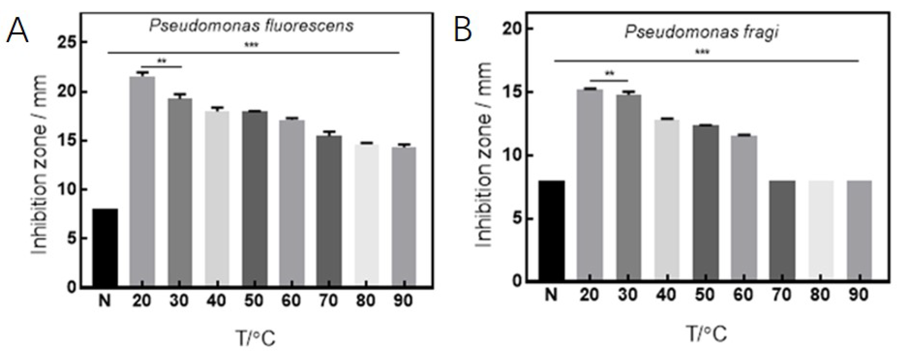
Finally, we turn to the thermal stability analysis and MIC determination. The experiments of antibacterial effects under various temperature conditions are illustrated in Figure 3. It can be observed that the antibacterial substance has strong stability of inhibition when the temperature is between 20 °C and 60 °C, and the best antibacterial performance is obtained at the temperature of 20 °C. The antibacterial protein or small peptide for P. fragi will degenerate such that there are no inhibition effects once the temperature is greater than 90 °C. However, P. fluorescens is still suppressed at 70 °C, which implies that it is influenced by the organic acid, as we said before. As is shown in Figure 4, the diameter of the inhibition zone is in proportion to the diluted concentration, and the antibacterial substances cannot show their effects of suppression once the concentration is less than 2 mg/mL. Thus, the MIC of CFS of D. algida on P. fragi and P. fluorescens can be determined as 2 mg/mL.

3.2. Antibacterial Effects under Various Culture Conditions

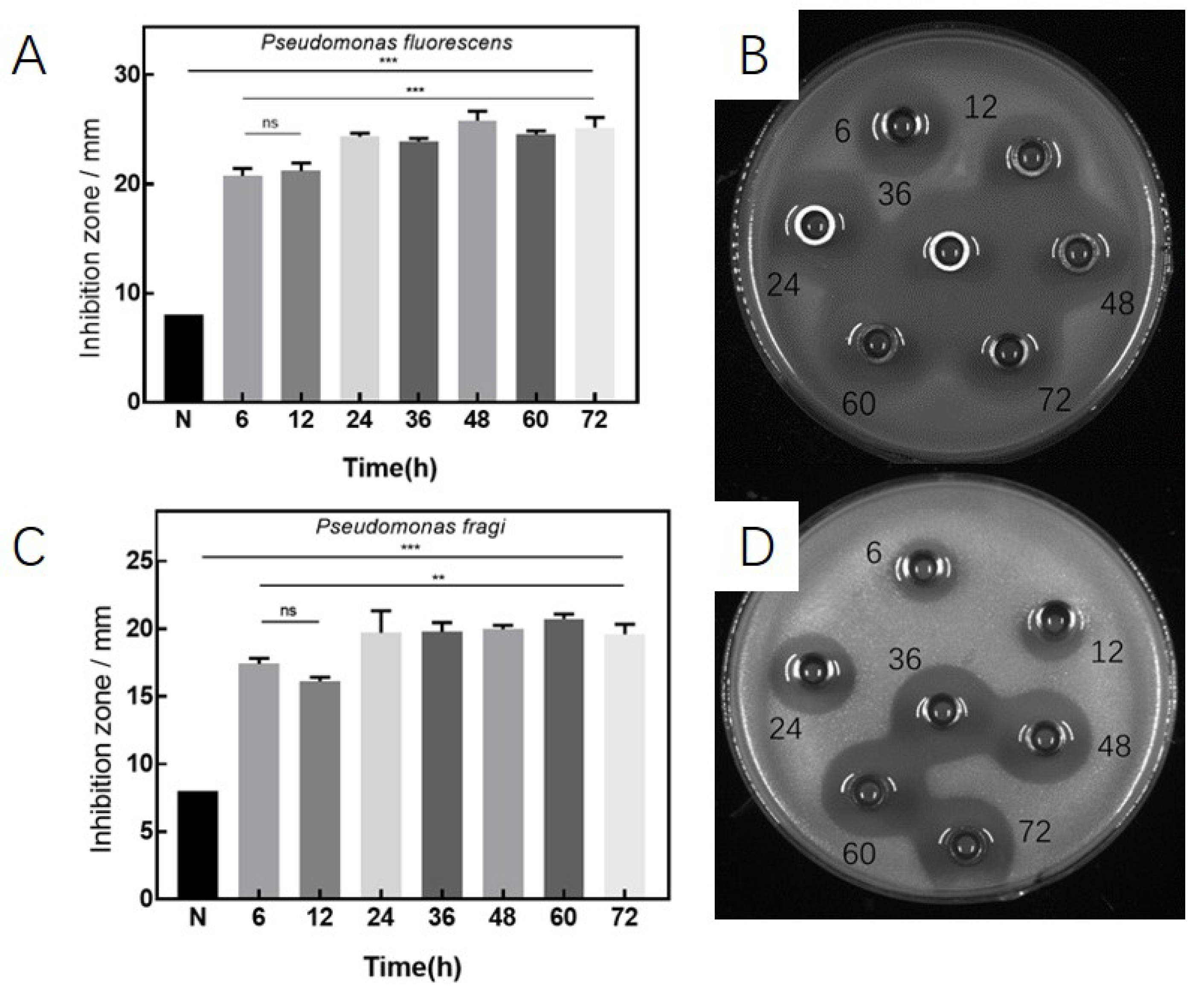
In Figure 5, the antibacterial effects are shown at different culture times, which demonstrates that the antibacterial substance is continuously accumulated during the growth of D. algida. Since the antibacterial substance is not generated after 48 h, which corresponds to the decline phase of the growth curve, one can have that the antibacterial substance is the secreted metabolites during the growth of D. algida.
Under the various salinities, as depicted in Figure 6, the generation of the antibacterial substance is in different cases. Compared with the blank control group, the cultured D. algida using mediums with salinities of 0–10% (w/v) shows inhibition effects, which decrease as the salinity increases. The growth of D. algida is inhibited by the salinity, instead of suppressing the effects of the antibacterial substance by the great salinity.
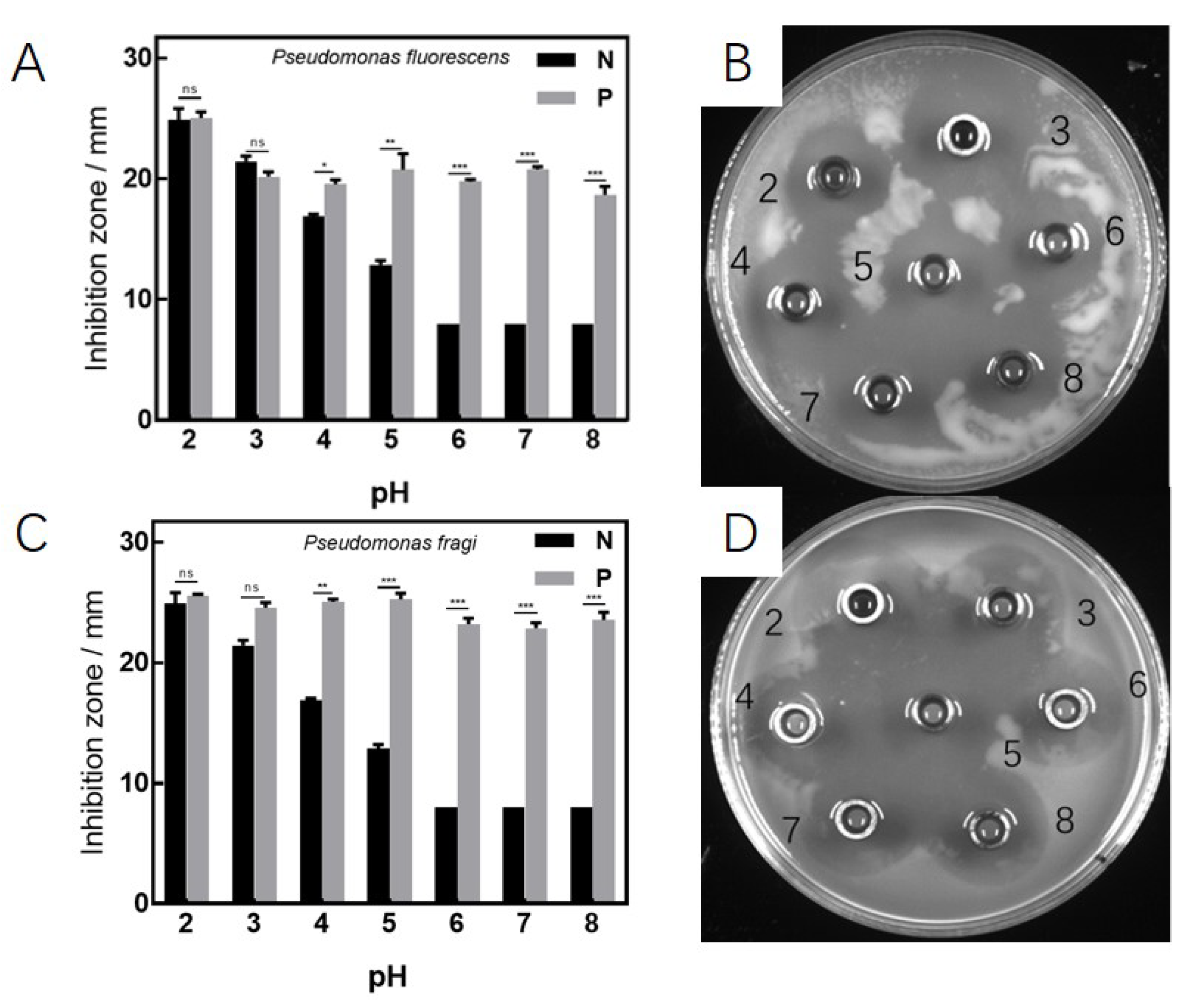
In addition, in Figure 7, it can be observed that, compared with the blank control group, the inhibition effects of the generated antibacterial substance existing in the CFS are fairly different, where the CFS has strong activity, and their inhibition effects are the same under different conditions of pH. Thus, the antibacterial substance produced by D. algida has the strong properties of both acid resistance and alkali resistance.

3.3. Antibacterial Effects on Biofilm of P. fluorescens and P. fragi

The influence of D. algida on the development stage of the biofilm of P. fluorescens is shown in Figure 8A. It can be seen that the biofilm significantly increases in contrast with the blank control at the concentration of 1 MIC, which may be caused from the fact that the CFS leads to the stress response of P. fluorescens, yielding the increase in the thickness of the biofilm. Hence, the CFS cannot reduce the produced biofilm of P. fluorescens. Moreover, as shown in Figure 8B, by the treatment of CFS of 0.5 MIC and 1 MIC, the biofilm of P. fragi is eliminated with a clearance rate of 69.73% and 83.33%, respectively, as compared to the blank control.
The elimination effects on the produced biofilm of P. fluorescens are shown in Figure 9A, where the biofilm cannot be eliminated under the treatment of any concentration of CFS. In addition, for P. fragi, as depicted in Figure 9B, the biofilm is eliminated obviously using the CFS of both 0.5 MIC and 1 MIC, and it almost disappears via 1 MIC.
Figure 10 shows the obtainment of the biofilms of P. fluorescens and P. fragi, respectively, observed by SEM. Different from the blank control that produces both a well-structured biofilm and bacteria with full form, by injecting the CFS of 0.5 MIC, 1 MIC, and 2 MIC, all bacteria are wizened and tend to rupture, and the holes are formatted on the biofilm. Then, the inhibition mechanism of CFS generates holes instead of decreasing the thickness of the biofilm of P. fluorescens to suppress its generation. For P. fragi, under 0.5 MIC and 1 MIC, the thickness of the biofilm is significantly reduced and the form of the bacteria is changed, while quite many holes are generated by the treatment of 2 MIC, which demonstrates its effects of decreasing biofilm.

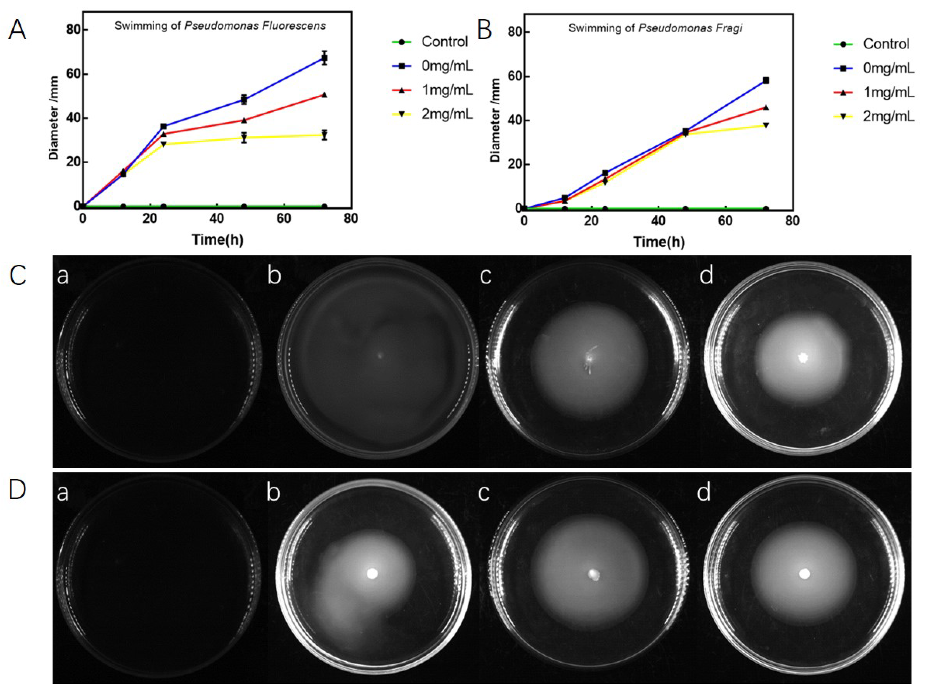
3.4. Antibacterial Effects on Swarming and Swimming of P. fluorescens and P. fragi

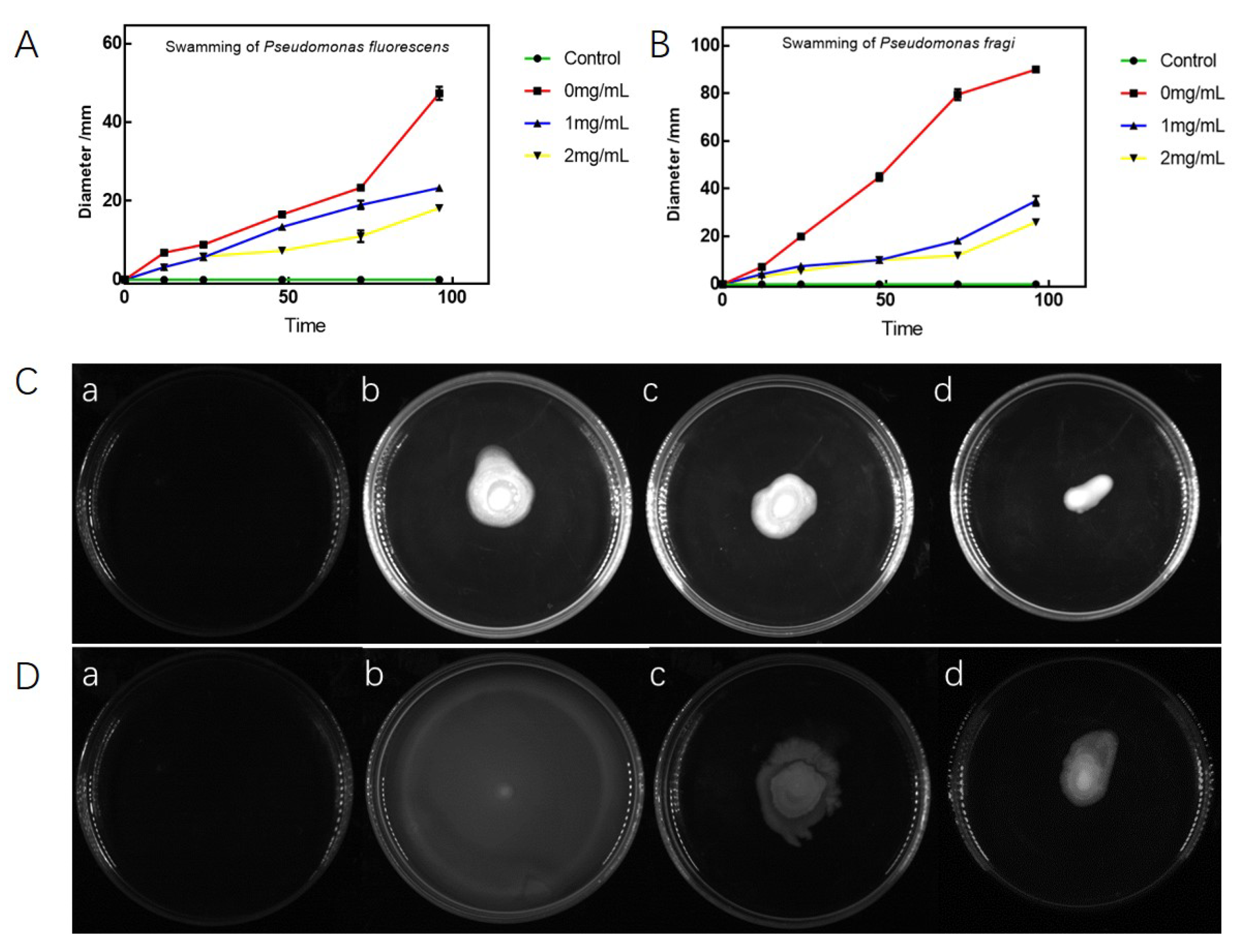
The abilities of swarming and swimming of the target Pseudomonas, i.e., P. fluorescens and P. fragi, via the treatment of the CFS of D. algida are shown in Figure 11 and Figure 12, respectively. By the treatment using 1 MIC CFS for 96 h, the capabilities of both swarming and swimming of P. fluorescens decrease obviously, where the migration diameter is fairly less than the one of the blank control (see Figure 11C and Figure 12C). Additionally, it can be observed that the inhibition effects of the CFS are in direct proportion to both treatment time and concentration. Hence, the CFS of D. algida has the property of inhibiting the motility of P. fluorescens.
For P. fragi, compared to the blank control, during the treatment process of 96 h, the swarming capability of P. fragi decreases significantly, which obtains the inhibition rates of 67.78% and 75.56% in the cases of using CFS with the concentration of 0.5 MIC and 1 MIC, respectively (see Figure 11D and Figure 12D), whereas the swimming capability of P. fragi is suppressed slightly via the CFS, as shown in Figure 12B. Thus, the CFS of D. algida can inhibit the motility of P. fragi by mainly suppressing its swarming capability.

3.5. Discussion

As common probiotics for food safety, various kinds of Lactobacillus and their applications have attracted extensive attention. The CFS of Lactobacillus contains the substances such as lactic acid, acetic acid, and protein, so that it can be utilized as an inhibitor for pathogenic bacteria and spoilage organisms, e.g., Streptococcus mutans [20], P. aeruginosa [36], Klebsiella pneumonia [37], Porphyromonas gingivalis [38], and Listeria monocytogenes [39], etc. In this paper, the target Pseudomonas, i.e., P. fragi and P. fluorescens, are psychrotrophic, which means they can live in cold environments, yielding the problem of the contamination of chilled meat and fresh milk. Hence, it is significant to find the inherent mechanism to inhibit the target pseudomonas using D. algida, a psychrotrophic Lactobacillus.
From Section 3.1, there exists a special acidoid, protein, or small peptide in D. algida which can inhibit the target psychrotrophic Pseudomonas, i.e., P. fragi and P. fluorescens, and it is sensitive to protease and temperature. Since the antibacterial effects are achieved by organic acid (lactic acid and acetic acid) [32], bacteriocin [40], or novel antibacterial molecules [41] that are different from any carboxylic acid in the existing studies about the investigation of inhibition mechanism of other Lactobacillus on Pseudomonas, it can be reasonably regarded that the antibacterial substance is one/some novel bacteriocin or organic acid. It is worth noting that, in this paper, the diameters of the inhibition zones of D. algida on P. fragi and P. fluorescens are 21.36 mm and 33.75 mm, respectively. Compared to the existing study [33] that obtains an inhibition zone with a diameter of 19 mm using Lactobacillus rhamnosus GG, larger inhibition zones are obtained by adopting D. algida, which demonstrates a better antibacterial capability of D. algida against the target psychrotrophic Pseudomonas. Although the antibacterial substance shall be further investigated in detail, D. algida can be widely applied by virtue of its following antibacterial effects and mechanisms.
(i): By the results in Section 3.2, the antibacterial effects of D. algida can be obtained in various conditions of pH and salinity, and it will not decrease by decomposing antibacterial substances as the culture time goes on. Since environments with various pH and salinity will be faced to keep the taste of food and the effects of drugs, D. algida is more applicable as compared to other Lactobacillus in the transportation and storage of foods or drugs, let alone in the low-temperature environments that are an inherent advantage of the psychrotrophic Lactobacillus. Hence, the application of D. algida shall be further developed for the food and drug industry as a novel inhibitor of P. fragi and P. fluorescens.
(ii): The CFS of D. algida can inhibit the biofilm of P. fragi by the elimination of the produced biofilm, instead of the suppression of the biofilm’s generation, whereas the biofilm of P. fragi can be suppressed in both of the aforementioned cases (see Section 3.3). Note that the reason for the situation that CFS cannot suppress or decrease the generation of the biofilm of P. fragi may be the existence of the disease-causing protein excreted during cold storage. Considering the results in [42], where P. fragi is inhibited in a sterilization way, one can have that there may exist a new sterilization mechanism of the CFS on P. fragi, which shall be further developed. Since the bacteria with biofilm is more drug-fast than the one without the biofilm [43], the suppression of the generation of the biofilm or the elimination of the produced one is significant to prevent the contamination of Pseudomonas. In the existing study [44], the inhibition rate of the spent culture fluid of Lactobacillus rhamnosus GG on the biofilm of P. aeruginosa is 32%, while the inhibition rates of the CFS of D. algida on the biofilm of P. fragi with the concentration of 1 mg/mL and 2 mg/mL are 69.37% and 83.33%, respectively. Hence, the CFS of D. algida obtains a better inhibition performance, which shows the possibility of contamination prevention via D. algida.
(iii): It is well known that the strong swimming and swarming abilities of the infected bacteria will lead to great financial loss, which should be avoided. Since the CFS of D. algida can suppress the swarming of P. fragi and both the swimming and swarming of P. fluorescens based on the results in Section 3.4, D. algida can be utilized as an effective inhibitor to psychrotrophic Pseudomonas, where it can be further developed based on the results we obtained.

4. Conclusions

In this paper, the antibacterial mechanisms of D. algida on two kinds of psychrotrophic Pseudomonas, including P. fragi and P. fluorescens, are investigated. Using the CFS of D. algida, the antibacterial substances are verified to be certain acidoids or small peptides produced by D. algida, which has strong inhibition stability in various pH and salinity. It is also found that D. algida suppresses P. fragi by reducing the swarming of P. fragi and generating holes on the produced biofilm, instead of inhibiting its generation, whereas D. algida can absolutely suppress P. fluorescens in the terms not only by inhibiting the growth and damaging the obtained biofilm, but also by alleviating the mobility, including both swarming and swimming. It can be seen that D. algida can be widely utilized as an inhibitor to psychrotrophic Pseudomonas in the food industry, while the antibacterial substance of D. algida can be further developed in detail via technologies of separation and purification.

Author Contributions

Conceptualization, Y.S. and S.L.; methodology, Y.S. and S.L.; software, Y.S. and Z.Z.; validation, Y.S. and T.W.; formal analysis, Y.S., T.M., and J.S. (Jie Sun); investigation, Y.S.; resources, J.S. (Jinzhu Song); data curation, J.S. (Jinzhu Song) and T.M.; writing—original draft preparation, Y.S.; writing—review and editing, Y.S.; visualization, Y.S.; supervision, J.S. (Jinzhu Song); project administration, J.S. (Jinzhu Song); funding acquisition, J.S (Jinzhu Song). All authors have read and agreed to the published version of the manuscript.

Funding

This research was funded by Heilongjiang Touyan Team under Grant HITTY-20190034.

Institutional Review Board Statement

Not applicable.

Informed Consent Statement

Not applicable.

Data Availability Statement

The data presented in this study are available on request from the corresponding author.

Conflicts of Interest

The authors declare no conflict of interest. All authors consent to publication of this manuscript.

References

  1. Ramos, A.N.; Cabral, M.E.; Noseda, D.; Bosch, A.; Yantorno, O.M.; Valdez, J.C. Antipathogenic properties of Lactobacillus plantarum on Pseudomonas aeruginosa: The potential use of its supernatants in the treatment of infected chronic wounds. Wound Repair Regen. 2012, 20, 552–562. [Google Scholar] [CrossRef] [PubMed]
  2. Jeyanathan, A.; Ramalhete, R.; Blunn, G.; Gibbs, H.; Pumilia, C.A.; Meckmongkol, T.; Lovejoy, J.; Coathup, M.J. Lactobacillus cell-free supernatant as a novel bioagent and biosurfactant against Pseudomonas aeruginosa in the prevention and treatment of orthopedic implant infection. J. Biomed. Mater. Res. Part B 2021, 109, 1634–1643. [Google Scholar] [CrossRef] [PubMed]
  3. Dambrauskienė, A.; Adukauskienė, D.; Jeroch, J.; Vitkauskienė, A. Pseudomonas aeruginosa bacteremia: Associations with a source of infection and antibiotic resistance. Med. Lith. 2009, 45, 1. [Google Scholar] [CrossRef] [Green Version]
  4. Lenzmeier, T.D.; Mudaliar, N.S.; Stanbro, J.A.; Watters, C.; Ahmad, A.; Simons, M.P.; Ventolini, G.; Zak, J.C.; Colmer, H.J.A.; Hamood, A.N. Application of Lactobacillus gasseri 63 AM supernatant to Pseudomonas aeruginosa-infected wounds prevents sepsis in murine models of thermal injury and dorsal excision. J. Med. Microbiol. 2019, 68, 1560–1572. [Google Scholar] [CrossRef]
  5. Ahmed, N.; Khalid, H.; Mushtaq, M.; Basha, S.; Rabaan, A.A.; Garout, M.; Halwani, M.A.; Al, M.A.; Alhumaid, S.; Al, A.Z.; et al. The molecular characterization of virulence determinants and antibiotic resistance patterns in human bacterial uropathogens. Antibiotics 2022, 11, 516. [Google Scholar] [CrossRef]
  6. Park, Y.; Koo, S.H. Epidemiology, molecular characteristics, and virulence factors of carbapenem-resistant Pseudomonas aeruginosa isolated from patients with urinary tract infections. Infect. Drug Resist. 2022, 15, 141–151. [Google Scholar] [CrossRef]
  7. Zhang, Z.C.; Wu, R.Y.; Gui, M.; Jiang, Z.J.; Li, P.L. Identification of the specific spoilage organism in farmed sturgeon (Acipenser baerii) fillets and its associated quality and flavour change during ice storage. Foods 2021, 10, 2021. [Google Scholar] [CrossRef]
  8. Bellassi, P.; Rocchetti, G.; Morelli, L.; Senizza, B.; Lucini, L.; Cappa, F. A milk foodomics investigation into the effect of Pseudomonas fluorescens growth under cold chain conditions. Foods 2021, 10, 1173. [Google Scholar] [CrossRef]
  9. Dunstall, G.; Rowe, M.T.; Wisdom, G.B.; Kilpatrick, D. Effect of quorum sensing agents on the growth kinetics of Pseudomonas spp. of raw milk origin. J. Dairy Res. 2005, 72, 276–280. [Google Scholar] [CrossRef]
  10. Chierici, M.; Picozzi, C.; LaSpina, M.G.; Orsi, C.; Vigentini, I.; Zambrini, V.; Foschino, R. Strain diversity of Pseudomonas fluorescens group with Potential blue pigment phenotype isolated from dairy products. J. Food Prot. 2016, 79, 1430–1435. [Google Scholar] [CrossRef]
  11. Wang, G.Y.; Wang, H.H.; Han, Y.W.; Xing, T.; Ye, K.P.; Xu, X.L.; Zhou, G.H. Evaluation of the spoilage potential of bacteria isolated from chilled chicken in vitro and in situ. Food Microbiol. 2017, 63, 139–146. [Google Scholar] [CrossRef] [PubMed]
  12. Almeida, K.M.D.; Bruzaroski, S.R.; Zanol, D.; Melo, M.D.; Santos, J.S.D.; Alegro, L.C.A.; Botaro, B.G.; Santana, E.H.W.D. Pseudomonas spp. and P. fluorescens: Population in refrigerated raw milk. Cienc. Rural 2017, 47, 2017. [Google Scholar] [CrossRef] [Green Version]
  13. Khailova, L.; Baird, C.H.; Rush, A.A.; McNamee, E.N.; Wischmeyer, P.E. Lactobacillus rhamnosus GG improves outcome in experimental pseudomonas aeruginosa pneumonia: Potential role of regulatory T cells. Shock 2013, 40, 496–503. [Google Scholar] [CrossRef] [PubMed] [Green Version]
  14. Duan, X.X.; Chen, S.Y.; Duan, S.; Lan, C.M.; Yang, Z.N.; Cao, Y.; Miao, J.Y. Antibiotic activities of the natural antimicrobial substance produced by Lactobacillus paracasei FX-6 against Pseudomonas putida. LWT-Food Sci. Technol. 2020, 123, 109096. [Google Scholar] [CrossRef]
  15. Duan, X.X.; Duan, S.; Wang, Q.E.; Ji, R.; Cao, Y.; Miao, J.Y. Effects of the natural antimicrobial substance from Lactobacillus paracasei FX-6 on shelf life and microbial composition in chicken breast during refrigerated storage. Food Control 2020, 109, 106906. [Google Scholar] [CrossRef]
  16. Forestier, C.; Guelon, D.; Cluytens, V.; Gillart, T.; Sirot, J.; DeChamps, C. Oral probiotic and prevention of Pseudomonas aeruginosa infections: A randomized, double-blind, placebo-controlled pilot study in intensive care unit patients. Crit. Care 2008, 12, R69. [Google Scholar] [CrossRef] [Green Version]
  17. Alexandre, Y.; LeBerre, R.; Barbier, G.; LeBlay, G. Screening of Lactobacillus spp. for the prevention of Pseudomonas aeruginosa pulmonary infections. BMC Microbiol. 2014, 14, 107. [Google Scholar] [CrossRef] [Green Version]
  18. Valdez, J.C.; Peral, M.C.; Rachid, M.; Santana, M.; Perdigon, G. Interference of Lactobacillus plantarum with Pseudomonas aeruginosa in vitro and in infected burns: The potential use of probiotics in wound treatment. Clin. Microbiol. Infect. 2005, 11, 472–479. [Google Scholar] [CrossRef] [Green Version]
  19. Scillato, M.; Spitale, A.; Mongelli, G.; Privitera, G.F.; Mangano, K.; Cianci, A.; Stefani, S.; Santagati, M. Antimicrobial properties of Lactobacillus cell-free supernatants against multidrug-resistant urogenital pathogens. MicrobiologyOpen 2021, 10, e1173. [Google Scholar] [CrossRef]
  20. Wasfi, R.; AbdElRahman, O.A.; Zafer, M.M.; Ashour, H.M. Probiotic Lactobacillus sp. inhibit growth, biofilm formation and gene expression of caries-inducing Streptococcus mutans. J. Cell. Mol. Med. 2018, 22, 1972–1983. [Google Scholar] [CrossRef] [Green Version]
  21. Kato, Y.; Sakala, R.M.; Hayashidani, H.; Kiuchi, A.; Kaneuchi, C.; Ogawa, M. Lactobacillus algidus sp. nov., a psychrophilic lactic acid bacterium isolated from vacuum-packaged refrigerated beef. Int. J. Syst. Evol. Microbiol. 2000, 50, 1143–1149. [Google Scholar] [CrossRef] [PubMed] [Green Version]
  22. Zheng, J.S.; Wittouck, S.; Salvetti, E.; Franz, C.M.; Harris, H.; Mattarelli, P.; OToole, P.W.; Pot, B.; Vandamme, P.; Walter, J.; et al. A taxonomic note on the genus Lactobacillus: Description of 23 novel genera, emended description of the genus Lactobacillus Beijerinck 1901, and union of Lactobacillaceae and Leuconostocaceae. Int. J. Syst. Evol. Microbiol. 2020, 70, 2782–2858. [Google Scholar] [CrossRef] [PubMed]
  23. Sun, Z.H.; Harris, H.; McCann, A.; Guo, C.Y.; Argimón, S.; Zhang, W.Y.; Yang, X.W.; Jeffery, I.B.; Cooney, J.C.; Kagawa, T.F.; et al. Expanding the biotechnology potential of lactobacilli through comparative genomics of 213 strains and associated genera. Nat. Commun. 2015, 6, 8322. [Google Scholar] [CrossRef] [PubMed]
  24. Poirier, S.; Coeuret, G.; Champomier, V.M.C.; Chaillou, S. Draft genome sequences of nine strains of Brochothrix thermosphacta, Carnobacterium divergens, Lactobacillus algidus, Lactobacillus fuchuensis, Lactococcus piscium, Leuconostoc gelidum subsp. gasicomitatum, Pseudomonas lundensis, and Weissella viridescens, a collection of Psychrotrophic species involved in meat and seafood spoilage. Genome Announc. 2018, 6, e00479-18. [Google Scholar]
  25. Säde, E.; Johansson, P.; Heinonen, T.; Hultman, J.; Björkroth, J. Growth and metabolic characteristics of fastidious meat-derived Lactobacillus algidus strains. Int. J. Food Microbiol. 2020, 313, 108379. [Google Scholar] [CrossRef]
  26. Okaro, U.; Green, R.; Mohapatra, S.; Anderson, B. The trimeric autotransporter adhesin BadA is required for in vitro biofilm formation by Bartonella henselae. NPJ Biofilms Microbiomes. 2019, 5, 10. [Google Scholar] [CrossRef] [Green Version]
  27. Matsuo, S.; Wada, K.; Sadahira, T.; Mitsuhata, R.; Nasu, Y. Impact of Lactobacillus probiotics on biofilm formed by Pseudomonas aeruginosa. Eur. Urol. Suppl. 2019, 18, e548. [Google Scholar] [CrossRef]
  28. Wickramasinghe, N.N.; Hlaing, M.M.; Ravensdale, J.T.; Coorey, R.; Chandry, P.S.; Dykes, G.A. Characterization of the biofilm matrix composition of psychrotrophic, meat spoilage pseudomonads. Sci. Rep. 2020, 10, 16457. [Google Scholar] [CrossRef]
  29. Hossain, M.I.; Mizan, M.F.R.; Roy, P.K.; Nahar, S.; Toushik, S.H.; Ashrafudoulla, M.; Jahid, I.K.; Lee, J.; Ha, S.D. Listeria monocytogenes biofilm inhibition on food contact surfaces by application of postbiotics from Lactobacillus curvatus B.67 and Lactobacillus plantarum M.2. Food Res. Int. 2021, 148, 110595. [Google Scholar] [CrossRef]
  30. Wickramasinghe, N.N.; Ravensdale, J.; Coorey, R.; Dykes, G.A.; Chandry, P.S. Transcriptional profiling of biofilms formed on chilled beef by psychrotrophic meat spoilage bacterium, Pseudomonas fragi 1793. Biofilm 2021, 3, 100045. [Google Scholar] [CrossRef]
  31. Cui, T.Q.; Bai, F.L.; Sun, M.T.; Lv, X.R.; Li, X.P.; Zhang, D.F.; Du, H. Lactobacillus crustorum ZHG 2-1 as novel quorum-quenching bacteria reducing virulence factors and biofilms formation of Pseudomonas aeruginosa. LWT-Food Sci. Technol. 2020, 117, 108696. [Google Scholar] [CrossRef]
  32. Layus, B.I.; Gerez, C.L.; Rodriguez, A.V. Antibacterial activity of Lactobacillus plantarum CRL 759 against methicillin-resistant Staphylococcus aureus and Pseudomonas aeruginosa. Arab. J. Sci. Eng. 2020, 45, 4503–4510. [Google Scholar] [CrossRef]
  33. deAlcântara, A.L.D.A.; Bruzaroski, S.R.; Luiz, L.L.; de Souza, C.H.B.; Poli-Frederico, R.C.; Fagnani, R.; deSantana, E.H.W. Antimicrobial activity of Lactobacillus rhamnosus against Pseudomonas fluorescens and Pseudomonas putida from raw milk. J. Food Process. Preserv. 2019, 43, e14082. [Google Scholar]
  34. Dai, M.; Li, Y.; Xu, L.; Wu, D.; Zhou, Q.; Li, P.; Gu, Q. A novel bacteriocin from Lactobacillus pentosus ZFM94 and its antibacterial mode of action. Front. Nutr. 2021, 8, 710862. [Google Scholar] [CrossRef] [PubMed]
  35. Lv, X.; Ma, H.; Sun, M.; Lin, Y.; Bai, F.; Li, J.; Zhang, B. A novel bacteriocin DY4-2 produced by Lactobacillus plantarum from cutlassfish and its application as bio-preservative for the control of Pseudomonas fluorescens in fresh turbot (Scophthalmus maximus) fillets. Food Control 2018, 89, 22–31. [Google Scholar] [CrossRef]
  36. Aman, M.; Aneeqha, N.; Bristi, K.; Deeksha, J.; Afza, N.; Sindhuja, V.; Shastry, R.P. Lactic acid bacteria inhibits quorum sensing and biofilm formation of Pseudomonas aeruginosa strain JUPG01 isolated from rancid butter. Biocatal. Agric. Biotechnol. 2021, 36, 102115. [Google Scholar] [CrossRef]
  37. El-Mokhtar, M.A.; Hassanein, K.M.; Ahmed, A.S.; Gad, G.F.M.; Amin, M.M.; Hassanein, O.F.E. Antagonistic activities of cell-free supernatants of lactobacilli against extended-spectrum β-lactamase producing Klebsiella pneumoniae and Pseudomonas aeruginosa. Infect. Drug Resist. 2020, 13, 543. [Google Scholar] [CrossRef] [Green Version]
  38. Yang, K.M.; Kim, J.S.; Kim, H.S.; Kim, Y.Y.; Oh, J.K.; Jung, H.W.; Park, D.S.; Bae, K.H. Lactobacillus reuteri AN417 cell-free culture supernatant as a novel antibacterial agent targeting oral pathogenic bacteria. Sci. Rep. 2021, 11, 1631. [Google Scholar] [CrossRef]
  39. Liu, Y.; Bu, Y.; Li, J.; Liu, Y.; Liu, A.; Gong, P.; Liu, T.; Zhang, L.; Wang, S.; Yi, H. Inhibition activity of plantaricin Q7 produced by Lactobacillus plantarum Q7 against Listeria monocytogenes and its biofilm. Fermentation. 2022, 8, 75. [Google Scholar] [CrossRef]
  40. Zhu, Y.Y.; Zhou, Q.Q.; Li, P.; Gu, Q. Purification, characterization, and mode of action of Paracin 54, a novel bacteriocin against Staphylococci. Appl. Microbiol. Biotechnol. 2021, 105, 6735–6748. [Google Scholar] [CrossRef]
  41. Marques, V.D.; Franzolin, M.R.; Sanabani, S.S.; Vigerelli, H.; Piazza, R.M.F.; Pimenta, D.C.; Venâncio, T.; Neves, I.V.; deSousa, S.H.G.; dosSantos, C.D.; et al. A new class of antimicrobial molecules derived from kefir, effective againstPseudomonas aeruginosaand methicillin resistantStaphylococcus aureus (MRSA) strains. Sci. Rep. 2020, 10, 17434. [Google Scholar] [CrossRef] [PubMed]
  42. Chappell, T.C.; Nair, N.U. Engineered lactobacilli display anti-biofilm and growth suppressing activities against Pseudomonas aeruginosa. npj Biofilms Microbomes. 2020, 6, 48. [Google Scholar] [CrossRef] [PubMed]
  43. Vuotto, C.; Longo, F.; Donelli, G. Probiotics to counteract biofilm-associated infections: Promising and conflicting data. Int. J. Oral Sci. 2014, 6, 189–194. [Google Scholar] [CrossRef] [PubMed] [Green Version]
  44. Alhubail, M.; O’Neill, C.A.; Mcbain, A.; Cruickshank, S. The spent culture fluid of Bifidobacterium longum, Lactobacillus rhamnosus GG and Escherichia coli Nissle 1917 inhibits the growth and biofilm formation of Pseudomonas aeruginosa. J. Investig. Dermatol. 2019, 139, S329. [Google Scholar] [CrossRef] [Green Version]
Figure 1. Antibacterial effects of D. algida against the target Pseudomonas, where (A,B) are the diameter and appearance, respectively, of the produced inhibition zone of P. fluorescens, and (C,D) are the diameter and appearance, respectively, of the produced inhibition zone of P. fragi. The data are shown as mean ± SD (n = 3).
Fermentation 08 00298 g001
Figure 2. Antibacterial effects of lactic acid against the target Pseudomonas, where (A) shows the diameter of the inhibition zone, and (B,C) are the appearence of the inhibition zones of P. fluorescens and P. fragi, respectively. The data are shown as mean ± SD (n = 3), *** p < 0.001.
Fermentation 08 00298 g002
Figure 3. Antibacterial effects of D. algida via the treatment at different temperatures against the target Pseudomonas, where (A,B) are P. fluorescens and P. fragi, respectively. The data are shown as mean ± SD (n = 3), ** p < 0.01, *** p < 0.001.
Fermentation 08 00298 g003
Figure 4. Antibacterial effects of D. algida when the used CFS are with different concentrations against the target Pseudomonas, where (A,B) are P. fluorescens and P. fragi, respectively. The data are shown as mean ± SD (n = 3).
Fermentation 08 00298 g004
Figure 5. Antibacterial effects of D. algida under different culture times against the target Pseudomonas, where (A,B) and (C,D) are P. fluorescens and P. fragi, respectively. The numbers in (B,D) stand for the culture time. The data are shown as mean ± SD (n = 3), ** p < 0.01, *** p < 0.001.
Fermentation 08 00298 g005
Figure 6. Antibacterial effects under different salinities of D. algida on the target Pseudomonas, where (A,B) are the diameter and appearance, respectively, of the produced inhibition zone of P. fluorescens, and (C,D) are the diameter and appearance, respectively, of the produced inhibition zone of P. fragi. The numbers in (B,D) stand for the salinities. The data are shown as mean ± SD (n = 3), *** p < 0.001.
Fermentation 08 00298 g006
Figure 7. Antibacterial . effects under different pH of D. algida against the target Pseudomonas, where (A,B) are the diameter and appearance, respectively, of the produced inhibition zone of P. fluorescens, and (C,D) are the diameter and appearance, respectively, of the produced inhibition zone of P. fragi. The numbers in (B) and (D) stand for the pH. The data are shown as mean ± SD (n = 3), * p < 0.05, ** p < 0.01, *** p < 0.001.
Fermentation 08 00298 g007
Figure 8. Antibacterial effects of D. algida on the biofilm during the development stage of the target Pseudomonas, where (A,B) are the absorbance values of P. fluorescens and P. fragi, respectively, under different MIC. The data are shown as mean ± SD (n = 3).
Fermentation 08 00298 g008
Figure 9. Antibacterial effects of D. algida on the generated biofilm of the target Pseudomonas, where (A,B) are the absorbance values of P. fluorescens and P. fragi, respectively, under different MIC. The data are shown as mean ± SD (n = 3), *** p < 0.001.
Fermentation 08 00298 g009
Figure 10. Antibacterial effects D. algida on biofilm appearance observed by SEM (40,000× magnification) of the target Pseudomonas, where (AD) are the blank control, 0.5 MIC, 1 MIC, 2 MIC, respectively, of P. fluorescens, while (EH) are the blank control, 0.5 MIC, 1 MIC, 2 MIC, respectively, of P. fragi.
Fermentation 08 00298 g010
Figure 11. Antibacterial effects of D. algida on the swarming of the target Pseudomonas, where (A,C) are the diameter and appearance, respectively, of the inhibition zone of P. fluorescens, while (B,D) are the diameter and appearance, respectively, of the inhibition zone of P. fragi. The symbols (a–d) in the subfigures (C,D) represent control group, 0 MIC, 0.5 MIC, and 1 MIC, respectively. The data are shown as mean ± SD (n = 3).
Fermentation 08 00298 g011
Figure 12. Antibacterial effects of D. algida on the swimming of the target Pseudomonas, where (A,C) are the diameter and appearance, respectively, of the inhibition zone of P. fluorescens, while (B,D) are the diameter and appearance, respectively, of the inhibition zone of P. fragi. The symbols (a–d) in the subfigures (C,D) represent the control group, 0 MIC, 0.5 MIC, and 1 MIC, respectively. The data are shown as mean ± SD (n = 3).
Fermentation 08 00298 g012
Table 1. The de Mann Rogosa Sharpe (MRS) medium components.
CompositionContentCompositionContent
Peptone10.00 gTriammonium citrate2.00 g
Beef paste10.00 gDipotassium hydrogen phosphate2.00 g
Yeast powder4.00 gManganese sulfate0.04 g
Glucose20.00 gTween-801.00 g
Magnesium sulfate0.20 gWater1.00 L
Sodium acetate5.00 gpH5.70 ± 0.20
Table 2. Protease validation experiment.
GroupP. fluorescensP. fragi
ControlTreatmentControlTreatment
Proteinase K22.625 ± 0.072 *17.703 ± 0.39522.355 ± 0.757 *10.625 ± 0.022
Trypsin22.694 ± 1.62021.045 ± 0.07223.697 ± 0.985 *17.634 ± 0.548
Papain21.965 ±0.72120.926 ± 0.37125.021 ± 0.003 *16.583 ± 0.021
α -Amylase21.683 ± 0.07621.633 ± 0.01625.470 ± 0.36825.750 ± 0.013
Lipase21.866 ± 0.06121.454 ± 0.04524.863 ± 0.69724.637 ± 0.036
Catalase22.701 ± 0.00322.407 ± 0.00426.131 ± 0.05125.189 ± 0.095
The control and treatment denotes the blank control group and the group with the treatment of enzymes, where the data was shown as mean ± SD (n = 3), and * stands for the difference between control and treatment (p < 0.01).
Publisher’s Note: MDPI stays neutral with regard to jurisdictional claims in published maps and institutional affiliations.