Article

Physicochemical, Functional, and Technological Properties of Protein Hydrolysates Obtained by Microbial Fermentation of Broiler Chicken Gizzards

1
Department of Food and Biotechnology, South Ural State University (National Research University), 76 Lenin Avenue, 454080 Chelyabinsk, Russia
2
Department of Scientific Research, V. M. Gorbatov Federal Research Center for Food Systems, 26 Talalikhina St., 109316 Moscow, Russia
3
Nanotechnology Research & Education Centre, South Ural State University (National Research University), 76 Lenin Avenue, 454080 Chelyabinsk, Russia
4
Department of Scientific Research, K. G. Razumovsky Moscow State University of Technologies and Management (The First Cossack University), 73 Zemlyanoy Val, 109004 Moscow, Russia
5
Center for Biotechnology of Animal Reproduction, South Ural State Agrarian University, 13 Gagarina St., Troitsk, 457100 Chelyabinsk, Russia
*
Author to whom correspondence should be addressed.
Fermentation 2022, 8(7), 317; https://doi.org/10.3390/fermentation8070317
Submission received: 2 June 2022 / Revised: 30 June 2022 / Accepted: 4 July 2022 / Published: 6 July 2022
(This article belongs to the Special Issue Assessment of the Quality and Safety of Fermented Foods)

Abstract

:
Fermentation is an economical method for obtaining protein hydrolysates. The purpose of the scientific research was to perform a comprehensive analysis of the physicochemical, technological, and functional properties of protein hydrolysates obtained by microbial fermentation. The research results showed that hydrolysates fermented with propionic acid bacteria and bifidobacteria have better physicochemical and technological indicators compared to the control sample. Significant increases in water-holding and fat-holding capacities (by 1.8–2.1 times and 1.5–2.5 times, respectively), as well as fat-emulsifying ability (by 12.8–29.8%) in experimental samples were found. Hydrolysates obtained by fermentation effectively inhibit the growth of Escherichia coli and Staphylococcus aureus. The thermal analysis showed a sufficiently high-thermal stability of the obtained protein hydrolysates. In hydrolysates fermented by bacterial culture, the removal of physico-mechanical and osmotically bound moisture occurred at temperatures of 110 °C and 115 °C, respectively, and in whey protein hydrolysate at a temperature of 100 °C. The release of chemically bound moisture was observed at a temperature of 170 °C for fermented hydrolysates and at 155 °C for the control sample. The results proved that fermented protein products are characterized by high functional properties, antioxidant and antimicrobial activity, and can be used as natural food additives and preservatives.

1. Introduction

Innovative biotechnological developments allow for the creation of ingredients with high added value from by-products formed during the slaughter and processing of animals [1,2,3]. Most slaughter products obtained are high in collagen. Collagen-containing raw materials are characterized by a stable, resistant structure and require complex multi-stage biotechnological processing. In the process of such treatment, mainly enzymatic protein hydrolysates are obtained from meat raw materials and offal [4]. Many researchers have noted the potential of chicken by-products as substrates for the production of protein hydrolysates with various functional properties and biological activity [5,6,7]. Protein hydrolysates are obtained in different ways, but the most economical and environmentally friendly is the enzymatic hydrolysis of protein raw materials, entailing the use of enzymes to break down molecules and change the chemical, functional, and sensory properties of proteins without reducing their nutritional value. Poultry by-products, such as gizzards and combs, are also promising for the production of protein hydrolysates by means of enzymatic hydrolysis [8,9]. These by-products contain muscle, fat, and connective tissues, which are differentially modified by the action of microbial enzyme systems [10].
Protein hydrolysates are complex and multicomponent systems consisting of several fractions: amino acids and lower peptides in solution, polypeptide aggregates, and colloidal nanoparticles and insoluble microparticles [11]. According to the degree of hydrolysis, we can differentiate partial hydrolysates containing long peptides and a minimum amount of free amino acids and deep hydrolysates represented by short chain peptides and free amino acids [12,13]. Depending on the degree of hydrolysis, the functional and technological indicators of the resulting hydrolysates vary significantly, which allows for the regulation of the depth of hydrolysis depending on the subsequent direction of use.
Fermentation can be carried out both with the use of enzyme preparations and enzyme systems of microorganisms that produce directly in the substrate, not only multidirectional enzymes but also other organic substances that contribute to the biotransformation of the substrate composition. Thus, lactic and propionic acid bacteria can be used for microbial fermentation. Proteolytic enzymes, as well as lactic and propionic acids, which accumulate during the metabolism of propionic acid bacteria and lactic acid bacteria, play a decisive role in changing the structure of by-products during microbial fermentation [14]. The processing of meat raw materials with organic acids has a pronounced effect on muscle fibers and connective tissue and increases tenderness [15].
Scientific literature notes the prospects for the use of fermentation, including microbial, and the resulting hydrolysis products to increase the shelf life of food systems [16,17,18,19]. Fermentation products include protein hydrolysates and biologically active peptides, which can be used as natural food preservatives as a result of their high antioxidant and antimicrobial activity. Their introduction into food systems can improve the storage capacity of food and reduce the risk of biological hazard [20,21,22]. Protein hydrolysates containing antioxidant and antimicrobial peptides can be used as biologically active ingredients in foods to provide health benefits and extend shelf life [23]. In addition, the prospects of including hydrolysates in the composition of packaging materials as active ingredients have been proven [24].
The functional and technological properties of protein hydrolysates are of great importance, such as water-holding and fat-holding capacities, foaming and emulsifying abilities, solubility, and viscosity. These properties depend on the structure and degree of hydrolysis of the protein product, and they are involved in the formation of the required parameters of the food systems [9]. The technological characteristics of hydrolysates are improved as a result of protein splitting into fragments with a lower molecular weight [25].
Based on the foregoing, the purpose of the scientific research was to perform a comprehensive analysis of the physicochemical, technological, antimicrobial, and antioxidant properties of protein hydrolysates obtained by microbial fermentation of the broiler chicken gizzards.

2. Materials and Methods

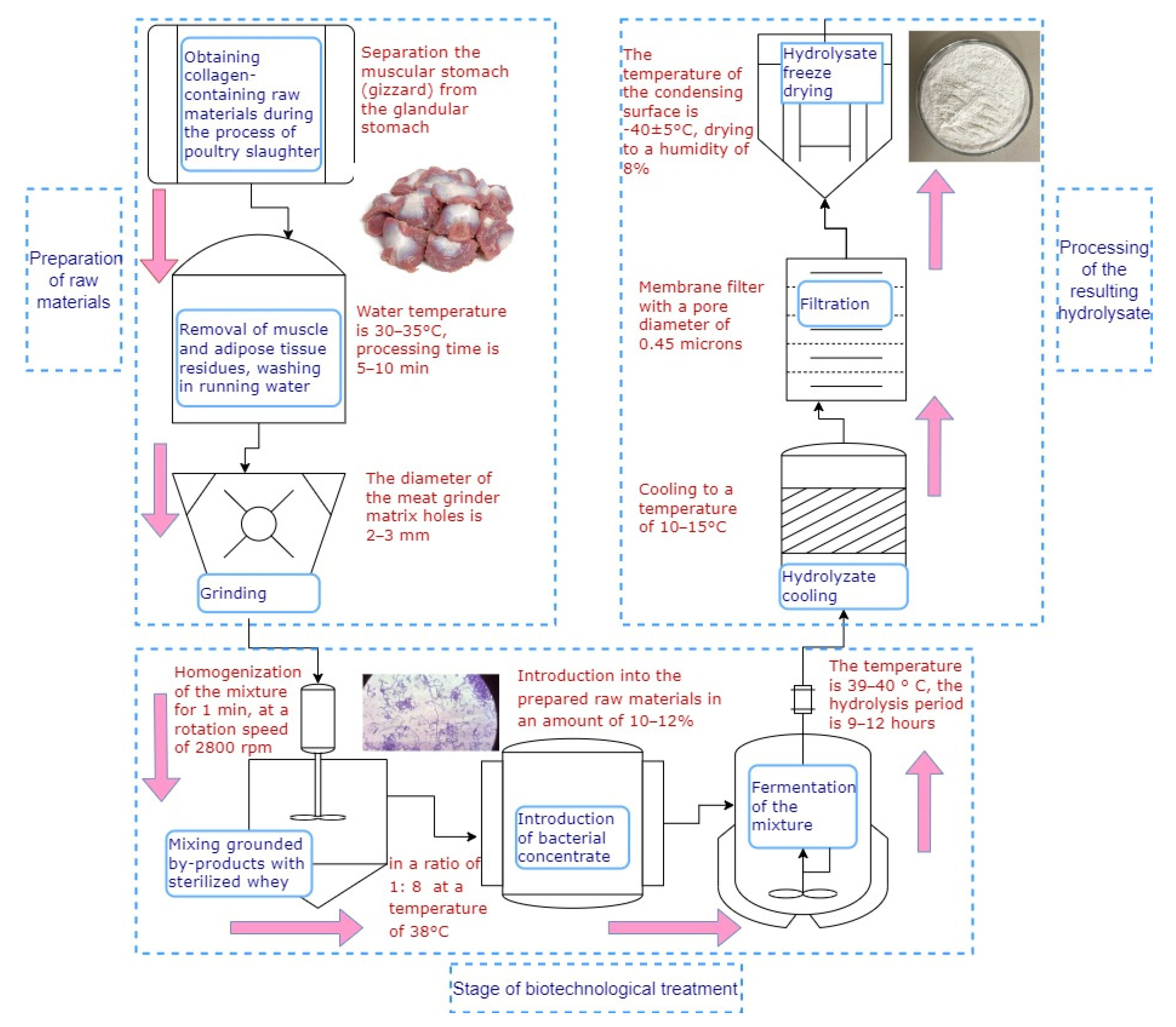
2.1. Raw Materials and Ingredients

The raw material used to obtain the hydrolysates was the muscle stomachs obtained by slaughtering broiler chickens (ROSS 308) at the age of 41 days at a poultry farm (Chelyabinsk region). The muscular stomach (gizzard) was separated from the glandular stomach, washed, packaged in plastic bags, and frozen at −18 °C. Frozen gizzards were transported in a refrigerator to the laboratory of the Food and Biotechnology Department within 2 h for further processing. The whey was collected after the production of soft cheese, it was transported in a refrigerated polymer barrel to the laboratory, and it was stored in the refrigerator at a temperature of 3 ± 1 °C for no more than 24 h before use. The whey was used with bacterial concentrates for the treatment of the gizzards.
The biotechnological processing of the experimental samples was performed in curd whey with the addition of microorganism cultures: Bifidobacteria liquid concentrate (BLC); and Propionix liquid concentrated starter culture (Propionix LCSC) manufactured by Propionix (Moscow, Russia).
BLC is a concentrated microbial mass of the Bifidobacterium longum B379M strain with an activity of 1011–1012 CFU/cm3. Propionix LCSC is a concentrated microbial mass of the Propionibacterium freudenreichii subsp. strain shermanii-KM 186, the bacteria of which are in the active form—1010–1011 CFU/cm3. The manufacturer recommends storing direct-introduced starters at a temperature of −18 °C for six months.

2.2. Preparation of Protein Hydrolysate

Based on the results of previous studies and the optimal parameters of hydrolysis [26], the protein hydrolysate from the broiler chicken gizzards was obtained by means of microbial fermentation based on the technological scheme presented in Figure 1.
The key stages of the production process were washing, grinding, and degreasing the stomachs, the actual stage of fermenting with a bacterial concentrate, and the final processing of the resulting protein component. Fermentation was carried out under the following parameters determined in previous studies: temperature, 39–40 °C; hydrolysis time, 9–12 h; concentration of the bacterial concentrate in the substrate, 10–12% [26]. The degree of hydrolysis was 63.9% when treated with Propionix LCSC and 56.4% when treated with BLC. The control sample was hydrolyzed in serum without the addition of bacteria. The liquid hydrolysate obtained after the fermentation was dried and ground to a powder state. The resulting protein hydrolysate was a fine white powder without taste, with a neutral odor. Its physicochemical, technological, antioxidant, and antimicrobial properties were subjected to a comprehensive study.

2.3. Determination of Proximate Composition

The determination of the chemical composition of hydrolysates was conducted in five repetitions according to the methods described by AOAC (1995). The total nitrogen content was assayed by the Kjeldahl method with nitrogen converted to equivalent protein content using a factor of 6.25 (Methods 992.15 and 992.23). Moisture was determined according to Method 950.46 B. Total fat was determined via the Soxhlet method (Methods 920.39 C and 960.39). The ash content was determined via the dry-ashing method (Method 920.153).

2.4. Determination of Functional and Technological Properties

The fat-holding capacity (FHC) was determined as follows. A 5 g portion of the hydrolysate was placed in a weighed graduated tube, and 30 cm3 of refined sunflower oil with a density of 0.63 g/cm3 was added and mixed in a homogenizer for 1 min at a speed of 1800 rpm. After 30-min exposure, the tubes were centrifuged for 15 min at a speed of 4000 rpm. To determine the FHC by the weight method (g/g), the test tube was weighed with protein and oil. After the removal of non-adsorbed oil, the residue in the test tube was weighed. To determine the FHC by the volumetric method (%), the total volume of the mixture in the test tube and the volume of the oil remaining unadsorbed were measured. FHC was calculated by means of the formulas:
FHC, g/g of fat:
F H C = ( A B ) / C
where A is the mass of the tube with hydrolysate and oil, g;
B—the mass of the tube with hydrolysate and adsorbed oil, g;
C—suspension of hydrolysate, g.
FHC, %:
F H C = ( 30 V ) × ρ   C × 100
where V is the volume of oil remaining unadsorbed, cm3;
ρ—is the relative density of the oil, g/cm3.
The water-holding capacity (WHC) was determined similarly by adding water to the hydrolysate instead of oil.
The fat-emulsifying ability (FEA) (%) was determined as follows. A portion of hydrolysate of 7 g was mixed in a blender with 100 cm3 of distilled water for 1 min. Then 100 cm3 of refined sunflower oil was added and emulsified in a blender for 5 min. The resulting emulsion was centrifuged for 5 min at 4000 rpm. FEA calculated by means of the formula:
F E A = ( V e / V o   ) × 100
where Ve is the volume of the emulsified layer, cm3;
Vo is the total volume of the mixture, cm3.
The stability of the emulsion (SE) was determined by heating the emulsion at a temperature of 80 °C for 30 min and then cooling with water for 15 min. Then, the tubes with the emulsion were centrifuged at a speed of 4000 rpm for 5 min. Next, the volume of the emulsified layer was determined, and the SE (%) was calculated by means of the formula:
S E = ( V 1 / V 2   ) × 100
where V1 is the volume of emulsified oil, mL;
V2 total volume of the emulsion, mL.
The foaming capacity (FC) was determined as follows. A portion of the hydrolysate weighing 7 g was ground to a homogeneous suspension with 25 cm3 of distilled water. The suspension was quantitatively transferred to a measuring cylinder, and the liquid volume was adjusted to 300 cm3. The FC was calculated by means of the formula:
F C = ( H f / H l   ) × 100
where Hf is the height of the foam layer, mm;
Hl is the initial height of the liquid, mm.
The resistance of the foam (FR) was determined after exposure for 15 min. The height of the remaining foam was measured, and FR was calculated by means of the following formula:
F R = ( H e / H f   ) × 100
where He is the height of the foam layer after exposure, mm.
To determine the solubility (S), a portion of hydrolysate weighing 10 g was dissolved in 100 cm3 of distilled water heated in a thermostat to 24 °C and stirred in a laboratory mixer for 90 s. Then, the solution was transferred to test tubes and centrifuged for 5 min at 4000 rpm. The top layer of liquid was decanted and the mass of the test tube with precipitate was measured. S was calculated by means of the following formula:
S = 100 [ M s M o m × 100 % ]
where Ms is the mass of the solution in the test tube, g;
Mo is the mass of the precipitate in the test tube, g;
m is the mass of the hydrolysate sample, g.

2.5. Determination of Antioxidant Activity

The ability of protein hydrolysates to remove DPPH free radicals was determined in accordance with the method described in [27]. To carry out the reaction, 20 cm3 of methyl alcohol was added to 0.5 g of the sample. The extraction was carried out with stirring at a speed of 150 rpm for 6 h at 20 °C. An amount of 1 mL of the extract was mixed with 1 cm3 of DPPH solution in methanol and incubated in the dark for 30 min. Then, the optical density was measured at 515 nm on a Jenway 6404 UV/Vis spectrophotometer (Great Britain). The total antioxidant activity (AOA) was calculated by means of the formula:
A O A = [ A k A i A k × 100 % ]
where Ak is the optical density value for the control sample;
Ai is the optical density value for the test sample.

2.6. Determination of Microbiological Indicators

Studies of microbiological parameters of hydrolysates were carried out using microbiological rapid tests Petritest™ (Research and Production Association “Alternativa”, Russia). A total of 10 g of hydrolysates were taken to prepare initial dilutions. From the initial dilution of the hydrolyzate, a series of tenfold dilution was prepared in accordance with the data of TR CU 021/2011. Saline solution was used for dilution.
To determine the total viable counts (TVC) of the hydrolyzate, Petritest™ was used. To identify and determine the total coliforms count (TCC) content in 1 g, PetritestTM was used, which contains an indicator for staining colonies of enterobacteria in red. The study was carried out as follows. A total of 0.2 cm3 of the dilution was added to the open surface of the substrate in Petritest™, the lid was then closed with latches, and the test liquid was distributed evenly over the surface of the nutrient medium. Petritests were placed in a thermostat and incubated at a temperature of (36 ± 1) °C for (12–24) h. Then, the number of colonies was counted, selecting for counting Petritests on which 15 to 300 colonies were grown. The result was multiplied by the value of the corresponding dilution, and the total viable counts or the total coliforms count in 0.2 cm3 of the sample was obtained. The results were multiplied by five to bring the results to 1 cm3 according to the manufacturer’s recommendations.
To determine yeasts and molds, the study was carried out similarly to those described above; the tests were incubated at a temperature of (24 ± 1) °C for 24 h (for preliminary accounting) and 120 h (for final accounting). Then, the numbers of colonies of yeasts and molds were counted separately. For quantitative calculation, Petritests were used on which 15 to 150 yeast colonies and 5 to 50 mold colonies were grown. The result was multiplied by the value of the corresponding dilution, and the number of yeasts or molds per 0.2 cm3 (g) of the sample was obtained. The result was multiplied by five to bring the results to 1 cm3.

2.7. Determination of Antimicrobial Activity

Antimicrobial properties were evaluated by measuring the growth inhibition zone of Escherichia coli and Staphylococcus aureus cultures under the action of discs moistened with 1% hydrolysate solutions. To obtain pure cultures, BD Microtrol discs were used with a pure culture of Escherichia coli ATCC 8739 microorganisms and with a pure culture of Staphylococcus aureus ATCC 6538 microorganisms.
To determine the antibacterial properties of hydrolysates, their 1% solution was pre-pared as follows: 1 g of powdered hydrolysate was poured into 100 mL of distilled water and stirred until the hydrolysate was completely dissolved. Then, paper discs 10 mm in diameter were moistened in a dispersed solution of the hydrolysates and were placed on the surface of Petri dishes with meat-peptone agar on which Escherichia coli/Staphylococcus aureus culture was applied with a spatula.
Dishes with samples were placed in a thermostat and cultivated at a temperature of 37 °C for 48–72 h.
When the growth of the Escherichia coli/Staphylococcus aureus culture appeared on the surface of the Petri dishes, the width of the zone of inhibition of the development of microorganisms by hydrolysate solutions was fixed.

2.8. Differential-Scanning Calorimetry (DSC) and Thermal Gravimetric Analysis

Differential-scanning calorimetry (DSC) and thermal gravimetric analysis (TGA) of hydrolysates were performed using a simultaneous thermal analyzer (TG-DSC), Netzsch STA 449F1 “Jupiter” (Selb, Germany), in corundum crucibles at heating from 30 °C to 300 °C. The heating rate was 10 °C per minute. The measurements were carried out in an argon medium. Using an analog-to-digital converter, curves were obtained in digital form. Before analysis, the hydrolysates were dissolved in water at a ratio of 1:2. As a comparison of the technological properties of the hydrolysates with other protein supplements, whey protein hydrolyzate (HWP) (Optipep, Ballineen, Ireland) was used, which was also hydrated.

2.9. Analysis of Average Particle Size

Determination of the average hydrodynamic diameter (µm) of hydrolysates was carried out by laser diffraction using a particle size analyzer, Microtrac S3500 (Katowice, Poand).

2.10. Statistical Analysis

Analyses were performed in five replicates. The results were expressed as the mean values of the five replicates ± the standard deviation. Probability values of p ≤ 0.05 were taken to indicate statistical significance. The results were subjected to the one-way ANOVA and the post hoc Tukey test using the free web-based software offered by Assaad et al. [28]. Statistica 10.0 software was used to analyze the data normality and homogeneity of variance.

3. Results and Discussions

3.1. Physicochemical and Technological Indicators of Protein Hydrolysates

The research results showed that hydrolysates obtained by fermentation of raw materials with bacterial cultures have better physicochemical and technological indicators when compared to the control sample fermented in whey (Table 1 and Table 2). Thus, it was found that with an increase in the content of the protein fraction, significant increases in water-holding and fat-holding capacities (by 1.8–2.1 times and 1.5–2.5 times, respectively), as well as fat-emulsifying ability and foaming capacity (by 12.8–29.8%) were observed in experimental samples.
The high ability of biostructures of hydrolysates to retain water and fat molecules and increase foaming can be explained by the accumulation of free amino acids and peptides during microbial fermentation, capable of stabilizing food systems. Low-molecular-weight peptides are characterized by the considerable amount of active hydrophilic groups, which can capture water more efficiently than peptides with a large molecular weight [29].
The data obtained is consistent with the results of a previous study which found an increase in the content of free amino acids and the dispersion of hydrolysates during fermentation by microorganisms [26,30]. The presence of peptides with low-molecular weight in protein hydrolysates contributes to increases in water-holding and fat-holding capacities.
Comparing our research results with the data of other authors, we can note the high technological indicators of the studied protein hydrolysates. Egg protein hydrolysates produced by fermentation with pancreatin, pepsin, and chymotrypsin showed lower levels of WHC (from 1 to 3.109 g/g for albumin hydrolyzate) and FHC (from 0.298 to 0.842 g/g); emulsifying ability did not differ significantly from our results (in average 55%) [31].
It has been noted that the cleavage of proteins to peptides and smaller structures leads to an increase in the solubility of dry products [32]. This can be explained by the effect of enzymatic hydrolysis on the molecular size and hydrophobicity. This means that these factors change the structure of peptides and thereby lead to a change in their solubility [33]. The results of the studies carried out confirm the production of hydrolysates with high solubility in the process of microbial fermentation. Another important factor which affects the increase in solubility is the change in the proportion of hydrophilic and hydrophobic bonds in the peptide structure during fermentation [32].

3.2. Differential-Scanning Calorimetry (DSC) and Thermal Gravimetric Analysis

The functional indicators of technological additives depend on the ratio of free and bound water with biopolymers [34]. To study the behavior of hydrolysates during heat treatment, the amounts of free and bound moisture in the samples were studied by differential-scanning calorimetry (DSC) and thermogravimetry (TG). The DSC method allows the thermal effects of the transformations of substances under the influence of temperature to be registered, and the TG method allows changes in mass to be established [34,35].
The thermoanalytical curves used for estimation of the data show the change in temperature, sample mass, rate of change in mass and temperature—TG, DSC, DTG, DDSC (Figure 2). All the curves obtained had characteristic temperatures determined by the peak of the endothermic effect, accompanied by moisture evaporation.
For all three samples the curves of dependence of mass changes on temperature (DDSC) were characterized by a similar configuration. However, for HWP, the mass changes were more uniform when heated, without significant drops and endothermic effects. This is clearly due to the homogeneous composition of the additive. This contrasts with hydrolysates, which did not pass the purification stage. Their composition contains not only simple peptides and free amino acids but also other organic substances formed during the metabolism of bacteria—lactic, propionic, and acetic acids, which began to transform and volatilize with an increase in the heating temperature of the samples. A significant endothermic minimum was observed on the DSC curve at a temperature that corresponded to the processes of dehydration of the product. This was also accompanied by a loss of sample mass associated with the transformation of substances and the release of gaseous fractions [36].
To obtain the dehydration temperature intervals at the same rate, a graphical dependence (−lgα) on the value of 1000/T was constructed (Figure 3).
Regarding the hydrolysates studied herein, four linear intervals were obtained. The rate of moisture loss was approximately identical but occurred at different temperature ranges. However, the general trend of the dehydration process for all three samples was analogous. As an interval with a zero mark, a period of moisture loss was set when the device was heated to 30 °C. It was during this period that free moisture evaporated intensively during argon purging. In the first two sections, moisture loss proceeded more intensively, and then, a slowdown of dehydration was observed with the onset of bound moisture loss. The results of the studies, reflecting the kinetics of dehydration of the studied samples, are presented in Table 3.
In protein hydrolysates fermented by Propionix LCSC and BLC, the removal of physico-mechanical and osmotically bound moisture with low binding energy from the protein product occurred at temperatures of 110 and 115 °C, respectively. In whey protein hydrolysate (control), this occurred at a temperature of 100 °C.
Upon further heating of the samples (interval II), the deployment of peptide side chains occurred. Moisture was released due to the destruction of hydrophobic interactions of proteins with water (adsorption-bound moisture). For hydrolysates fermented by Propionix LCSC and BLC bacteria, as well as for control samples of hydrolysates (WPH), the temperature range for removing adsorption moisture was 120–125 °C. The following intervals characterized the removal of chemically bound moisture at a temperature of 170 °C for fermented hydrolysates; and at 155 °C for WPH sample. It was at these temperatures that intensive thermal decomposition of the product components with the release of gaseous substances occurred. Thus, the thermal analysis showed a sufficiently high-thermal stability of the obtained protein hydrolysates. Once again this proves the prospects of their use in the technology of emulsified meat products.

3.3. Antimicrobial Activity

One common method for evaluating the antimicrobial activity of hydrolysates and peptides is the agar diffusion assay or zone of inhibition assay [37]. With an increase in the diameter of the formed zone of inhibition, the antimicrobial effect of the hydrolysate increases [38].
When determining the antimicrobial properties of protein hydrolysates, 1% solutions of the studied samples were applied to paper disks. After exposure in a thermostat, the width of the inhibition zone of Escherichia coli and Staphylococcus aureus growth was fixed (Table 4).
When analyzing the inhibition zone of the growth of pathogenic microorganisms, significant differences were determined between the antimicrobial activity of hydrolysates fermented by Propionix LCSC and BLC and hydrolysate fermented without probiotic microorganisms (control). According to the results, hydrolysates obtained by fermentation with propionic acid bacteria and bifidobacteria inhibit the growth of Escherichia coli and Staphylococcus aureus but to varying degrees. The data obtained is consistent with the research results of numerous authors [39,40,41] and confirm the effectiveness of the use of bioactive hydrolysates against pathogens of food toxicoinfections.
The effects of different doses porcine blood hydrolysates on microbial growth in pork emulsions were determined; it was found that the addition of the hydrolysate in the emulsion led to antimicrobial proliferation during storage [42]. In a study on the effect of porcine plasma hydrolysates on emulsion-type pork sausages, antimicrobial properties appeared after addition of at least 20 g/kg. The authors noted that antimicrobial activity depends on the level of concentration of hydrolysates, as well as on the characteristics (net charge, hydrophobicity, and length) of hydrolyzed peptides [43]. Peptides with high antimicrobial and antioxidant potential were found in hydrolysates from Cynoscion guatucupa. The authors of the study note the possible use of antimicrobial peptides to extend the shelf life of foods, as they may help inhibit the growth of microorganisms [44].

3.4. Antioxidant Properties

The radical-scavenging activities on DPPH were measured spectrophotometrically in the prepared extracts of hydrolysates. Figure 4 shows the results of the antioxidant activity of DPPH, which reflects the high activity of removing DPPH radicals. It was established that protein hydrolysates have high-antiradical activity and can be used as part of functional foods. At the same time, the hydrolysate obtained by fermentation of by-products by means of bifidobacteria showed the highest value of antioxidant activity of 76.5% (p ≤ 0.05). Moreover, when determining an average hydrodynamic diameter in hydrolysates, an inverse correlation was established. With a decrease in the particle size in the protein hydrolysate, the antiradical activity of DPPH increased.
Similar results confirming the efficiency of fermentation for obtaining protein hydrolysates with high-antioxidant activity were obtained by several scientists. The hydrolysate obtained by fermentation of the Nile perch by-products by Bacillus anthracis and Bacillus fusiformis had a DPPH of 80.1% [45]. High-antioxidant activity, about 87%, was found for the enzymatic protein hydrolysate of sardinella by-products with a low degree of hydrolysis, about 6% [46].
When compared with the research results of authors who produced protein hydrolysates from by-products by enzymatic hydrolysis, the antioxidant activity in the obtained hydrolysates during microbial fermentation was significantly higher. Thus, hydrolysates of proteins from pork liver obtained by fermentation of raw materials with commercial proteases (alkalase, trypsin, and papain) showed DPPH levels from 40.3 to 57.5% [47]. According to other authors, similar hydrolysates obtained by fermentation with alkalase and papain had DPPH at 42 and 37%, respectively [48]. In hydrolysates produced by autolytic degradation of connective tissue protein, gallbladder, intestine, and spleen, the level of antioxidant activity was 43.9% [49].
In the literature, the amino acids tyrosine, tryptophan, methionine, lysine, cysteine, and histidine have been noted to induce antioxidant activity of protein hydrolysates [21,29]. The antioxidant activity of histidine-containing peptides is explained by the ability to release hydrogen and capture peroxyl radicals of lipids. Amino acids with aromatic residues improve the antiradical properties of peptides because they can give protons to electron-deficient radicals [50].
Due to its direct interaction with radicals, the SH-group in cysteine has an antioxidant effect in itself [51]. Higher inhibitory activity of DPPH for peptide fractions of 5–10 kDa was shown due to a higher content of hydrophobic aliphatic (valine, isoleucine, and leucine) and hydrophobic aromatic (phenylanaline and tyrosine) amino acid residues, when compared to fractions of 1–3 and 3–5 kDa [21]. Our earlier data on the increase in the content of free amino acids in hydrolysates, including leucine, isoleucine, valine, phenylalanine, and tyrosine [30], during microbial fermentation and the high DPPH values of hydrolysates are consistent with the theoretical aspects of the formation of antioxidant properties.

3.5. Microbiological Indicators

The microbiological indicators studied in the control and experimental samples of protein hydrolysates showed their high microbiological purity (Table 5). According to the requirements of microbiological food safety, protein hydrolysates should contain no more than 1 × 10⁴ of the total number of viable microorganisms (TVC), and the content of E. coli (TCC), yeast, and mold is not allowed in 1 g of the product [52].

4. Conclusions

The research results showed that hydrolysates obtained by fermentation with propionic acid bacteria and bifidobacteria have better physicochemical and technological indicators compared to the control sample. Significant increases in water-holding and fat-holding capacities, as well as fat-emulsifying ability and foaming capacity, were observed in experimental samples with an increase in the content of the protein fraction. Hydrolysates obtained by fermentation with propionic acid bacteria and bifidobacteria effectively inhibit the growth of Escherichia coli and Staphylococcus aureus. The thermal analysis showed a sufficiently high-thermal stability of the obtained protein hydrolysates. Unexpressed sensory indicators and high-technological indicators, as well as high-antioxidant activity and thermal stability, allow us to recommend the use of protein hydrolysates based on collagen-containing raw materials in the production of a wide range of products, including for functional and specialized purposes. The fermented protein products are characterized by high-functional properties and antioxidant and antimicrobial activity and can be used as natural food additives and preservatives. Due to the high level of antiradical activity and the ability to stabilize the composition of the food system over the recommended shelf life, the developed protein hydrolysates can improve the stability of individual essential nutrients in the structure of the products and, accordingly, increase their functional properties.

Author Contributions

Conceptualization, O.Z.; methodology, O.Z. and M.R.; investigation, O.Z., S.M., D.G., M.K. and P.B.; data curation and analysis, O.Z., S.M. and M.R.; supervision, O.Z. and M.R.; writing—original draft, O.Z. and S.M.; writing—article and editing O.Z. and S.M. All authors have read and agreed to the published version of the manuscript.

Funding

The research was funded by a grant of the Ministry of Science and Higher Education of the Russian Federation for large scientific projects in priority areas of scientific and technological development (subsidy identifier 075-15-2020-775).

Institutional Review Board Statement

Not applicable.

Informed Consent Statement

Not applicable.

Data Availability Statement

Data will be made available upon request to the corresponding authors.

Acknowledgments

We would like to thank managers of Nanotechnology Research & Education Centre of South Ural State University for their technical support during this work.

Conflicts of Interest

The authors declare no conflict of interest.

References

  1. Arihara, K.; Yokoyamaa, I.; Ohatab, M. Bioactivities generated from meat proteins by enzymatic hydrolysis and the Maillard reaction. Meat Sci. 2021, 180, 108561. [Google Scholar] [CrossRef]
  2. Thoresenb, P.; García Álvareza, R.; Risa Vakaa, M.; Rustadb, T.; Sonea, I.; Noriega Fernándeza, E. Potential of innovative pre-treatment technologies for the revalorisation of residual materials from the chicken industry through enzymatic hydrolysis. Innov. Food Sci. Emerg. Technol. 2020, 64, 102377. [Google Scholar] [CrossRef]
  3. Hong, H.; Fan, H.; Chalamaiah, M.; Wu, J. Preparation of low-molecular-weight, collagen hydrolysates (peptides): Current progress, challenges, and future perspectives. Food Chem. 2019, 301, 125222. [Google Scholar] [CrossRef] [PubMed]
  4. Toldra, F.; Reig, M.; Mora, L. Management of meat by- and co-products for an improved meat processing sustainability. Meat Sci. 2021, 181, 108608. [Google Scholar] [CrossRef]
  5. Rathina Raj, K.; Mahendrakar, N.S. Effect of ensiling and organic solvents treatment on proteolytic enzymes of layer chicken intestine. J. Food Sci. Technol. 2010, 47, 320–324. [Google Scholar] [CrossRef] [Green Version]
  6. Lafarga, T.; Hayes, M. Bioactive peptides from meat muscle and by-products: Generation, functionality and application as functional ingredients. Meat Sci. 2014, 98, 227–239. [Google Scholar] [CrossRef] [PubMed]
  7. Santos, B.A.S.; Azambuja, S.P.H.; Ávila, P.F.; Pacheco, M.T.B.; Goldbeck, R. n-Butanol production by Saccharomyces cerevisiae from protein-rich agro-industrial by-products. Braz. J. Microbial. 2020, 51, 1655–1664. [Google Scholar] [CrossRef] [PubMed]
  8. Soares, M.; Rezende, P.C.; Corrêa, N.M.; Rocha, J.S.; Martins, M.A.; Andrade, T.C.; Fracalossi, D.M.; Vieira, F.N. Protein hydrolysates from poultry by-product and swine liver as an alternative dietary protein source for the Pacific white shrimp. Aquac. Rep. 2020, 17, 100344. [Google Scholar] [CrossRef]
  9. Lasekan, A.; Bakar, F.A.; Hashim, D. Potential of chicken by-products as sources of useful biological resources. Waste Manag. 2013, 33, 552–565. [Google Scholar] [CrossRef]
  10. Vikman, Y.M.; Siipola, V.; Kanerva, H.; Šližyte, R.; Wikberg, H. Poultry By-products as a Potential Source of Nutrients. Adv. Recycl. Waste Manag. 2017, 2, 142. [Google Scholar] [CrossRef]
  11. Tram, N.X.T.; Ishikawa, K.; Minh, T.H.; Benson, D.; Tsuru, K. Characterization of carbonate apatite derived from chicken bone and its in-vitro evaluation using MC3T3-E1 cells. Mater. Res. Express 2021, 8, 025401. [Google Scholar] [CrossRef]
  12. Mozuriene, E.; Bartkiene, E.; Krungleviciute, V.; Zadeike, D.; Juodeikiene, G.; Damasius, J.; Baltusnikiene, A. Effect of natural marinade based on lactic acid bacteria on pork meat quality parameters and biogenic amine contents. LWT Food Sci. Technol. 2016, 69, 319–326. [Google Scholar] [CrossRef]
  13. Heyman, M. Evaluation of the impact of food technology on the allergenicity of cow’s milk proteins. Proc. Nutr. Soc. 1999, 58, 587–592. [Google Scholar] [CrossRef] [PubMed] [Green Version]
  14. Halavach, T.N.; Kurchenko, V.P. Milk protein hydrolysis with enzyme preparation and proteolytic systems of lactic acid bacteria. Tr. BGU 2012, 7, 106–126. (In Russian) [Google Scholar]
  15. Chakka, A.K.; Elias, M.; Jini, R.; Sakhare, P.Z.; Bhaskar, N. In-vitro antioxidant and antibacterial properties of fermentatively and enzymatically prepared chicken liver protein hydrolysates. J. Food Sci. Technol. 2015, 52, 8059–8067. [Google Scholar] [CrossRef] [Green Version]
  16. Vlahova-Vangelova, D.B.; Dragoev, S.G.; Balev, D.K.; Assenova, B.K.; Amirhanov, K.J. Quality, Microstructure, and Technological Properties of Sheep Meat Marinated in Three Different Ways. J. Food Qual. 2017, 2017, 5631532. [Google Scholar] [CrossRef] [Green Version]
  17. Hou, Y.; Wu, Z.; Dai, Z.; Wang, G.; Wu, G. Proteinhydrolysates in animal nutrition: Industrial production, bioactive peptides, and functional significance. J. Anim. Sci. Biotechnol. 2017, 8, 24. [Google Scholar] [CrossRef] [Green Version]
  18. Izzo, L.; Luz, C.; Ritieni, A.; Mañes, J.; Meca, G. Whey fermented by using Lactobacillus plantarum strains: A promising approach to increase the shelf life of pita bread. J. Dairy Sci. 2020, 103, 5906–5915. [Google Scholar] [CrossRef]
  19. Kęska, P.; Wójciak, K.M.; Stadnik, J. Bioactive peptides from beef products fermented by acid whey—In vitro and in silico study. Sci. Agric. 2019, 76, 311–320. [Google Scholar] [CrossRef]
  20. Wan, M.Y.; Dong, G.; Yang, B.Q.; Feng, H. Identification and characterization of a novel antioxidant peptide from feather keratin hydrolysate. Biotechnol. Lett. 2016, 38, 643–649. [Google Scholar] [CrossRef]
  21. Mundi, S.; Aluko, R.E. Inhibitory properties of kidney bean protein hydrolysate and its membrane fractions against renin, angiotensin converting enzyme, and free radicals. Austin J. Nutr. Food Sci. 2014, 2, 1008–1019. [Google Scholar]
  22. Tadesse, S.A.; Emire, S.A. Production and processing of antioxidant bioactive peptides: A driving force for the functional food market. Heliyon 2020, 6, e04765. [Google Scholar] [CrossRef] [PubMed]
  23. Unsal, M.; Aktas, N. Fractionation and characterization of edible sheep tail fat. Meat Sci. 2003, 63, 235. [Google Scholar] [CrossRef]
  24. Tkaczewska, J. Peptides and protein hydrolysates as food preservatives and bioactive components of edible films and coatings-A review. Trends Food Sci. Technol. 2020, 106, 298–311. [Google Scholar] [CrossRef]
  25. Di Bernardini, R.; Harnedy, P.; Bolton, D.; Kerry, J.; O’Neill, E.; Mullen, A.M.; Hayes, M. Antioxidant and antimicrobial peptidic hydrolysates from muscle protein sources and by-products. Food Chem. 2011, 124, 1296–1307. [Google Scholar] [CrossRef]
  26. Zinina, O.; Merenkova, S.; Galimov, D. Optimization of microbial hydrolysis parameters of poultry by-products using probiotic microorganisms to obtain protein hydrolysates. Fermentation 2021, 7, 122. [Google Scholar] [CrossRef]
  27. Brand-Williams, W.; Cuvelier, M.; Berset, C. Use of a free radical method to evaluate antioxidant activity. LWT 1995, 28, 25–30. [Google Scholar] [CrossRef]
  28. Assaad, H.; Zhou, L.; Carroll, R.J.; Wu, G. Rapid publication-ready MS-Word tables for one-way ANOVA. SpringerPlus 2014, 3, 474. [Google Scholar] [CrossRef] [Green Version]
  29. Karami, Z.; Akbariadergani, B. Bioactive food derived peptides: A review on correlation between structure of bioactive peptides and their functional properties. J. Food Sci. Technol. 2019, 56, 535–547. [Google Scholar] [CrossRef]
  30. Zinina, O.V.; Merenkova, S.P.; Knyazeva, A.A.; Marushkevich, M.A.; Gavrilova, K.S. Microbial fermentation of poultry by-products. Bull. South Ural. State Univ. Ser. Food Biotechnol. 2021, 9, 77–89. (In Russian) [Google Scholar] [CrossRef]
  31. Karpukhina, P.A.; Krasnoshtanova, A.A. Protein fractions poultry egg white hydrolysis and measurement of its functional and technological properties. Success Chem. Chem. Technol. 2021, 35, 76–78. (In Russian) [Google Scholar]
  32. Gbogouri, G.A.; Linder, M.; Fanni, J.; Parmentier, M. Influence of hydrolysis degree on the functional properties of salmon byproduct hydrolysates. J. Food Sci. 2004, 69, 615–622. [Google Scholar] [CrossRef]
  33. Klompong, V.; Benjakul, S.; Kantachote, D.; Shahidi, F. Antioxidative activity and functional properties of protein hydrolysate of yellow stripe trevally (Selaroides leptolepis) as influenced by the degree of hydrolysis and enzyme type. Food Chem. 2007, 102, 1317–1327. [Google Scholar] [CrossRef]
  34. Glotova, I.A.; Litovkin, A.N.; Artemov, E.S.; Ermolova, A.V.; Shakhov, S.V. Research of the dehydration processes of biopolymer systems in poultry products. Polythematic Online Sci. J. Kuban State Agrar. Univ. 2016, 121, 45. [Google Scholar] [CrossRef]
  35. Nilova, L.; Naumenko, N.; Kalinina, I. A study of the forms of bound water in bread and bakery products using differential thermal analysis. Agron. Res. 2017, 15, 1386–1398. [Google Scholar]
  36. Sultanova, S.A.; Safarov, J.E.; Ikromova, S. The study of moisture bond forms in edible vegetable raw materials by differential thermal analysis. Int. J. Adv. Sci Technol. 2020, 29, 5839–5845. [Google Scholar]
  37. Hickey, R.M.; Twomey, D.P.; Ross, R.P.; Hill, C. Production of enterolysin A by a raw milk enterococcal isolate exhibiting multiple virulence factors. Microbiology 2003, 149, 655–664. [Google Scholar] [CrossRef] [Green Version]
  38. Hayes, M.; Ross, R.P.; Fitzgerald, G.F.; Hill, C.; Stanton, C. Casein-derived antimicrobial peptides generated by Lactobacillus acidophilus DPC6026. Appl. Environ. Microbiol. 2006, 72, 2260–2264. [Google Scholar] [CrossRef] [Green Version]
  39. Alemán, A.; Giménez, B.; Pérez-Santin, E.; Gómez-Guillén, M.C.; Montero, P. Contribution of Leu and Hyp residues to antioxidant and ACE-inhibitory activities of peptide sequences isolated from squid gelatin hydrolysate. Food Chem. 2011, 125, 334–341. [Google Scholar] [CrossRef] [Green Version]
  40. Iwaniak, A.; Minkiewicz, P.; Darewicz, M.; Hrynkiewicz, M. Food protein originating peptides as tastants—Physiological, technological, sensory, and bioinformatic approaches. Food Res. Int. 2016, 89, 27–38. [Google Scholar] [CrossRef]
  41. O’Sullivan, S.M.; Lafarga, T.; Hayes, M.; O’Brien, N.M. Bioactivity of bovine lung hydrolysates prepared using papain, pepsin, and Alcalase. J. Food Biochem. 2017, 41, 12406. [Google Scholar] [CrossRef]
  42. Verma, A.K.; Chatli, M.K.; Mehta, N.; Kumar, P. Assessment of physicochemical, antioxidant and antimicrobial activity of porcine blood protein hydrolysate in pork emulsion stored under aerobic packaging condition at 4 ± 1 °C. LWT 2018, 88, 71–79. [Google Scholar] [CrossRef]
  43. Jin, S.-K.; Choi, J.-S.; Kim, G.-D. Effect of porcine plasma hydrolysate on physicochemical, antioxidant, and antimicrobial properties of emulsion-type pork sausage during cold storage. Meat Sci. 2021, 171, 108293. [Google Scholar] [CrossRef] [PubMed]
  44. Lima, K.O.; de Quadros, C.d.C.; da Rocha, M.; de Lacerda, J.T.J.G.; Juliano, M.A.; Dias, M.; Mendes, M.A.; Prentice, C. Bioactivity and bioaccessibility of protein hydrolyzates from industrial byproducts of Stripped weakfish (Cynoscion guatucupa). LWT 2019, 111, 408–413. [Google Scholar] [CrossRef]
  45. Mhina, C.F.; Jung, H.Y.; Kim, J.K. Recovery of antioxidant and antimicrobial peptides through the reutilization of Nile perch wastewater by biodegradation using two Bacillus species. Chemosphere 2020, 253, 126728. [Google Scholar] [CrossRef] [PubMed]
  46. Bougatef, A.; Nedjar-Arroume, N.; Manni, L.; Ravallec, R.; Barkia, A.; Guillochon, D.; Nasri, M. Purification and identification of novel antioxidant peptides from enzymatic hydrolysates of sardinelle (Sardinella aurita) by-products proteins. Food Chem. 2010, 118, 559–565. [Google Scholar] [CrossRef]
  47. Verma, A.K.; Chatli, M.K.; Kumar, P.A.V.A.N.; Mehta, N. Antioxidant and antimicrobial activity of protein hydrolysate extracted from porcine liver. Indian J. Anim. Sci. 2017, 87, 711–717. [Google Scholar]
  48. Yu, H.C.; Hsu, J.L.; Chang, C.I.; Tan, F.J. Antioxidant properties of porcine liver proteins hydrolyzed using Monascus purpureus. Food Sci. Biotechnol. 2017, 26, 1217–1225. [Google Scholar] [CrossRef]
  49. Jamdar, S.N.; Rajalakshmi, V.; Sharma, A. Antioxidant and ace inhibitory properties of poultry viscera protein hydrolysate and its peptide fractions. J. Food Biochem. 2012, 36, 494–501. [Google Scholar] [CrossRef]
  50. Qian, Z.-J.; Jung, W.-K.; Kim, S.-K. Free radical scavenging activity of a novel antioxidative peptide purified from hydrolysate of bullfrog skin, Rana catesbeiana Shaw. Bioresour. Technol. 2008, 99, 1690–1698. [Google Scholar] [CrossRef]
  51. Rajapakse, N.; Mendis, E.; Jung, W.-K.; Je, J.-Y.; Kim, S.-K. Purification of a radical scavenging peptide from fermented mussel sauce and its antioxidant properties. Food Res. Int. 2005, 38, 175–182. [Google Scholar] [CrossRef]
  52. Sheveleva, S.A.; Kuvaeva, I.B.; Efimochkina, N.R.; Minaeva, L.P. Microbiological safety of food: Development of normative and methodological base. Probl. Nutr. 2020, 89, 125–145. (In Russian) [Google Scholar] [CrossRef]
Figure 1. Process flow diagram for obtaining protein hydrolysate from the broiler chicken gizzards.
Fermentation 08 00317 g001
Figure 2. Dependences of sample mass change (TG), mass change rate (DTG), temperature (DSC), and temperature change rate (DDSC) measured by thermal gravimetric and differential-scanning calorimetry analysis.
Fermentation 08 00317 g002aFermentation 08 00317 g002b
Figure 3. Dependence of (−lgα) on the value 1000/T during heating the studied samples.
Fermentation 08 00317 g003aFermentation 08 00317 g003b
Figure 4. Antioxidant activity DPPH (%) of protein hydrolysates depending on the average particle diameter.
Fermentation 08 00317 g004
Table 1. Physicochemical indicators of protein hydrolysates.
IndicatorControl
Hydrolysate
Hydrolysate
Fermented
by Propionix LCSC
Hydrolysate
Fermented
by BLC
Mass fraction of protein, %36.26 ± 0.015 b49.79 ± 0.040 a51.42 ± 0.128 a
Mass fraction of fat, %2.61 ± 0.006 a2.58 ± 0.007 a2.62 ± 0.004 a
Mass fraction of moisture, %7.91 ± 0.019 a7.54 ± 0.013 a7.34 ± 0.013 a
Mass fraction of ash, %6.03 ± 0.007 a6.29 ± 0.010 a6.18 ± 0.012 a
Results are represented as means (n = 5) ± standard deviation. Means in a row without a common superscript letter differ (p < 0.05) as analyzed by one-way ANOVA.
Table 2. Technological indicators of protein hydrolysates.
IndicatorControl
Hydrolysate
Hydrolysate
Fermented by
Propionix LCSC
Hydrolysate
Fermented by BLC
FHC,
%
g/g
139.5 ± 2.1 c
4.05 ± 0.02 c
220.5 ± 1.8 b
6.52 ± 0.01 b
351.1 ± 3.2 a
10.35 ± 0.02 a
WHC,
%
g/g
170.3 ± 2.2 c
2.43 ± 0.01 b
315.0 ± 2.4 b
4.27 ± 0.01 a
363.0 ± 2.6 a
4.92 ± 0.02 a
FEA, %47 ± 1.0 b53 ± 1.0 b61 ± 1.5 a
SE, %24 ± 1.0 b36 ± 1.5 a42 ± 1.0 a
FC, %240 ± 2.0 b275 ± 3.0 a310 ± 2.0 a
FR, %120 ± 1.0 b132 ± 1.0 b160 ± 2.0 a
S, %88.9 ± 1.1 a90.1 ± 1.5 a91.4 ± 0.8 a
Results are represented as means (n = 5) ± standard deviation. Means in a row without a common superscript letter differ (p < 0.05) as analyzed by one-way ANOVA.
Table 3. Kinetics of dehydration of the studied samples.
Sample Number of IntervalTemperature IntervalMass Losses, %
°CK
Hydrolysate
fermented by BLC
0up to 30up to 30328.4
I30–115303–38815.5
II115–125388–3981.2
III125–170398–4439.5
IV170–250443–5236.1
Hydrolysate
fermented
by Propionix LCSC
0up to 30up to 30319.1
I30–110303–38321.7
II110–120383–3933.9
III120–170393–4431.9
IV170–250443–5236.2
Whey protein
hydrolysate
0up to 30up to 30332.8
I30–100303–37310.7
II100–125373–3980.3
III125–155398–4281.5
IV155–240428–5137.1
Table 4. The width of the inhibition zone of pathogenic microorganism’s growth.
Type of HydrolysateThe Width of the Inhibition Zone for the Microorganism, mm
Escherichia coliStaphylococcus aureus
Control
hydrolysate
2.0 ± 0.45 b1.0 ± 0.48 c
Hydrolysate
fermented by BLC
5.0 ± 0.52 a5.0 ± 0.54 a
Hydrolysate
fermented by Propionix LCSC
4.0 ± 0.46 a3.0 ± 0.52 b
Effect of hydrolysates on culture Fermentation 08 00317 i001 Fermentation 08 00317 i002
Results are represented as means (n = 5) ± standard deviation. Means in a column without a common superscript letter differ (p < 0.05) as analyzed by one-way ANOVA. K—control hydrolysate, P—hydrolysate fermented by Propionix LCSC, B—hydrolysate fermented by BLC.
Table 5. Microbiological indicators of protein hydrolysates.
Indicator,
CFU/g
Control
Hydrolysate
Hydrolysate
Fermented
by Propionix LCSC
Hydrolysate
Fermented
by BLC
Total viable counts (TVC) ((1.5 ± 0.05) × 102) a((1.0 ± 0.03) × 102) b((1.5 ± 0.04) × 102) a
Total coliforms count (TCC) not detectednot detectednot detected
Moldnot detectednot detectednot detected
Yeastsnot detectednot detectednot detected
Results are represented as means (n = 5) ± standard deviation. Means in a column without a common superscript letter differ (p < 0.05) as analyzed by one-way ANOVA.
Publisher’s Note: MDPI stays neutral with regard to jurisdictional claims in published maps and institutional affiliations.

