Article

Comparative Evaluation of Secreted Plant Carotenoid Cleavage Dioxygenase 1 (CCD1) Enzymes in Saccharomyces cerevisiae

ARC Centre of Excellence in Synthetic Biology, Macquarie University, Sydney 2109, Australia
*
Author to whom correspondence should be addressed.
Fermentation 2022, 8(8), 395; https://doi.org/10.3390/fermentation8080395
Submission received: 4 May 2022 / Revised: 8 August 2022 / Accepted: 12 August 2022 / Published: 15 August 2022
(This article belongs to the Special Issue Wine Microbiology)

Abstract

:
Enabling technologies in synthetic biology now present the opportunity to engineer wine yeast for enhanced novel aromas. In doing so, improved wine products will increase the desirability of wine for the consumer and add value to the winemaker. The action of the enzyme carotenoid cleavage dioxygenase 1 (CCD1) on β-carotene to produce β-ionone is of interest to improve the aroma and flavour of the wine. Engineering the yeast, Saccharomyces cerevisiae, to produce higher concentrations of CCD1 in grape-must presents an opportunity to increase the levels of this volatile organic compound, thus enhancing the organoleptic properties of wine. To this end, four phylogenetically diverse plant CCD1 genes were synthesised with a secretion signal peptide and transformed into S. cerevisiae. The relative ability of each enzyme secreted into the yeast supernatant to cleave the deep orange C40 β-carotene was determined by spectrophotometry; furthermore, the by-product of such cleavage, the highly aromatic C13 β-ionone, was assessed by head-space solid-phase micro-extraction, with analysis and detection by GCMS. Reduction in β-carotene levels and release of β-ionone from the supernatant were validated by LCMS detection of CCD1. These experiments demonstrated that expression in yeast of the CCD1s derived from Petunia hybrida and Vitis vinifera and their subsequent secretion into the medium provided superior efficacy in both β-carotene reduction and β-ionone liberation. We anticipate this knowledge being of benefit to future winemakers in producing a vinous product with enhanced organoleptic properties.

1. Introduction

The numerous volatile compounds inherent to the composition of wine impart a bouquet of aromas that improve the sensory and organoleptic qualities of this beverage consumed around the world [1]. These volatiles contribute to the final desirable bouquet of the beverage [2]. One of the most important and best studied of these compounds is β-ionone [3], which is distinguished both by its characteristic and pleasant aroma and accentuated by a low aroma threshold, thus contributing to the overall appeal of the wine.
β-Ionone is a C13 apocarotenoid derived from the oxidative enzymatic cleavage of β-carotene by carotenoid cleavage dioxygenase (CCD) enzymes [4]. The CCD enzyme family currently consists of CCD1, CCD2, CCD4, CCD7, and CCD8 and is related to the earlier characterised family of 9-cis-epoxycarotenoid-dioxygenases (NCEDs), both grouped under the umbrella of carotenoid cleavage oxygenases (CCO). In Vitis vinifera (Vv), the European grape, CCD1s are found in the hydrophilic cytoplasm of the berry meso- and endocarp cells [5]. CCD1s are found in many other plants and micro-organisms, together with their isoforms or homologues in some mammals [4]. Grapes also produce the hydrophobic C40 tetraterpenoid β-carotene in plastidial compartments, mainly in its skin, with β-carotene contributing 36% of total carotenoids in the grape berry [6], with total carotenoids amounting to between 800 μg/kg and 2500 μg/kg [7,8]. Furthermore, the average β-carotene content of three red grape varieties at maturity was reported as 469 μg/kg [9], while that of a mature white grape variety was reported as 230 μg/kg [10]. This β-carotene can then act as a substrate for CCD1 once the compartmental barriers of a plant structure are disrupted when grape berries are crushed in preparation for fermentation into wine. The expression of the grape CCD1 gene, VvCCD1, is induced during all the growth stages of the grape berry development [11], from small, hard berries through veraison (the stage for the start of softening and colour change) to the full ripening of the large, soft berry, but expression peaks at veraison then levels off [5,11,12,13,14]. During the contact of the CCD1 enzyme with β-carotene in the grape-must following crushing and the latter’s ensuing cleavage, the highly aromatic C13 apocarotenoid, β-ionone, is liberated. This volatile aroma compound enhances the organoleptic qualities of any resultant wine with its violet/berry/woody fragrance [15] made more potent because of its low aroma threshold of 0.007 nL/L in water [16]. Levels of β-ionone remain constant during different harvest periods when picked at differing stages of maturity, only decreasing slightly in the last of six stages [17]. However, it is suggested that with climate change and global warming, the higher levels of ethanol may affect the volatility and perception of aroma compounds, with decreases in such volatiles such as β-ionone with increasing grape maturity [18].
In inoculated wine fermentations, the yeast Saccharomyces cerevisiae is usually the fermentative microorganism used. In contrast, ‘wild’ or ‘natural’ fermentations, where several different types of indigenous or autochthonous yeast may be present on the grape skin surface, are defined by an initial fermentation from the wild yeast present. Because of its greater tolerance to rising levels of ethanol, S. cerevisiae eventually comes to dominate and convert the remaining sugars into ethanol. Yeasts also play an important part in the biotransformation of soluble must compounds into volatile aromas and wine flavours [2,19]. Since S. cerevisiae does not produce either β-carotene or the native CCD1 enzyme, the presence of β-ionone in wine is solely dependent on grape-derived precursors.
The natural occurrence of β-ionone in wine is thus limited by its dependence on the levels of these two components in the grape berry: β-carotene and CCD1. The levels of these can vary with climatic and soil conditions, sunlight and shade, the depth of colour in the grape berry, and even the variety of V. vinifera [20,21]. Even if detectable levels of β-ionone are produced, it may be rendered undetectable by the fermentation temperature and other winemaking processes. To ensure detectable β-ionone in the final bottled wine, we propose that wine yeast can be manipulated to provide a guaranteed exogenous level of this volatile organic compound through heterologous expression of CCD1. In this study, we evaluated different plant CCD1s secreted from S. cerevisiae for their ability to produce β-ionone through the cleavage of the substrate, β-carotene; four phylogenetically separated and diverse CCD1s were selected to provide a range of enzymes for comparison. Firstly, we demonstrated that the secreted enzymes acted on the substrate β-carotene, solubilised in the inoculating medium, and, secondly, a product of the cleavage of β-carotene in this medium by CCD1 was β-ionone.

2. Materials and Methods

2.1. Media

Ingredients were sourced from Sigma-Aldrich, Australia, unless noted otherwise. Luria Bertani (LB) medium was prepared from 1% peptone (Amyl Media Pty Ltd., Dandenong, Australia), 0.5% yeast extract, and 1% sodium chloride. YPD medium was prepared with 1% yeast extract, 2% peptone (Amyl Media Pty Ltd., Dandenong, Australia), and 2% glucose. Agar plates of the base media was made by the incorporation of 2% bacteriological agar. To select and maintain plasmids, YPD media was supplemented with Hygromycin B Gold (InvivoGen, San Diego, CA, USA) for a working concentration of 200 μg/mL. A minimal medium without uracil, YNB-ura, was prepared using 1.36% yeast nitrogen base without amino acids (containing ammonium sulfate), 2% succinic acid, 1.2% sodium hydroxide, 0.192% yeast synthetic drop-out medium supplements without uracil, and 2% glucose. The pH of the media was then adjusted to 6.0 with sodium hydroxide pearls.

2.2. CCD1s Gene Synthesis and Episomal Plasmid Construction

The four plant CCD1 genes plus ZCD (zeaxanthin cleavage dioxygenase), a truncated form of CCD4, which is here used as a non-CCD1 comparator, were based on the GenBank entries listed in Table 1. Each was codon optimised for S. cerevisiae, synthesised by GeneWiz (South Plainfield, NJ, USA), and cloned into pUC57 cloning vectors:
Each codon-optimised gene incorporated a secretion signal (ss) sequence from the Trichoderma reesei β-xylanase (Xyn2) [22,23] on the 5′-end of each ORF. The five genes were liberated from the pUC57 plasmids using AscI and PacI restriction enzymes and cloned into the pMU1531 plasmids linearised with the same sites, directly downstream of the ENO1 promoter. The pMU1531 plasmid also contained a URA3 gene and the hygromycin resistance marker, hph, for selection. To confirm correct integration, each gene was amplified via PCR using the primers listed in Table 2 and validated using agarose gel electrophoresis. Each gene was then ligated into pMU1531 [24,25] to create the pMU1531_Rd-ssCCD1, pMU1531_Vv-ssCCD1, pMU1531_Ph-ssCCD1, pMU1531_Cs-ssCCD1, and pMU1531_Cs-ssZCD expression plasmids. Each plasmid was then transformed into the S. cerevisiae strain, BY4741, using the LiAc/ssDNA/PEG method [26] to generate BY4741+pMU1531_ Rd-ssCCD1, BY4741+pMU1531_Vv-ssCCD1, BY4741+pMU1531_Ph-ssCCD1, BY4741+pMU1531_Cs-ssCCD1, and BY4741+pMU1531_Cs-ssZCD, plus BY4741 transformed with an empty plasmid pMU1531 as a negative control. The resultant transformants were plated out on YPD hygromycin selection plates with positive colonies validated by PCR using primers specific for each CCD1 gene (Table 2).

2.3. Construction of a CCD1 Phylogenetic Tree

The phylogenetic tree construction software used was MAFFT V7.310 for protein alignment with G-INS-I global alignment and phylip output format. Phylip output was imported into PhyML (v20180427) and computed with default parameters with AA data type for proteins. The tree file was imported into iTOL for visualisation and coloured online.

2.4. LCMS Validation of Heterologous CCD1 Secretion

Each yeast strain was incubated in 5 mL of YNB-ura for 72 h at 30 °C with shaking. After pelleting cells, 4 mL of the supernatant was concentrated to 500 uL in a Pierce Concentrator (PES, 10K MWCO, 0.5 mL (ThermoScientific, Horsham, UK)) with 200 µL of this concentrate used for protein precipitation via the chloroform-methanol method; following precipitation and washing with ice-cold methanol, the pellets were dried in a 65 °C oven for 15 min. LCMS analysis was then performed by Sydney Mass Spectrometry (University of Sydney, Sydney, Australia). Samples were reconstituted with 50 µL of digestion buffer (6 M urea, 2 M thiourea, 100 mM HEPES buffer, pH 7.5) and then reduced with dithiothreitol and alkylated with iodoacetamide diluted with 25 mM NH4HCO3, then after the addition of 0.8 µg porcine trypsin they were incubated overnight at 37 °C. Peptides were concentrated and desalted using C18 Zip-Tips (Millipore, Bedford, MA, USA) as per the manufacturer’s instructions. Peptides were resuspended in 50 µL 3% (v/v) acetonitrile/0.1% (v/v) formic acid, briefly sonicated and centrifuged at 16,000× g for 5 min. Samples were separated by nano-LC using an Ultimate 3000 HPLC and autosampler system (Dionex, Amsterdam, The Netherlands) coupled to an in-house-built fritless nano 75 μm × 30 cm column packed with ReproSil Pur 120 C18 stationary phase (1.9 μm, Dr, Maisch GmbH, Amerbu, Germany). The LC was coupled to a qExactive Plus Orbitrap mass spectrometer (Thermo Fisher Scientific, Scoresby, Australia). Column voltage was 2300 V, and the heated capillary was set to 275 °C. The data were analysed using Proteome Discoverer vr 2.4 (Thermo) and Mascot vr 2.7 (Matrix Science, London, UK).

2.5. Spectrophotometry of β-Carotene Reduction in the Supernatant of β-Carotene-Containing Medium (BCM)

We developed a medium designed to improve the solubility of the hydrophobic β-carotene (designated ‘BCM’ for β-carotene medium). This formulation was based on YPD but included the addition of 50 μM FeSO4·7H2O (modified from Vogel et al. [27] and cognisant of the work by Baldermann et al. [28]). β-Carotene (Tokyo Chemical Industries Co Ltd. (TCI), Tokyo, Japan; purity > 97%, Catalogue Number C0560) was dissolved into a mixture of Tween 40 and DMSO and filtered through a 0.20 μm membrane and added to the autoclaved media. The formulation was 0.0125% β-carotene (62.5 mg), 1% Yeast Extract (5 g), 2% Peptone (10 g), 2% D-glucose (10 g), 50 μM FeSO4·7H2O (6.875 mg), 1.25% Tween 40 (6.25 g), 5% DMSO (25 mL), MilliQ Water to 500 mL.
Each yeast strain was incubated in YPD + hygromycin at 30 °C with shaking at 200 rpm for 4 h. From these six samples, 100 µL-based volumes, normalised to an OD600 of 0.5, were added to 1000 µL of BCM in triplicates in a 24-well plate and incubated with a breathable cover (AeraSeal sterile film, Sigma-Aldrich, St. Louis, MO, USA) at 30 °C with shaking at 200 rpm for 24 h. Cells were pelleted at 1500× g for 3 min, and 600 µL of the supernatant from each well was transferred to a new 24-well plate, and the absorbance read at 450 nm in BioTek Synergy H1 spectrophotometer (BioTek Instruments Inc., Winooski, VT, USA).

2.6. Head-Space Solid-Phase Micro-Extraction (HS-SPME) for Detection of β-Ionone by GCMS

Precultures of the strains secreting the different CCDs were grown with the selection at 30 °C with shaking at 200 rpm for 72 h. The six samples were inoculated to achieve an OD600 of 0.5 with 500 µL being added to 2.0 mL of ‘BCM’ in 20 mL HS-SPME glass vials with steel screw caps (Shimadzu Corporation, Kyoto, Japan) and incubated overnight at 30 °C. Following incubation, the vials were transferred to a water bath maintained at 30 °C. After 10 min temperature equilibration, the septum of the head-space vial was pierced, and the pre-conditioned SPME 100 µm PDMS fibre (Supelco, Bellefonte, PA, USA) mounted in a manual holder (Supelco, Bellefonte, PA, USA) was exposed for 20 min for adsorption of head-space gases [29,30,31,32]. The fibre was then withdrawn from the vial, inserted into the GC injection port of the GCMS-QP2010 (Shimadzu Corporation, Kyoto, Japan), and exposed for 12 min during the GCMS run, during which time the β-ionone was desorbed from the fibre and registered as a dominant peak at 177 m/z on the mass spectrum [28]. The β-ionone peaks were normalised, and the resultant intensities were compared. The analysis of the six samples was repeated, producing triplicates of each sample. The GCMS parameters used are listed below (Table 3), with variations between and within other references using similar HS-SPME with GCMS methods [29,31,32,33,34,35].

3. Results and Discussion

3.1. Construction of a Phylogenetic Tree for Selection of Four CCD1s

Construction of the phylogenetic tree (Figure 1) enabled the selection of four phylogenetically separated and diverse CCD1s to be used for comparison of their enzymatic activity in cleaving the substrate β-carotene and their concomitant efficacy in the generation of β-ionone.

3.2. Spectrophotometry of β-Carotene Levels in the Supernatant of β-Carotene-Containing Medium (BCM)

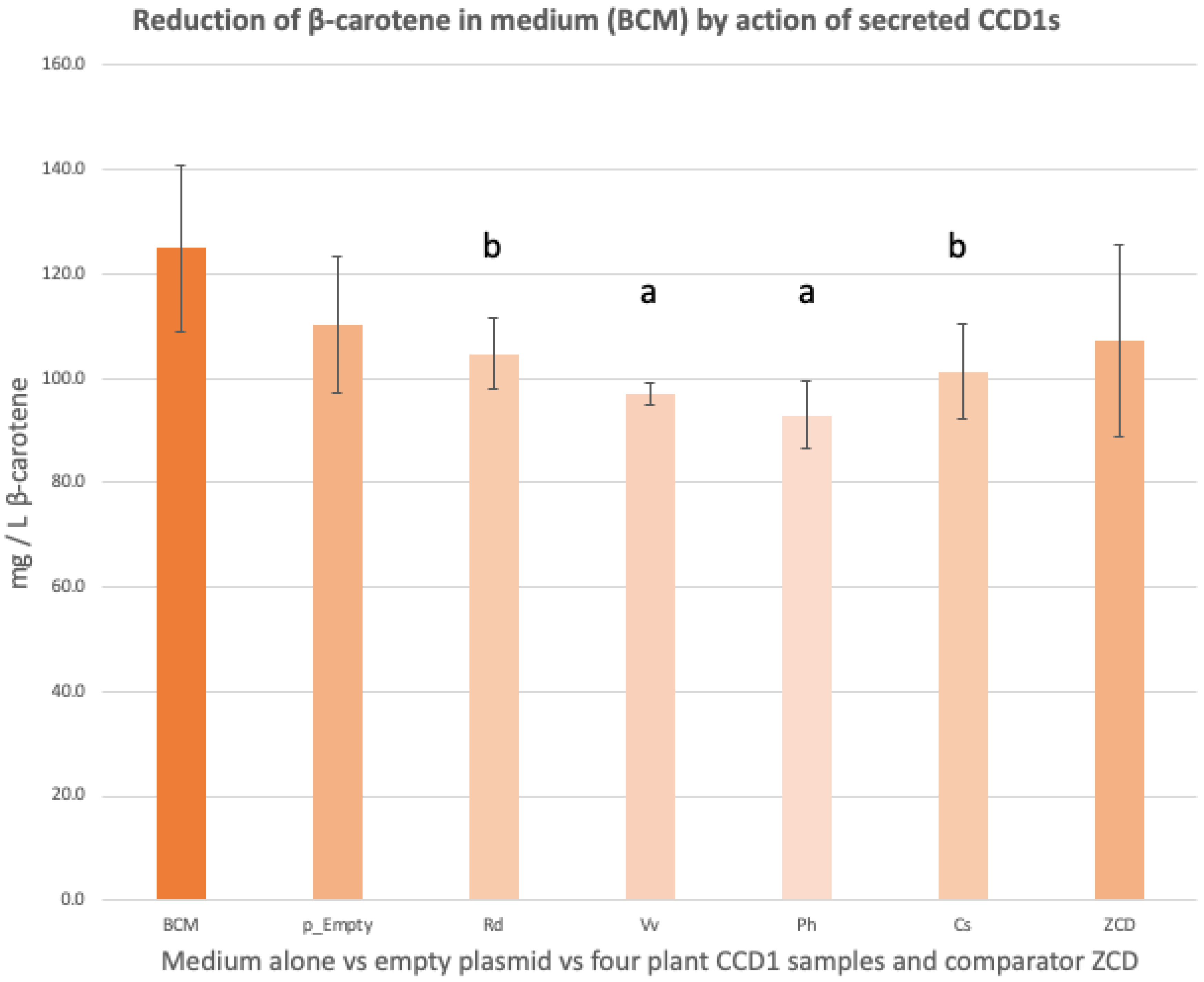
All strains grown in BCM reduced β-carotene levels after 24 h incubation (Figure 2). t-tests performed comparing the BCM sample to the CCD1s from different plant species demonstrate varying levels of activity or ability to cleave the substrate, β-carotene, with Ph and Vv registering a p-value of <0.05 versus <0.1 for Rd and Cs. The secreted ZCD, whose main substrate in planta is zeaxanthin rather than β-carotene, did not demonstrate a significant reduction (p > 0.1), while the control sample (BY4741—empty plasmid) similarly showed a decrease in the base level of β-carotene, possibly from auto-oxidation or the action of native yeast enzymes [34] but, again, this difference was not significant when compared to the BCM control. The secreted Petunia hybrida and Vitis vinifera CCD1s had the greatest effect in reducing levels of β-carotene compared to the remaining plant CCD1s and ZCD.

3.3. GCMS of β-Ionone from Head-Space Solid-Phase Micro-Extraction (HS-SPME)

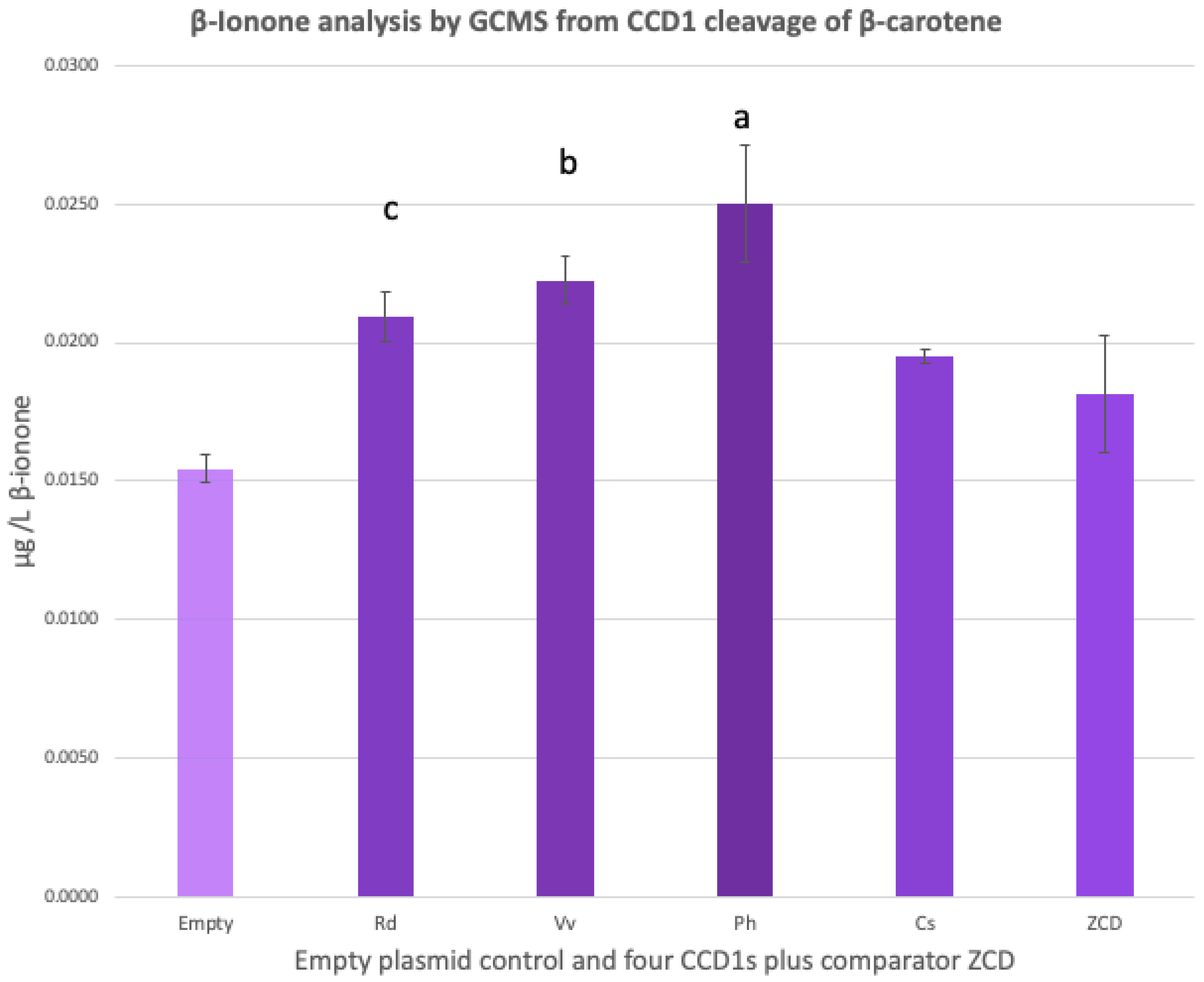
To validate the production of β-ionone, GCMS analyses were performed on triplicate samples. The results obtained were calculated as average values and these results demonstrated, prima facie, a greater release of β-ionone from the Petunia hybrida sample of BY4741_pMU1532_Ph ssCCD1, closely followed by that of Vitis vinifera, with Rd, Cs, and ZCD next. Overall, our results show a significant difference in the detection of β-ionone from the action of CCD1 compared to the control. However, there was no significant difference between Ph and Vv but significant differences between Ph and Rd, Cs and ZCD; there is a significant difference in the detection of β-ionone between the empty plasmid and the four CCD1s but not the ZCD, as determined by a t-test (Figure 3). As was seen earlier with the detection of β-carotene by spectrophotometry, the yeast with the empty plasmid also displayed some action in reducing β-carotene in a carotene-containing medium; this is mimicked in the detection of cleaved β-ionone and, again, such presence is suggested by the action of other non-CCD1 enzymes and/or by non-enzymatic reactions such as auto-oxidation [33]. However, all CCD samples had higher-than-baseline degradation values, as seen in the yeast sample with the empty plasmid.
These GCMS results for the detection of cleaved β-ionone from β-carotene by carotenoid cleavage dioxygenase 1 enzymes show an inverse correlation of the spectrophotometric analysis of β-carotene with the Ph sample being the most active, followed by Vv, with a lesser effect from Rd and Cs and a minimal result for ZCD.

3.4. Liquid Chromatography-Mass Spectrometry (LCMS) Analysis of Extracellularly Secreted CCD1s

To validate the secretion of CCD1 from the yeast cell, LCMS was performed for the detection of protein from the supernatant of the incubated yeast samples and revealed that CCD1 and ZCD enzymes were secreted from the yeast cell into the incubating medium (see supplementary data for an example of detection of secreted CCD1, Table S1). Furthermore, no CCD1 protein was detected from the empty plasmid control (Table S2). These results validated the actions of CCD1 enzymes on the extracellular substrate in the β-carotene-containing medium, both to lower the levels of β-carotene as determined by spectrophotometry and to liberate β-ionone detected by GCMS.

3.5. Comparison of the Efficacy of the Four Secreted Plant CCD1s

Comparative analysis of four phylogenetically diverse plant CCD1s demonstrated that the enzyme expressed and secreted from the Petunia hybrida gene had the best efficacy on the cleavage of β-carotene to release β-ionone but is closely followed by that from the Vitis vinifera CCD1-expressing strain. This result is in agreement with another study of the comparative efficacy of CCD1s from plant origins, which showed that the CCD1 from Petunia hybrida had a higher selectivity for β-carotene than that from Vitis vinifera, as well as other plant CCD1s [36]. The CCD1 from Rosa damascena and from Crocus sativus would appear to be slightly less responsive in their cleavage effectiveness; in fact, Jeffrey Czajka and colleagues suggest that only slight improvements can be achieved by substituting enzymes from different plant CCD1s with those of PhCCD1 or VvCCD1 [37]. While the patterns of efficacy are consistent in our experiments with PhCCD1 and VvCCD1 leading the way, in both cases of β-carotene reduction and release of β-ionone, the differences between these two types were found to be not statistically significant. Therefore, if any wine yeast were to be constructed, such as to provide higher levels of β-ionone, the use of a VvCCD1 gene would be more acceptable than that of the PhCCD1 gene, given that the Vv proteins would be already found in wine. Ultimately, however, most proteins are precipitated out of wine through interaction with tannins (red wines) or through fining agents (white wines) in the final stages of winemaking. It is recognised that the laws governing genetically modified organisms (GMO) vary from country to country around the world, particularly in North America (more accepting) versus Europe (less accepting). The arguments surrounding GMOs in wine and foodstuffs in general have been canvassed in some detail in a previous paper [4].

4. Conclusions

β-Ionone represents one of the most important aroma components in the multitudinous array of chemicals contributing to the olfactory experience of wine. However, its enzymatic release through the action of endogenous enzymes within wine grapes is minimal. As has been shown in this study, the fermenting yeast Saccharomyces cerevisiae can be manipulated at the molecular level to deliver higher levels of this aromatic organic compound and, thus, could be of significant advantage to the winemaker. In these experiments, we designed and secreted several heterologous CCD1s in yeast and demonstrated that these secreted CCD1s were functional and able to release β-ionone from exogenous β-carotene. Thus, the future of winemaking may be augmented by the use of such constructed yeast strains, adding options to the armamentarium of the winemaker to enhance the organoleptic properties of the resultant vinous product for the benefit and enjoyment of the consumer.

Supplementary Materials

The following supporting information can be downloaded at: https://www.mdpi.com/article/10.3390/fermentation8080395/s1, Table S1: Results table of an example of CCD1 protein detection by LCMS, Petunia x hybrida PhCCD1 with MW 61.3 kDa, line 6731. Table S2: Results table indicating no detection by LCMS of any CCD1 protein secreted from the empty plasmid (p_Empty) between the range of CCD1 sizes used: MW 61.1 kDa–62.4 kDa.

Author Contributions

Each of the authors contributed to the following: conceptualisation, J.J.B.T. and H.K.; writing—original draft preparation, J.J.B.T.; writing—review and editing, J.J.B.T., H.K., R.S.K.W., I.S.P. and I.T.P.; visualisation, J.J.B.T. All authors have read and agreed to the published version of the manuscript.

Funding

This research was conducted by the Australian Research Council Centre of Excellence in Synthetic Biology (project number CE200100029) and funded by the Australian Government.

Acknowledgments

We wish to acknowledge the help and support given to us by Donald Cameron, Applied BioSciences, Macquarie University, during the GCMS analysis of samples for β-ionone detection. Geraldine Sullivan is acknowledged for her kind assistance in helping to construct the CCD1 phylogenetic tree and producing the p-values for the graphs of β-carotene and β-ionone. This research was facilitated by access to Sydney Mass Spectrometry, a core research facility at the University of Sydney.

Conflicts of Interest

The authors declare no conflict of interest.

References

  1. Petronilho, S.; Lopez, R.; Ferreira, V.; Coimbra, M.A.; Rocha, S.M. Revealing the usefulness of aroma networks to explain wine aroma properties: A case study of Portuguese wines. Molecules 2020, 25, 272. [Google Scholar] [CrossRef] [PubMed]
  2. Robinson, A.L.; Boss, P.K.; Solomon, P.S.; Trengove, R.D.; Heymann, H.; Ebeler, S.E. Origins of grape and wine aroma. Part 1. Chemical components and viticultural impacts. Am. J. Enol. Vitic. 2014, 65, 1–24. [Google Scholar] [CrossRef]
  3. Cataldo, V.F.; Lopez, J.; Carcamo, M.; Agosin, E. Chemical vs. biotechnological synthesis of C13-apocarotenoids: Current methods, applications and perspectives. Appl. Microbiol. Biotechnol. 2016, 100, 5703–5718. [Google Scholar] [CrossRef]
  4. Timmins, J.J.B.; Kroukamp, H.; Paulsen, I.T.; Pretorius, I.S. The sensory significance of apocarotenoids in wine: Importance of carotenoid cleavage dioxygenase 1 (CCD1) in the production of β-ionone. Molecules 2020, 25, 2779–2793. [Google Scholar] [CrossRef] [PubMed]
  5. Mathieu, S.; Terrier, N.; Procureur, J.; Bigey, F.; Gunata, Z. A carotenoid cleavage dioxygenase from Vitis vinifera L.: Functional characterisation and expression during grape berry development in relation to C13-norisoprenoid accumulation. J. Exp. Bot. 2005, 56, 2721–2731. [Google Scholar] [CrossRef] [PubMed]
  6. Baumes, R.; Wirth, J.; Bureau, S.; Gunata, Y.; Razungles, A. Biogeneration of C13-norisoprenoid compounds: Experiments supportive for an apo-carotenoid pathway in grapevines. Anal. Chim. Acta 2002, 458, 3–14. [Google Scholar] [CrossRef]
  7. Razungles, A.; Bayonove, C.L.; Cordonnier, R.E.; Baumes, R.L. Etude des caroténoïdes du raisin à maturité. Vitis 1987, 26, 183–191. [Google Scholar]
  8. Mendes-Pinto, M.M. Carotenoid breakdown products–the norisoprenoids–in wine aroma. Arch. Biochem. Biophys. 2009, 483, 236–245. [Google Scholar] [CrossRef]
  9. Razungles, A.; Bayonove, C.L.; Cordonnier, R.E.; Sapis, J.C. Grape carotenoids: Changes during the maturation period and localisation in mature berries. Am. J. Enol. Vitic. 1988, 39, 44–48. [Google Scholar]
  10. Saini, R.K.; Nile, S.H.; Park, S.W. Carotenoids from fruits and vegetables: Chemistry, analysis, occurrence, bioavailability and biological activities. Food Res. Int. 2015, 76, 735–750. [Google Scholar] [CrossRef]
  11. Chen, W.-K.; Yu, K.-J.; Liu, B.; Lan, Y.-B.; Sun, R.-Z.; Li, Q.; He, F.; Pan, Q.-H.; Duan, C.-Q.; Wang, J. Comparison of transcriptional expression patterns of carotenoid metabolism in ‘Cabernet Sauvignon’ grapes from two regions with distinct climate. J. Plant Physiol. 2017, 213, 75–86. [Google Scholar] [CrossRef] [PubMed]
  12. Young, P.R.; Lashbrooke, J.G.; Alexandersson, E.; Jacobson, D.; Moser, C.; Velasco, R.; Vivier, M.A. The genes and enzymes of the carotenoid metabolic pathway in Vitis vinifera L. BMC Genom. 2012, 13, 1–17. [Google Scholar] [CrossRef] [PubMed]
  13. Lashbrooke, J.G.; Young, P.R.; Dockrall, S.J.; Vasanth, K.; Vivier, M.A. Functional characterization of three members of the Vitis vinifera L. carotenoid cleavage dioxygenase gene family. BMC Plant Biol. 2013, 13, 156. [Google Scholar] [CrossRef]
  14. Kambiranda, D.; Basha, S.M.; Singh, R.K.; He, H.; Calvin, K.; Mercer, R. In depth proteome analysis of ripening Muscadine grape berry cv. Carlos reveals proteins associated with flavour and aroma compounds. J. Proteome Res. 2016, 15, 2910–2923. [Google Scholar] [CrossRef]
  15. Beekwilder, J.; van Rossum, H.M.; Koopman, F.; Sonntag, F.; Buchhaupt, M.; Schrader, J.; Hall, R.D.; Bosch, D.; Pronk, J.T.; van Maris, A.J.A.; et al. Polycistronic expression of a β-carotene biosynthetic pathway in Saccharomyces cerevisiae coupled to β-ionone production. J. Biotech. 2014, 192, 383–392. [Google Scholar] [CrossRef] [PubMed]
  16. Ohloff, G. Importance of minor components in flavors and fragrances. Perfum. Flavor. 1978, 3, 11–22. [Google Scholar]
  17. Canuti, V.; Conversano, M.; Li Calzi, M.; Heymann, H.; Matthews, M.A.; Ebeler, S.E. Headspace solid-phase microextraction–gas chromatography–mass spectro- metry for profiling free volatile compounds in Cabernet Sauvignon grapes and wines. J. Chromatogr. A 2009, 1216, 3012–3022. [Google Scholar] [CrossRef]
  18. Asproudi, A.; Ferrandino, A.; Bonello, F.; Vaudano, E.; Pollon, M.; Petrozziello, M. Key norisoprenoid compounds in wines from early-harvested grapes in view of climate change. J. Food Chem. 2018, 268, 143–152. [Google Scholar] [CrossRef]
  19. Swiegers, J.H.; Bartowsky, E.J.; Henschke, P.A.; Pretorius, I.S. Yeast and bacterial modulation of wine aroma and flavour. Aust. J. Grape Wine Res. 2005, 11, 139–173. [Google Scholar] [CrossRef]
  20. Pedroza, M.A.; Zalacain, A.; Lara, J.F.; Salinas, M.R. Global grape aroma potential and its individual analysis by SBSE-GC-MS. Food Res. Int. 2010, 43, 1003–1008. [Google Scholar] [CrossRef]
  21. Marais, J.; van Wyk, C.J.; Rapp, A. Carotenoid levels in maturing grapes as affected by climatic regions, sunlight and shade. S. Afr. J. Enol. Vitic. 1991, 12, 64–69. [Google Scholar] [CrossRef]
  22. la Grange, D.C.; Pretorius, I.S.; van Zyl, W.H. Expression of a Trichoderma reesei β-xylanase gene (XYN2) in Saccharomyces cerevisiae. Appl. Environ. Microbiol. 1996, 62, 1036–1044. [Google Scholar] [CrossRef]
  23. Nevalainen, H.; Peterson, R. Making recombinant proteins in filamentous fungi–are we expecting too much? Front. Microbiol. 2014, 5, 75. [Google Scholar] [PubMed]
  24. Brevnova, E.; McBride, J.; Wiswall, E.; Wenger, K.; Caiazza, N.; Hau, H.; Argyros, A.; Agbogbo, F.; Rice, C.F.; Barrett, T.; et al. Yeast Expressing Saccharolytic Enzymes for Consolidated Bioprocessing Using Starch and. Cellulose. Patent No. WO/2011/153516, 2013. [Google Scholar]
  25. Kroukamp, H.; den Haan, R.; la Grange, D.C.; Sibanda, N.; Foulquié-Moreno, M.R.; Thevelein, J.M.; van Zyl, W.H. Strain breeding enhanced heterologous cellobiohydrolase secretion by Saccharomyces cerevisiae in a protein specific manner. Biotechnol. J. 2017, 12, 1700346. [Google Scholar] [CrossRef]
  26. Gietz, R.D.; Schiestl, R.H. High-efficiency yeast transformation using the LiAc/SS carrier DNA/PEG method. Nat. Protoc. 2007, 2, 31–35. [Google Scholar] [CrossRef] [PubMed]
  27. Vogel, J.T.; Tan, B.-C.; McCarty, D.R.; Klee, H.J. The carotenoid cleavage dioxygenase 1 enzyme has broad substrate specificity, cleaving multiple carotenoids at two different bond positions. J. Biol. Chem. 2008, 283, 11364–11373. [Google Scholar] [CrossRef]
  28. Baldermann, S.; Kato, M.; Kurosawa, M.; Kurobayashi, Y.; Fujita, A.; Fleischmann, P.; Watanabe, N. Functional characterization of a carotenoid cleavage dioxygenase 1 and its relation to the carotenoid accumulation and volatile emission during the floral development of Osmanthus fragrans Lour. J. Exp. Bot. 2010, 61, 2967–2977. [Google Scholar] [CrossRef]
  29. Soria, A.C.; Sanz, J.; Villamiel, M. Analysis of volatiles in dehydrated carrot samples by solid-phase microextraction followed by GC-MS. J. Sep. Sci. 2008, 31, 3548–3555. [Google Scholar] [CrossRef]
  30. Yamamoto, M.; Baldermann, S.; Yoshikawa, K.; Fujita, A.; Mase, N.; Watanabe, N. Determination of volatile compounds in four commercial samples of Japanese green algae using solid phase microextraction gas chromatography mass spectrometry. Sci. World J. 2014, 2014, 289780. [Google Scholar] [CrossRef]
  31. Langen, J.; Wegmann-Herr, P.; Schmarr, H.-G. Quantitative determination of a-ionone, β-ionone and β-damascenone and enantiodifferentiation of a-ionone in wine for authenticity control using multidimensional gas chromatography with tandem mass spectrometric detection. Ann. Bioanal. Chem. 2016, 408, 6483–6496. [Google Scholar] [CrossRef]
  32. Maruti, A.; Duran-Guerrero, E.; Barroso, C.G.; Castro, R. Optimisation of a multiple headspace sorptive extraction method coupled to gas chromatography-mass spectrometry for the determination of volatile compounds in macroalgae. J. Chromatogr. A 2018, 1551, 41–51. [Google Scholar] [CrossRef] [PubMed]
  33. Huang, F.-C.; Horvath, G.; Molnar, P.; Turcsi, E.; Deli, J.; Schrader, J.; Sandmann, G.; Schmidt, H.; Schwab, W. Substrate promiscuity of RdCCD1, a carotenoid cleavage oxygenase from Rosa damascena. Phytochemistry 2009, 70, 457–464. [Google Scholar] [CrossRef] [PubMed]
  34. Ding, Z.; Peng, S.; Xia, W.; Zheng, H.; Chen, X.; Yin, L. Analysis of five earthy-musty odorants in environmental water by HS-SPME/GC-MS. Int. J. Anal. Chem. 2014, 2014, 697260. [Google Scholar] [CrossRef] [PubMed]
  35. Nawade, B.; Shaltiel-Harpaz, L.; Yahyaa, M.; Bosamia, T.C.; Kabaha, A.; Kedoshim, R.; Zohar, M.; Isaacson, T.; Ibdah, M. Analysis of apocarotenoid volatiles during the development of Ficus carica fruits and characterisation of carotenoid cleavage dioxygenase genes. Plant Sci. 2020, 290, 110292. [Google Scholar] [CrossRef] [PubMed]
  36. Zhang, C.; Chen, X.; Lindley, N.D.; Too, H.-P. A “plug-n-play” modular metabolic system for the production of apocarotenoids. Biotechnol. Bioeng. 2018, 115, 174–183. [Google Scholar] [CrossRef]
  37. Czajka, J.J.; Nathenson, J.A.; Benites, V.T.; Baidoo, E.E.K.; Cheng, Q.; Wang, Y.; Tang, Y.J. Engineering the oleaginous yeast Yarrowia lipolytica to produce the aroma compound β-ionone. Microb. Cell Factories 2018, 17, 136. [Google Scholar] [CrossRef]
Figure 1. The nominated four phylogenetically separated CCD1s (in red) within a selected range of other plant CCD1s; the numerical scale represents the proportion of genetic variation. The biological names of the plants from the CCD1 abbreviation are: RdCCD1 from Rosa x damascena; PmCCD1 from Prunus mume; MtCCD1 from Medicago truncatula; NnCCD1 from Nelumbo nucifera; VvCCD1 from Vitis vinifera; OfCCD1 from Osmanthus fragrans; CcCCD1 from Coffea canephora; PhCCD1 from Petunia x hybrida; ZmCCD1 from Zea mays; CsCCD1 from Crocus sativus.
Fermentation 08 00395 g001
Figure 2. Graph of comparative reductions in β-carotene in a β-carotene-rich medium (BCM) through cleavage by four ssCCD1s—Rd, Vv, Ph, Cs—against controls. t-tests performed on the results demonstrated a difference between the BCM and the four CCD1s, with Ph and Vv having a p-value of <0.05 (a) and Rd and Cs with a p-value of <0.1 (b); there were no significant differences between the BCM and p_Empty sample and the BCM and ZCD sample.
Fermentation 08 00395 g002
Figure 3. GCMS analysis of β-ionone, following secretion of CCD1s into β-carotene-containing medium. Results for p-values (calculated using Anova and TukeyHSD functions from statistics package in R version 4.0.3), showed p < 0.0001 between empty plasmid and Ph sample (a); p < 0.001 between empty plasmid and Vv sample (b); p < 0.01 between empty plasmid and Rd sample (c). A t-test performed on the results demonstrated a significant difference between the empty plasmid and the four CCD1s but no significant difference between the ZCD sample and the empty plasmid.
Fermentation 08 00395 g003
Table 1. GenBank Accession Numbers of plant genes used in this study.
GenBank Accession NumberGene NameGene SizePlant Origin
EU327776.1Rd ssCCD11659 bpRosa × damascena
KF008001.1Vv ssCCD11629 bpVitis vinifera
AY576003.1Ph ssCCD11641 bpPetunia × hybrida
AJ132927.1Cs ssCCD11686 bpCrocus sativus
AJ489276.1Cs ssZCD1186 bpCrocus sativus
Table 2. Primers used in this study.
Type of PrimerSequence of Primer
Rd-ssCCD1 ORF_R5′-AAG AAC AAG CAA AGT TCT AA-3′
Vv-ssCCD1 ORF_R5′-AAG AAC AAG CAA AAC TTT GA-3′
Ph-ssCCD1 ORF_R5′-AAG AAC AAG CCA AAC TGT GA-3′
Cs-ssCCD1 ORF_R5′-TGC TGG ATC ATT GCA GTA CC-3′
Cs-ssZCD ORF_R5′-CCT CTC CAT ATT CGC TGC CA-3′
ENO1p_all-CCD ORF_F5′-ACA CAA ACA CTA AAT CAA AG-3′
Table 3. Parameters used in GCMS analysis of β-ionone.
GC Parameters
Start temperature: 50 °CInjection temp: 270 °CSampling time: 1.0 to 1.5 min
Flow control: 69 cm/sPressure: 100 kPaPurge flow: on at 3.0 mL/min
Linear velocity: 47 cm/sMode: SpitlessSplit ratio: −1.0
MS Parameters
Ion source temp: 200 °CInterface temp: 270 °CSolvent cut-off time: 1.5 min
Threshold: 1000 countsStart m/z: 45.0End m/z: 350.0
Start time: 2.0 minEnd time: 12.0 minElectron ionisation: 70 eV
Acquisition mode: scanInterval: 0.15 sScan speed: 2500
Publisher’s Note: MDPI stays neutral with regard to jurisdictional claims in published maps and institutional affiliations.