Article

Microbial Diversity of Marula Wine during Spontaneous Fermentation

by
Evelyn Maluleke
,
Maleho Annastasia Lekganyane
and
Kgabo L. Maureen Moganedi
*
Department of Biochemistry, Microbiology and Biotechnology, University of Limpopo, Sovenga 0727, South Africa
*
Author to whom correspondence should be addressed.
Fermentation 2023, 9(10), 862; https://doi.org/10.3390/fermentation9100862
Submission received: 21 August 2023 / Revised: 16 September 2023 / Accepted: 19 September 2023 / Published: 22 September 2023
(This article belongs to the Section Fermentation for Food and Beverages)

Abstract

:
Marula wine is produced from ripe fruits of the Sclerocarya birrea subspecies caffra tree through spontaneous fermentation. A few culture-based studies have shown that the fermentation is largely driven by yeasts, although, in the early stages, some lactic acid bacteria (LAB) and acetic acid bacteria may be detected. Some of the microbes may produce undesirable metabolites that lead to the spoilage and short shelf life of the wine. However, there is generally limited information on the microbial composition and its contribution to the chemical characteristics of the resultant marula wine. The aim of this study was to characterise the microbial population of marula wine from different localities in the Limpopo province, South Africa. MALDI-TOF and amplicon sequencing technique were used to identify microbial strains and to determine their diversity and changes in the different stages of fermentation. The phylogenetic relationships of LAB and S. cerevisiae were analysed using multilocus sequence typing. Bacterial species that were common in the different marula wines included Gluconobacter oxydans, Lactiplantibacillus plantarum, Levilactobacillus brevis, Lacitilactobacillus nagelii, Lentilactobacillus kefiri and Lentilactobacillus parabuchneri, and the yeasts were Hanseniaspora guiliermondii, Saccharomyces cerevisiae, Rhodotorula mucilaginosa and Pichia kudriavzevii. The MLST data indicated common microbiota from different marula wines with low intraspecific diversity, suggesting that the LAB and S. cerevisiae strains that are mainly responsible for the spontaneous fermentation of marula wine are similar irrespective of the geographical differences and production preferences.

1. Introduction

Marula tree, scientifically known as Sclerocarya birrea, is a widespread species found throughout the semi-arid savannas of sub-Saharan Africa. Its natural habitat extends from Senegal to Ethiopia in the north; southward to KwaZulu-Natal in South Africa; and eastward to Namibia, Angola, and the southern part of the Democratic Republic of Congo. There are three recognised subspecies of S. birrea, namely, caffra (Sond.) Kokwaro, multifoliolata (Engl.) Kokwaro, and birrea. The subspecies caffra is the most important and dominant subspecies in southern Africa. It occurs in Zimbabwe, Botswana, Namibia, South Africa and Swaziland. In South Africa, the tree is common in KwaZulu–Natal, Mpumalanga, Limpopo and North-West provinces [1,2]. In most cases, female trees bloom between September and December, and the fruit ripens between January and March of each year. The fruit size varies but is the size of a plum in approximation. S. birrea fruits abscise before ripening, at which stage the skin colour is green and the fruit is firm. The fruits ripen on the ground and attain a thick yellow peel and translucent whitish flesh. The fruit is characterised by a bitter-sweet taste but of pleasant flavour when fully ripe. The marula fruit contains high levels of vitamin C as well as other antioxidants. On average, the ripe fruit has 168 mg of vitamin C per 100 g, which is comparable to what is found in guava and approximately three times better than oranges [2,3].
Marula wine is brewed naturally by the yeasts and bacteria found on the marula fruit [4], which are reportedly introduced by Drosophila flies [2] and handling during manual extraction of the juice. During spontaneous fermentation, the diverse microbiome on the fruit surfaces plays a variety of roles [5], which can positively contribute to fermentation or produce spoilage effect in the wine. Alcoholic fermentation is primarily a result of the activities of yeasts [6,7]. In red grape wine production, lactic acid bacteria (LAB) are involved in the process of malolactic fermentation, which follows the alcoholic fermentation carried out by yeasts. LAB produce chemical compounds such as ethyl lactate and higher alcohols that contribute to the wine aroma [8,9,10]. The process involves converting the dicarboxylic l-malic acid (malate) into monocarboxylic l-lactic acid (lactate) and carbon dioxide [11]. Acetic acid bacteria (AAB) such as Acetobacter pasteurianus and Gluconobacter oxydans convert ethanol to acetic acid, carbon dioxide and water. Researchers have reported that these microorganisms usually dominate the late stage of fermentation and can result in a vinegary taste, a character emanating from the production of acetic acid and other volatiles [12,13].
A variety of bacteria and yeasts have been observed during fermentation of marula juice to wine. These include Saccharomyces sp., coliforms, lactic acid bacteria and acetic acid bacteria [4,14]. Marula wine is regarded as a source of social stability by the African community since it brings the community together during cultural ceremonies. However, the production of marula wine has become an enterprise in the African communities, and the income generated solely supports the livelihoods of the families [15]. Spontaneous or traditionally fermented beverages and foods have a short shelf life, which makes it difficult to produce and market these products at a large scale [16]. To achieve large-scale production without compromising the typical properties of marula wine and prevent spoilage during storage and maturation, it is imperative to know the type and evolution of the microorganisms involved in the fermentation process [17]. In this study, the microbial communities present during fermentation of marula wine were characterised using the high-throughput techniques such as next-generation sequencing and multilocus sequence typing (MLST). These techniques that identify microorganisms within mixed cultures are revolutionising the study of microbial ecology because of their high level of accuracy and reliability. Lactobacillus spp. and acetic acid bacteria have previously been successfully characterised using MLST [18,19,20,21]. The aim of this study was to characterise the microbial population of marula wine from different localities in Limpopo province, South Africa. MALDI-TOF and amplicon sequencing technique were used to identify microbial strains and to determine their β-Diversity and changes in the different stages of fermentation.

2. Materials and Methods

Three marula wines (5 L) produced following the same recipe and all using spontaneous fermentation were collected from different brewers in the two locations in Limpopo province, South Africa. These include the Makhushane village in Phalaborwa (23°58′32.08″ S 31°02′42.82″ E) and The Oaks village in Tzaneen (24.3624° S, 30.6753° E). The two wines collected from the two unrelated brewers at The Oaks were labelled MST and SKB wines, and the wine collected from Makhushane village was labelled MLT wine. The three marula wine brewers and the two locations were selected randomly without any particular preference. The wines were collected at different fermentation periods depending on availability. The MST wine was collected on the 3rd day of fermentation, and the SKB wine was collected on the 14th day, whereas MLT wine was collected after preparation (day 0). Ripe marula fruits were collected from the University of Limpopo (23.8888° S, 29.7386° E) grounds to produce the 4th wine for comparison, and this was labelled the LAB-UL wine. Marula fruits and wines used in this study were collected during March 2016 and February 2017. The fruits were collected into sterilised plastic buckets and were transported to the lab for analysis. For the microbial molecular typing work, the samples previously collected from the marula wines obtained from Mentz (MEN) (23°54′3.54″ S 29°46′28.02″ E) and Sekhukhune (SKK) (24°20′35.84″ S 30°5′33.07″ E) localities were included, whereas the SKB sample was excluded since it lacked data for the early stage of fermentation. Figure 1 represents the intricate process of marula wine fermentation and sampling. Additionally, it outlines the comprehensive microbial identification strategies employed to elucidate the microbial communities driving this fermentation process.

2.1. Preparation of Marula Wine

Ripe marula fruits from trees within the grounds of the University of Limpopo, Mankweng, Limpopo province, South Africa (23°53′19.7″ S 29°44′19.0″ E) were collected and used to prepare a wine under laboratory conditions. The trees are well marked and labelled with indigenous and scientific names. This wine was termed the LAB-UL wine. Ethanol (70%, v/v) was used to clean all the utensils used for fruit processing. A generic recipe typically used in the preparation of marula wines in the communities was followed. Briefly, metal forks were used to remove the thick skin from the fruits. The juice was manually squeezed from the fruits and collected into a clean sterilised plastic bucket, and the kernels with fibrous flesh still attached were put in a separate plastic bucket. The kernels were then covered with just enough potable tap water, and a wooden spoon was used to knead through the kernels in order to release any remaining juice and pulp. This was then mixed with the juice, and the mixture was covered with a bucket lid and left to ferment at an ambient temperature. The thick layer on top of the clarifying fermenting juice was continuously removed until there was no further formation of the blob.

2.2. Sampling of the Marula Wine

Fifty-millilitre samples of fermenting marula wines were collected throughout fermentation at 2-day intervals, beginning with day 0, until the ferments reached the state at which avid consumers of the wine considered it to be unpalatable. The samples were centrifuged at 5000 rpm for 10 min to collect the microbial cells. The cell pellets were mixed with sterile fifty percent glycerol at a ratio of 1:1 and stored at −80 °C as stock culture for microbial analysis.

2.3. Isolation of Bacteria from Marula Wine

LAB and AAB were isolated from the stock cultures using a spread-plate method. A volume of 100 µL of 10−2 and 10−3 culture dilutions prepared with 0.85% sterile saline was spread-plated onto de Man Rogosa and Sharpe (MRS) agar (Biolab, Merck, South Africa) and Wallerstein differential agar (WLD) (Sigma Aldrich, Steinheim, Germany) for the isolation of Lactobacillus species. MRS agar plates were incubated at 30 °C and WLD agar plates at 35 °C. All the cultures were incubated anaerobically with Anaerocult® (Merck) in a closed container for 48 to 72 h. AAB were isolated on WLD that was incubated aerobically at 35 °C. Following incubation, colonies were differentiated based on morphology and then enumerated, and 10% of each type was sub-cultured on the same media for purification and identification.

2.4. Isolation of Yeasts from the Marula Wine

Tenfold serial dilutions of the glycerol stock culture suspension were prepared with 0.85% sterile saline solution, and the spread plate method was used for the cultivation of yeasts on Wallerstein Nutrient agar (WLN) and YPD media (Sigma Aldrich, Steinheim, Germany). The cultures were incubated aerobically at 30 °C for 42 to 72 h. Colonies were recorded according to morphology and enumerated. Ten percent of each morphological subgroup was sub-cultured on the same medium to identify the culturable fraction of the microbiota.

2.5. Identification of the Microbial Isolates

a.
MALDI-TOF Biotyping Technology
The biotyper function of an Ultraflex MALDI-TOF (Bruker Daltonik GmbH, Bremen, Germany) was used for identification of the purified bacterial isolates. A colony of an actively growing bacterial culture was suspended in 300 µL of sterile deionised water in a microcentrifuge tube. This was followed by the addition of 900 µL of absolute ethanol. The mixture was mixed thoroughly and centrifuged at 2200 rpm for 2 min. The supernatant was decanted, and the pellet was allowed to dry at room temperature. A volume of 5 µL of 70% formic acid was added to the pellet and mixed well with a vortex. Equal volume of absolute acetonitrile (Sigma Aldrich, Steinheim, Germany) was added, mixed well and centrifuged for 2 min at 2200 rpm. One microliter of the supernatant was spotted onto the MALDI-TOF target plate and allowed to dry. The samples were overlaid with 1 µL of matrix (α-cyano-4-hydroxycinnamic acid (HCCA) in 50% acetonitrile and 1.5% trifluoroacetic acid) and dried at room temperature. The samples were applied to an Ultraflex LT mass spectrometer (Bruker Daltonik), and the results were analysed by MALDI Biotyper 3.0 software (Bruker Daltonik) in the automatic mode at manufacturer’s settings. A good identification was signified by a minimum score of 1.700 [22].
b.
Next-Generation Sequencing Technology
  • DNA Extraction and Amplification of the 16S rDNA and ITS Regions
This analysis sought to identify and compare microbial populations and diversity in the marula wines during various stages of fermentation. Two sets of microbial cultures from different marula wines (Mentz and Sekhukhune) that were previously collected from an earlier study in our laboratory were also included in the analysis alongside the four wine samples from the current study, namely, MST, SKB, MLT and LAB-UL. DNA was extracted from 1.5 mL of marula wines culture samples that was collected at different intervals during fermentation. DNeasy® PowerSoil® kit was used to extract DNA (QIAGEN, Hilden, Germany) following the manufacturer’s instructions. The DNA was stored at −20 °C until further analysis.
The ITS 3 (5′GCATCGATGAAGAACGCAGC′3) and ITS 4 (5′TCCTCCGCTTATTGATATGC′3) primers were used for yeast characterisation (White et al., 1990 [23]) and the 16S rDNA sequence 27F (5′AGAGTTTGATCNTGGCTCAG′3) and 1492R (5′-TACGGYTACCTTGTTACG-3′) primers (Lane, 1991 [24]) were used for the bacteria.
PCR amplification was performed in a 25 µL volume that contained 20.5 µL of PCR master mix and 4.5 µL of the DNA template. The PCR master mix was prepared with 7.5 µL molecular water, 5 µL of 5 × buffer, 5 µL each of the primers, and 0.5 µL of 2.5 units/µL HotStart HiFidelity DNA polymerase (QIAGEN, Hilden, Germany). A negative control was prepared by replacing DNA template with molecular water. Amplification was carried out as follows: an initial denaturation at 95 °C for 5 min; then 30 cycles of denaturation at 94 °C for 15 s, annealing at 55 °C for 1 min, extension at 72 °C for 30 s and a final extension at 72 °C for 10 min. GeneAmp PCR system 9600 thermal cycler was used for amplification. Gel electrophoresis was performed with 1.5% agarose gel and 1 × TAE buffer at 80 volts for 30 min to check the presence and integrity of the amplicons. A 100 bp DNA ladder (Inqaba Biotec, Pretoria, South Africa) was used for sizing of the bands. The gel was viewed under UV light.
  • Sequence Processing, Operational Taxonomic Units (OTUs) Clustering
The amplicons were diluted with 10 mM Tris pH 8.5 to 4 nM, and 5 μL from each library were mixed for pooling libraries with unique indices. All the libraries were pooled for one MiSeq run. Pooled PCR amplicons were purified and indexed using the Nextera XT primers (Illumina Inc., San Diego, CA, USA). Thereafter, the amplicon library was quantified, normalised and pooled prior to loading on the MiSeq flow cell for a 2 × 300 paired-end sequencing. Sequence reads were then de-multiplexed using the on-system MiSeq reporter software. The raw data files (ITS and 16S) are available at NCBI under the BioProject accessions PRJNA1019785 and PRJNA1019803, respectively. The quality of the reads was initially checked using the FastQC software (v 0.11.5, Babraham Institute, Cambridge, Bioinformatics, UK) prior to assembling the forward and reverse reads by using PANDAseq [25]. Assembled reads were then clustered into operational taxonomic units (OTUs) by using the “pick_open.reference_otus.py” script in QIIME [26] and aligning against the Silva rRNA database (release 128) [27] by using usearch61 [26] and PyNAST aligner [24]. The OTU table generated from the clustering step was first rarefied, prior to summarising the taxa. β-diversity (principal component analysis (PCA)) analysis were calculated using QIIME software (Version 1.8.0). Following data analysis, SKB data was disregarded due to low OTUs identified; i.e., the SKB wine sample was excluded in this analysis because it showed very low microbial diversity, noting that it was collected on the 14th day of fermentation from the brewer. Therefore, the present data only reported on the following wines: LAB-UL, MLT, MST, Mentz and Sekhukhune.

2.6. Characterisation of Common Microbiota from Different Marula Wines

The lactic acid bacteria and yeast isolates from various marula wines (see Table 1) that were assigned to the same species were further characterised via multilocus sequence typing (MLST) for genetic relatedness. The selection of isolates was influenced by identification to the same species, and in some instances, variable culture morphologies were used for same species. Pure cultures of 20 Lactobacillus spp. (6 L. plantarum, 8 L. brevis and 6 L. buchneri) and 13 S. cerevisiae isolates were selected for this analysis.
Each selected gene locus was amplified with the corresponding primers using the genomic DNAs of Lactobacillus isolates (clpx, groel, mure, phes, pyrg and ucrc) or S. cerevisiae (ATF1, ITS1, NUP116, RPN2, STE50 and YBL081W) as templates. The PCR reactions were performed in final volumes of 10 µL following the manufacturer’s guidelines (EconoTaq PLUS 2X Master Mix). Thermal cycling conditions included an initial denaturation at 94 °C for 2 min, 30 cycles of 94 °C for 30 s, annealing at 50 to 65 °C (both yeasts and bacteria) for 30 s and 72 °C for 30 s. The PCR products were analysed via electrophoresis on a 1.2% agarose gel. PCR products were cleaned using ExoSAP-IT™ PCR Product Cleanup reagent (ThermoFisher Scientific, Johannesburg, South Africa). DNA sequencing was then carried out with the ABI V3.1 Big dye kit according to the manufacturer’s instructions and the labelled products were cleaned with the Zymo Seq clean-up kit.
Sequence trimming and alignments were performed with ClustalX algorithm of the MEGA software [28,29]. The evolutionary history was inferred with the Maximum Likelihood method based on the Jukes–Cantor model [30]. The percentage of trees in which the associated taxa clustered together is shown next to the branches. The initial trees for the heuristic search were obtained automatically by applying Neighbour-Joining and BioNJ algorithms to a matrix of pairwise distances that was estimated with the Maximum Composite Likelihood (MCL) approach, followed by selecting the topology with superior log likelihood value [31]. Evolutionary analyses were conducted in MEGA7 [31]. This service was provided by Inqaba Biotechnical Industries (Pty) Ltd., Pretoria, South Africa.

3. Results

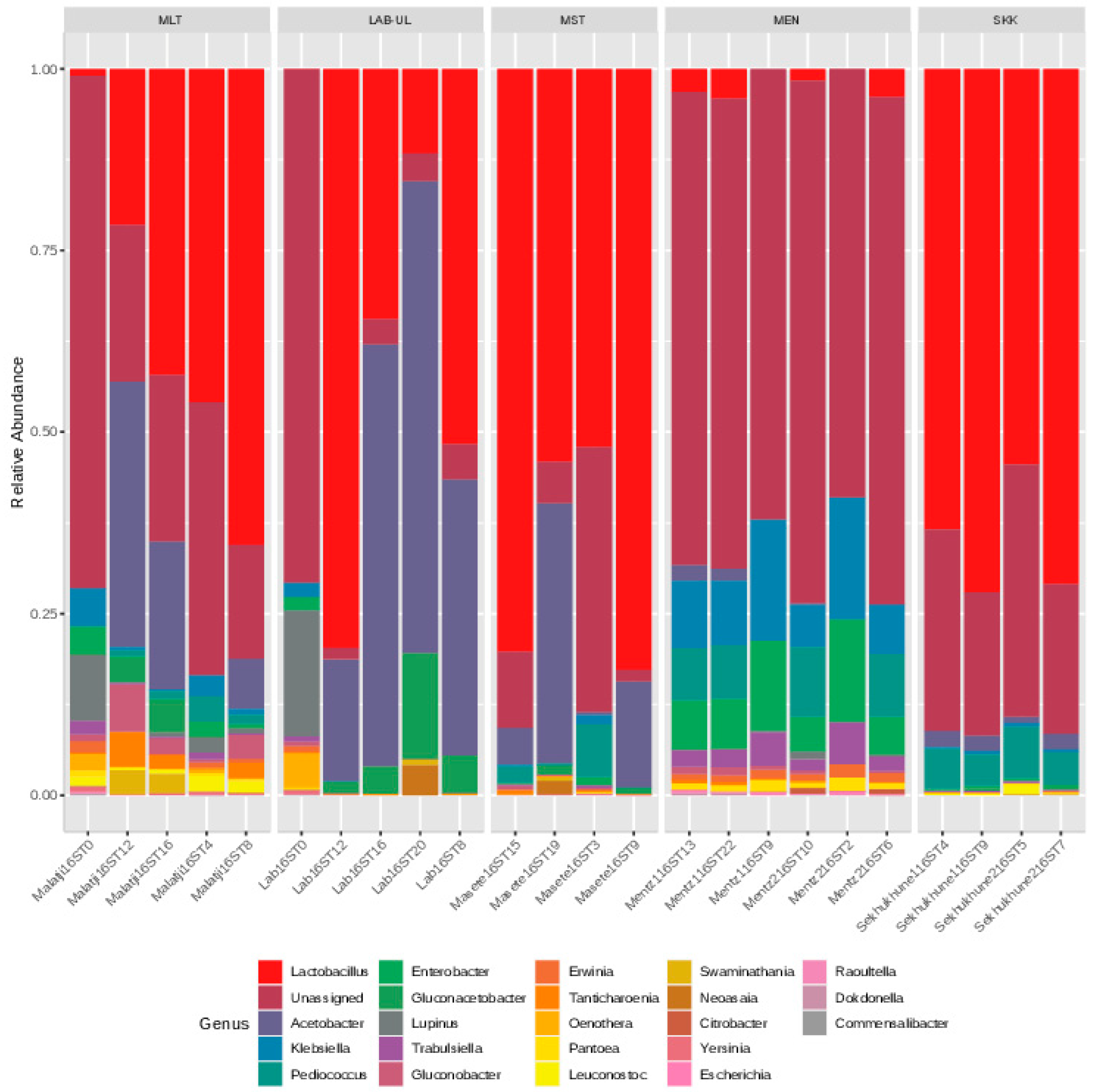
3.1. Bacterial Microbiome in the Marula Wines

Microbial diversity analysis was performed on four different wines to understand microbial changes during spontaneous fermentation of marula wine. The same bacterial species and similar trends were observed in all the four wines (MLT, LAB-UL, MST and SKB). The lactic acid bacterial species Lactiplantibacillus plantarum, Levilactobacillus brevis, Lacitilactobacillus nagelii, Lentilactobacillus kefiri and Lentilactobacillus parabuchneri were detected throughout the fermentation period, while the acetic acid bacteria such as Gluconobacter oxydans and Acetobacter pasteurianus were detected during the late stages of fermentation, i.e., between days 8 and 18 (Figure 2). Non-contributing environmental species such as Bacillus subtilis and Bacillus cereus were also detected in the marula wines during the early stages of fermentation (days 0–4).
Various non-fermentative and fermentative bacterial species were identified (Figure 2). Lactic acid bacteria were observed to be the dominant bacteria throughout the fermentation study period of the different marula wines.

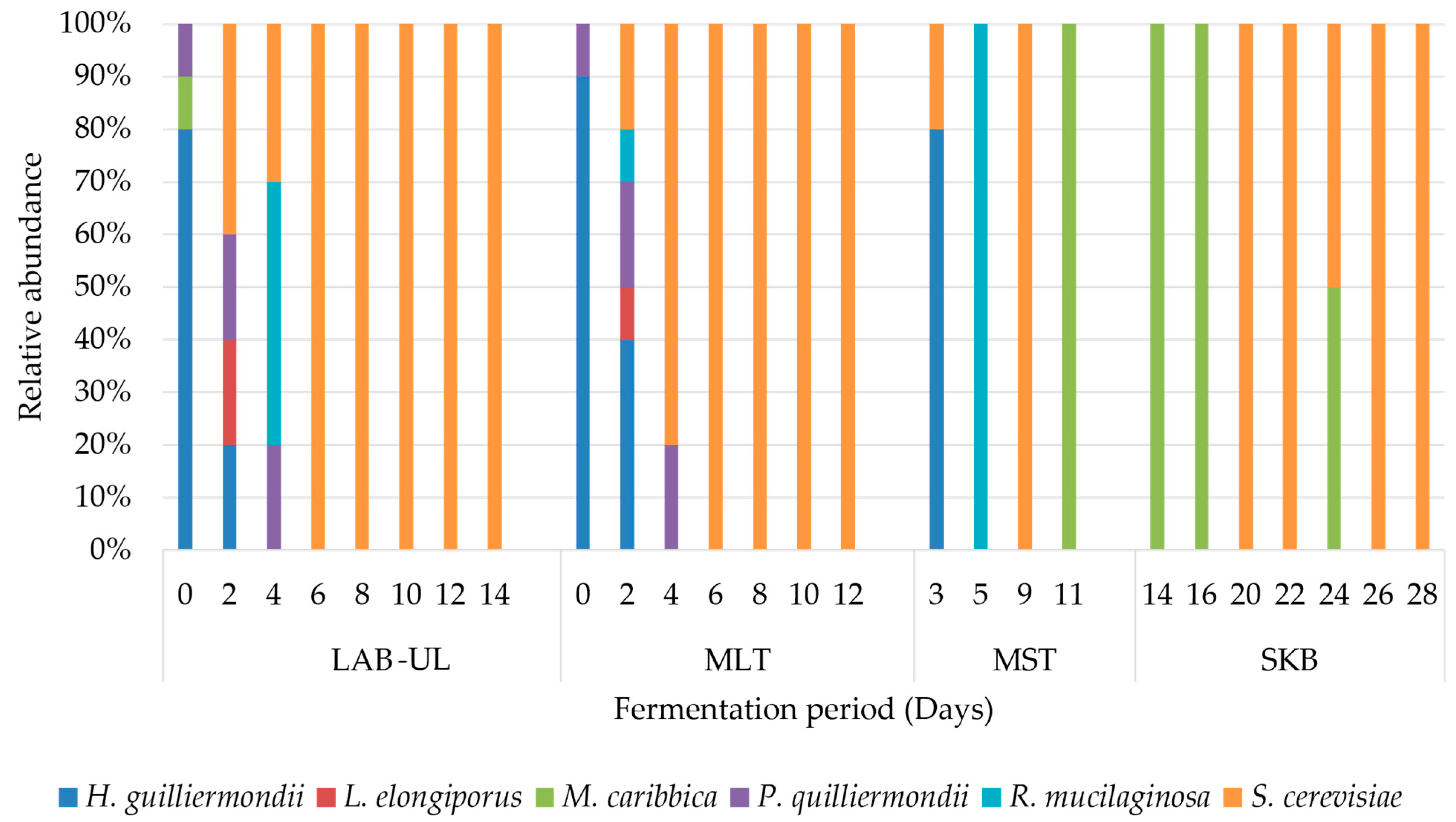
3.2. Yeast Microbiota Present in the Wine during Fermentation

Fermenting and non-fermenting yeasts were detected during spontaneous fermentation of marula juice to wine, with divergence at specific stages during the fermentation period. Non-fermenting yeasts were detected in the marula juice (day 0) and the early stages of fermentation (days 2 and 4), wherein the yeasts such as Hanseniaspora guilliermondii, Rhodotorula mucilaginosa and Meyerozyma caribbica were the dominant species (Figure 3). The mid- and late stages of fermentation were dominated by Saccharomyces cerevisiae. Meyerozyma caribbica was also detected at the late stage (days 11–24) in some of the wine samples (MST and SKB wines).
Non-Saccharomyces yeasts are known for initiating spontaneous fermentation before Saccharomyces yeast dominates for alcoholic fermentation.

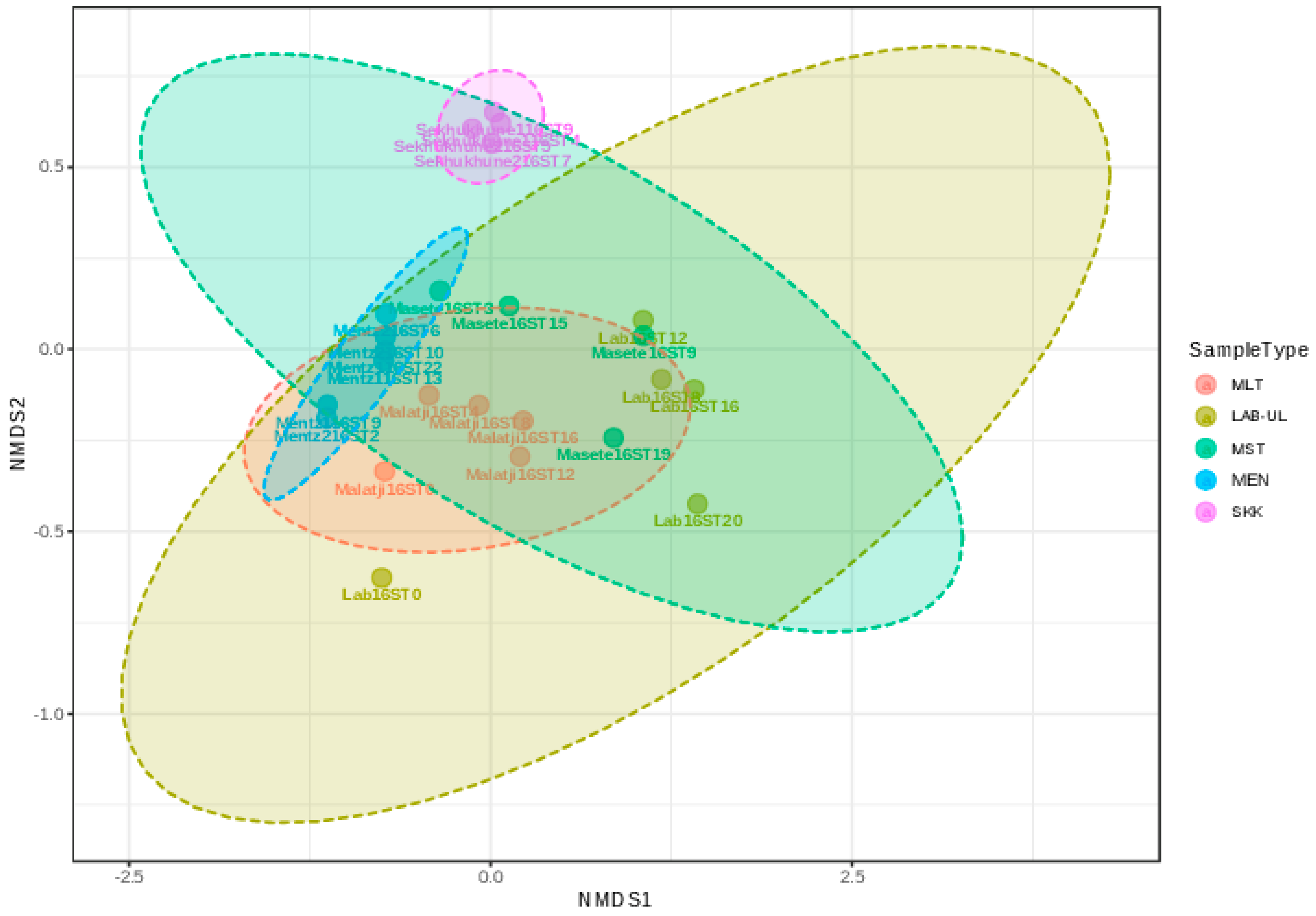
3.3. Microbial Taxonomic Composition of Various Marula Wine during Fermentation

Taxonomic composition analysis of the LAB-UL, MLT, MST, MEN, and SKK marula wines (e.g., Malatji16S10, where the name before 16S indicate the wine name, ‘16S’ demonstrates 16S data, and ‘10’ indicates day 10 of fermentation) was evaluated using microbiome analysis. The five wines were evaluated to decipher bacterial community changes that take place during spontaneous fermentation of marula juice to wine. Three phyla were detected across all the five wines at varying degrees of abundance. The two most dominant phyla were Firmicutes and Proteobacteria across the five wines, while Cyanobacteria were detected from only three wines, namely, LAB-UL, MLT and Mentz wines, with low abundance (Figure 4). The SK and MST wines were dominated by the Firmicutes, while Mentz wine was dominated by Proteobacteria.
The dominant orders were Lactobacillales (phylum Firmicutes) and Enterobacterales (phylum Proteobacteria). Rhodospirillales, Streptophyta, Rickettsiales, Anthomonadales and Acromonales, which all belong to the Proteobacteria phylum except for Streptophyta, were also detected at low abundance (Figure 5). The MLT wine revealed a mixed microbial community represented by all the different orders mentioned above. However, the SKK wine was dominated mostly by the Lactobacillales. On the other hand, Mentz wine (MEN) was dominated by Enterobacterales with very low counts of Rhodospirillales and Streptophyta. The LAB-UL wine was dominated by the Lactobacillales and Rhodospirillales. However, LAB16ST0 exhibited a microbial community that contains Lactobacillales, Enterobacterales, Rhodospirillales, Streptophyta, Rickettsiales and Anthomonadales. Overall, the presence of Lactobacillales correlated with MALDI-TOF data, which revealed LAB species as the dominant microbial group (Figure 2).
The analysis of the microbial communities of marula wines revealed the presence of various genera (Figure 6). The Enterobacteriales and Rhodospirillales which contain the genera of enteric and acetic acid bacteria, respectively, had the most diverse composition. The Lactobacillus, Pediococcus and Leuconostoc genera were the only Lactobacillales in all the wines. However, the genus Lactobacillus dominated almost all the wines except the Mentz (MEN) wine. Of the seven genera of the acetic acid bacteria that belong to the order Rhodospirillales, the genus Gluconobacter was the dominant one, and it showed presence throughout the fermentation period. This was followed by Acetobacter genus. Notably, the two genera Acetobacter and Gluconobacter are commonly implicated in the spoilage of wines.
Several genera such as Enterobacter, Escherichia, Citrobacter, Klebsiella, Erwinia, Raoultella, Yersinia, Pantoea and Trabulsiella from the order Enterobacteriales were detected in all the wines, albeit in small numbers. The genera from the orders Rickettsiales, Anthomonadales and Aeromonadales were not specifically detected (Figure 6).
β-Diversity of the wines samples (LAB-UL, MLT, MST, MEN and SKK) was measured for comparison of the LAB strains isolated from the marula wine samples. The different LAB isolates clustered according to origin of the wines. The lactic acid bacteria isolated from Sekhukhune wine (SKK) formed a tight group and were far apart from the other wines, whilst the isolates from the other wines formed loose clusters (Figure 7). Microbial similarities between the LAB-UL, MLT, MST and MEN wines were observed. However, LAB-UL wine had a unique community, indicated by LAB16ST0.

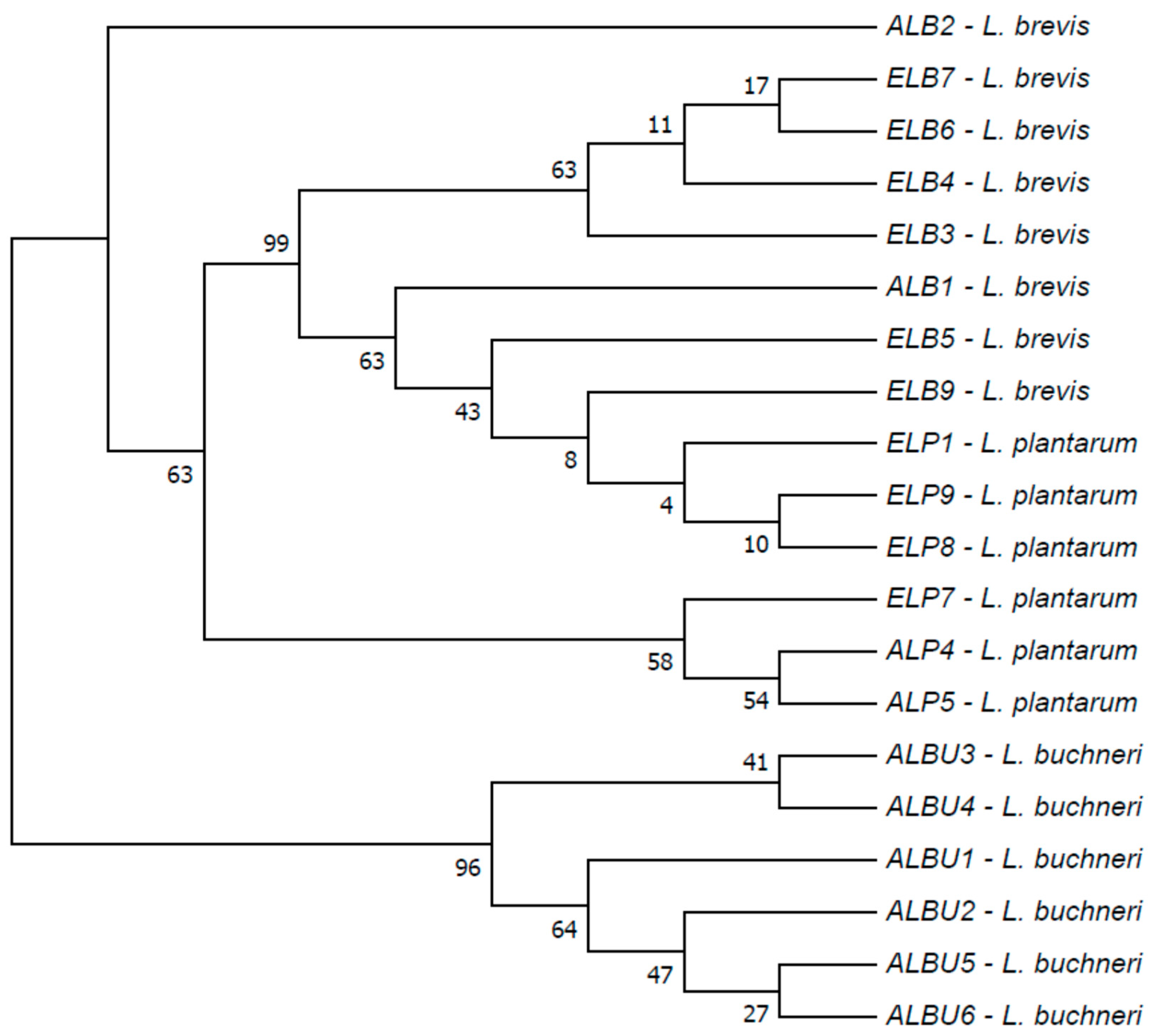
3.4. Characterisation of Common Microbiota from Different Marula Wines

The LAB and yeast isolates that were common in all the marula wines from different localities were characterised for strain typing and phylogenetic relationship. Intraspecific relatedness amongst the different species of Lactobacillus was apparent in the clusters that formed according to the specific source of marula wine and by the species type. The isolates at the same species from the same marula wine grouped closely together prior to broadly clustering with the isolates of the same species from different marula wines (Figure 8). This is congruent with the observed dispersion by the principal component analysis (Figure 7). Contrary to the relationships observed with the LAB strains, the S. cerevisiae strains showed varying levels of genetic relatedness. The yeasts clustered loosely by the type of wine they were isolated from; hence, a low intraspecific relatedness was observed (Figure 9).

4. Discussion

This study evaluated the microbial diversity of bacteria and yeasts that were isolated during fermentation of marula wines which were obtained from the different localities of Limpopo province, South Africa. The purpose was to obtain a profile of the contributing yeasts and bacteria and ultimately associate the microbiota to the characteristic taste and aroma of a typical marula wine in future studies. One marula wine was produced by the authors in the laboratory, whereas the other wines used in this study were prepared by marula wine brewers in their homes. The wines used in this study were all produced through a spontaneous fermentation process.
Spontaneous fermentation of alcoholic beverages is naturally driven by microorganisms that are present in the must/juice or on the fruit skin. Other possible sources of microorganisms involved in spontaneous fermented beverages include the materials and utensils used during extraction and fermentation of marula fruit juice [32,33,34]. The microbiological profiling of marula wines revealed the presence of various bacterial and yeast species, with some occurring throughout the fermentation period.
Various lactic acid bacteria, acetic acid bacteria and few Enterobacter species were detected. Of concern was the presence of different genera of the enteric bacteria, though occurring only during the early stages of fermentation, since the species such as E. cloacae and Raoultella omithinolytica do not survive in acidic and alcoholic environments that result due to active fermentative metabolism [35,36,37]. The presence of these microorganisms could be an indication of contamination of the wine during harvesting, handling and processing of the marula fruits. Proper hygienic etiquette is often lacking during preparations of traditional ferments and microbial contamination from the environment is possible [38]. These enteric bacteria were eliminated in the early stages of fermentation, and hence, they do not pose a serious health hazard as fermentation of marula wine takes days to complete prior to consumption.
The presence of lactic acid bacteria and acetic acid bacteria during the spontaneous fermentation period was previously reported [39]. The Lactobacillus species naturally survives in acidic and alcoholic environments [40], and hence, their presence throughout the fermentation process of marula wine is not surprising. Lactic acid bacteria such as L. plantarum, L. paracasei, L. brevis, L. curvatis, L. sharpie and L. rauturi were detected at various stages of fermentation in marula wine fermentation. L. plantarum has been reported to be one of the active species in grape wines, and it is responsible for acidification [41]. Generally, lactic acid bacteria produce organic acids such as lactic acids, citric acids, succinic acids, formic acid and propionic acid, which improve the aroma and extend the shelf life of the wine, in addition to improving the nutritional value of fermented beverages [42,43].
The occurrence of Acetobacteriaceae was observed from the early stages of fermentation but at a very low level. However, their accumulation during the late stages of fermentation was attributed to their ability to survive acidic and alcoholic environments [44], especially noting that this species can oxidise ethanol to acetic acid. This trend was reported previously in spontaneous fermentation of Ghanaian cocoa [45]. The two species that were largely isolated from the marula wines were A. pasteurianus and G. oxydans. These two species are common spoilage microorganisms during storage and ageing of wines due to their ability to metabolise ethanol [46]. The obligate aerobe G. oxydans utilises ethanol as its primary substrate to produce acetic acid, and its continuous presence in wines was previously reported [47].
Non-Saccharomyces yeasts such as Hanseniaspora guilliermondii and Pichia guilliermondii (Meyerozyma guilliermondii) dominated the early stages of fermentation; these yeasts have been reported to inhabit the fruit skins and be transferred to the juice during juice extractions [32]. The non-fermenting yeasts H. guilliermondii and P. guilliermondii have low tolerance for ethanol [48,49], which explains their distribution only in the early stages of fermentation. Non-fermenting yeasts play a vital role in the initial stages of fermentation. They are involved in the initial conversion of sugars, production of essential metabolites and lowering of pH which all favour the growth of the fermenting yeasts Saccharomyces [50]. Saccharomyces cerevisiae was detected from 2nd day of fermentation and it quickly dominated the fermenting matrix. This infers that the initial activities of the non-fermenting yeasts provided a conducive environment for the growth of S. cerevisiae that grows optimally in slight acidic environments of pH 5, which supports alcoholic fermentation [51]. The dominant activity of the fermenting yeast S. cerevisiae is often accompanied by an increase in alcohol levels in the fermenting matrix and inadvertently, the numbers of the non-fermenting yeast strains would decline due to sensitivity to high alcohol concentrations. S. cerevisiae also contributes various metabolites that influence wine quality and aroma [52,53].
As revealed in the current study, there is a great diversity of microorganisms that are present in the fermenting marula wine, more specifically at the beginning of fermentation. However, convergence was observed when fermentation progressed wherein non-fermenting bacterial and yeast species disappeared. The occurrence of various microorganisms at specific fermentation stages could be due to the production of various metabolites such as organic acids from dominating lactic and acetic acid bacterial species and ethanol from S. cerevisiae. The lowering of acidity and the presence of alcohol in the fermenting matrix render the medium selective against most microorganisms, especially those of the enteric bacterial species. Several bacterial species such as B. ambifaria, A. koreens, S. xylosus and B. anthina, among others, were detected at low numbers at specific fermentation periods and their presence could be considered sporadic.
β-diversity was calculated to estimate the microbial dynamics of various marula wines. The obtained data showed that microbial communities were different among the five marula wines. Each locality had a unique or regional microbial signature for its LAB isolates, with the LAB-UL wine showing a loose lactic acid bacterial character with wider dispersion. As such, the phylogenetic analysis revealed less genetic variation among the selected lactic acid bacteria and Saccharomyces yeasts. Generally, the common microbiome obtained from various marula wines indicated a low level of relatedness. There is a dearth of knowledge on the geographical differentiation of microbial communities associated with the marula fruit. The data obtained indicate that the different marula wines-producing communities in the Limpopo province possess less distinct, distinguishable microbial patterns (both bacteria and yeast microbiome). Lactic acid bacteria and S. cerevisiae strains that contributed to the fermentation of the marula wines were closely related within their types, irrespective of the origin of the marula wine although slight variations were observed within S. cerevisiae isolates. The geographical distribution did not place excessive evolutionary pressure on these microbes. Noting that the wines were produced through spontaneous fermentation, this infers habitation of marula fruits by similar types of fermenting microbiome. Strain typing is crucial when studying the diversity of microorganisms that contribute to food production. By knowing the strain type of the fermenting microbiota in marula wine, a consistent quality wine can be produced, and the shelf life can be extended by eliminating the spoilage bacteria.

5. Conclusions

This study elucidated the microbial diversity driving marula wine fermentation in different Limpopo province localities. Common yeast strains, including Saccharomyces cerevisiae and Hanseniaspora guiliermondii, were identified, alongside key bacterial species like Gluconobacter oxydans and Lactiplantibacillus plantarum. Remarkably, this microbial composition remained relatively consistent across regions, indicating a stable fermentation process. These findings lay a foundation for future strategies to optimize marula wine production and improve its quality and longevity.

Author Contributions

K.L.M.M. conceived and designed the study. E.M. executed the study and analysed the results. M.A.L. commissioned the marula wines from the communities and assisted in some of the experimental work. E.M. prepared the manuscript. All the authors contributed to the finalisation and refining of the manuscript for publication. All authors have read and agreed to the published version of the manuscript.

Funding

This work was supported by the National Research Foundation of South Africa under grant number 93178.

Institutional Review Board Statement

Not applicable.

Informed Consent Statement

Not applicable.

Data Availability Statement

Not applicable.

Acknowledgments

Authors are grateful to Danie La Grange and Marlin Mert, who assisted with performing the NGS for marula wine samples at the North West University, RSA. We would like to thank Evodia Setati from Stellenbosch University, RSA for using her laboratory for yeast identification.

Conflicts of Interest

The authors declare no conflict of interest.

References

  1. Xaba, P.; Moll, E. Marula: Gardening with traditionally useful indigenous plants. Veld Flora 2011, 97, 76–77. [Google Scholar]
  2. Hiwilepo-van Hal, P.; Bille, P.G.; Verkerk, R.; Dekker, M. The effect of temperature and time on the quality of naturally fermented marula (Sclerocarya birrea subsp. caffra) juice. LWT-Food Sci. Technol. 2013, 53, 70–75. [Google Scholar] [CrossRef]
  3. Simatende, P.; Gadaga, T.H.; Nkambule, S.J.; Siwela, M. Methods of preparation of Swazi traditional fermented foods. J. Ethnic Foods 2015, 2, 119–125. [Google Scholar] [CrossRef]
  4. Nyanga, L.K.; Nout, M.J.; Gadaga, T.H.; Theelen, B.; Boekhout, T.; Zwietering, M.H. Yeasts and lactic acid bacteria microbiota from masau (Ziziphus mauritiana) fruits and their fermented fruit pulp in Zimbabwe. Int. J. Food Microbiol. 2007, 120, 159–166. [Google Scholar] [CrossRef] [PubMed]
  5. De Vuyst, L.; Weckx, S. The cocoa bean fermentation process: From ecosystem analysis to starter culture development. J. Appl. Microbiol. 2016, 121, 5–17. [Google Scholar] [CrossRef] [PubMed]
  6. Barata, A.; Malfeito-Ferreira, M.; Loureiro, V. The microbial ecology of wine grape berries. Int. J. Food Microbiol. 2012, 153, 243–259. [Google Scholar] [CrossRef]
  7. Maicas, S. The role of yeasts in fermentation processes. Microorganism 2020, 8, 1142. [Google Scholar] [CrossRef] [PubMed]
  8. Bartowsky, E.J. Bacterial spoilage of wine and approaches to minimize it. Lett. Appl. Microbiol. 2009, 48, 149–156. [Google Scholar] [CrossRef] [PubMed]
  9. Bleve, G.; Tufariello, M.; Vetrano, C.; Mita, G.; Grieco, F. Simultaneous alcoholic and malolactic fermentations by Saccharomyces cerevisiae and Oenococcus oeni cells co-immobilized in alginate beads. Front. Microbiol. 2016, 7, 943. [Google Scholar] [CrossRef]
  10. Langer, E.M. Molecular Ferment: The Rise and Proliferation of Yeast Model Organism Research; University of California: San Francisco, CA, USA, 2016. [Google Scholar]
  11. Liu, W.; Li, H.; Jiang, D.; Zhang, Y.; Zhang, S.; Sun, S. Effect of Saccharomyces cerevisiae, Torulaspora delbrueckii and malolactic fermentation on fermentation kinetics and sensory property of black raspberry wines. Food Microbiol. 2020, 91, 103551. [Google Scholar] [CrossRef]
  12. Bartowsky, E.J.; Henschke, P.A. Acetic acid bacteria spoilage of bottled red wine a review. Int. J. Food Microbiol. 2008, 125, 60–70. [Google Scholar] [CrossRef] [PubMed]
  13. Juvonen, R.; Virkajarv, I.V.; Priha, O.; Laitila, A. Microbiological Spoilage and Safety Risks in Non-Beer Beverages; VTT Tiedotteita—Research Notes: Espoo, Finland, 2011; pp. 29–34. [Google Scholar]
  14. Gadaga, T.H.; Mutukumira, A.N.; Narvhus, J.A.; Feresu, S.B. A review of traditional fermented foods and beverages of Zimbabwe. Int. J. Food Microbiol. 1999, 53, 1–11. [Google Scholar] [CrossRef] [PubMed]
  15. Nwonwu, F.O. The socio-economic and economic relevance of the marula tree and its sustainable use in South Africa. Afr. Insight 2006, 36, 249–265. [Google Scholar]
  16. Kutyauripo, J.; Parawira, W.; Tinofa, S.; Kudita, I.; Ndengu, C. Investigation of shelf-life extension of sorghum beer (Chibuku) by removing the second conversion of malt. Int. J. Food Microbiol. 2009, 129, 271–276. [Google Scholar] [CrossRef]
  17. Bottari, B. Culture Independent Approach for the Evaluation of Presence and Activity of Microorganisms in Food. Doctoral Thesis, Università degli Studi di Parma, Dipartimento di Sanità Pubblica, Italy, 2009. Available online: https://hdl.handle.net/1889/1024 (accessed on 5 March 2023).
  18. Calmin, G.; Lefort, F.; Belbahri, L. Multi-Loci Sequence Typing (MLST) for two lacto-acid bacteria (LAB) species: Pediococcus áparvulus and P. ádamnosus. Mol. Biotechnol. 2008, 40, 170–179. [Google Scholar] [CrossRef]
  19. Dušková, M.; Šedo, O.; Kšicová, K.; Zdráhal, Z.; Karpíšková, R. Identification of Lactobacilli isolated from food by genotypic methods and MALDI-TOF MS. Int. J. Food Microbiol. 2012, 159, 107–114. [Google Scholar] [CrossRef]
  20. Carvalho, B.F.; Ávila, C.L.S.; Bernardes, T.F.; Pereira, M.N.; Santos, C.; Schwan, R.F. Fermentation profile and identification of lactic acid bacteria and yeasts of rehydrated corn kernel silage. J. Appl. Microbiol. 2017, 122, 589–600. [Google Scholar] [CrossRef]
  21. Rodrigues, N.P.A.; Garcia, E.F.; De Souza, E.L. Selection of Lactic Acid Bacteria with Promising Probiotic Aptitudes from Fruit and Ability to Survive in Different Food Matrices. Braz. J. Microbiol. 2021, 52, 2257–2269. [Google Scholar] [CrossRef]
  22. Bruker Daltonik, G. Bruker Guide to MALDI Sample Preparation. 2015. Available online: https://researchservices.pitt.edu/sites/default/files/Bruker_Guide%20for%20MALDI_Sample_Preparation.pdf (accessed on 10 July 2016).
  23. White, T.J.; Bruns, T.; Lee, S.J.W.T.; Taylor, J. Amplification and direct sequencing of fungal ribosomal RNA genes for phylogenetics. In PCR Protocols: A Guide to Methods and Applications; Elsevier: Amsterdam, The Netherlands, 1990; pp. 315–322. [Google Scholar] [CrossRef]
  24. Lane, D.J. 16S/23S rRNA Sequencing. In Nucleic Acid Techniques in Bacterial Systematic; Stackebrandt, E., Goodfellow, M., Eds.; John Wiley and Sons: New York, NY, USA, 1991; pp. 115–175. [Google Scholar]
  25. Masella, A.P.; Bartram, A.K.; Truszkowski, J.; Brown, D.G.; Neufeld, J.D. PANDAseq: Paired-End Assembler for Illumina Sequences. BMC Bioinform. 2012, 13, 31. [Google Scholar] [CrossRef]
  26. Caporaso, J.G.; Kuczynski, J.; Stombaugh, J.; Bittinger, K.; Bushman, F.D.; Costello, E.K.; Fierer, N.; Peña, A.G.; Goodrich, J.K.; Gordon, J.I.; et al. QIIME allows analysis of high-throughput community sequencing data. Nat. Methods 2010, 7, 335–336. [Google Scholar] [CrossRef]
  27. Quast, C.; Pruesse, E.; Yilmaz, P.; Gerken, J.; Schweer, T.; Yarza, P.; Peplies, J.; Glöckner, F.O. The SILVA ribosomal RNA gene database project: Improved data processing and web-based tools. Nucleic Acids Res. 2013, 41, 590–596. [Google Scholar] [CrossRef] [PubMed]
  28. Edgar, R.C. Search and Clustering Orders of Magnitude Faster than BLAST. Bioinformatics 2010, 26, 2460–2461. [Google Scholar] [CrossRef] [PubMed]
  29. Larkin, M.A.; Blackshields, G.; Brown, N.P.; Chenna, R.; McGettigan, P.A.; McWilliam, H.; Valentin, F.; Wallace, I.M.; Wilm, A.; Lopez, R.; et al. Clustal W and Clustal X version 2.0. Bioinformatics 2007, 23, 2947–2948. [Google Scholar] [CrossRef]
  30. Kumar, S.; Stecher, G.; Tamura, K. MEGA7: Molecular Evolutionary Genetics Analysis version 7.0 for bigger datasets. Mol. Biol. Evol. 2016, 33, 1870–1874. [Google Scholar] [CrossRef] [PubMed]
  31. Kimura, M. A simple method for estimating evolutionary rate of base substitutions through comparative studies of nucleotide sequences. J. Mol. Evol. 1980, 16, 111–120. [Google Scholar] [CrossRef]
  32. Ashenafi, M. A review on the microbiology of indigenous fermented foods and beverages of Ethiopia. Ethiop. J. Biol. Sci. 2006, 5, 189–245. [Google Scholar]
  33. Walker, G.M.; Stewart, G.G. Saccharomyces cerevisiae in the production of fermented beverages. Beverages 2016, 2, 30. [Google Scholar] [CrossRef]
  34. Motlhanka, K.; Zhou, N.; Lebani, K. Microbial and chemical diversity of traditional non-cereal based alcoholic beverages of Sub-Saharan Africa. Beverages 2018, 4, 36. [Google Scholar] [CrossRef]
  35. Bokulich, N.A.; Bamforth, C.W. The microbiology of malting and brewing. Microbiol. Mol. Biol. Rev. 2013, 77, 157–172. [Google Scholar] [CrossRef]
  36. Felšöciová, S.; Kowalczewski, P.Ł.; Krajčovič, T.; Dráb, Š.; Kačániová, M. Quantitative and Qualitative Composition of Bacterial Communities of Malting Barley Grain and Malt during Long-Term Storage. Agronomy 2020, 10, 1301. [Google Scholar] [CrossRef]
  37. Nemo, R.; Bacha, K. Microbial, Physicochemical and Proximate Analysis of Selected Ethiopian Traditional Fermented Beverages. Lebensm.-Wiss. Technol. 2020, 131, 109713. [Google Scholar] [CrossRef]
  38. Schutte, L.M. Isolation and Identification of the Microbial Consortium Present in Fermented Milks from Sub-Saharan Africa. Masters of Science in Food Science, Stellenbosch University. 2013. Available online: https://scholar.sun.ac.za/bitstream/handle/10019.1/80020/schutte_isolation_2013.pdf?sequence=2 (accessed on 21 July 2016).
  39. Dlamini, N.R.; Dube, S. Studies on the physico-chemical, nutritional and microbiological changes during the traditional preparation of Marula wine in Gwanda, Zimbabwe. Nutr. Food Sci. 2008, 38, 61–69. [Google Scholar] [CrossRef]
  40. Costantini, A.; Garcia-Moruno, E.; Moreno-Arribas, M.V. Biochemical Transformations Produced by Malolactic Fermentation. In Wine Chemistry and Biochemistry; Springer: New York, NY, USA, 2008; pp. 27–57. [Google Scholar] [CrossRef]
  41. Berbegal, C.; Peña, N.; Russo, P.; Grieco, F.; Pardo, I.; Ferrer, S.; Spano, G.; Capozzi, V. Technological properties of Lactobacillus plantarum strains isolated from grape must fermentation. Food Microbiol. 2016, 57, 187–194. [Google Scholar] [CrossRef] [PubMed]
  42. Raj, T.; Chandrasekhar, K.; Kumar, A.N.; Kim, S.H. Recent biotechnological trends in lactic acid bacterial fermentation for food processing industries. Syst. Microbiol. Biomanuf. 2021, 2, 14–40. [Google Scholar] [CrossRef]
  43. Bangar, S.P.; Suri, S.; Trif, M.; Özogul, F. Organic Acids Production from Lactic Acid Bacteria: A Preservation Approach. Food Biosci. 2022, 46, 101615. [Google Scholar] [CrossRef]
  44. Kersters, K.; Lisdiyanti, P.; Komagata, K.; Swings, J. The family acetobacteraceae: The genera Acetobacter, Acidomonas, Asaia, Gluconacetobacter, Gluconobacter, and Kozakia. Prokaryotes 2006, 5, 163–200. [Google Scholar] [CrossRef]
  45. Nielsen, D.S.; Teniola, O.D.; Ban-Koffi, L.; Owusu, M.; Andersson, T.S.; Holzapfel, W.H. The microbiology of Ghanaian cocoa fermentations analysed using culture-dependent and culture-independent methods. Int. J. Food Microbiol. 2007, 114, 168–186. [Google Scholar] [CrossRef]
  46. Jackson, R.S. Wine Science: Principles and Applications; Elsevier Science Publishing: London, UK, 2008. [Google Scholar]
  47. Joyeux, A.; Lafon-Lafourcade, S.; Ribéreau-Gayon, P. Evolution of acetic acid bacteria during fermentation and storage of wine. Appl. Environ. Microbiol. 1984, 48, 153–156. [Google Scholar] [CrossRef]
  48. Fleet, G.H. Yeast and wine flavour. Int. J. Food Microbiol. 2003, 86, 11–22. [Google Scholar] [CrossRef]
  49. Moreira, N.; Mendes, F.; de Pinho, P.G.; Hogg, T.; Vasconcelos, I. Heavy sulphur compounds, higher alcohols and esters production profile of Hanseniaspora uvarum and Hanseniaspora guilliermondii grown as pure and mixed cultures in grape must. Int. J. Food Microbiol. 2008, 124, 231–238. [Google Scholar] [CrossRef]
  50. García, M.; Esteve-Zarzoso, B.; Arroyo, T. Non-Saccharomyces yeasts: Biotechnological role for wine production. In Grape and Wine Biotechnology; Books on Demand: Norderstedt, Germany, 2016; Volume 10, p. 64957. [Google Scholar] [CrossRef]
  51. Pretorius, I.S. Tailoring wine yeast for the new millennium: Novel approaches to the ancient art of winemaking. Yeast 2000, 16, 675–729. [Google Scholar] [CrossRef] [PubMed]
  52. Parapouli, M.; Vasileiadis, A.; Afendra, A.S.; Hatziloukas, E. Saccharomyces cerevisiae and its industrial applications. AIMS Microbiol. 2020, 6, 1–31. [Google Scholar] [CrossRef] [PubMed]
  53. Aldrete-Tapia, J.A.; Escalante-Minakata, P.; Miranda-Castilleja, D.E.; Hernández-Iturriaga, M. Fermentation conditions for yeast selection and effect of yeast–bacterial interaction in developing a starter culture for tequila fermentation. J. Food Sci. 2022, 87, 5089–5098. [Google Scholar] [CrossRef] [PubMed]
Figure 1. Schematic diagram illustrating marula wine fermentation and comprehensive microbial identification strategies.
Fermentation 09 00862 g001
Figure 2. Profile and abundance of the bacterial species detected in the marula wines (MLT—Malatji wine; LAB-UL—Laboratory; MST—Masete; SKB—Sekgobela) at different stages of spontaneous fermentation.
Fermentation 09 00862 g002
Figure 3. Profile of yeast isolates of four different wines (LAB-UL—Laboratory; MLT—Malatji; MST—Masete; SKB—Sekgobela) at different intervals during spontaneous fermentation.
Fermentation 09 00862 g003
Figure 4. Relative abundance of bacteria associated with marula wines (MLT—Malatji; LAB-UL—Laboratory; MST—Masete; MEN—Mentz; SKK—Sekhukhune) during spontaneous fermentation at the phylum level.
Fermentation 09 00862 g004
Figure 5. Relative abundance of Bacteria associated with marula wines (MLT—Malatji; LAB-UL—Laboratory; MST—Masete; MEN—Mentz; SKK—Sekhukhune) during spontaneous fermentation at order level.
Fermentation 09 00862 g005
Figure 6. Relative abundance of bacteria associated with marula wines (MLT—Malatji; LAB-UL—Laboratory; MST—Masete; MEN—Mentz; SKK—Sekhukhune) during spontaneous fermentation at the genus level.
Fermentation 09 00862 g006
Figure 7. β-diversity comparisons among lactic acid bacteria communities of the marula wines, Malatji (MLT), Laboratory (LAB), Masete (MST), Mentz (MEN) and Sekhukhune (SK).
Fermentation 09 00862 g007
Figure 8. Maximum likelihood tree for Lactobacillus isolates of marula wines. A total of 1000 bootstrap replicates were applied. Percentage likelihood is shown at node branches.
Fermentation 09 00862 g008
Figure 9. Maximum likelihood tree for Saccharomyces yeast isolates of marula wines. A total of 1000 bootstrap replicates were applied. Percentage likelihood is shown at node branches.
Fermentation 09 00862 g009
Table 1. Bacterial and yeasts strains characterised via the MLST technique.
Strain IDStrain CodeStrain Origin (as Origin of the Wine)
L. plantarumELP1, ELP8, ELP9,LAB-UL
L. plantarumELP7MLT
L. plantarumALP4MOSHIRA *
L. plantarumALP5DENILTON *
L. brevisALB1MOSHIRA *
L. brevisALB2DENILTON *
L. brevisELB3, ELB4, ELB5, ELB6, ELB7, ELB9LAB-UL
L. buchneriALBU1, ALBU2, ALBU5, ALBU6MOSHIRA *
L. buchneriALBU3, ALBU4DENILTON *
S. cerevisiaeEL0DG4, MWRS5, MGS1, EL2DS1, MCS5, MLG1, MRW1, MRWR1, MWRS4, MRW3LAB-UL
S. cerevisiaeES20DG, EM6RW1MLT
S. cerevisiaeES22DGSKB
* Obtained from culture collection of previous marula wine isolates in our laboratory.
Disclaimer/Publisher’s Note: The statements, opinions and data contained in all publications are solely those of the individual author(s) and contributor(s) and not of MDPI and/or the editor(s). MDPI and/or the editor(s) disclaim responsibility for any injury to people or property resulting from any ideas, methods, instructions or products referred to in the content.

