Review

In Pursuit of Understanding the Rumen Microbiome

1
Division of Applied Life Science (BK21 Four), ABC-RLRC, PMBBRC, Gyeongsang National University, Jinju 52828, Republic of Korea
2
Division of Life Science, ABC-RLRC, PMBBRC, Gyeongsang National University, Jinju 52828, Republic of Korea
*
Authors to whom correspondence should be addressed.
These authors contributed equally to this work.
Fermentation 2023, 9(2), 114; https://doi.org/10.3390/fermentation9020114
Submission received: 30 December 2022 / Revised: 20 January 2023 / Accepted: 23 January 2023 / Published: 25 January 2023
(This article belongs to the Special Issue Rumen Fermentation)

Abstract

:
The rumen plays an indispensable role in ruminants to utilize ligno-cellulosic material and convert non-protein nitrogen into nutrients otherwise unavailable for human consumption. Recent advancements in the sequencing technology and omics approach have provided profound insights into the rumen world, wherein a consortium of archaea, bacteria, protozoa, fungi, and viruses exist and interact. These ruminal microbes alter the ruminal environment and execute several interlinked metabolic cascades that produce substrates for the host’s energy and body requirements. Methane is emitted as a by-product during this complex fermentation process in ruminants leading to a loss in productivity while negatively impacting the environment. As ruminants play an ever-increasing role in our food supply chain, manipulating the rumen is the critical step towards maximizing the ruminant product’s nutritional value while reducing its carbon footprint. Diet is the most straightforward way to alter the rumen microbiome, possibly in conjunction with phytobiotics and probiotics as feed supplements. Early life interventions allow the manipulation of microbial population structure and function that could persist later on in adult life. It has also been proven that the host exerts influence on the rumen microbiome as a heritable trait. The goal of this review is to provide a better understanding of the rumen, its key organisms, and its development to better identify, characterize, and engineer the rumen microbiome for efficient feed conversion and methane reduction.

1. Introduction

Ruminants are cloven-hoofed mammals of the Artiodactyla order, with domesticated cattle, sheep, and goats comprising 95% of the total ruminant population [1]. They do not produce cellulolytic or hemicellulolytic enzymes, but rely on the cooperative works among rumen microbes to degrade complex plant polysaccharides [2]. The rumen microbiome is the most diverse gut ecosystem in the animal kingdom and is composed of bacteria, protozoa, fungi, archaea, and phages [3,4]. Anaerobic rumen fermentation by a complex group of rumen microbes converts indigestible forages, food by-products, and non-protein nitrogen into high-grade meat and dairy products for human consumption [5]. The primary products of the rumen fermentation are short volatile fatty acids (SVFAs; predominantly acetate, propionate, and butyrate) and microbial crude protein. The SVFAs serve as an essential energy source, providing up to 70–80% of the host energy requirements [6] and, in the process, generates ATP for the synthesis of microbial cellular protein. Upon digestion, this microbial protein supplies 60% to 85% of the amino acids reaching the small intestine [7].
However, methane is generated during this fermentative process, which is then eructed into the environment, increasing methane concentration in the atmosphere [8]. This gas by-product is a short-lived climate pollutant with a lifetime of only 12.5 years in the atmosphere and 80 times more potent than carbon dioxide over 10–20 years [9,10]. According to Environmental Protection Agency (EPA) and National Oceanic and Atmospheric Administration (NOAA), atmospheric concentrations of the major greenhouse gases such as carbon dioxide and methane have increased since 1950 from 350 to 410 ppm (28%) and 1100 to over 1900 ppb (70%), respectively [11,12]. Hence, methane emission into the atmosphere causes the earth a surge in global average temperature and brings host animals a loss of 2–12% energy which could otherwise be used for meat and milk production [13,14]. Moreover, the world’s population is set to reach 9.8 billion in 2050 [15], increasing the demand for dairy and meat products by 1.04 million tons and 465 million tons, respectively. In addition, population growth and rapid urbanization can further intensify the challenges to tackling food insecurity, causing the food system to face increased demands for animal source foods [16]. Hence, to ensure sustainable global food supply, it is highly critical to understand the rumen microbiome and its role in feed digestion as well as methane production (Figure 1). This review article aims to provide a brief overview of rumen fermentation, microbial communities, and how the rumen microbiome is affected by various factors.

2. Rumen Development

The digestive system of an adult ruminant consists of four compartments: rumen, reticulum, omasum, and abomasum [17]. At birth, all four compartments except the abomasum are anatomically undeveloped and metabolically non-functional [18]. The development of rumen is greatly affected by the nature of diet or feeding method. Hence, special care should be taken when transitioning from a liquid (milk) to a solid diet, as improper development of the rumen can impair critical functions, such as immune system, absorption, transportation, and metabolism of short-chain fatty acids [19,20]. As stated by Heinrichs [21], a smooth metabolic and physiological transition from a monogastric to a ruminant animal requires the development of the reticulorumen and its associated microorganisms. As a result of the rudimentary state of the reticulorumen and omasum, the presence of the esophageal groove, and the development of intestinal enzymatic state, newly born ruminants function as monogastric animals until these systems are fully developed [21,22]. When the calves are born, the weights of reticulorumen, omasum, and abomasum make up 38%, 13%, and 49% of the entire stomach weight, respectively. When the digestive system fully develops, their weight proportions will change to 67%, 18%, and 15% of the stomach weight, respectively (Figure 2) [23].
Rumen development can be categorized into three phases: (i) non-rumination phase (from birth to 21 days), (ii) transitional phase (from 21 to 56 days), and (iii) rumination phase (from 56 days onward) [24]. During the rumen development, specific physiological or functional events occur, such as anatomical development, where the rumen mass and papillae grow; functional achievement, where the rumen achieves its enzymatic and fermentation activity; and establishment of rumen microbiota during which microbes begin to colonize [25]. Colonizing the gastrointestinal tract (GIT) by rumen microbiome is critical for normal neonate health, development, and intestinal mucosal immunity. There is a consensus that microbial colonization in the rumen occurs immediately after birth [26,27]. However, recent studies have demonstrated that microbes exist in the GIT of ruminant animals even before their birth and provided more profound insights into the dynamic fluctuations in the microbial community from fetus to adulthood [28,29].

3. Rumen Microbiome

Enormous efforts have been made to study the composition of the rumen microbial community and its dynamics with parameters such as diet, age, and host species. Since the 1940s, Robert Hungate, the father of rumen microbiology [30], pioneered this field and laid out many fundamental tenets in understanding rumen fermentation using culture-based techniques [31]. Recent advancements in culture-independent high-throughput sequencing technologies have greatly expanded the scope of the rumen microbiome enabling better analysis of the structure and function of the rumen ecosystem.

3.1. Bacteria

Bacteria is the most abundant, diverse, and metabolically active group [32] among other rumen microbes, accounting for approximately 50–70% of the rumen microbial population with 1010–1011 bacterial cells per gram of rumen content [4,13]. Their diversity and abundance in the rumen have been studied through a meta-analysis of 16S ribosomal RNA (rRNA) gene sequences [13]. Most studies have used genus-level identification for taxonomic assignment of 16S rRNA sequencing data from rumen samples primarily due to the short-length sequencing reads and lack of reference genome, making the resolution for the species level identification difficult and unreliable [33,34]. Guo et al. could identify slightly over 1% of the total OTUs to species level in Holstein cows ranging in age from 1 week to 5 years old [35]. However, it is estimated that there are over 7000 species of bacteria representing over 19 diverse phyla in the rumen [36]. In the studies reported to date, Firmicutes and Bacteroidetes are consistently the most predominant phyla, followed by Proteobacteria in the ruminants from cattle, such as dairy cows [37,38], buffalo [39], sheep [40], and yaks [41] to non-domesticated ruminants, such as Cervids [42]. The Hungate 1000 project in 2012 produced 480 bacterial genomes from diverse rumen samples. It revealed that members of the Firmicutes and Bacteroidetes phyla predominate in the rumen, contributing 68% and 12.8% of the Hungate genome sequences, respectively [43]. More recently, the Global Rumen Census report covered 742 samples across 32 different species from different geographical regions. It revealed that the 30 most abundant bacterial groups were all found in almost all samples accounting for 89.4% of all sequence data. Moreover, the structure of these core bacterial groups is strikingly similar in all parts of the world [44]. Several groups of bacteria belonging to phyla, such as Actinobacteria, Acidobacteria, Tenericutes, Spirochaetes, and Verrucomicrobia, have been found in the rumen in lower populations. Several species from genera, such as Ruminococcus, Butyrivibrio, Prevotella, Fibrobacter, Coprococcus, Porphyromonas, and Butyrivibrio, constitute the core rumen microbiome. They have also been found across different GIT segments (abomasum, duodenum, and rectum) [27]. Many rumen bacteria harbor several genes encoding various carbohydrate-active enzymes (CAZymes) that act synergistically to degrade plant lignocellulose [45]. For instance, metagenomic analysis revealed that the majority of the CAZyme-encoding gene fragments detected in sheep rumen samples belonged to the genera, such as Prevotella, Bacteroides, Fibrobacter, Ruminococcus, and Alistipes. CAZyme-encoding gene fragments found in this study were carbohydrate binding modules (CBM), carbohydrate esterases (CE), glycoside hydrolases (GH), glycosyl transferases (GT), and polysaccharide lyases (PL) families [46].

3.2. Archaea

Members of the archaeal domain make up less than 3.3% of the total rumen rRNA (both 16S and 18S), of which the majority belong to a group of methane producers known as methanogen [47]. Despite their low population, many groups have made an overwhelming effort for decades to suppress the growth of this specific group of microbes by using numerous biological and chemical additives [48,49,50]. Methanogens are classified as free-living, epithelial, and protozoa-associated depending on their growth type [51]. The population of metabolically active methanogens associated with protozoa was found to be the highest because protozoa house an active archaeal population on both their interior and outer surface [52]. Methanogens utilize H2 as an energy source [53] to reduce methane derivatives (methylotrophic), carbon dioxide (hydrogenotrophic), and acetate (acetoclastic) to CH4, which is essential to prevent the accumulation of reducing equivalents and the overall inhibition of ruminal fermentation [47]. Of all three, hydrogenotrophic methanogen remains the most abundant member constituting about 78% of the total archaea, followed by methylotrophic methanogens accounting for up to 22%, while acetoclastic methanogen is the rarest among all [54]. Similar to bacteria, the structure of the archaeal community is surprisingly similar across ruminant species and all regions of the world [44,55]. However, the rumen archaea are much less diverse than bacteria, probably due to the limited number of substrates they can use [56]. The archaeal domain consists of seven orders: Methanobacteriales, Methanosarcinales, Methanomicrobiales, Methanococcales, Methanomicrobiales, Methanopyrales, and Methanomassiliicoccales [57]. Methanobrevibacter was consistently found to be the most dominant and ubiquitous genus across all rumen samples, followed by Methanosphaera, Methanomicrobium, and members from Thermoplasmatales [58,59]. A report on the initial colonization of methanogens in the rumen has revealed that metabolically active methanogens, such as Methanomicrobiales mobile, Methanoccocales votae, and Methanobrevibacter spp., were detected in the rumen of calves from as early as 20 min after birth [60]. Wang et al. observed active members of the genera Methanobrevibacter, Methanomethylophilus, and Methanosphaera colonizing the rumen of goats one day after birth [53]. Hence, these findings suggest the existence of alternative hydrogen providers in the rumen to support the growth of methanogens during these early stages of postnatal development [60].

3.3. Fungi

Orpin first discovered the presence of anaerobic rumen fungi in 1975. He revealed multiple aspects of the rumen fungi, including their anaerobic nature, the presence of chitin in the cell wall, and their colonization of the plant fiber [61]. Orpin also successfully isolated the anaerobic fungus Neocallimastix jrontalis from the rumen of sheep and suggested that it was closely related to chytrid fungi, which was later formally assigned to a new family Neocallimastigaceae [62]. Since then, much attention has been given to this unique phylum, mainly due to its role in efficiently degrading recalcitrant plant material [63]. The fungal community represents about 10–20% of the rumen microbiome based on rRNA transcript, with significant fluctuation depending on the diet [64]. Host species affect the structure of the fungal community, as Guo Wei et al. showed that alpha-diversity indices in Tibetan yak are significantly greater than in sheep [65]. Phyla, such as Neocallimastigomycota, Basidiomycota, and Ascomycota, are known to exist in the rumen, and Neocallimastigomycota is the most dominant phylum across ruminant species, including cattle, sheep, yak, and camel [65,66,67]. Many fungal genera, such as Neocallimastix, Piromyces, Anaeromyces, Caecomyces, Orpinomyces, and Cyllamyces, have been described and characterized in detail based on their thallus morphology and zoospore ultrastructure. Even though microscopic data have been critical for fungal taxonomic classification in the past, this approach resulted in numerous complications due to the wide morphological variations, the absence of zoosporogenesis in some polycentric species, and similarities in morphological features. Hence, recent studies have adopted the rRNA operon-based analysis (using internal transcribed spacer 1 or D1/D2 region of the large 28S rRNA subunit) as a phylogenetic marker or a broader, whole-genome phylogenomic approach [68,69]. Rumen fungi synthesize high levels of cellulases, hemicellulases, and xylanases and perform a critical role in the initial colonization of lignin-containing tissues of forage and degradation of the plant cell wall with the help of hyphae [61,70]. It is increasingly becoming clear that anaerobic rumen fungi express CAZymes that allow fungi to become key players in digesting the plant cell wall in a strictly anaerobic rumen ecosystem. These crucial sets of enzymes have been preserved throughout the evolution of the rumen fungi, especially the Neocallimastigomycota phylum, facilitating this group of microorganisms to survive in the competitive rumen environment [63,71,72]. Similar to bacteria and archaea, the abundance, richness, and activity of the rumen fungi in the rumen depend heavily on the diet, host phylogeny, host genetics, and other rumen microorganisms [69].

3.4. Protozoa

Rumen protozoa are primarily ciliates, strictly anaerobic, and constitute the eukaryotic portion of the rumen microbiome along with fungi. Ciliate protozoa were the first microorganisms to be documented from rumen samples [73], and microscopy has been the chosen method in identifying the rumen protozoa for many decades [73]. The role of ciliate protozoa in rumen fiber degradation still needs to be better understood, as it has proven impossible to maintain rumen protozoa in axenic culture [74]. The population of protozoa in the rumen varies between 104 and 106 cells per mL of ruminal content and represents up to 50% of the ruminal biomass [74]. In several reports on protozoal community structure, Entodinium is the most dominant genus (1.17 × 10⁶ cells/mL of rumen liquor) in the rumen accounting for up to 90% of the total protozoal population across different ruminants. Other protozoal genera, such as Dasytricha, Ostracodinium, Diplodinium, Diploplastron, Eudiplodinium, Epidinium, Ophryoscolex, and Polyplastron, have also been found in the rumen [73,75,76,77,78]. Depending on their morphological features, ciliate protozoa can be classified into Holotrich and Entodiniomorphid protozoa. Entodiniomorphid protozoa contain a firm pellicle and cilia on the peristome, whereas the Holotrich protozoa are entirely covered with cilia [79]. Ciliate protozoa are characterized by their ability to harbor both epi- and endo-symbiotic methanogens [52]. The robust functional association between methanogens and protozoa community via interspecies hydrogen transfer significantly contributes to the overall CH4 production in the rumen as demonstrated by the elimination of certain protozoa or defaunation [78]. The defaunation in the rumen could increase microbial protein supply by up to 30% and reduces methanogenesis by up to 11% [74]. These ciliates have a specialized organelle, known as hydrogenosome, that can metabolize pyruvate, synthesizing acetate, hydrogen, and carbon dioxide. Acetate produced during this fermentation reaction is then used by the ciliate protozoa as a carbon and energy source, while the methanogen uses H2 and CO2 for methanogenesis [47,80]. Ciliate protozoa contribute to feed degradation directly by breaking down plant fibers as they are known to produce a significantly large quantity of hydrolytic enzymes and indirectly by associating with other rumen microbes [81,82,83]. According to metagenomic and phylogenetic analysis of over 4000 Expressed Sequence Tag libraries, there is an extensive horizontal gene transfer (HGT) from bacteria and archaea to rumen ciliate protozoa involving numerous essential genes encoding for enzymes integral in carbohydrate metabolism and transportation. Hence, this suggests the close interspecies interaction in the rumen and evolutionary response by the ciliate protozoal to the carbohydrate-rich environment of the rumen [82,84].

3.5. Virus

The presence of viruses in the rumen was first discovered in the 1960s using rumen fluid from cattle and rumen isolates of Serratia and Streptococcus as bacterial hosts [85]. Another early pioneer study in 1969 showed six distinct morphological types of bacteriophages found in bovine rumen contents using an electron microscope [86]. Now it is known that the rumen harbors a dense (107–109 particles per gram) and diverse population of viruses co-existing with other rumen microbes [87,88]. However, rumen viral populations are still the least explored and understood compared to other rumen microbial populations. This could be due to various challenges involved in the isolation and characterization process as the isolation of viruses requires the availability of susceptible microbial hosts [89]. Viral sequencing is limited because it requires intact viral particles from environmental samples. A relatively low number of available virus sequences and a high percentage of uncharacterized viral genes further limit genomic/transcriptomic studies. This makes it difficult to annotate gene functions and viral taxonomy [87]. Nevertheless, phages particularly bacteriophages and archaeaphages are becoming an increasingly prominent focus of study due to their potential role in microbial lysis, gene transfer, animal production, and health [90,91]. Based on the recent comprehensive metagenomic analysis by Berg et al., 28,000 diverse viral genotypes were identified and reported that prophages were significantly more abundant than lytic phages (approximately 2:1) in the bovine rumen virome. The metagenomic study also revealed that Siphoviridae was the most dominant viral family, followed by Myoviridae and Podoviridae [92]. Several studies have reported that the most dominant bacterial hosts belong to phyla, such as Firmicutes, Proteobacteria, and Bacteroidetes [92,93]. Since phages can directly integrate into the host genome, they are generally considered to be involved in the mechanism of genetic exchange. This facilitates HGT with other groups of rumen microbes, such as bacteria and archaea [94]. It has been shown that there is a high degree of sequence similarity in almost all putative mobile elements detected in rumen microbial genomes with rumen virome [92]. Viruses are a crucial component of the rumen microbiome and have impacts on substrate availability, nutrient cycling, and genetic exchange with other rumen microbes through HGT [95]. However, more research efforts are still needed to provide insights into the overall significance of phage-host interactions, their activation mechanisms in the rumen, and the biological and physiochemical properties of rumen viruses.

4. Rumen Fermentation-Metabolic Cascades

The microbial community of rumen executes complex metabolic cascades in a coordinated fashion, through which continual cross-feeding among the rumen microbiota occurs [96]. Due to sizeable functional redundancy, Moraïs & Mizrahi proposed a concept of functional groups by combining several groups of microbes with similar metabolic activity [97]. Rumen metabolic events begin with the microbial degradation of plant polymers into smaller soluble sugars. They yield vast arrays of metabolites, some channel into the host animal, serving up to 70% of its energy needs [6,98]. A symbiotic relationship between the microorganisms that produce fibrolytic enzymes and the host animal that provides an anaerobic fermentation chamber results in the effective digestion of a fiber-rich diet [2,99]. The entire rumen fermentation is categorized into three stages by the cognate functionality of microbial communities according to the assortment of their input and output metabolites (Figure 3) [96,97]. During the first stage, adherent microbes colonize the plant macromolecules, breaking down the cell wall using enzymes and releasing carbohydrate polymers, such as cellulose, hemicellulose, and starch [100,101]. The multienzyme complex molecular structure, Cellulosome, synthesized by many fibrolytic rumen organisms, facilitates the adherence of microorganisms to plant cells [102]. Since cellulose is the most abundant carbohydrate polymer accounting for up to 40% of the total dry matter and over 50% of the plant cell wall, cellulose degradation is the most crucial process for providing energy to ruminant animals [103]. Degradation of cellulose and hemicellulose into soluble sugars (hexose and pentose) is facilitated by prokaryotic (bacteria) and eukaryotic (fungi and protozoa) microorganisms [61]. In the second stage, rumen microbes metabolize the soluble sugars via various pathways, such as Embden-Meyerhof-Parnas (EMP) or pentose phosphate pathway (PPP), resulting in the excretion of SVFAs, organic acids, other metabolites, and gases, such as carbon dioxide and hydrogen. This crucial fermentative process is conducted by various bacteria (Fibrobacter succinogenes, Butyrivibrio fibrisolvens, and Ruminococci albus), protozoa (Entodinium caudatum, Enoploplastron triloricatum, and Eudiplodinium medium), and fungi (Neocallimastix frontalis) [2]. The host absorbs volatile fatty acids via monocarboxylate transporters (MCTs), which is a principal feature of the evolved synergistic relationship between the microbes and host [98,104]. Electron disposal is the final rumen fermentation process, where methanogenic archaea consume most H2 generated from the second stage of fermentation. During the oxidation of sugars to metabolites, such as acetyl-coA, NAD+ is reduced to NADH. The reduced NADH must be reoxidized to NAD+ to allow continuous fermentation. In the rumen’s anaerobic condition, electron acceptor, such as oxygen, is absent and hence, methanogens primarily use CO2 (hydrogenotrophic methanogens) as an electron acceptor. Methanogenic archaea can also reduce methyl compounds (methylotrophic methanogenesis), and acetate (acetoclastic) to methane [105,106]. This fundamental metabolic process is situated at the end of rumen electron flow and conducted predominantly by a few genera of the Methanobacteriales and Methanomicrobiales orders from the archaeal domain [107]. Moreover, intercellular H2 transfer between methanogens and the fermentative community of bacteria, fungi, and protozoa regulates the H2 concentration in the rumen, as traces of H2 can significantly affect the rumen fermentation. Slight increase in the hydrogen partial pressure can inhibit the ability of many rumen microbes to generate electron carriers and disrupt the microbial metabolism and growth [108,109]. Other electron disposal pathways include nitrate and sulfate reduction to ammonia and sulfide, respectively [107].

5. Factors Affecting Rumen Microbiome

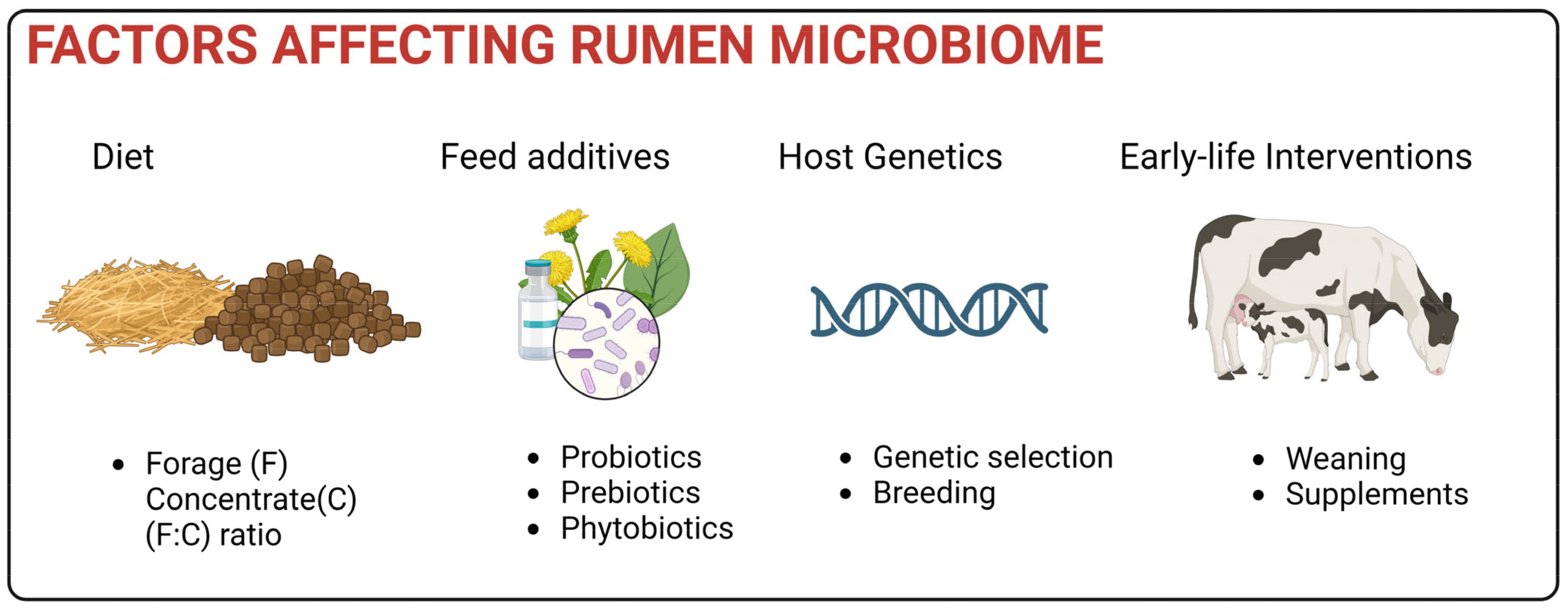
Several key factors that influence rumen microbiota and fermentation, including diet, feed additives, the host, and other early life interventions, are discussed below (Figure 4).

5.1. Impact of Diet

Diet is one of the major factors affecting ruminal microbial activity [44], influencing the host’s feed efficiency and nutrient supply. Hence, an appropriate diet is essential for a healthy rumen environment that improves rumen microbiota and promotes ruminant growth and development. At 28 days of age, calves fed with milk and concentrate had a higher relative abundance of methanogens and bacteria known to degrade readily fermentable carbohydrates than milk fed-only calves [110]. Forages, which mainly consist of neutral detergent fiber (NDF), form the basis of the adult ruminant diet [111]. The amount of NDF provides an approximation of the concentration of cellulose, hemicellulose and lignin [112,113]. The ruminants are usually fed with a mixture of forage, and starch-rich concentrate feeds to balance their energy, protein, minerals, and vitamin requirements. When adult dairy cows were fed an NDF-rich diet than a starch-rich diet, there was an increase in the biodiversity of bacteria and fungi, and the rumen concentrations of protozoa, anaerobic fungi, and methanogens in the cows. This was attributed to NDF-rich diets providing less acidic conditions which microorganisms can grow faster and generating a more comprehensive range of cellulosic and heteropolysaccharide substrates than starch diets [114]. Conversely, in diets with an increasing proportion of starch-rich concentrates, ruminal pH decreases linearly due to the excessive production of SVFAs by rumen microorganisms as there was a larger amount of fermentable carbohydrates [115]. This subsequently causes ruminal acidosis leading to dysbiosis affecting bacterial richness and diversity negatively [114,115,116,117]. However, propionate concentration increased along with total protein in blood serum, which led to higher animal productivity [118,119]. In yaks, amylolytic Bacteroidota and cellulolytic Firmicutes decreased with increasing starch-rich concentrate levels [117]. In another study by Zhang et al., Holstein heifers fed with a high concentrate diet negatively impacted bacterial and archaeal richness and diversity but not anaerobic fungi richness and diversity [115]. Overall, an increase in the proportion of fiber in the diet results in a corresponding increase in the abundance and diversity of ruminal bacteria, fungi, and protozoa [120], but decreases when dietary F:C ratio is lowered. These findings showed how rumen microbiota changes according to the diet provided.

5.2. Impact of host

There has been a growing consensus in recent mammalian host-microbial interaction studies [121,122,123] regarding the nature of the microbial composition of the gastrointestinal tract as a polygenic trait. Various genome-wide association studies have been conducted to identify host chromosomal regions that influence microbiome composition and function in the rumen. In a cohort study of 709 beef cattle, it was found that 19 single nucleotide polymorphisms (SNPs) located on 12 bovine chromosomes were associated with 14 rumen microbial taxa, of which five were known quantitative trait loci for feed efficiency in cattle [124]. Breeding has also demonstrated a significant correlation between host and microbes. In a unique multibreed of Angus-Brahman herd from 100% Angus (Bos taurus) to 100% Brahman (Bos indicus), about 30% of the microbial community was found to be significantly associated with breed composition. Especially, SNP markers located in or near mucin-coding were strongly correlated with breed composition and contributed to the differences in the relative abundance of mucin-degrading bacteria (Clostridium, Rikenellaceae, and Akkermansia). Mucin is a critical component of the GI tract defense system in which elevated presence of mucin-degrading bacteria can cause increased susceptibility to GI pathogens [125]. In a separate study, rumen metabolite and rumen microbiome patterns among sika deer (Cervus nippon) and elk (Cervus elaphus) hybrids showed strong correlation along with changes in the amount of SVFAs and amino acids. Pathways associated with alanine, arginine, proline, and phenylalanine were enriched, which correlated positively with the abundance in Prevotella spp., Acetitomaculum spp., Quinella spp., Succinivibrio spp. and Ruminobacter spp. The rumen microbiota in the hybrids differed from that in their parents as well. This suggests that there is a significant effect of host genetics on the rumen microbiome that may have resulted from vertical transmission [126].
Moreover, the ruminal features of the host include not just the host’s genetic components but also the heritability of its rumen microbiota [127]. Li et al. found out that Bacteroidetes, the predominant bacterial phylum had low heritability estimates in cattle and were primarily affected by dietary interventions. On the other hand, the phylum Firmicutes, usually composed of fibrolytic organisms involved in starch hydrolysis that could produce acetate, formate, and succinate [128], had moderate heritability estimates. Major butyrate producers, such as Butyrivibrio [129] under the family Lachnospiraceae, were not heritable in the rumen [124]. Nevertheless, these suggests that some rumen microbial features are heritable. Therefore, genetic selection and breeding can be applied to modify the rumen microbial taxa, but it is unlikely to exert any effect if driven by external factors, such as diet.
As much as gut microbes are influenced by hosts, another key aspect of gut biology is the regulation of host physiology by intestinal microbes. In several studies, gut microbes whether indigenous or pathogenic, have been shown to influence intestinal stem cell (ISC) activity and change intestinal morphology [130]. In mice fed with Bifidobacterium and Lactobacillus spp., the diet significantly increased ISC proliferation, resulting in improved gut barrier function, while conferring protection against gut injury induced by radiation exposure and chemotherapy drug treatment [131]. The colonization of Bacteroides thetaiotaomicron, a prominent member of the intestine microflora in humans and mice, upregulates genes involved with intestinal transport and barrier function [132]. Intestinal microbes also regulate immune response as the ubiquitous Bacteroides fragilis corrected systemic Tcell deficiencies in germ-free mice [133]. Ruminants often experience microbiome dysbiosis including gastrointestinal associated diseases or metabolic disorders such as ruminal acidosis and ketosis [27]. These diseases reduce ruminal epithelial cell proliferation and threaten the integrity of the rumen [134], which can be repaired by supplementation of specific microbial strains. However, information regarding specific ruminal bacteria and their interaction with ISC remains to be further elucidated. Regardless, there has already been increasing evidence of continuous interaction between gut microbiota and intestinal stem cells forming a niche that is vital to maintaining a healthy epithelial environment [135,136].

5.3. Feed Additives

Feed additives, such as probiotics, prebiotics, and phytobiotics, have been increasingly used to promote the health of ruminants and minimize methane emissions from the rumen. These additives modulate the rumen microbial community and are essential to animal nutrition. Probiotics are “live microorganisms which, when administered in adequate amounts, confer a health benefit to the host” [137], and act to outcompete and replace pathogenic bacteria in the gastrointestinal tract. They are involved in food digestion and the secretion of organic acids and several metabolites which regulate the rumen microbial community [138], leading to increased animal productivity [139]. The most commonly used probiotic preparations are the lactic acid bacteria (Lactobacillus, Streptococci, Bifidobacteria), Propionibacterium, Enterococcus, the fungi yeast Saccharomyces, and filamentous fungi Aspergillus [140]. In goats, the addition of Lactobacillus rhamnosus and Enterococcus faecalis favored the dominance of beneficial fibrolytic or cellulolytic bacteria regardless of dietary treatment [141], which is observed similarly in cattle using yeast as a probiotic [142]. A multispecies probiotic mixture of Bacillus subtilis, Lactobacillus acidophilus, and Saccharomyces cerevisiae was inoculated into newborn female calves, significantly leading to higher average daily gain in the first eight weeks after birth while improving immune function and decreasing the incidence of diarrhea. It also resulted in an increased relative abundance of fiber-degrading Ruminococcaceae and Bifidobacterium, which plays a vital role in immune support [143]. Nowadays, probiotics are supplemented with prebiotics to boost their effect. Prebiotics are non-digestible dietary substances that stimulate the activity of beneficial microbes in the gut [144]. These are usually oligosaccharides which, when fermented by probiotics, confer benefits on the host [145,146] by altering the gastrointestinal microflora [147].
Alternatively, natural additives of plant origin, such as essential oils or herbs, are used as candidates for reducing methane emissions, as antimicrobials and for improving animal productivity [148]. Recent issues with antimicrobial drug residues polluting the environment have highlighted the role of phytobiotics or phytonutrients as a replacement to modulate rumen fermentation and influence the microbiota structure [149]. These include tannins, flavonoids, and essential oils, which are plant secondary metabolites with anti-inflammatory, antioxidant, and antimicrobial properties [150]. Orzuna-Orzuna et al. performed a meta-analysis on the effect of tannins showing decreased methane production with no apparent effect on weight gain, feed consumption, and feed efficiency on beef cattle across 32 studies [151]. However, some sources of tannins, such as Leucaena leucocephala, Acacia negra, and Uncaria gambir, exert antimicrobial activity against the rumen protozoa and methanogenic bacteria [151].
On the other hand, essential oils can increase dry matter intake and daily weight gain of beef cattle. At the same time, it improves feed efficiency, likely attributed to its positive effects on rumen fungal and fibrolytic bacterial populations [152]. Phytobiotic-rich herbal extracts contain an extensive array of organic compounds that may be useful in animal nutrition. In dairy cows, rosemary extract influenced the microbiota of dairy cows, especially the abundance of the genus Prevotella. It not only exerted anti-inflammatory and antioxidative properties on the cattle, but also increased propionate production and maintained pH stability in the rumen [153]. The flavonoid-rich alcohol extract of a Mongolian medicinal herb Allium mongolicum Regel (AME), increased the relative abundance of fibrolytic bacteria, but decreased those bacteria associated with propionate production, such as Prevotella, Succiniclasticum, and Selenomonas, leading to a decreased propionate production in lambs. Furthermore, AME supplementation did not affect the palatability in the diet but promoted the secretion of an insulin-like growth factor and adrenocorticotropic hormone leading to a significant increase in the average daily weight gain [154]. In a separate study by Stefanska et al. a combination of phytobiotics and multi-strain probiotics containing Lactobacillus strains was used as a supplement in neonatal calves. The combination led to a robust rumen microbiome increasing total bacteria while enhancing calf health and growth performance in the process [155].

5.4. Early Life Interventions

The gastrointestinal tract (GI) of newly born calves has long been considered sterile and microbial colonization starts immediately after contact with the dam’s vaginal canal, fecal material, saliva, skin, and colostrum milk [156]. However, it has been recently challenged by increasing evidence of vertical transmission from the placenta, umbilical cord, or amniotic fluid of dams during the gestation period [157,158,159]. Microbes were found in the GI tract of young Holstein calves as early as 20 min after birth, indicating microbial colonization before or during birth [60]. This suggests that the maternal calf can influence the bovine fetal gut microbiome as early as the pregnancy stage. Mineral supplementation of the dam did improve the richness and diversity of the fetal gut microbiome during the gestation period [160]. Similarly, supplementation of methionine enriched microbes and metabolites that regulate vital metabolic pathways typically associated with healthy calves [161]. This could be the first step in engineering the rumen microbiome, influencing the microbiota community composition that could persist for better health and enhance animal productivity later in life.
The rumen and its microbiota changes throughout the first year of the calf’s life as it grows, with various microbial groups beginning to occupy and colonize quickly. This development stage includes the critical transition phase of weaning. As feeding shifts, the microbial diversity increases as it is weaned as early as 7 up to 17 weeks of age. As the ruminant is weaned, this decreases Actinobacteria, which are essential early colonizers for converting milk components in the neonate’s gut. On the other hand, fiber degrading Bacteriodetes and Fibrobacteres increase along with the replacement of milk with a total mixed ration (TMR), including grass silage [162]. Even the mode of feeding (suckled vs. bottle-fed) can alter the microbiome. Bottle-feeding delayed the onset of an anaerobic environment in the gut along with higher levels of Escherichia/Shigella suggesting an increased number of potential pathogens [163].
This period of instability in the gastrointestinal tract during the process of weaning provides an opportunity for manipulation wherein supplements can be added to program the rumen microbiome development [22]. Palma-Hidalgo et al. inoculated fresh rumen fluid from adult Murciano-Granadina goats into 80 newborn goat kids from day one until 11 weeks of age, resulting in an accelerated rumen microbial development showing the greater presence of plant degraders Rikenellaceae and Fibrobacter. This is a sign of a highly matured bacterial community of strict anaerobes capable of degrading recalcitrant fiber facilitating the transition from liquid to solid feeding with minimized weaning stress [164]. This highlights that the manipulation of the microbial population of the rumen is achievable before birth and early life interventions can result in increased productivity and improved health that can last a lifetime.

6. Conclusions

The global food system relies heavily on the production of ruminant livestock, which generates copious amounts of methane. This results in lower productivity and higher costs due to a significant loss of feed energy. Rumen fermentation produces SVFAs, microbial crude proteins, and vitamins by digesting plant fibers, non-protein-nitrogen, and other organic matter in the diet. The rumen microorganism influence gut metabolism as well as nutrient absorption, immune response, and health of the host. Recent advances in molecular and sequencing technology have revolutionized the way in which microbial ecology is studied. In the past, 16S rDNA sequencing has been instrumental in understanding the taxonomic composition of the rumen microbiome. Next-generation sequencing (NGS) techniques have enabled scientists to analyze and understand complex microbial communities from wider and deeper perspective. Through the use of metagenomics and metatranscriptomics analysis, we can identify the key group of microbes that perform a significant role in various processes, including cellulose degradation, fermentation, acetogenesis, and methanogenesis. This will enable us to modulate the structure of the rumen microbiome. Metatranscriptomics has been extensively used to detect key enzymes, such as CAZymes, involved in the degradation of plant biomass. The identification of novel and efficient fiber-degrading enzymes through functional metagenomics and metatranscriptomics could provide insight into strategies to improve ruminal feed conversion.
Despite decades of rumen research, current culture collections do not represent the typical composition of a rumen. Several rumen microbes remain unidentified, and the cultivation of rumen microbes is not completely understood. The identification, isolation, and characterization of ruminal microorganisms, both taxonomically and biochemically, is still a matter of research, particularly in the area of culturomics. With advances in the omics approach, we will be able to improve animal feed efficiency and health, and reduce methane emissions from ruminants.

Author Contributions

R.A.S. and T.T. wrote the manuscripts; M.-K.K., M.K. and S.-W.K. reviewed and edited the manuscript. All authors have read and agreed to the published version of the manuscript.

Funding

This work was supported by following grand agencies; The National Research Foundation of Korea (NRF) grant funded by the Korea government (MSIT) (No. 2021R1A5A8029490 and 2022M3A9I3018121); The Cooperative Research Program for Agriculture Science and Technology Development (Project No. PJ01577902), Rural Development Administration, Republic of Korea; the Technology Development Program (grant number, 20014582) funded by the Ministry of Trade, Industry and Energy (MOTIE, Korea); the research grant of the Gyeongsang National University in 2022.

Institutional Review Board Statement

Not applicable.

Informed Consent Statement

Not applicable.

Data Availability Statement

Not applicable.

Conflicts of Interest

The authors declare no conflict of interest.

References

  1. Hackmann, T.J.; Spain, J.N. Invited Review: Ruminant Ecology and Evolution: Perspectives Useful to Ruminant Livestock Research and Production. J. Dairy Sci. 2010, 93, 1320–1334. [Google Scholar] [CrossRef]
  2. Castillo-González, A.R.; Burrola-Barraza, M.E.; Domínguez-Viveros, J.; Chávez-Martínez, A. Rumen microorganisms and fermentation. Arch. Med. Vet. 2014, 46, 349–361. [Google Scholar] [CrossRef] [Green Version]
  3. Huws, S.A.; Creevey, C.J.; Oyama, L.B.; Mizrahi, I.; Denman, S.E.; Popova, M.; Muñoz-Tamayo, R.; Forano, E.; Waters, S.M.; Hess, M.; et al. Addressing Global Ruminant Agricultural Challenges through Understanding the Rumen Microbiome: Past, Present, and Future. Front. Microbiol. 2018, 9, 2161. [Google Scholar] [CrossRef] [PubMed]
  4. Newbold, C.J.; Ramos-Morales, E. Review: Ruminal Microbiome and Microbial Metabolome: Effects of Diet and Ruminant Host. Animal 2020, 14, s78–s86. [Google Scholar] [CrossRef] [PubMed] [Green Version]
  5. Gruninger, R.J.; Ribeiro, G.O.; Cameron, A.; McAllister, T.A. Invited Review: Application of Meta-Omics to Understand the Dynamic Nature of the Rumen Microbiome and How It Responds to Diet in Ruminants. Animal 2019, 13, 1843–1854. [Google Scholar] [CrossRef] [Green Version]
  6. Bergman, E.N. Energy Contributions of Volatile Fatty Acids from the Gastrointestinal Tract in Various Species. Physiol. Rev. 1990, 70, 567–590. [Google Scholar] [CrossRef] [Green Version]
  7. Hackmann, T.J.; Firkins, J.L. Maximizing Efficiency of Rumen Microbial Protein Production. Front. Microbiol. 2015, 6, 465. [Google Scholar] [CrossRef] [Green Version]
  8. Tseten, T.; Sanjorjo, R.A.; Kwon, M.; Kim, S.W. Strategies to Mitigate Enteric Methane Emissions from Ruminant Animals. J. Microbiol. Biotechnol. 2022, 32, 269–277. [Google Scholar] [CrossRef]
  9. Jangir, K.; Gujar, B. Role of Livestock in Global Warming. Pharma Innov. J. 1934, 11, 1934–1938. [Google Scholar]
  10. Arndt, C.; Hristov, A.N.; Price, W.J.; McClelland, S.C.; Pelaez, A.M.; Cueva, S.F.; Oh, J.; Dijkstra, J.; Bannink, A.; Bayat, A.R.; et al. Full Adoption of the Most Effective Strategies to Mitigate Methane Emissions by Ruminants Can Help Meet the 1.5 °C Target by 2030 but Not 2050. Proc. Natl. Acad. Sci. USA 2022, 119, e2111294119. [Google Scholar] [CrossRef]
  11. Lan, X.; Thoning, K.W.; Dlugokencky, E.J. Trends in Globally-Averaged CH4, N2O, and SF6; Determined from NOAA Global Monitoring Laboratory Measurements; NOAA Global Monitoring Laboratory Measurements: Boulder, CO, USA, 2022. [Google Scholar] [CrossRef]
  12. U.S. Environmental Protection Agency Office of Atmospheric Protection Greenhouse Gas Reporting Program (GHGRP). Climate Change Indicators: Atmospheric Concentrations of Greenhouse Gases. 2022. Available online: https://www.epa.gov/climate-indicators (accessed on 6 November 2022).
  13. Matthews, C.; Crispie, F.; Lewis, E.; Reid, M.; O’Toole, P.W.; Cotter, P.D. The Rumen Microbiome: A Crucial Consideration When Optimising Milk and Meat Production and Nitrogen Utilisation Efficiency. Gut Microbes 2019, 10, 115–132. [Google Scholar] [CrossRef] [PubMed]
  14. Ribeiro Pereira, L.G.; Vet, M.; Machado, F.S.; Campos, M.M.; Guimaraes Júnior, R.; Tomich, T.R.; Reis, L.G.; Coombs, C.; Vet St, M. Enteric methane mitigation strategies in ruminants: A review. Rev. Colomb. Cienc. Pecu. 2015, 28, 124–143. [Google Scholar] [CrossRef] [Green Version]
  15. Rate, N.M. World Population Prospects: The 2017 Revision; United Nations: New York, NY, USA, 2017. [Google Scholar]
  16. Hatab, A.A.; Cavinato, M.E.R.; Lagerkvist, C.J. Urbanization, Livestock Systems and Food Security in Developing Countries: A Systematic Review of the Literature. Food Secur. 2019, 11, 279–299. [Google Scholar] [CrossRef] [Green Version]
  17. Melletti, M.; Burton, J. Ecology, Evolution and Behaviour of Wild Cattle: Implications for Conservation; Cambridge University Press: Cambridge, UK, 2014; ISBN 9781139568098. [Google Scholar]
  18. Pradesh, M.; Baghel, I.R.; Thakur, D.; Govil, K.; Yadav, D.S.; Patil, A.K.; Nayak, S.; Baghel, R.; Yadav, P.K. Feeding Management for Early Rumen Development in Calves. J. Entomol. Zool. Stud. 2017, 5, 1132–1139. [Google Scholar]
  19. Górka, P.; Kowalski, Z.M.; Pietrzak, P.; Kotunia, A.; Jagusiak, W.; Zabielski, R. Is Rumen Development in Newborn Calves Affected by Different Liquid Feeds and Small Intestine Development? J. Dairy Sci. 2011, 94, 3002–3013. [Google Scholar] [CrossRef] [PubMed] [Green Version]
  20. Baldwin, R.L.; McLeod, K.R.; Klotz, J.L.; Heitmann, R.N. Rumen Development, Intestinal Growth and Hepatic Metabolism in the Pre- and Postweaning Ruminant. J. Dairy Sci. 2004, 87, E55–E65. [Google Scholar] [CrossRef] [Green Version]
  21. Heinrichs, A.J.; Lesmeister, K.E. Rumen Development in the Dairy Calf. Calf and heifer rearing: Principles of rearing the modern dairy heifer from calf to calving. In Proceedings of the 60th University of Nottingham Easter School in Agricultural Science, Nottingham, UK, 23–24 March 2004; pp. 53–65. [Google Scholar]
  22. Yáñez-Ruiz, D.R.; Abecia, L.; Newbold, C.J. Manipulating Rumen Microbiome and Fermentation through Interventions during Early Life: A Review. Front. Microbiol. 2015, 6, 1133. [Google Scholar] [CrossRef] [Green Version]
  23. Diao, Q.; Zhang, R.; Fu, T. Review of Strategies to Promote Rumen Development in Calves. Animals 2019, 9, 490. [Google Scholar] [CrossRef] [PubMed] [Green Version]
  24. Zhang, Y.; Choi, S.H.; Nogoy, K.M.; Liang, S. Review: The Development of the Gastrointestinal Tract Microbiota and Intervention in Neonatal Ruminants. Animal 2021, 15, 100316. [Google Scholar] [CrossRef]
  25. Jiao, J.; Li, X.; Beauchemin, K.A.; Tan, Z.; Tang, S.; Zhou, C. Rumen Development Process in Goats as Affected by Supplemental Feeding v. Grazing: Age-Related Anatomic Development, Functional Achievement and Microbial Colonisation. Br. J. Nutr. 2015, 113, 888–900. [Google Scholar] [CrossRef] [Green Version]
  26. Malmuthuge, N.; Liang, G.; Guan, L.L. Regulation of Rumen Development in Neonatal Ruminants through Microbial Metagenomes and Host Transcriptomes. Genome Biol. 2019, 20, 172. [Google Scholar] [CrossRef] [Green Version]
  27. Khalil, A.; Batool, A.; Arif, S. Healthy Cattle Microbiome and Dysbiosis in Diseased Phenotypes. Ruminants 2022, 2, 134–156. [Google Scholar] [CrossRef]
  28. Zou, X.; Liu, G.; Meng, F.; Hong, L.; Li, Y.; Lian, Z.; Yang, Z.; Luo, C.; Liu, D. Exploring the Rumen and Cecum Microbial Community from Fetus to Adulthood in Goat. Animals 2020, 10, 1639. [Google Scholar] [CrossRef] [PubMed]
  29. Guzman, C.E.; Wood, J.L.; Egidi, E.; White-Monsant, A.C.; Semenec, L.; Grommen, S.V.H.; Hill-Yardin, E.L.; de Groef, B.; Franks, A.E. A Pioneer Calf Foetus Microbiome. Sci. Rep. 2020, 10, 17712. [Google Scholar] [CrossRef] [PubMed]
  30. Krause, D.O.; Nagaraja, T.G.; Wright, A.D.G.; Callaway, T.R. Board-Invited Review: Rumen Microbiology: Leading the Way in Microbial Ecology1,2. J. Anim. Sci. 2013, 91, 331–341. [Google Scholar] [CrossRef] [PubMed]
  31. Chung, K.-T.; Bryant, M.P.; Robert, E. Hungate: Pioneer of Anaerobic Microbial Ecology. Anaerobe 1997, 3, 213–217. [Google Scholar] [CrossRef]
  32. Deusch, S.; Camarinha-Silva, A.; Conrad, J.; Beifuss, U.; Rodehutscord, M.; Seifert, J. A Structural and Functional Elucidation of the Rumen Microbiome Influenced by Various Diets and Microenvironments. Front. Microbiol. 2017, 8, 1605. [Google Scholar] [CrossRef] [PubMed] [Green Version]
  33. Henderson, G.; Yilmaz, P.; Kumar, S.; Forster, R.J.; Kelly, W.J.; Leahy, S.C.; Guan, L.L.; Janssen, P.H. Improved Taxonomic Assignment of Rumen Bacterial 16S RRNA Sequences Using a Revised SILVA Taxonomic Framework. PeerJ 2019, 2019, e6496. [Google Scholar] [CrossRef] [PubMed]
  34. Neves, A.L.A.; Li, F.; Ghoshal, B.; McAllister, T.; Guan, L.L. Enhancing the Resolution of Rumen Microbial Classification from Metatranscriptomic Data Using Kraken and Mothur. Front. Microbiol. 2017, 8, 2445. [Google Scholar] [CrossRef] [PubMed]
  35. Guo, C.Y.; Ji, S.K.; Yan, H.; Wang, Y.J.; Liu, J.J.; Cao, Z.J.; Yang, H.J.; Zhang, W.J.; Li, S.L. Dynamic Change of the Gastrointestinal Bacterial Ecology in Cows from Birth to Adulthood. Microbiol. Open 2020, 9, e1119. [Google Scholar] [CrossRef]
  36. Chaucheyras-Durand, F.; Ossa, F. REVIEW: The Rumen Microbiome: Composition, Abundance, Diversity, and New Investigative Tools. Prof. Anim. Sci. 2014, 30, 1–12. [Google Scholar] [CrossRef]
  37. Hennessy, M.L.; Indugu, N.; Vecchiarelli, B.; Bender, J.; Pappalardo, C.; Leibstein, M.; Toth, J.; Katepalli, A.; Garapati, S.; Pitta, D. Temporal Changes in the Fecal Bacterial Community in Holstein Dairy Calves from Birth through the Transition to a Solid Diet. PLoS ONE 2020, 15, e0238882. [Google Scholar] [CrossRef] [PubMed]
  38. Xue, M.Y.; Sun, H.Z.; Wu, X.H.; Guan, L.L.; Liu, J.X. Assessment of Rumen Bacteria in Dairy Cows with Varied Milk Protein Yield. J. Dairy Sci. 2019, 102, 5031–5041. [Google Scholar] [CrossRef] [PubMed]
  39. Koringa, P.G.; Thakkar, J.R.; Pandit, R.J.; Hinsu, A.T.; Parekh, M.J.; Shah, R.K.; Jakhesara, S.J.; Joshi, C.G. Metagenomic Characterisation of Ruminal Bacterial Diversity in Buffaloes from Birth to Adulthood Using 16S RRNA Gene Amplicon Sequencing. Funct. Integr. Genom. 2019, 19, 237–247. [Google Scholar] [CrossRef]
  40. Rabee, A.E.; Kewan, K.Z.; Sabra, E.A.; el Shaer, H.M.; Lamara, M. Rumen Bacterial Community Profile and Fermentation in Barki Sheep Fed Olive Cake and Date Palm Byproducts. PeerJ 2021, 9, e12447. [Google Scholar] [CrossRef]
  41. Guo, W.; Li, Y.; Wang, L.; Wang, J.; Xu, Q.; Yan, T.; Xue, B. Evaluation of Composition and Individual Variability of Rumen Microbiota in Yaks by 16S RRNA High-Throughput Sequencing Technology. Anaerobe 2015, 34, 74–79. [Google Scholar] [CrossRef] [PubMed]
  42. Gruninger, R.J.; Sensen, C.W.; McAllister, T.A.; Forster, R.J. Diversity of Rumen Bacteria in Canadian Cervids. PLoS ONE 2014, 9, e89682. [Google Scholar] [CrossRef]
  43. Seshadri, R.; Leahy, S.C.; Attwood, G.T.; Teh, K.H.; Lambie, S.C.; Cookson, A.L.; Eloe-Fadrosh, E.A.; Pavlopoulos, G.A.; Hadjithomas, M.; Varghese, N.J.; et al. Cultivation and Sequencing of Rumen Microbiome Members from the Hungate1000 Collection. Nat. Biotechnol. 2018, 36, 359–367. [Google Scholar] [CrossRef]
  44. Henderson, G.; Cox, F.; Ganesh, S.; Jonker, A.; Young, W.; Janssen, P.H.; Abecia, L.; Angarita, E.; Aravena, P.; Arenas, G.N.; et al. Rumen Microbial Community Composition Varies with Diet and Host, but a Core Microbiome Is Found across a Wide Geographical Range. Sci. Rep. 2015, 5, 14567. [Google Scholar] [CrossRef] [Green Version]
  45. Bhujbal, S.K.; Ghosh, P.; Vijay, V.K.; Rathour, R.; Kumar, M.; Singh, L.; Kapley, A. Biotechnological Potential of Rumen Microbiota for Sustainable Bioconversion of Lignocellulosic Waste to Biofuels and Value-Added Products. Sci. Total Environ. 2022, 814, 152773. [Google Scholar] [CrossRef] [PubMed]
  46. Su, M.; Hao, Z.; Shi, H.; Li, T.; Wang, H.; Li, Q.; Zhang, Y.; Ma, Y. Metagenomic Analysis Revealed Differences in Composition and Function Between Liquid-Associated and Solid-Associated Microorganisms of Sheep Rumen. Front. Microbiol. 2022, 13, 851567. [Google Scholar] [CrossRef] [PubMed]
  47. Patra, A.; Park, T.; Kim, M.; Yu, Z. Rumen Methanogens and Mitigation of Methane Emission by Anti-Methanogenic Compounds and Substances. J. Anim. Sci. Biotechnol. 2017, 8, 13. [Google Scholar] [CrossRef] [PubMed] [Green Version]
  48. Adetunji, C.O.; Olaniyan, O.T.; Dash, R.; Varma, A. The Process of Methanogenesis by Rumen Microorganisms: State of Art. In Animal Manure; Soil Biology; Springer: Cham, Switzerland, 2022; pp. 13–20. [Google Scholar]
  49. Glasson, C.R.K.; Kinley, R.D.; de Nys, R.; King, N.; Adams, S.L.; Packer, M.A.; Svenson, J.; Eason, C.T.; Magnusson, M. Benefits and Risks of Including the Bromoform Containing Seaweed Asparagopsis in Feed for the Reduction of Methane Production from Ruminants. Algal Res. 2022, 64, 102673. [Google Scholar] [CrossRef]
  50. Bauchop, T. Inhibition of Rumen Methanogenesis by Methane Analogues. J. Bacteriol. 1967, 94, 171–175. [Google Scholar] [CrossRef] [Green Version]
  51. Janssen, P.H.; Kirs, M. Structure of the Archaeal Community of the Rumen. Appl. Environ. Microbiol. 2008, 74, 3619–3625. [Google Scholar] [CrossRef] [Green Version]
  52. Levy, B.; Jami, E. Exploring the Prokaryotic Community Associated with the Rumen Ciliate Protozoa Population. Front. Microbiol. 2018, 9, 2526. [Google Scholar] [CrossRef] [Green Version]
  53. Wang, Z.; Elekwachi, C.O.; Jiao, J.; Wang, M.; Tang, S.; Zhou, C.; Tan, Z.; Forster, R.J. Investigation and Manipulation of Metabolically Active Methanogen Community Composition during Rumen Development in Black Goats. Sci. Rep. 2017, 7, 422. [Google Scholar] [CrossRef] [PubMed] [Green Version]
  54. Beauchemin, K.A.; Ungerfeld, E.M.; Eckard, R.J.; Wang, M. Review: Fifty Years of Research on Rumen Methanogenesis: Lessons Learned and Future Challenges for Mitigation. Animal 2020, 14, S2–S16. [Google Scholar] [CrossRef] [Green Version]
  55. Kittelmann, S.; Seedorf, H.; Walters, W.A.; Clemente, J.C.; Knight, R.; Gordon, J.I.; Janssen, P.H. Simultaneous Amplicon Sequencing to Explore Co-Occurrence Patterns of Bacterial, Archaeal and Eukaryotic Microorganisms in Rumen Microbial Communities. PLoS ONE 2013, 8, e47879. [Google Scholar] [CrossRef] [Green Version]
  56. Aller, J.Y.; Kemp, P.F. Are Archaea Inherently Less Diverse than Bacteria in the Same Environments? FEMS Microbiol. Ecol. 2008, 65, 74–87. [Google Scholar] [CrossRef] [PubMed]
  57. Vanwonterghem, I.; Evans, P.N.; Parks, D.H.; Jensen, P.D.; Woodcroft, B.J.; Hugenholtz, P.; Tyson, G.W. Methylotrophic Methanogenesis Discovered in the Archaeal Phylum Verstraetearchaeota. Nat. Microbiol. 2016, 1, 16170. [Google Scholar] [CrossRef] [PubMed] [Green Version]
  58. Sundset, M.A.; Edwards, J.E.; Cheng, Y.F.; Senosiain, R.S.; Fraile, M.N.; Northwood, K.S.; Praesteng, K.E.; Glad, T.; Mathiesen, S.D.; Wright, A.-D.G. Rumen Microbial Diversity in Svalbard Reindeer, with Particular Emphasis on Methanogenic Archaea. FEMS Microbiol. Ecol. 2009, 70, 553–562. [Google Scholar] [CrossRef] [Green Version]
  59. Hook, S.E.; Wright, A.D.G.; McBride, B.W. Methanogens: Methane Producers of the Rumen and Mitigation Strategies. Archaea 2010, 2010, 945785. [Google Scholar] [CrossRef] [PubMed] [Green Version]
  60. Guzman, C.E.; Bereza-Malcolm, L.T.; de Groef, B.; Franks, A.E. Presence of Selected Methanogens, Fibrolytic Bacteria, and Proteobacteria in the Gastrointestinal Tract of Neonatal Dairy Calves from Birth to 72 Hours. PLoS ONE 2015, 10, e0133048. [Google Scholar] [CrossRef] [Green Version]
  61. Akin, D.E.; Borneman, W.S. Role of Rumen Fungi in Fiber Degradation. J. Dairy Sci. 1990, 73, 3023–3032. [Google Scholar] [CrossRef] [PubMed]
  62. Ho, Y.W.; Khoo, I.Y.S.; Tan, S.G.; Abdullah, N.; Jalaludin, S.; Kudo, H. Isozyme Analysis of Anaerobic Rumen Fungi and Their Relationship to Aerobic Chytrids. Microbiology 1994, 140, 1495–1504. [Google Scholar] [CrossRef] [PubMed] [Green Version]
  63. Hagen, L.H.; Brooke, C.G.; Shaw, C.A.; Norbeck, A.D.; Piao, H.; Arntzen, M.; Olson, H.M.; Copeland, A.; Isern, N.; Shukla, A.; et al. Proteome Specialization of Anaerobic Fungi during Ruminal Degradation of Recalcitrant Plant Fiber. ISME J. 2021, 15, 421–434. [Google Scholar] [CrossRef]
  64. Elekwachi, C.O.; Wang, Z.; Wu, X.; Rabee, A.; Forster, R.J. Total RRNA-Seq Analysis Gives Insight into Bacterial, Fungal, Protozoal and Archaeal Communities in the Rumen Using an Optimized RNA Isolation Method. Front. Microbiol. 2017, 8, 1814. [Google Scholar] [CrossRef]
  65. Guo, W.; Wang, W.; Bi, S.; Long, R.; Ullah, F.; Shafiq, M.; Zhou, M.; Zhang, Y. Characterization of Anaerobic Rumen Fungal Community Composition in Yak, Tibetan Sheep and Small Tail Han Sheep Grazing on the Qinghai-Tibetan Plateau. Animals 2020, 10, 144. [Google Scholar] [CrossRef] [Green Version]
  66. Rabee, A.E.; Forster, R.J.; Elekwachi, C.O.; Kewan, K.Z.; Sabra, E.A.; Shawket, S.M.; Mahrous, H.A.; Khamiss, O.A. Community Structure and Fibrolytic Activities of Anaerobic Rumen Fungi in Dromedary Camels. J. Basic Microbiol. 2019, 59, 101–110. [Google Scholar] [CrossRef] [Green Version]
  67. Wang, H.; Li, P.; Liu, X.; Zhang, C.; Lu, Q.; Xi, D.; Yang, R.; Wang, S.; Bai, W.; Yang, Z.; et al. The Composition of Fungal Communities in the Rumen of Gayals (Bos Frontalis), Yaks (Bos Grunniens), and Yunnan and Tibetan Yellow Cattle (Bos Taurs). Pol. J. Microbiol. 2019, 68, 505–514. [Google Scholar] [CrossRef] [PubMed] [Green Version]
  68. Elshahed, M.S.; Hanafy, R.A.; Cheng, Y.; Dagar, S.S.; Edwards, J.E.; Flad, V.; Fliegerová, K.O.; Griffith, G.W.; Kittelmann, S.; Lebuhn, M.; et al. Characterization and Rank Assignment Criteria for the Anaerobic Fungi (Neocallimastigomycota). Int. J. Syst. Evol. Microbiol. 2022, 72, 005449. [Google Scholar] [CrossRef] [PubMed]
  69. Hess, M.; Paul, S.S.; Puniya, A.K.; van der Giezen, M.; Shaw, C.; Edwards, J.E.; Fliegerová, K. Anaerobic Fungi: Past, Present, and Future. Front. Microbiol. 2020, 11, 584893. [Google Scholar] [CrossRef] [PubMed]
  70. Edwards, J.E.; Kingston-Smith, A.H.; Jimenez, H.R.; Huws, S.A.; Skøt, K.P.; Griffith, G.W.; McEwan, N.R.; Theodorou, M.K. Dynamics of Initial Colonization of Nonconserved Perennial Ryegrass by Anaerobic Fungi in the Bovine Rumen. FEMS Microbiol. Ecol. 2008, 66, 537–545. [Google Scholar] [CrossRef] [PubMed]
  71. Garcia-Vallvé, S.; Romeu, A.; Palau, J. Horizontal Gene Transfer of Glycosyl Hydrolases of the Rumen Fungi. Mol. Biol. Evol. 2000, 17, 352–361. [Google Scholar] [CrossRef] [PubMed] [Green Version]
  72. Gruninger, R.J.; Nguyen, T.T.M.; Reid, I.D.; Yanke, J.L.; Wang, P.; Abbott, D.W.; Tsang, A.; McAllister, T. Application of Transcriptomics to Compare the Carbohydrate Active Enzymes That Are Expressed by Diverse Genera of Anaerobic Fungi to Degrade Plant Cell Wall Carbohydrates. Front. Microbiol. 2018, 9, 1581. [Google Scholar] [CrossRef] [PubMed] [Green Version]
  73. Duarte, E.R.; Abrão, F.O.; Ribeiro, I.C.O.; Vieira, E.A.; Nigri, A.C.; Silva, K.L.; Júnior, G.F.V.; Barreto, S.M.P.; Geraseev, L.C. Rumen Protozoa of Different Ages of Beef Cattle Raised in Tropical Pastures during the Dry Season. J. Appl. Anim. Res. 2018, 46, 1457–1461. [Google Scholar] [CrossRef] [Green Version]
  74. Newbold, C.J.; de la Fuente, G.; Belanche, A.; Ramos-Morales, E.; McEwan, N.R. The Role of Ciliate Protozoa in the Rumen. Front. Microbiol. 2015, 6, 1313. [Google Scholar] [CrossRef] [Green Version]
  75. dos Reis, C.C.; Maeda, E.M.; Cedrola, F.; Martins, E.N.; de Paula, F.M.; Martinele, I. Diet and Breed Alter Community Structures of Rumen Protozoa in Cattle Subjected to Different Feeding Systems. Semin. Ciênc. Agrár. 2019, 40, 909–918. [Google Scholar] [CrossRef] [Green Version]
  76. Francisco, A.E.; Santos-Silva, J.M.; Portugal, A.P.v.; Alves, S.P.; Bessa, R.J.B. Relationship between Rumen Ciliate Protozoa and Biohydrogenation Fatty Acid Profile in Rumen and Meat of Lambs. PLoS ONE 2019, 14, e0221996. [Google Scholar] [CrossRef] [Green Version]
  77. Ayemele, A.G.; Ma, L.; Park, T.; Xu, J.; Yu, Z.; Bu, D. Giant Milkweed (Calotropis Gigantea): A New Plant Resource to Inhibit Protozoa and Decrease Ammoniagenesis of Rumen Microbiota in Vitro without Impairing Fermentation. Sci. Total Environ. 2020, 743, 140665. [Google Scholar] [CrossRef] [PubMed]
  78. Tan, C.; Ramírez-Restrepo, C.A.; Shah, A.M.; Hu, R.; Bell, M.; Wang, Z.; McSweeney, C. The Community Structure and Microbial Linkage of Rumen Protozoa and Methanogens in Response to the Addition of Tea Seed Saponins in the Diet of Beef Cattle. J. Anim. Sci. Biotechnol. 2020, 11, 80. [Google Scholar] [CrossRef] [PubMed]
  79. Coleman, G.S. Rumen ciliate protozoa. Adv. Parasitol. 1980, 18, 121–173. [Google Scholar] [CrossRef] [PubMed]
  80. Paul, R.G.; Williams, A.G.; Butler, R.D. Hydrogenosomes in the Rumen Entodiniomorphid Ciliate Polyplastron Multivesiculatum. J. Gen. Microbiol. 1990, 136, 1981–1989. [Google Scholar] [CrossRef] [PubMed] [Green Version]
  81. Park, T.; Yu, Z. Aerobic Cultivation of Anaerobic Rumen Protozoa, Entodinium Caudatum and Epidinium Caudatum. J. Microbiol. Methods 2018, 152, 186–193. [Google Scholar] [CrossRef]
  82. Williams, C.L.; Thomas, B.J.; McEwan, N.R.; Rees Stevens, P.; Creevey, C.J.; Huws, S.A. Rumen Protozoa Play a Significant Role in Fungal Predation and Plant Carbohydrate Breakdown. Front. Microbiol. 2020, 11, 720. [Google Scholar] [CrossRef]
  83. Findley, S.D.; Mormile, M.R.; Sommer-Hurley, A.; Zhang, X.-C.; Tipton, P.; Arnett, K.; Porter, J.H.; Kerley, M.; Stacey, G. Activity-Based Metagenomic Screening and Biochemical Characterization of Bovine Ruminal Protozoan Glycoside Hydrolases. Appl. Environ. Microbiol. 2011, 77, 8106–8113. [Google Scholar] [CrossRef] [Green Version]
  84. Ricard, G.; McEwan, N.R.; Dutilh, B.E.; Jouany, J.-P.; Macheboeuf, D.; Mitsumori, M.; McIntosh, F.M.; Michalowski, T.; Nagamine, T.; Nelson, N.; et al. Horizontal Gene Transfer from Bacteria to Rumen Ciliates Indicates Adaptation to Their Anaerobic, Carbohydrates-Rich Environment. BMC Genom. 2006, 7, 22. [Google Scholar] [CrossRef] [Green Version]
  85. Adams, J.C.; Gazaway, J.A.; Brailsford, M.D.; Hartman, P.A.; Jacobson, N.L. Isolation of Bacteriophages from the Bovine Rumen. Experientia 1966, 22, 717–718. [Google Scholar] [CrossRef]
  86. Paynter, M.J.B.; Ewert, D.L.; Chalupa, W. Some Morphological Types of Bacteriophages in Bovine Rumen Contents. Appl. Microbiol. 1969, 18, 942–943. [Google Scholar] [CrossRef]
  87. Gilbert, R.A.; Townsend, E.M.; Crew, K.S.; Hitch, T.C.A.; Friedersdorff, J.C.A.; Creevey, C.J.; Pope, P.B.; Ouwerkerk, D.; Jameson, E. Rumen Virus Populations: Technological Advances Enhancing Current Understanding. Front. Microbiol. 2020, 11, 450. [Google Scholar] [CrossRef] [Green Version]
  88. Lobo, R.R.; Faciola, A.P. Ruminal Phages—A Review. Front. Microbiol. 2021, 12, 763416. [Google Scholar] [CrossRef] [PubMed]
  89. Gilbert, R.A.; Klieve, A.V. Ruminal Viruses (Bacteriophages, Archaeaphages). In Rumen Microbiology: From Evolution to Revolution; Springer: New Delhi, India, 2015; pp. 121–141. [Google Scholar]
  90. Alemneh, T.; Getabalew, M. Strategies to Reduce Methane Emission in Ruminants. Int. J. Ecol. 2019, 6, 16–22. [Google Scholar]
  91. Gilbert, R.; Ouwerkerk, D. The Genetics of Rumen Phage Populations. Proceedings 2019, 36, 165. [Google Scholar] [CrossRef] [Green Version]
  92. Berg Miller, M.E.; Yeoman, C.J.; Chia, N.; Tringe, S.G.; Angly, F.E.; Edwards, R.A.; Flint, H.J.; Lamed, R.; Bayer, E.A.; White, B.A. Phage-Bacteria Relationships and CRISPR Elements Revealed by a Metagenomic Survey of the Rumen Microbiome. Environ. Microbiol. 2012, 14, 207–227. [Google Scholar] [CrossRef] [PubMed]
  93. Friedersdorff, J.C.A.; Kingston-Smith, A.H.; Pachebat, J.A.; Cookson, A.R.; Rooke, D.; Creevey, C.J. The Isolation and Genome Sequencing of Five Novel Bacteriophages From the Rumen Active Against Butyrivibrio Fibrisolvens. Front. Microbiol. 2020, 11, 1588. [Google Scholar] [CrossRef]
  94. Rohwer, F.; Thurber, R.V. Viruses Manipulate the Marine Environment. Nature 2009, 459, 207–212. [Google Scholar] [CrossRef]
  95. Flint, H.J.; Scott, K.P. Genetics of Rumen Microorganisms: Gene Transfer, Genetic Analysis and Strain Manipulation. In Ruminant Physiology: Digestion, Metabolism, Growth and Reproduction; CABI: Oxfordshire, UK, 2000; pp. 389–408. [Google Scholar]
  96. Mizrahi, I.; Wallace, R.J.; Moraïs, S. The Rumen Microbiome: Balancing Food Security and Environmental Impacts. Nat. Rev. Microbiol. 2021, 19, 553–566. [Google Scholar] [CrossRef]
  97. Moraïs, S.; Mizrahi, I. The Road Not Taken: The Rumen Microbiome, Functional Groups, and Community States. Trends Microbiol. 2019, 27, 538–549. [Google Scholar] [CrossRef]
  98. Gleason, C.B.; Beckett, L.M.; White, R.R. Rumen Fermentation and Epithelial Gene Expression Responses to Diet Ingredients Designed to Differ in Ruminally Degradable Protein and Fiber Supplies. Sci. Rep. 2022, 12, 2933. [Google Scholar] [CrossRef]
  99. Russell, J.B.; Muck, R.E.; Weimer, P.J. Quantitative Analysis of Cellulose Degradation and Growth of Cellulolytic Bacteria in the Rumen. FEMS Microbiol. Ecol. 2009, 67, 183–197. [Google Scholar] [CrossRef]
  100. Russell, J.B.; Hespell, R.B. Microbial Rumen Fermentation. J. Dairy Sci. 1981, 64, 1153–1169. [Google Scholar] [CrossRef]
  101. Zhang, Z.; Gao, X.; Dong, W.; Huang, B.; Wang, Y.; Zhu, M.; Wang, C. Plant Cell Wall Breakdown by Hindgut Microorganisms: Can We Get Scientific Insights From Rumen Microorganisms? J. Equine Vet. Sci. 2022, 115, 104027. [Google Scholar] [CrossRef]
  102. Krause, D.O.; Denman, S.E.; Mackie, R.I.; Morrison, M.; Rae, A.L.; Attwood, G.T.; McSweeney, C.S. Opportunities to Improve Fiber Degradation in the Rumen: Microbiology, Ecology, and Genomics. FEMS Microbiol. Rev. 2003, 27, 663–693. [Google Scholar] [CrossRef] [PubMed] [Green Version]
  103. Weimer, P.J. Cellulose Degradation by Ruminal Microorganisms. Crit. Rev. Biotechnol. 1992, 12, 189–223. [Google Scholar] [CrossRef]
  104. Bainbridge, M.L.; Saldinger, L.K.; Barlow, J.W.; Alvez, J.P.; Roman, J.; Kraft, J. Alteration of Rumen Bacteria and Protozoa through Grazing Regime as a Tool to Enhance the Bioactive Fatty Acid Content of Bovine Milk. Front. Microbiol. 2018, 9, 904. [Google Scholar] [CrossRef] [Green Version]
  105. Kelly, W.J.; Mackie, R.I.; Attwood, G.T.; Janssen, P.H.; McAllister, T.A.; Leahy, S.C. Hydrogen and Formate Production and Utilisation in the Rumen and the Human Colon. Anim. Microbiome 2022, 4, 22. [Google Scholar] [CrossRef] [PubMed]
  106. Buan, N.R.; Metcalf, W.W. Methanogenesis by Methanosarcina Acetivorans Involves Two Structurally and Functionally Distinct Classes of Heterodisulfide Reductase. Mol. Microbiol. 2010, 75, 843–853. [Google Scholar] [CrossRef]
  107. Leahy, S.C.; Janssen, P.H.; Attwood, G.T.; Mackie, R.I.; Mcallister, T.A.; Kelly, W.J. Electron Flow: Key to Mitigating Ruminant Methanogenesis. Trends Microbiol. 2022, 30, 209–212. [Google Scholar] [CrossRef]
  108. McAllister, T.A.; Newbold, C.J. Redirecting Rumen Fermentation to Reduce Methanogenesis. Aust. J. Exp. Agric. 2008, 48, 7–13. [Google Scholar] [CrossRef]
  109. Thauer, R.K.; Kaster, A.K.; Seedorf, H.; Buckel, W.; Hedderich, R. Methanogenic Archaea: Ecologically Relevant Differences in Energy Conservation. Nat. Rev. Microbiol. 2008, 6, 579–591. [Google Scholar] [CrossRef]
  110. Dias, J.; Marcondes, M.I.; Noronha, M.F.; Resende, R.T.; Machado, F.S.; Mantovani, H.C.; Dill-McFarland, K.A.; Suen, G. Effect of Pre-Weaning Diet on the Ruminal Archaeal, Bacterial, and Fungal Communities of Dairy Calves. Front. Microbiol. 2017, 8, 1553. [Google Scholar] [CrossRef] [Green Version]
  111. Dijkstra, J.; Tamminga, S. Simulation of the Effects of Diet on the Contribution of Rumen Protozoa to Degradation of Fibre in the Rumen. Br. J. Nutr. 1995, 74, 617–634. [Google Scholar] [CrossRef] [Green Version]
  112. Rayburn, E.B.; Sharpe, P. Introduction to Pasture Ecology. In Horse Pasture Management; Academic Press: Cambridge, MA, USA, 2019; pp. 81–91. [Google Scholar]
  113. Diatta, A.A.; Min, D.; Jagadish, S.V.K. Drought Stress Responses in Non-Transgenic and Transgenic Alfalfa—Current Status and Future Research Directions. Adv. Agron. 2021, 170, 35–100. [Google Scholar]
  114. Belanche, A.; Doreau, M.; Edwards, J.E.; Moorby, J.M.; Pinloche, E.; Newbold, C.J. Shifts in the Rumen Microbiota Due to the Type of Carbohydrate and Level of Protein Ingested by Dairy Cattle Are Associated with Changes in Rumen Fermentation. J. Nutr. 2012, 142, 1684–1692. [Google Scholar] [CrossRef] [Green Version]
  115. Neubauer, V.; Petri, R.M.; Humer, E.; Kröger, I.; Reisinger, N.; Baumgartner, W.; Wagner, M.; Zebeli, Q. Starch-Rich Diet Induced Rumen Acidosis and Hindgut Dysbiosis in Dairy Cows of Different Lactations. Animals 2020, 10, 1727. [Google Scholar] [CrossRef]
  116. Zhang, J.; Shi, H.; Wang, Y.; Li, S.; Cao, Z.; Ji, S.; He, Y.; Zhang, H. Effect of Dietary Forage to Concentrate Ratios on Dynamic Profile Changes and Interactions of Ruminal Microbiota and Metabolites in Holstein Heifers. Front. Microbiol. 2017, 8, 2206. [Google Scholar] [CrossRef] [Green Version]
  117. Pang, K.; Chai, S.; Yang, Y.; Wang, X.; Liu, S.; Wang, S. Dietary Forage to Concentrate Ratios Impact on Yak Ruminal Microbiota and Metabolites. Front. Microbiol. 2022, 13, 964564. [Google Scholar] [CrossRef]
  118. Chen, H.; Wang, C.; Huasai, S.; Chen, A. Effects of Dietary Forage to Concentrate Ratio on Nutrient Digestibility, Ruminal Fermentation and Rumen Bacterial Composition in Angus Cows. Sci. Rep. 2021, 11, 17023. [Google Scholar] [CrossRef]
  119. Chen, G.J.; Song, S.D.; Wang, B.X.; Zhang, Z.F.; Peng, Z.L.; Guo, C.H.; Zhong, J.C.; Wang, Y. Effects of Forage:Concentrate Ratio on Growth Performance, Ruminal Fermentation and Blood Metabolites in Housing-Feeding Yaks. Asian Australas. J. Anim. Sci. 2015, 28, 1736–1741. [Google Scholar] [CrossRef] [Green Version]
  120. Wang, L.; Li, Y.; Zhang, Y.; Wang, L. The Effects of Different Concentrate-to-Forage Ratio Diets on Rumen Bacterial Microbiota and the Structures of Holstein Cows during the Feeding Cycle. Animals 2020, 10, 957. [Google Scholar] [CrossRef]
  121. Meijerink, E.; Neuenschwander, S.; Fries, R.; Dinter, A.; Bertschinger, H.U.; Stranzinger, G.; Vögeli, P. A DNA Polymorphism Influencing α(1,2)Fucosyltransferase Activity of the Pig FUT1 Enzyme Determines Susceptibility of Small Intestinal Epithelium to Escherichia Coli F18 Adhesion. Immunogenetics 2000, 52, 129–136. [Google Scholar] [CrossRef]
  122. McKnite, A.M.; Perez-Munoz, M.E.; Lu, L.; Williams, E.G.; Brewer, S.; Andreux, P.A.; Bastiaansen, J.W.M.; Wang, X.; Kachman, S.D.; Auwerx, J.; et al. Murine Gut Microbiota Is Defined by Host Genetics and Modulates Variation of Metabolic Traits. PLoS ONE 2012, 7, e39191. [Google Scholar] [CrossRef] [Green Version]
  123. Suzuki, T.A.; Phifer-Rixey, M.; Mack, K.L.; Sheehan, M.J.; Lin, D.; Bi, K.; Nachman, M.W. Host Genetic Determinants of the Gut Microbiota of Wild Mice. Mol. Ecol. 2019, 28, 3197–3207. [Google Scholar] [CrossRef]
  124. Li, F.; Li, C.; Chen, Y.; Liu, J.; Zhang, C.; Irving, B.; Fitzsimmons, C.; Plastow, G.; Guan, L.L. Host Genetics Influence the Rumen Microbiota and Heritable Rumen Microbial Features Associate with Feed Efficiency in Cattle. Microbiome 2019, 7, 92. [Google Scholar] [CrossRef] [Green Version]
  125. Fan, P.; Bian, B.; Teng, L.; Nelson, C.D.; Driver, J.; Elzo, M.A.; Jeong, K.C. Host Genetic Effects upon the Early Gut Microbiota in a Bovine Model with Graduated Spectrum of Genetic Variation. ISME J. 2020, 14, 302–317. [Google Scholar] [CrossRef] [Green Version]
  126. Li, Z.; Wright, A.-D.G.; Si, H.; Wang, X.; Qian, W.; Zhang, Z.; Li, G. Changes in the Rumen Microbiome and Metabolites Reveal the Effect of Host Genetics on Hybrid Crosses. Environ. Microbiol. Rep. 2016, 8, 1016–1023. [Google Scholar] [CrossRef]
  127. Wallace, R.J.; Sasson, G.; Garnsworthy, P.C.; Tapio, I.; Gregson, E.; Bani, P.; Huhtanen, P.; Bayat, A.R.; Strozzi, F.; Biscarini, F.; et al. A Heritable Subset of the Core Rumen Microbiome Dictates Dairy Cow Productivity and Emissions. Sci. Adv. 2019, 5, eaav8391. [Google Scholar] [CrossRef] [Green Version]
  128. Mukhopadhya, I.; Moraïs, S.; Laverde-Gomez, J.; Sheridan, P.O.; Walker, A.W.; Kelly, W.; Klieve, A.V.; Ouwerkerk, D.; Duncan, S.H.; Louis, P.; et al. Sporulation Capability and Amylosome Conservation among Diverse Human Colonic and Rumen Isolates of the Keystone Starch-Degrader Ruminococcus bromii. Environ. Microbiol. 2018, 20, 324–336. [Google Scholar] [CrossRef] [Green Version]
  129. Li, R.W.; Wu, S.; Baldwin, R.L.; Li, W.; Li, C. Perturbation Dynamics of the Rumen Microbiota in Response to Exogenous Butyrate. PLoS ONE 2012, 7, e29392. [Google Scholar] [CrossRef] [Green Version]
  130. Liu, X.; Nagy, P.; Bonfini, A.; Houtz, P.; Bing, X.-L.; Yang, X.; Buchon, N. Microbes Affect Gut Epithelial Cell Composition through Immune-Dependent Regulation of Intestinal Stem Cell Differentiation. Cell Rep. 2022, 38, 110572. [Google Scholar] [CrossRef]
  131. Lee, Y.-S.; Kim, T.-Y.; Kim, Y.; Lee, S.-H.; Kim, S.; Kang, S.W.; Yang, J.-Y.; Baek, I.-J.; Sung, Y.H.; Park, Y.-Y.; et al. Microbiota-Derived Lactate Accelerates Intestinal Stem-Cell-Mediated Epithelial Development. Cell Host Microbe 2018, 24, 833–846. [Google Scholar] [CrossRef] [Green Version]
  132. Mazmanian, S.K.; Liu, C.H.; Tzianabos, A.O.; Kasper, D.L. An Immunomodulatory Molecule of Symbiotic Bacteria Directs Maturation of the Host Immune System. Cell 2005, 122, 107–118. [Google Scholar] [CrossRef] [Green Version]
  133. Zocco, M.A.; Ainora, M.E.; Gasbarrini, G.; Gasbarrini, A. Bacteroides Thetaiotaomicron in the Gut: Molecular Aspects of Their Interaction. Dig. Liver Dis. 2007, 39, 707–712. [Google Scholar] [CrossRef]
  134. Nishihara, K.; Suzuki, Y.; Roh, S. Ruminal Epithelial Insulin-like Growth Factor-binding Proteins 2, 3, and 6 Are Associated with Epithelial Cell Proliferation. Anim. Sci. J. 2020, 91, e13422. [Google Scholar] [CrossRef]
  135. Markandey, M.; Bajaj, A.; Ilott, N.E.; Kedia, S.; Travis, S.; Powrie, F.; Ahuja, V. Gut Microbiota: Sculptors of the Intestinal Stem Cell Niche in Health and Inflammatory Bowel Disease. Gut Microbes 2021, 13, 1990827. [Google Scholar] [CrossRef]
  136. Ma, N.; Chen, X.; Johnston, L.J.; Ma, X. Gut Microbiota-stem Cell Niche Crosstalk: A New Territory for Maintaining Intestinal Homeostasis. iMeta 2022, 1, e54. [Google Scholar] [CrossRef]
  137. Hill, C.; Guarner, F.; Reid, G.; Gibson, G.R.; Merenstein, D.J.; Pot, B.; Morelli, L.; Canani, R.B.; Flint, H.J.; Salminen, S.; et al. The International Scientific Association for Probiotics and Prebiotics Consensus Statement on the Scope and Appropriate Use of the Term Probiotic. Nat. Rev. Gastroenterol. Hepatol. 2014, 11, 506–514. [Google Scholar] [CrossRef] [Green Version]
  138. Michalak, M.; Wojnarowski, K.; Cholewińska, P.; Szeligowska, N.; Bawej, M.; Pacoń, J. Selected Alternative Feed Additives Used to Manipulate the Rumen Microbiome. Animals 2021, 11, 1542. [Google Scholar] [CrossRef]
  139. Mani, S.; Aiyegoro, O.A.; Adeleke, M.A. Characterization of Rumen Microbiota of Two Sheep Breeds Supplemented with Direct-Fed Lactic Acid Bacteria. Front. Vet. Sci. 2021, 7, 570074. [Google Scholar] [CrossRef]
  140. Kulkarni, N.A.; Chethan, H.S.; Srivastava, R.; Gabbur, A.B. Role of Probiotics in Ruminant Nutrition as Natural Modulators of Health and Productivity of Animals in Tropical Countries: An Overview. Trop. Anim. Health Prod. 2022, 54, 110. [Google Scholar] [CrossRef]
  141. Maake, T.W.; Aiyegoro, O.A.; Adeleke, M.A. Effects of Lactobacillus Rhamnosus and Enterococcus Faecalis Supplementation as Direct-Fed Microbials on Rumen Microbiota of Boer and Speckled Goat Breeds. Vet. Sci. 2021, 8, 103. [Google Scholar] [CrossRef]
  142. Pinloche, E.; McEwan, N.; Marden, J.-P.; Bayourthe, C.; Auclair, E.; Newbold, C.J. The Effects of a Probiotic Yeast on the Bacterial Diversity and Population Structure in the Rumen of Cattle. PLoS ONE 2013, 8, e67824. [Google Scholar] [CrossRef] [Green Version]
  143. Wu, Y.; Wang, L.; Luo, R.; Chen, H.; Nie, C.; Niu, J.; Chen, C.; Xu, Y.; Li, X.; Zhang, W. Effect of a Multispecies Probiotic Mixture on the Growth and Incidence of Diarrhea, Immune Function, and Fecal Microbiota of Pre-Weaning Dairy Calves. Front. Microbiol. 2021, 12, 681014. [Google Scholar] [CrossRef]
  144. Davani-Davari, D.; Negahdaripour, M.; Karimzadeh, I.; Seifan, M.; Mohkam, M.; Masoumi, S.; Berenjian, A.; Ghasemi, Y. Prebiotics: Definition, Types, Sources, Mechanisms, and Clinical Applications. Foods 2019, 8, 92. [Google Scholar] [CrossRef] [Green Version]
  145. Estrada-Angulo, A.; Zapata-Ramírez, O.; Castro-Pérez, B.I.; Urías-Estrada, J.D.; Gaxiola-Camacho, S.; Angulo-Montoya, C.; Ríos-Rincón, F.G.; Barreras, A.; Zinn, R.A.; Leyva-Morales, J.B.; et al. The Effects of Single or Combined Supplementation of Probiotics and Prebiotics on Growth Performance, Dietary Energetics, Carcass Traits, and Visceral Mass in Lambs Finished under Subtropical Climate Conditions. Biology 2021, 10, 1137. [Google Scholar] [CrossRef]
  146. Lucey, P.M.; Lean, I.J.; Aly, S.S.; Golder, H.M.; Block, E.; Thompson, J.S.; Rossow, H.A. Effects of Mannan-Oligosaccharide and Bacillus Subtilis Supplementation to Preweaning Holstein Dairy Heifers on Body Weight Gain, Diarrhea, and Shedding of Fecal Pathogens. J. Dairy Sci. 2021, 104, 4290–4302. [Google Scholar] [CrossRef]
  147. Baker, L.M.; Kraft, J.; Karnezos, T.P.; Greenwood, S.L. Review: The Effects of Dietary Yeast and Yeast-Derived Extracts on Rumen Microbiota and Their Function. Anim. Feed Sci. Technol. 2022, 294, 115476. [Google Scholar] [CrossRef]
  148. Calsamiglia, S.; Busquet, M.; Cardozo, P.W.; Castillejos, L.; Ferret, A. Invited Review: Essential Oils as Modifiers of Rumen Microbial Fermentation. J. Dairy Sci. 2007, 90, 2580–2595. [Google Scholar] [CrossRef] [Green Version]
  149. Oh, J.; Wall, E.H.; Bravo, D.M.; Hristov, A.N. Host-Mediated Effects of Phytonutrients in Ruminants: A Review. J. Dairy Sci. 2017, 100, 5974–5983. [Google Scholar] [CrossRef] [Green Version]
  150. Ku-Vera, J.C.; Jiménez-Ocampo, R.; Valencia-Salazar, S.S.; Montoya-Flores, M.D.; Molina-Botero, I.C.; Arango, J.; Gómez-Bravo, C.A.; Aguilar-Pérez, C.F.; Solorio-Sánchez, F.J. Role of Secondary Plant Metabolites on Enteric Methane Mitigation in Ruminants. Front. Vet. Sci. 2020, 7, 584. [Google Scholar] [CrossRef]
  151. Orzuna-Orzuna, J.; Dorantes-Iturbide, G.; Lara-Bueno, A.; Mendoza-Martínez, G.; Miranda-Romero, L.; Hernández-García, P. Effects of Dietary Tannins’ Supplementation on Growth Performance, Rumen Fermentation, and Enteric Methane Emissions in Beef Cattle: A Meta-Analysis. Sustainability 2021, 13, 7410. [Google Scholar] [CrossRef]
  152. Orzuna-Orzuna, J.F.; Dorantes-Iturbide, G.; Lara-Bueno, A.; Miranda-Romero, L.A.; Mendoza-Martínez, G.D.; Santiago-Figueroa, I. A Meta-Analysis of Essential Oils Use for Beef Cattle Feed: Rumen Fermentation, Blood Metabolites, Meat Quality, Performance and, Environmental and Economic Impact. Fermentation 2022, 8, 254. [Google Scholar] [CrossRef]
  153. Kong, F.; Wang, S.; Dai, D.; Cao, Z.; Wang, Y.; Li, S.; Wang, W. Preliminary Investigation of the Effects of Rosemary Extract Supplementation on Milk Production and Rumen Fermentation in High-Producing Dairy Cows. Antioxidants 2022, 11, 1715. [Google Scholar] [CrossRef]
  154. Zhao, Y.; Zhang, Y.; Khas, E.; Ao, C.; Bai, C. Effects of Allium Mongolicum Regel Ethanol Extract on Three Flavor-Related Rumen Branched-Chain Fatty Acids, Rumen Fermentation and Rumen Bacteria in Lambs. Front. Microbiol. 2022, 13, 978057. [Google Scholar] [CrossRef]
  155. Stefańska, B.; Sroka, J.; Katzer, F.; Goliński, P.; Nowak, W. The Effect of Probiotics, Phytobiotics and Their Combination as Feed Additives in the Diet of Dairy Calves on Performance, Rumen Fermentation and Blood Metabolites during the Preweaning Period. Anim. Feed Sci. Technol. 2021, 272, 114738. [Google Scholar] [CrossRef]
  156. Steele, M.A.; Penner, G.B.; Chaucheyras-Durand, F.; Guan, L.L. Development and Physiology of the Rumen and the Lower Gut: Targets for Improving Gut Health. J. Dairy Sci. 2016, 99, 4955–4966. [Google Scholar] [CrossRef] [Green Version]
  157. Aagaard, K.; Ma, J.; Antony, K.M.; Ganu, R.; Petrosino, J.; Versalovic, J. The Placenta Harbors a Unique Microbiome. Sci. Transl. Med. 2014, 6, 237ra65. [Google Scholar] [CrossRef] [Green Version]
  158. Arshad, M.A.; Hassan, F.; Rehman, M.S.; Huws, S.A.; Cheng, Y.; Din, A.U. Gut Microbiome Colonization and Development in Neonatal Ruminants: Strategies, Prospects, and Opportunities. Anim. Nutr. 2021, 7, 883–895. [Google Scholar] [CrossRef]
  159. Zhu, H.; Yang, M.; Loor, J.J.; Elolimy, A.; Li, L.; Xu, C.; Wang, W.; Yin, S.; Qu, Y. Analysis of Cow-Calf Microbiome Transfer Routes and Microbiome Diversity in the Newborn Holstein Dairy Calf Hindgut. Front. Nutr. 2021, 8, 736270. [Google Scholar] [CrossRef]
  160. Hummel, G.; Woodruff, K.; Austin, K.; Knuth, R.; Lake, S.; Cunningham-Hollinger, H. Late Gestation Maternal Feed Restriction Decreases Microbial Diversity of the Placenta While Mineral Supplementation Improves Richness of the Fetal Gut Microbiome in Cattle. Animals 2021, 11, 2219. [Google Scholar] [CrossRef]
  161. Elolimy, A.; Alharthi, A.; Zeineldin, M.; Parys, C.; Helmbrecht, A.; Loor, J.J. Supply of Methionine during Late-Pregnancy Alters Fecal Microbiota and Metabolome in Neonatal Dairy Calves Without Changes in Daily Feed Intake. Front. Microbiol. 2019, 10, 2159. [Google Scholar] [CrossRef]
  162. Amin, N.; Schwarzkopf, S.; Kinoshita, A.; Tröscher-Mußotter, J.; Dänicke, S.; Camarinha-Silva, A.; Huber, K.; Frahm, J.; Seifert, J. Evolution of Rumen and Oral Microbiota in Calves Is Influenced by Age and Time of Weaning. Anim. Microbiome 2021, 3, 31. [Google Scholar] [CrossRef]
  163. Bi, Y.; Cox, M.S.; Zhang, F.; Suen, G.; Zhang, N.; Tu, Y.; Diao, Q. Feeding Modes Shape the Acquisition and Structure of the Initial Gut Microbiota in Newborn Lambs. Environ. Microbiol. 2019, 21, 2333–2346. [Google Scholar] [CrossRef] [Green Version]
  164. Palma-Hidalgo, J.M.; Jiménez, E.; Popova, M.; Morgavi, D.P.; Martín-García, A.I.; Yáñez-Ruiz, D.R.; Belanche, A. Inoculation with Rumen Fluid in Early Life Accelerates the Rumen Microbial Development and Favours the Weaning Process in Goats. Anim. Microbiome 2021, 3, 11. [Google Scholar] [CrossRef]
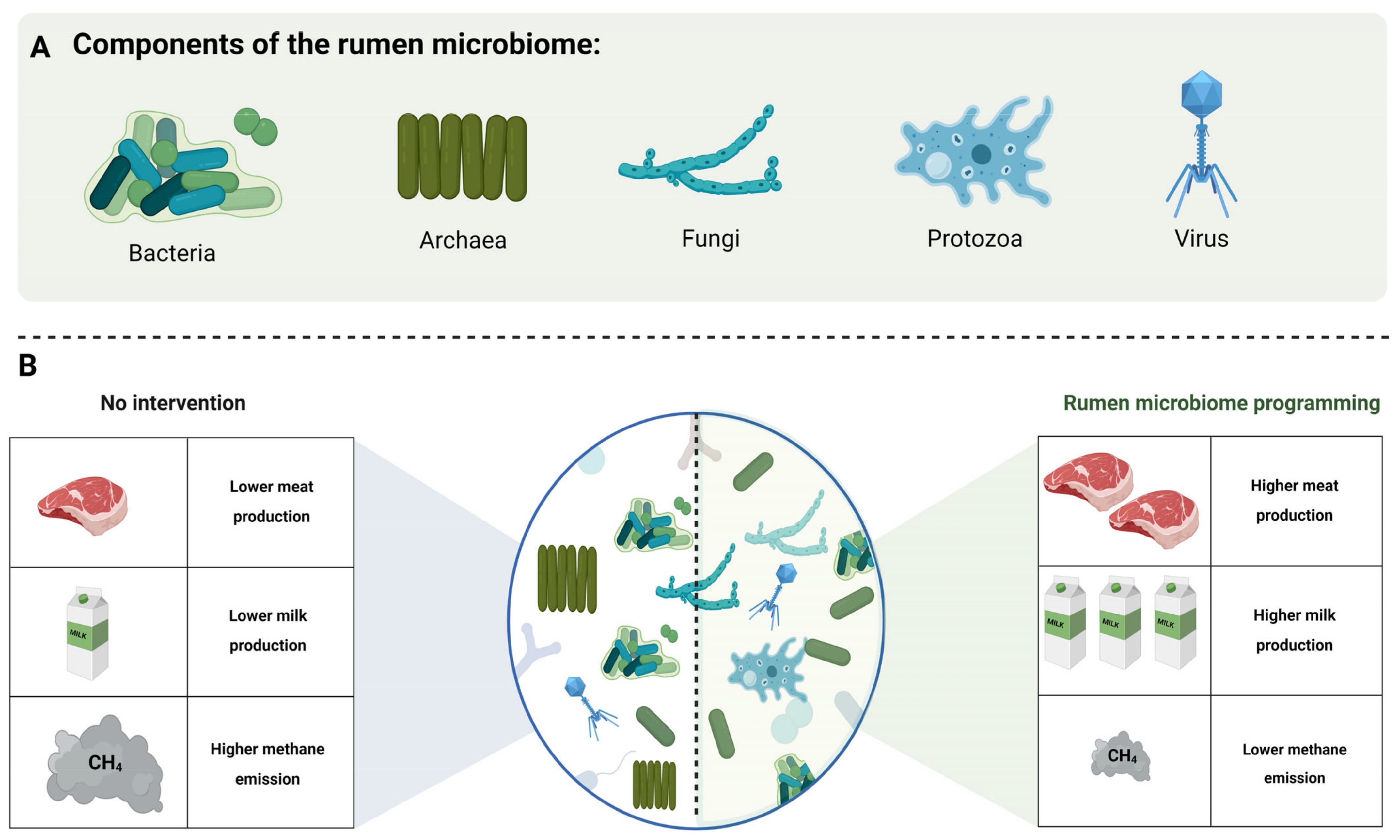
Figure 1. (A) Components of the rumen microbiome: bacteria, archaea, fungi, protozoa, and virus. (B) Improvement in the milk and meat production and reduction in methane emission through rumen microbiome programming.
Fermentation 09 00114 g001
Figure 2. Transitioning of ruminant digestive system from early life to maturity [23].
Fermentation 09 00114 g002
Figure 3. Rumen metabolic cascades represented by three stages of metabolic events: polymer degradation (I), rumen fermentation (II), and electron disposal (III). Blue hexagon and purple pentagon represent hexose and pentose sugar, respectively. Dark grey circle and blue circle represent methyl and carboxyl group, respectively. Circles with different color represent different elements: brown (carbon), yellow (oxygen), green (hydrogen), light grey (nitrogen), and pink (sulfur). SVFA: Short volatile fatty acids. MCT: monocarboxylate transporter.
Fermentation 09 00114 g003
Figure 4. Factors affecting the rumen microbiome: Diet is the major factor affecting rumen microbiome. Other factors include supplementation of feed additives, host genetics, and early life interventions.
Fermentation 09 00114 g004
Disclaimer/Publisher’s Note: The statements, opinions and data contained in all publications are solely those of the individual author(s) and contributor(s) and not of MDPI and/or the editor(s). MDPI and/or the editor(s) disclaim responsibility for any injury to people or property resulting from any ideas, methods, instructions or products referred to in the content.